Journal Description
Journal of Clinical & Translational Ophthalmology
is an international, peer-reviewed, open access journal on ophthalmology published quarterly online by MDPI.
- Open Access— free for readers, with article processing charges (APC) paid by authors or their institutions.
- Rapid Publication: manuscripts are peer-reviewed and a first decision is provided to authors approximately 40.3 days after submission; acceptance to publication is undertaken in 4.5 days (median values for papers published in this journal in the first half of 2026).
- Recognition of Reviewers: APC discount vouchers, optional signed peer review, and reviewer names published annually in the journal.
- Journal of Clinical & Translational Ophthalmology is a companion journal of JCM.
Latest Articles
Peripheral Exudative Hemorrhagic Chorioretinopathy: Current Concepts, Multimodal Imaging, Differential Diagnosis and Emerging Therapeutic Perspectives
J. Clin. Transl. Ophthalmol. 2026, 4(3), 21; https://doi.org/10.3390/jcto4030021 - 17 Aug 2026
Abstract
►
Show Figures
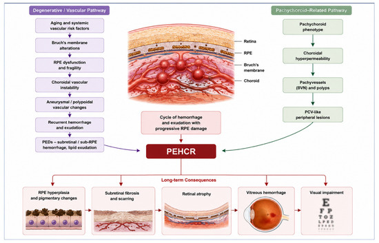
Peripheral exudative hemorrhagic chorioretinopathy (PEHCR) is an uncommon but likely underrecognized degenerative retinal disorder characterized by peripheral subretinal and sub-retinal pigment epithelium hemorrhagic and exudative lesions, predominantly affecting elderly individuals. The condition frequently mimics choroidal melanoma and other peripheral retinal diseases, often resulting
[...] Read more.
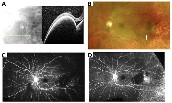
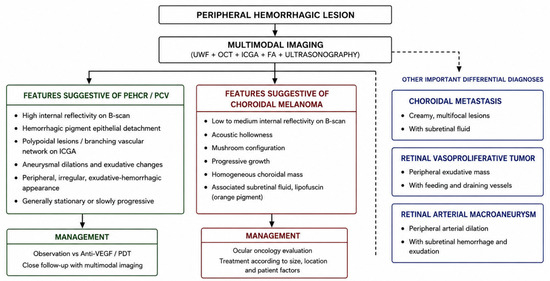
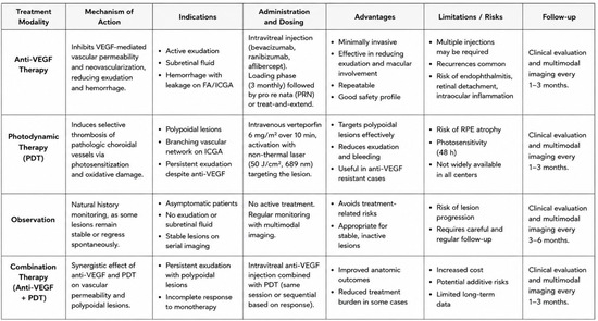
Peripheral exudative hemorrhagic chorioretinopathy (PEHCR) is an uncommon but likely underrecognized degenerative retinal disorder characterized by peripheral subretinal and sub-retinal pigment epithelium hemorrhagic and exudative lesions, predominantly affecting elderly individuals. The condition frequently mimics choroidal melanoma and other peripheral retinal diseases, often resulting in diagnostic uncertainty and occasionally unnecessary invasive procedures. Recent advances in multimodal imaging have substantially improved the recognition and characterization of PEHCR, suggesting possible associations with polypoidal choroidal vasculopathy and the pachychoroid disease spectrum. A narrative literature review was performed using PubMed/MEDLINE, Scopus, Embase, and Web of Science. Original studies, retrospective case series, multimodal imaging reports, and relevant review articles published in English were included according to their relevance to PEHCR epidemiology, imaging, pathogenesis, differential diagnosis, and treatment. Particular attention was devoted to recent developments in ultra-widefield imaging, optical coherence tomography, indocyanine green angiography, and optical coherence tomography angiography. PEHCR typically presents with peripheral subretinal hemorrhage, exudation, pigment epithelial detachments, and occasionally vitreous hemorrhage. Multimodal imaging, particularly ultra-widefield imaging and ICGA, has substantially improved the diagnosis of PEHCR and supports its relationship with the pachychoroid-PCV spectrum. Although many patients can be managed conservatively, individualized treatment based on lesion activity remains essential, while prospective studies are needed to establish evidence-based management strategies.
Full article
Open AccessArticle
Comparing Patient Experiences in Ophthalmic Workflows with Virtual Reality Versus In-Office Standard Testing: A Preliminary Survey Study
by
Margarita Labkovich, Randal A. Serafini, Andrew J. Warburton, Christopher Cheng, Oluwafeyikemi O. Okome, Vicente N. Navarro, Harsha Reddy and James G. Chelnis
J. Clin. Transl. Ophthalmol. 2026, 4(3), 20; https://doi.org/10.3390/jcto4030020 - 29 Jul 2026
Abstract
►▼
Show Figures
Background/Aims: This study set out to compare patient experiences and attitudes toward virtual reality (VR) versus in-office ophthalmic functional testing, as well as their eye exam visit preferences. Methods: A total of 64 patients undergoing a comparative clinical trial including VR and in-office
[...] Read more.
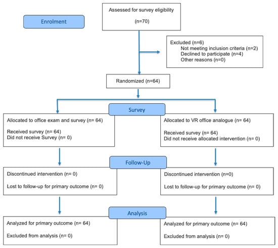
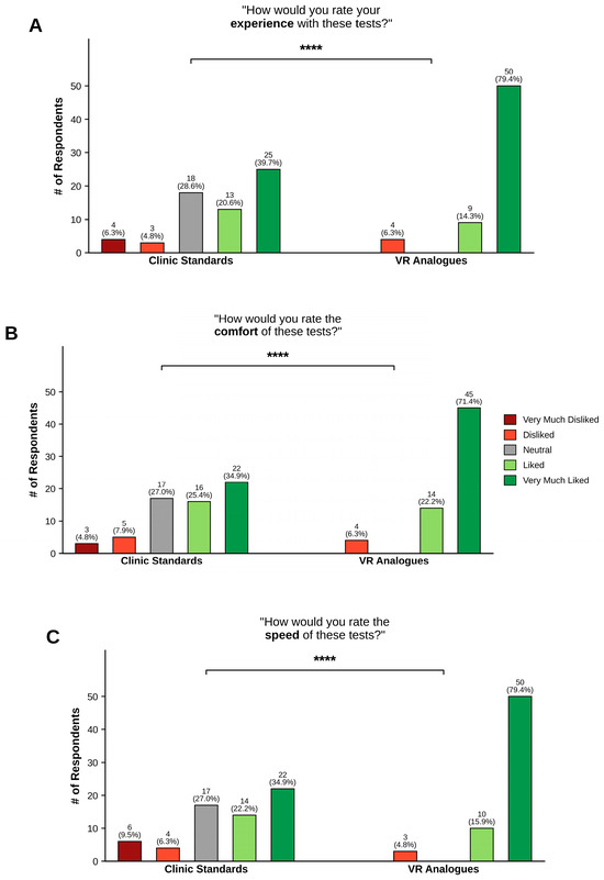
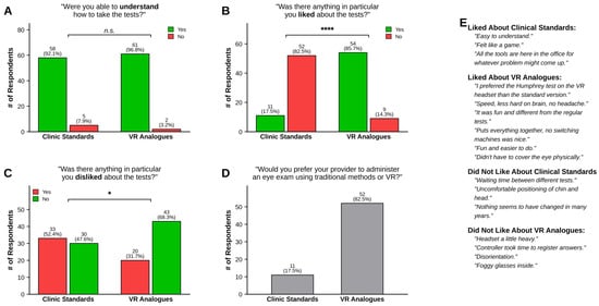
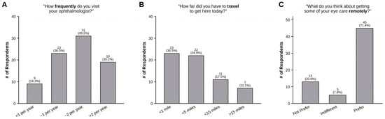
Background/Aims: This study set out to compare patient experiences and attitudes toward virtual reality (VR) versus in-office ophthalmic functional testing, as well as their eye exam visit preferences. Methods: A total of 64 patients undergoing a comparative clinical trial including VR and in-office versions of 24-2 visual field perimetry, Pelli–Robson contrast sensitivity, ETDRS visual acuity, Ishihara plates, and Amsler Grid tests were given a survey that included Likert-scaled questions regarding overall experience, speed, and comfort of broad VR testing versus in-office tests, as well as binary modality preference, like or dislike of various exam modality aspects, ease of understanding each test, distance traveled to their appointment, visit frequency, and remote care preferences. Results: In terms of overall experience, 59 (93.7%) participants rated VR as “Liked” or “Very Much Liked” compared to 38 (60.3%) for in-office tests (χ2 p < 0.0001). Regarding comfort, 59 (93.7%) and 38 (60.3%) participants at least “Liked” VR and in-office tests, respectively (χ2 p < 0.0001). For speed, 60 (95.2%) and 36 (57.1%) participants at least “Liked” VR and in-office tests, respectively (χ2 p < 0.0001). Accordingly, 11 (17.5%) patients liked all aspects of in-office tests, while 54 (85.7%) patients said the same for VR (χ2 p < 0.0001). Overall, 33 (52.4%) patients disliked something about in-office tests, while 20 (31.7%) said the same for VR (χ2 p < 0.0001). There were no differences in ability to understand tests between the two modalities. We noted a variable distribution of distance traveled and visits per year in our population, yet there was also a strong preference for implementing more remote eye testing options. Conclusions: Development and implementation of virtual reality analogs to in-office, functional ophthalmic exams can improve patient experience and facilitate remote care delivery.
Full article

Figure 1
Open AccessCase Report
Boston Type I Keratoprosthesis with Pars Plana Vitrectomy and Silicone Oil Tamponade for Prephthisical Eyes: A Case Series
by
Parvin Aghayeva, Irwin Leventer, Peter Y. Chang and Stephen D. Anesi
J. Clin. Transl. Ophthalmol. 2026, 4(3), 19; https://doi.org/10.3390/jcto4030019 - 15 Jul 2026
Abstract
►▼
Show Figures
Background: Potential vision rehabilitation in patients with hypotony and pre-phthisis remains one of the most challenging issues of ophthalmology. Boston type I keratoprosthesis (KPro) combined with pars plana vitrectomy (PPV) and silicone oil injection (SOI) represents a potential approach to this problem in
[...] Read more.
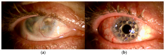
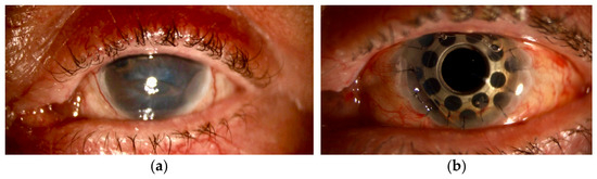
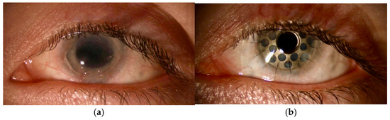
Background: Potential vision rehabilitation in patients with hypotony and pre-phthisis remains one of the most challenging issues of ophthalmology. Boston type I keratoprosthesis (KPro) combined with pars plana vitrectomy (PPV) and silicone oil injection (SOI) represents a potential approach to this problem in patients believed to have recoverable vision. This study presents a case series of three patients who underwent Boston type I KPro implantation with simultaneous silicone oil tamponade at our center. Methods: We retrospectively reviewed medical records of three patients with prephthisical changes who underwent Boston type I KPro and PPV/SOI. Preoperative and postoperative visual acuity (VA), anatomical stability, and postoperative complications were assessed over a follow-up period. Three eyes of three patients were included. All eyes had severe corneal pathology with sufficient wet surface. Two of three eyes had corneal decompensation secondary to intraocular inflammation, one eye had penetrating trauma-related corneal scarring. All three eyes underwent simultaneous KPro implantation with combined PPV/SOI. Results: Postoperatively, all eyes demonstrated improvement in best-corrected visual acuity (BCVA) from hand motion (HM) to 20/1000, with final BCVA ranging from counting fingers at 2 feet (CF2) to 20/150 and no evidence of progressive phthisical changes over a follow-up period of 23–66 months. Conclusions: Boston type I KPro implantation combined with PPV/SOI can be a viable approach for preserving anatomical integrity and improving visual function in patients with prephthisical eyes and perceived recoverable-vision potential.
Full article

Figure 1
Open AccessArticle
Giving the Side-Eye: Asymmetrical Response of the Tear Film Margins to Lateral Gaze Changes
by
Timon Ax, Fabian N. Fries, Tomas L. Bothe, Francesc March de Ribot, Slade O. Jensen, Thomas J. Millar and Berthold Seitz
J. Clin. Transl. Ophthalmol. 2026, 4(3), 18; https://doi.org/10.3390/jcto4030018 - 2 Jul 2026
Abstract
►▼
Show Figures
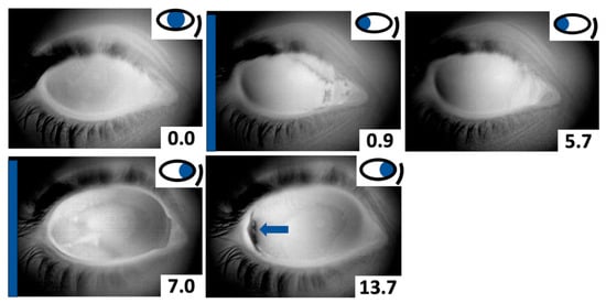
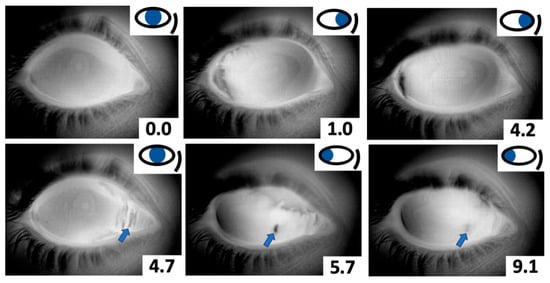
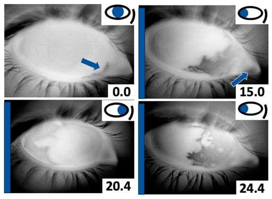
Background: Lateral eye movements are part of natural ocular motility, and their effect on tear film behavior is largely unknown because they are usually not evaluated during tear film examinations, where patients are required to look straight ahead. This study aimed to determine
[...] Read more.
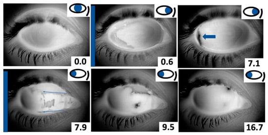
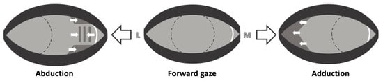
Background: Lateral eye movements are part of natural ocular motility, and their effect on tear film behavior is largely unknown because they are usually not evaluated during tear film examinations, where patients are required to look straight ahead. This study aimed to determine what happens to the tear film during lateral eye movements in the absence of blinking. Methods: Tear film dynamics during a sequence of open-eye lateral gaze maneuvers were recorded using an infrared camera system (TearView). Results: The study included 15 healthy participants (5 female; median age 26) who had no ocular surface-related complaints. It was observed that lateral eye movements exposed new ocular surface areas not previously covered by the tear film. The tear film margins reacted to lateral eye movements by spreading towards the newly uncovered ocular surface areas, thereby recoating them. This response was asymmetrical, with a tear reservoir in the lacus lacrimalis of the medial canthus providing additional tear film spread only during ocular abduction. Conclusions: These findings expand our understanding of tear film physiology beyond static gaze. They might also bear implications for dry eye disease and oculoplastic surgical techniques affecting the medial canthus.
Full article

Figure 1
Open AccessArticle
Early Versus Delayed Introduction of Faricimab for Initially Treatment-Naïve Diabetic Macular Edema: A Real-World Pilot Study
by
Tanya Gupta, Benjamin Setters, Lama Hanbali, Shruti Wadhwa, Michael W. Daniels, Wei Wang, Charles Barr, Melis Kabaalioglu Guner, SriniVas R. Sadda and Aditya Verma
J. Clin. Transl. Ophthalmol. 2026, 4(3), 17; https://doi.org/10.3390/jcto4030017 - 30 Jun 2026
Abstract
►▼
Show Figures
Background: Faricimab is one of the most potent anti-vascular endothelial growth factors used in the management of diabetic macular edema (DME). However, real-world benefits regarding its timing and efficacy are still being explored. Methods: This retrospective non-randomized pilot study aimed to evaluate the
[...] Read more.
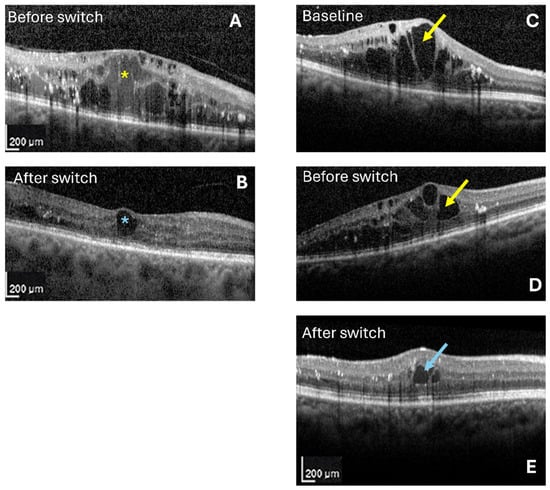
Background: Faricimab is one of the most potent anti-vascular endothelial growth factors used in the management of diabetic macular edema (DME). However, real-world benefits regarding its timing and efficacy are still being explored. Methods: This retrospective non-randomized pilot study aimed to evaluate the efficacy of intravitreal faricimab in the treatment of DME. Eyes initially treatment-naïve for DME with a follow-up of 1 year were grouped as: group 1, where faricimab was introduced within the first six months after the start of treatment; group 2, where it was initiated six or more months after treatment with other drugs. Study parameters included changes in best corrected visual acuity (BCVA) and optical coherence tomography based structural parameters within the 6 × 6 mm optical coherence tomography (OCT) scan regions. Results: Forty-two eyes from 26 patients were analyzed. No statistically significant differences were observed between the groups in cluster-weighted proportions of intra- or sub-retinal fluid, retinal thickness or volume parameters, although group 1 showed modest numerical benefits. SRF showed a trend towards qualitative reduction in group 1, although IRF showed persistence in both groups. Adjusted linear mixed-effects modeling demonstrated no significant impact of early faricimab initiation on functional and anatomical outcomes, which appeared to be influenced by the baseline BCVA, glycemic control, and the number of injections, nullifying the benefits. Conclusions: Faricimab demonstrated modest anatomical improvements with earlier treatment in eyes initially treatment-naïve for DME. Further prospective studies are indicated to assess the treatment strategy and the timing of introduction with faricimab in such eyes.
Full article

Figure 1
Open AccessArticle
Clinical Profile of Conjunctival and Eyelid Lesions in Patients Referred to the Ophthalmology Service of the Brazilian Public Health System (SUS)
by
Diego Brito Mascarós, Priscilla Luppi Ballalai, Gabrielle Aredes Leal, Vinicius Portela Correia, Diego Leite Gava, Thais Moura Gascón, Samantha Sanches de Carvalho, Glaucia Luciano da Veiga, Fernando Luiz Affonso Fonseca and Vagner Loduca Lima
J. Clin. Transl. Ophthalmol. 2026, 4(2), 16; https://doi.org/10.3390/jcto4020016 - 16 Jun 2026
Abstract
Introduction: Conjunctival and eyelid lesions encompass a broad spectrum of conditions, ranging from benign entities to malignant neoplasms, and may significantly impact ocular health and quality of life. Their occurrence is influenced by environmental exposure, demographic characteristics, and access to healthcare services. In
[...] Read more.
Introduction: Conjunctival and eyelid lesions encompass a broad spectrum of conditions, ranging from benign entities to malignant neoplasms, and may significantly impact ocular health and quality of life. Their occurrence is influenced by environmental exposure, demographic characteristics, and access to healthcare services. In public health settings, delayed diagnosis and limited access to specialized care may contribute to disease progression. Objective: To characterize the clinical and epidemiological profile of conjunctival and eyelid lesions in patients treated at a referral ophthalmology service within the Brazilian Unified Health System (SUS). Methods: This retrospective observational study reviewed medical records of patients diagnosed with conjunctival and/or eyelid lesions during ophthalmologic screening campaigns. Clinical and demographic variables were analyzed using descriptive statistics to assess prevalence and distribution patterns. Results: A total of 66 patients were included. Conjunctival involvement predominated (78.8%), with the nasal region being the most frequently affected location (62.1%). Pterygium was the most prevalent diagnosis (31.8%), followed by racial melanosis (15.2%) and melanocytic nevus (12.1%). No statistically significant differences were observed according to sex or lesion laterality. However, a significant association was identified between lesion type and anatomical location. Conclusions: Conjunctival and eyelid lesions in this population were predominantly benign and associated with demographic and environmental factors. These findings highlight the importance of structured screening strategies and early diagnosis to improve clinical outcomes and support public health policies aimed at reducing the burden of ocular disease within the SUS.
Full article
(This article belongs to the Special Issue Advances in the Diagnosis and Treatment of Ocular Surface Tumors and Diseases)
Open AccessArticle
Predictors of Receiving Surgical Treatment for Neovascular Glaucoma in the California Medicare Population
by
Justin S. Yun, Ken Kitayama, Deyu Pan, Fei Yu and Victoria L. Tseng
J. Clin. Transl. Ophthalmol. 2026, 4(2), 15; https://doi.org/10.3390/jcto4020015 - 8 Jun 2026
Abstract
Background: Population-level predictors of intraocular pressure (IOP)-lowering surgery for neovascular glaucoma (NVG) are understudied. This study examines factors associated with IOP-lowering surgery in California (CA) Medicare beneficiaries with NVG. Methods: The study population included all 2019 CA Medicare beneficiaries with NVG. Covariates included
[...] Read more.
Background: Population-level predictors of intraocular pressure (IOP)-lowering surgery for neovascular glaucoma (NVG) are understudied. This study examines factors associated with IOP-lowering surgery in California (CA) Medicare beneficiaries with NVG. Methods: The study population included all 2019 CA Medicare beneficiaries with NVG. Covariates included age, sex, race/ethnicity, history of treatments for retinal ischemia, dual Medicare/Medicaid eligibility, Social Vulnerability Index score, and Charlson Comorbidity Index (CCI) score. Outcomes included incidence of trabeculectomy, tube shunt, minimally invasive glaucoma surgery, cyclophotocoagulation (CPC), or any IOP-lowering surgery. Results: Of 1843 beneficiaries, 264 (14.3%) had IOP-lowering surgeries. In multivariable logistic regression including all covariates, CCI ≥ 5 versus 0 was associated with lower odds of any IOP-lowering surgery and of each type of surgery except CPC (adjusted odds ratio [aOR] = 0.47, 95% confidence interval [CI] = 0.29, 0.75 for any versus no surgery; aOR = 1.35, CI = 0.51, 3.60 for CPC). Compared to Non-Hispanic White, racial and ethnic minorities had increased odds of trabeculectomy (aOR = 3.77, CI = 1.05, 13.57 for Black; aOR = 2.69, CI = 1.04, 6.92 for Hispanic) and tube shunt (aOR = 2.62, CI = 1.27, 5.41 for Other/Unknown). Beneficiaries 75–79 versus 65–69 years old had decreased odds of trabeculectomy (aOR = 0.21, CI = 0.05, 0.98). Conclusions: Among CA Medicare beneficiaries, higher systemic disease burden was associated with a decreased likelihood of surgery for NVG, while racial and ethnic minorities were more likely to undergo certain procedures. These findings suggest surgical risk stratification and treatment pattern disparities for individuals with NVG.
Full article
Open AccessArticle
Outcome of Unilateral Lateral Rectus Resection in Patients with Age-Related Distance Esotropia
by
Shuan Dai and Irina Effendi-Tenang
J. Clin. Transl. Ophthalmol. 2026, 4(2), 14; https://doi.org/10.3390/jcto4020014 - 21 May 2026
Abstract
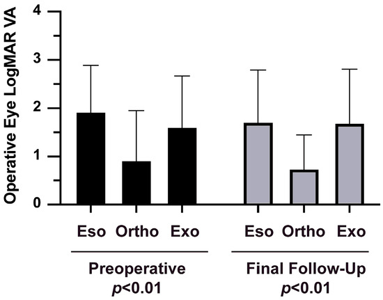
Purpose: We sought to evaluate the surgical outcomes of unilateral lateral rectus (LR) resection among patients with age-related distance esotropia (ARDE). Methods: We conducted a retrospective review of 39 patients who underwent unilateral LR resection for ARDE between January 2019 and November 2024
[...] Read more.
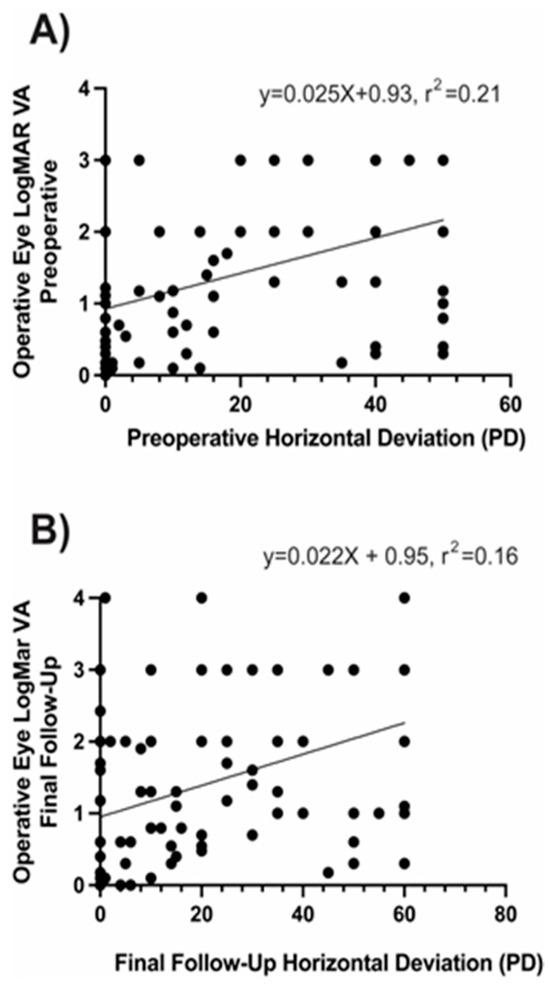
Purpose: We sought to evaluate the surgical outcomes of unilateral lateral rectus (LR) resection among patients with age-related distance esotropia (ARDE). Methods: We conducted a retrospective review of 39 patients who underwent unilateral LR resection for ARDE between January 2019 and November 2024 performed by a single surgeon at a tertiary eye centre in Brisbane, Queensland. The inclusion criteria included symptomatic horizontal diplopia at distance, comitant esotropia that was at least 25% greater at a distance than nearby, and absence of an abduction deficit or neurologic abnormality. In all but one procedure, an adjustable-suture technique was used. The amount of LR resection was based on a 1 mm per 4 prism dioptre (PD) personal surgical table. Pre- and postoperative measurements of deviation close up at near (33 cm) and at a distance (6 m) were obtained using prism and alternate cover testing. Results: The mean (±SD) patient age was 68.1 ± 9.7 years (range: 44–91). The mean preoperative esotropia measured 14.3 ± 2.7 PD at a distance and 4.7 ± 3.3 PD nearby. The mean LR resection was 3.8 ± 0.7 mm (range: 3.0–6.0 mm), with a mean follow-up of 18 months (range: 9–26). Diplopia was resolved in 97% (38/39) of the patients. At the final follow-up, 85% (33/39) were orthophoric at a distance, and all were orthophoric nearby. Five patients (13%) required postoperative adjustment, four of whom achieved orthophoria. Three patients had minimal residual esotropia (<5 PD). Conclusions: Unilateral LR resection with adjustable sutures provides a safe, effective, and targeted surgical option for ARDE, achieving high rates of distance orthophoria and diplopia resolution without compromising near alignment. Prospective long-term studies are warranted to confirm durability and refine surgical dosing.
Full article
Open AccessReview
Ophthalmic Effects of Recreational (“Party”) Drugs: Clinical and Translational Perspectives
by
Vinoth Navaratnam, Jurgen Baumann and Maneli Mozaffarieh
J. Clin. Transl. Ophthalmol. 2026, 4(2), 13; https://doi.org/10.3390/jcto4020013 - 12 May 2026
Abstract
►▼
Show Figures
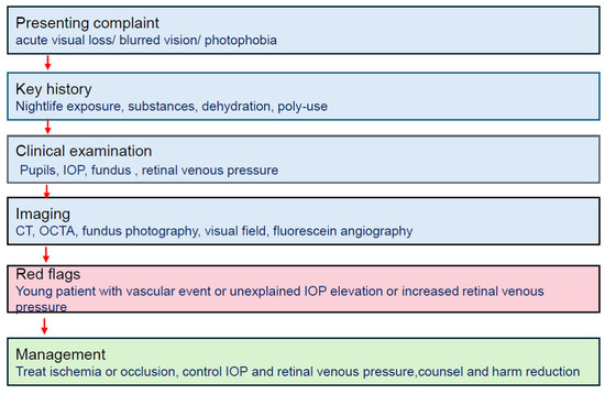
Recreational (“party”) drug use is prevalent in social environments and is increasingly relevant in ophthalmic care. While the neurological and cardiovascular consequences of these subokstances are well documented, their ocular and visual effects may not be fully recognized or consistently reported in clinical
[...] Read more.
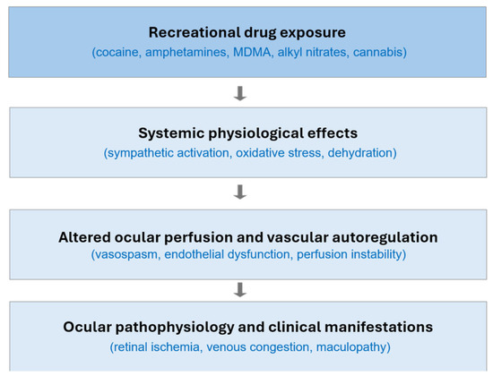
Recreational (“party”) drug use is prevalent in social environments and is increasingly relevant in ophthalmic care. While the neurological and cardiovascular consequences of these subokstances are well documented, their ocular and visual effects may not be fully recognized or consistently reported in clinical practice. This invited narrative review summarizes clinical observations and translational mechanisms underlying ophthalmic manifestations associated with commonly used recreational substances, including sympathomimetic stimulants (cocaine, amphetamines), empathogens (3,4-methylenedioxymethamphetamine (MDMA), inhalants (alkyl nitrites, “poppers”), and cannabinoids (cannabis/Δ9-tetrahydrocannabinol (THC)). Particular focus is placed on vascular dysregulation, altered ocular perfusion pressure, venous outflow impairment, oxidative stress, and neuro-ophthalmic dysfunction. Characteristic presentations, diagnostic pitfalls, and management considerations are discussed. Improved awareness of drug-related ocular effects may facilitate earlier recognition of such conditions and help reduce the risk of visual complications. Other recreational substances, including hallucinogens and emerging psychoactive compounds, may also have ocular effects, although current evidence remains limited.
Full article

Figure 1
Open AccessReview
Strategies for Conformer and Prosthetic Therapy in Children with Acquired Eye Loss and Congenital Microphthalmia or Anophthalmia
by
Vita Louisa Sophie Dingerkus, Kathleeya Nan Stang-Veldhouse, Brian Sloan and Keith Raymond Pine
J. Clin. Transl. Ophthalmol. 2026, 4(2), 12; https://doi.org/10.3390/jcto4020012 - 28 Apr 2026
Abstract
Early eye loss, congenital microphthalmia, and anophthalmia can significantly disrupt facial and psychological development in children. Timely intervention with conformers and ocular prostheses is essential for stimulating orbital growth and supporting healthy psychosocial development. This review presents evidence-based guidelines for ocularists, physicians, and
[...] Read more.
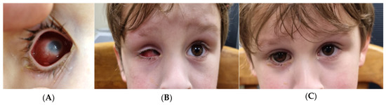
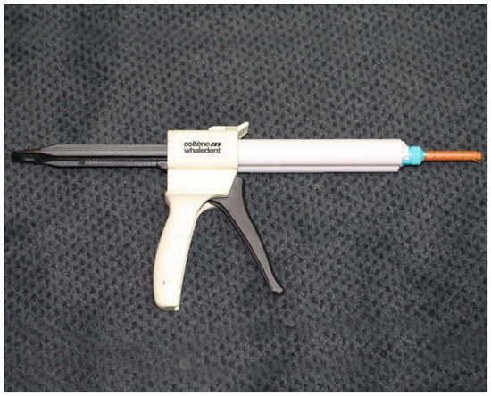
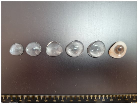
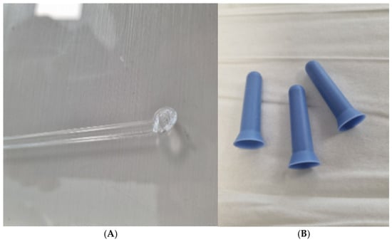
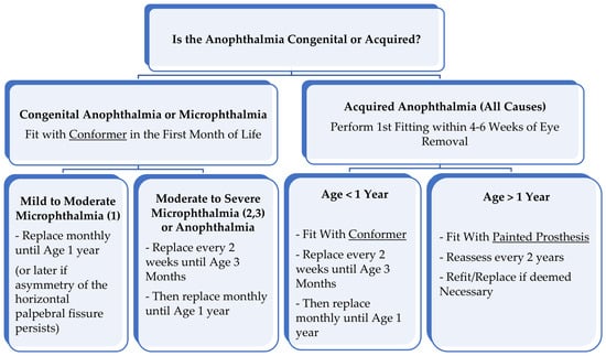
Early eye loss, congenital microphthalmia, and anophthalmia can significantly disrupt facial and psychological development in children. Timely intervention with conformers and ocular prostheses is essential for stimulating orbital growth and supporting healthy psychosocial development. This review presents evidence-based guidelines for ocularists, physicians, and allied professionals on fitting conformers and prostheses in young children, emphasizing the need for individualized treatment based on anatomical severity and age. Recommendations include initial conformer fitting within the first month of life for congenital cases or 4–6 weeks post-surgery in acquired cases, with frequent early replacements. For microphthalmia, moderate-to-severe cases require treatment similar to congenital anophthalmia cases; mild-to-moderate cases treatment within months; and mild cases are usually managed individually without urgency. A cosmetic prosthesis is advised the latest after the first year, as growth slows and self-awareness develops. Regular follow-up and adjustments support functional and psychosocial outcomes. We advocate for standardized care protocols to ensure equitable access and consistent long-term results across healthcare systems.
Full article
(This article belongs to the Special Issue Advances in Oculoplastic Surgery: From Eyelid Reconstruction to Innovative Techniques)
►▼
Show Figures

Figure 1
Open AccessArticle
Structural and Computational Validation of a Novel Titanium Scleral Buckle Implant for Posterior Pole Retinal Detachment
by
Ahmet Turer, Tugce Ilayda Turer and Levent Akduman
J. Clin. Transl. Ophthalmol. 2026, 4(2), 11; https://doi.org/10.3390/jcto4020011 - 17 Apr 2026
Abstract
►▼
Show Figures
Background: A novel titanium scleral buckle implant (TSBI) was developed for the treatment of posterior pole retinal detachments, analytically modeled and structurally tested as part of preclinical approval studies. The strength and stiffness requirements to apply pressure for retinal reattachment also suggested potential
[...] Read more.
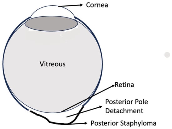
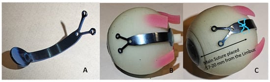
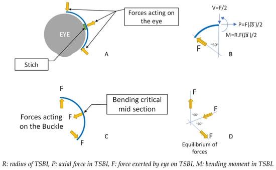
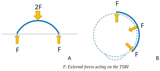
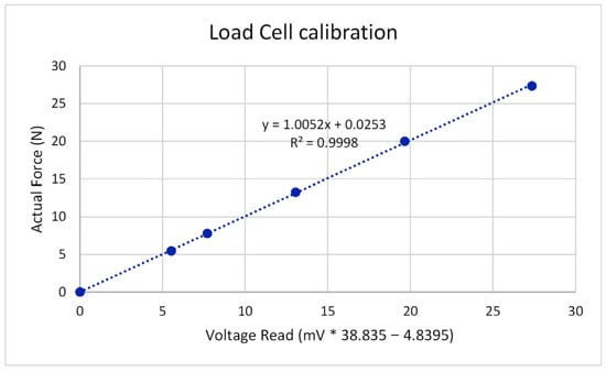
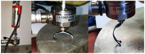
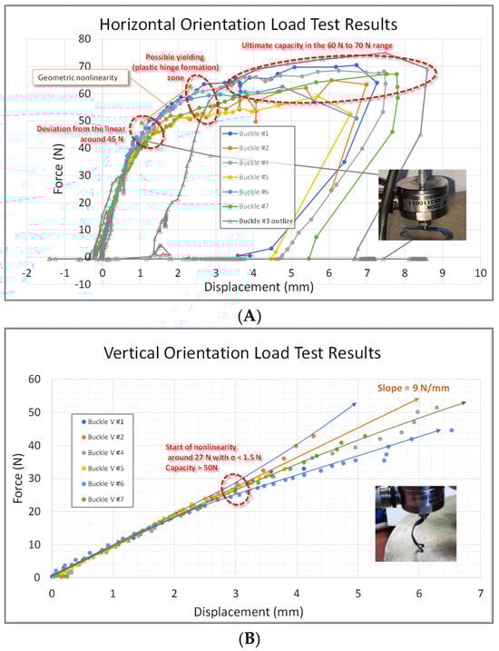
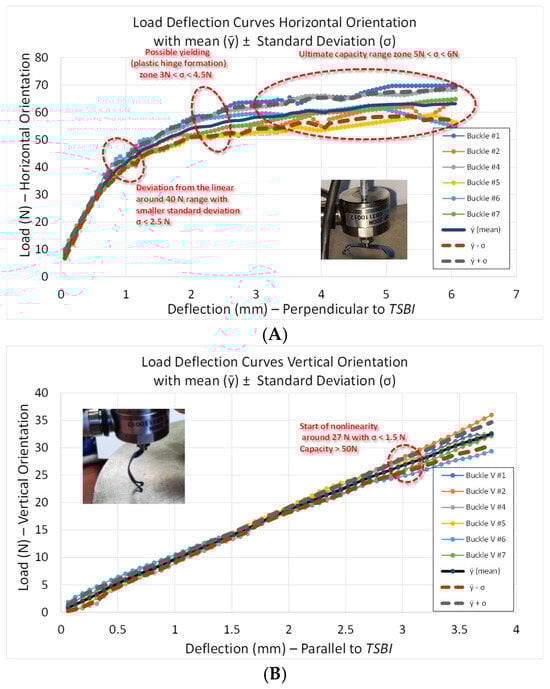
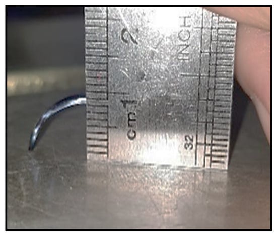
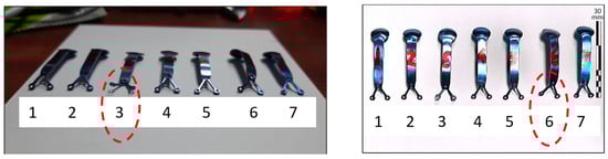
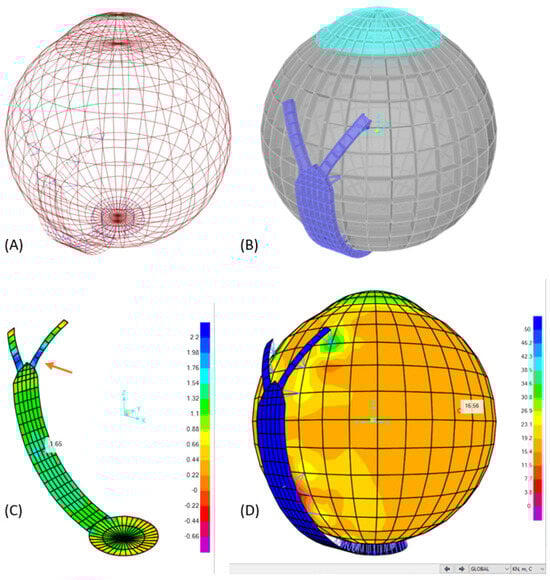
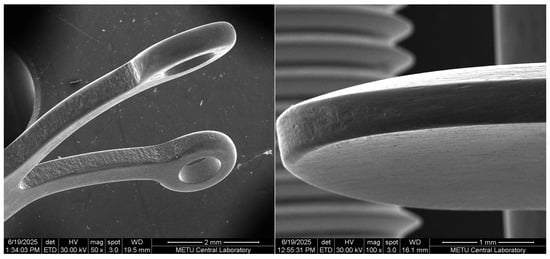
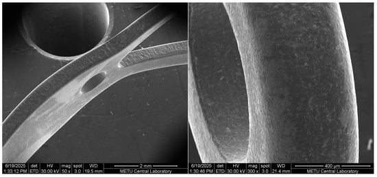
Background: A novel titanium scleral buckle implant (TSBI) was developed for the treatment of posterior pole retinal detachments, analytically modeled and structurally tested as part of preclinical approval studies. The strength and stiffness requirements to apply pressure for retinal reattachment also suggested potential benefits for correcting high myopia greater than 8 diopters. Methods: Laboratory load testing and analytical calculations were complemented by nonlinear finite element modeling (FEM), applied for the first time to capture the interaction between the highly deformed myopic eye and the TSBI. Simulations were used to visualize posterior pole indentation and force distribution across anatomical regions. Seven TSBI units were tested in the transverse direction and six in the longitudinal direction. Results: The simulations confirmed that stable indentation is maintained even in areas distant from the sutures. The TSBI’s minimum midspan bending capacity was 40 N at yield and 60 N at ultimate. These values, together with FEM predictions, demonstrated a very large safety margin and showed that the implant deforms insignificantly under high intraocular pressure changes. Conclusions: The TSBI withstands ocular forces, cushions the sclera safely, and retains its geometry, a behavior that may differ from softer buckle materials, which can exhibit time-dependent deformation under sustained loading. Early controlled clinical applications outside the USA, followed for over three years, further validate its safety and potential effectiveness.
Full article

Figure 1
Open AccessCase Report
Chronic Posterior Segment Involvement with Retinal Nerve Fiber Layer and Ganglion Cell Loss in Graft-Versus-Host Disease: A Case Report
by
Alba Chiara Termite, Gabriele Bruno, Silvana Guerriero, Pasquale Viggiano, Giacomo Boscia, Paola Carluccio, Giovanni Alessio and Francesco Boscia
J. Clin. Transl. Ophthalmol. 2026, 4(2), 10; https://doi.org/10.3390/jcto4020010 - 15 Apr 2026
Abstract
►▼
Show Figures
Background: Graft-versus-host disease is the most common complication after allogeneic hematopoietic stem cell transplantation. While ocular graft-versus-host disease typically manifests as dry eye syndrome and anterior segment involvement, posterior segment complications are rare. Previously reported posterior segment complications in graft-versus-host disease have been
[...] Read more.
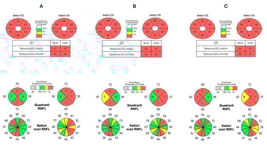
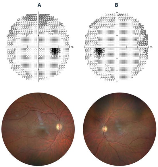
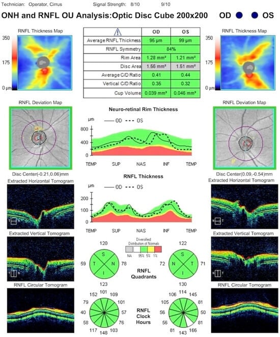
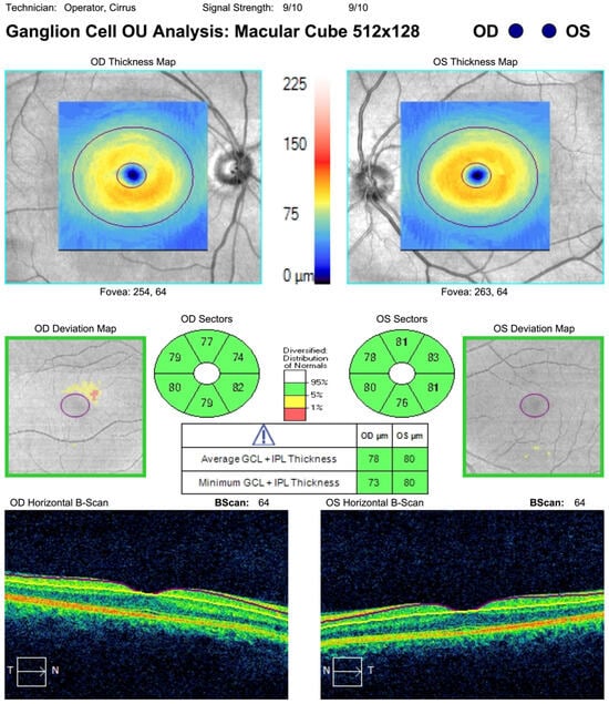
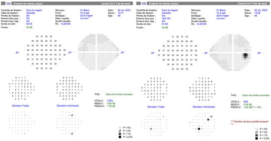
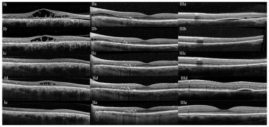
Background: Graft-versus-host disease is the most common complication after allogeneic hematopoietic stem cell transplantation. While ocular graft-versus-host disease typically manifests as dry eye syndrome and anterior segment involvement, posterior segment complications are rare. Previously reported posterior segment complications in graft-versus-host disease have been limited to acute presentations with significant functional visual impairment. Methods: A 41-year-old man developed progressive retinal nerve fiber layer and ganglion cell layer loss four years after allogeneic hematopoietic stem cell transplantation for acute myeloid leukemia. The patient had established chronic graft-versus-host disease with cutaneous involvement and ocular surface disease. Results: Despite preserved visual acuity and visual fields, and only subtle functional involvement on visual evoked potentials, optical coherence tomography revealed significant reduction in retinal nerve fiber layer thickness and ganglion cell layer. Magnetic resonance imaging showed no optic nerve or brain abnormalities. Conclusions: This case describes an uncommon presentation of chronic, subclinical posterior segment involvement in chronic GVHD and suggests that optical coherence tomography may detect progressive structural retinal changes in the absence of clinically evident visual impairment, supporting its potential role in longitudinal monitoring.
Full article

Figure 1
Open AccessArticle
Enhancing Ophthalmologists’ Accuracy in Detecting Convergence Insufficiency Using AI-Derived Graphical Outputs
by
Ahmad Khatib, Haneen Jabaly-Habib, Shmuel Raz and Ilan Shimshoni
J. Clin. Transl. Ophthalmol. 2026, 4(2), 9; https://doi.org/10.3390/jcto4020009 - 24 Mar 2026
Cited by 1
Abstract
►▼
Show Figures
Background: Accurate evaluation of the Near Point of Convergence (NPC) is essential for diagnosing and managing convergence insufficiency (CI). Conventional assessment relies on the patient’s verbal feedback and the examiner’s visual observation, making it subjective and examiner-dependent. The AI-based MobileS platform, previously validated
[...] Read more.
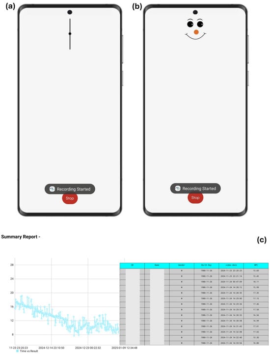
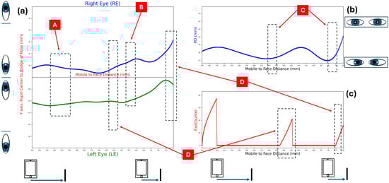
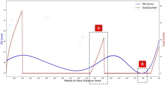
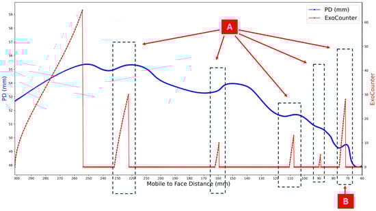
Background: Accurate evaluation of the Near Point of Convergence (NPC) is essential for diagnosing and managing convergence insufficiency (CI). Conventional assessment relies on the patient’s verbal feedback and the examiner’s visual observation, making it subjective and examiner-dependent. The AI-based MobileS platform, previously validated for both diagnosis and home-based therapy of CI, enables smartphone-based measurement and visualisation of NPC through eye tracking, without the need for verbal responses or additional equipment. This study, the third stage of our research programme, examined how ophthalmologists interpret NPC data when presented as videos versus AI-derived graphs. Methods: Twenty-two ophthalmologists completed an online questionnaire with 20 NPC test cases from the validated MobileS database, presented as both silent videos and AI-derived graphs. Accuracy was analysed using mixed-effects logistic regression, and continuous error was assessed using clustered bootstrap. Results: Graph-based interpretation showed higher odds of accurate NPC identification than video-based interpretation at the primary ±5 mm threshold (OR = 19.7, 95% CI: 13.50–28.74; p < 0.0001). Absolute error was lower for graphs than videos (Graphs − Videos: −22.73 mm; 95% CI: −26.88 to −18.59; p < 0.0001). “Uncertain” responses occurred in 28.2% of video-based assessments and 0% of graph-based assessments. Off-target errors decreased from 50.2% (videos) to 3.6% (graphs). Conclusions: AI-derived graphs of eye-movement data were associated with improved NPC estimation, suggesting a potential role in supporting clinical and tele-ophthalmology workflows.
Full article

Figure 1

Open AccessArticle
Virtual Reality Enables Rapid and Multi-Faceted Vision Screening in a Pilot Study
by
Margarita Labkovich, Andrew J. Warburton, Christopher P. Cheng, Oluwafeyikemi O. Okome, Vicente Navarro, Randal A. Serafini, Aly A. Valliani, Harsha Reddy and James Chelnis
J. Clin. Transl. Ophthalmol. 2026, 4(1), 8; https://doi.org/10.3390/jcto4010008 - 18 Mar 2026
Abstract
►▼
Show Figures
Background: Given global population growth and aging, it is imperative to prioritize early eye disease detection and treatment. However, as patient volume increases, providers are facing a shortage of workforce capacity, particularly in areas where eye doctors are already scarce, making it
[...] Read more.
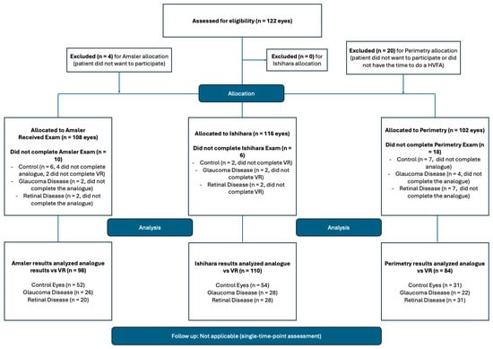
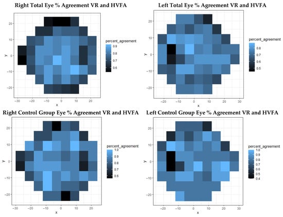
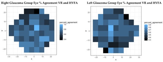
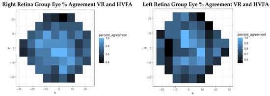
Background: Given global population growth and aging, it is imperative to prioritize early eye disease detection and treatment. However, as patient volume increases, providers are facing a shortage of workforce capacity, particularly in areas where eye doctors are already scarce, making it important to consider alternative innovative solutions that could help increase eye screening capabilities. This study compared virtual reality (VR) platform of vision screening exams that are used to evaluate ocular health, such as 24-2 perimetry, Ishihara tiles, and the Amsler grid, against their in-clinic counterparts. Methods: A total of 86 subjects were recruited from Mount Sinai’s ophthalmology clinic (New York, USA) for a comparison trial that was internally controlled across healthy eyes and those with glaucoma and retinal diseases. VR and in-office tests were administered to the patients during their clinical visit, including 24-2 perimetry, Ishihara tiles, and the Amsler grid in a randomized order, and the results were compared for each test. Results: Perimetry results from Humphrey Visual Field Analyzer (HVFA) and VR suprathreshold testing demonstrated a good sensitivity both overall (80% OD, 84% OS) and across control (86% OD, 89% OS), glaucoma (69% OD, 78% OS), and retinal disease (76% OD, 80% OS) groups. A Garway-Heath anatomical map showed an overall 70–80% agreement. Ishihara plate tests did not show a significant difference between the two testing modalities (p = 0.12; Mann–Whitney U test), which remained true across all groups. Amsler grid testing differences were also non-significant within each subgroup (p = 0.81; Mann–Whitney U test). Patient time required to complete VR exams was significantly improved (p < 0.0001; Welch’s t-test) compared to the clinical standard tests. Conclusions: All VR-based exams tested in this study showed high sensitivity and percent agreement when compared to their in-office standards. Given the results of this study, VR has a promising potential in visual function screening, which, in addition to its portable design and easy use, could assist eye doctors in screening for prevalent diseases such as glaucoma and retinal conditions. Translational Relevance: VR-based vision exams that test vision fields, color vision and visual distortions provide comparable results in healthy patients, as well as those with glaucoma and retinal diseases, indicating its potential as a screening technology for different ocular pathologies. Given VR’s portable and low-profile features, it is important to consider leveraging VR to augment delivery of vision care.
Full article

Figure 1
Open AccessArticle
Omega-3 Fatty Acid Supplementation and Incident Age-Related Macular Degeneration in a United States Veteran Population: A Retrospective Cohort Study
by
Angela A. Cao, Anders D. Westanmo, Amy A. Gravely and Karen R. Armbrust
J. Clin. Transl. Ophthalmol. 2026, 4(1), 7; https://doi.org/10.3390/jcto4010007 - 9 Mar 2026
Abstract
►▼
Show Figures
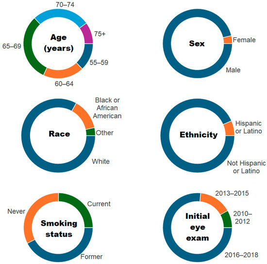
Background: Higher intake of omega-3 fatty acids from food sources is associated with a reduced risk of age-related macular degeneration (AMD), but there are no recommended parameters for omega-3 fatty acid supplementation. The purpose of our study was to investigate whether fish oil
[...] Read more.
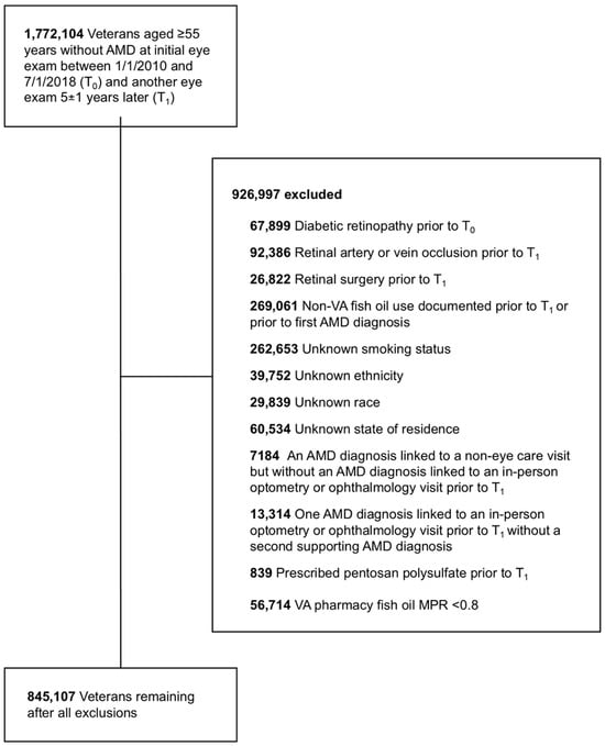
Background: Higher intake of omega-3 fatty acids from food sources is associated with a reduced risk of age-related macular degeneration (AMD), but there are no recommended parameters for omega-3 fatty acid supplementation. The purpose of our study was to investigate whether fish oil supplementation protects against development of AMD in the US Veteran Affairs (VA) Corporate Data Warehouse. Methods: Patients ≥ 55 years without an AMD diagnosis at the initial eye examination who also had a 5-year follow up eye examination were included in this retrospective cohort study. Patients receiving fish oil supplementation from the VA pharmacy were categorized into the fish oil exposure group and matched 1:1 to a non-exposure control group. All patients were assessed for AMD development at the 5-year follow up eye examination. Results: AMD developed in 772 of 16,172 patients (4.8%) with fish oil exposure and 635 of 16,093 (3.9%) without fish oil exposure (relative risk (RR) = 1.21, 95% confidence interval (CI) = 1.09–1.34, p = 0.0003). Compared to controls, AMD risk was similar in low-dose (RR = 0.91; 95% CI = 0.65–1.27) and moderate-dose (RR = 0.93; 95% CI = 0.79–1.10) fish oil supplementation but higher in high-dose fish oil supplementation (RR = 1.32; 95% CI = 1.19–1.47). Conclusions: Fish oil supplementation did not protect against AMD in a US veteran population.
Full article

Figure 1

Open AccessArticle
Anterior Segment Variations After Cataract Surgery in Subjects with Primary Open-Angle Glaucoma
by
Antonio Martínez-Abad, Maria José García-Corral, Pilar Yébana-Rubio, Ana Siverio-Colomina, Lucía Rial-Álvarez, Pedro Amat-Peral, Maria Luisa Ramón, Jorge Alió-del-Barrio and Mario Cantó-Cerdán
J. Clin. Transl. Ophthalmol. 2026, 4(1), 6; https://doi.org/10.3390/jcto4010006 - 13 Feb 2026
Abstract
►▼
Show Figures
Background: The impact of cataract surgery on anterior segment structures in subjects affected by primary open-angle glaucoma (POAG) remains incompletely understood. The aim of this study was to characterize changes in anterior segment parameters after cataract surgery in POAG subjects compared with non-glaucoma
[...] Read more.
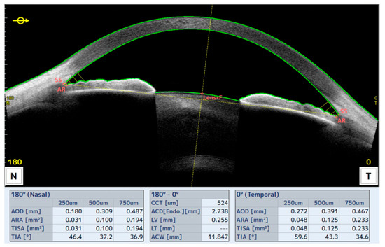
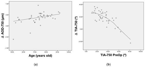
Background: The impact of cataract surgery on anterior segment structures in subjects affected by primary open-angle glaucoma (POAG) remains incompletely understood. The aim of this study was to characterize changes in anterior segment parameters after cataract surgery in POAG subjects compared with non-glaucoma subjects (control). Methods: A prospective comparative study was conducted, including patients scheduled for cataract surgery who underwent a comprehensive ophthalmic examination before and after surgery using anterior segment optical coherence tomography (OCT). Longitudinal changes and between-group comparisons were analyzed. Results: The results demonstrated a significant enlargement of the anterior chamber and iridocorneal angles after surgery in both groups (p < 0.05), with no significant differences between POAG and control eyes (p > 0.05). Several preoperative parameters, particularly angle-related metrics, were correlated with postoperative anatomical changes, suggesting their potential role as predictors of anterior segment variation (R > 0.50; p < 0.05). Conclusions: In conclusion, this study provides a detailed characterization of anterior segment changes in POAG subjects following cataract surgery, demonstrating anterior chamber enlargement comparable to that observed in non-glaucoma eyes, which may assist clinicians in the management of glaucoma.
Full article

Figure 1

Open AccessArticle
Impact of Attending Surgeon Experience on Resident-Performed Cataract Surgery Outcomes
by
Paras P. Shah, Jamie M. Nord, Sarangdev Vaidya, Daniel Zhu, Jules A. Winokur and Isha Cheela
J. Clin. Transl. Ophthalmol. 2026, 4(1), 5; https://doi.org/10.3390/jcto4010005 - 30 Jan 2026
Abstract
►▼
Show Figures
Purpose: This study evaluates the association between supervising attending surgeons’ post-residency experience and complication rates during resident-performed phacoemulsification (cataract extraction) surgeries, and to determine whether this relationship changes as the academic year progresses. Methods: A retrospective analysis of 1263 cataract surgeries performed by
[...] Read more.
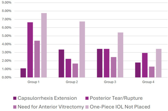
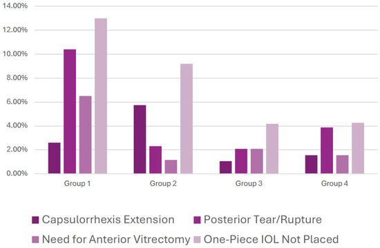
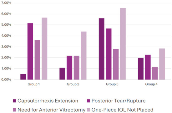
Purpose: This study evaluates the association between supervising attending surgeons’ post-residency experience and complication rates during resident-performed phacoemulsification (cataract extraction) surgeries, and to determine whether this relationship changes as the academic year progresses. Methods: A retrospective analysis of 1263 cataract surgeries performed by eight PGY-4 residents under 14 board-certified attendings was conducted at a New York City residency program over two years. Attendings were divided into four groups based on years of post-residency experience. Primary complications included posterior capsule (PC) tears, anterior vitrectomy (AV), capsulorrhexis extensions (CE), and inability to place a one-piece intraocular lens (IOL). Chi-square analyses compared complication rates between attending groups overall, and between the first and second halves of the academic year. Results: A total of 167 primary complications (13.2%) were identified. Attendings with the fewest years of experience (Group 1) supervised significantly more cases with PC tears (χ2 = 8.173, p = 0.004), AV usage (χ2 = 7.748, p = 0.005), and inability to place a one-piece IOL (χ2 = 4.753, p = 0.029), particularly during the first half of the academic year. Notably, supervising attending experience was not correlated with resident complications in the second half of the academic year. Conclusions: Early in the academic year, less experienced attendings supervised cases with higher complication rates, underscoring the critical role of strategic case assignment and targeted mentorship during early surgical training. These findings suggest that aligning resident progression with appropriate supervision can enhance outcomes and support skill development, optimizing both education and patient safety.
Full article

Figure 1

Open AccessArticle
Comparison of Visual Acuity and Strabismus Pre- and Post-Baerveldt 350 Glaucoma Drainage Device Placement in Refractory Childhood Glaucomas
by
Adam Jacobson, Elizabeth M. Bolton and Brenda L. Bohnsack
J. Clin. Transl. Ophthalmol. 2026, 4(1), 4; https://doi.org/10.3390/jcto4010004 - 19 Jan 2026
Abstract
►▼
Show Figures
Objective: Assess visual acuity (VA) and strabismus changes in children after Baerveldt 350 (BV350) device placement. Methods and Analysis: Retrospective cohort study of children (<21 years of age) who had superotemporal BV350 placement (2011–2023) and >6-month follow-up. Ocular diagnoses, surgical details,
[...] Read more.
Objective: Assess visual acuity (VA) and strabismus changes in children after Baerveldt 350 (BV350) device placement. Methods and Analysis: Retrospective cohort study of children (<21 years of age) who had superotemporal BV350 placement (2011–2023) and >6-month follow-up. Ocular diagnoses, surgical details, and preoperative and final follow-up exam findings were collected. In bilateral cases, first eye implanted was included in analysis. Results: Ninety-seven patients underwent BV350 surgery with median age of 6.7 (interquartile (IQR) 3.1, 11.2) years and with a median of 4.2 (IQR 1.8, 6.8) years of follow-up. Most common glaucomas were secondary to non-acquired ocular anomaly (n = 31) or primary congenital glaucoma (n = 21). There was no difference in preoperative and final VA (p = 0.6583). Twenty-seven (28%) and twenty-five (26%) patients were orthophoric preoperatively and at final follow-up, respectively. Orthophoria at final follow-up was associated with preoperative (odds ratio (OR)1.8 [1.2, 2.9]) and final VA (OR1.5 [1.1, 2.3]). At final follow-up, 13 patients (13%) and 19 patients (20%) showed worsened or improved horizontal deviation (>10 prism diopter (PD) change), respectively. No patients reported postoperative diplopia. Only four patients, all with esotropia, underwent subsequent strabismus surgery. Conclusions: Children who underwent BV350 placement did not have significant change in VA, and a high percentage of patients had strabismus prior to (72%) and following (74%) glaucoma surgery. Orthophoria was associated with better VA. The majority of patients did not show worsening of strabismus postoperatively, and none reported diplopia.
Full article

Figure 1
Open AccessCase Report
Inaugural Sixth Nerve Palsy in a Patient with Neuroborreliosis: A Case Report
by
Yasmine Lahrichi, Jean-Marie Rakic and Anne-Catherine Chapelle
J. Clin. Transl. Ophthalmol. 2026, 4(1), 3; https://doi.org/10.3390/jcto4010003 - 17 Jan 2026
Abstract
►▼
Show Figures
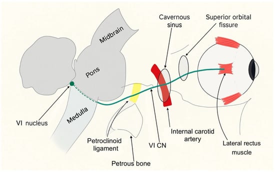
Background: We report an uncommon presentation of Lyme disease and highlight the importance of a detailed history in a patient with new-onset sixth nerve palsy. Methods: Case report and literature review. Results: A 46-year-old man receiving infliximab presented to the ophthalmology emergency department
[...] Read more.
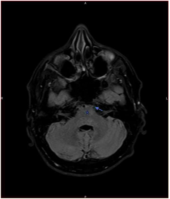
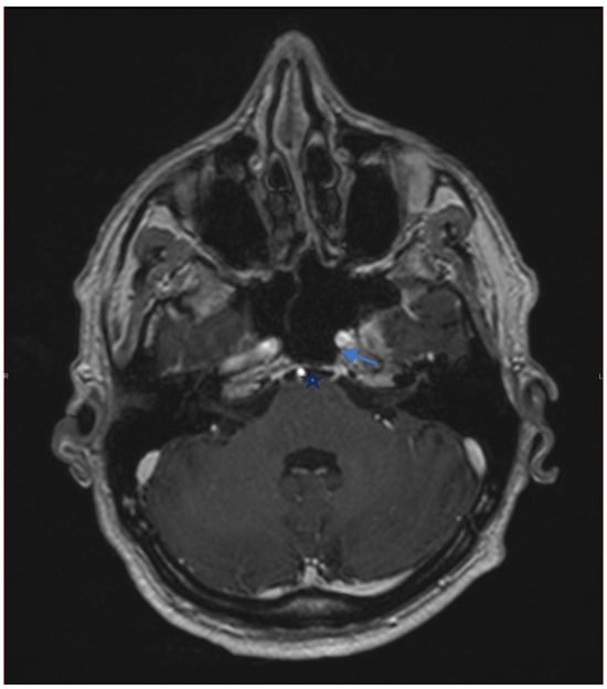
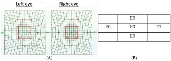
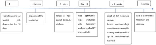
Background: We report an uncommon presentation of Lyme disease and highlight the importance of a detailed history in a patient with new-onset sixth nerve palsy. Methods: Case report and literature review. Results: A 46-year-old man receiving infliximab presented to the ophthalmology emergency department with horizontal binocular diplopia. History revealed a diffuse headache that had begun three weeks earlier. Ophthalmologic examination demonstrated a left sixth cranial nerve palsy. The workup showed positive Borrelia serum IgG, which was interpreted as a likely false-positive result given the limited specificity of serologic testing. At follow-up, the patient reported left-sided peripheral facial palsy, and worsening headache and diplopia. Further history revealed prior erythema migrans treated with doxycycline four months earlier. Considering these new findings, a lumbar puncture was performed and demonstrated intrathecal production of Borrelia antibodies. Neuroborreliosis, a neurologic involvement secondary to systemic infection by the spirochete Borrelia burgdorferi, was diagnosed. The patient was treated with oral doxycycline for 28 days with complete resolution of symptoms. Conclusions: Lyme disease may present with progressive neuro-ophthalmologic symptoms, underscoring the crucial role of ophthalmologists in its diagnosis. Moreover, immunosuppression may delay diagnosis and allow neurological progression, highlighting the need for careful history taking and close follow-up.
Full article

Figure 1

Open AccessArticle
Difluprednate and Loratadine in the Treatment of Pachychoroid Disease Spectrum
by
Emile R. Vieta-Ferrer, Adrian Au, Jeeyun Ahn and Michael B. Gorin
J. Clin. Transl. Ophthalmol. 2026, 4(1), 2; https://doi.org/10.3390/jcto4010002 - 29 Dec 2025
Abstract
Background: The recently defined pachychoroid disease spectrum (PDS), which includes central serous chorioretinopathy (CSCR), is a group of retinal disorders that share the common characteristic of a thick, dilated, hyperpermeable choroid. This study aimed to evaluate the efficacy of difluprednate and loratadine in
[...] Read more.
Background: The recently defined pachychoroid disease spectrum (PDS), which includes central serous chorioretinopathy (CSCR), is a group of retinal disorders that share the common characteristic of a thick, dilated, hyperpermeable choroid. This study aimed to evaluate the efficacy of difluprednate and loratadine in the treatment of pachychoroid disease spectrum (PDS). Methods: A retrospective study of 27 eyes from 19 patients with macular edema secondary to chronic PDS were treated with topical difluprednate and oral loratadine at a tertiary medical center. Visual acuity and optical coherence tomography (OCT) images were analyzed at baseline, 1-, 2-, 3-, 6-, 12-month, and final follow-up. Baseline was defined as the initiation of topical difluprednate. Patients with neovascularization or who had other concurrent treatments for PDS were excluded. Subfoveal choroidal thickness was measured at each time point. Response was defined as eyes that showed a reduction in intra- or subretinal fluid. Results: All 27 eyes studied responded to treatment. Of these, 70.4% resolved by 4 months and 81.5% by 6 months, with 52.2% of these patients having recurrences related to cessation or tapering of topical steroids. Visual acuity remained stable (p > 0.05) while subfoveal choroidal thickness decreased compared to baseline (p < 0.001) across all time points. Eleven (40.7%) of the eyes developed increased intraocular pressure, for which seven (25.9%) required incisional surgery. Conclusions: Chronic PDS can be treated with a combination of topical difluprednate and oral antihistamines to reduce retinal edema and subfoveal choroidal thickness. The effectiveness of therapy could be linked to the regulation of mast cell degranulation, necessitating a well-powered prospective randomized clinical trial.
Full article
(This article belongs to the Special Issue Retinal Diseases: Recent Advances in Diagnosis and Treatment)
►▼
Show Figures

Figure 1
Highly Accessed Articles
Latest Books
E-Mail Alert
News
Topics
Special Issues
Special Issue in
JCTO
Clinical Implications of Pupil Function: Optical Quality, Visual Performance, and Neurological Biomarkers
Guest Editors: Pablo De Gracia, Héctor Soriano BaronDeadline: 20 October 2026
Special Issue in
JCTO
Advances in Oculoplastic Surgery: From Eyelid Reconstruction to Innovative Techniques
Guest Editor: Argyrios TzamalisDeadline: 25 October 2026
Special Issue in
JCTO
Advances in Strabismus and Amblyopia
Guest Editor: Vijay Tailor-HamblinDeadline: 25 February 2027