
Journal Description
Dentistry Journal
Dentistry Journal
is an international, peer-reviewed, open access journal on dentistry, published monthly online by MDPI.
- Open Access— free for readers, with article processing charges (APC) paid by authors or their institutions.
- High Visibility: indexed within Scopus, ESCI (Web of Science), PubMed, PMC, and other databases.
- Journal Rank: JCR - Q1 (Dentistry, Oral Surgery and Medicine) / CiteScore - Q1 (General Dentistry)
- Rapid Publication: manuscripts are peer-reviewed and a first decision is provided to authors approximately 25.7 days after submission; acceptance to publication is undertaken in 5.9 days (median values for papers published in this journal in the first half of 2026).
- Recognition of Reviewers: reviewers who provide timely, thorough peer-review reports receive vouchers entitling them to a discount on the APC of their next publication in any MDPI journal, in appreciation of the work done.
Impact Factor:
3.4 (2025);
5-Year Impact Factor:
3.5 (2025)
Latest Articles
Digitized Physical Impressions Optimize Apical Adaptation of Bio-Root Inlays for Apexification: A Comparison of Four Impression Techniques
Dent. J. 2026, 14(9), 568; https://doi.org/10.3390/dj14090568 - 4 Sep 2026
Abstract
►
Show Figures
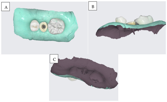
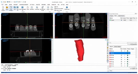
Objectives: To evaluate the three-dimensional adaptation of prefabricated Bio-Root inlays fabricated from impressions of simulated immature permanent root canals using four techniques: direct intraoral scanning (A), a light-body rubber impression digitized with an intraoral scanner (B), cone-beam computed tomography (CBCT)-based design (C),
[...] Read more.
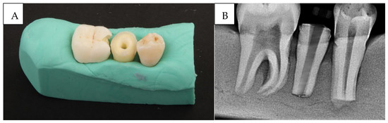
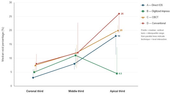
Objectives: To evaluate the three-dimensional adaptation of prefabricated Bio-Root inlays fabricated from impressions of simulated immature permanent root canals using four techniques: direct intraoral scanning (A), a light-body rubber impression digitized with an intraoral scanner (B), cone-beam computed tomography (CBCT)-based design (C), and a conventional indirect technique (D). Methods: Ten standardized simulated immature roots (extracted mandibular premolars prepared to a 1.5 mm apical diameter and 10 mm length) were used in a within-specimen comparative design, with each root receiving inlays fabricated by all four techniques. After fabrication and seating of the bioceramic inlays, the void percentage around each inlay was quantified by CBCT at the coronal, middle, and apical thirds relative to the baseline empty-canal volume. Data were analyzed using the Friedman, Wilcoxon signed-rank (Bonferroni-corrected), and linear mixed-model tests (α = 0.05). Results: Technique significantly affected void percentage (F(3,98) = 12.87, p < 0.001). Pooled across all canal thirds, Technique B had the lowest median void percentage (6.00 [7.00]), followed by A (8.00 [8.75]), C (12.00 [12.50]), and D (12.00 [19.00]). Independently of technique, the median void percentage increased from the coronal (6.00) to the middle (11.00) and apical (19.00) thirds. Differences among techniques were non-significant coronally (p = 0.437) but significant in the middle (p = 0.014) and apical (p < 0.001) thirds, with Technique B providing the best apical adaptation. Conclusions: The impression method significantly affected inlay adaptation. A light-body rubber impression digitized with an intraoral scanner yielded the best overall and apical fit, supporting digital workflows, particularly those using digitized physical impressions, for apexification. Adaptation deteriorated toward the apex regardless of technique.
Full article
Open AccessArticle
Electrochemical and Surface Characterization of Nickel-Containing Orthodontic Archwires Under In Vitro and In Vivo Conditions
by
Angelina Stoyanova-Ivanova, Velizar Georgiev, Petar Lilov, Todor Vlakhov, Laura Andreeva, Valeri Petrov, Mirela Georgieva and Jorge N. R. Martins
Dent. J. 2026, 14(9), 567; https://doi.org/10.3390/dj14090567 - 4 Sep 2026
Abstract
►▼
Show Figures
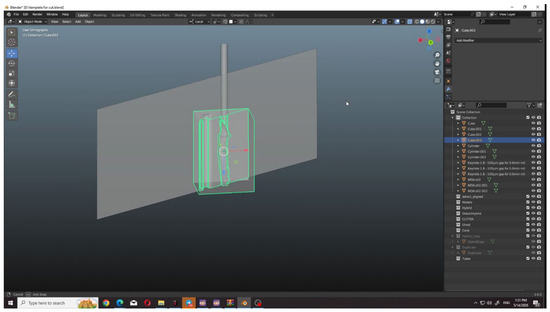
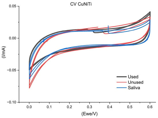
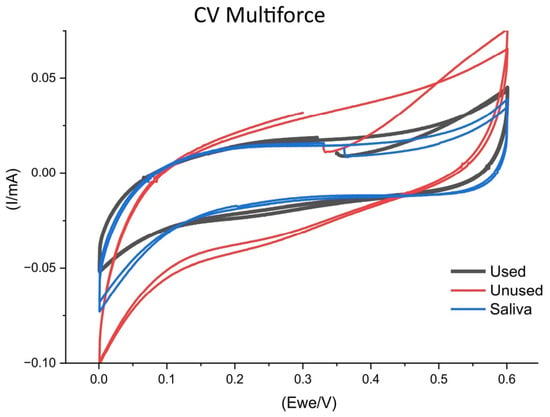
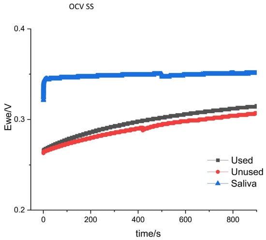
Objectives: To evaluate the corrosion behavior and surface characteristics of four nickel-containing orthodontic archwires (stainless steel (SS), superelastic nickel–titanium (NiTi), copper–nickel–titanium (CuNiTi), and multiforce NiTi) under unused, in vitro artificial saliva-immersed, and clinically used (in vivo) conditions. Methods: Rectangular SS, NiTi, CuNiTi, and
[...] Read more.
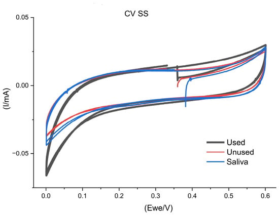
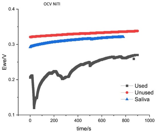
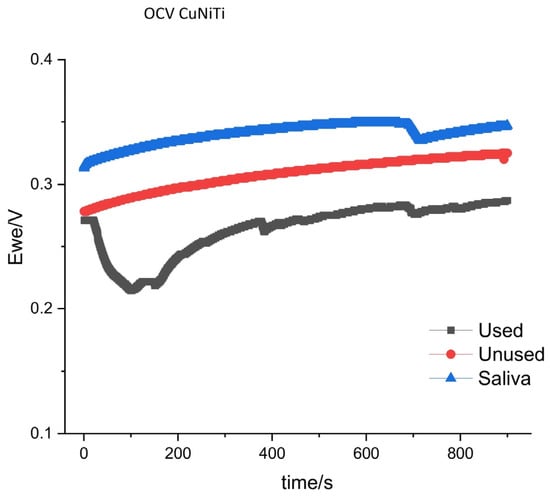
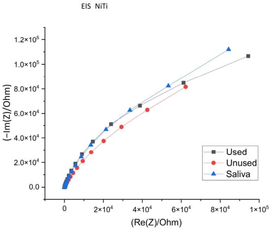
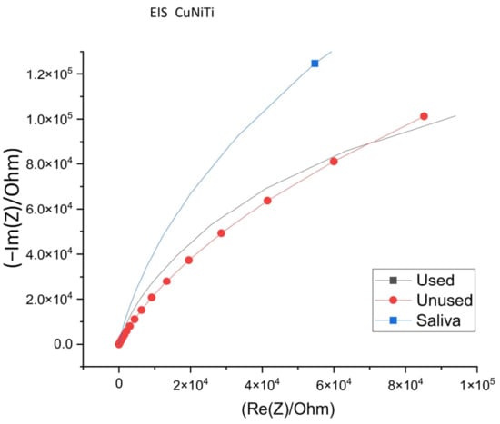
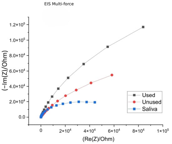
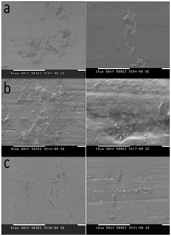
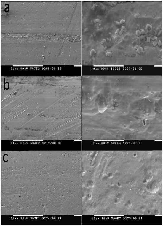
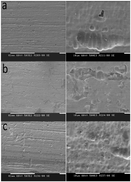
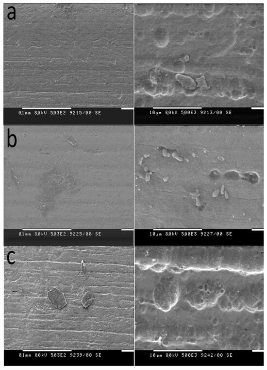
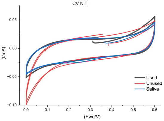
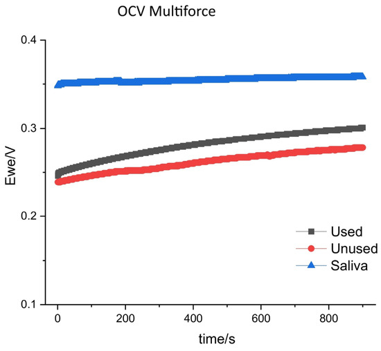
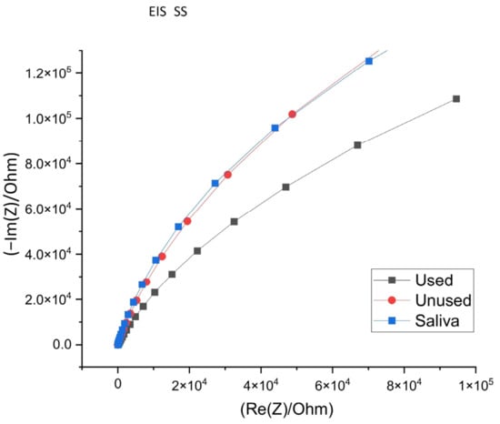
Objectives: To evaluate the corrosion behavior and surface characteristics of four nickel-containing orthodontic archwires (stainless steel (SS), superelastic nickel–titanium (NiTi), copper–nickel–titanium (CuNiTi), and multiforce NiTi) under unused, in vitro artificial saliva-immersed, and clinically used (in vivo) conditions. Methods: Rectangular SS, NiTi, CuNiTi, and multiforce NiTi archwires were analyzed in the following three conditions: as received, after one week of immersion in artificial saliva (pH 6.4), and after clinical use for 6–8 weeks. Corrosion behavior was assessed using cyclic voltammetry (CV), open-circuit voltammetry (OCV), and electrochemical impedance spectroscopy (EIS). Surface morphology was examined by scanning electron microscopy (SEM). Results: Corrosion behavior was dependent on archwire type and exposure condition. SS archwires exhibited reduced impedance response after clinical use, indicating passive-film degradation. In the clinically used NiTi specimen, pronounced electrochemical instability was observed, characterized by a deep OCV transient, slow repassivation, and SEM evidence compatible with localized pitting corrosion. In the CuNiTi specimens, minimal differences were observed between the unused and clinically used conditions, which may be consistent with stable passive-film integrity in these specimens. In the multiforce specimen, clinical use was associated with a higher impedance response than the unused and saliva-immersed conditions of that same specimen. In the saliva-immersed specimens, possible passive-film formation and a higher impedance response were observed relative to the other conditions of the same specimens, but these did not reproduce the electrochemical and morphological changes observed after clinical use. Conclusions: In vitro artificial saliva immersion does not reliably replicate in vivo aging of nickel-containing orthodontic archwires. Corrosion behavior evolves during clinical service in an archwire-specific manner, with NiTi archwires showing susceptibility to clinically induced surface degradation.
Full article

Figure 1
Open AccessArticle
The Agreement Between Preoperative Digital Radiographic Measurements and Clinically Fitted Stainless Steel Crown Size in Primary Molars: A Retrospective Study
by
Mayar Alharbi, Eman Bakhurji, Ashwin Shetty, Mohammed Alsaati and Yousef Alyousef
Dent. J. 2026, 14(9), 566; https://doi.org/10.3390/dj14090566 - 4 Sep 2026
Abstract
►▼
Show Figures
Background/Objectives: Accurate selection of stainless steel crown (SSC) size is essential for successful restoration of primary molars. Conventional crown selection relies on a trial-and-error approach, which may increase chair time and reduce clinical efficiency. Preoperative digital radiographic measurements may provide an objective adjunct
[...] Read more.
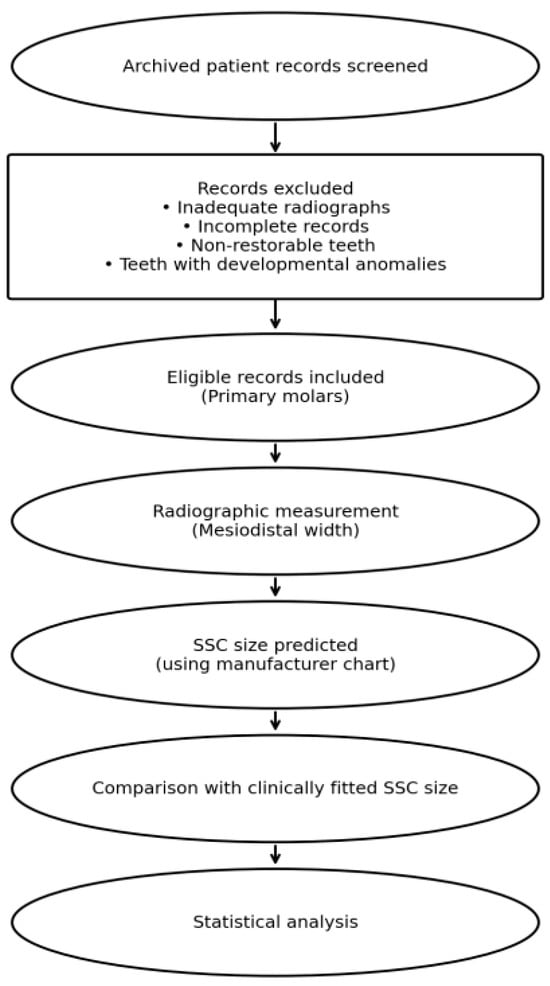
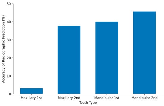
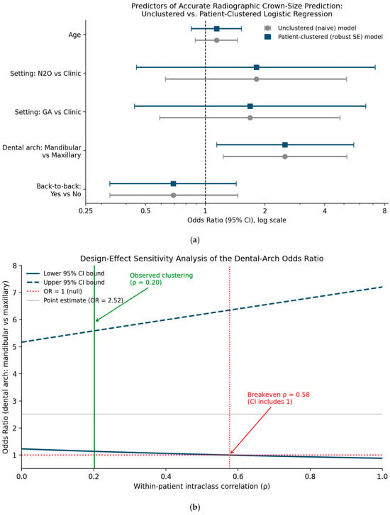
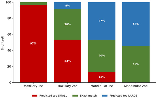
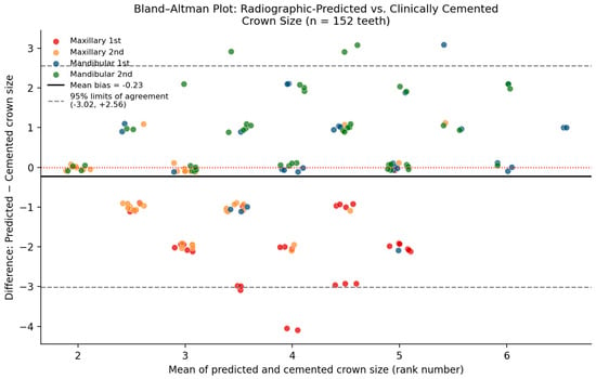
Background/Objectives: Accurate selection of stainless steel crown (SSC) size is essential for successful restoration of primary molars. Conventional crown selection relies on a trial-and-error approach, which may increase chair time and reduce clinical efficiency. Preoperative digital radiographic measurements may provide an objective adjunct for predicting SSC size; however, evidence supporting their agreement with clinically selected crown sizes remains limited. This study aimed to evaluate the agreement between preoperative digital radiographic measurements and clinically fitted SSC size in primary molars, and to assess whether this agreement is influenced by dental arch or tooth type. Methods: This retrospective cross-sectional study analyzed archived patient records and preoperative digital radiographs of pediatric patients who received SSCs on primary molars. Mesiodistal tooth dimensions were measured and converted to SSC sizes according to manufacturer guidelines. The clinically fitted and cemented crown size, as recorded in the patient chart, served as the reference standard against which radiographically predicted size was compared. Intra-examiner reliability was assessed using the intraclass correlation coefficient. Associations were analyzed using chi-square tests and binomial logistic regression (p ≤ 0.05). Given repeated teeth within patients, the primary logistic regression model was additionally re-fit using generalized estimating equations (GEEs) with an exchangeable working correlation structure and robust standard errors, and prediction error was further characterized using near-miss accuracy, direction of bias, weighted kappa, and Bland–Altman analysis. Results: A total of 152 primary molars from 71 patients were included. Intra-examiner reliability was excellent (ICC = 0.99). Radiographic prediction agreed with the clinically fitted crown size in 33.6% of cases, and 69.7% of predictions fell within one crown size of the cemented crown. Agreement was significantly higher for mandibular molars than maxillary molars (p = 0.010) and varied by tooth type (p < 0.001). Mandibular molars were more likely to show accurate prediction (OR = 2.52; 95% CI: 1.23–5.17); this association remained statistically significant after accounting for within-patient clustering using GEEs with an exchangeable correlation structure (OR = 2.46; 95% CI: 1.10–5.49; p = 0.029). Maxillary first molars showed a systematic under-prediction of exactly two crown sizes in 97% of cases (mean bias −2.00), identifying a specific, correctable calibration problem. Conclusions: Preoperative digital radiographic measurements show moderate agreement with clinically fitted crown size and may serve as a useful adjunct to clinical judgment for SSC size selection, particularly for mandibular molars. Because the reference standard reflects clinical judgment itself rather than an independent anatomical gold standard, these findings should be interpreted as a concordance analysis, and further prospective work is needed before efficiency benefits can be confirmed.
Full article

Figure 1
Open AccessArticle
Flexural Stiffness and Force Retention of Six 3D-Printed Thermoplastics for Orthodontic Appliances: An In Vitro Screening Study
by
Marco Serafin, Marina Borgese, Elisa Boccalari, Gilberto Binda, Mario Raspanti and Piero Antonio Zecca
Dent. J. 2026, 14(9), 565; https://doi.org/10.3390/dj14090565 - 4 Sep 2026
Abstract
►▼
Show Figures
Background/Objectives: Directly printed orthodontic appliances rely mostly on vat-photopolymerisation resins; pre-polymerised thermoplastics processed by fused deposition modelling (FDM) and selective laser sintering (SLS) are a less-explored, single-component route, but are poorly characterised. This study compared six thermoplastic material–process systems, supplied for biomedical or
[...] Read more.
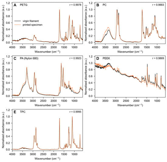
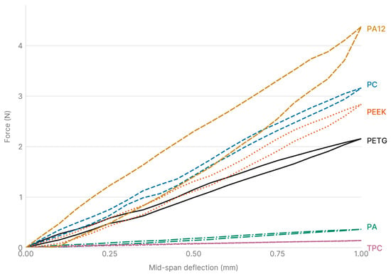
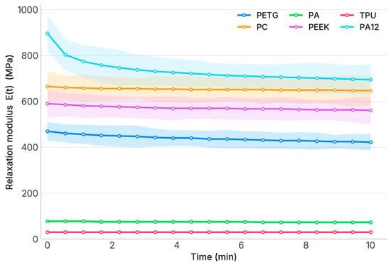
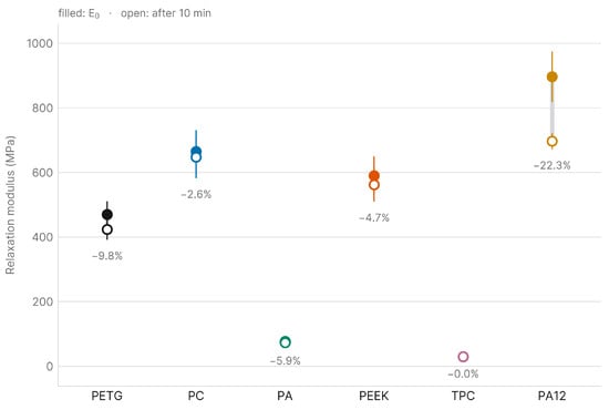
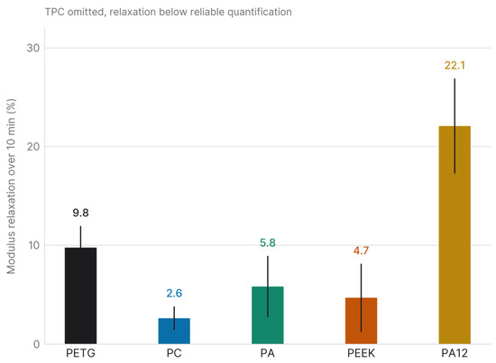
Background/Objectives: Directly printed orthodontic appliances rely mostly on vat-photopolymerisation resins; pre-polymerised thermoplastics processed by fused deposition modelling (FDM) and selective laser sintering (SLS) are a less-explored, single-component route, but are poorly characterised. This study compared six thermoplastic material–process systems, supplied for biomedical or dental use, using a single intraoral-simulating protocol. Methods: Thin specimens (25 × 5 × 0.6 mm) of polyethylene terephthalate glycol (PETG), polycarbonate (PC), polyamide (PA), polyether-ether-ketone (PEEK) and a thermoplastic copolyester elastomer (TPC) printed by FDM, and of polyamide 12 (PA12) by SLS, underwent three-point bending and 10 min stress relaxation at 37 °C in artificial saliva (n = 5). Differences were assessed using the Kruskal–Wallis test with Dunn post hoc comparisons (Holm correction), and Fourier-transform infrared (FTIR) spectroscopy was used to compare virgin and printed materials for the five FDM polymers. Results: Apparent flexural moduli were 989 (PA12), 750 (PC), 687 (PEEK), 509 (PETG), 85 (PA) and 32 MPa (TPC), a very large material effect (ε2 = 0.95), with post hoc comparisons separating only the extremes of that range. Relaxation over 10 min ranged from 2.6% (PC) to 22.1% (PA12) among the five materials in which it could be quantified, with TPC falling below the load-cell resolution; expressed as force, PA12 lost the most (0.86 N) and yet still delivered 3.01 N, which was above the initial force of PEEK, PETG, PA and TPC, though not separable from that of PC, which retained 97.4% of its own. Virgin and printed spectra were concordant (Pearson r ≥ 0.986, against ≤ 0.80 between different materials). Conclusions: The PA12/SLS system was the stiffest, followed by PC and PEEK, but, as the only laser-sintered material, its polymer and process effects cannot be separated. PETG and PC combine useful stiffness with accessible FDM; PA and TPC are far more compliant. These specimen-level findings provide a screening basis for selecting material–process systems for printed orthodontic appliances, preceding device-level validation.
Full article

Figure 1
Open AccessArticle
Efficacy of Dual-Light Photodynamic Therapy on Oral Health in Adolescents Undergoing Fixed Orthodontic Treatment: A Randomized, Assessor-Blinded and Controlled Trial
by
Katja Hashemi Elses, Tommi Pätilä and Ann-Marie Roos Jansåker
Dent. J. 2026, 14(9), 564; https://doi.org/10.3390/dj14090564 - 4 Sep 2026
Abstract
Background/Objectives: Patients with fixed orthodontic appliances are at increased risk of plaque accumulation, gingivitis, and caries because brackets and bands hinder mechanical hygiene. This study evaluated whether regular home use of dual-light antibacterial photodynamic therapy (aPDT) supports oral hygiene during fixed orthodontic
[...] Read more.
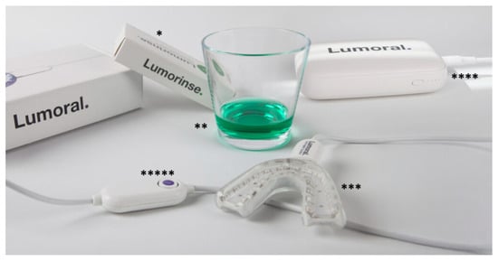
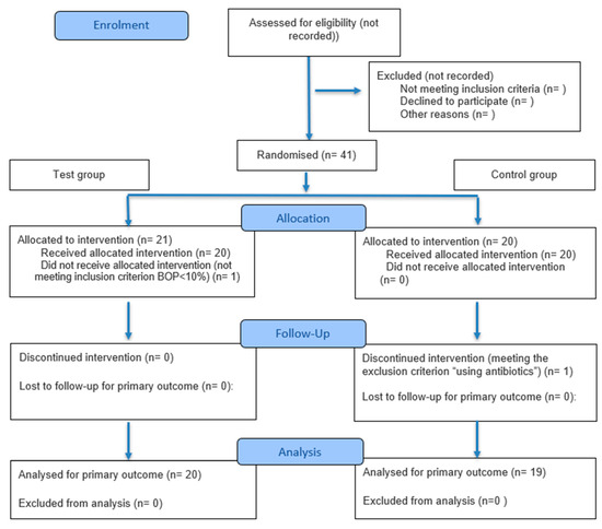
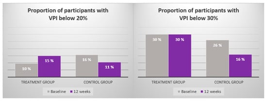
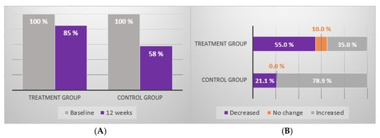
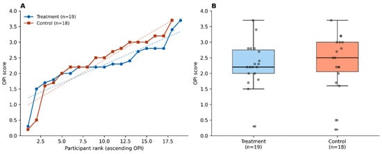
Background/Objectives: Patients with fixed orthodontic appliances are at increased risk of plaque accumulation, gingivitis, and caries because brackets and bands hinder mechanical hygiene. This study evaluated whether regular home use of dual-light antibacterial photodynamic therapy (aPDT) supports oral hygiene during fixed orthodontic treatment in adolescents. Methods: In a single-center, randomized, assessor-blinded trial, forty-one adolescents starting fixed orthodontic treatment were randomised to a Control group (standard hygiene) or a Test group (standard hygiene plus home-use dual-light aPDT). Visible Plaque Index (VPI%), Bleeding on Probing (BOP%), and Orthodontic Plaque Index (OPI) were assessed at baseline and 12 weeks by an assessor blinded to allocation. After two protocol-driven exclusions, 39 participants (20 Test group, 19 Control group) were analysed. Site-level proportions were compared using the chi-square test. Results: VPI decreased in both groups, with a larger mean within-group reduction in the Test group (−7.0%) than in the Control group (−3.7%); exploratory site-level analysis showed a greater reduction in plaque-positive sites in the Test group at 12 weeks (p < 0.001). BOP increased in both groups but to a smaller extent in the Test group (mean +2.4% versus +5.7%), and a higher proportion of Test-group participants maintained BOP ≤ 10% (85% versus 58%); bleeding-positive sites were lower in the Test group at 12 weeks (p < 0.001). The Test group also showed a flatter distribution of OPI scores around orthodontic brackets. No device- or protocol-related adverse events were reported. Conclusions: In adolescents undergoing fixed orthodontic treatment, regular home use of dual-light aPDT was safe and associated with better site-level plaque and bleeding control, with directional but non-significant trends at the participant level. The findings support further evaluation of dual-light aPDT as an adjunct to mechanical hygiene during this vulnerable treatment period.
Full article
(This article belongs to the Section Oral Hygiene, Periodontology and Peri-implant Diseases)
►▼
Show Figures

Figure 1
Open AccessArticle
Optical Attenuation Analysis of Biofilm Formation on Dental Prostheses Using Time-Domain Optical Coherence Tomography and Atomic Force Microscopy
by
Maria Cecilia Galvez, Aaron Mharl Barlisan, Emmanuelle Hermosilla, Mark Nickole Tabafa, Ernest Macalalad, Jose Esmeria Jr., Edgar Vallar, Seiroh Okaneya, Jumar Cadondon and Tatsuo Shiina
Dent. J. 2026, 14(9), 563; https://doi.org/10.3390/dj14090563 - 3 Sep 2026
Abstract
Background/Objectives: Dental biofilm formation on prosthetic materials contributes to oral diseases and material degradation. This pilot study evaluated the feasibility of combining time-domain optical coherence tomography (TD-OCT) and non-contact atomic force microscopy (NC-AFM) to characterize biofilm-associated optical and surface changes in dental prosthesis
[...] Read more.
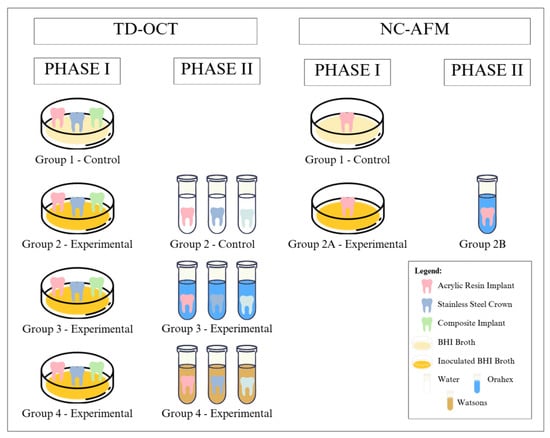
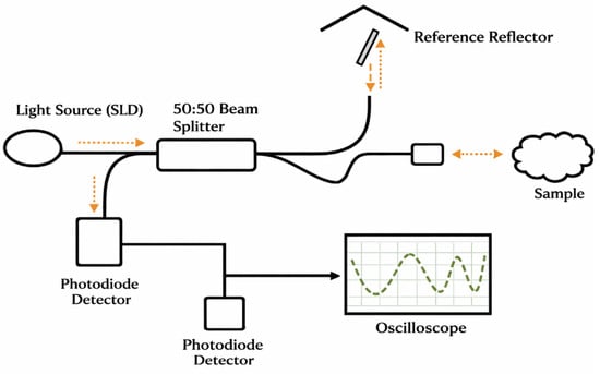
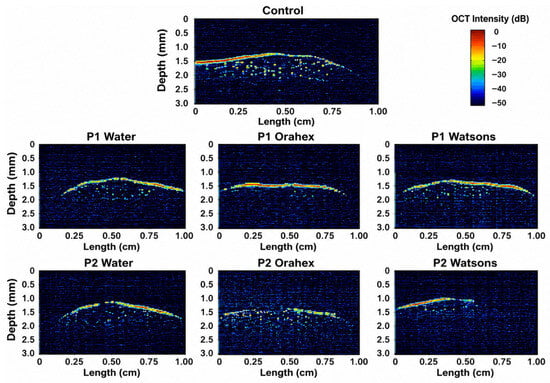
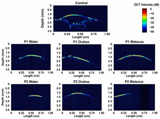
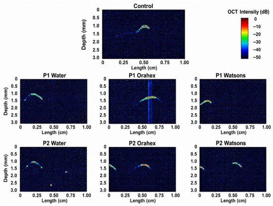
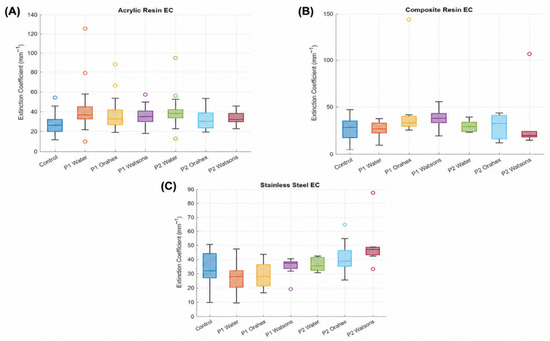
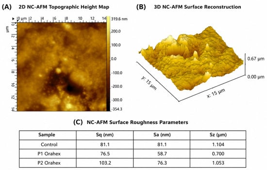
Background/Objectives: Dental biofilm formation on prosthetic materials contributes to oral diseases and material degradation. This pilot study evaluated the feasibility of combining time-domain optical coherence tomography (TD-OCT) and non-contact atomic force microscopy (NC-AFM) to characterize biofilm-associated optical and surface changes in dental prosthesis materials following antiseptic treatment. Methods: Acrylic resin, composite resin, and stainless-steel specimens were inoculated with Streptococcus mutans biofilms and exposed to water, Orahex, and Watsons mouthwash. TD-OCT was used to obtain extinction coefficient (EC) and optical thickness measurements, while NC-AFM was performed on Orahex-treated specimens to assess nanoscale surface morphology. EC distributions were analyzed descriptively from valid TD-OCT A-scans. Results: Within the examined specimens, EC distributions varied following biofilm formation and antiseptic treatment, with material-dependent responses. Composite resin exhibited the clearest reduction in EC following Watsons treatment, whereas acrylic resin and stainless-steel specimens showed more variable responses. Physical thickness remained relatively stable, while NC-AFM observations of the Orahex-treated specimens supported the TD-OCT findings by demonstrating corresponding surface morphological changes. Conclusions: This pilot study demonstrates the preliminary feasibility of integrating TD-OCT with NC-AFM for non-destructive evaluation of biofilm-associated optical and surface changes in dental prosthesis materials. The findings are descriptive and exploratory, highlighting the potential of TD-OCT for monitoring prosthetic materials while emphasizing the need for future studies incorporating independent biological replicates and complementary biological validation techniques.
Full article
(This article belongs to the Special Issue Advances in Dental Imaging: Innovations and Applications)
►▼
Show Figures

Figure 1
Open AccessSystematic Review
Periodontal and Peri-Implant Status Among Users of Different Inhaled Tobacco and Nicotine Products: A Systematic Review
by
Maria Pia Di Palo, Federica Di Spirito, Francesco Giordano, Giuseppina De Benedetto, Roberto Lo Giudice, Federica Piedepalumbo, Gabriele Balsamo, Colomba Pessolano, Marina Garofano, Andrea Marino and Alessia Bramanti
Dent. J. 2026, 14(9), 562; https://doi.org/10.3390/dj14090562 - 3 Sep 2026
Abstract
Background/Objectives: Periodontal and peri-implant diseases are influenced by several risk factors, including smoking. While cigarette smoking (CS) is a well-established risk factor, the impact of heated tobacco products (HTPs), electronic cigarettes (E-Cigs), and waterpipe (WS) remains less clear. This systematic review aimed
[...] Read more.
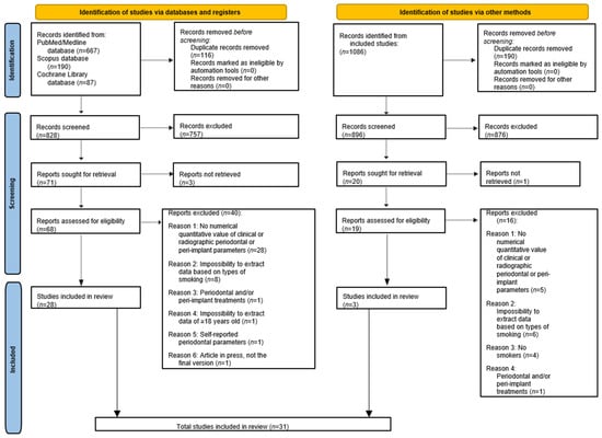
Background/Objectives: Periodontal and peri-implant diseases are influenced by several risk factors, including smoking. While cigarette smoking (CS) is a well-established risk factor, the impact of heated tobacco products (HTPs), electronic cigarettes (E-Cigs), and waterpipe (WS) remains less clear. This systematic review aimed to update the current evidence evaluating the clinical, radiographical, and inflammatory biomarkers of periodontal and peri-implant status among different untreated adult users of inhaled tobacco and nicotine products. Methods: A systematic search of PubMed/MEDLINE, Scopus, and Cochrane Library was performed for studies published between April 2022 and December 2025. Data were extracted and synthesized qualitatively according to the study protocol (PROSPERO: CRD420261284851). Studies were qualitatively assessed using the Risk-of-Bias in Nonrandomized-I. Results: Thirty-one studies including 3106 subjects were analyzed. CS consistently showed the worst periodontal and peri-implant status, with higher plaque indices, probing depth (PD), clinical attachment level (CAL), marginal bone loss (MBL) and more missing teeth. WS showed parameters comparable to CS and worse than non-smokers, with particularly high plaque accumulation and probing depth, as well as a pro-inflammatory biomarker profile. E-Cigs and HTPs generally exhibited intermediate status; E-Cigs showed increased PD and CAL compared with non-smokers, with elevated bleeding on probing despite a lower gingival index, whereas MBL was notably higher, showing the highest overall values among all smoking categories, exceeding CS. HTPs presented slightly less harmful outcomes than other smokers, but the evidence was limited. Biomarker analysis indicated elevated pro-inflammatory and tissue-destructive markers, particularly in CS and WS. Conclusions: Smoking negatively affects periodontal and peri-implant status, with CS exhibiting the worst impact. E-Cigs and HTPs appear less harmful but are not risk-free. Longitudinal studies are warranted to define long-term effects, particularly on peri-implant status, for which robust evidence was restricted to CS.
Full article
(This article belongs to the Special Issue Advances in Periodontal and Peri-Implant Tissues Health Management: 2nd Edition)
►▼
Show Figures

Figure 1
Open AccessReview
A Proposed Parameter-Based Planning Framework for Inferior Alveolar Nerve Repositioning
by
Fares Kablan, Asaf Zigron, Idan Redenski, Shadi Daoud, Amjad Shhadeh, Adeeb Zoabi and Samer Srouji
Dent. J. 2026, 14(9), 561; https://doi.org/10.3390/dj14090561 - 3 Sep 2026
Abstract
Background: Rehabilitation of the severely atrophic posterior mandible remains challenging when the residual bone height above the inferior alveolar nerve (IAN) is insufficient for conventional implant placement. In such cases, IAN lateralization or transposition may facilitate placement of longer implants and support
[...] Read more.
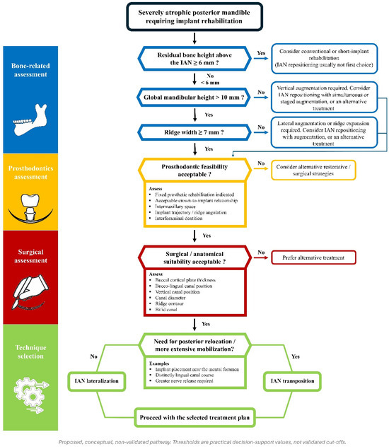
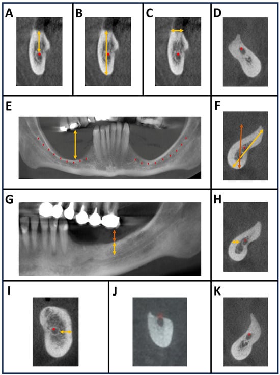
Background: Rehabilitation of the severely atrophic posterior mandible remains challenging when the residual bone height above the inferior alveolar nerve (IAN) is insufficient for conventional implant placement. In such cases, IAN lateralization or transposition may facilitate placement of longer implants and support fixed prosthetic rehabilitation. However, these procedures require careful case selection because of their technical complexity and the risk of neurosensory complications. Objective: This narrative review presents a literature-informed, expert-derived, parameter-based clinical decision framework for preoperative planning of IAN lateralization and transposition in the severely atrophic posterior mandible. Methods: A focused appraisal of the literature was performed to identify anatomical, prosthodontic, and surgical parameters relevant to IAN repositioning. Parameters were selected according to their recurrent relevance to surgical feasibility, implant positioning, procedural safety, and restorative planning, and were integrated with the authors’ clinical experience to develop a structured decision-making framework. No formal risk-of-bias or certainty-of-evidence assessment was performed. Each parameter was assigned an evidence-source category and a threshold status to make its evidentiary basis explicit. Results: The proposed framework organizes treatment planning into three domains: bone-related, prosthodontic, and surgical considerations. Key parameters include residual bone height above the IAN canal, global mandibular height, ridge width, bone density, crown-to-implant ratio, intermaxillary space, ridge angulation, buccal cortical plate thickness, and the three-dimensional course of the IAN. Additional parameters include interforaminal dentition, implant trajectory, canal diameter, bifid canal anatomy, and ridge contour. Collectively, these variables are integrated into a stepwise decision pathway to help determine whether IAN repositioning is appropriate, whether lateralization or transposition is preferable, and when alternative approaches, such as short implants, ridge splitting, or vertical augmentation, may be more suitable. The framework links restorative feasibility and surgical risk to the choice of technique within a single sequential pathway, and reports the evidence basis of each decision point. Conclusions: The proposed framework should not be regarded as a validated clinical decision tool; prospective clinical validation and consensus-based refinement are required before routine clinical implementation. The framework is intended to structure and guide multidisciplinary assessment and discussion of cases; it should not independently determine treatment selection or replace individualized clinical judgment.
Full article
(This article belongs to the Section Oral and Maxillofacial Surgery)
►▼
Show Figures

Figure 1
Open AccessArticle
Screening ‘Gum Disease or Problem’ Status Using Molar Probing Depths: Model Development and Validation in NHANES Young to Middle-Aged Adults
by
Miika Penttala
Dent. J. 2026, 14(9), 560; https://doi.org/10.3390/dj14090560 - 3 Sep 2026
Abstract
Background/Objectives: A parsimonious multivariable model was engineered and externally validated using dental electronic health records to screen for a clinician-evaluated “gum disease or related problem” status. Designed for seamless electronic integration, a positive screening result triggers a targeted referral—and the model is well-suited
[...] Read more.
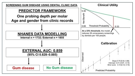
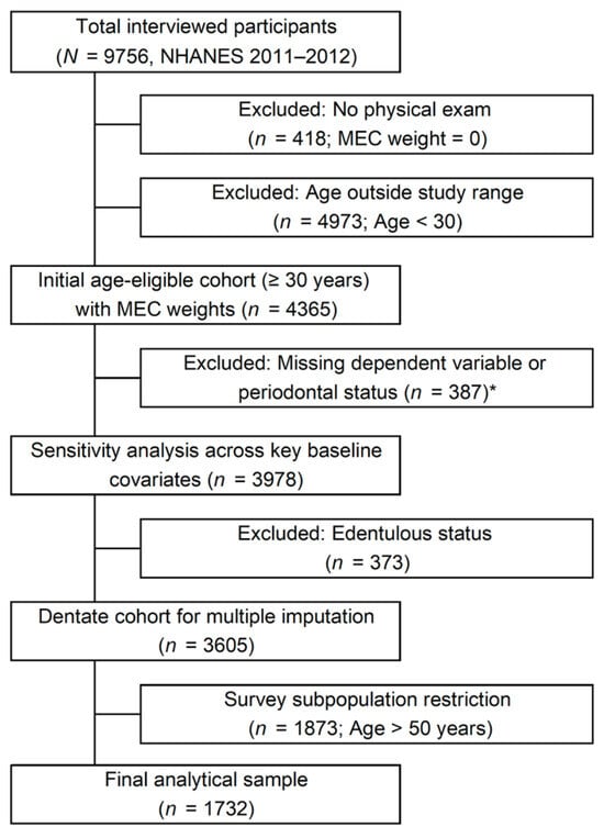
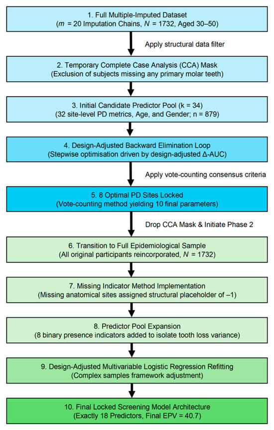
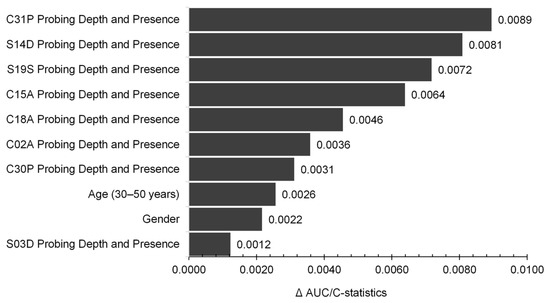
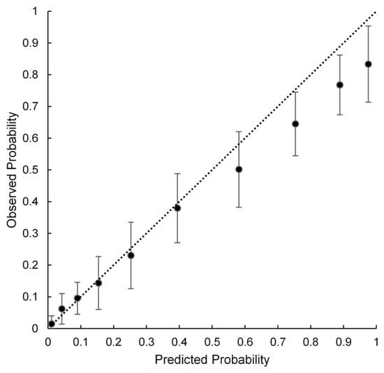
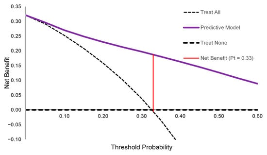
Background/Objectives: A parsimonious multivariable model was engineered and externally validated using dental electronic health records to screen for a clinician-evaluated “gum disease or related problem” status. Designed for seamless electronic integration, a positive screening result triggers a targeted referral—and the model is well-suited for dental hygienist use— while providing clinical decision support for dentists during definitive diagnoses. This automated support standardises workflows, maximises team efficiency, and supports data-driven clinical decision-making. Methods: This cross-sectional study developed the model using the US National Health and Nutrition Examination Survey (NHANES) 2011–2012 cohort (n = 1732) and independently validated it on the 2013–2014 replication cycle (n = 1855) for the high-risk 30–50-year-old demographic. Survey-weighted logistic regression incorporated age, gender, and eight targeted molar probing depth (PD) measurements paired with binary indicators for tooth presence to predict whether an individual has a gum disease or related problem. Results: External validation demonstrated high parameter generalisability, yielding a pooled population-level AUC of 0.859 (95% CI: 0.828–0.885). At the prevalence-matched threshold of Pt = 0.367, the framework yielded a sensitivity of 78.39% (95% CI: 69.48–85.29%) and a specificity of 79.10% (95% CI: 74.55–83.04%). Demonstrating public health utility via Decision Curve Analysis, the model avoided approximately 39 unnecessary clinical examinations by a dentist per 100 individuals at a practical clinical decision threshold of 0.33, achieving a substantial pooled net benefit compared with a treat-all strategy without compromising case-detection metrics. Conclusions: This scalable screening model offers a highly practical pathway for gum disease and related problem interception. Optimised for seamless software integration, the framework facilitates rapid, hygienist-led risk stratification without increasing chairside burden, and delivers objective data streams to reinforce the dentist’s definitive diagnostic workflow.
Full article
(This article belongs to the Section Preventive Dentistry)
►▼
Show Figures

Graphical abstract
Open AccessArticle
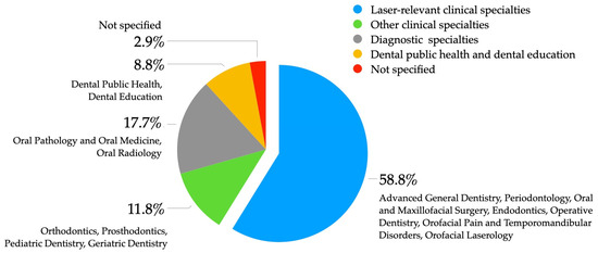
Institutional Readiness for Orofacial Laserology Education in Thailand: A National Cross-Sectional Survey of Dental Schools
by
Suwat Tanya, Chattarika Domdok and Patcharawan Srisilapanan
Dent. J. 2026, 14(9), 559; https://doi.org/10.3390/dj14090559 - 2 Sep 2026
Abstract
Background/Objectives: Orofacial laserology is being increasingly incorporated into contemporary dental practice, yet its integration into dental education remains inconsistent. National evidence regarding institutional readiness for integrating orofacial laserology into dental curricula remains limited. This study examined the current status of orofacial laserology education
[...] Read more.
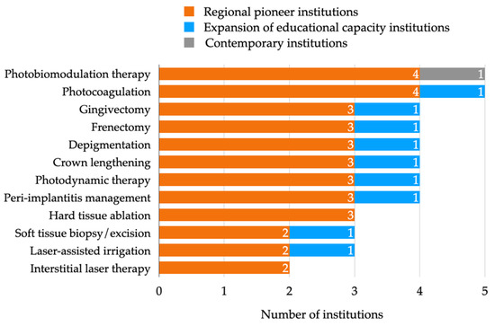
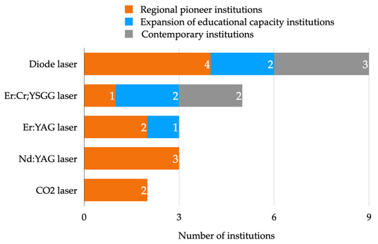
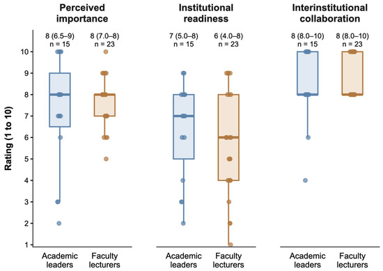
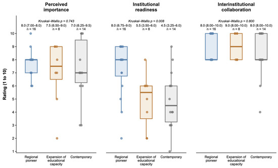
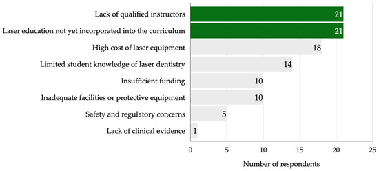
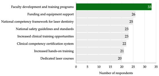
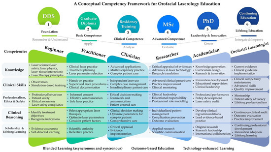
Background/Objectives: Orofacial laserology is being increasingly incorporated into contemporary dental practice, yet its integration into dental education remains inconsistent. National evidence regarding institutional readiness for integrating orofacial laserology into dental curricula remains limited. This study examined the current status of orofacial laserology education in Thailand. Methods: A national cross-sectional survey was conducted between May and July 2026 among all accredited Thai dental schools. Academic leaders and full-time lecturers completed a structured online questionnaire addressing curriculum implementation, educational delivery, competency assessment, institutional readiness, perceived barriers, and future priorities. Descriptive and non-parametric statistical analyses were performed. Results: Fifteen of 18 dental schools (83.3%) participated, yielding 38 respondents. Undergraduate orofacial laserology education showed limited curricular integration, whereas postgraduate programs demonstrated broader implementation, particularly in hands-on, supervised clinical, and competency-based training. Institutional readiness did not differ between academic leaders and full-time lecturers (p > 0.05). Across historical phases of institutional establishment, perceived importance (p = 0.743) and interinstitutional collaboration (p = 0.800) were comparable, whereas institutional readiness differed significantly (p = 0.008), with regional pioneer dental schools reporting higher readiness than expansion-phase and contemporary dental schools. The principal barriers were insufficient qualified instructors, limited curriculum integration, and high equipment costs. Faculty development, funding and equipment support, and national competency frameworks were identified as the highest priorities. Conclusions: Orofacial laserology education in Thailand remains heterogeneous, with broader implementation in postgraduate than undergraduate programs. Strengthening faculty capacity, competency-based curricula, institutional support, and national collaboration may facilitate more consistent integration of orofacial laserology into dental curricula.
Full article
(This article belongs to the Section Lasers in Dentistry)
►▼
Show Figures

Figure 1
Open AccessReview
Artificial Intelligence and Digital Workflow in Craniofacial Bone Tissue Engineering: From Cone-Beam Computed Tomography (CBCT) to Personalized Bioceramic Implants
by
Amirhossein Bahador, Seyed Ali Mostafavi Moghaddam, Hamid Mojtahedi, Lotfollah Kamali Hakim and Hamid Tebyaniyan
Dent. J. 2026, 14(9), 558; https://doi.org/10.3390/dj14090558 - 2 Sep 2026
Abstract
Background: Reconstruction of craniofacial bone defects caused by tumor removal, infection, congenital anomalies, or trauma remains a major challenge. Recent advances in artificial intelligence (AI), digital imaging, and additive manufacturing have enabled personalized treatment strategies. This review discusses AI-driven workflows from cone-beam computed
[...] Read more.
Background: Reconstruction of craniofacial bone defects caused by tumor removal, infection, congenital anomalies, or trauma remains a major challenge. Recent advances in artificial intelligence (AI), digital imaging, and additive manufacturing have enabled personalized treatment strategies. This review discusses AI-driven workflows from cone-beam computed tomography (CBCT) image acquisition to implant fabrication using patient-specific bioceramics. Methods: Several electronic databases were searched, including Scopus, PubMed, ScienceDirect, and Web of Science. A peer-reviewed article published between 2015 and 2026 was considered for inclusion. This review provides an overview of AI applications in craniofacial tissue engineering, including CBCT image segmentation, three-dimensional reconstruction, virtual surgical planning, computer-aided design/computer-aided manufacturing (CAD/CAM), topology optimization, finite element analysis, and three-dimensional bioceramic scaffold printing. Results: AI improves accuracy, efficiency, and reproducibility of craniofacial reconstructions when used in conjunction with AI-assisted workflows. Through automated image segmentation and anatomical modeling, operator dependence can be reduced, and predictive algorithms can optimize biomechanical properties. CAD/CAM and 3D printing are used to manufacture bioceramic implants with controlled porosity, mechanical integrity, and enhanced osteoconductive properties. AI-driven predictive models have demonstrated potential for supporting material selection, manufacturing quality control, and treatment planning; however, their ability to reliably predict long-term postoperative outcomes requires further validation through prospective clinical studies. Data standardization, algorithm transparency, regulatory approval, clinical trial validation, and ethical considerations remain challenges. Conclusions: AI, CBCT imaging, computational modeling, and advanced bioceramic manufacturing are being combined to create an end-to-end digital workflow for personalized reconstruction. Clinical studies must be conducted prospectively to maximize the therapeutic potential of these technologies.
Full article
(This article belongs to the Special Issue Digital Implantology in Dentistry)
Open AccessArticle
Subjective Xerostomia and Chairside Salivary Dysfunction in Institutionalized Older Adults: Concordance and Exploratory Derivation of a Five-Item Short Form
by
Mădălina Monica Bicheru, Andreea Zamfirescu, Elena Preoteasa, Adrian Iustin Georgevici, Nicolae Vladimir Bicheru and Cristina Teodora Preoteasa
Dent. J. 2026, 14(9), 557; https://doi.org/10.3390/dj14090557 - 2 Sep 2026
Abstract
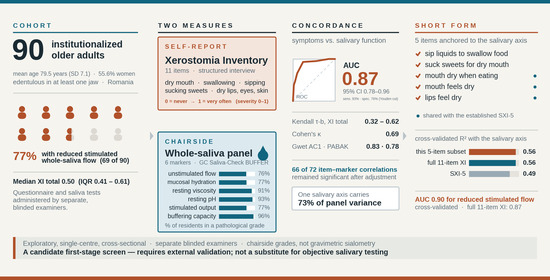
Background/Objectives: In institutionalized older adults, we examined the concordance between self-reported xerostomia and chairside salivary dysfunction and explored whether a smaller subset of Xerostomia Inventory items could retain comparable screening performance. Methods: This cross-sectional study included institutionalized older adults. The 11-item
[...] Read more.
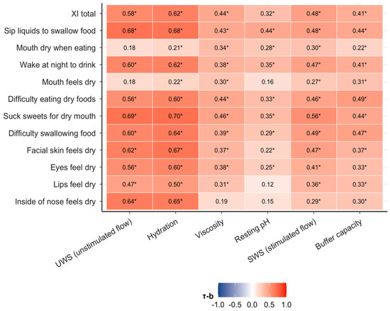
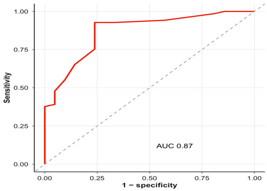
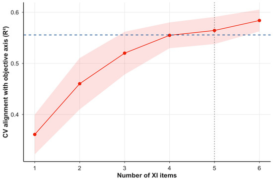
Background/Objectives: In institutionalized older adults, we examined the concordance between self-reported xerostomia and chairside salivary dysfunction and explored whether a smaller subset of Xerostomia Inventory items could retain comparable screening performance. Methods: This cross-sectional study included institutionalized older adults. The 11-item Xerostomia Inventory was compared with a six-marker chairside whole-saliva panel, with all variables scaled 0 (normal) to 1 (pathological). We measured how closely symptoms tracked each objective marker, how well the questionnaire identified hyposalivation, and whether fewer questions could be comparable with the full 11-item inventory. Results: The study included 90 participants, with a mean age of 79.5 years; 55.6% were women. Symptoms tracked every marker in the expected direction (correlation τ-b 0.32 to 0.62). The questionnaire identified objective hyposalivation with an area under the ROC curve of 0.87 (95% CI 0.78–0.96) and substantial chance-corrected agreement (Cohen’s κ 0.69, 95% CI 0.51–0.87). A five-item short form reproduced the objective findings as well as the full questionnaire did (cross-validated R2 0.56 ± 0.03 versus 0.56). Conclusions: In this high-prevalence institutional cohort, self-reported symptoms agreed with graded salivary function more closely than the older discordance literature would suggest, though still within the range of recent studies rather than beyond it. A five-item subset, chosen for its alignment with the objective salivary axis rather than for internal consistency, recovered as much as the full inventory, which points to a short, function-oriented score as a plausible first-stage screen. We would treat this short form as a candidate first-stage screening approach that requires external validation, rather than as a replacement for objective salivary assessment or as a validated replacement for the Xerostomia Inventory.
Full article
(This article belongs to the Special Issue Geriatric Dentistry: Innovations, Challenges, and Future Directions)
►▼
Show Figures

Graphical abstract
Open AccessArticle
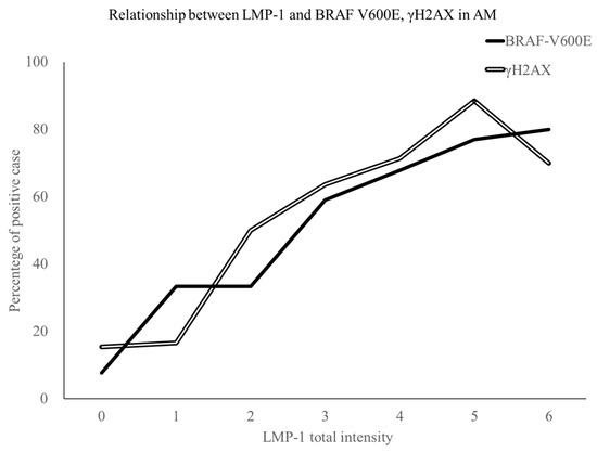
Relationship Between Epstein–Barr Virus LMP-1 and BRAF-V600E Mutant Protein Expression in Ameloblastoma
by
Kenko Okamoto, Michiko Nishimura, Yuji Miyazaki, Miyako Hoshino, Shinnichi Sakamoto, Miki Haruyama, Fumio Ide, Nobuharu Yamamoto and Kentaro Kikuchi
Dent. J. 2026, 14(9), 556; https://doi.org/10.3390/dj14090556 - 2 Sep 2026
Abstract
►▼
Show Figures
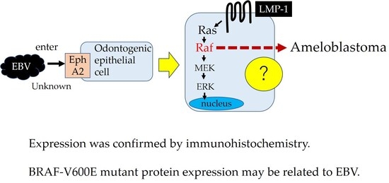
Background: Epstein–Barr virus (EBV) enters the human body via saliva. EBV-encoded latent membrane protein-1 (LMP-1) activates the mitogen-activated protein kinase (MAPK) pathway. MAPK signaling pathway activation due to the BRAF-V600E mutation is widely known as a major event in the pathogenesis of
[...] Read more.
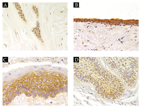
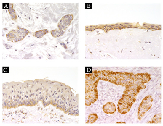
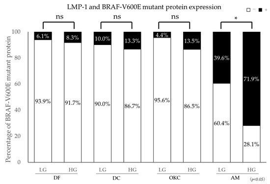
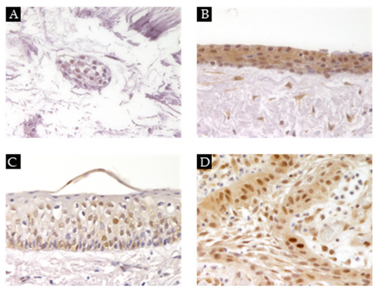
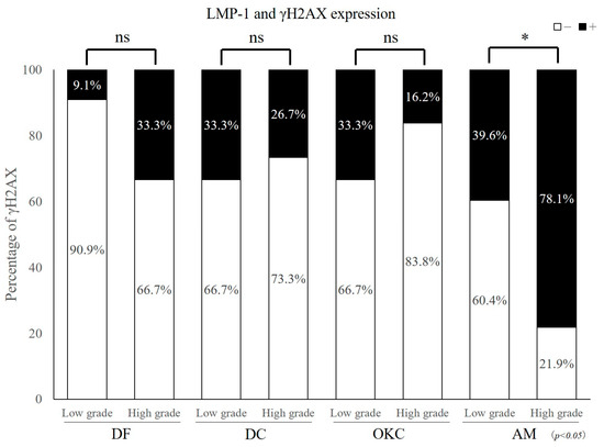
Background: Epstein–Barr virus (EBV) enters the human body via saliva. EBV-encoded latent membrane protein-1 (LMP-1) activates the mitogen-activated protein kinase (MAPK) pathway. MAPK signaling pathway activation due to the BRAF-V600E mutation is widely known as a major event in the pathogenesis of ameloblastoma. It is unclear whether EBV infects ameloblastoma and is involved in the pathogenesis of ameloblastoma. We investigated the relationship between LMP-1 and BRAF-V600E mutant protein expression in ameloblastoma by immunohistochemistry. Methods: We examined a total of 334 samples (ameloblastoma 117 samples, odontogenic keratocyst 127 samples, dentigerous cysts 45 samples, and dental follicle 45 samples). Ephrin type-A receptor 2 (EphA2), BRAF-V600E mutant protein, and γH2AX expression were confirmed by immunohistochemistry. We confirmed that the positive cases of BRAF-V600E mutant protein and γH2AX segregated based on LMP-1 expression: high grade (HG) and low grade (LG). Results: EphA2 expression was high in all lesions. The positive rate of BRAF-V600E mutant protein expression was significantly higher in ameloblastoma, and the positive rate of BRAF-V600E mutant protein in LMP-1 HG cases (71.9%) was significantly greater than in LG cases (39.6%) in ameloblastoma. The positive rate of γH2AX expression was significantly higher in ameloblastoma than in the other lesions. The positive rate of BRAF-V600E mutant protein in LMP-1 HG cases (71.9%) was significantly greater than in LG cases (39.6%) in ameloblastoma. Conclusions: BRAF-V600E mutant protein and γH2AX expression in ameloblastoma were significantly higher in LMP-1 HG cases than in LG cases.
Full article

Graphical abstract
Open AccessReview
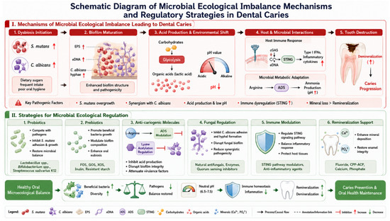
Recent Advances in Caries Prevention and Non-Invasive Management: From Ecological Modulation to Precision Interventions
by
Yanfei Yin, Wenxia Li, Wei Zhang, Ying Huang, Lingying Fan, Xiaolin Wei and Dongyang Ma
Dent. J. 2026, 14(9), 555; https://doi.org/10.3390/dj14090555 - 2 Sep 2026
Abstract
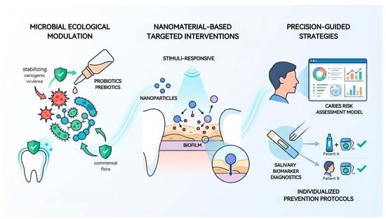
Background: Dental caries remains a globally prevalent biofilm-mediated disease driven by microbial dysbiosis and progressive enamel demineralization. The traditional “drill and fill” paradigm has increasingly given way to biologically informed preventive frameworks emphasizing ecological management. Methods: This narrative review synthesizes 134 studies (2016–2026)
[...] Read more.
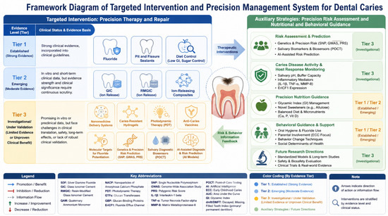
Background: Dental caries remains a globally prevalent biofilm-mediated disease driven by microbial dysbiosis and progressive enamel demineralization. The traditional “drill and fill” paradigm has increasingly given way to biologically informed preventive frameworks emphasizing ecological management. Methods: This narrative review synthesizes 134 studies (2016–2026) retrieved from PubMed, Web of Science, Scopus, and Google Scholar to evaluate emerging caries prevention strategies across three domains. Results: Three interrelated domains of advancement were identified. First, microbial ecological modulation—including probiotics, prebiotics, and targeted antimicrobial peptides—aims to restore oral microbiome homeostasis by disrupting cariogenic virulence without eliminating commensal flora. Second, nanomaterial-based targeted interventions utilizing stimuli-responsive carriers and biofilm-penetrating nanoparticles enable site-specific drug delivery with enhanced therapeutic efficacy. Third, precision-guided strategies incorporating caries risk assessment models and salivary biomarker diagnostics facilitate individualized prevention protocols. Conclusions: These findings demonstrate a fundamental paradigm shift from indiscriminate microbial eradication to equilibrium-driven, ecology-centered disease management. Future directions include AI-assisted personalized prevention, chairside microbiome diagnostics, and smart responsive biomaterials for sustained caries control.
Full article
(This article belongs to the Special Issue Caries Risk Assessment and Preventive Care Protocols)
►▼
Show Figures

Graphical abstract
Open AccessArticle
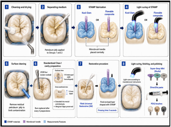
Effects of STAMP Techniques and Conventional Carving on Marginal Adaptation of Class I Composite Resin Restorations: An In Vitro Study
by
Benjamin Allen Bingham, Suchunya Rose Bumrung, Hassan Ziada and Neamat Hassan Abubakr
Dent. J. 2026, 14(9), 554; https://doi.org/10.3390/dj14090554 - 2 Sep 2026
Abstract
►▼
Show Figures
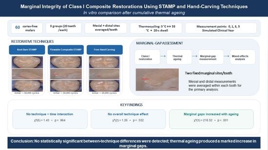
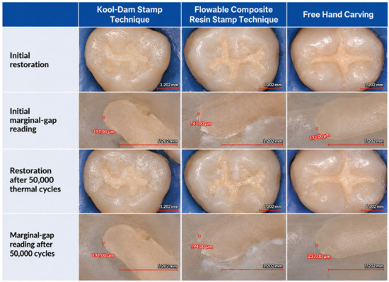
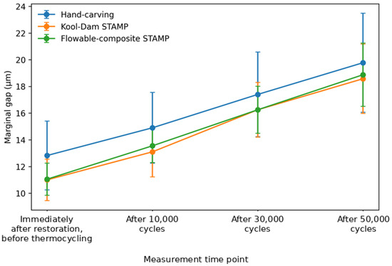
Background: Accurate reproduction of occlusal anatomy is a critical determinant of the clinical performance of posterior composite restorations. The present study compares marginal adaptation in Class I composite restorations completed using a Kool-Dam STAMP, a flowable-composite STAMP, or conventional hand-carving techniques. Methods
[...] Read more.
Background: Accurate reproduction of occlusal anatomy is a critical determinant of the clinical performance of posterior composite restorations. The present study compares marginal adaptation in Class I composite restorations completed using a Kool-Dam STAMP, a flowable-composite STAMP, or conventional hand-carving techniques. Methods: Sixty extracted caries-free molars were randomly allocated to three experimental groups (n = 20). Two fixed marginal sites per tooth (mesial and distal) were measured at four measurement time points: immediately after restoration, before thermocycling, and after 10,000, 30,000, and 50,000 cumulative thermal cycles (5 °C/55 °C; 20 s dwell time). The two site-level values were averaged within each tooth for the primary analysis. A linear mixed-effects model included technique, measurement time point, and the technique-by-time-point interaction, with a random intercept for tooth. Results: No significant interaction was observed between restorative technique and measurement time point, indicating that changes in marginal gap over time were not statistically significant among the three techniques (likelihood-ratio χ2(6) = 1.43, p = 0.964). The overall effect of the technique was also not significant (χ2(2) = 1.26, p = 0.532). In contrast, marginal-gap measurements increased considerably across the four measurement time points (χ2(3) = 218.52, p < 0.001), indicating progressive increases in marginal gaps with thermal ageing. Following the Holm adjustment, no pairwise comparisons between techniques at any individual time point achieved statistical significance. Conclusions: In the present in vitro study, no statistically significant differences in marginal-gap values were detected among the Kool-Dam STAMP, flowable-composite STAMP, and conventional hand-carving techniques. The study was not designed to demonstrate equivalence or noninferiority; therefore, the absence of a statistically significant difference should not be interpreted as evidence that the techniques are equivalent. These findings suggest that the STAMP approach may be a useful technique for reproducing occlusal morphology while maintaining marginal fit. However, further clinical studies are needed to confirm its long-term performance in vivo.
Full article

Graphical abstract
Open AccessArticle
Reliability of Root Canal Curvature Measurements and Canal Visibility Ratings over Repeated Calibration Rounds
by
Mounir Ajjan Alhadid, Sabina Noreen Wuersching, Falk Schwendicke, Chantal Herbst and Sascha R. Herbst
Dent. J. 2026, 14(9), 553; https://doi.org/10.3390/dj14090553 - 2 Sep 2026
Abstract
►▼
Show Figures
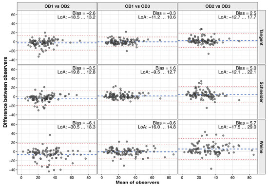
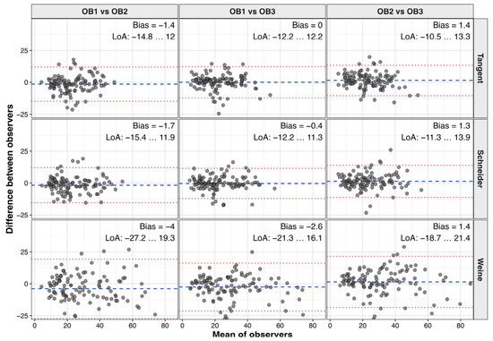
Objectives: To evaluate the inter- and intra-observer reliability of three methods for measuring root canal curvature and the reliability of canal visibility ratings on periapical radiographs over repeated calibration rounds. Materials and Methods: A total of 360 clinical periapical radiographs were divided into
[...] Read more.
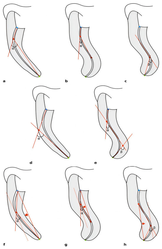
Objectives: To evaluate the inter- and intra-observer reliability of three methods for measuring root canal curvature and the reliability of canal visibility ratings on periapical radiographs over repeated calibration rounds. Materials and Methods: A total of 360 clinical periapical radiographs were divided into six batches and independently evaluated by three observers. Root canal curvature was measured using the Schneider, Weine, and a novel tangency-based method, while canal visibility was classified on a three-category scale. Inter- and intra-observer reliability were assessed using intraclass correlation coefficients (ICC). Agreement between observers was evaluated using Bland–Altman analysis, and differences between measurement methods were compared using repeated-measures analysis. The association between canal visibility and inter-observer measurement differences was assessed using Spearman rank correlation. Results: Inter-observer reliability for curvature measurements was predominantly good to excellent across all methods, with no consistent improvement over successive calibration rounds. Intra-observer reliability, assessed in one observer, was excellent (ICC 0.90–0.97). Measurement method significantly influenced angle values, with the Weine method producing larger angles than the Schneider and tangency-based methods, whereas the latter two showed comparable results. Agreement on canal visibility remained fair to moderate, and reduced canal visibility showed a weak but significant association with greater inter-observer differences in curvature measurements. Conclusions: All three methods demonstrated comparable inter-observer reliability, although the Weine method yielded systematically larger curvature angles. Agreement did not consistently improve over repeated calibration rounds. Reduced canal visibility may increase measurement variability and should be considered when interpreting root canal curvature on periapical radiographs.
Full article

Figure 1

Open AccessArticle
Soft Skills in the Training of Future Dentists: Perceptions and Perspectives of Final-Year Students
by
Cristina Gena Dascalu, Magda-Ecaterina Antohe, Diana Tatarciuc, Roxana Ionela Vasluianu, Cristina Iordache, Anca Vitalariu, Odette Elena Luca and Georgeta Liliana Foia
Dent. J. 2026, 14(9), 552; https://doi.org/10.3390/dj14090552 - 2 Sep 2026
Abstract
The practice of contemporary dentistry requires not only technical skills but also the development of soft skills, such as communication, empathy, and ethical and professional values. Although the importance of these skills is increasingly recognized, the perceptions of dental students in Romania regarding
[...] Read more.
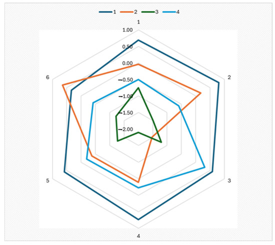
The practice of contemporary dentistry requires not only technical skills but also the development of soft skills, such as communication, empathy, and ethical and professional values. Although the importance of these skills is increasingly recognized, the perceptions of dental students in Romania regarding their relevance to their future professional practice remain insufficiently explored. Objectives: The study aimed to assess the perceptions of final-year students at the Faculty of Dentistry in Iași regarding the importance of soft skills, in relation to their demographic characteristics, professional motivations, and preferences regarding their future practice settings, as well as to identify distinct profiles through cluster analysis. Material and Methods: The study included 205 students in their fourth through sixth years at the Faculty of Dentistry in Iași, who anonymously completed a 26-item questionnaire rated on a Likert scale from 1 to 5. The statistical analysis included the chi-square test, Ward’s method, and k-means clustering. Results: Ethical and professional values, artistic skills, and empathy were most frequently rated as “very important.” Female students assigned significantly higher scores than male students to empathy, ethical and professional values, personal characteristics, and communication (p < 0.05–0.001). Professional independence, financial stability, and professional prestige were the main motivations for career choice. Cluster analysis identified four distinct profiles, with significant differences based on gender, professional motivations, and preferences regarding future practice. Conclusions: Students place a high value on soft skills, with differences based on gender and professional profile. The results support the integration of these skills into university education, to complement specialized clinical training.
Full article
(This article belongs to the Special Issue Dental Education: Innovation and Challenge)
►▼
Show Figures

Figure 1
Open AccessArticle
Agreement Between Cone Beam Computed Tomography, Bone Sounding, Pulse–Echo Ultrasound, and Caliper Measurements for Gingival Thickness Assessment: A Method-Comparison Study
by
Selma Dervisbegovic, Reinhard Gruber, Andrea Gamper, Stefan Nemec, Xiaohui Rausch-Fan and Gabriella Dvorak
Dent. J. 2026, 14(9), 551; https://doi.org/10.3390/dj14090551 - 2 Sep 2026
Abstract
►▼
Show Figures
Background/Objectives: Accurate assessment of gingival thickness (GT) is important for periodontal and implant treatment planning. Methods: In this method-comparison study based on prospectively collected clinical data from 2012, GT was assessed at standardized buccal sites using cone beam computed tomography (CBCT),
[...] Read more.
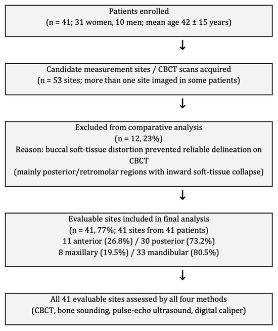
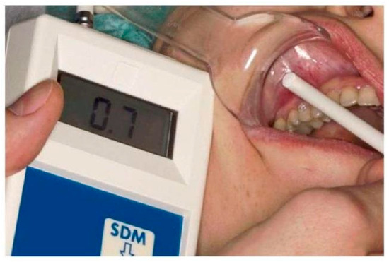
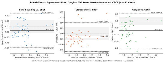
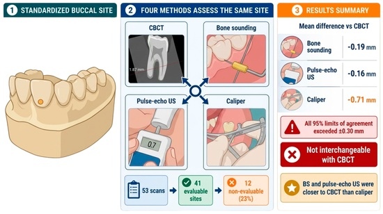
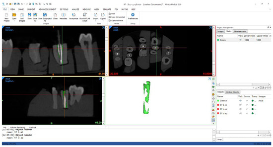
Background/Objectives: Accurate assessment of gingival thickness (GT) is important for periodontal and implant treatment planning. Methods: In this method-comparison study based on prospectively collected clinical data from 2012, GT was assessed at standardized buccal sites using cone beam computed tomography (CBCT), bone sounding (BS), an earlier-generation pulse–echo ultrasound (US) device, and direct digital caliper (CA) measurement. Of 53 acquired CBCT scans, 12 (23%) were non-evaluable because the buccal soft tissues could not be reliably delineated, leaving 41 sites from 41 patients for analysis. A ±0.30 mm clinical comparison margin, based on the difference used in the original sample-size calculation, was applied to interpret agreement. Results: Mean comparator-minus-CBCT biases were −0.19 mm for BS (95% limits of agreement [LoA], −0.98 to 0.61 mm), −0.16 mm for US (95% LoA, −0.83 to 0.51 mm), and −0.71 mm for CA (95% LoA, −1.99 to 0.56 mm). Although BS and the tested US device showed smaller average differences from CBCT than CA, the LoA for all comparisons exceeded this margin. Phenotype classification was exploratory because only four sites were classified as thin by CBCT. Conclusions: BS and the tested pulse–echo US device showed smaller average differences from CBCT than CA; however, the limits of agreement for all comparisons exceeded this margin, so none of the comparator methods can be considered interchangeable with CBCT. The 23% non-evaluable CBCT rate and the device- and protocol-specific nature of the findings limit their generalizability to current-generation systems.
Full article

Graphical abstract
Open AccessArticle
Five-Year Retrospective Radiographic Analysis of Peri-Implant Bone Stability: A Comparison Between Two Socket Preservation Protocols and Spontaneous Healing
by
Esteban Colombo, Enrica Giammarinaro, Maria Menini, Paolo Pesce and Nicola De Angelis
Dent. J. 2026, 14(9), 550; https://doi.org/10.3390/dj14090550 - 1 Sep 2026
Abstract
Objectives: The primary objective of this retrospective pilot study was to evaluate the long-term radiographic stability of peri-implant marginal bone levels (MBLs) at a five-year follow-up, comparing two different socket preservation protocols against spontaneous healing. Materials and Methods: A total of 24 patients
[...] Read more.
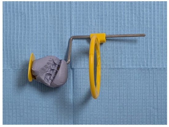
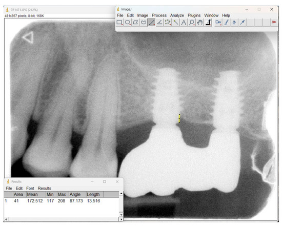
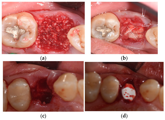
Objectives: The primary objective of this retrospective pilot study was to evaluate the long-term radiographic stability of peri-implant marginal bone levels (MBLs) at a five-year follow-up, comparing two different socket preservation protocols against spontaneous healing. Materials and Methods: A total of 24 patients who underwent tooth extraction followed by delayed implant placement were divided into three groups of 8 subjects each. Test Group 1 was treated with a heterologous cortico-cancellous bone graft covered by a slow-resorbing bovine pericardium membrane. Test Group 2 received the same bone graft but covered by a rapidly resorbing native porcine collagen membrane. The Control Group was left to heal spontaneously. Marginal bone loss (MBL) was calculated radiographically by measuring the difference in bone levels between the time of osseointegration (T0) and the 5-year follow-up (T1). For each implant, mesial and distal MBL values were averaged to obtain a single mean per-implant MBL value for group-level analysis. Results: The 5-year implant survival rate was 100% across all analyzed groups. Test Group 1 demonstrated the highest peri-implant bone stability, recording a mean MBL of mm. In contrast, Test Group 2 and the Control Group exhibited greater resorption, with mean MBL values of mm and mm, respectively. Descriptive analysis indicated a trend toward improved peri-implant bone preservation in the group treated with biomaterial and a slow-resorbing pericardium membrane compared to both the short-resorbing collagen membrane group and the spontaneous healing control group. Conclusions: Within the limitations of this pilot study, the results suggest that the choice of barrier membrane in socket preservation procedures could affect 5-year marginal bone stability. The application of a slow-resorbing membrane provided a prolonged barrier action and showed a descriptive trend of lower mean marginal bone loss. However, the extremely small sample size precludes establishing clinical superiority over standard collagen membranes or spontaneous healing. Further randomized clinical trials with larger cohorts are necessary to confirm these long-term findings.
Full article
(This article belongs to the Special Issue 3D Printing in Dentistry: Materials, Devices and Technologies)
►▼
Show Figures

Figure 1
Open AccessArticle
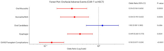
Chimeric Antigen Receptor T-Cell Therapy Is Associated with Low Absolute Rates of Orofacial Adverse Events and Significantly Less When Compared to Hemopoietic Stem Cell Therapy
by
Stella O. Oyewole, Emma Butler, Sagun D. Goyal and Adepitan A. Owosho
Dent. J. 2026, 14(9), 549; https://doi.org/10.3390/dj14090549 - 1 Sep 2026
Abstract
Background/Objectives: Chimeric antigen receptor T-cell (CAR-T) therapy has been approved for the management of relapsed and refractory hematologic malignancies, transforming the oncologic landscape and producing durable remissions in patient populations with limited alternatives. The systemic adverse events of CAR-T are well characterized; however,
[...] Read more.
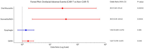
Background/Objectives: Chimeric antigen receptor T-cell (CAR-T) therapy has been approved for the management of relapsed and refractory hematologic malignancies, transforming the oncologic landscape and producing durable remissions in patient populations with limited alternatives. The systemic adverse events of CAR-T are well characterized; however, the orofacial adverse events have not been well described. The objective of this study is to leverage a large, de-identified, real-world dataset to (1) estimate the prevalence of orofacial adverse events following CAR-T, (2) compare event rates with the general population, and (3) directly contrast the orofacial toxicity burden of CAR-T with that observed after hemopoietic stem cell transplant (HSCT). Methods: We performed a retrospective cohort study using de-identified electronic health record data from TriNetX. CAR-T and HSCT cohorts were identified via RxNorm and procedure codes; a non-exposed control cohort was included. Patients with prior orofacial conditions or confounding therapies were excluded. New adverse orofacial events within one year were identified by International Classification of Diseases, 10th Revision (ICD-10) codes. Cohorts were 1:1 propensity-matched by age and sex; associations were estimated as odds ratios with two-sided 95% CIs. Results: In 1142 CAR-T recipients (mean age of 62), gastroesophageal reflux disease (GERD) was most frequent (5.18%). Oral mucosal events included stomatitis in 1.52%, mucositis in 1.31%, and lichenoid reactions in 1.03%. Dysphagia occurred in 1.8% and oral candidiasis in 1.6%. Several severe oral conditions were absent. Compared with the general population (CART vs. general population): mucositis—[12/995 vs. 0/1112] (OR 28.3)—and stomatitis—[16/987 vs. 0/1119] (OR 38)—risks were significantly increased, while CAR-T patients had significant lower risks of mucosal complications than HSCT recipients in this analysis (CART vs. HSCT): mucositis—[1.21% vs. 3.72%] (OR 0.316) and stomatitis—[1.42% vs. 3.91%] (OR 0.354). Conclusions: CAR-T therapy carries a distinct and generally lower orofacial toxicity burden than HSCT, but targeted dental assessment and prospective surveillance remain important to optimize supportive care for cellular therapy recipients.
Full article
(This article belongs to the Special Issue Dental Oncology: 2nd Edition)
►▼
Show Figures

Figure 1
Journal Menu
► ▼ Journal Menu-
- Dentistry Journal Home
- Aims & Scope
- Editorial Board
- Reviewer Board
- Topical Advisory Panel
- Instructions for Authors
- Special Issues
- Topics
- Sections & Collections
- Article Processing Charge
- Indexing & Archiving
- Editor’s Choice Articles
- Most Cited & Viewed
- Journal Statistics
- Journal History
- Journal Awards
- Conferences
- Editorial Office
Journal Browser
► ▼ Journal BrowserHighly Accessed Articles
Latest Books
E-Mail Alert
News
5 September 2026
MDPI Open Science Insights: Academic Publishing Workshop at the University of Toronto–Faculty of Dentistry, 23 September 2026
1 September 2026
MDPI INSIGHTS: The CEO’s Letter #38 – 2 Million Published Articles, Outstanding Reviewers, Michele Parrinello Award, AIS 2026 & WSF-12
Topics
Topic in
Applied Sciences, Dentistry Journal, Polymers, Applied Biosciences, Bioengineering, Materials
Advances in Biomaterials—2nd Edition
Topic Editors: Satoshi Komasa, Yoshiro Tahara, Tohru Sekino, Hideaki Sato, Yoshiya Hashimoto, Tetsuya AdachiDeadline: 31 December 2026
Conferences
Special Issues
Special Issue in
Dentistry Journal
Oral Health and Public Healthcare Systems: Challenges and Innovations in National Health Services
Guest Editors: Simone Gallo, Francesco CavarraDeadline: 10 September 2026
Special Issue in
Dentistry Journal
Geriatric Dentistry: Innovations, Challenges, and Future Directions
Guest Editor: Jung-Tae LeeDeadline: 10 September 2026
Special Issue in
Dentistry Journal
Current Research and Clinical Practice in Digital Orthodontics
Guest Editor: Britta A. JungDeadline: 20 September 2026
Special Issue in
Dentistry Journal
Dental Materials Design and Application
Guest Editor: Walter DukićDeadline: 25 September 2026
Topical Collections
Topical Collection in
Dentistry Journal
Novel Ceramic Materials in Dentistry
Collection Editors: Roberto Sorrentino, Gianrico Spagnuolo
Topical Collection in
Dentistry Journal
Dental Traumatology and Sport Dentistry
Collection Editor: Enrico Spinas