
Acute Kidney Injury Associated with Severe Leptospirosis: Fatal Re-Emerging Disease in Latin America
 , , ,
, , ,
Abstract
:1. Introduction
2. Etiology
3. Pathophysiology of Leptospirosis and Acute Kidney Injury
4. Clinical Manifestations
5. Diagnosis
5.1. Cellular Immunoglobulin Mucin Domain 1 (KIM-1)
5.2. Monocyte Chemoattractant Protein 1 (MCP-1)
5.3. Neutrophil Gelatinase-Associated Lipocalin (NGAL)
5.4. Urinary N-Acetyl-B-D-glucosaminidase (NAG)
6. Management Strategies
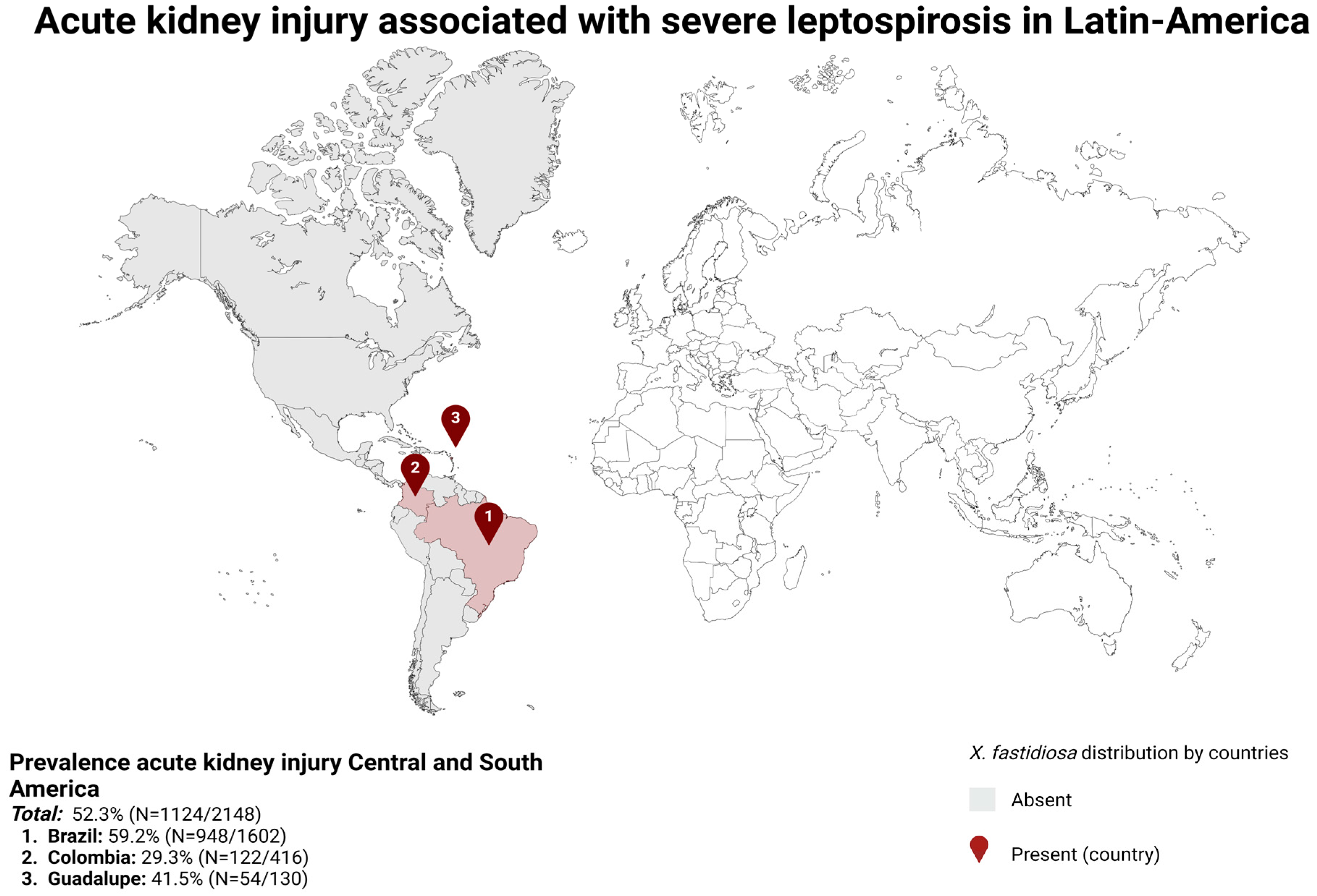
7. Overview of Acute Kidney Injury Secondary to Severe Leptospirosis in Latin America
7.1. Overall Findings
7.2. Epidemiology of Acute Kidney Injury
7.3. Renal Replacement Therapy
7.4. Mortality
8. Conclusions
Author Contributions
Funding
Institutional Review Board Statement
Informed Consent Statement
Data Availability Statement
Conflicts of Interest
References
- Chou, L.-F.; Yang, H.-Y.; Hung, C.-C.; Tian, Y.-C.; Hsu, S.-H.; Yang, C.-W. Leptospirosis kidney disease: Evolution from acute to chronic kidney disease. Biomed. J. 2023, 46, 100595. [Google Scholar] [CrossRef]
- Schneider, M.C.; Leonel, D.G.; Hamrick, P.N.; de Caldas, E.P.; Velásquez, R.T.; Paez, F.A.M.; Arrebato, J.C.G.; Gerger, A.; Pereira, M.M.; Aldighieri, S. Leptospirosis in Latin America: Exploring the first set of regional data. Rev. Panam. Salud. Publica/Pan. Am. J. Public Health 2017, 41, e81. [Google Scholar] [CrossRef]
- Calvopiña, M.; Vásconez, E.; Coral-Almeida, M.; Romero-Alvarez, D.; Garcia-Bereguiain, M.A.; Orlando, A. Leptospirosis: Morbidity, mortality, and spatial distribution of hospitalized cases in Ecuador. A nationwide study 2000–2020. PLoS Neglect. Trop. Dis. 2022, 16, e0010430. [Google Scholar] [CrossRef]
- Philip, N.; Than, L.T.L.; Shah, A.M.; Yuhana, M.Y.; Sekawi, Z.; Neela, V.K. Predictors of severe leptospirosis: A multicentre observational study from Central Malaysia. BMC Infect. Dis. 2021, 21, 1081. [Google Scholar] [CrossRef]
- Agampodi, S.; Gunarathna, S.; Lee, J.-S.; Excler, J.-L. Global, regional, and country-level cost of leptospirosis due to loss of productivity in humans. PLoS Neglect. Trop. Dis. 2023, 17, e0011291. [Google Scholar] [CrossRef]
- Daher, E.D.F.; de Abreu, K.L.S.; Junior, G.B.d.S. Insuficiência renal aguda associada à leptospirose. Braz. J. Nephrol. 2010, 32, 408–415. [Google Scholar] [CrossRef]
- Tubiana, S.; Mikulski, M.; Becam, J.; Lacassin, F.; Lefèvre, P.; Gourinat, A.-C.; Goarant, C.; D’Ortenzio, E. Risk Factors and Predictors of Severe Leptospirosis in New Caledonia. PLoS Neglect. Trop. Dis. 2013, 7, e1991. [Google Scholar] [CrossRef]
- Pongpan, S.; Thanatrakolsri, P.; Vittaporn, S.; Khamnuan, P.; Daraswang, P. Prognostic Factors for Leptospirosis Infection Severity. Trop. Med. Infect. Dis. 2023, 8, 112. [Google Scholar] [CrossRef]
- Temeiam, N.; Jareinpituk, S.; Phinyo, P.; Patumanond, J.; Srisawat, N. Development and Validation of a simple score for diagnosis of Leptospirosis at outpatient departments. PLoS Neglect. Trop. Dis. 2020, 14, e0007977. [Google Scholar] [CrossRef] [PubMed]
- Sukmark, T.; Lumlertgul, N.; Peerapornratana, S.; Khositrangsikun, K.; Tungsanga, K.; Sitprija, V.; Srisawat, N.; Thai-Lepto AKI Study Group. Thai-Lepto-on-admission probability (THAI-LEPTO) score as an early tool for initial diagnosis of leptospirosis: Result from Thai-Lepto AKI study group. PLoS Neglect. Trop. Dis. 2018, 12, e0006319. [Google Scholar] [CrossRef] [PubMed]
- Meneses, G.C.; Gomes, P.E.A.d.C.; Galdino, G.S.; Bezerra, G.F.; Santos, R.S.d.S.; Martins, A.M.C.; Junior, G.B.d.S.; Libório, A.B.; Neto, R.d.J.P.; Daher, E.D.F. Endothelial biomarkers as predictors for haemodialysis need in severe leptospirosis patients (Weil’s disease). Trop. Med. Int. Health 2022, 27, 727–734. [Google Scholar] [CrossRef]
- Yamaguchi, T.; Higa, N.; Okura, N.; Matsumoto, A.; Hermawan, I.; Yamashiro, T.; Suzuki, T.; Toma, C. Characterizing interactions of Leptospira interrogans with proximal renal tubule epithelial cells. BMC Microbiol. 2018, 18, 64. [Google Scholar] [CrossRef]
- Junior, G.B.d.S.; Srisawat, N.; Galdino, G.S.; Macedo, S.; Pinto, J.R.; Farias, G.M.N.; Alencar, R.L.; Neto, R.d.J.P.; Barros, E.J.G.; Daher, E.D.F. Acute kidney injury in leptospirosis: Overview and perspectives. Asian Pac. J. Trop. Med. 2018, 11, 549–554. [Google Scholar] [CrossRef]
- Abreu, P.A.E.; Seguro, A.C.; Canale, D.; da Silva, A.M.G.; Matos, L.D.R.B.; Gotti, T.B.; Monaris, D.; de Jesus, D.A.; Vasconcellos, S.A.; de Brito, T.; et al. Lp25 membrane protein from pathogenic Leptospira spp. is associated with rhabdomyolysis and oliguric acute kidney injury in a guinea pig model of leptospirosis. PLoS Neglect. Trop. Dis. 2017, 11, e0005615. [Google Scholar] [CrossRef]
- Karnik, N.D.; Patankar, A.S. Leptospirosis in Intensive Care Unit. Indian J. Crit. Care Med. 2021, 25, S134–S137. [Google Scholar] [CrossRef]
- Andrade, L.A.A.; Cleto, S.A.A.; Seguro, A.C. Door-to-Dialysis Time and Daily Hemodialysis in Patients with Leptospirosis. Clin. J. Am. Soc. Nephrol. 2007, 2, 739–744. [Google Scholar] [CrossRef]
- Daher, E.D.F.; de Carvalho, G.S.G.; Soares, D.d.S.; Mendes, M.H.; Filho, S.L.A.P.; Rocha, H.A.L.; Junior, G.B.d.S. Changing patterns in leptospirosis: A three-decade study in Brazil. Int. J. Infect. Dis. 2017, 60, 4–10. [Google Scholar] [CrossRef]
- Herath, N.J.; Kularatne, S.A.; Weerakoon, K.G.; Wazil, A.; Subasinghe, N.; Ratnatunga, N.V. Long term outcome of acute kidney injury due to leptospirosis? A longitudinal study in Sri Lanka. BMC Res. Notes 2014, 7, 398. [Google Scholar] [CrossRef]
- Yang, H.-Y.; Hung, C.-C.; Liu, S.-H.; Guo, Y.-G.; Chen, Y.-C.; Ko, Y.-C.; Huang, C.-T.; Chou, L.-F.; Tian, Y.-C.; Chang, M.-Y.; et al. Overlooked Risk for Chronic Kidney Disease after Leptospiral Infection: A Population-Based Survey and Epidemiological Cohort Evidence. PLoS Neglect. Trop. Dis. 2015, 9, e0004105. [Google Scholar] [CrossRef]
- Atasoyu, E.M.; Turhan, V.; Unver, S.; Evrenkaya, T.R.; Yildirim, S. A case of leptospirosis presenting with end-stage renal failure. Nephrol. Dial. Transplant. 2005, 20, 2290–2292. [Google Scholar] [CrossRef] [PubMed]
- Haake, D.A.; Levett, P.N. Leptospirosis in Humans. Leptospira Leptospirosis 2015, 387, 65–97. [Google Scholar] [CrossRef]
- Picardeau, M. Diagnosis and epidemiology of leptospirosis. Méd. Mal. Infect. 2013, 43, 1–9. [Google Scholar] [CrossRef]
- Budihal, S.V. Leptospirosis Diagnosis: Competancy of Various Laboratory Tests. J. Clin. Diagn. Res. 2014, 8, 199–202. [Google Scholar] [CrossRef]
- Thibeaux, R.; Girault, D.; Bierque, E.; Soupé-Gilbert, M.-E.; Rettinger, A.; Douyère, A.; Meyer, M.; Iraola, G.; Picardeau, M.; Goarant, C. Biodiversity of Environmental Leptospira: Improving Identification and Revisiting the Diagnosis. Front. Microbiol. 2018, 9, 816. [Google Scholar] [CrossRef]
- Adler, B.; de la Pena Moctezuma, A. Leptospira and leptospirosis. Vet. Microbiol. 2010, 140, 287–296. [Google Scholar] [CrossRef]
- Uribe-Restrepo, P.; Munoz-Zanzi, C.; Agudelo-Flórez, P. Kidney Injury Biomarkers in Leptospirosis. Rev. Soc. Bras. Med. Trop. 2023, 56, e0260-2022. [Google Scholar] [CrossRef]
- Tian, Y.-C. Leptospirosis and Kidney Fibrosis. In Leptospirosis and the Kidney; Karger Publishers: Basel, Switzerland, 2019; pp. 57–64. [Google Scholar] [CrossRef]
- Reis, E.A.G.; Hagan, J.E.; Ribeiro, G.S.; Teixeira-Carvalho, A.; Martins-Filho, O.A.; Montgomery, R.R.; Shaw, A.C.; Ko, A.I.; Reis, M.G. Cytokine Response Signatures in Disease Progression and Development of Severe Clinical Outcomes for Leptospirosis. PLoS Neglect. Trop. Dis. 2013, 7, e2457. [Google Scholar] [CrossRef]
- Barnett, J.K.; Barnett, D.; Bolin, C.A.; Summers, T.A.; Wagar, E.A.; Cheville, N.F.; Hartskeerl, R.A.; Haake, D.A. Expression and Distribution of Leptospiral Outer Membrane Components during Renal Infection of Hamsters. Infect. Immun. 1999, 67, 853–861. [Google Scholar] [CrossRef]
- Srisawat, N.; Sitprija, V. Leptospirosis and the Kidney: An Overview. In Leptospirosis and the Kidney; Karger Publishers: Basel, Switzerland, 2019; pp. 1–9. [Google Scholar] [CrossRef]
- Tanaka, K.; Tanabe, K.; Nishii, N.; Takiue, K.; Sugiyama, H.; Wada, J. Sustained Tubulointerstitial Inflammation in Kidney with Severe Leptospirosis. Intern. Med. 2017, 56, 1179–1184. [Google Scholar] [CrossRef]
- Senavirathna, I.; Rathish, D.; Agampodi, S. Cytokine response in human leptospirosis with different clinical outcomes: A systematic review. BMC Infect. Dis. 2020, 20, 268. [Google Scholar] [CrossRef]
- Nisansala, T.; Weerasekera, M.; Ranasinghe, N.; Marasinghe, C.; Gamage, C.; Fernando, N.; Gunasekara, C. Contributing role of TNF, IL-10, sTNFR1 and TNF gene polymorphisms in disease severity of leptospirosis. Med. Microbiol. Immunol. 2021, 210, 211–219. [Google Scholar] [CrossRef] [PubMed]
- Araujo, E.R.; Seguro, A.C.; Spichler, A.; Magaldi, A.J.; Volpini, R.A.; De Brito, T. Acute kidney injury in human leptospirosis: An immunohistochemical study with pathophysiological correlation. Virchows Arch. 2010, 456, 367–375. [Google Scholar] [CrossRef] [PubMed]
- Sitprija, V. Altered fluid, electrolyte and mineral status in tropical disease, with an emphasis on malaria and leptospirosis. Nat. Clin. Pract. Nephrol. 2008, 4, 91–101. [Google Scholar] [CrossRef] [PubMed]
- Khositseth, S.; Sudjaritjan, N.; Tananchai, P.; Ong-Ajyuth, S.; Sitprija, V.; Thongboonkerd, V. Renal magnesium wasting and tubular dysfunction in leptospirosis. Nephrol. Dial. Transplant. 2007, 23, 952–958. [Google Scholar] [CrossRef] [PubMed]
- Siriwanij, T.; Suttinont, C.; Tantawichien, T.; Chusil, S.; Kanjanabuch, T.; Sitprija, V. Haemodynamics in leptospirosis: Effects of plasmapheresis and continuous venovenous haemofiltration. Nephrology 2005, 10, 1–6. [Google Scholar] [CrossRef] [PubMed]
- Wu, M.-S.; Yang, C.-W.; Pan, M.-J.; Chang, C.-T.; Chen, Y.-C. Reduced renal Na+-K+-Cl− co-transporter activity and inhibited NKCC2 mRNA expression by Leptospira shermani: From bed-side to bench. Nephrol. Dial. Transplant. 2004, 19, 2472–2479. [Google Scholar] [CrossRef] [PubMed]
- Vinh, T.; Adler, B.; Faine, S. Glycolipoprotein Cytotoxin from Leptospira interrogans serovar copenhageni. Microbiology 1986, 132, 111–123. [Google Scholar] [CrossRef]
- Pereira, M.; Andrade, J.; Lacerda, M.D.; Batoréu, N.; Marchevsky, R.; dos Santos, R.R. Demonstration of leptospiral antigens on tissues using monoclonal antibodies and avidin-biotin peroxidase staining. Exp. Toxicol. Pathol. 1997, 49, 505–511. [Google Scholar] [CrossRef]
- Younes-Ibrahim, M.; Burth, P.; Castro-Faria, M.; Cheval, L.; Buffin-Meyer, B.; Marsy, S.; Doucet, A. Effect of Leptospira interrogans Endotoxin on Renal Tubular Na,K-ATPase and H,K-ATPase Activities. Ann. N. Y. Acad. Sci. 1997, 834, 684–686. [Google Scholar] [CrossRef]
- Gonçalves-De-Albuquerque, C.F.; Burth, P.; Silva, A.R.; de Moraes, I.M.M.; Oliveira, F.M.d.J.; Santelli, R.; Freire, A.S.; Bozza, P.T.; Younes-Ibrahim, M.; de Castro-Faria-Neto, H.C.; et al. Oleic acid inhibits lung Na/K-ATPase in mice and induces injury with lipid body formation in leukocytes and eicosanoid production. J. Inflamm. 2013, 10, 34. [Google Scholar] [CrossRef]
- Martins, C.A.; dos Santos, M.C.B.; Gonçalves-De-Albuquerque, C.F.; Castro-Faria-Neto, H.C.; Castro-Faria, M.V.; Burth, P.; Younes-Ibrahim, M. The relationship of oleic acid/albumin molar ratio and clinical outcomes in leptospirosis. Heliyon 2021, 7, e06420. [Google Scholar] [CrossRef] [PubMed]
- Gonçalves-De-Albuquerque, C.F.; da Cunha, C.M.C.; de Castro, L.V.G.; Martins, C.d.A.; Barnese, M.R.C.; Burth, P.; Younes-Ibrahim, M. Cellular Pathophysiology of Leptospirosis: Role of Na/K-ATPase. Microorganisms 2023, 11, 1695. [Google Scholar] [CrossRef] [PubMed]
- Younes-Ibrahim, M.; Buffin-Meyer, B.; Cheval, L.; Burth, P.; Castro-Faria, M.; Barlet-Bas, C.; Marsy, S.; Doucet, A. Na,K-ATPase: A molecular target for Leptospira interrogans endotoxin. Braz. J. Med. Biol. Res. 1997, 30, 213–223. [Google Scholar] [CrossRef] [PubMed]
- Lacroix-Lamandé, S.; D’andon, M.F.; Michel, E.; Ratet, G.; Philpott, D.J.; Girardin, S.E.; Boneca, I.G.; Vandewalle, A.; Werts, C. Downregulation of the Na/K-ATPase Pump by Leptospiral Glycolipoprotein Activates the NLRP3 Inflammasome. J. Immunol. 2012, 188, 2805–2814. [Google Scholar] [CrossRef] [PubMed]
- Dorigatti, F.; Brunialti, M.; Romero, E.; Kallas, E.; Salomão, R. Leptospira interrogans activation of peripheral blood monocyte glycolipoprotein demonstrated in whole blood by the release of IL-6. Braz. J. Med. Biol. Res. 2005, 38, 909–914. [Google Scholar] [CrossRef] [PubMed]
- Diament, D.; Brunialti, M.K.C.; Romero, E.C.; Kallas, E.G.; Salomao, R. Peripheral Blood Mononuclear Cell Activation Induced by Leptospira interrogans Glycolipoprotein. Infect. Immun. 2002, 70, 1677–1683. [Google Scholar] [CrossRef] [PubMed]
- Garcia, M.; Gopalakrishna, K.V. A Case of Imported Leptospirosis: Rhabdomyolysis and Severe Hyperbilirubinemia in a Traveler Returning from Puerto Rico. Cureus 2023, 15, e34690. [Google Scholar] [CrossRef] [PubMed]
- Bienaimé, F.; Muorah, M.; Yammine, L.; Burtin, M.; Nguyen, C.; Baron, W.; Garbay, S.; Viau, A.; Broueilh, M.; Blanc, T.; et al. Stat3 Controls Tubulointerstitial Communication during CKD. J. Am. Soc. Nephrol. 2016, 27, 3690–3705. [Google Scholar] [CrossRef]
- Tian, Y.-C.; Chen, Y.-C.; Hung, C.-C.; Chang, C.-T.; Wu, M.-S.; Phillips, A.O.; Yang, C.-W. Leptospiral Outer Membrane Protein Induces Extracellular Matrix Accumulation through a TGF-β1/Smad-Dependent Pathway. J. Am. Soc. Nephrol. 2006, 17, 2792–2798. [Google Scholar] [CrossRef]
- Kurzhagen, J.T.; Dellepiane, S.; Cantaluppi, V.; Rabb, H. AKI: An increasingly recognized risk factor for CKD development and progression. J. Nephrol. 2020, 33, 1171–1187. [Google Scholar] [CrossRef]
- Yu, S.M.-W.; Bonventre, J.V. Acute kidney injury and maladaptive tubular repair leading to renal fibrosis. Curr. Opin. Nephrol. Hypertens. 2020, 29, 310–318. [Google Scholar] [CrossRef]
- Sant’Anna, R.; Vieira, A.; Oliveira, J.; Lilenbaum, W. Asymptomatic leptospiral infection is associated with canine chronic kidney disease. Comp. Immunol. Microbiol. Infect. Dis. 2018, 62, 64–67. [Google Scholar] [CrossRef]
- He, L.; Wei, Q.; Liu, J.; Yi, M.; Liu, Y.; Liu, H.; Sun, L.; Peng, Y.; Liu, F.; Venkatachalam, M.A.; et al. AKI on CKD: Heightened injury, suppressed repair, and the underlying mechanisms. Kidney Int. 2017, 92, 1071–1083. [Google Scholar] [CrossRef]
- Caravaca-Fontán, F.; Fernández-Juárez, G.; Praga, M. Acute kidney injury in interstitial nephritis. Curr. Opin. Crit. Care 2019, 25, 558–564. [Google Scholar] [CrossRef]
- Kusirisin, P.; Junior, G.B.d.S.; Sitprija, V.; Srisawat, N. Acute kidney injury in the tropics. Nephrology 2023, 28, 5–20. [Google Scholar] [CrossRef]
- Chancharoenthana, W.; Leelahavanichkul, A.; Schultz, M.J.; Dondorp, A.M. Going Micro in Leptospirosis Kidney Disease. Cells 2022, 11, 698. [Google Scholar] [CrossRef]
- Yang, H.-Y.; Yen, T.-H.; Lin, C.-Y.; Chen, Y.-C.; Pan, M.-J.; Lee, C.-H.; Yu, C.-C.; Wu, M.-S.; Wu, S.-S.; Weng, C.-H.; et al. Early Identification of Leptospirosis as an Ignored Cause of Multiple Organ Dysfunction Syndrome. Shock 2012, 38, 24–29. [Google Scholar] [CrossRef]
- Nabity, S.A.; Hagan, J.E.; Araújo, G.; Damião, A.O.; Cruz, J.S.; Nery, N.; Wunder, E.A.; Reis, M.G.; Ko, A.I.; Ribeiro, G.S. Prospective evaluation of accuracy and clinical utility of the Dual Path Platform (DPP) assay for the point-of-care diagnosis of leptospirosis in hospitalized patients. PLOS Neglect. Trop. Dis. 2018, 12, e0006285. [Google Scholar] [CrossRef]
- Kamath, N.; Iyengar, A. Infections and the kidney: A tale from the tropics. Pediatr. Nephrol. 2017, 33, 1317–1326. [Google Scholar] [CrossRef]
- Daher, E.; Abdulkader, R.C.; Zanetta, D.M.; Cavalcante, M.B. Risk factors for death and changing patterns in leptospirosis acute renal failure. Am. J. Trop. Med. Hyg. 1999, 61, 630–634. [Google Scholar] [CrossRef]
- Nisansala, T.; Weerasekera, M.; Ranasinghe, N.; Marasinghe, C.; Gamage, C.; Fernando, N.; Gunasekara, C. Importance of KIM-1 and MCP-1 in Determining the Leptospirosis-Associated AKI: A Sri Lankan Study. BioMed Res. Int. 2021, 2021, 1752904. [Google Scholar] [CrossRef]
- Daher, E.D.F.; Soares, D.S.; Fernandes, A.T.B.D.M.; Girão, M.M.V.; Sidrim, P.R.; Pereira, E.D.B.; Rocha, N.A.; da Silva, G.B. Risk factors for intensive care unit admission in patients with severe leptospirosis: A comparative study according to patients’ severity. BMC Infect. Dis. 2015, 16, 40. [Google Scholar] [CrossRef]
- Júnior, G.B.S.; Abreu, K.L.S.; Mota, R.M.; Barreto, A.G.; Araújo, S.M.; Rocha, H.A.; Libório, A.B.; Daher, E.F. RIFLE and Acute Kidney Injury Network classifications predict mortality in leptospirosis-associated acute kidney injury. Nephrology 2011, 16, 269–276. [Google Scholar] [CrossRef]
- Cerqueira, T.B.; Athanazio, D.A.; Spichler, A.S.; Seguro, A.C. Renal involvement in leptospirosis--new insights into pathophysiology and treatment. Braz. J. Infect. Dis. 2008, 12, 248–252. [Google Scholar] [CrossRef]
- Lombi, F.; Muryan, A.; Canzonieri, R.; Trimarchi, H. Biomarcadores en la lesión renal aguda: Paradigma o evidencia? Nefrología 2016, 36, 339–346. [Google Scholar] [CrossRef]
- Rico-Fontalvo, J.; Aroca-Martínez, G.; Daza-Arnedo, R.; Cabrales, J.; Rodríguez-Yanez, T.; Cardona-Blanco, M.; Montejo-Hernández, J.; Barrios, D.R.; Patiño-Patiño, J.; Rodríguez, E.O. Novel Biomarkers of Diabetic Kidney Disease. Biomolecules 2023, 13, 633. [Google Scholar] [CrossRef]
- Dase, J.; Rasyid, H.; Masadah, R.; Cangara, M.H.; Bukhari, A.; Dwiyanti, R.; Hatta, M. Analysis of mRNA and protein kidney injury Molecule-1 (KIM-1) expression in a kidney model during the initiation phase of ischemia reperfusion injury. Ann. Med. Surg. 2022, 75, 103373. [Google Scholar] [CrossRef]
- Bonventre, J.V. Kidney injury molecule-1 (KIM-1): A urinary biomarker and much more. Nephrol. Dial. Transplant. 2009, 24, 3265–3268. [Google Scholar] [CrossRef]
- Munshi, R.; Johnson, A.; Siew, E.D.; Ikizler, T.A.; Ware, L.B.; Wurfel, M.M.; Himmelfarb, J.; Zager, R.A. MCP-1 Gene Activation Marks Acute Kidney Injury. J. Am. Soc. Nephrol. 2011, 22, 165–175. [Google Scholar] [CrossRef]
- Srisawat, N.; Praditpornsilpa, K.; Patarakul, K.; Techapornrung, M.; Daraswang, T.; Sukmark, T.; Khositrangsikun, K.; Fakthongyoo, A.; Oranrigsupak, P.; Praderm, L.; et al. Neutrophil Gelatinase Associated Lipocalin (NGAL) in Leptospirosis Acute Kidney Injury: A Multicenter Study in Thailand. PLoS ONE 2015, 10, e0143367. [Google Scholar] [CrossRef]
- Ajjimarungsi, A.; Bhurayanontachai, R.; Chusri, S. Clinical characteristics, outcomes, and predictors of leptospirosis in patients admitted to the medical intensive care unit: A retrospective analysis. J. Infect. Public Health 2020, 13, 2055–2061. [Google Scholar] [CrossRef]
- Panaphut, T.; Domrongkitchaiporn, S.; Vibhagool, A.; Thinkamrop, B.; Susaengrat, W. Ceftriaxone Compared with Sodium Penicillin G for Treatment of Severe Leptospirosis. Clin. Infect. Dis. 2003, 36, 1507–1513. [Google Scholar] [CrossRef]
- Daher, E.F.; Silva, G.B.; de Abreu, K.L.S.; Mota, R.M.S.; Batista, D.V.; Rocha, N.A.; Araújo, S.M.H.A.; Libório, A.B. Leptospirosis-associated acute kidney injury: Penicillin at the late stage is still controversial. J. Clin. Pharm. Ther. 2011, 37, 420–425. [Google Scholar] [CrossRef]
- Barrera, E.L.P.; Piruccini, S.B.; Rodríguez, K.; Duarte, C.; Torres, M.; Undurraga, E.A. Demographic and clinical risk factors associated with severity of lab-confirmed human leptospirosis in Colombia, 2015–2020. PLoS Neglect. Trop. Dis. 2023, 17, e0011454. [Google Scholar] [CrossRef]
- Smith, S.; Liu, Y.-H.; Carter, A.; Kennedy, B.J.; Dermedgoglou, A.; Poulgrain, S.S.; Paavola, M.P.; Minto, T.L.; Luc, M.; Hanson, J. Severe leptospirosis in tropical Australia: Optimising intensive care unit management to reduce mortality. PLoS Neglect. Trop. Dis. 2019, 13, e0007929. [Google Scholar] [CrossRef]
- Rajapakse, S. Leptospirosis: Clinical aspects. Clin. Med. 2022, 22, 14–17. [Google Scholar] [CrossRef]
- Marotto, P.C.F.; Nascimento, C.M.R.; Eluf-Neto, J.; Marotto, M.S.; Andrade, L.; Sztajnbok, J.; Seguro, A.C. Acute Lung Injury in Leptospirosis: Clinical and Laboratory Features, Outcome, and Factors Associated with Mortality. Clin. Infect. Dis. 1999, 29, 1561–1563. [Google Scholar] [CrossRef]
- Rhodes, A.; Evans, L.E.; Alhazzani, W.; Levy, M.M.; Antonelli, M.; Ferrer, R.; Kumar, A.; Sevransky, J.E.; Sprung, C.L.; Nunnally, M.E.; et al. Surviving Sepsis Campaign: International Guidelines for Management of Sepsis and Septic Shock: 2016. Intensive Care Med. 2017, 43, 304–377. [Google Scholar] [CrossRef]
- Edwards, C.N.; Everard, C.O.R.; Callender, J.; Nicholson, G.D.; Hassell, T.A. Thrombocytopenia in Leptospirosis: The Absence of Evidence for Disseminated Intravascular Coagulation. Am. J. Trop. Med. Hyg. 1986, 35, 352–354. [Google Scholar] [CrossRef]
- Faucher, J.-F.; Hoen, B.; Estavoyer, J.-M. The management of leptospirosis. Expert Opin. Pharmacother. 2004, 5, 819–827. [Google Scholar] [CrossRef]
- Sitprija, V.; Losuwanrak, K.; Kanjanabuch, T. Leptospiral Nephropathy. Semin. Nephrol. 2003, 23, 42–48. [Google Scholar] [CrossRef]
- Kularathna, M.D.S.V.; Kularatne, S.A.M.; Pathirage, M.; Nanayakkara, P.T.M.A. Severe leptospirosis complicated with multiorgan dysfunction successfully managed with plasma exchange: A case report. J. Med. Case Rep. 2021, 15, 584. [Google Scholar] [CrossRef]
- Bourquin, V.; Ponte, B.; Hirschel, B.; Pugin, J.; Martin, P.-Y.; Saudan, P. Severe Leptospirosis with Multiple Organ Failure Successfully Treated by Plasma Exchange and High-Volume Hemofiltration. Case Rep. Nephrol. 2011, 2011, 817414. [Google Scholar] [CrossRef]
- Cerdas-Quesada, C. Potential benefits of plasma exchange by apheresis on the treatment of severe Icteric Leptospirosis: Case report and literature review. Transfus. Apher. Sci. 2011, 45, 191–194. [Google Scholar] [CrossRef]
- Taylor, D.; Karamadoukis, L. Plasma exchange in severe leptospirosis with multi-organ failure: A case report. J. Med. Case Rep. 2013, 7, 169. [Google Scholar] [CrossRef]
- Fonseka, C.L.; Lekamwasam, S. Role of Plasmapheresis and Extracorporeal Membrane Oxygenation in the Treatment of Leptospirosis Complicated with Pulmonary Hemorrhages. J. Trop. Med. 2018, 2018, 4520185. [Google Scholar] [CrossRef]
- Delmas, B.; Jabot, J.; Chanareille, P.; Ferdynus, C.; Allyn, J.; Allou, N.; Raffray, L.; Gaüzere, B.-A.; Martinet, O.; Vandroux, D. Leptospirosis in ICU: A Retrospective Study of 134 Consecutive Admissions. Crit. Care Med. 2018, 46, 93–99. [Google Scholar] [CrossRef]
- Peces, R. Acute renal failure in severe leptospirosis. Nephrol. Dial. Transplant. 2003, 18, 1235–1236. [Google Scholar] [CrossRef]
- Wiwanitkit, V. Letter to the Editor: “Peritoneal Dialysis in Leptospirosis-Induced Acute Renal Failure: An Appraisal on Thai Patients”. Ren. Fail. 2006, 28, 201. [Google Scholar] [CrossRef]
- Matthay, M.A.; Arabi, Y.; Arroliga, A.C.; Bernard, G.; Bersten, A.D.; Brochard, L.J.; Calfee, C.S.; Combes, A.; Daniel, B.M.; Ferguson, N.D.; et al. A New Global Definition of Acute Respiratory Distress Syndrome. Am. J. Respir. Crit. Care Med. 2023, 209, 37–47. [Google Scholar] [CrossRef]
- Laffey, J.G.; Bellani, G.; Pham, T.; Fan, E.; Madotto, F.; Bajwa, E.K.; Brochard, L.; Clarkson, K.; Esteban, A.; Gattinoni, L.; et al. Potentially modifiable factors contributing to outcome from acute respiratory distress syndrome: The LUNG SAFE study. Intensiv. Care Med. 2016, 42, 1865–1876. [Google Scholar] [CrossRef]
- Meyer, N.J.; Gattinoni, L.; Calfee, C.S. Acute respiratory distress syndrome. Lancet 2021, 398, 622–637. [Google Scholar] [CrossRef]
- Chiumello, D.; Carlesso, E.; Cadringher, P.; Caironi, P.; Valenza, F.; Polli, F.; Tallarini, F.; Cozzi, P.; Cressoni, M.; Colombo, A.; et al. Lung Stress and Strain during Mechanical Ventilation for Acute Respiratory Distress Syndrome. Am. J. Respir. Crit. Care Med. 2008, 178, 346–355. [Google Scholar] [CrossRef]
- Neto, A.S.; Deliberato, R.O.; Johnson, A.E.W.; Bos, L.D.; Amorim, P.; Pereira, S.M.; Cazati, D.C.; Cordioli, R.L.; Correa, T.D.; Pollard, T.J.; et al. Mechanical power of ventilation is associated with mortality in critically ill patients: An analysis of patients in two observational cohorts. Intensive Care Med. 2018, 44, 1914–1922. [Google Scholar] [CrossRef] [PubMed]
- Papazian, L.; Forel, J.-M.; Gacouin, A.; Penot-Ragon, C.; Perrin, G.; Loundou, A.; Jaber, S.; Arnal, J.-M.; Perez, D.; Seghboyan, J.-M.; et al. Neuromuscular Blockers in Early Acute Respiratory Distress Syndrome. N. Engl. J. Med. 2010, 363, 1107–1116. [Google Scholar] [CrossRef]
- Guérin, C.; Reignier, J.; Richard, J.-C.; Beuret, P.; Gacouin, A.; Boulain, T.; Mercier, E.; Badet, M.; Mercat, A.; Baudin, O.; et al. Prone Positioning in Severe Acute Respiratory Distress Syndrome. N. Engl. J. Med. 2013, 368, 2159–2168. [Google Scholar] [CrossRef] [PubMed]
- Abrams, D.; Agerstrand, C.L.; Biscotti, M.; Burkart, K.M.; Bacchetta, M.; Brodie, D. Extracorporeal Membrane Oxygenation in the Management of Diffuse Alveolar Hemorrhage. ASAIO J. 2015, 61, 216–218. [Google Scholar] [CrossRef]
- Gouveia, E.L.; Metcalfe, J.; de Carvalho, A.L.F.; Aires, T.S.; Villasboas-Bisneto, J.C.; Queirroz, A.; Santos, A.C.; Salgado, K.; Reis, M.G.; Ko, A.I. Leptospirosis-associated Severe Pulmonary Hemorrhagic Syndrome, Salvador, Brazil. Emerg. Infect. Dis. 2008, 14, 505–508. [Google Scholar] [CrossRef] [PubMed]
- Vieira, S.R.R.; Brauner, J.S. Leptospirosis as a cause of acute respiratory failure: Clinical features and outcome in 35 critical care patients. Braz. J. Infect. Dis. 2002, 6, 135–139. [Google Scholar] [CrossRef]
- Echeverri-Toro, L.M.; Penagos, S.; Castañeda, L.; Villa, P.; Atehortúa, S.; Ramírez, F.; Restrepo, C.; Ospina, S.; Agudelo, Y.; Hidrón, A.; et al. Sociodemographic and clinical characteristics of patients infected with Leptospira spp. treated at four hospitals in Medellín, Colombia, 2008–2013. Biomedica 2017, 37, 62–67. [Google Scholar] [CrossRef]
- Daher, E.F.; Silva, J.G.B.; Karbage, N.N.; Carvalho, J.P.C.; Kataoka, R.S.; Silva, E.C.; Magalhães, M.M.; Mota, R.M.; Araújo, S.M.; Gutiérrez-Adrianzén, O.A.; et al. Predictors of Oliguric Acute Kidney Injury in Leptospirosis. Nephron Clin. Pract. 2009, 112, c25–c30. [Google Scholar] [CrossRef] [PubMed]
- Daher, E.F.; Lima, R.S.; Júnior, G.B.S.; Silva, E.C.; Karbage, N.N.; Kataoka, R.S.; Júnior, P.C.C.; Magalhães, M.M.; Mota, R.M.; Libório, A.B. Clinical presentation of leptospirosis: A retrospective study of 201 patients in a metropolitan city of Brazil. Braz. J. Infect. Dis. 2010, 14, 3–10. [Google Scholar] [CrossRef] [PubMed]
- Herrmann-Storck, C.; Louis, M.S.; Foucand, T.; Lamaury, I.; Deloumeaux, J.; Baranton, G.; Simonetti, M.; Sertour, N.; Nicolas, M.; Salin, J.; et al. Severe Leptospirosis in Hospitalized Patients, Guadeloupe. Emerg. Infect. Dis. 2010, 16, 331–334. [Google Scholar] [CrossRef] [PubMed]
- Damasco, P.V.; Ávila, C.A.L.; Barbosa, A.T.; Carvalho, M.d.M.R.; Pereira, G.M.B.; de Lemos, E.R.S.; Bóia, M.N.; Pereira, M.M. Atypical lymphocytosis in leptospirosis: A cohort of hospitalized cases between 1996 and 2009 in State of Rio de Janeiro, Brazil. Rev. Soc. Bras. Med. Trop. 2011, 44, 611–615. [Google Scholar] [CrossRef] [PubMed]
- Echeverri, L.M.; Atehortúa, S.; Ospina, S. Leptospirosis con inmunoglobulina M positiva en pacientes hospitalizados en una institución de tercer nivel de Medellín, Colombia, en 2009. Infectio 2011, 15, 118–123. [Google Scholar] [CrossRef]
- Daher, E.F.; Silva, G.B.; Silveira, C.O.; Falcão, F.S.; Alves, M.P.; Mota, J.A.A.A.; Lima, J.B.; Mota, R.M.S.; Vieira, A.P.F.; Neto, R.d.J.P.; et al. Factors associated with thrombocytopenia in severe leptospirosis (Weil’s disease). Clinics 2014, 69, 106–110. [Google Scholar] [CrossRef] [PubMed]
- Sharp, T.M.; García, B.R.; Pérez-Padilla, J.; Galloway, R.L.; Guerra, M.; Ryff, K.R.; Haberling, D.; Ramakrishnan, S.; Shadomy, S.; Blau, D.; et al. Early Indicators of Fatal Leptospirosis during the 2010 Epidemic in Puerto Rico. PLoS Neglect. Trop. Dis. 2016, 10, e0004482. [Google Scholar] [CrossRef] [PubMed]
- Cleto, S.A.; Rodrigues, C.E.; Malaque, C.M.; Sztajnbok, J.; Seguro, A.C.; Andrade, L. Hemodiafiltration Decreases Serum Levels of Inflammatory Mediators in Severe Leptospirosis: A Prospective Study. PLoS ONE 2016, 11, e0160010. [Google Scholar] [CrossRef]
- Daher, E.D.F.; Soares, D.d.S.; Galdino, G.S.; Macedo, S.; Gomes, P.E.A.d.C.; Neto, R.d.J.P.; Junior, G.B.d.S. Leptospirosis in the elderly: The role of age as a predictor of poor outcomes in hospitalized patients. Pathog. Glob. Health 2019, 113, 117–123. [Google Scholar] [CrossRef]


| Author | Type Study | AKI (%) | Hydroelectrolyte Imbalance (Na or K) | Oliguria 1 (%) | Dialysis (%) | Global Mortality (%) | Country |
|---|---|---|---|---|---|---|---|
| Daher et al., 1999 [62] | Retrospective Cohort | - | No | n = 103/110 (93.6) | n = 89/110 (81) | n = 24/110 (22) | Brazil |
| Andrade et al., 2007 [16] | Retrospective Cohort | - | No | - | Total: n = 33/33 (100) DAdD: n = 15/33 (45.5) PaDD: n = 18/33 (54.5) | Total: n = 13/33 (39.4) DAdD: n = 10/15 (66.7) PaDD: n = 3/18 (16.7) | Brazil |
| Daher et al., 2009 [103] | Retrospective Cohort | - | Yes | n = 64/196 (32.7) | Total: n = 103/196 (52) Oliguric 1: n = 43/103 (41.7) Nonoliguric: n = 60/103 (58.3) | Total: n = 27/196 (14) Oliguric 1: n = 17/64 (27) Nonoliguric: n = 10/132 (8) | Brazil |
| Daher et al., 2010 [104] | Retrospective Cohort | n = 175/201 (87) | Yes | n = 64/201 (31.8) | n = 103/201 (51.2) | n = 31/201 (15.4) | Brazil |
| Herrmann-Storck et al., 2010 [105] | Retrospective Cohort | n = 54/130 (41.5) | No | n = 34/128 (26.6) | n = 10/110 (9.09) | n = 6/110 (5.45) | Guadalupe |
| Damasco et al., 2011 February [106] | Retrospective | n = 13/27 (48.1) | - | - | n = 3/27 (11.1) | n = 3/27 (11.1) | Brazil |
| Echeverri et al., 2011 June [107] | Case series | n = 2/14 (14.3) | - | - | n = 1/14 (7.1) | n = 2/14 (14.3) | Colombia |
| Silva Júnior et al., 2011 [65] | Retrospective Cohort | RIFLE: n = 237/287 (82) AKIN: n = 242/287 (84) | Yes | n = 55/287 (19) | n = 105/287 (36.6) | Total: n = 37/287 (13) Dialysis n = 21/37 (56.7) | Brazil |
| Reis et al., 2013 Septmeber [28] | Cases and controls | - | - | n = 65/172 (37.8) | n = 37/172 (21.5) | n = 25/172 (14.5) | Brazil |
| Daher et al., 2014 February [108] | Retrospective Cohort | n = 301/374 (80.5) | - | n = 79/374 (21.2) | n = 124/374 (33.2) | n = 47/374 (12.5) | Brazil |
| Daher et al., 2016 February [64] | Cross-sectional | n = 162/206 (78.6) | Yes | n = 42/206 (20.4) | n = 80/206 (38.8) | n = 26/206 (12.7) | Brazil |
| Sharp et al., 2016 February [109] | Cases and controls | - | - | - | n = 11/173 (6.36) | n = 21/173 (12.1) | Puerto rico |
| Cleto S et al., 2016 Ago [110] | Prospective | - | - | - | Total: n = 39/138 (28.3) SLED: n = 19/39 (48.7) SLEDf: n = 20/39 (51.3) | Total: n = 6/138 (4.3) SLED: n = 3/19 (15.8) SLEDf n = 3/20 (15) | Brazil |
| Echeverri-Toro et al., 2017 [102] | Cross-sectional | n = 60/201 (29.9) | - | - | n = 14/119 (11.8) | n = 6/119 (5) | Colombia |
| Daher et al., 2017 [17] | Retrospective Cohort | - | Yes | n = 130/507 (25.6) | n = 193/507 (38.1) | n = 72/507 (14.2) | Brazil |
| Daher et al., 2019 [111] | Retrospective Cohort | n = 60/507 (76.1) | - | n = 127/207 (25) | n = 193/507 (39.2) | n = 75/507 (14.8) | Brazil |
| Meneses et al., 2022 [11] | Prospective | - | - | n = 4/27 (14.8) | n = 12/27 (44) | n = 2/27 (7.4) | Brazil |
| Parra et al., 2023 [76] | Retrospective Cohort | n = 60/201 (29.9) | - | n = 40/201 (19.9) | n = 37/201 (18.4) | n = 17/201 (8.5) | Colombia |
Disclaimer/Publisher’s Note: The statements, opinions and data contained in all publications are solely those of the individual author(s) and contributor(s) and not of MDPI and/or the editor(s). MDPI and/or the editor(s) disclaim responsibility for any injury to people or property resulting from any ideas, methods, instructions or products referred to in the content. |
© 2024 by the authors. Licensee MDPI, Basel, Switzerland. This article is an open access article distributed under the terms and conditions of the Creative Commons Attribution (CC BY) license (https://creativecommons.org/licenses/by/4.0/).
Share and Cite
Osorio-Rodríguez, E.; Rodelo-Barrios, D.; Rebolledo-Maldonado, C.; Polo-Barranco, A.; Patiño-Patiño, J.; Aldana-Roa, M.; Sánchez-Daza, V.; Sierra-Ordoñez, E.; Bettin-Martínez, A. Acute Kidney Injury Associated with Severe Leptospirosis: Fatal Re-Emerging Disease in Latin America. Kidney Dial. 2024, 4, 78-92. https://doi.org/10.3390/kidneydial4020006
Osorio-Rodríguez E, Rodelo-Barrios D, Rebolledo-Maldonado C, Polo-Barranco A, Patiño-Patiño J, Aldana-Roa M, Sánchez-Daza V, Sierra-Ordoñez E, Bettin-Martínez A. Acute Kidney Injury Associated with Severe Leptospirosis: Fatal Re-Emerging Disease in Latin America. Kidney and Dialysis. 2024; 4(2):78-92. https://doi.org/10.3390/kidneydial4020006
Chicago/Turabian StyleOsorio-Rodríguez, Elber, Dairo Rodelo-Barrios, Carlos Rebolledo-Maldonado, Alberto Polo-Barranco, Jhonny Patiño-Patiño, Mauricio Aldana-Roa, Valeria Sánchez-Daza, Emily Sierra-Ordoñez, and Alfonso Bettin-Martínez. 2024. "Acute Kidney Injury Associated with Severe Leptospirosis: Fatal Re-Emerging Disease in Latin America" Kidney and Dialysis 4, no. 2: 78-92. https://doi.org/10.3390/kidneydial4020006