Article

Surgical and Rehabilitative Treatment of Misdiagnosed Posterior Dislocation of the Shoulder: Case Series

Department of General Surgery and Medical Surgical Specialties, Section of Orthopedics and Traumatology, AOU Policlinico-Vittorio Emanuele, University of Catania, 95100 Catania, Italy
*
Author to whom correspondence should be addressed.
J. Funct. Morphol. Kinesiol. 2018, 3(2), 30; https://doi.org/10.3390/jfmk3020030
Submission received: 27 April 2018 / Revised: 19 May 2018 / Accepted: 24 May 2018 / Published: 26 May 2018

Abstract

:
Background: Posterior gleno-humeral joint (GHJ) dislocation is a rare injury accounting for 2.7–3% of all shoulder dislocations. Early detection is the key for an effective treatment. Poor clinical outcomes or prosthetic solutions depend on the severity of the lesions at the time of trauma and worsening of the McLaughlin lesion during the months when the dislocation remains unrecognized or untreated. The aim of the study is to report our experience in treating, and clinically and radiographically evaluate the outcomes of, four neglected posterior GHJ dislocations. Methods: Four patients with inveterate posterior dislocation of the shoulder were evaluated. In three cases, the technique of McLaughlin was performed, and in one case, plication of the subscapularis tendon was performed. During the follow up, patients were clinically evaluated using the Constant shoulder score; an imaging investigation, using X-ray integrated with computed tomography(CT) and magnetic resonance imaging (MRI), was assessed. Results: Following the Constant criteria, treated shoulders reported an excellent outcome with an average score of 91. The mean score of untreated shoulders was 92.5. The humeral appearance that resulted was radiographically well-structured. Surgical treatment with McLaughlin lesion or transposition of the subscapularis were satisfactory from a subjective and objective point of view. To perform surgery soon as possible after the traumatic episode is considered a positive prognostic factor.

1. Introduction

Posterior glenohumeral joint (GHJ) dislocation is a rare injury accounting for 2.7–3% of all shoulder dislocations [1,2]. Its reported prevalence is about 1.1/100,000 cases per year, with two peaks of incidence in males: one between 20 and 49 years of age, and the other in patients aged over 70 years [3].
Based on a recent study made by Robison et al. [3], the dislocation of the glenohumeral joint usually occurs after a high-energy trauma (67% of cases) directly or indirectly acting on the shoulder and the superior limb. Several different mechanisms of injury have been reported; most frequently, a high-energy trauma with the shoulder in adduction, flexion, and internal rotation is the cause of a posterior dislocation [4]. It could also occur as a rare complication of convulsive seizures or electrocution [5,6,7]. The sudden and violent muscular contractions derived from these conditions may lead to a superior and posterior movement of the humeral head because of spasms in the internal rotatory muscles (dorsal, pectoralis major, subscapularis), which overwhelm the weaker external rotatory muscles (supraspinatus, infraspinatus, teres minor).
Posterior GHJ dislocation could be associated with a fracture [8]. In particular, posterior GHJ dislocation fracture represents 0.9% of all dislocation fractures of the shoulders [4]. Bone fracture presents in 34% of the cases; the neck and lesser and greater tuberosity are the most common [8]. A reverse Hill–Sachs lesion, also called a McLaughlin lesion, is defined as an impaction fracture of the anteromedial aspect of the humeral head lesion and has been reported in 29% of cases. A rotator cuff tear was involved in 13–20% of the patients [8,9].
At clinical evaluation, patients usually present with the arm positioned in adduction and internal rotation with a limitation of extra-rotation, a posterior humeral head prominence, and an anterior prominence due to the coracoid process. In 60–80% of patients, this injury could remain unobserved [10,11,12], worsening clinical symptoms with a possible assurance of the chronic condition of instability, capsular-tendinous retraction, or osteonecrosis of the humeral head. An accurate diagnosis is possible with an adequate physical examination and an imaging investigation. There is strong evidence that the use of an anteroposterior (AP) (Figure 1) and Velpeau radiograph in all cases of suspected posterior GHJ dislocation is essential for the diagnosis [3,13,14,15]. In selected cases of a suspected Hill–Sachs lesion, an axillary or a modified axillary view [16] could be useful [12]. A CT scan, especially when fracture is associated, and an MRI, when a cuff tear is suspected, may complete the diagnosis [9,15].
Several treatments have been proposed for posterior GHJ dislocations and their associated injuries. The recent evidence supports an early reduction of the dislocation and a conservative treatment for elderly patients. If there is an associated bone injury, such as a Hill–Sachs lesion, a bone fracture, or shoulder instability, surgical treatment of the injury must be performed early to satisfy the functional requirement of the patient [15]. In a reducible posterior acute dislocation with an osteochondral defect less than 20%, conservative treatment, consisting of a reduction under sedation followed by immobilisation with a brace, is preferred [15]. If the osteochondral defect is between 20% and 45% of the articular surface, to avoid instability in intrarotation, surgical treatment is required. The McLaughlin technique consists of a transposition of the subscapularis tendon sutured into the reverse Hill–Sachs lesion using bone drill holes [16]. In a major defect, the Neer technique (also known as the modified McLaughlin procedure) proposes the detachment of the subscapularis through an osteotomy and a fixation with cancellous screws [11]. Both techniques are effective in acute and neglected dislocations [17,18]. In smaller bone defects, it is possible to transfer a plication of the subscapularis tendon and to fix it through arthroscopic suture anchors [19,20]. For osteochondral defects greater than 45%, shoulder arthroplasty [21,22], derotation osteotomy [23] of the humeral neck [23], or a transplant of the humeral defect with an allograft or with an autograft [24,25] could be performed.
The aim of the study is to report on the surgical treatment of neglected GHJ dislocation in four patients and evaluate the clinical and radiographic outcomes.

2. Materials and Methods

From January 2012 to April 2015, four patients, two men and two women, with posterior dislocation of the shoulder (Table 1) were treated; in two cases, the right shoulder was involved, and in only one case was the dominant arm involved. This study was approved by the university institutional review board, and all subjects provided informed consent before testing.
The average age of the patients was 46.5 ± 8.7 years (range 34–54 years). In all cases, the mechanism of injury was a direct force applied anteriorly on the shoulder.
The diagnosis of posterior dislocation was obtained after an average of 24.25 ± 31.35 days (range 2–70) from trauma.
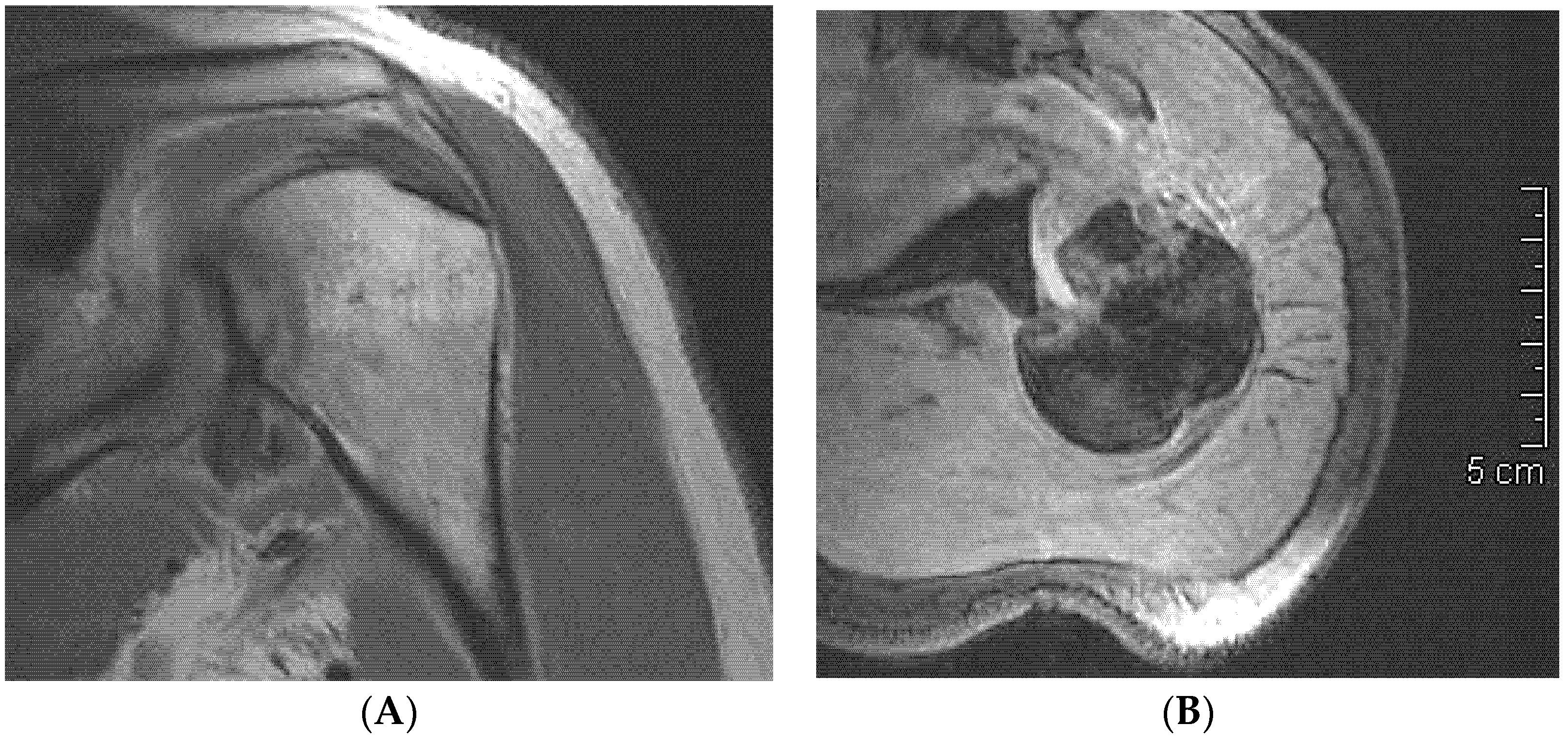
The pre-operative evaluation of the humeral head defects, individuated through CT and/or MRI (Figure 2A,B), was performed using the Randelli classification [26]. In all cases, the defects were 20–50% and classified as first group.

2.1. Surgical Technique

All of the cases were treated by the same surgeon after an average time of 37.75 ± 45.81 days (range 9–106). In three cases, the technique used was a filling of the anterior defect of the humeral head with a transposition of the subscapularis tendon (McLaughlin technique) sutured through trans osseous wires. In the other case, a plication of the subscapularis tendon fixed with a suture anchor was performed.

2.2. Post-Operative Care and Rehabilitation

All of the patients were immobilized with an orthosis, with the arm positioned in external rotation (0–45°) and abduction (0–90°), for a period of 4 weeks to aid in the healing of the posterior capsule [15,26]. In this phase, pendulum exercises and elbow motion three times per day were encouraged. After the immobilization period, the patients underwent a rehabilitative treatment to allow for an early recovery of the functionality of the joint within 3–4 months.
At 4 weeks, unlimited progressive range of motion is initiated as well as isometric posterior rotator cuff strengthening. Progressively, after 1 week from the treatment, a passive and successively active mobility of the shoulder must be executed until complete mobility is achieved approximately after three months [27].

2.3. Clinical and Radiographic Assessment

The clinical evaluation was based on the Constant shoulder score [28], which is associated with an objective evaluation of shoulder stability. The mean difference (ΔConstant) between the involved and the not involved shoulder was reported. The imaging evaluation was based on radiographic exams and an MRI at 1 year from surgery.

3. Results

The average time of follow up was 2.3 ± 0.4 years (range 1.7–2.6 years). The Constant shoulder score assessed at the time of follow up was excellent, with a mean Constant score of 91 (range 83–98) for the treated shoulder and 92.5 ± 6.16 (range 86–98) for the non-involved limb. The mean Constant was 92.5 ± 4.99 (range 86–98).
Analysing the Constant shoulder score, pain was observed in all of the patients only during a great physical effort, being absent at rest and moderate during sleep. In one patient was observed a major night pain in the treated shoulder.
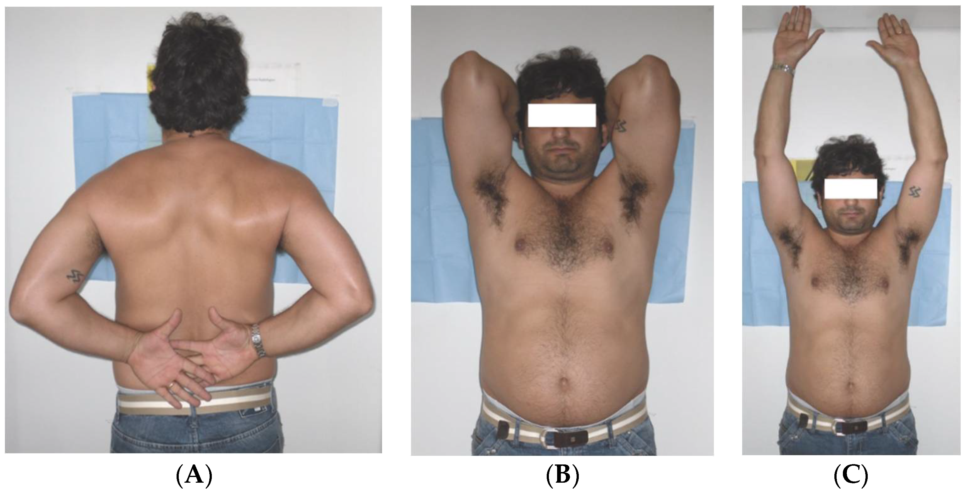
At the physical exam, the functional aspect of the upper limb for three patients was fully recovered; nobody referred to a limitation in the recovery of daily activities or in sport activities. Only one patient referred to a mild functional limitation in the treated shoulder. All of the patients showed good strength in both upper limbs. The range of movement of the treated limbs was satisfactory (Table 2). In particular, an optimal recovery of the movement of active anterior elevation (AAE), abduction, and extra rotation (RE 1 and 2) was observed. A moderate limitation in the movement of intra rotation was evidenced in three patients, being mild in only one patient, who reported a better functional recovery (Figure 3A–C).
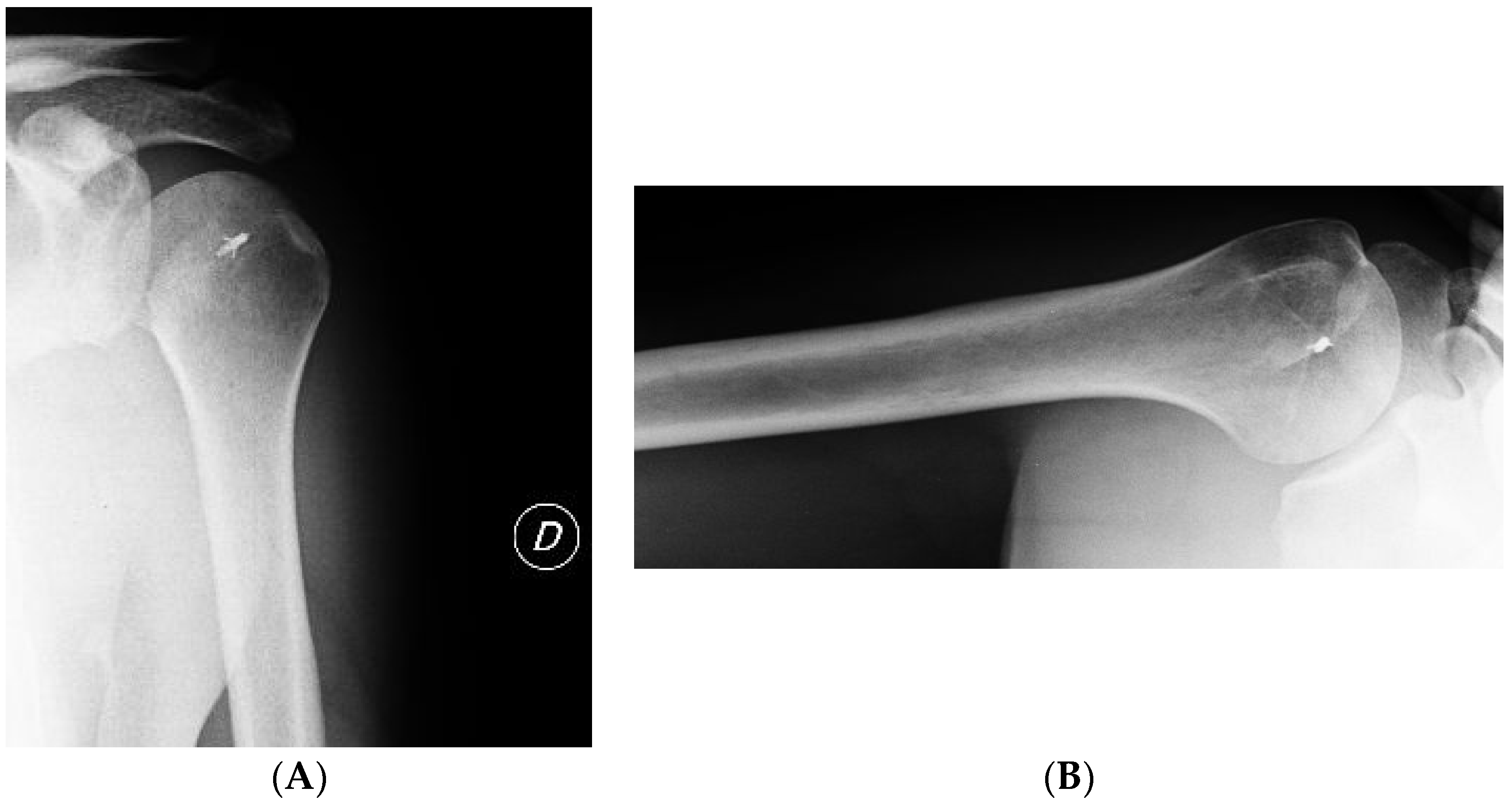
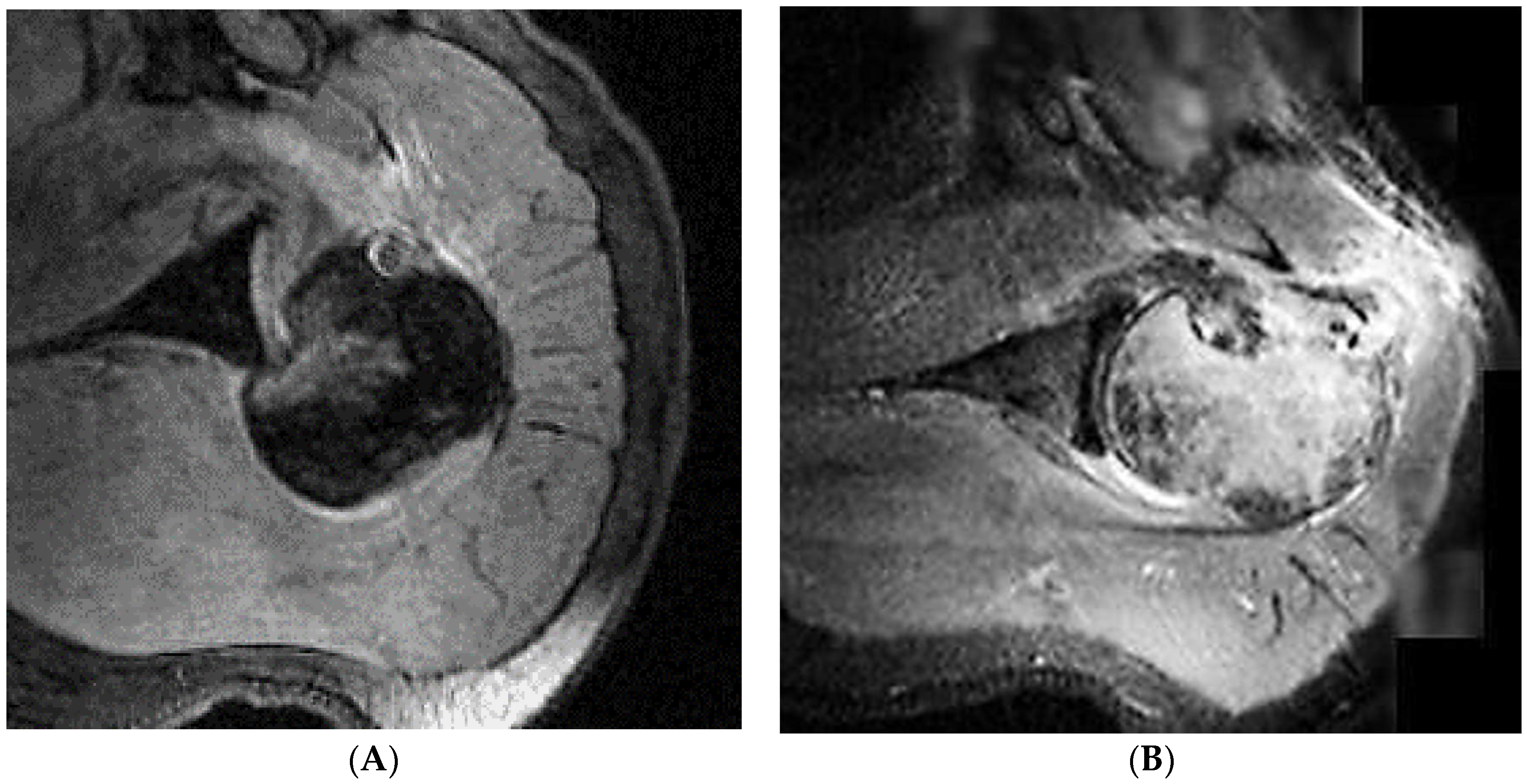
During the clinical exam, good stability for all the treated shoulders was observed. Three patients were very satisfied and one was satisfied. The radiographic evaluation did not report in any case anatomical disposition (loss of the bone contact) of osteoarthritis or other associated lesions (Figure 4 and Figure 5).

4. Discussion

A unidirectional posterior dislocation of the shoulder, caused by a traumatic episode, is frequently associated with an osteochondral defect of the anterior part of the humeral head, which is also known as an inverse Hill–Sachs lesion [8]. This defect involves the articular surface and, due to repetitive movement of internal rotation of the humeral head, could be increased until the degeneration of the entire joint. This lesion is encountered in almost 10% of the articular surface after an acute trauma [16].
Currently, the most useful classification, from the point of view of the choice of therapy, is the Randelli Classification [26], which is based on a defect of the humeral head observed through RX, CT, and MRI imaging. These lesions are classified into four types. In Type 1, the fracture is caused by an anterior impact to the humeral head, is also known as a McLaughlin lesion, and involves from 20% to 50% of the humeral head profile. In Type 2, the articular lesion involves more than 50% of the humeral head surface. Type 3 is an authentic fracture and dislocation of the anatomical neck of the humeral head, which is rotated and posterior dislocated. Type 4 is a multifragmentary fracture and dislocation of the humeral head in circumstances where the articular structure is destructed.
Every radiographic exam of a traumatized shoulder must include AP and Velpeau views [3,11,13]. The AP view, at a first glance, seems normal or shows an imperceptible alteration; its observation is difficult to study because it could be easily confused with a shoulder subdislocation. Indirect signs that can be seen include the lightbulb sign, loss of the half-moon sign, and the trough line [13,14]. A CT scan offers a good definition of the bone lesions, which determine the choice of the treatment. It should always be made in the emergency department in the presence of unclear radiographs showing underdiagnosed or underestimated lesions. It provides a correct and precise representation of the dimensions and shape of the humeral surface and determines the grade of the degeneration in the humeral surface. A three-dimensional reconstruction could be useful in programs for operative reconstruction [29,30].
The indication for the treatment of this pathology varies with the extension of the defect, the duration of the dislocation, and the general condition of the patient. A recent systematic review of 242 neglected cases of posterior dislocation of the GHJ reported an average age of 47.6 and an average interval between injury and diagnosis of 5.9 months [31]. In our series, we reported an average diagnosis delay of 1 month. While our study has the major limit of a small sample, we could assume that our excellent results depend on an early diagnosis. Other series reported in the literature showed how a posterior GHJ dislocation with an associated bone fracture [32], or a reverse Hill–Sachs lesion lower than 50% [33], could be treated with a treatment within 5 months. However, there is also evidence of good results with a later diagnosis with a great functional improvement. In particular, a study of 2005 [18] showed a case of neglected posterior shoulder dislocation, which underwent surgery with a delay of 19 months, having an optimal functional recovery.
In conclusion, our findings encourage the development of standardized protocols for shoulder trauma to avoid misdiagnosis and complications. Acute lesions are easier to treat and have a better success rate. We must underline how early diagnosis is paramount for an effective treatment of a posterior dislocation or of a dislocation with an associated fracture. It is necessary for every orthopaedic surgeon to understand the importance of two orthogonal projections of the shoulder to avoid misidentified lesions. Poor results depend on the gravity of the lesions immediately after the trauma, on the amplification of the McLaughlin lesions, and on humeral suffering due to a mismatch diagnosis. Our study has demonstrated how, even though an inverse Hill–Sachs lesion may be found to be between 25–50%, treatment with the transposition of the sub scapularis tendon is satisfying as shown by the Constant shoulder score.

Author Contributions

V.P. and G.T. conceived and designed the study; V.F.C. performed the surgical procedures; S.M. and D.C. analyzed the data; G.S. supervised the manuscript; E.C. wrote the paper.

Conflicts of Interest

The authors declare no conflict of interest.

References

  1. Shields, D.W.; Jefferies, J.G.; Brooksbank, A.J.; Millar, N.; Jenkins, P.J. Epidemiology of glenohumeral dislocation and subsequent instability in an urban population. J. Shoulder Elbow Surg. 2018, 27, 189–195. [Google Scholar] [CrossRef] [PubMed]
  2. Robinson, C.M.; Aderinto, J. Posterior shoulder dislocations and fracture-dislocations. J. Bone Jt. Surg. Am. 2005, 87, 639–650. [Google Scholar] [CrossRef] [PubMed]
  3. Robinson, C.M.; Seah, M.; Akhtar, M.A. The Epidemiology, Risk of Recurrence, and Functional Outcome after an Acute Traumatic Posterior Dislocation of the Shoulder. J. Bone Jt. Surg. Am. 2011, 93, 1605–1613. [Google Scholar] [CrossRef] [PubMed]
  4. Robinson, C.M.; Akhtar, A.; Mitchell, M.; Beavis, C. Complex posterior fracture-dislocation of the shoulder: Epidemiology, injury patterns, and results of operative treatment. J. Bone Jt. Surg. Am. 2007, 89, 1454–1466. [Google Scholar] [CrossRef]
  5. Goudie, E.B.; Murray, I.R.; Robinson, C.M. Instability of the shoulder following seizures. J. Bone Jt. Surg. Br. 2012, 94, 721–728. [Google Scholar] [CrossRef] [PubMed]
  6. Pushpakumara, J.; Sivathiran, S.; Roshan, L.; Gunatilake, S. Bilateral posterior fracture-dislocation of the shoulders following epileptic seizures: A case report and review of the literature. BMC Res. Notes 2015, 8, 704. [Google Scholar] [CrossRef] [PubMed]
  7. Betz, M.E.; Traub, S.J. Bilateral posterior shoulder dislocations following seizure. Intern. Emerg. Med. 2007, 2, 63–65. [Google Scholar] [CrossRef] [PubMed]
  8. Rouleau, D.M.; Hebert-Davies, J. Incidence of Associated Injury in Posterior Shoulder Dislocation. J. Orthop. Trauma 2012, 26, 246–251. [Google Scholar] [CrossRef] [PubMed]
  9. Saupe, N.; White, L.M.; Bleakney, R.; Schweitzer, M.E.; Recht, M.P.; Jost, B.; Zanetti, M. Acute Traumatic Posterior Shoulder Dislocation: MR Findings. Radiology 2008, 248, 185–193. [Google Scholar] [CrossRef] [PubMed]
  10. Walch, G.; Boileau, P.; Martin, B.; Dejour, H. Unreduced posterior luxations and fractures-luxations of the shoulder. Apropos of 30 cases. Rev. Chir. Orthop. Reparatrice Appar. Mot. 1990, 76, 546–558. [Google Scholar] [PubMed]
  11. Hawkins, R.J.; Neer, C.S.; Pianta, R.M.; Mendoza, F.X. Locked posterior dislocation of the shoulder. J. Bone Jt. Surg. Am. 1987, 69, 9–18. [Google Scholar] [CrossRef]
  12. Rowe, C.R.; Zarins, B. Chronic unreduced dislocations of the shoulder. J. Bone Jt. Surg. Am. 1982, 64, 494–505. [Google Scholar] [CrossRef]
  13. Kowalsky, M.S.; Levine, W.N. Traumatic Posterior Glenohumeral Dislocation: Classification, Pathoanatomy, Diagnosis, and Treatment. Orthop. Clin. N. Am. 2008, 39, 519–533. [Google Scholar] [CrossRef] [PubMed]
  14. Cisternino, S.J.; Rogers, L.F.; Stufflebam, B.C.; Kruglik, G.D. The trough line: a radiographic sign of posterior shoulder dislocation. Am. J. Roentgenol. 1978, 130, 951–954. [Google Scholar] [CrossRef] [PubMed]
  15. Rouleau, D.M.; Hebert-Davies, J.; Robinson, C.M. Acute traumatic posterior shoulder dislocation. J. Am. Acad. Orthop. Surg. 2014, 22, 145–152. [Google Scholar] [CrossRef] [PubMed]
  16. McLaughlin, H.L. Posterior dislocation of the shoulder. J. Bone Jt. Surg. Am. 1952, 24, 584–590. [Google Scholar] [CrossRef]
  17. Castagna, A.; Delle Rose, G.; Borroni, M.; Markopoulos, N.; Conti, M.; Maradei, L.; Garofalo, R. Modified MacLaughlin procedure in the treatment of neglected posterior dislocation of the shoulder. Chir. Organi Mov. 2009, 93 (Suppl. 1), S1–S5. [Google Scholar] [CrossRef] [PubMed]
  18. Delcogliano, A.; Caporaso, A.; Chiossi, S.; Menghi, A.; Cillo, M.; Delcogliano, M. Surgical management of chronic, unreduced posterior dislocation of the shoulder. Knee Surg. Sport Traumatol. Arthrosc. 2005, 13, 151–155. [Google Scholar] [CrossRef] [PubMed]
  19. Krackhardt, T.; Schewe, B.; Albrecht, D.; Weise, K. Arthroscopic fixation of the subscapularis tendon in the reverse Hill-Sachs lesion for traumatic unidirectional posterior dislocation of the shoulder. Arthroscopy 2006, 22, 227.e1–227.e6. [Google Scholar] [CrossRef] [PubMed]
  20. Jariwala, A.; Haines, S.; McLeod, G. Locked posterior dislocation of the shoulder with communition of the lesser tuberosity: A stabilisation technique. Eur. J. Orthop. Surg. Traumatol. 2008, 18, 377–379. [Google Scholar] [CrossRef]
  21. Drignei, M.; Scarlat, M.M. Treatment of chronic dislocations of the shoulder by reverse total shoulder arthroplasty: A clinical study of six cases. Eur. J. Orthop. Surg. Traumatol. 2009, 19, 541–546. [Google Scholar] [CrossRef]
  22. Gerber, C.; Pennington, S.D.; Yian, E.H.; Pfirrmann, C.A.W.; Werner, C.M.L.; Zumstein, M.A. Lesser Tuberosity Osteotomy for Total Shoulder Arthroplasty: Surgical Technique. J. Bone Jt. Surg. Am. 2006, 88 Pt 2 (Suppl. 1), 170–177. [Google Scholar] [CrossRef]
  23. Keppler, P.; Holz, U.; Thielemann, F.W.; Meinig, R. Locked posterior dislocation of the shoulder: Treatment using rotational osteotomy of the humerus. J. Orthop. Trauma 1994, 8, 286–292. [Google Scholar] [CrossRef] [PubMed]
  24. Gerber, C.; Lambert, S.M. Allograft reconstruction of segmental defects of the humeral head for the treatment of chronic locked posterior dislocation of the shoulder. J. Bone Jt. Surg. Am. 1996, 78, 376–382. [Google Scholar] [CrossRef]
  25. Diklic, I.D.; Ganic, Z.D.; Blagojevic, Z.D.; Nho, S.J.; Romeo, A.A. Treatment of locked chronic posterior dislocation of the shoulder by reconstruction of the defect in the humeral head with an allograft. J. Bone Jt. Surg. Br. 2010, 92, 71–76. [Google Scholar] [CrossRef] [PubMed]
  26. Randelli, M. La Fracture-Luxation Postérieure de L’épaule: Nouveaux Éléments de Classification et Thérapeutiques; 2e congrès de la Societé Europèenne de Chirurgie de l’épaule et du coude: Berne, Switzerland, 1988; Volume 2. [Google Scholar]
  27. Alepuz, E.S.; Pérez-Barquero, J.A.; Jorge, N.J.; García, F.L.; Baixauli, V.C. Treatment of The Posterior Unstable Shoulder. Open Orthop. J. 2017, 11, 826–847. [Google Scholar] [CrossRef] [PubMed]
  28. McIntyre, K.; Bélanger, A.; Dhir, J.; Somerville, L.; Watson, L.; Willis, M.; Sadi, J. Evidence-based conservative rehabilitation for posterior glenohumeral instability: A systematic review. Phys. Ther. Sport 2016, 22, 94–100. [Google Scholar] [CrossRef] [PubMed]
  29. Constant, C.R.; Murley, A.H. A Clinical method of functional assessment of the shoulder. Clin. Orthop. Relat. Res. 1987, 214, 160–164. [Google Scholar] [CrossRef]
  30. Aparicio, G.; Calvo, E.; Bonilla, L.; Espejo, L.; Box, R. Neglected traumatic posterior dislocations of the shoulder: Controversies on indications for treatment and new CT scan findings. J. Orthop. Sci. 2000, 5, 37–42. [Google Scholar] [PubMed]
  31. Xu, W.; Huang, L.X.; Guo, J.J.; Jiang, D.H.; Zhang, Y.; Yang, H.L. Neglected posterior dislocation of the shoulder: A systematic literature review. J. Orthop. Transl. 2015, 3, 89–94. [Google Scholar] [CrossRef]
  32. Bekmezci, T.; Altan, E. Management and Prognostic Factors for Delayed Reconstruction of Neglected Posterior Shoulder Fracture-Dislocation. Arch. Trauma Res. 2015, 4, e29903. [Google Scholar] [CrossRef] [PubMed]
  33. Kokkalis, Z.T.; Mavrogenis, A.F.; Ballas, E.G.; Papanastasiou, J.; Papagelopoulos, P.J. Modified McLaughlin Technique for Neglected Locked Posterior Dislocation of the Shoulder. Orthopedics 2013, 36, e912–e916. [Google Scholar] [CrossRef] [PubMed]
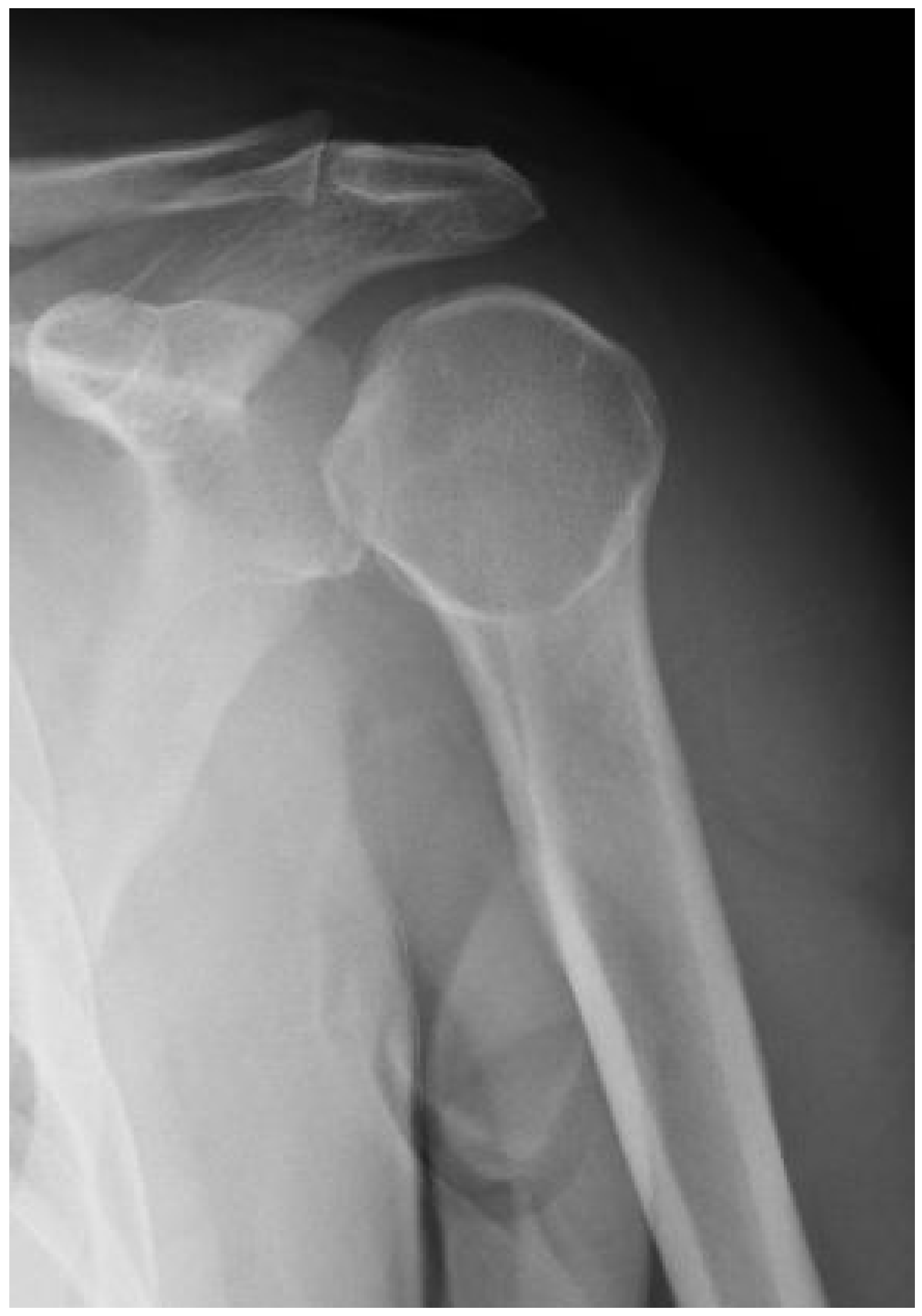
Figure 1. RX made in emergency: AP scan.
Jfmk 03 00030 g001
Figure 2. (A,B) Preoperational MRI showing the chondral lesion of the humeral head.
Jfmk 03 00030 g002
Figure 3. (A) Intra rotation at the Th10- level for both arms; (B) active anterior elevation (AAE) 180°; (C) extra rotation and abduction.
Jfmk 03 00030 g003
Figure 4. (A,B) Patient 4 post-surgery RX–AP and axial scans.
Jfmk 03 00030 g004
Figure 5. Patient 4: (A) Comparative evaluation through pre- and post-surgery MRI; (B) the filling defect of the humeral head is evident.
Jfmk 03 00030 g005
Table 1. Details of treated patients. TS = supraspinatus tendon LHB = long head biceps.
PatientsSex–Age (y)Days from the Trauma to the DiagnosisDays from the Trauma to the TreatmentKind of LesionTreatment
1 CPM–4919 days22 daysHill–Sachs reverse lesion <50%McLaughlin technique Subscapularis tendon transfer
2 DFF–4970 days106 daysHill–Sachs reverse lesion <50%Subscapularis tendon transfer
3 AAM–546 days14 daysHill–Sachs reverse lesion <50%, humeral insertional detachment of ST and LHBSubscapularis tendon transfer
4 CIM–342 days9 daysHill–Sachs reverse lesion <50%Repair through a plication of subscapularis tendon (McLaughlin modified technique)
Table 2. Constant shoulder score results in 2010. R: right; L: left; AAE: active anterior elevation; IR intrarotation; ER 1 extrarotation and adduction; ER 2 extrarotation and abduction.
PatientLimb OperatedConstant ScoreAbductionAAEIRER 1ER 2
Limb RLimb LLimb RLimb LLimb RLimb sinLimb RLimb LLimb RLimb LLimb RLimb L
1 CPLeft, not dominant9492>150°>150°180°180°T7T1140°45°90°90°
2 DFRight, not dominant9193>150°>150°180°180°L1T970°70°90°90°
3 AALeft, not dominant8683>150°>150°180°180°T10L445°45°90°85°
4 CIRight, dominant9898>150°>150°180°180°T11T1145°45°90°90°