Article

Synthesis, Characterization and Magnetic Studies of a Tetranuclear Manganese(II/IV) Compound Incorporating an Amino-Alcohol Derived Schiff Base

by
Madhusudan Nandy
1,2,*,
Shyamapada Shit
3,
Georgina Rosair
4 and
Carlos J. Gómez-García
5
1
Department of Chemistry, Jadavpur University, Raja S. C. Mullick Road, Kolkata 700032, India
2
Department of Basic Engineering Science and Humanities, Netaji Subhash Engineering College, Panchpota, Kolkata 700152, India
3
Department of Chemistry, Jalpaiguri Government Engineering College, Jalpaiguri 735102, India
4
School of Engineering and Physical Sciences, Heriot Watt University, Edinburgh EH144AS, UK
5
Instituto de Ciencia Molecular (ICMol), Departamento de Química Inorgánica, Universidad de Valencia, 46980 Paterna, Spain
*
Author to whom correspondence should be addressed.
Magnetochemistry 2018, 4(4), 57; https://doi.org/10.3390/magnetochemistry4040057
Submission received: 31 October 2018 / Revised: 2 December 2018 / Accepted: 6 December 2018 / Published: 11 December 2018
(This article belongs to the Special Issue A Themed Issue in Honor of Late Professor Samiran Mitra)

Abstract

:
A new tetranuclear mixed-valence manganese(II/IV) compound [MnIIMnIV3(μ-Cl)33-O)(L)3] (1) (where H3L = (3E)-3-((Z)-4-hydroxy-4-phenylbut-3-en-2-ylideneamino)propane-1,2-diol) has been synthesized and characterized by different physicochemical methods. Single crystal X-ray diffraction analysis reveals that 1 is a tetrahedral cluster consisting of a Mn4Cl3O4 core in which the only Mn(II) ion is joined through three μ2-O bridges to an equilateral triangle of Mn(IV) ions, which are connected by a μ3-O and three μ2-Cl bridges. The redox behavior of 1 was studied by cyclic voltammetry. Variable temperature magnetic susceptibility measurements of 1 revealed predominant antiferromagnetic coupling inside the Mn4Cl3O4 cluster.

Graphical Abstract

1. Introduction

Paramagnetic metal clusters are widely utilized in magnetic, electronic, optical, biological, and catalytic studies and applications [1,2]. Though researchers are engaged in the development of several synthetic strategies to design such complexes with high nuclearity, their synthesis is still challenging [3,4,5]. Polydentate ligands, functioning as chelators and bridges, are capable of coordinating with several metal centers and their pendant donating atoms can join other metal centers to construct various intricate structures [6,7]. Schiff bases derived from amino alcohols have proven to possess the ability to form polynuclear complexes [1,2], which play a significant role as biomimetic models of metallobiomolecules [8,9].
Catalytic and magnetic behaviors of many mixed-valence manganese complexes have been investigated using different synthetic strategies [4,10]. The most common oxidation state originates in these hetero-valence manganese complexes are MnII, MnIII and MnIV [11,12,13]. Due to the presence of numerous possibilities in different magnetic coupling between pairs of ions in various oxidation states (II/II, II/III, II/IV, III/III, III/IV and IV/IV pairs), very high values of total molecular spin (S) [14,15,16,17] are an expected phenomenon in these complexes. The molecular magnetism of mixed-valence tetranuclear manganese clusters has been explored, and has been examined as biomimetic models of PS II [1,2,14,15,16].
In this context, we have isolated a new mixed-valence manganese compound [MnIIMnIV3(μ-Cl)33-O)(L)3] (1), incorporating a new Schiff base ligand H3L (H3L = (3E)-3-((Z)-4-hydroxy-4-phenylbut-3-en-2-ylideneamino)propane-1,2-diol) (Scheme 1). The title compound has been characterized by micro-analytical analysis, FT-IR and UV/Visible spectroscopy, cyclic voltammetry and variable temperature magnetic measurements. The single crystal X-ray structure determination shows that 1 is a tetrahedral manganese cluster bearing a Mn4Cl3O4 core. The bond valence sum analysis (BVS) reveals different valence states in the manganese centers, with one Mn(II) and three Mn(IV) ions. The electrochemical characterization of the complex reveals a two-step single electron redox process involving the Mn(IV) centers. Variable temperature magnetic susceptibility measurements show predominant antiferromagnetic intra-cluster coupling in compound 1.

2. Results and Discussion

2.1. Discussion

Condensation of a mixture of equivalent amounts of Benzoyl acetone and 3-aminopropane-1,2-diol in methanol yield a new Schiff base ligand, (H3L = (3E)-3-((Z)-4-hydroxy-4-phenylbut-3-en-2-ylideneamino)propane-1,2-diol) (Scheme 1). A methanolic solution of H3L was mixed with MnCl2∙6H2O and LiOH at a 1:1:1 ratio in isopropanol. The resulting solution was refluxed for an hour and the solvent subsequently removed by slow evaporation, which yielded red block shaped crystals of [MnIIMnIV3(μ-Cl)33-O)(L)3] (1) after one week.

2.2. FT-IR Spectroscopy

The solid state FT-IR spectrum of H3L and 1 is shown in the supplementary materials (Figure S1). These spectral studies reveal that the azomethine (ν-CH=N-) stretching frequency of the H3L (1602 cm−1) was shifted to a lower energy in the complex and appeared at 1591 cm−1. The ν(Calk-O) stretching vibration of the H3L (1270 cm−1) was also moved to lower energies in the complex and appeared at 1112 cm−1. The stretching vibration corresponding to aliphatic-OH (ν-CH2-OH) of the H3L (centered a 3305 cm−1) was not observed in the spectrum of 1, confirming its deprotonation upon complexation. The binding of the ligand was further substantiated by the presence of a number of bands in the range 412–567 cm−1, assignable to the Mn–O and Mn–N bonds [18].

2.3. Electronic Spectrum

Electronic absorption spectra of H3L (Figure S2) and 1 (Figure 1) were recorded in acetonitrile at 300K. The free ligand showed two peaks at 242 and 372 nm, assignable to π→π* and n→π* transitions, respectively, which on chelation underwent red shift and appeared at 248 and 374 nm in the spectrum of 1. An additional shoulder appeared at 473 nm in the spectrum of 1 that may be considered as a combination of ligand to metal charge transfer (LMCT) and ligand-field transitions involving the manganese(IV) ions. Weak d-d transition bands for Mn(IV) ions are found at 850 and 966 nm, corresponding to 2T1g4A2g and 2Eg4A2g transitions, respectively [19].

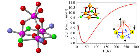
2.4. Crystal Structure Description

The single crystal X-ray diffraction study shows that compound 1 crystallizes in the trigonal space group R-3. Figure 2(left) shows a perspective view of 1 with atom labels. The compound possesses a tetra nuclear tetrahedral Mn4 structure (Figure 2(right)). Relevant bond distances and angles of 1 are listed in Table 1.
The tetranuclear mixed-valence manganese compound has a C3 symmetry and possesses a Mn(II) center(Mn2) on the C3 axis, three crystallographically equivalent Mn(IV) centers (Mn1, Mn11 and Mn12), three fully deprotonated crystallographically equivalent Schiff base ligands, three bridging Cl ligands, and a central oxido anion connecting the three Mn(IV) centers located on the C3 axis. The ligand L3− exhibits a μ-bridging tetradentate coordination mode of the type O3N (Scheme 2) and is coordinated to both types of Mn ions.
Thus, each of the three crystallographically equivalent Schiff base anions coordinated to one of the three Mn(IV) metals in a tridentate O2N meridional mode through its enolato oxygen (O11), the imine nitrogen (N7), and the internal alkoxido oxygen atom (O2). The distorted octahedral geometry of the Mn(IV) ions is completed by two of the three equivalent μ-bridging chlorido ligands (Cl1 and Cl11; symmetry code: 1 = 1 − y, x − y, z) and the μ3-oxido ligand (O1). Mn2 is coordinated by the three Schiff base anions via their terminal (O3) and internal (O2) alkoxido oxygen atoms, giving rise to a slightly distorted trigonal prismatic geometry. The coordination geometry, analyzed by the program SHAPE [20,21], confirms that of Mn1 adopts a distorted octahedral geometry whereas Mn2 displays a distorted trigonal prismatic geometry(Table 2). Each Mn(IV) is joined to the Mn(II) ion by the O2 atom with a Mn1–O2–Mn2 bond angle of 125.5(2)°. The three μ-bridging chlorido ligands connect two Mn(IV) atoms with a Mn1–Cl1–Mn12 (symmetry operation: 2 = 1 − x + y, 1 − x, z) bond angle of 74.20(9)° and form an equilateral triangle as a result of the C3 symmetry of the cluster. The oxido (O1) atom is located on the C3 axis at the center of the triangle and connected with three Mn(IV) atoms with a Mn11-O1-Mn12 bond angle of 119.00(7)°. The central core can be described as a tetrahedral Mn4 cluster formulated as Mn4Cl3O4, in which a Mn(II) ion is associated with threeμ2-O bridges to an equilateral triangle of Mn(IV) ions, which are connected by a μ3-O and three μ2-Cl bridges. The Mn(IV)–Mn(IV) and Mn(IV)–Mn(II) distances in the cluster are 3.2523(17) and 3.5801(17) Å, respectively. Crystal packing reveals that very weak non-classical intermolecular C-H···Cl hydrogen bonding between aromatic hydrogen (H13) and chloride ligand (Cl1) with bond dimension C13-H13···Cl1 = 3.742(13) Å leads to a 3D supramolecular structure in its solid state.
Although there are hundreds of discrete multinuclear Mn cluster compounds with oxido and/or chlorido bridges, the Cambridge Crystallographic Data Center CCDC search showed that compound 1 is the first structurally characterized tetranuclear Mn cluster with an MnIV3MnIICl3O4 core involving Mn(II) and Mn(IV) ions. Although there is a similar tetranuclear cluster formulated as [MnIII3MnIINaOCl4(HL1)3]·3MeCN (HL1 = 2-((E)-(2,3-dihydroxypropylimino)methyl)phenol, CCDC code: LIQXOA), it contains one Mn(II) and three Mn(III) ions, along with a Na+ cation connecting three Mn(III) atoms with a terminal Cl ligand connected to the Na+ ion [22]. Additionally, there are two structures [Na2Mn74-O)23-C9H7NO2Ph(OMe)-κ1-N,κ5-O,O′,O″}62-Cl)6]·CH3OH·CH3CN (C9H7NO2Ph(OMe) = R-2-((E)-(2-hydroxy-2-phenylethylimino)methyl)-6-methoxyphenol, CCDC code: WAWRIY) and [Na2Mn74-O)23-C9H7NO2Ph(OMe)-κ1-N,κ5-O,O′,O″}62-Cl)6]·CH3OH·H2O (C9H7NO2Ph(OMe) = R-2-((E)-(2-hydroxy-2-phenylethylimino)methyl)-6-methoxyphenol, CCDC code: WAWROE) containing a cluster formed by two NaMn4Cl3O4 clusters (along with one Mn(II) and three Mn(III), similar to LIQXOA) sharing a vertex to generate a sandglass shaped cluster of the type NaMn3Cl3O4-Mn-Cl3O4Mn3Na [23]. Finally, there are also two compounds, Na[Mn8Na24-O){μ3-C9H7NO2Ph(OMe)-κ1-N,κ5-O,O′,O″}63-O)(μ2-Cl)9]·CH3OH·H2O (C9H7NO2Ph(OMe) = 2-((E)-(2-hydroxy-2-phenylethylimino)methyl)-6-methoxyphenol, CCDC code: YEMBOK) and Na[Mn8Na24-O){μ3-C9H7NO2(Pri)(OMe)-κ1-N,κ5-O,O′,O″}63-O)(μ2-Cl)9]·CH3OH·H2O (C9H7NO2(Pri)(OMe) = 2-((E)-(2-hydroxy-3-methylbutylimino)methyl)-6-methoxyphenol, CCDC code: YEMBUQ) with a central cluster formed by two Mn4Cl3O4 cores, again with one Mn(II) ion and three Mn(III) ions, which are connected by a triple μ-Cl bridge through the two equilateral triangular faces [24]. Mn(IV)–Cl distances in the range 2.663(3)–2.719(3) Å observed in 1 are slightly higher in comparison to the Mn(III)–Cl distances found in the range 2.631(5)–2.712(3) Å for the reported mixed valence manganese complex (YEMBOK) [1] with a MnIIMnIII3(μ-Cl)33-O) triangular planar core. This can be attributed to the weak overlap between the Mn(IV) and Cl atom in the present compound, which probably arises due to the greater Mn(IV)…Mn(IV) separation (3.2523 (17) Å) within the MnIV3 triangular core in comparison the reported complex (YEMBOK), where Mn(III)…Mn(III) separation within the MnIII3 core are 3.252(1) and 3.248(2) Å. However, no such structure was found in CCDC bearing a similar core involving MnII and MnIV atoms.
The oxidation state of the manganese ions has been determined by the bond valence sum (BVS) calculation based on the correlation between bond valence and bond distance. Here, valence between the two atoms i and j can be calculated using the empirical formula [25,26].
S ij = exp ( R 0 R ij ) / b
where bond valence between two atoms i and j is represented by the term Sij. The observed bond length between i and j is symbolized by the term Rij. R0 and b are the tabulated bond valence parameters defined by Liu et al. [25]. The algebraic sum of Sij values of all the bonds (n) around an atom i gives the oxidation number of the atom i and is symbolized by Nij, which is given by the equation
N ij = Σ S ij
With this equation, the values obtained for compound 1 are Mn1/Mn11/Mn12= 4.224 and Mn2 = 2.208, which clearly confirms the oxidation states of both types of manganese center.

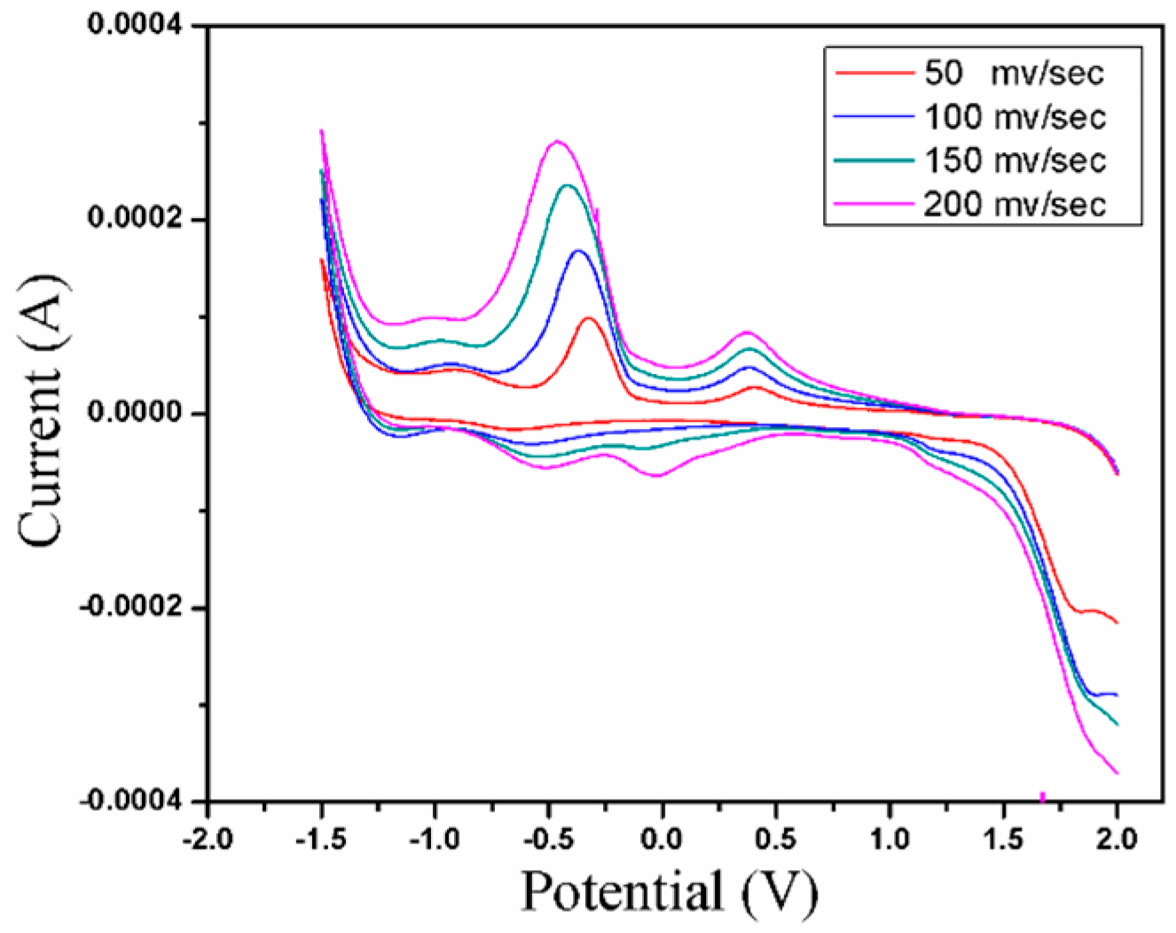
2.5. Electrochemistry

Figure 3 represents the cyclic voltammograms of compound 1, measured within the potential range −2.0 to +2.0 V using nBu4NClO4 as the supporting electrolyte at the following scan rates: 50, 100, 150 and 200 mV s−1. In the redox process, the tetranuclear compound 1 underwent two one-electron charge transfers, namely, (a) MnIIMnIV↔MnIIMnIII and (b) MnIIMnIII↔MnIIMnII. In the cathodic scan, the first peak, assigned to the MnIIMnIV→MnIIMnIII reduction, appeared at 408, 386, 386 and 375 mV, vs. saturated calomel electrode(SCE), for the scan rates 50, 100, 150 and 200 mV s−1, respectively. A second cathodic peak, attributed to the MnIIMnIII→MnIIMnII reduction, was observed at −328, −369, −414 and −458 mV vs. SCE, for the scan rates 50, 100, 150 and 200 mV s−1, respectively. The anodic scan showed one oxidation peak, attributed to the MnIIMnII→MnIIMnIII oxidation, at −639, −561, −539 and −520 mV, vs. SCE, for the same respective scan rates [27,28]. The reversibility of the redox processes is supported by the difference in the respective reduction to oxidation peaks which were found to be 311, 192, 125 and 69 mV when measured at scan rates: 50, 100, 150, 200 mVs−1, respectively. It was also found that with the increase of scan rate, the corresponding cathodic peak shifted to more negative values while the anodic peak shifted to more positive values with an increase of peak currents for both scans and, as a result, the Ipc/Ipa being greater than 1, which also confirms the irreversible nature of the redox processes [28].

2.6. Magnetic Study

The thermal variation of the molar magnetic susceptibility per manganese tetramer times the temperature (χmT) for compound 1 gives a room temperature value of ca. 10.9 cm3Kmol−1, which is a little above the expected value for three independent S = 3/2 manganese(IV) ions and one S = 5/2 manganese(II) ion. On cooling, χmT shows a gradual decrease already observed at room temperature and reaches a minimum value of ca. 7.0 cm3 K mol−1 at ca. 37 K (Figure 4). Below this temperature, χmT rises and reaches a maximum of ca. 8.7 cm3 K mol−1 at ca. 5 K. Below ca. 5 K, χmT sharply decreases and reaches a value of ca. 8.1 cm3 K mol−1 at 2 K. This behavior suggests that the predominant interaction inside the Mn4 cluster is antiferromagnetic, as shown by the continuous decrease of χmT, starting from room temperature to ca. 37 K. Since there are two different exchange coupling constants inside the Mn4 tetrahedron, if both are antiferromagnetic then they are expected to give rise to spin frustration, as not all pairwise interactions can present an antiferromagnetic interaction (see below). This spin frustration results in an intermediate spin ground state that gives rise to the observed increase of the magnetic moment below ca. 37 K. Finally, the decrease observed at very low temperatures can be attributed to the presence of a zero field splitting of the spin ground state and/or to the presence of weak antiferromagnetic inter-cluster coupling. The structure of the Mn4Cl3O4 cluster in 1 shows that the manganese(II) ion is connected to another three manganese(IV) ions, and that there are two crystallographically independent manganese ions (three Mn1 atoms, corresponding to S = 3/2 manganese(IV) ions and one Mn2 atom, which is a S = 5/2 manganese(II) ion). A close look at the structure of the cluster also shows that there are two different exchange pathways (J1, connecting the three Mn1 ions through a µ3-oxido and a µ-chlorido bridge; and J2, connecting the Mn2 ion with the three Mn1 ions through a single µ-oxido bridge; see inset in Figure 4). Accordingly, we have used a model that takes into account the magnetic exchange scheme displayed in the inset in Figure 4 using the package MAGPACK [29,30] with the Hamiltoninan H = −J1[S2S3+ S2S4 + S3S4] − J2[S1S2 + S1S3 + S1S4]. This model very satisfactorily reproduces the magnetic properties of complex 1 above the maximum at ca. 5K with g = 2.013, J1 = −3.8 cm−1 and J2 = −10.3 cm−1 (solid line in Figure 4).
The observed two weak antiferromagnetic exchange interactions inside the Mn4 tetramer leads to spin frustration. Thus, the stronger antiferromagnetic J2 coupling constant implies that the spins of the Mn2 ions must be antiparallel to the three Mn1 ions. Therefore, the spins of the three Mn1 ions should be parallel to each other, in conflict with the antiferromagnetic coupling observed for J1.
Finally, the weak couplings found for both exchange interactions are in agreement to those observed for other oxido-bridged Mn clusters and can be explained from previous magneto structural correlations that indicate that the coupling in this kind of Mn cluster with single µ-oxido bridges is weak and antiferromagnetic [31,32,33]. Unfortunately, the few related Mn4Cl3O clusters reported to date contain one Mn(II) and three Mn(III) ions and the magnetic couplings, although usually weak and antiferromagnetic, cannot be compared with those of 1 [22,23,24].

3. Materials and Methods

3.1. Starting Materials

All solvents and chemicals used in the synthesis were of analytical grade. Benzoyl acetone and 3-aminopropane-1,2-diol were received from Aldrich Chemical Co. Inc. (Sigma-Aldrich, St. Louis, MO, USA). MnCl2∙6H2O and LiOH were obtained from E. Merck, India.

3.2. Syntheses

3.2.1. Synthesis of ((3E)-3-((Z)-4-hydroxy-4-phenylbut-3-en-2-ylideneamino)propane-1,2-diol)

The Schiff base (H3L) was synthesized by reflux condensation of benzoyl acetone (0.162 g, 1 mmol) and 3-aminopropane-1,2-diol (0.091 g, 1 mmol) in 20 mL of methanol for an hour (Scheme 1). Pale yellow crystals of the ligand were obtained upon slow evaporation of the resulting solution. This was dried and stored in vacuo over CaCl2 for future use. Yield: 0.185 g (82%). C14H21NO3 (M = 251.32 g mol−1): Calculated: C, 66.91; H, 8.42; N, 5.57%. Found: C, 66.85; H, 8.37; N, 5.52%.FT-IR (cm−1): 3305s, 2940w, 2866vw, 1834vw, 1602s, 1536s, 1438w, 1328w, 1270s, 1046s, 929w, 838w, 755w, 713w, 672w, 555w, 480w. UV/Vis (nm): 242 (π→π*), 372 (n→π*). 1H NMR (CDCl3, 300 MHz) δ: 2.01 (s, 3H, -CH3), 3.35–3.47 (m, 3H, NCH2, -OH), 3.61–3.73 (m, 3H, -OCH2, OH), 3.95(s, 1H, O-CH), 5.65 (s, 1H, Vinylic-OH), 7.36–7.41 (m, 3H, Ar-H), 7.80 (d, J = 4.80 Hz, 2H, Ar-H), 11.46 (s, 1H, -OH) ppm (Figure S3).

3.2.2. Synthesis of [MnIIMnIV3(μ-Cl)33-O)(L)3] (1)

A methanolic solution (20 mL) of H3L (0.239 g, 1 mmol) was added slowly to a warm isopropanolic solution (10 mL) of MnCl2·6H2O (0.197 g, 1 mmol). Solid LiOH (0.041 g, 1mmol) was added to the mixture. The resulting brown solution was refluxed for an hour, resulting in a darkening of the solution. The solution was cooled and filtered.Slow evaporation of the solvent yielded X-ray diffraction quality red block shaped single crystals of 1 within a week. Yield: 65%. C39H42Cl3Mn4N3O10 (M = 1038.87gmol−1): Calculated: C, 45.09; H, 4.07; N, 4.04%. Found: C, 45.07; H, 4.03; N, 4.02%. FT-IR (cm−1): 3413s, 2936w, 1591s, 1570m, 1543w, 1512vs, 1487s, 1453s, 1439m, 1432m, 1395s, 1364w, 1299s, 1254w, 1215w, 1187vw, 1159vw, 1112s, 1073w, 1028s, 940s, 911m, 880w, 855w, 799vw, 772s, 704s, 679w, 647vw, 603m, 567w, 530w, 481w, 421w, 412w. UV/Vis (nm): 248 (π→π*), 374 (n→π*), 473 (LMCT/LFT), 850 (2T1g4A2g) and 966 nm (2Eg4A2g).

3.3. Physical Measurements

Elemental analyses (carbon, hydrogen, and nitrogen) were performed in a Perkin Elmer 2400 II elemental analyzer. The FT-IR spectrum of H3L and 1 were recorded on a Perkin Elmer Spectrum RX I FT-IR system using a KBr pellet in the range 4000–400 cm−1. The UV/Visible spectra of the H3L and compound 1 were recorded on a Perkin Elmer Lambda 40 UV/Vis spectrometer using HPLC grade acetonitrile in the range 200–1100 nm. Proton nuclear magnetic resonance spectrum of H3L was recorded in CDCl3 on a 300 MHz Bruker Fourier transform NMR spectrometer using (CH3)4Si(TMS) as internal standard. Electrochemical studies of compound 1 were carried out on a PAR VersaStat-potentiostat/Galvanostat II electrochemical analysis system using conventional three-electrode configurations in acetonitrile with nBu4NClO4 as the supporting electrolyte in dry argon atmosphere. Cyclic voltammetry was performed at scan rates of ν = 50, 100, 150 and 200 mV s−1 using platinized platinum millielectrode and saturated calomel electrode (SCE) as working and reference electrodes, respectively, along with a platinum counter electrode. Magnetic susceptibility measurements of polycrystalline sample of compound 1(with a mass of 44.76 mg) were performed in the temperature range 2–300 K with an applied magnetic field of 0.1 T on a Quantum Design MPMS-XL-5 SQUID susceptometer. The susceptibility data were corrected for the sample holders previously measured using the same conditions and for the diamagnetic contributions of the salt as deduced by using Pascal’s constant tables (χdia = −545.43 × 10⁻⁶cm3 mol−1) [34].

3.4. X-ray Crystallography

A red block-shaped X-ray diffraction quality single crystal of compound 1 was mounted on fine glass fiber and placed on a Bruker Apex2 CCD area detector diffractometer equipped with a graphite monochromated fine focus Mo-Kα(λ = 0.7173Å) sealed tube. Crystal data of compound 1 were collected using Apex2 (Bruker, 2011) software at a temperature of 100 K using φ and ω scans. Absorption correction on the collected data was performed by Multi-scan SADABS2008/1 (Bruker, 2008). R(int) was 0.0620 before and 0.0513 after correction. Further details are given in Supplementary Materials (Table S1). The crystal structure of compound 1 was solved employing Olex2 [35], with the SHELXS [36] structure solution program using Direct Methods and refined with the SHELXL [37] utilizing least Squares minimization. The anisotropic displacement parameters were refined successfully for all non-hydrogen atoms. For hydrogen atoms of the aromatic rings and the imino groups, the corresponding hydrogen atoms were placed geometrically and refined using isotropic displacement parameters. The molecular pictorial representations and crystallographic illustrations were made by OLEX2 [35]. There was unresolved solvent, which was probably a mixture of isopropanol and methanol. Attempts at modelling were made but no chemically sensible distances were found from the residual electron density and attempts at squeezing out the solvent were unsuccessful since it was close to the main molecule (in particular the Cl ligand).The supplementary crystallographic data for 1, [MnIIMnIV3(μ-Cl)33-O)(L)3](CCDC-1876188) is available free of charge via www.ccdc.cam.ac.uk/data_request/cif.

4. Conclusions

A new tetranuclear tetrahedral-shaped mixed valence manganese(II/IV) complex based on a bridging tetradentate Schiff base has been synthesized and characterized by spectroscopic methods. Single crystal X-ray diffraction studies reveal that Mn(IV) ions adopt a distorted octahedral geometry whereas the Mn(II) ion displays a slightly distorted trigonal prismatic geometry. The Mn(IV) ions embraced by three μ-chlorido and a single μ3-oxido bridge in a triangular plane are connected with the Mn(II) ion at the apex through endogenic μ-alkoxido bridges, resulting in a Mn4Cl3O4core. The magnetic susceptibility study shows a weak antiferromagnetic coupling inside the Mn4Cl3O4 core of this tetrameric cluster compound.

Supplementary Materials

The following supplementary data are available online. Figure S1: IR spectra of H3L and 1. Figure S2: Electronic spectrums of H3L. Figure S3: 1H NMR spectrum of H3L. Table S1: Crystallographic information and structure refinement parameters for 1.

Author Contributions

M.N. and S.S. planned and designed the experiments; M.N. performed the syntheses and carried out micro-analytical and spectroscopic characterization; G.R. performed crystallographic measurements and characterization; C.J.G.-G. carried out the magnetic studies; M.N. and S.S. wrote the paper.

Acknowledgements

M. Nandy is thankful to the CSIR, New Delhi, Government of India, for presenting a Senior Research Fellowship (SRF) to him [CSIR sanction No.09/096(0598)/2009-EMR-I]. S. Shit is also grateful to UGC, New Delhi, India for financial support (Minor Research Project No. F. PSW-65/12-13 (ERO))]. Financial assistance from the Spanish MINECO (project CTQ2017-87201-P AEI/FEDER, EU) and the Generalitat Valenciana (project Prometeo II/2014/076) are gratefully acknowledged.

Conflicts of Interest

The authors declare there are no conflicts of interest.

References

  1. Reed, C.J.; Agapie, T. Thermodynamics of Proton and Electron Transfer in Tetranuclear Clusters with Mn–OH2/OH Motifs Relevant to H2O Activation by the Oxygen Evolving Complex in Photosystem II. J. Am. Chem. Soc. 2018, 140, 10900–10908. [Google Scholar] [CrossRef] [PubMed]
  2. Nayak, S.; Nayek, H.P.; Dehnen, S.; Powell, A.K.; Reedijk, J. Trigonal propeller-shaped [MnIII3MIINa] complexes (M = Mn, Ca): Structural and functional models for the dioxygen evolving centre of PS II. Dalton Trans. 2011, 40, 2699–2702. [Google Scholar] [CrossRef] [PubMed]
  3. Kanady, J.S.; Tsui, E.Y.; Day, M.W.; Agapie, T. A synthetic model of the Mn3Ca subsite of the oxygen-evolving complex in photosystem II. Science 2011, 333, 733–736. [Google Scholar] [CrossRef] [PubMed]
  4. Maayan, G.; Gluz, N.; Christou, G. A bioinspired soluble manganese cluster as a water oxidation electrocatalyst with low overpotential. Nat. Catal. 2018, 1, 48–54. [Google Scholar] [CrossRef]
  5. Stamatatos, T.C.; Foguet-Albiol, D.; Stoumpos, C.C.; Raptopoulou, C.P.; Terzis, A.; Wernsdorfer, W.; Perlepes, S.P.; Christou, G. Initial example of a triangular single-molecule magnet from ligand-induced structural distortion of a [MnIII3O]7+ complex. J. Am. Chem. Soc. 2005, 127, 15380–15381. [Google Scholar] [CrossRef] [PubMed]
  6. Akhtar, M.N.; Lan, Y.; AlDamen, M.A.; Zheng, Y.-Z.; Anson, C.E.; Powell, A.K. Effect of ligand substitution on the SMM properties of three isostructural families of double-cubane Mn4Ln2 coordination clusters. Dalton Trans. 2018, 47, 3485–3495. [Google Scholar] [CrossRef] [PubMed]
  7. Shopov, D.Y.; Sharninghausen, L.S.; Sinha, S.B.; Borowski, J.E.; Mercado, B.Q.; Brudvig, G.W.; Crabtree, R.H. Synthesis of pyridine-alkoxide ligands for formation of polynuclear complexes. New J. Chem. 2017, 41, 6709–6719. [Google Scholar] [CrossRef]
  8. Semina, B.K.; Davletshinaa, L.N.; Seibertb, M.; Rubina, A.B. Creation of a 3Mn/1Fe cluster in the oxygen-evolving complex of photosystem II and investigation of its functional activity. J. Photochem. Photobiol. B Biol. 2018, 178, 192–200. [Google Scholar] [CrossRef]
  9. Paul, S.; Neese, F.; Pantazis, D.A. Structural models of the biological oxygen-evolving complex: Achievements, insights, and challenges for biomimicry. Green Chem. 2017, 19, 2309–2325. [Google Scholar] [CrossRef]
  10. Alaimo, A.A.; Koumousi, E.S.; Cunha-Silva, L.; McCormick, L.J.; Teat, S.J.; Psycharis, V.; Raptopoulou, C.P.; Mukherjee, S.; Li, C.; Das Gupta, S.; et al. Structural Diversities in Heterometallic Mn-Ca Cluster Chemistry from the Use of Salicylhydroxamic Acid: {MnIII4Ca2}, {MnII/III6Ca2}, {MnIII/IV8Ca}, and {MnIII8Ca2} Complexes with Relevance to Both High- and Low-Valent States of the Oxygen-Evolving Complex. Inorg. Chem. 2017, 56, 10760–10774. [Google Scholar] [CrossRef]
  11. Melnic, S.; Shova, S.; Benniston, A.C.; Waddell, P.G. Evolution of manganese–calcium cluster structures based on nitrogen and oxygen donor ligands. CrystEngComm 2017, 19, 3674–3681. [Google Scholar] [CrossRef]
  12. Mondal, D.; Majee, M.C. Synthesis and Structural Characterization of a New High-valent Bis(oxo)-bridged Manganese(IV) Complex and its Catechol Oxidase Activity. Inorg. Chim. Acta 2017, 465, 70–77. [Google Scholar] [CrossRef]
  13. Zhang, S.; Hua, Y.; Chen, Z.; Zhang, S.; Hai, H. Manganese trinuclear clusters based on Schiff base: Synthesis, characterization, magnetic and electrochemiluminescence properties. Inorg. Chim. Acta 2018, 471, 530–536. [Google Scholar] [CrossRef]
  14. Savva, M.; Skordi, K.; Fournet, A.D.; Thuijs, A.E.; Christou, G.; Perlepes, S.P.; Papatriantafyllopoulou, C.; Tasiopoulos, A.J. Heterometallic MnIII4Ln2 (Ln = Dy, Gd, Tb) Cross-Shaped Clusters and Their Homometallic MnIII4MnII2 Analogues. Inorg. Chem. 2017, 56, 5657–5668. [Google Scholar] [CrossRef] [PubMed]
  15. Adebayo, O.A.; Abboud, K.A.; Christou, G. New mixed-valence MnII4MnIV clusters from an unusual ligand transformation. Polyhedron 2017, 122, 71–78. [Google Scholar] [CrossRef]
  16. Vaddypally, S.; Jovinelli, D.J.; McKendry, I.G.; Zdilla, M.J. Covalent Metal—Metal-Bonded Mn4 Tetrahedron Inscribed within a Four-Coordinate Manganese Cubane Cluster, as Evidenced by Unexpected Temperature-Independent Diamagnetism. Inorg. Chem. 2017, 56, 3733–3737. [Google Scholar] [CrossRef] [PubMed]
  17. Tian, H.; Qiao, X.; Xie, C.-Z.; Ouyang, Y.; Xu, J. Synthesis, characterization, and magnetochemical properties of two Mn4 clusters derived from 2-pyridinecarboxaldehyde Schiff base ligands. J. Coord. Chem. 2017, 70, 1207–1220. [Google Scholar] [CrossRef]
  18. Nakamoto, K. Infrared and Raman Spectra of Inorganic and Coordination Compounds, 5th ed.; Part B; Wiley: New York, NY, USA, 1997. [Google Scholar]
  19. Lever, A.B.P. Inorganic Electronic Spectroscopy, 2nd ed.; Elsevier Science Publishers B.V.: Amsterdam, The Netherlands, 1984. [Google Scholar]
  20. Alvarez, S.; Avnir, D.; Llunell, M.; Pinsky, M. Continuous symmetry maps and shape classification. The case of six-coordinated metal compounds. New J. Chem. 2002, 26, 996–1009. [Google Scholar] [CrossRef]
  21. Llunell, M.; Casanova, D.; Cirera, J.; Bofill, J.M.; Alemany, P.; Alvarez, S.; Pinsky, M.; Avnir, D. SHAPE; Version 2.3; University of Barcelona: Barcelona, Spain; Hebrew University of Jerusalem: Jerusalem, Israel, 2013. [Google Scholar]
  22. Ding, C.; Gao, C.; Ng, S.; Wang, B.; Xie, Y. Polynuclear Complexes with Alkoxo and Phenoxo Bridges from In Situ Generated Hydroxy-Rich Schiff Base Ligands: Syntheses, Structures, and Magnetic Properties. Chem. Eur. J. 2013, 19, 9961–9972. [Google Scholar] [CrossRef] [PubMed]
  23. Escuer, A.; Mayans, J.; Font-Bardia, M.; Gorecki, M.; Di Bari, L. Syntheses, structures, and chiroptical and magnetic properties of chiral clusters built from Schiff bases: A novel [MnIIMnIII6NaI2] core. Dalton Trans. 2017, 46, 6514–6517. [Google Scholar] [CrossRef] [PubMed]
  24. Mayans, J.; Font-Bardia, M.; Escuer, A. Triple Halide Bridges in Chiral MnII2MnIII6NaI2 Cages: Structural and Magnetic Characterization. Inorg. Chem. 2018, 57, 926–929. [Google Scholar] [CrossRef] [PubMed]
  25. Liu, W.; Thorp, H.H. Bond Valence Sum Analysis of Metal-Ligand Bond Lengths in Metalloenzymes and Model Complexes. 2. Refined Distances and Other Enzymes. Inorg. Chem. 1993, 32, 4102–4105. [Google Scholar] [CrossRef]
  26. Brown, I.D. VALENCE: A program for calculating bond valences. J. Appl. Cryst. 1996, 29, 479–489. [Google Scholar] [CrossRef]
  27. Goodson, P.A.; Oki, A.R.; Glerup, J.; Hodgson, D.J. Design, synthesis, and characterization of bis(μ-oxo)dimanganese(III,III) complexes. Steric and electronic control of redox potentials. J. Am. Chem. Soc. 1990, 112, 6248–6254. [Google Scholar] [CrossRef]
  28. Pradeep, C.P.; Htwe, T.; Zacharias, P.S.; Das, S.K. First structurally characterized optically active mononuclear Mn(IV) complex: Synthesis, crystal structure and properties of [MnIVL2] {H2L = S-(−)-2-[(2-hydroxy-1-phenylethylimino)methyl]phenol}. New J. Chem. 2004, 28, 735–739. [Google Scholar] [CrossRef]
  29. Borrás-Almenar, J.J.; Clemente-Juan, J.M.; Coronado, E.; Tsukerblat, B.S. High-Nuclearity Magnetic Clusters: Generalized Spin Hamiltonian and Its Use for the Calculation of the Energy Levels, Bulk Magnetic Properties, and Inelastic Neutron Scattering Spectra. Inorg. Chem. 1999, 38, 6081–6088. [Google Scholar] [CrossRef] [PubMed]
  30. Borrás-Almenar, J.J.; Clemente-Juan, J.M.; Coronado, E.; Tsukerblat, B.S. MAGPACK1 A package to calculate the energy levels, bulk magnetic properties, and inelastic neutron scattering spectra of high nuclearity spin clusters. J. Comput. Chem. 2001, 22, 985–991. [Google Scholar] [CrossRef]
  31. Kahn, O. Molecular Magnetism; VCH Publishers: New York, NY, USA, 1993. [Google Scholar]
  32. Stoicescu, L.; Jeanson, A.; Duhayon, C.; Tesouro-Vallina, A.; Boudalis, A.K.; Costes, J.; Tuchagues, J. Synthesis of CdTe Nanocrystals Using Te Nanorods as the Te Source and the Formation of Microtubes with Red Fluorescence. Inorg. Chem. 2007, 46, 6902–6910. [Google Scholar] [CrossRef] [PubMed]
  33. Li, Y.; Wu, Q.; Lecren, L.; Clerac, R. Synthesis, structure and magnetism of new polynuclear transition metal aggregates assembled with Schiff-base ligand and anionic N-donor ligands. J. Mol. Struct. 2008, 890, 339–345. [Google Scholar] [CrossRef]
  34. Bain, G.A.; Berry, J.F. Diamagnetic Corrections and Pascal’s Constants. J. Chem. Educ. 2008, 85, 532–536. [Google Scholar] [CrossRef]
  35. Dolomanov, O.V.; Bourhis, L.J.; Gildea, R.J.; Howard, J.A.K.; Puschmann, H. OLEX2: A complete structure solution, refinement and analysis program. J. Appl. Cryst. 2009, 42, 339–341. [Google Scholar] [CrossRef]
  36. Sheldrick, G.M. A Short History of SHELX. Acta Cryst. 2008, A64, 112–122. [Google Scholar] [CrossRef] [PubMed]
  37. Sheldrick, G.M. SHELXL-97, Program for Crystal Structure Refinement; University of Göttingen: Göttingen, Germany, 1997. [Google Scholar]
Scheme 1. Schematic diagram for the synthesis of the Schiff base (H3L) and its deprotonated form (L3−).
Magnetochemistry 04 00057 sch001
Figure 1. UV/Visible spectrum of 1, recorded in acetonitrile at 300K. Inset: zoom of the high wavelength region showing the d-d band.
Magnetochemistry 04 00057 g001
Figure 2. (left) Perspective view of the compound 1 with atom labels. (right) View of atetrahedral Mn4 core with coordinating atoms in compound 1. Symmetry operations: 1 = 1 − y, x − y, z; 2 = 1 + y − x, 1 − x, z.
Magnetochemistry 04 00057 g002
Scheme 2. Coordination modes of deprotonated Schiff base ligand L3− with metal ion (M = Mn(IV) and M′ = Mn(II)) in 1.
Magnetochemistry 04 00057 sch002
Figure 3. Cyclic voltammograms of compound 1 plotted at scan rates of 50, 100, 150 and 200 mV s−1.
Magnetochemistry 04 00057 g003
Figure 4. χmT (per Mn4 cluster) vs. T plot for compound 1. Solid line indicates the best fit to the tetramer model (see text). Inset shows exchange scheme of the Mn4 cluster.
Magnetochemistry 04 00057 g004
Table 1. Relevant bond distances (Å) and angles (°) for compound 1.
Bond Lengths (Å)
Mn1-Cl12.663(3)Mn2-O22.134(5)
Mn1-O11.8873(11)Mn2-O32.115(8)
Mn1-O21.892(5)Mn2-O32.227(7)
Mn1-O111.890(4)Mn2-O312.228(7)
Mn1-N71.984(6)
Mn1-Cl112.719(3)
Bond Angles (°)
Cl1-Mn1-Cl11164.90(8)O21-Mn2-O293.30(18)
O1-Mn1-Cl183.38(6)O21-Mn2-O3147.6(4)
O1-Mn1-Cl1181.83(7)O22-Mn2-O3116.6(4)
O1-Mn1-O293.2(3)O2-Mn2-O374.1(2)
O1-Mn1-O1194.3(2)O32-Mn2-O385.0(3)
O1-Mn1-N7174.7(3)O31-Mn2-O385.0(3)
O2-Mn1-Cl191.20(18)O32-Mn2-O3185.0(3)
O2-Mn1-Cl1192.64(17)Mn1-Cl1-Mn1274.35(8)
O2-Mn1-N782.1(2)Mn11-O1-Mn12119.00(7)
O11-Mn1-Cl191.87(16)Mn1-O2-Mn2125.5(2)
O11-Mn1-Cl1186.22(16)
O11-Mn1-O2172.2(2)
O11-Mn1-N790.3(2)
N7-Mn1-Cl199.1(2)
N7-Mn1-Cl1195.9(2)
Symmetry operations: 1 = 1 − y, x − y, z; 2 = 1 − x + y, 1 − x, z.
Table 2. Shape values for the five possible coordination geometries found for coordination number six in Mn1 and Mn2 in compound 1. The minimum values are indicated in bold.
GeometrySymmetryMn1Mn2
HP-6D6h33.61736.993
PPY-6C5v25.91318.084
OC-6Oh3.5928.406
TPR-6D3h14.9422.205
JPPY-6C5v28.17822.087
HP-6 = Hexagon; PPY-6 = Pentagonal pyramid; OC-6 = Octahedron; TPR-6 = Trigonal prism; JPPY-6 = Johnson pentagonal pyramid J2.

