Article

Sustainable Characterization of Some Extracts of Origanum vulgare L. and Biosafety Evaluation Using Allium cepa Assay

by
Daniela Nicuță
1,
Luminița Grosu
2,
Irina-Claudia Alexa
2,* and
Adriana-Luminița Fînaru
2,*
1
Faculty of Sciences, Department of Biology, Ecology and Protection of Environment, “Vasile Alecsandri” University of Bacău, 157, Calea Mărăşeşti, 600115 Bacău, Romania
2
Faculty of Engineering, Department of Chemical and Food Engineering, “Vasile Alecsandri” University of Bacău, 157, Calea Mărăşeşti, 600115 Bacău, Romania
*
Authors to whom correspondence should be addressed.
Horticulturae 2024, 10(5), 504; https://doi.org/10.3390/horticulturae10050504
Submission received: 1 April 2024 / Revised: 8 May 2024 / Accepted: 10 May 2024 / Published: 14 May 2024

Abstract

:
Origanum vulgare L. is ethnomedicinally valuable against various diseases. In Romania, attention for the oregano extracts such as infusions, decoctions, or tinctures, which are very popular among consumers, is constantly increasing, mainly as an important therapeutic alternative. Therefore, this study was undertaken to evaluate the comparative cytotoxic and genotoxic effects of aqueous and hydroalcoholic extracts of local oregano using a sustainable method such as the Allium cepa assay. Two aqueous oregano extracts obtained by infusion (I01) and decoction (D02) and two hydroalcoholic extracts (E03—water/ethanol 80:20 v/v; E04—water/ethanol 60:40 v/v) were used in this study. Before performing the Allium cepa test, a phytochemical screening carried out using fast and efficient analytical methods (electrometry, colorimetry, UV-Vis spectrometry, and high-performance thin-layer chromatography/HPTLC) allowed the qualitative differences in the chemical profile of the investigated oregano extracts to be highlighted. The aqueous and hydroalcoholic oregano extracts were tested on root meristems of Allium cepa and the cytotoxicity and genotoxicity parameters evaluated were the mitotic index (MI) and chromosomal aberration (CA). The results revealed a decrease in MI for each analyzed sample, with hydroethanolic extract E04 showing the most significant effect on MI (9.66%, 3 times less than that of the control sample), followed by the D02 sample obtained by decoction. Chromosomal aberrations such as the ana-telophase with bridges, expelled chromosomes, or delayed chromosomes were observed in all four extracts. The frequency of cells with CA was higher in the case of samples treated with hydroalcoholic extracts compared to aqueous extracts. The experimental extraction conditions influenced the mitotic index, the varieties of identified chromosomal aberrations, and their frequency. Therefore, based on the result obtained in this study, it may be concluded that the Oregano vulgare L. extracts present cytotoxic and genotoxic effects on onion cells. The Allium cepa assay proves to be an easy-to-handle method, with reliable results, minimal cost, and environmental friendliness for the evaluation of the cytotoxic and genotoxic effects of oregano extracts.

1. Introduction

Traditional medicine has consistently been an alternative to allopathic medicine, with the use of medicinal plants as a primary cure being a current practice worldwide for about 85% of the world’s population [1]. Also, in Romania, traditional medicine that uses plant extracts still represents a significant part in the treatment of mild diseases due to their availability and lower costs [2,3,4]. Basically, through the whole evolution of Romanians, therapeutic medicinal plants have played an important role [2].
In the last decade, the investigation of the biological properties of medicinal plants has received increased attention, aiming on the one hand to identify bioactive compounds and to evaluate their therapeutic potential and possible synergistic actions, and on the other hand, to establish the safety profile or tolerability of these extracts and their implications in human health [5].
Since ancient times, the domestic preparation of aqueous extracts (infusion, decoction) or, in some cases, the hydroethanolic extracts of oregano (Origanum vulgare L.), one of the most familiar medicinal plants, has been used for their curative purposes with various healing properties (expectorant, digestive, antispasmodic, antioxidant, antibacterial, etc.) [6,7,8].
As a result, oregano species have so far been the subject of many investigations regarding their chemical composition and biological activity, where their rich content in flavonoids and phenolic acids was highlighted [9,10,11]. Attention for this plant is increasing not only in other countries, but also in Romania, mainly as an important therapeutic alternative. Investigations on indigenous O. vulgare were carried out by Romanian research teams using plant material from different regions of our country [12,13], including our team who was interested in improving the in vitro cultivation conditions of this plant [14].
Despite the important therapeutic advantages revealed in the literature for oregano alcoholic extracts [15,16] and essential oils [17,18,19], at the same time, it has been reported that some compounds such as phenolic monoterpenes present especially in oregano essential oil can be toxic if administered in excess or are not tolerated by some people, making it necessary to use with caution [19,20].
Therefore, it is important to carry out toxicity studies with appropriate methods and with the principal scientific objective being the establishment of the safety or tolerability profile of bioactive extracts from plants and the implications of their use on human health [20,21,22].
The toxicological methods used to assess the biosafety of drugs or plant-based dietary supplements frequently include brine shrimp, cell lines, or animals [22,23,24,25,26]. Knowing that toxicity evaluation involving animal experiments is limited by ethical and economic reasons, researchers turned their attention to the plant kingdom, where some plants proved to be suitable materials for testing the toxic potential of bioactive compounds [27,28,29,30].
Notable among the plant-based tests developed for toxicity screening is the Allium cepa test, which is based in particular on microscopic observations of the aberrations that occur in the root division zone during mitosis and cytokinesis and the subsequent effects on chromosomes, with plants of Allium cepa growing in direct contact with the evaluated substances [30,31].
With multiple advantages such as minimal costs, the easy preservation of plant material, high sensitivity, reproducibility, and environmental friendliness, this in vivo model—Allium cepa—has been successfully applied by many researchers mainly to evaluate the genotoxic potential of medicinal plants [32,33,34,35].
Only a few studies concerning the in vivo cytotoxic and genotoxic potential of aqueous or hydroalcoholic extracts of oregano [36,37] have been reported, with most of the research being related to the cytotoxic effect of oregano essential oils or alcoholic extracts [38,39]. For example, Dragoeva et al. evaluated the allelopathic activity of cold-water extracts [36] or oregano water infusions [37] made from the aerial parts of O. vulgare ssp. vulgare using the Allium cepa assay.
To the best of our knowledge, no in vivo study using the Allium cepa assay comparing aqueous oregano extracts with hydroalcoholic ones have been reported so far. This aspect is relevant because, in traditional Romanian medicine, aqueous extracts such as infusions and decoctions and hydroalcoholic extracts such as tinctures are very popular among consumers, being cheap and easy to prepare compared to essential oils which are more expensive and require specific acquisition methods.
As part of our ongoing study on acquisition and valorization in sustainable conditions of bioactive plant extracts from local raw materials, the assessment of the genetic safety of extracts using an eco-compatible method represents an important step. From this perspective, the present research aims to evaluate the biosafety potential of Origanum vulgare L. extracts obtained under different conditions, by identification of the mitotic index (MI) and chromosomal aberrations (CAs), using a sustainable method. Moreover, the application of fast and efficient physicochemical methods will allow for comparing the phytochemical profile of the investigated oregano extracts.
Therefore, this study was undertaken to evaluate the comparative cytotoxic and genotoxic effects of aqueous and hydroalcoholic extracts of the local oregano Allium cepa assay.

2. Materials and Methods

2.1. Oregano Extract Preparation

Aerial parts (leaves and flowers, separated from branches) of Origanum vulgare L., previously dried, were provided by AdNatura (Hărman, Braşov, Romania), notified by the National Service for Medicinal Plants, Aromatic and Hive Products [40], with the source of investigated oregano being different from those used in previous Romanian studies [12,13].
The plant material was used as such without further grinding, exhibiting a degree of grinding suitable for the extraction process. The moisture content of oregano plant material was 7.82 ± 0.10%.
Commercial ethyl alcohol of agricultural origin, 96°, supplied by S.C. Prodalcom S.A. Botoşani, Romania [41] was used for hydroalcoholic extracts.
Commercial Bucovina non-carbonated mineral water (Vatra Dornei, Suceava, Romania) [42] was used for aqueous extracts.
The ratio of plant material to extractant used for extraction was 1:10 v/v, using 2 g of plant (approximately the content of a tea bag) and 200 mL of water/solvent (the content of a cup) as in the case of home preparation (traditional medicine).
Four extractions were performed from Origanum vulgare L. under specific conditions, resulting in two aqueous extracts (I01 and D02) and two hydroethanolic extracts (E03 and E04), respectively.
All samples were prepared in triplicates.
Aqueous extracts were obtained by infusion and decoction:
For infusion, an oregano sample was added to boiling water and allowed to stand at room temperature for 5 min [16]. After filtration, the I01 extract sample was obtained.
For decoction preparation, the sample was added to warm water (heated on a hot plate) and boiled for 5 min. The mixture was left to stand for another 5 min and then filtered [16]. Extract D02 was obtained.
Hydroalcoholic extraction was carried out by using two hydroalcoholic mixtures (water/ethanol 80:20 and 60:40 v/v, respectively). The samples were stirred at 25 °C and 150 rpm for 30 min. After filtration through Whatman paper, the E03 (80:20 v/v) and E04 (60:40 v/v) extracts were obtained.
The extracts were stored at 4 °C in the refrigerator for 2–3 days until use. The filtrates were used as such, without further concentration.

2.2. Oregano Extract Analysis

From every prepared oregano extract, the dry matter (DM) content was determined from the moisture content according to the AOAC procedure by using a Moisture Analyser (KERN MLB 50-3, Balingen, Germany) [43].
Four physicochemical parameters of the extracts, pH, conductivity (EC), total dissolved solids (TDS), and salinity (SAL), were measured by the electrometric method [44,45] using the Thermo Scientific™ Orion™ Versa Star Pro™ Multiparameter Benchtop Meter (Thermo Fisher Scientific, Waltham, MA, USA) provided with a ROSS Ultra pH/ATC Trode electrode and DuraProbe conductivity cell 013005MD.
All measurements were performed in triplicate.
Total polyphenols content (TPC) was evaluated using the Folin–Ciocalteu method [17,46]. The results are expressed as mg gallic acid equivalents (GAE)/mL using an equation obtained from a calibration curve presented in Figure 1. The absorption was determined spectrophotometrically, by using a UV/VIS Spectrophotometer (Shimadzu UV-1280, Kyoto, Japan), at 750 nm.
In addition, by UV-Vis scanning of the oregano extracts in the range of 190–440 nm, the fingerprint in the representative range of 190–440 nm was recorded.
Color evaluation of oregano extract samples was performed by calculating the color intensity (CI) as a marker for color expression according to the Glories method [47]. The procedure is usually used for the chromatic characteristics of wine [48] and was recently extended to the color evaluation of different juices [49] or the addition of medicinal plants to the functional beverages [50].
According to this method, the color intensity represents the sum of the absorbance at 420 nm (yellow), 520 nm (red), and 620 nm (blue), respectively, thus reflecting the contribution of different pigment categories to the expressed color.
All spectrophotometric absorbance readings were taken using a UV/VIS Spectrophotometer (Shimadzu UV-1280, Kyoto, Japan).
The high-performance thin-layer chromatography (HPTLC) technique was used for a rapid estimation of the extract [51,52,53,54]. Plant extracts were spotted as 8 mm bands on 20 cm × 10 cm HPTLC silica gel 60 F254 plates from Merck (Darmstadt, Germany) using a Semi Auto Sample Applicator CAMAG® Linomat 5 (CAMAG, Muttenz, Switzerland). The plates were developed through a developing chamber with ethyl acetate–water–formic acid (85:15:10, v/v/v) as the developing solvent [54]. The chromatograms were evaluated using a CAMAG TLC Visualizer and scanned using a CAMAG TLC Scanner 3 UV with visionCats CAMAG HPTLC software (version 3.0) under UV light (λ = 254 nm and 366 nm).

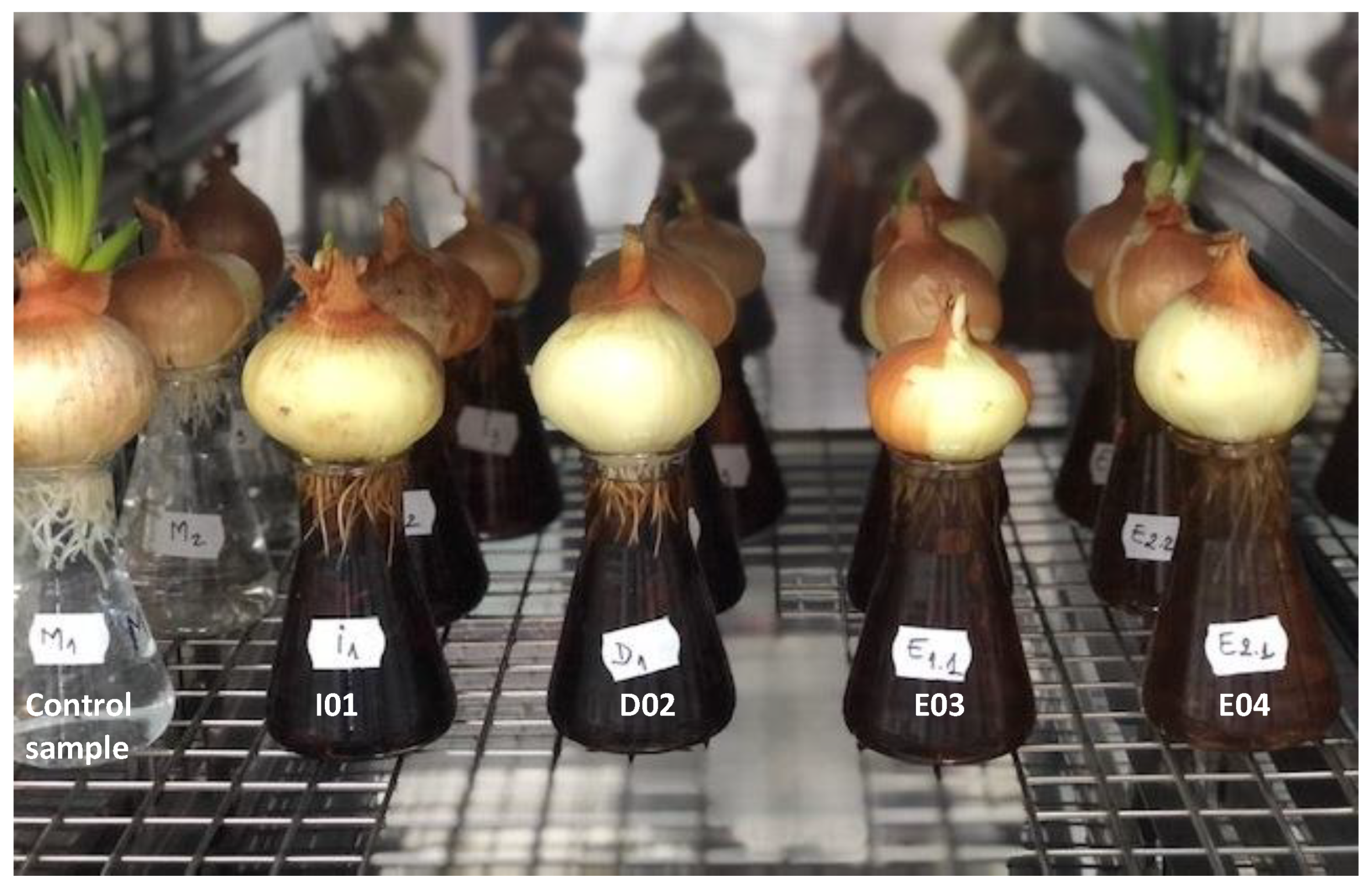
2.3. Allium Cepa Assay

For the Allium cepa test, onion bulbs were purchased from a local market, chosen at about the same size, and the outer scales of the bulbs were carefully removed [32,33,34]. A total of 15 onion bulbs were placed in tap water for 72 h. The next day, a series of 3 bulbs were maintained in the four oregano extracts as such, without any dilution, in a SANYO growth chamber (model MLR-351, Osaka, Japan) in controlled conditions for 24 h in the dark, at 22 °C (Figure 2). As a control, a series of 3 bulbs was placed in water.
For cytogenetic investigations regarding the effect of different oregano extracts on cell division in onion roots, 0.8–1.5 cm roots were harvested. In order to make the cytogenetic preparations, the roots were subjected to processing steps. Roots were fixed in Farmer’s solution, for 18 h in the refrigerator. In order to macerate the pectic substances in the cell wall, to color the genetic material, and for a good display of the cells on the slide, hydrolysis of the plant material samples was achieved. Thus, the roots were treated with a 50% HCl solution, for 10 min, at room temperature.
The roots were immersed in carbol-fuchsin staining solution 10%, leading to a red-violet coloration of the chromosomes. The samples were kept in the refrigerator for 72 h.
Preparation for microscopic evaluation was carried out by the “squash” technique: one root was placed on each microscopic slide, in a drop of 55% acetic acid aqueous solution.

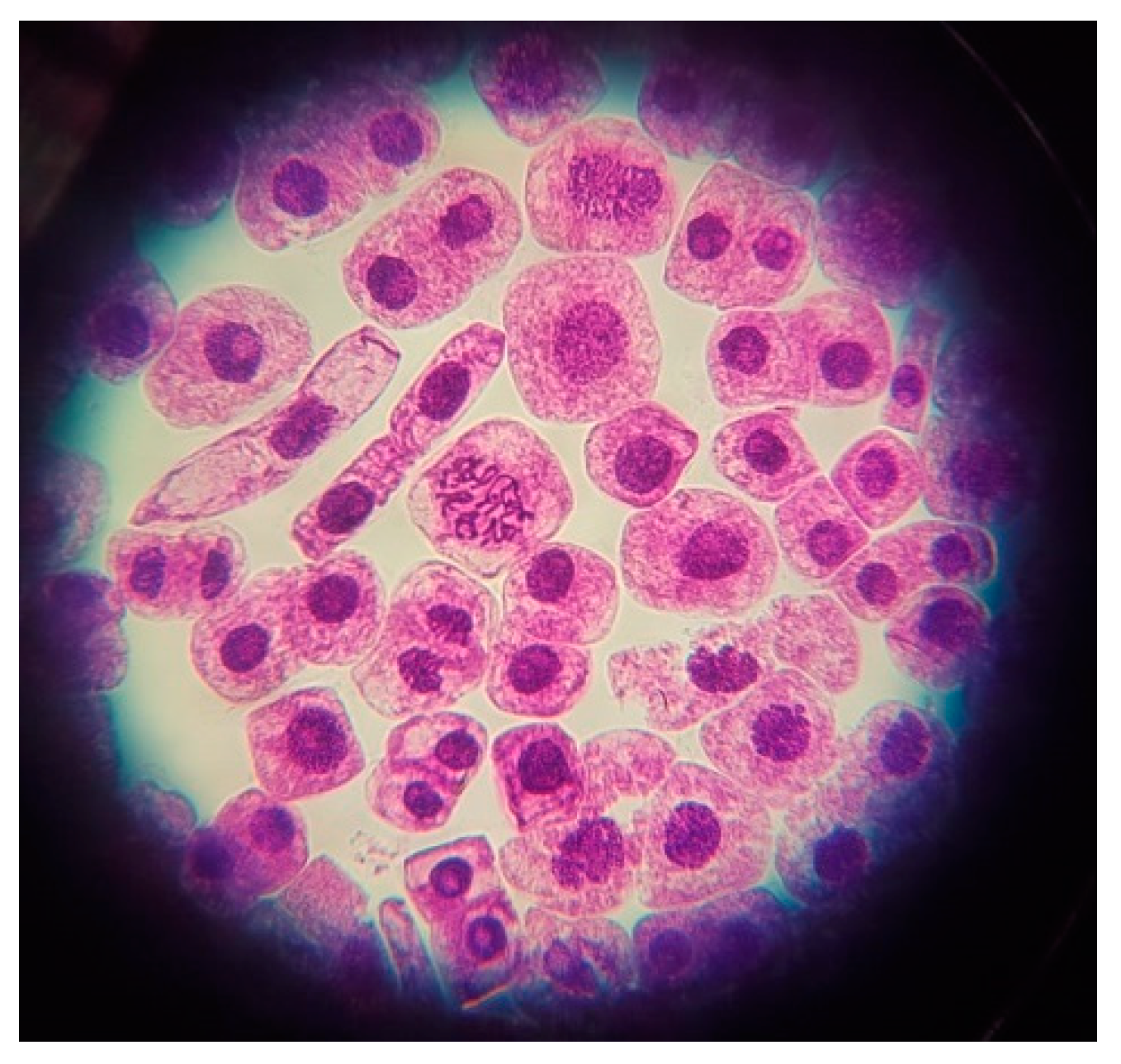
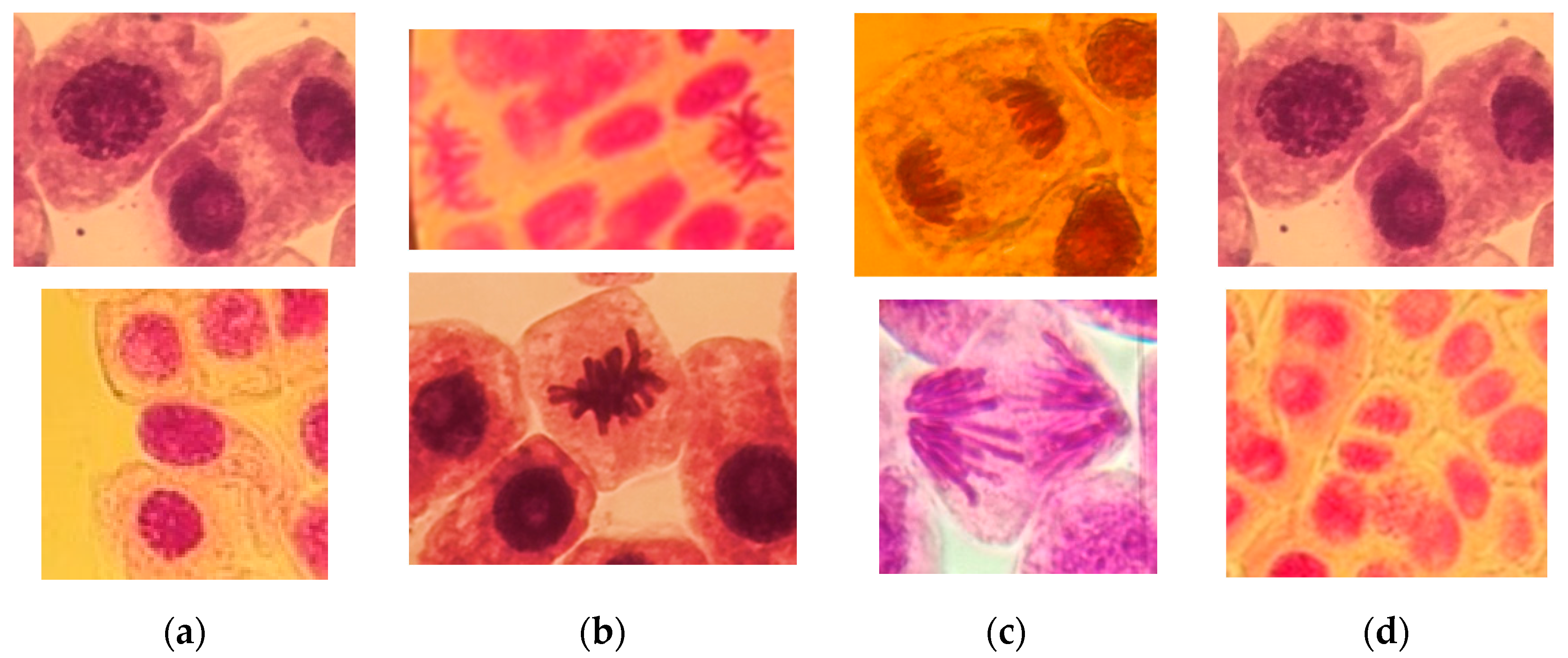
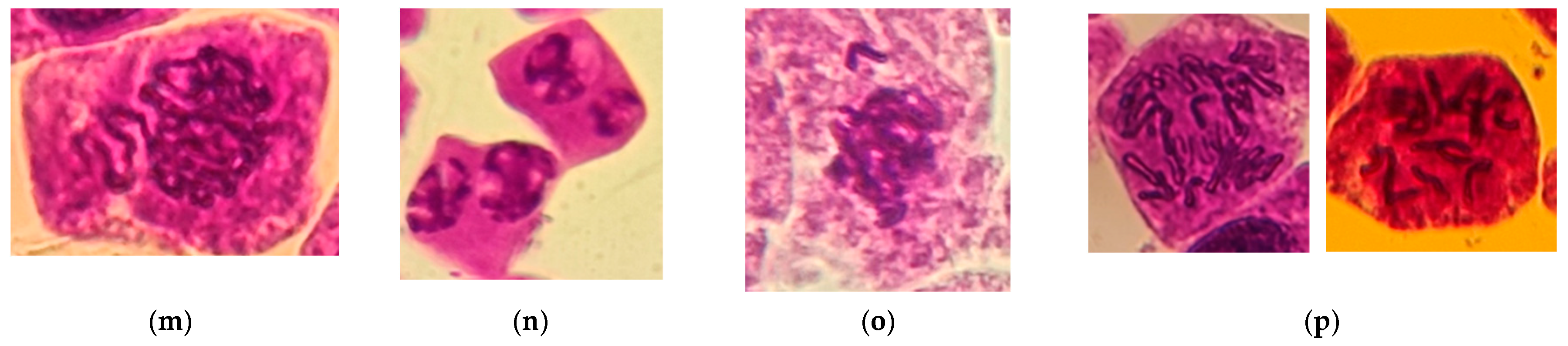
2.4. Cytogenetic Analysis

The cytogenetic analysis of microscopic slides consisted of highlighting the number and types of cells/microscopic field in order to calculate the mitotic index (MI), the frequency of cells in the division phases, and the types of chromosomal aberrations (CAs) observed in cells in mitosis. In the case of interphases, the cells with micronuclei were highlighted as well.
Six slides were prepared for each type of extract (two slides/bulb/flask) and for the control sample.
The slides were analyzed using a microscope OPTIKA (model M-699, Ponteranica, Italy) at 40× and 100× magnification.
For mitotic index calculation and chromosomal aberration, 30 microscopic fields were examined for each variant.
Three replicates were evaluated for each variant and scoring was performed for two roots per replicate (an average of 3500–4500 cells).
The mitotic index represents the ratio between the cells in mitosis and the total number of analyzed cells and was calculated according to Equation (1), where I is the interphase, P is the prophase, M is the metaphase, A is the anaphase, and T is the telophase:
% MI = (P + M + A + T)/(I + P + M + A + T) × 100,
For each type of extract, the frequency of chromosomal aberrations was calculated based on the number of aberrant cells per total cells in division according to Equation (2).
% CA = abnormal cells observed/total number of cells in division × 100,

2.5. Statistical Analysis

The data are expressed as mean ± standard deviation (SD) for all groups of investigated samples.
The statistical significance was assessed by one-way analysis of variance (ANOVA) (p < 0.05), in order to detect significant differences of samples.
A Pearson correlation analysis of the data generated was carried out using the Microsoft EXCEL 2010 Statistical Tool Package.
For the one-way analysis of variance (ANOVA) and graphical representation, the Microsoft Office Excel 2013 (15.0.5589.1000) software of the Windows XP operating system and Origin 2024 10.1.0.170 (Academic) OriginLab Corporation were used.

3. Results and Discussion

3.1. Oregano Extract Analysis

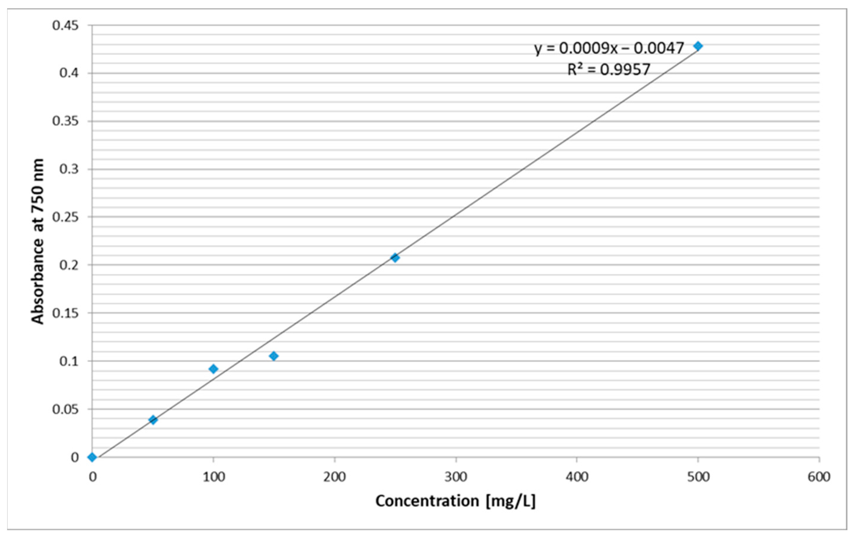
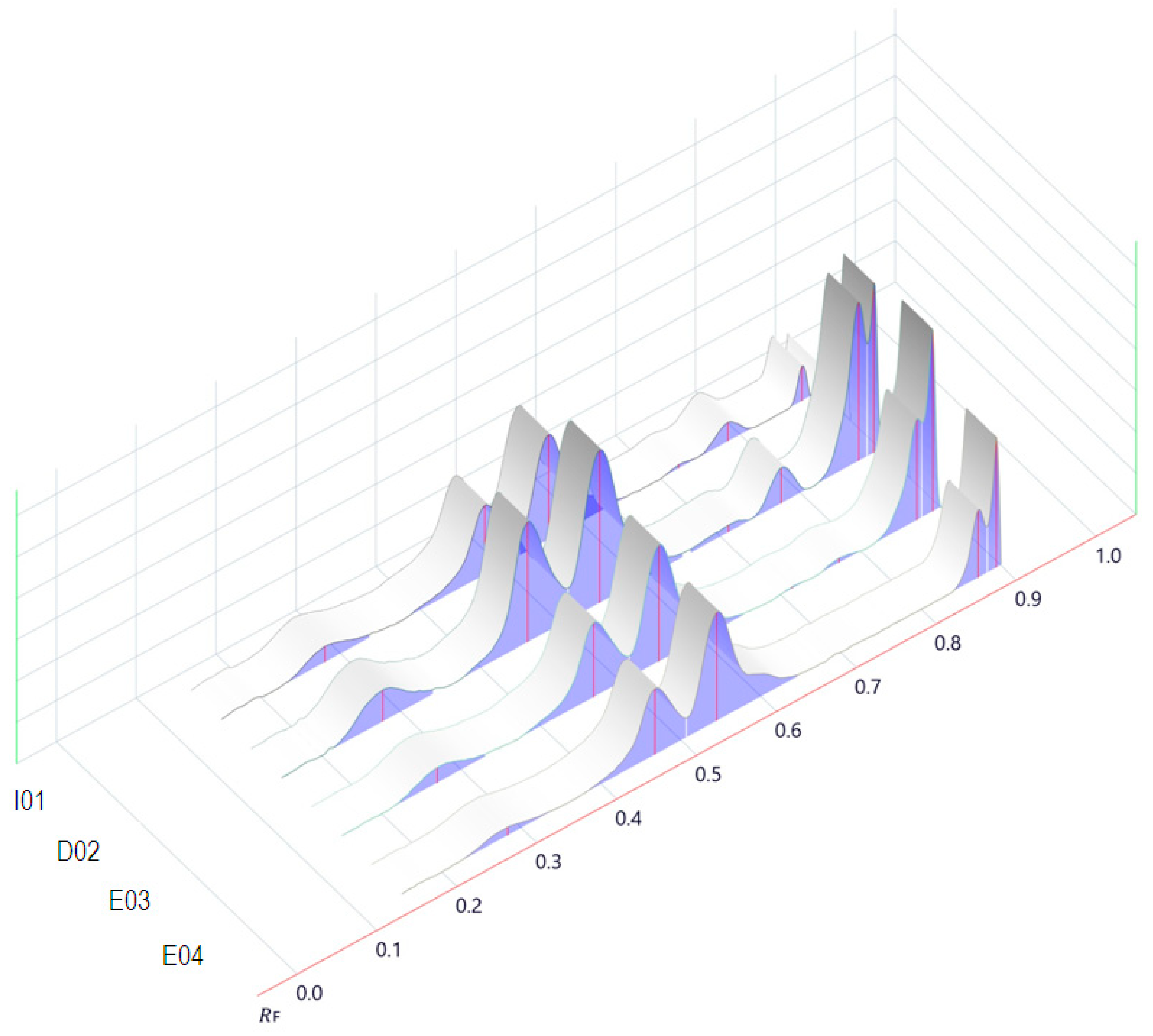
3.1.1. Physicochemical Parameters, Total Phenolic Content, and Color Intensity of Origanum vulgare L. Extracts

The oregano aqueous and hydroalcoholic extracts were analyzed to estimate some physicochemical characteristics before in vivo biological evaluations.
The results regarding the evaluation of the four types of extracts from the point of view of some representative physicochemical parameters such as dry matter (DM), pH, electrical conductivity (EC), total dissolved solids (TDS), and salinity (SAL), the content of phenolic compounds (TPC), and the color intensity (CI) are summarized in Table 1 and graphically represented on the Schoeller–Berkaloff diagram (Figure 3).
It can be noted that the higher dry matter content is in extract I01 obtained by the infusion technique, whereas hydroalcoholic extract E04 presented the lowest dry matter.
Concerning the pH values, both aqueous extracts are slightly acidic, while the hydroethanolic extracts are neutral.
In the case of EC, TDS, and SAL, it is observed that the aqueous extracts (I01 and D02) present the highest values, while for the hydroalcoholic extracts, these values are reduced by 50–60% for E03 and by 75–80% for the extraction solvent with the highest ethanol content (E04).
It is also noted that the highest TPC value is obtained in the case of using water as a solvent and applying the decoction extraction method (sample D02), while increasing the proportion of ethanol in the hydroalcoholic mixture (sample E04, water/ethanol 60:40 v/v) leads to a reduction in the TPC content by 50% (Table 1 and Figure 3).
Knowing that TDS is a measure of combined inorganic and organic compounds dissolved in solution and that salinity is a strong contributor to conductivity, a Pearson correlation analysis of the data generated by measuring seven parameters for oregano extracts is presented in Table 2.
A very strong positive correlation (0.999) was observed between three physicochemical parameters, EC-TDS, EC-SAL, and TDS-SAL, respectively (0.938–0.941), for TPC-EC, TPC-TDS, and TPC-SAL.
On the other hand, it can be seen that these four parameters (EC, TDS, SAL, and TPC) are significantly negatively correlated with pH (r = −0.934 ÷ −0.774).
TPC and CI are moderately positive correlated, with the Pearson coefficient being 0.672, and a weak positive correlation (0.162–0.385) was registered in the case of DM and CI with EC, TDS, and SAL.
The small variations in conductivity for aqueous extracts (I01; D02) and also for hydroethanolic extracts (E03; E04) in close correlation with the variation in TDS, SAL, TPC, and CI are a reflection of the stability of the chemical composition of these extracts (Table 1 and Table 2).
In addition, the logarithmic representation of these data (Figure 3) allows a better visual comparison, highlighting the relationship between the seven indicators selected for monitoring the influence of the extraction conditions on the chemical profile of the extracts, as well as the therapeutic potential.

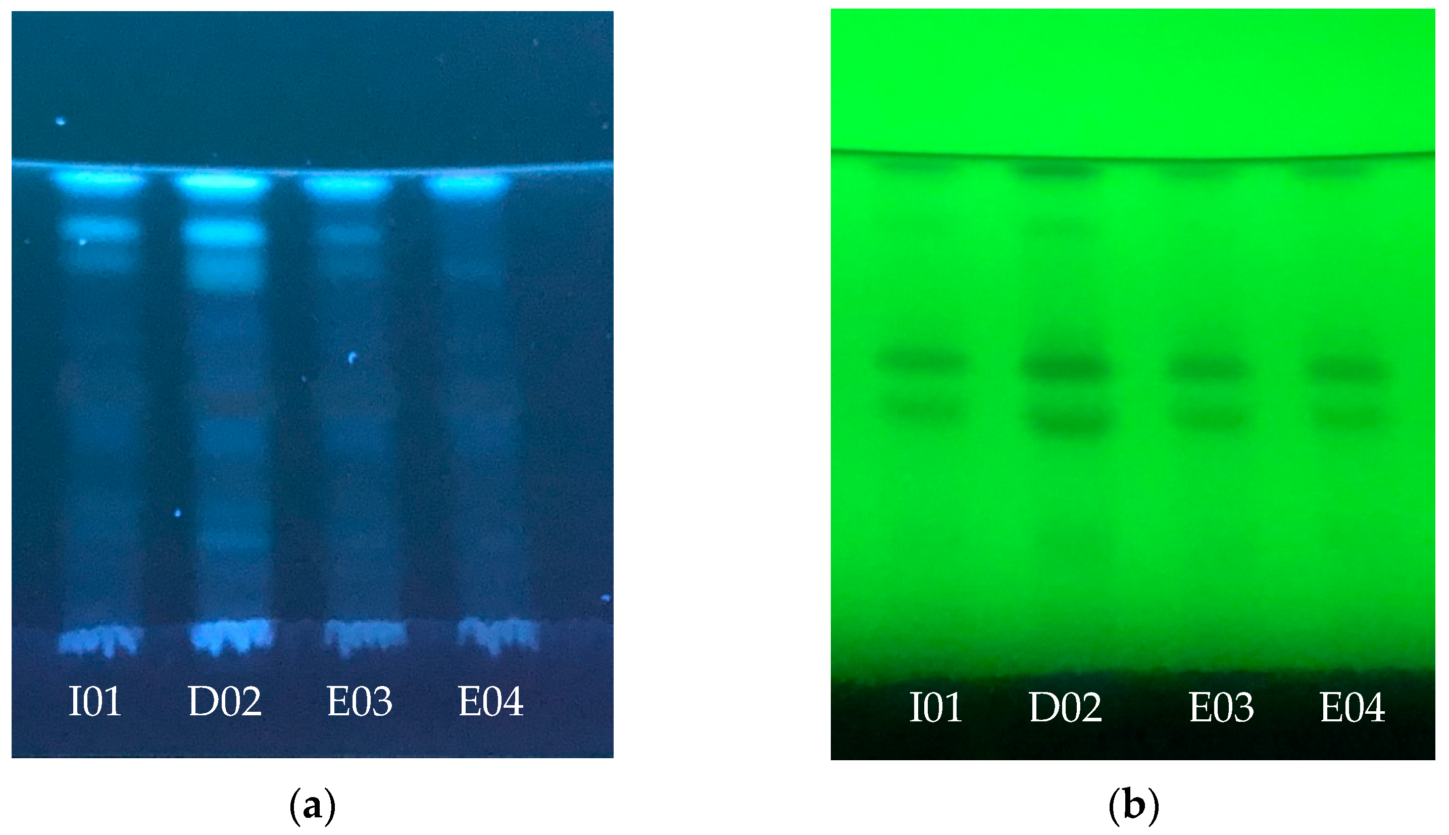
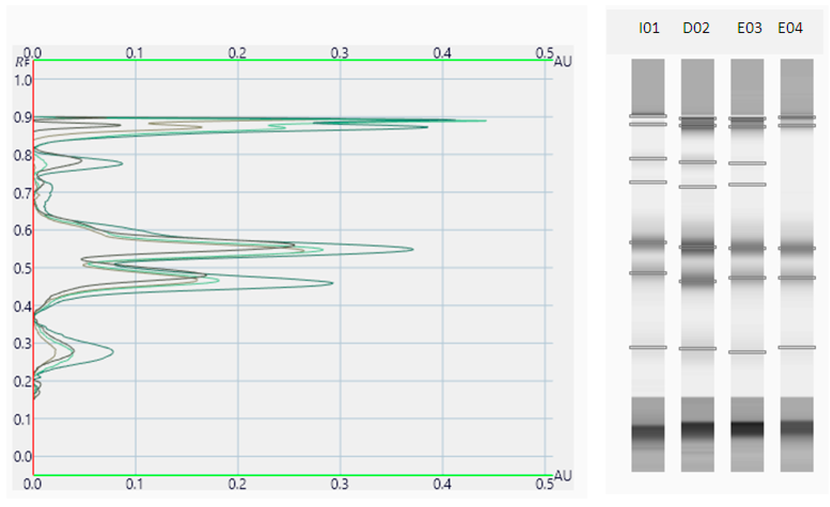
3.1.2. Phytochemical Screening of Origanum vulgare L. Extracts

After UV-Vis scanning of the samples, a comparative spectrophotometric fingerprint of oregano extracts was recorded in the representative 190–440 nm range (Figure 4). It can be noted that the samples present a similar profile with a relatively significant difference in the 260–360 nm range. Phenolic acids such as caffeic and gallic acid are highly absorbing and highly overlapping in this range [55,56], showing that these compounds are present in the oregano extract samples.
In addition to the spectrophotometric analysis, the HPTLC test of oregano extracts was also performed. HPTLC represents a relatively simple, low-cost, and reproducible test method that can provide essential information regarding the comparative compositional quality of plant extracts.
HPTLC silica gel plates developed with ethyl acetate–water–formic acid (85:15:10, v/v/v) at 366 and 254 nm are presented in Figure 5.
The images of the HPTLC plate captured were converted to videodensitograms. The effect of the extraction conditions and solvents on the qualitative profiles of oregano solvents can be observed in Figure 6 and Figure 7.
After development, differences were revealed in the qualitative profiles of the aqueous and hydroalcoholic oregano extracts in both illumination conditions.
At 366 nm, the same intensive blue bands at RF 0.90 were detected in all samples.
In the case of aqueous extracts (I01 and D02), some intensive bands were observed also at RF 0.87, while, by comparison, the corresponding bands for hydroalcoholic extracts (E03 and E04) were much less intensive.
At RF 0.78, for both aqueous samples, bands were noticed in the case of the D02 sample obtained by decoction, with that band being more intense. This aspect can be easily confirmed by the densitograms presented in Figure 6 and Figure 7.
From the densitograms, a peak corresponding to the RF of 0.29 can be observed (rutin RF) [54], being quite visible, especially in the case of sample D02.
Also, at 254 nm, dark bands (almost black) at RF 0.47 and 0.55 (hyperoside RF) [54] were observed in all samples. The intensities of the bands were higher for sample D02. The HPTLC method provides chromatographic fingerprints for the oregano extract samples, with qualitative and quantitative differences being observed between the aqueous samples compared to the hydroalcoholic ones. It has been observed that unlike the neutral pH and the presence of ethanol in the extraction medium, an aqueous medium and slightly acidic pH favor the extraction of polyphenols.
In accordance with the data from the literature, the applied extraction procedure using different solvents and their mixtures influences the phytochemical profile of the sample both qualitatively and quantitatively [10,57].
For the isolation of phenolic acids, including chlorogenic acid, gallic acid, ferulic acid, caffeic acid, etc., water has been proven as a proficient extraction solvent. In addition, in the water extracts of various oregano species, rutin, quercetin, luteolin-7-O-glucoside, apigenin-7-O-glucoside, rosmarinic acid, and luteolin were also identified [10,58].
Through comparative analysis of the aqueous extract densitograms, it can be observed that the two extracts have the same qualitative profile, but that they differ significantly from a quantitative point of view. The densitograms of hydroalcoholic extracts are almost similar, and some small differences can be observed.

3.2. Cytotoxicity of the Oregano Extracts (Mitotic Index MI)

As a first general observation, the microscopic evaluation of root-tips of Allium cepa indicated that the tested extracts of Origanum vulgare did not inhibit the cell division process in onion root meristems, with cells being observed in all mitotic phases.
The cytotoxic effects of oregano extracts were evaluated by calculating the mitotic index (MI) and the frequency of cells in the division phases (Figure 8).
The mitotic index (MI) is a convenient tool used to measure the number of dividing cells, with the degree of cytotoxicity of an agent being quantified by the MI decrease or increase. Both higher and lower MIs compared to the control can be related to an alteration of mitosis mechanisms as a result of cytotoxic effects. If the MI of the studied samples is increasing compared to the control, the samples induce stimulatory effects on cell division, while if the MI is decreasing, it shows inhibitory effects. The inhibition could be interpreted as a delay in cell proliferation, cellular damage, or death.
The obtained data of mitotic index and the number of cells in the division phases are presented in Table 3.
A decrease in the percentage mitotic index (% MI) value was observed for each analyzed sample, whether it is an aqueous extract or hydroalcoholic extract compared to the control sample. This indicates that oregano extracts induce a mitodepressive effect, with the aspect also highlighted by the research carried out by Dragoeva et al. [36,37].
The aqueous extract obtained by infusion I01 showed a % mitotic index (% MI) of 24.15, almost similar to the value of the blank sample (28.54%). Similar results of MI were obtained by Dragoeva et al. in the case of oregano cold-water extracts, with the mitotic index being reduced by 15% compared to the control [36]. The mitotic activity in Allium cepa cells in the presence of oregano hot-water extracts from our study sample I01 is comparable with the mitotic activity reported by Dragoeva et al. for onion roots treated with oregano cold-water extracts (Table 4).
Instead, the mitotic index for the D02 sample obtained by decoction was significantly lower than that of the control sample (16.25%). This fact could be due partly to the high total polyphenols content and total dissolved solids from the sample obtained through the decoction aspect that correlates with the data obtained for the measured parameters presented in Table 1. This can be compared to the result of Dragoeva et al. [37], who also observed a significant decrease in mitotic index after the treatment of onion roots with water infusions obtained from oregano dried stems, leaves, and flowers (Table 4).
This might be due to the heat involved in the extraction process since this would have probably caused a better extraction of more constituents than for the infused extract.
Regarding the hydroalcoholic extracts, it was found that the presence of alcohol in the extraction solvent affects cell division. Thus, in both hydroalcoholic samples E03 and E04, the mitotic index was lower compared to the control sample. It can be observed that in the case of sample E04, a higher concentration of alcohol used for extraction leads to a significant decrease in the mitotic index (9.66%, approximately 3 times less compared to the control sample). This obtained result is in agreement with other research findings concerning other plant species [32].
Regarding the frequency of cells in the division phases, some changes in the ratio were observed depending on the extract used.
In all samples analyzed, the highest percentage was calculated for cells in the prophase (82.70–94.49%), followed by cells in the metaphase (2.35–6.62%), telophase (2.01–6.41%), and anaphase (0.91–4.27%). Analyzing the results, it can be noted that the percentage of anaphase cells decreased in the case of aqueous samples compared to the control, while for the hydroalcoholic samples, it increased. Concerning the percentage of telophase cells, an increase was observed only in the case of hydroalcoholic sample E04, while for I01, D02, and E03 samples, the telophase index was lower than that of the control. On the contrary, in the studies carried out by the Bulgarian researchers, the effect caused by oregano cold-water extracts [36] or oregano water infusion [37] was an increase in the anaphase index and a simultaneous decrease in the telophase index. The interference in the cell cycle kinetics is accepted as a sign of cytotoxic influence.
The different stages of normal mitotic division are presented in Figure 9.

3.3. Genototoxicity of the Oregano Extracts (Chromosomal Aberration CA)

Chromosomal aberrations represent changes in chromosome structure as a result of a break or exchange of chromosomal material due to exposure to various mutagen agents. If a significant increase in cells with chromosomal aberrations is observed, this suggests that some compounds present in the extract plants could be responsible for producing the aberrations.
For all samples of oregano extracts, along with normal cells, some cells with chromosomal aberrations were also highlighted.
The genotoxic effects in onion root meristems including the percentage of chromosomal aberrations and the various types of chromosomal abnormalities are summarized in Table 5 and graphically represented in Figure 10.
The percentage chromosomal aberrations for all the samples were found to be less than 4.50% at the highest concentration.
The lowest percentage of cells with chromosomal aberrations was recorded in the case of the control sample (0.93%).
Concerning the aqueous extracts, the I02 sample obtained by infusion presented a similar value to that of the control sample (1.01%), while in the case of the D02 sample obtained by decoction, the number of cells with abnormalities doubled (1.99%) compared to the control.
For hydroalcoholic extracts, the highest value of cells with aberrations was highlighted in sample E04 (4.5%), while sample E03 presented 3.52% of total aberrant cells, a value almost 4 times higher compared to the control sample.
Nearly comparable results were observed in Dragoeva et al.’s studies [36,37]. The treatment with oregano extracts (with cold or hot water) considerably increased the percent of chromosome aberrations in comparison to the control. In our study, as shown above, the significant increase in chromosomal aberration number was not as high in the aqueous extracts but was in the hydroalcoholic ones (Table 4 and Table 5).
Various types of chromosomal abnormalities were observed, especially in the ana-telophase (A-T) of mitotic cells such as A-T with bridges, A-T with delayed chromosomes, A-T with expulsed chromosomes, and A-T with fragments. The frequency of these abnormal cell types varied depending on the type of extract applied to the onion roots.
Among all the chromosomal aberrations recorded on the cytogenetic slides, A-T cells with bridges were the most frequent, in all samples analyzed, including the control. The highest percentage of bridged A-T was calculated for sample E04 (1.01%), followed by sample D02 (0.86%) and sample E03 (0.72%). The lowest value of A-T with bridges was represented by sample I01 (0.27%), while in the control sample, the calculated value was 0.33%.
A high rate of abnormal A-T cells was also represented by the ana-telophase with expulsed chromosomes. The most numerous were identified in sample E04 (0.88%) followed by sample E03 (0.55%). In the case of aqueous extracts, the most numerous A-T with expulsed chromosomes were observed in the decoction sample (0.37%), while in the infusion sample, the percentage was slightly lower (0.22%) compared to the control one (0.28%).
A-T with delayed chromosomes were also detected, with the most representative sample being hydroalcoholic extract sample E04 (0.44%), closely followed by sample E03 (0.41%) and aqueous extract D02 (0.34%). In the case of infusion sample I01, the amount of A-T with delayed chromosomes (0.04%) was lower than that of the control sample (0.10%).
Another type of aberration was represented by A-T cells that showed chromosome fragments. Such cells were observed in a high percentage in the E04 hydroalcoholic extract (0.35%), while in the other tested extracts, the number of A-T with fragments was significantly lower (0.03–0.05%). In the control sample, A-T with fragments were not present.
In all analyzed samples, abnormal cells were also identified in other phases of the mitotic division, namely in the metaphase and prophase.
Thus, cells with non-oriented chromosomes or expulsed chromosomes were observed in the metaphase. Only in the case of the hydroalcoholic extracts were aberrant cells in the metaphase highlighted; in the other samples, this type of abnormal cell was absent. The most numerous aberrant metaphases were recorded in the case of sample E03 (1.06%), followed by E04 (0.55%).
Prophase cells with a micronucleus and some spindle disturbance were also registered for all samples. An increased number of micronuclei in the prophase were observed especially in samples E03 and E04.
The chromosomal aberrations identified by Dragoeva et al. [36,37] refer only to spindle abnormalities in the metaphase and ana-telophase, being the most frequently observed in the treated cells with oregano extracts (with cold or hot water), followed by bridges and fragments in the ana-telophase. This constitutes an argument of the fact that the types of identified aberrations and their frequency depend on the biological material used as the test plant (onion variety), as well as on the experimental conditions for obtaining medicinal plant extracts.
The hydroalcoholic extracts also presented some cells whose nuclei had a “mace” appearance, multipolar anaphases, and a prophase with “buds” (data not reported in Table 5).
Figure 11 shows various types of chromosomal aberrations observed in the Allium cepa assay with tested extracts of Origanum vulgare.

4. Conclusions

The present work describes the evaluation of the biosafety potentials of some extracts of Origanum vulgare L., by the Allium cepa test, as well their physicochemical and phytochemical investigations using simple, fast, and environmentally friendly analytical techniques (electrometry, colorimetry, UV-Vis spectrometry, and high-performance thin-layer chromatography/HPTLC).
Both the monitoring of the seven parameters for obtaining oregano extracts and the phytochemical screening indicate that the highest content in bioactive compounds was obtained when using water as a solvent and applying the decoction extraction method, while increasing the proportion of ethanol in the mixture of hydroalcoholic led to a 50% reduction in TPC content. In addition, the presence of two important flavonoid glycosides with significant pharmacological activities, namely rutin (especially in the case of sample D02) and hyperoside (in all samples), was highlighted by HPTLC screening.
Concerning the biosafety evaluation, it can be concluded that the aqueous and hydroethanolic extracts of Origanum vulgare L. may have potential cytotoxic and genotoxic effects, especially in the case of hydroethanolic extracts.
The cell division process was not affected and all phases of division were identified, mostly being represented by prophases, followed by metaphases, telophases, and, in a much smaller percentage, anaphases. The mitotic index was significantly reduced especially in the case of the E04 hydroethanolic extract obtained with water/ethanol 60:40 v/v (MI = 9.66%, approximately 3 times less compared to the control sample).
The hydroalcoholic extracts induce an obvious genotoxic effect on the onion cells, since, in the studied samples, the most numerous abnormal cells were highlighted both in the ana-telophase and in the metaphase and prophase. The frequency of cells with chromosomal aberrations was higher in the case of samples treated with hydroalcoholic extracts (4.5% in the case of E04) than the aqueous extracts when compared to the control sample.
The obtained results in the present investigation demonstrate that the biological material used for the Allium cepa test, the source of oregano plants submitted to extraction, and the experimental conditions influence the mitotic index, the varieties of identified chromosomal aberrations, and their frequency.
Therefore, it may be concluded that Oregano vulgare L. extracts present cytotoxic and genotoxic effects on onion cells. Hence, the use of oregano in traditional medicine must be carried out with caution, especially in the case of hydroalcoholic extracts with a higher alcohol proportion, as well as in the case of decoction.
The present study has allowed the in vivo evaluation of the cytotoxicity and genotoxicity potential of aqueous and hydroethanolic oregano extracts using the Allium cepa assay, a fast and easy-to-handle method, with reliable results, minimal cost, and environmental friendliness.

Author Contributions

Conceptualization, D.N., L.G., I.-C.A. and A.-L.F.; methodology, D.N., L.G. and I.-C.A.; software, I.-C.A.; writing—original draft preparation, D.N., L.G. and I.-C.A.; writing—review and editing, I.-C.A. and A.-L.F.; visualization, D.N., L.G., I.-C.A. and A.-L.F.; supervision, A.-L.F.; project administration, A.-L.F.; funding acquisition, A.-L.F. All authors have read and agreed to the published version of the manuscript.

Funding

This research received no external funding.

Data Availability Statement

The original contributions presented in the study are included in the article, further inquiries can be directed to the corresponding authors.

Acknowledgments

The authors are grateful for the technical and material support provided by the CAIP Research Center and the School of Doctoral Studies of “Vasile Alecsandri” University of Bacău.

Conflicts of Interest

The authors declare no conflicts of interest.

References

  1. Fitzgerald, M.; Heinrich, M.; Booker, A. Medicinal plant analysis: A historical and regional discussion of emergent complex techniques. Front. Pharmacol. 2020, 10, 1–24. [Google Scholar] [CrossRef] [PubMed]
  2. Segneanu, A.-E.; Cepan, C.; Grozescu, I.; Cziple, F.; Olariu, S.; Ratiu, S.; Lazar, V.; Murariu, S.M.; Velciov, S.M.; Marti, T.D. Therapeutic use of some Romanian medicinal plants. In Pharmacognosy—Medicinal Plants; Perveen, A., Al-Taweel, A., Eds.; InTech: London, UK, 2019; pp. 1–17. [Google Scholar] [CrossRef]
  3. Fierascu, R.C.; Fierascu, I.; Ortan, A.; Avramescu, S.M.; Dinu-Pirvu, C.E.; Ionescu, D. Romanian aromatic and medicinal plants: From tradition to science. In Aromatic and Medicinal Plants—Back to Nature; El-Shemy, H., Ed.; InTech: London, UK, 2017; pp. 151–173. [Google Scholar] [CrossRef]
  4. Horablaga, N.M.; Cozma, A.; Alexa, E.; Obistioiu, D.; Cocan, I.; Poiana, M.-A.; Lalescu, D.; Pop, G.; Imbrea, I.M.; Buzna, C. Influence of sample preparation/extraction method on the phytochemical profile and antimicrobial activities of 12 commonly consumed medicinal plants in Romania. Appl. Sci. 2023, 13, 2530. [Google Scholar] [CrossRef]
  5. Sarkar, A.; Sarkar, D.; Poddar, K. Plant metabolites as new leads to drug discovery: Approaches and challenges. In Medicinal Plants—Chemistry, Pharmacology, and Therapeutic Applications, 1st ed.; Swamy, M.K., Patra, J.K., Rudramurthy, G.R., Eds.; CRC Press: Boca Raton, FL, USA, 2019; pp. 61–70. [Google Scholar] [CrossRef]
  6. Singletary, K. Oregano—Overview of the literature on health benefits. Nutr. Today 2010, 45, 129–138. [Google Scholar] [CrossRef]
  7. Bharti, V.; Vasudeva, N. Oreganum vulgare linn. leaf: An extensive pharmacognostical and phytochemical quality assessment. Adv. Pharm. Bull. 2013, 3, 277–281. [Google Scholar] [CrossRef] [PubMed]
  8. Coccimiglio, J.; Alipour, M.; Jiang, Z.-H.; Gottardo, C.; Suntres, Z. Antioxidant, antibacterial, and cytotoxic activities of the ethanolic Origanum vulgare extract and its major constituents. Oxid. Med. Cell. Longev. 2016, 2016, 1404505. [Google Scholar] [CrossRef] [PubMed]
  9. Gutierrez-Grijalva, E.P.; Picos-Salas, M.A.; Leyva-López, N.; Criollo-Mendoza, M.S.; Vazquez-Olivo, G.; Heredia, J.B. Flavonoids and phenolic acids from oregano: Occurrence, biological activity and health benefits. Plants 2018, 7, 2. [Google Scholar] [CrossRef]
  10. Hrncic, M.K.; Cör, D.; Simonovska, J.; Knez, Ž.; Kavrakovski, Z.; Rafajlovska, V. Extraction techniques and analytical methods for characterization of active compounds in Origanum species. Molecules 2020, 25, 4735. [Google Scholar] [CrossRef] [PubMed]
  11. Abdelaali, B.; El Menyiy, N.; El Omari, N.; Benali, T.; Guaouguaou, F.E.; Salhi, N.; Naceiri Mrabti, H.; Bouyahya, A. Phytochemistry, toxicology, and pharmacological properties of Origanum elongatum. Evid.-Based Complement. Altern. Med. 2021, 2021, 6658593. [Google Scholar] [CrossRef]
  12. Oniga, I.; Pușcaș, C.; Silaghi-Dumitrescu, R.; Olah, N.-K.; Sevastre, B.; Marica, R.; Marcus, I.; Sevastre-Berghian, A.C.; Benedec, D.; Pop, C.E.; et al. Origanum vulgare ssp. vulgare: Chemical Composition and Biological Studies. Molecules 2018, 23, 2077. [Google Scholar] [CrossRef]
  13. Gîrd, C.E.; Duţu, L.E.; Costea, T.; Nencu, I.; Popescu, M.L.; Tudorel, O.O. Preliminary research concerning the obtaining of herbal extracts with potential neuroprotective activity note I. Obtaining and characterization of a selective Origanum vulgare L. dry extract. Farmacia 2016, 64, 680–687. [Google Scholar]
  14. Bran, P.E.; Nicuță, D.; Grosu, L.; Patriciu, O.-I.; Alexa, I.-C. Investigation regarding the potential application of grape pomace extracts on in vitro plant growth and development. Ovidius Univ. Ann. Chem. 2022, 33, 135–142. [Google Scholar] [CrossRef]
  15. de Almeida, P.; Blanco-Pascual, N.; Rosolen, D.; Cisilotto, J.; Creczynski-Pasa, T.; Laurindo, J. Antioxidant and antifungal properties of essential oils of oregano (Origanum vulgare) and mint (Mentha arvensis) against Aspergillus flavus and Penicillium commune for use in food preservation. Food Sci. Technol. 2022, 42, e64921. [Google Scholar] [CrossRef]
  16. Martins, N.; Barros, L.; Santos-Buelga, C.; Henriques, M.; Silva, S.; Ferreira, I. Decoction, infusion and hydroalcoholic extract of Origanum vulgare L.: Different performances regarding bioactivity and phenolic compounds. Food Chem. 2014, 158, 73–80. [Google Scholar] [CrossRef] [PubMed]
  17. Teixeira, B.; Marques, A.; Ramos, C.; Serrano, C.; Matos, O.; Neng, N.R.; Nogueira, J.M.; Saraiva, J.A.; Nunesa, M.L. Chemical composition and bioactivity of different oregano (Origanum vulgare) extracts and essential oil. J. Sci. Food Agric. 2013, 93, 2707–2714. [Google Scholar] [CrossRef]
  18. Rostro-Alanis, M.d.J.; Báez-González, J.; Torres-Alvarez, C.; Parra-Saldívar, R.; Rodriguez-Rodriguez, J.; Castillo, S. Chemical composition and biological activities of oregano essential oil and its fractions obtained by vacuum distillation. Molecules 2019, 24, 1904. [Google Scholar] [CrossRef]
  19. Leyva-López, N.; Gutiérrez-Grijalva, E.P.; Vazquez-Olivo, G.; Basilio Heredia, B.J. Essential oils of oregano: Biological activity beyond their antimicrobial properties. Molecules 2017, 22, 989. [Google Scholar] [CrossRef] [PubMed]
  20. Bakkali, F.; Averbeck, S.; Averbeck, D.; Idaomar, M. Biological effects of essential oils—A review. Food Chem. Toxicol. 2008, 46, 446–475. [Google Scholar] [CrossRef] [PubMed]
  21. Rehman, M.H.; Saleem, U.; Ahmad, B.; Rashid, M. Phytochemical and toxicological evaluation of Zephyranthes citrine. Front. Pharmacol. 2022, 13, 1007310. [Google Scholar] [CrossRef]
  22. Elshafie, H.S.; Armentano, M.F.; Monica, C.; Bufo, S.A.; De Feo, V.; Camele, I. Cytotoxic activity of Origanum Vulgare L. on hepatocellular carcinoma cell line HepG2 and evaluation of its biological activity. Molecules 2017, 22, 1435. [Google Scholar] [CrossRef]
  23. Grbović, F.; Stanković, M.S.; Ćurčić, M.; Đorđević, N.; Šeklić, D.; Topuzović, M.; Marković, S. In Vitro cytotoxic activity of Origanum vulgare L. on HCT-116 and MDA-MB-231 Cell Lines. Plants 2013, 2, 371–378. [Google Scholar] [CrossRef]
  24. Pour, B.M.; Latha, L.Y.; Sasidharan, S. Cytotoxicity and oral acute toxicity studies of Lantana camara leaf extract. Molecules 2011, 16, 3663–3674. [Google Scholar] [CrossRef] [PubMed]
  25. Jiang, N.; Naz, S.; Ma, Y.; Ullah, Q.; Khan, M.Z.; Wang, J.; Lu, X.; Luosang, D.-Z.; Tabassum, S.; Chatha, A.M.M.; et al. An overview of comet assay application for detecting DNA damage in aquatic animals. Agriculture 2023, 13, 623. [Google Scholar] [CrossRef]
  26. Shokrzadeh, M.; Habibi, E.; Shadboorestan, A.; Chabra, A.; Ahmadi, A. The protective effects of Origanum vulgare L. extract on genetic damage of cyclophosphamide in mice blood lymphocytes using micronucleus test. Pharm. Biomed. Res. 2020, 6, 297–302. [Google Scholar] [CrossRef]
  27. McKinnon, K.M. Flow cytometry: An Overview. Curr. Protoc. Immunol. 2019, 120, 511–5111. [Google Scholar] [CrossRef] [PubMed]
  28. Klein, P.; Chauvey, L.; Kallerhoff, J.; Pinelli, E.; Morard, M.; Silvestre, J. A tool derived from the Vicia faba micronucleus assay, to assess genotoxicity, cytotoxicity or biostimulation of novel compound used in agriculture. Agronomy 2021, 11, 321. [Google Scholar] [CrossRef]
  29. Santos, C.L.V.; Pourrut, B.; Ferreira de Oliveira, J.M.P. The use of comet assay in plant toxicology: Recent advances. Front. Genet. 2015, 6, 216. [Google Scholar] [CrossRef] [PubMed]
  30. Ciobanu, D.G. A review about phytotoxicity with a focus on the Allium test. Biostudent 2019, 2, 65–74. Available online: https://cbg.uvt.ro/wp-content/uploads/2021/10/A-review-about-phytotoxicity-with-a-focus-on-the-Allium-testBIOSTUDENT_December2019_Ciobanu_65-74.pdf (accessed on 15 November 2023).
  31. Bonciu, E.; Firbas, P.; Fontanetti, C.S.; Wusheng, J.; Karaismailoğlu, M.C.; Liu, D.; Menicucci, F.; Pesnya, D.S.; Popescu, A.; Romanovsky, A.V.; et al. An evaluation for the standardization of the Allium cepa test as cytotoxicity and genotoxicity assay. Caryologia 2018, 71, 191–209. [Google Scholar] [CrossRef]
  32. Owolarafe, T.A.; Salawu, K.; Ihegboro, G.O.; Ononamadu, C.J.; Alhassan, A.J.; Wudil, A.M. Investigation of cytotoxicity potential of different extracts of Ziziphus mauritiana (Lam) leaf Allium cepa model. Toxicol. Rep. 2020, 7, 816–821. [Google Scholar] [CrossRef]
  33. Ihegboroa, G.O.; Alhassanb, A.J.; Ononamadua, C.J.; Owolarafea, T.A.; Sule, M.S. Evaluation of the biosafety potentials of methanol extracts/fractions of Tapinanthus bangwensis and Moringa oleifera leaves using Allium cepa model. Toxicol. Rep. 2020, 7, 671–679. [Google Scholar] [CrossRef]
  34. Chukwujekwu, J.C.; Van Staden, J. Cytotoxic and genotoxic effects of water extract of Distephanus angulifolius on Allium cepa Linn. S. Afr. J. Bot. 2014, 92, 147–150. [Google Scholar] [CrossRef]
  35. Akinboro, A.; Bakare, A.A. Cytotoxic and genotoxic effects of aqueous extracts of five medicinal plants on Allium cepa Linn. J. Ethnopharmacol. 2007, 112, 470–475. [Google Scholar] [CrossRef] [PubMed]
  36. Dragoeva, A.P.; Koleva, V.P.; Nanova, Z.D.; Kaschieva, M.Z. Allelopathy of cold water extracts from Origanum vulgare ssp. vulgare L. JACEN 2014, 3, 144–150. [Google Scholar] [CrossRef]
  37. Dragoeva, A.P.; Koleva, V.P.; Nanova, Z.D.; Kaschieva, M.Z.; Yotova, I.R. Allelopathic and cytotoxic activity of Origanum Vulgare ssp. vulgare growing wild in Bulgaria. Chem. Bulg. J. Sci. Educ. 2014, 23, 914–924. Available online: https://www.researchgate.net/publication/287350745 (accessed on 15 November 2023).
  38. Economou, G.; Travlos, I.S.; Folinas, A.; Karamanos, A.J. Greek oregano (Origanum vulgare ssp. hirtum) as allelopathic plant. JFAE 2007, 5, 348–351. Available online: https://www.wflpublisher.com/Abstract/790 (accessed on 16 November 2023).
  39. Grondona, E.; Gatti, G.; López, A.G.; Sánchez, L.R.; Rivero, V.; Pessah, O.; Zunino, M.P.; Ponce, A.A. Bio-efficacy of the essential oil of oregano (Origanum vulgare Lamiaceae. ssp. Hirtum). Plant. Foods Hum. Nutr. 2014, 69, 351–357. [Google Scholar] [CrossRef]
  40. AdNatura. Available online: https://adnatura.ro/magazin/ceaiuri-vrac/ceaiuri-simple-vrac/ceai-de-sovarf/ (accessed on 13 March 2024).
  41. Prodalcom. Available online: https://prodalcom.ro/alcohol-production/ (accessed on 12 March 2024).
  42. Apa Bucovina. Available online: https://apabucovina.ro/ (accessed on 12 March 2024).
  43. Official Methods of Analysis of AOAC International (22). AOAC Official Method 922.01 Sampling of Plants. AOAC 930.04 Loss on Drying (Moisture) in Plants; Latimer, G.W., Jr., Beine, R.L., Eds.; Oxford University Press: Oxford, UK, 2023. [Google Scholar] [CrossRef]
  44. Eaton, A.D.; Clesceri, L.S.; Rice, E.W.; Greenberg, A.W. Standard Methods for the Examination of Water and Wastewater, 21st ed.; American Public Health Association (APHA) Press: Washington, DC, USA, 2005. [Google Scholar]
  45. Saad, R.; Asmani, F.; Saad, M.; Hussain, M.; Khan, J.; Kaleemullah, M.; Bin Othman, N.; Tofigh, A.; Yusuf, E. A new approach for predicting antioxidant property of herbal extracts. Int. J. Pharmacogn. Phytochem. Res. 2015, 7, 166–174. Available online: http://impactfactor.org/PDF/IJPPR/7/IJPPR,Vol7,Issue1,Article25.pdf (accessed on 20 November 2023).
  46. Cioroi, M. Study on total polyphenols and reducing power of aqueous extracts from selected Lamiaceae species. J. Agroaliment. Process Technol. 2009, 15, 521–524. Available online: https://journal-of-agroalimentary.ro/admin/articole/91369L8_Maria_Cioroi_Vol.4_521-524.pdf (accessed on 23 November 2023).
  47. Glories, Y. La couleur des vins rouges. 2e partie: Mesure, origine et interpretation. J. Int. Sci. Vigne du Vin 1984, 18, 253–271. [Google Scholar] [CrossRef]
  48. Fredes, S.N.; Ruiz, L.Á.; Recio, J.A. Modeling phenols, anthocyanins and color intensity of wine using pre-harvest Sentinel-2 images. Remote Sens. 2021, 13, 4951. [Google Scholar] [CrossRef]
  49. Remini, H.; Mertz, C.; Belbahi, A.; Achir, N.; Dornier, M.; Madani, K. Degradation kinetic modelling of ascorbic acid and colour intensity in pasteurized blood orange juice during storage. Food Chem. 2015, 173, 665–673. [Google Scholar] [CrossRef] [PubMed]
  50. Teneva, D.; Pencheva, D.; Petrova, A.; Ognyanov, M.; Georgiev, Y.; Denev, P. Addition of medicinal plants increases antioxidant activity, color, and anthocyanin stability of black chokeberry (Aronia melanocarpa) functional beverages. Plants 2022, 11, 243. [Google Scholar] [CrossRef] [PubMed]
  51. Reich, E.; Schibli, A. HPTLC for the Analysis of Medicinal Plants. In High-Performance Thin-Layer Chromatography for the Analysis of Medicinal Plants; Thieme: New York, NY, USA, 2007; pp. 160–174. [Google Scholar] [CrossRef]
  52. Nowak, R.; Wojciak-Kosior, M.; Sowa, I.; Sokolowska-Krzaczek, A.; Pietrzak, W.; Szczodra, A.; Kocjan, R. HPTLC-densitometry determination of triterpenic acids in Origanum vulgare, Rosmarinus officinalis and Syzygium aromaticum. Acta Pol. Pharm.-Drug Res. 2013, 70, 413–418. Available online: https://www.ptfarm.pl/pub/File/Acta_Poloniae/2013/3/413.pdf (accessed on 15 November 2023).
  53. Juga, U.; Glavnika, V.; Kranjca, E.; Vovk, I. HPTLC–densitometric and HPTLC–MS methods for analysis of flavonoids. J. Liq. Chromatogr. Relat. Technol. 2018, 41, 329–341. [Google Scholar] [CrossRef]
  54. Zekic, J.; Vovk, I.; Glavnik, V. Extraction and analyses of flavonoids and phenolic acids from Canadian Goldenrod and Giant Goldenrod. Forests 2021, 12, 40. [Google Scholar] [CrossRef]
  55. Kachbi, A.; Abdelfettah-Kara, D.; Benamor, M.; Senhadji-Kebiche, O. Simultaneous spectrometric determination of caffeic acid, gallic acid, and quercetin in some aromatic herbs, using chemometric tools. J. Korean Chem. Soc. 2021, 65, 254–259. [Google Scholar] [CrossRef]
  56. Kaeswurm, J.A.H.; Scharinger, A.; Teipel, J.; Buchweitz, M. Absorption coefficients of phenolic structures in different solvents routinely used for experiments. Molecules 2021, 26, 4656. [Google Scholar] [CrossRef]
  57. Cervato, G.; Carabelli, M.; Gervasio, S.; Cittera, A.; Cazzola, R.; Cestaro, B. Antioxidant properties of oregano (Origanum vulgare) leaf extracts. J. Food Biochem. 2000, 24, 453–465. [Google Scholar] [CrossRef]
  58. Kaliora, A.C.; Kogiannou, D.A.; Kefalas, P.; Papassideri, I.S.; Kalogeropoulos, N. Phenolic profiles and antioxidant and anticarcinogenic activities of Greek herbal infusions; balancing delight and chemoprevention? Food Chem. 2014, 142, 233–241. [Google Scholar] [CrossRef]
Figure 1. Calibration curve of standard gallic acid for determination of TPC.
Horticulturae 10 00504 g001
Figure 2. Bulbs of Allium cepa exposed to Origanum vulgare aqueous and hydroethanolic extracts.
Horticulturae 10 00504 g002
Figure 3. Schoeller–Berkaloff plots of the seven parameters monitored for the oregano aqueous extracts (I01; D02) and hydroethanolic extracts (E03; E04).
Horticulturae 10 00504 g003
Figure 4. UV spectra of Origanum vulgare aqueous and hydroethanolic extracts.
Horticulturae 10 00504 g004
Figure 5. HPTLC chromatograms of oregano extracts at λ = 366 nm (a) and at λ = 254 nm (b).
Horticulturae 10 00504 g005
Figure 6. Videodensitogram of Origanum vulgare aqueous and hydroethanolic extracts.
Horticulturae 10 00504 g006
Figure 7. Densitogram of Origanum vulgare aqueous and hydroethanolic extracts.
Horticulturae 10 00504 g007
Figure 8. Microscopic field of view used for counting the dividing cells.
Horticulturae 10 00504 g008
Figure 9. Normal stages of mitotic division in cells of Allium cepa treated with extracts of Origanum vulgare L.: (a) prophase; (b) metaphase; (c) anaphase; (d) telophase.
Horticulturae 10 00504 g009
Figure 10. Multilayer diagram representation of chromosomal aberrations of Allium cepa root cells induced by Origanum vulgare aqueous and hydroethanolic extracts.
Horticulturae 10 00504 g010
Figure 11. Cells with chromosomal aberrations of Allium cepa treated with Origanum vulgare L. aqueous and hydroethanolic extracts: (a) telophase with delayed chromosome; (b) anaphase with delayed chromosome; (c) telophase with expulsed chromosome; (d) anaphase with expulsed chromosome; (e) anaphase with expulsed chromosome and fragment; (f) anaphase with bridges; (g) prophase with “buds”; (h) prophase with one or two micronuclei; (i) disorganized metaphase; (j) metaphase with expulsed chromosome; (k) multipolar anaphases; (l) spindle disturbance in late prophase; (m) prophase with expelled chromosomes; (n) interphase with micronuclei; (o) metaphase with expulsed chromosome; (p) non-oriented chromosome in metaphase.
Horticulturae 10 00504 g011aHorticulturae 10 00504 g011b
Table 1. Physicochemical parameters, total phenolic content, and color intensity of oregano aqueous extracts (I01; D02) and hydroethanolic extracts (E03; E04).
CharacteristicsI01D02E03E04
DM [%]0.48 ± 0.02 **0.36 ± 0.04 **0.42 ± 0.07 **0.34 ± 0.05 **
pH at 25 °C5.69 ± 0.002 **5.98 ± 0.006 **6.93 ± 0.001 **7.25 ± 0.003 *
EC [μS/cm]638.26 ± 0.50 **768.50 ± 0.26 **329.73 ± 0.41 **164.06 ± 0.49 **
TDS [ppm]312.06 ± 0.35 *376.16 ± 0.25 *157.96 ± 3.53 *79.65 ± 0.49 *
SAL [psu]0.31 ± 0.0005 *0.37 ± 0.0005 *0.16 ± 0.0006 *0.08 ± 0.0006 *
TPC [mg GAE/100 mL]24.66 ± 0.01 *31.36 ± 0.009 *22.70 ± 0.038 *15.93 ± 0.003 *
CI a1.66 ± 0.001 **3.21 ± 0.004 **2.88 ± 0.004 **1.74 ± 0.002 **
I01—aqueous extract obtained by infusion; D02—aqueous extract obtained by decoction. E03—hydroethanolic extract obtained with water/ethanol 80:20 v/v; E04—hydroethanolic extract obtained with water/ethanol 60:40 v/v. Each value represents the average of three measurements. Values are expressed as mean ± SD. Values are significantly different according to one-way ANOVA (* p < 0.05; ** p < 0.001). a Calculated as sum of the absorbance at 420, 520, and 620 nm (values registered for each absorbance not shown).
Table 2. Pearson correlation coefficient matrix of seven measured parameters for oregano extracts, aqueous extracts (I01; D02), and hydroethanolic extracts (E03; E04).
pHDMECTDSSALTPCCI
pH1
DM−0.5491
EC−0.9340.3031
TDS−0.9350.2990.9991
SAL−0.9320.2940.9990.9991
TPC−0.7740.1620.9400.9380.9411
CI−0.059−0.2850.3820.3770.3850.6721
Table 3. Mitotic index and percentages of cells in different phases of Allium cepa roots exposed to Origanum vulgare aqueous and hydroethanolic extracts.
Control SampleI01D02E03E04
Mitotic index28.54 ± 2.43 *24.15 ± 2.12 *16.25 ± 1.40 *22.12 ± 2.64 *9.66 ± 0.91 *
Mitotic phase
% Prophase91.83 ± 3.1594.49 ± 4.0893.80 ± 2.7792.27 ± 0.8882.70 ± 1.38
% Metaphase2.81 ± 0.772.35 ± 0.623.22 ± 0.612.91 ± 0.306.62 ± 0.17
% Anaphase1.74 ± 0.430.91 ± 0.250.97 ± 0.441.91 ± 0.454.27 ± 0.79
% Telophase3.61 ± 0.892.25 ± 0.572.01 ± 0.542.91 ± 0.516.41 ± 0.80
I01—aqueous extract obtained by infusion; D02—aqueous extract obtained by decoction. E03—hydroethanolic extract obtained with water/ethanol 80:20 v/v; E04—hydroethanolic extract obtained with water/ethanol 60:40 v/v. Each value represents the average of three measurements. Values are expressed as mean ± SD. * Values are significantly different according to one-way ANOVA (p < 0.05).
Table 4. Comparison of the results for MI and CA (Allium cepa roots) with literature data depending on the extraction conditions.
Present WorkDragoeva et al.
SamplesI01D02E03E04OCWE * [36]OWI ** [37]
Extraction conditionsoregano2 g3.5 g
solvent200 mL
boiling water ***
200 mL HA
mixture a
200 mL HA
mixture b
1000 mL cold
distilled water
1000 mL boiling distilled water
time5 min
at rt
Boiled for 5 min and left for 5 min at rtstirred at rt at 150 rpm for
30 min
24 h
at rt
60 min
at rt
MI %24.15 ± 2.1216.25 ± 1.4022.12 ± 2.649.66 ± 0.915.01 ± 0.223.71 ± 0.19
CA %1.01 ± 0.031.99 ± 0.023.52 ± 0.054.50 ± 0.045.02 ± 0.236.10 ± 0.24
* Oregano cold-water extracts; ** oregano water infusions; *** non-carbonated mineral water. rt—room temperature. a HA—hydroalcoholic mixture (water/ethanol 80:20 v/v). b HA—hydroalcoholic mixture (water/ethanol 60:40 v/v).
Table 5. Chromosomal aberrations of Allium cepa root cells induced by Origanum vulgare aqueous and hydroethanolic extracts.
Chromosomal AberrationsControl SampleI01D02E03E04
Total aberrant cells [%]0.93 ± 0.021.01 ± 0.031.99 ± 0.023.52 ± 0.054.50 ± 0.04
Spindle disturbance at late prophase0.180.080.180.130.12
Micronucleus at prophase0.040.350.190.621.09
Non-oriented (NO) chromosome at metaphase0001.060.55
Ana-telophase with bridges (A-T bridges)0.330.270.860.721.10
Ana-telophase with delayed chromosome (A-T-DC)0.100.040.340.410.44
Ana-telophase with expulsed chromosome (A-T-EC)0.280.220.370.550.88
Ana-telophase with chromosomal fragments (A-T fragments)00.050.050.030.32
I01—aqueous extract obtained by infusion; D02—aqueous extract obtained by decoction; E03—hydroethanolic extract obtained with water/ethanol 80:20 v/v; E04—hydroethanolic extract obtained with water/ethanol 60:40 v/v. Each value represents the average of three measurements. Values are expressed as mean ± SD.
Disclaimer/Publisher’s Note: The statements, opinions and data contained in all publications are solely those of the individual author(s) and contributor(s) and not of MDPI and/or the editor(s). MDPI and/or the editor(s) disclaim responsibility for any injury to people or property resulting from any ideas, methods, instructions or products referred to in the content.

