Article

Rumen Metaproteomics Highlight the Unique Contributions of Microbe-Derived Extracellular and Intracellular Proteins for In Vitro Ruminal Fermentation

College of Animal Science and Technology, Northwest A&F University, Xianyang 712100, China
*
Authors to whom correspondence should be addressed.
Fermentation 2022, 8(8), 394; https://doi.org/10.3390/fermentation8080394
Submission received: 25 June 2022 / Revised: 5 August 2022 / Accepted: 12 August 2022 / Published: 15 August 2022
(This article belongs to the Special Issue In Vitro Fermentation)

Abstract

:
Rumen microorganisms can be used in in vitro anaerobic fermentation to encourage the sustainable exploitation of agricultural wastes. However, the understanding of active microbiota under in vitro ruminal fermentation conditions is still insufficient. To investigate how rumen microbes actively participate in the fermentation process in vitro, we resolved the metaproteome generated from ruminal fermentation broth after seven days of in vitro incubation. Herein, the sample-specific database for metaproteomic analysis was constructed according to the metagenomic data of in vitro ruminal fermentation. Based on the sample-specific database, we found in the metaproteome that Bacteroidetes and Firmicutes_A were the most active in protein expression, and over 50% of these proteins were assigned to gene categories involved in energy conversion and basic structures. On the other hand, a variety of bacteria-derived extracellular proteins, which contained carbohydrate-active enzyme domains, were found in the extracellular proteome of fermentation broth. Additionally, the bacterial intracellular/surface moonlighting proteins (ISMPs) and proteins of outer membrane vesicles were detected in the extracellular proteome, and these ISMPs were involved in maintaining microbial population size through potential adherence to substrates. The metaproteomic characterizations of microbial intracellular/extracellular proteins provide new insights into the ability of the rumen microbiome to maintain in vitro ruminal fermentation.

1. Introduction

One of the main reasons for developing rumen microbiota as the initial inoculum for in vitro anaerobic fermentation, which offers a promising solution for the reuse of crop residues in vitro, is the ability of the rumen microbiota to convert plant-based feedstuff into organic substances such as short-chain fatty acids. Previous research has demonstrated that the rumen ecosystem is made up of bacteria, protozoa, archaea, fungus, and viruses that cooperate to break down the carbohydrates in plant-based feedstuff and produce nutrients for ruminants [1]. Rumen bacteria have been extensively investigated up to this point, and metagenomic binning was used to identify the rumen bacterium genomes from the metagenomic data. At the same time, the carbohydrate-active enzymes in these bacterial genomes have been examined [2,3]. Extracellular cellulosome secretion was another significant method of cellulose degradation for rumen bacteria, in addition to cellulose degradation in the periplasmic space [4]. However, rumen fungi also contribute significantly to the breakdown of plant-based feedstock by expressing a wide range of intracellular/extracellular enzymes that are active [5]. To better reproduce the rumen cellulose-degradation system in vitro, it is essential to study these active intracellular/extracellular proteins produced by rumen microorganisms.
While limiting the growth and utilization of the rumen microbial community, the complex rumen microbial community structures make it more challenging for us to analyze the mechanism for cellulose and other plant polymer degradation in in vitro ruminal fermentation. A variety of in vitro fermentation systems and multi-omics approaches allow us to further investigate the process and mechanism of in vitro ruminal fermentation [6,7,8]. The rumen simulation technique (RUSITEC) system has been used to study the changes of rumen microorganisms under different experimental conditions [9]. In previous experiments, methane production in RUSITEC was decreased by adding feed additives and ozone, and short-chain fatty acid production increased in RUSITEC when rumen content and other content from various habitats were used as the initial inoculum [10,11]. Therefore, these microbial communities still have the capacity to transform cellulose and other plant polymers into other high-value organics when subjected to in vitro ruminal fermentation. However, the microorganisms and proteins that continue to play an active role under in vitro conditions have not been paid enough attention. Studying the roles that these rumen microorganisms and extracellular proteins play in the process of in vitro substrate degradation can accelerate the application of rumen microbial communities under in vitro conditions.
Due to its direct insight into the functional aspects of microbial communities, metaproteomics was widely used to examine the composition and function of anaerobic microbial communities [12]. The complex elements in rumen protein samples, however, restricted the interpretation of metaproteomic data. According to previous research, using the appropriate rumen metagenomic database or lengthening the LC-MS/MS analysis duration can both enhance the study of the rumen metaproteome [13]. Therefore, combining RUSITEC and metaproteome to explore the active rumen microbial communities and microbe-derived proteins under in vitro conditions is critical for understanding the active microbiome in the in vitro ruminal fermentation process.
The present work aims to investigate the microbial functional proteins and microbe-derived extracellular proteins in in vitro ruminal fermentation and further describe the potential functions of these proteins. Herein, we detected the total microbial proteins and microbe-derived proteins in RUSITEC samples using the metagenome. Furthermore, an in vitro ruminal sample-specific database covering bacteria, fungi, and protozoa was constructed for the improvement of metaproteomic analysis efficiency. Then, the taxonomic origin and function annotation of these proteins identified from different metagenome of fermentation broths were further analyzed to characterize the potential roles these proteins play in the process of in vitro ruminal fermentation. This study improved our understanding of the function of microbe-derived extracellular and intracellular proteins under in vitro ruminal fermentation conditions and provided a theoretical basis for the utilization of rumen microbial communities to degrade plant polymers under in vitro conditions.

2. Materials and Methods

2.1. Sample Collection and Preparation

The rumen contents of cows, goats, and co-inoculum were used to inoculate the RUSITEC system to produce the fermentation broth samples used for metaproteomic analysis. The initial inocula were obtained from 2 non-lactating goats and 2 non-lactating cows, kept over 2 years at the same farm under the same housing conditions. The basal diets consisted of dried whole-plant corn and concentrate (60% corn grain, 12% wheat bran, 20% soybean meal, 3% rapeseed meal, 1.5% calcium, 1.5% salt, and 2% premix). Equivalent biomass amounts were provided to each reactor as mentioned previously, for the co-inoculum, the rumen microorganisms of cows and goats were mixed in equal biomass as measured by DNA concentration.
The study was conducted using RUSITEC, as described by Adebayo Arowolo et al. [14]. Briefly, the RUSITEC system is a dual-flow continuous culture system for ruminal in vitro fermentation, which mainly consists of six fermenters with a volume of 1000 mL each per tank, stirrer, water-cooled overflow bottle, and information collection device. Additionally, the RUSITEC system configured liquid/solid/gas discharge devices, and other devices, such as automatic temperature controller and chyme collecting device.
To reduce biological variance, the fermentation broths from four independent biological replicates were pooled. RUSITEC samples belonging to the cow, goat, and co-inoculum groups were acquired for the subsequent preprocess. Proteins were extracted from three different fermentation broth samples using two different methods for the detection of total microbial proteins and microbe-derived extracellular proteins according to the previous method [15,16], respectively. Briefly, the sample was first filtered by cheesecloth gauze and washed twice with phosphate-buffered saline (PBS, Solarbio, Beijing, China). The filtrate was collected, centrifuged at 300× g for 10 min, and collected the supernatant and precipitate. The supernatant was put on ice for subsequent mixing of supernatant. In order to collect attached microorganisms on precipitate, the precipitate was washed twice with 2 mL PBS and repeated the above centrifugation steps to collect the supernatant. Then, three collected supernatants were combined and divided evenly into two parts (about 10 mL) for two protein extraction procedures.
Protocol 1 was modified from an already developed method [15]. Briefly, the preprocessed fermentation broth samples were centrifuged at 16,000× g at 4 °C for 20 min to collect the precipitate, and transfer precipitate to sodium dodecyl sulfate (SDS)-based protein lysis buffer (containing 4% SDS (w/v) in 50 mM pH 8.0 Tris-HCl buffer and protease inhibitor). The lysates were subjected to three ultrasonications (30 s each with 1 min interval on ice) using VCX105 SONICS (Sonics and Materials, Inc.) with an amplitude of 35%, and the supernatant was then collected by centrifugation at 16,000× g for 20 min. Then, five-fold volume of precooled acetone was added to the supernatant for precipitating proteins overnight at −20 °C, the precipitate was collected after centrifugation at 16,000× g for 20 min and washed twice with precooled acetone. The final protein pellet was stored at −80 °C for subsequent mass spectrometry analysis.
Protocol 2 was modified from previously described methods [16,17], which were used to collect microbial extracellular proteins from preprocessed fermentation broth samples. First, the preprocessed fermentation broth samples were filtered using 0.22 μm filter assembly (Millex-GP). After collecting the filtrate, the proteins in the filtrate were precipitated with trichloroacetic acid (TCA, 10% w/v) overnight at 4 °C, and then the precipitate was washed with precooled acetone at 16,000× g at 4 °C for 20 min. Finally, the collected precipitate was stored at −80 °C and handled within 1 week after collection.

2.2. Mass Spectrometry

The protein samples were processed as previously described [15], namely, processed and digested with trypsin enzyme (Promega) in solution. Briefly, 10 mM dithiothreitol (DTT) and 20 mM iodoacetamide (IAA) was added to the protein solution, respectively. After incubation, 1 μg of trypsin was added to the solution and digested overnight at 37 °C with agitation. The final concentration was reconstituted with 0.1% formic acid solution and then desalted on a 100 μL C18 column (ThermoFisher Scientific Inc., Shanghai, China). The collected filtrate was analyzed by Orbitrap Fusion mass spectrometer (ThermoFisher Scientific Inc.). The chromatographic columns (300-mm internal diameter × 35 mm; and 75-mm internal diameter × 150 mm) were used, these chromatographic columns were packed with Acclaim PepMap RPLC C18, 5 mm,100 A and 3 mm, 100 A, respectively. Using 0.1% methanoic acid, and 0.1% methanoic acid and 80% acetonitrile as mobile phase A and mobile phase B, respectively. The flow rate was adjusted to 300 nL/min, and analysis time, 0 min (12% phase B), 45 min (38% phase B), 47 min (100% phase B), and 60 min (100% phase B). Default parameters were used for MS scan.

2.3. Sample-Specific Metagenomic Database Constructions

In order to create a sample-specific database for the analysis of metaproteomics data, previous metagenome and metagenome-assembled genomes (MAGs) of in vitro ruminal fermentation (Project ID in China National GeneBank (CNGB): CNP0002970) [18] were used for database construction. These samples used for metagenome analysis correspond to those used for proteome analysis. We initially detected eukaryotic sequences in these metagenomic data using the EukDetect pipeline [19] with the reference database EukDetect database V2. The corresponding reference genome sequences of identified eukaryotes were obtained from the National Center for Biotechnology Information (NCBI), and the protein sequences in these genomes were further predicted by August [20]. Then, we merged these protein sequences into the protein dataset of 1677 MAGs [18], and finally, formed an in vitro ruminal sample-specific database covering bacteria, fungi, and protozoa for follow-up metaproteomic analysis.

2.4. Metaproteomic Data Analysis

Mass spectrometric data were analyzed in Proteome Discover 2.2 software (ThermoFisher Scientific Inc., Shanghai, China). The cleavage site of trypsin was set as a parameter, and a fragment ion mass tolerance of 0.8 Da and a parent ion tolerance of 10.0 PPM were also added as parameters. Additionally, we specified the oxidation of methionine and the oxidation of the carbamoyl methyl of cysteine as variable modifications. Then, the Sequest algorithm was set up to search the sample-specific database. Protein groups with high confidence and at least 1 unique peptide were collected for subsequent analysis.

2.5. Identified Protein Function Annotation

Using KofamKOALA [21], the protein sequences in the sample-specific database underwent Kyoto Encyclopedia of Genes and Genomes (KEGG) pathway analysis, and these proteins were assigned carbohydrate-active enzymes (CAZyme) with the CAZy database using diamond [22]. According to the sequence accession, the functional annotation and microbial origin details of the proteins discovered in the metaproteomic data were taken from the protein database. All protein domains were classified in different clusters of the orthologous group (COG) using diamond [22]. Then, a COG matrix was derived with bacterial phylum in rows and the count of the proteins assigned to COG categories in each phylum as columns. Enrichment analysis was performed using Fisher’s exact test with FDR (false discovery rate) corrected p-values. Finally, these results are visualized with ggplot2 (https://ggplot2.tidyverse.org, accessed on 3 May 2022).

3. Results and Discussion

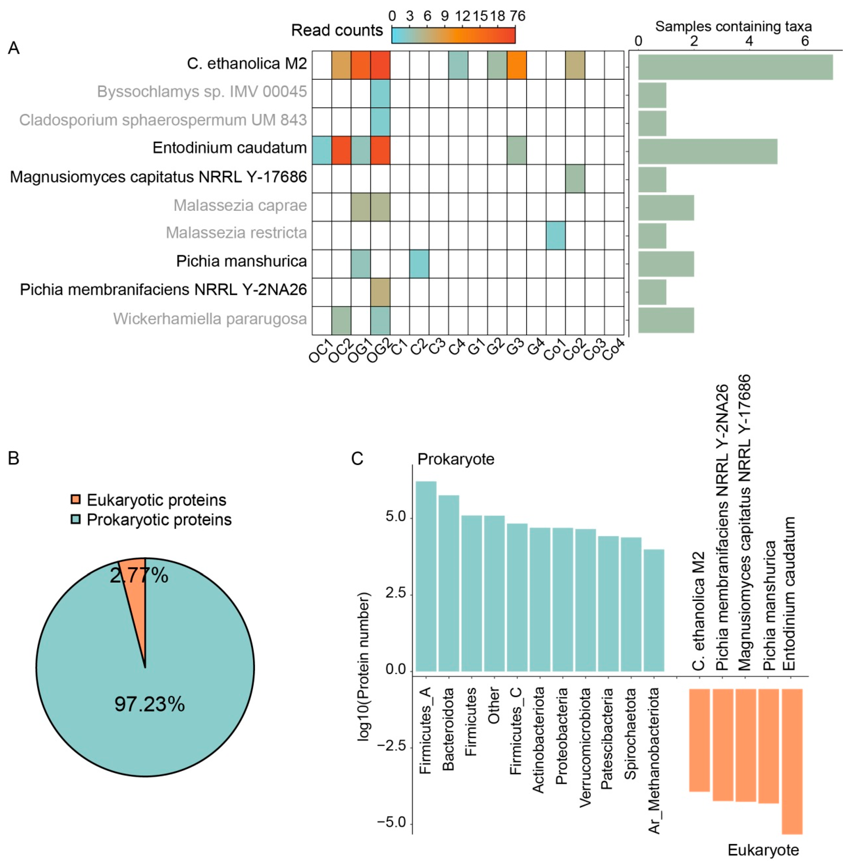
3.1. Sample-Specific Reference Database Covering Rumen Bacteria, Fungi, and Protozoa

The metaproteome sets higher requirements for the database utilized in the search process. Large or unspecific databases result in a significantly larger search space and would also increase the false positive rate [5]. To overcome this, sample-specific metagenomic data were used in the construction of in vitro ruminal fermentation-specific reference database in this study. To further improve the metaproteomic search database, the genomes of rumen eukaryotes that were included in sample-specific metagenomic data were added to the protein sequence database.
According to the output of the EukDetect pipeline, the eukaryotic species contained in the metagenomic data under in vitro fermentation conditions, including Candida ethanolica M2 (C. ethanolica M2), Byssochlamys sp. IMV 00045, Cladosporium sphaerospermum UM 843, Entodinium caudatum, Magnusiomyces capitatus NRRL Y-17686, Malassezia caprae, Malassezia restricta, Pichia manshurica, Pichia membranifaciens NRRL Y-2026, and Wickerhamiella pararugosa. We further screened these results in combination with previous studies on the rumen fungi and protozoa [23,24], and finally retained C. ethanolica M2 (Taxonomy ID: 1213352), Entodinium caudatum (Taxonomy ID: 47911), Magnusiomyces capitatus NRRL Y-17686 (Taxonomy ID: 1294685), Pichia manshurica (Taxonomy ID: 121235), and Pichia membranifaciens NRRL Y-2026 (Taxonomy ID: 763406) as the extension of the sample-specific database for subsequent proteomic data analysis. Among them, Entodinium caudatum and C. ethanolica M2 were observed in five and seven of the 16 samples, respectively. The results in these data all satisfied the screening threshold of EukDetect [18], and the coverage of marker genes of Entodinium caudatum mapped by these metagenomic data ranged from 8% to 76% (Figure 1A). Figure 1A demonstrates that more eukaryotic species and raw reads were found in the metagenomic data for the initial inoculum, but fewer eukaryote-related raw read counts were found as fermentation progressed.
Then, we assembled the respective genomes of these discovered eukaryotes, created a collection of 77,229 predicted protein sequences, and combined these protein sequences with a prokaryotic protein set of 1677 MAGs that had previously been generated using metagenomic binning [19]. A sample-specific database containing 2,789,316 protein sequences was constructed for the analysis of in vitro ruminal fermentation metaproteomic data. The final sample-specific database consisted of 2.77% prokaryotic sequences and 97.23% eukaryotic sequences (Figure 1B). Figure 1C showed the taxonomic origins of protein sequences contained in database, the eukaryotic protein sequences were mainly derived from the Entodinium caudatum, while the prokaryotic protein sequences were mainly from Firmicutes_A and Bacteroidetes. Although rumen fungus found it challenging to sustain large abundances under the conditions of in vitro ruminal fermentation, this is the reason for the lower fungal sequence in the database [25].
The quantity of proteins discovered from the metaproteomic data directly depends on how comprehensive the database is [5]. This result provided a preliminary description of eukaryotic species in in vitro ruminal fermentation, illustrated the rare eukaryotic species in continuous culture systems, and provided a reference for the expansion of sample-specific databases. To a certain extent, the false positive rate in the process of protein search was controlled and increased the detection rate of microbial proteins/microbial-derived extracellular proteins.

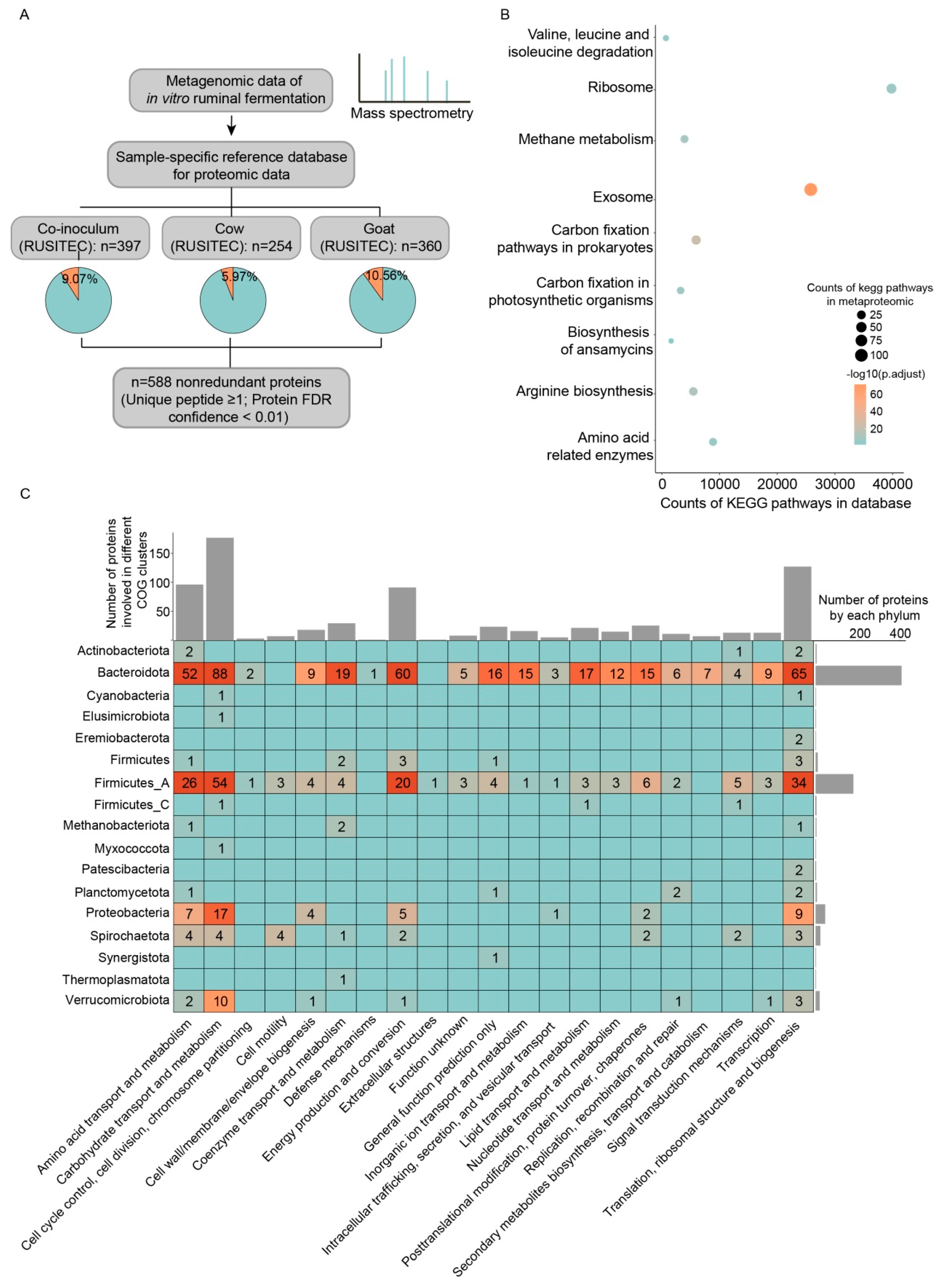
3.2. Metaproteomic Characterization of Microorganisms in In Vitro Ruminal Fermentation Broth

The metaproteomic data corresponding to the total microbial protein in the fermentation broths were searched with the sample-specific database. We obtained 357 (goat), 253 (cow), 396 (co-inoculum) microbial protein with high confidence and ≥1 unique peptide from three metaproteomic data, and the eukaryotic proteins accounted for 9.80%, 5.53%, and 8.83%, respectively (Figure 2A). After dereplication, 584 proteins from various microorganisms were recovered, 49 of these protein sequences came from eukaryotes, making up 8.40% of the total, and the remaining protein sequences came from bacterial or archaeal MAGs. Rumen Entodinium caudatum was previously thought to account for around 50% of rumen biomass [26]. However, the less raw reads and corresponding peptides of Entodinium caudatum were found in the metagenomic and metaproteomic data under in vitro ruminal fermentation conditions. Although we have constructed a sample-specific database to contain as many rumen microbial protein sequences as possible, the counts of identified proteins were still limited. This may be caused by discarding more contigs in the analysis of metagenomic binning. Similarly, the time length of mass spectrometry may also cause such a result. In follow-up studies, it will be worth exploring different analyses of mass spectrometry and the more comprehensive database to optimize the metaproteomic result of rumen micoorganisms. However, these proteins identified by current metaproteome could be accurately mapped to strain-level MAGs in the database, and it would allow us to perform subsequent functional annotation and analysis of these proteins. These results further suggested that the populations of Entodinium caudatum declined after a stable period in the RUSITEC system.
To discover functional properties, further annotation and enrichment analysis of the KEGG pathway associated with these discovered proteins were conducted. We found that 9 KEGG pathways were overrepresented in the identified protein set compared with reference database, such as exosome, methane metabolism, ribosome, and amino acid metabolism-related enzymes (Figure 2B). The KEGG pathways related to amino acid metabolism and ribosome were two important pathways in microorganisms to maintain normal cell function. It is worth noting that under in vitro fermentation conditions, the exosome pathway was overrepresented in metaproteome compared with database, and there were 107 identified proteins from different bacteria related to exosome pathway, reflecting that bacteria were active in producing extracellular active proteins under the conditions of in vitro ruminal fermentation, and the active exosome pathway may play an important role in the degradation of substrates.
Next, we assigned the bacterial proteins identified from the metaproteomic data to the corresponding microbial origins and annotated these proteins with COG database to further explore the role of bacteria in in vitro fermentation (Figure 2C). It was found that more than 82% of the identified proteins were from Bacteroidetes (57.3%) and Firmicutes_A (25.2%), which was similar to the result of a previous study on metaproteomic data of rumen natural fermentation [27], indicating that Firmicutes_A and Bacteroidetes remained the predominant active microbiota under in vitro ruminal fermentation conditions. Among all proteins, more than 55% of the proteins were assigned to the gene categories related to bacterial growth and energy metabolism, such as translation, ribosomal structure, and biogenesis (18.0%), energy production and conversion (12.9%), and carbohydrate transport and metabolism (25.0%). These results summarized the in vitro ruminal fermentation metaproteomic characterization, emphasized the dominant position of Bacteroidea and Firmicutes_A in protein expression and the activity of prokaryotes in secreting extracellular proteins, which reinforced our understanding of the active microbial structure and the bacterial functional characteristics under in vitro ruminal fermentation.

3.3. Carbohydrate Active Enzymes Contained in Microbe-Derived Extracellular Proteins

Extracellular proteome data from microbes revealed that the sample from the co-inoculum group had the highest number of identified proteins (519), while the sample from the cow group had the lowest number (278). Among the extracellular proteins obtained in three samples, 35–44% were derived from eukaryotes, and the protein sequences of Entodinium caudatum accounted for more than 90% of all eukaryotic identified protein sequences (Figure 3A). Nonetheless, the interpretation of the identified eukaryotic microbe-derived extracellular proteins still requires caution, especially for a large number of sequences from Entodinium caudatum, which may come from the intracellular proteins released after cell autolysis [28,29].
In order to clarify the information of the identified prokaryotic extracellular proteins, we first screened these proteins with high confidence, and further filtered out the corresponding proteins detected in less than two samples, and finally obtained 158 candidate prokaryotic extracellular proteins from three in vitro ruminal fermentation broth samples. The candidate extracellular proteins were further annotated. Except for the proteins with the description information of “hypothetical protein”, the protein with the highest proportion was TonB-dependent receptor SusC (15%), which contained 12 TonB-dependent receptors SusC from 11 different prokaryotic MAGs (Figure 3B), and this protein has been proved that it was the main component of bacterial outer membrane vesicles [30].
Microorganisms secrete a variety of extracellular active substances, including outer membrane vesicles, cellulosomes, etc. [31]. These extracellular active substances play a key role in interaction with the environment [31,32]. Collecting these extracellular active substances by effective means is also one of the keys to developing rumen cellulose degradation resources in vitro. We further detected the carbohydrate active enzyme domains contained in the extracellular proteins identified from the fermentation broth proteomic. However, we eventually discovered that 10 different extracellular proteins had domains related to carbohydrate-active enzymes (Table 1). We speculated that these enzymes may belong to the extracellular complexes, such as cellulosome, while the functions and the composition of the entire complex still require further experimental verification.
According to the results of taxonomic origins and COG annotation of bacteria-derived extracellular proteins, the proteins from Bacteroides and Firmicutes_A accounted for 78% of all extracellular proteins in proteomic data, demonstrating that these two phyla’s proteins dominated the performance of extracellular functions. For all bacteria-derived extracellular proteins, 37.7% of proteins were assigned functions related to carbohydrate transport and metabolism (Figure 3C). Compared with the metaproteomic data corresponding to the total bacterial proteins in result 3.2, the number of extracellular proteins assigned to translation, ribosomal structure and biogenesis was reduced to 5% in the extracellular proteomic. To our knowledge, this is the first study reporting the extracellular proteome of rumen microbial community, and the result revealed a scene of active extracellular proteins of rumen microbial community under in vitro fermentation conditions and identified that bacterial-derived extracellular proteins play a role in substrate degradation.

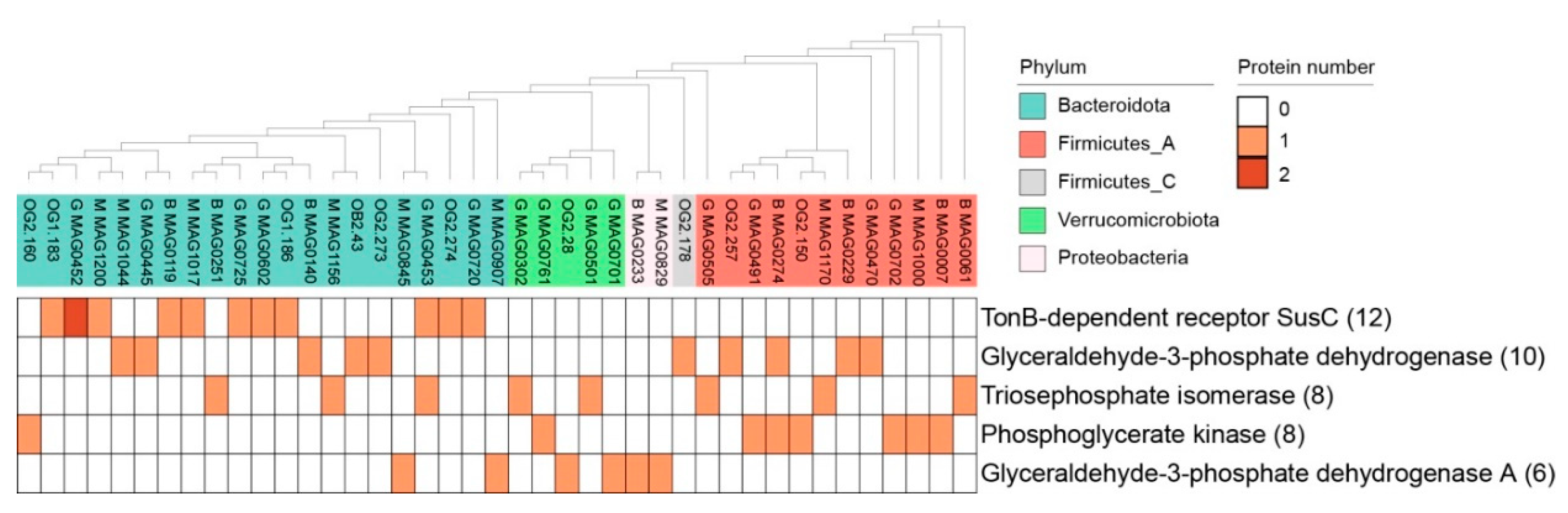
3.4. The Intracellular/Surface Moonlighting Proteins in Extracellular Proteomic of In Vitro Ruminal Fermentation Broth

Among the 210 nonredundant candidate extracellular protein sequences discovered using extracellular proteomic data, 73 distinct protein sequences came from prokaryotes. These proteins included a range of proteins involved in carbohydrate metabolism, outer membrane proteins, and other biological processes. The top five proteins with the highest occurrence frequency were TonB-dependent receptor SusC, Glyceraldehyde-3-phosphate dehydrogenase (GAPDH), Triosephosphate isomerase, Phosphoglycerate kinase, Glyceraldehyde-3-phosphate dehydrogenase A (GAPDHA), which were derived from five different bacterial phyla (Bacteroidota, Firmicutes_A, Firmicutes_C, Verrucomicrobiota, and Proteobacteria, respectively) (Figure 4). Except for TonB-dependent receptor SusC, the other four proteins are all involved in the sugar metabolism pathway [33,34]. Previous research has revealed that these bacterial housekeeping proteins involved in glucose metabolism, which were also referred to as intracellular/surface moonlighting proteins, had a function in bacterial adhesion (ISMP) [35]. For instance, the glycolytic enzyme GAPDH, which has a second role on the surface of pathogenic streptococci, was the first one to be identified as ISMP [36]. The studies on proteins, such as phosphoglycerate kinase acting as ISMP, were also reported previously [37]. We reported the ISMPs observed in in vitro ruminal fermentation broth, and under in vitro conditions, these proteins may play an important role in substrate adhesion, which may contribute to the substrate degradation for their corresponding microbial origins. However, the process by which these proteins secrete to extracellular has not been well investigated. This may require a unique type of secretion system [35]. We further speculated that the expression of these ISMPs related to adhesion results in microbial attachment and colonization on plant cell wall material that is likely too large in particle size to be expelled from the fermenter, ultimately maintaining the population of these microbial communities. Therefore, these results suggested that rumen bacterial ISMPs play an important role in substrate adhesion and maintenance of bacterial populations.
The extracellular TonB-dependent receptor SusC in the fermentation broth acquired in this study is all from the Bacteroidetes, and it was the protein with the highest frequency among the microbial-derived proteins in our investigation generated from prokaryotes. In a prior study, TonB-dependent receptor SusC was abundant in bacterial extracellular vesicles [30], and this protein contained a carbohydrate enzyme domain, which encodes a starch utilization locus [38]. In our research, the fermentation broth proteomic data revealed the presence of the TonB-dependent receptor SusC from Bacteroidea. These findings show that under in vitro ruminal fermentation conditions, Bacteroidetes secreted the TonB-dependent receptor SusC or extracellular vesicles containing this protein into the extracellular space and may have had a role in starch degradation in the fermentation broth.

4. Conclusions

The research on in vitro ruminal fermentation has gained more attention recently, while the understanding of the function of microbial intracellular and extracellular proteins in the process of in vitro fermentation is limited to a few studies. Our results demonstrated that the metaproteomic approach allows an in-depth investigation of the functional characteristics of microbial functional proteins under the conditions of in vitro ruminal fermentation. Bacteroidetes and Firmicutes_A were most active in the expression of extracellular/intracellular proteins, which were mainly involved in maintaining basic metabolism and carbohydrate degradation. In addition, the rumen bacteria-derived ISMPs obtained from in vitro metaproteome showed an adherent phenotype related to substrate adhesion, and the adherent strategy of rumen bacteria was a potential unique way to sustain their population size in the RUSITEC system. In general, these results characterized the functional proteins and active microbiome under the conditions of in vitro ruminal fermentation and emphasized the role of extracellular proteins in in vitro ruminal fermentation. This study improved the understanding of the mechanism of substrate degradation by rumen microbial community and further accelerated the development of cellulose-degrading resources in rumen microorganisms under in vitro conditions.

Author Contributions

Conceptualization, Y.J. and Z.L.; methodology, T.S. and T.Z.; investigation, X.G., T.S., Y.L. and X.W.; resources, Z.L. and T.Z.; data curation, T.S.; writing—original draft preparation, T.S. and X.G.; writing review and editing, Y.J. and Z.L.; visualization, T.S. and X.G.; supervision, Y.J. and Z.L.; All authors have read and agreed to the published version of the manuscript.

Funding

This research was funded by the National Natural Science Foundation of China, 31822052.

Institutional Review Board Statement

Not applicable.

Informed Consent Statement

Not applicable.

Data Availability Statement

Not applicable.

Acknowledgments

This work was supported by China National GeneBank (CNGB).

Conflicts of Interest

The authors declare no conflict of interest.

References

  1. Calsamiglia, S.; Busquet, M.; Cardozo, P.W.; Castillejos, L.; Ferret, A. Invited review: Essential oils as modifiers of rumen microbial fermentation. J. Dairy Sci. 2007, 90, 2580–2595. [Google Scholar] [CrossRef] [PubMed]
  2. Svartstrom, O.; Alneberg, J.; Terrapon, N.; Lombard, V.; de Bruijn, I.; Malmsten, J.; Dalin, A.M.; El Muller, E.; Shah, P.; Wilmes, P.; et al. Ninety-nine de novo assembled genomes from the moose (Alces alces) rumen microbiome provide new insights into microbial plant biomass degradation. ISME J. 2017, 11, 2538–2551. [Google Scholar] [CrossRef] [PubMed]
  3. Stewart, R.D.; Auffret, M.D.; Warr, A.; Walker, A.W.; Roehe, R.; Watson, M. Compendium of 4,941 rumen metagenome-assembled genomes for rumen microbiome biology and enzyme discovery. Nat. Biotechnol. 2019, 37, 953–961. [Google Scholar] [CrossRef]
  4. Artzi, L.; Bayer, E.A.; Morais, S. Cellulosomes: Bacterial nanomachines for dismantling plant polysaccharides. Nat. Rev. Microbiol. 2017, 15, 83–95. [Google Scholar] [CrossRef] [PubMed]
  5. Hagen, L.H.; Brooke, C.G.; Shaw, C.A.; Norbeck, A.D.; Piao, H.; Arntzen, M.O.; Olson, H.M.; Copeland, A.; Isern, N.; Shukla, A.; et al. Proteome specialization of anaerobic fungi during ruminal degradation of recalcitrant plant fiber. ISME J. 2021, 15, 421–434. [Google Scholar] [CrossRef] [PubMed]
  6. Elliott, C.L.; Edwards, J.E.; Wilkinson, T.J.; Allison, G.G.; McCaffrey, K.; Scott, M.B.; Rees-Stevens, P.; Kingston-Smith, A.H.; Huws, S.A. Using ‘Omic Approaches to Compare Temporal Bacterial Colonization of Lolium perenne, Lotus corniculatus, and Trifolium pratense in the Rumen. Front. Microbiol. 2018, 9, 2184. [Google Scholar] [CrossRef]
  7. Wei, X.; Ouyang, K.; Long, T.; Liu, Z.; Li, Y.; Qiu, Q. Dynamic Variations in Rumen Fermentation Characteristics and Bacterial Community Composition during In Vitro Fermentation. Fermentation 2022, 8, 276. [Google Scholar] [CrossRef]
  8. Wang, Y.-L.; Wang, W.-K.; Wu, Q.-C.; Zhang, F.; Li, W.-J.; Yang, Z.-M.; Bo, Y.-K.; Yang, H.-J. The Effect of Different Lactic Acid Bacteria Inoculants on Silage Quality, Phenolic Acid Profiles, Bacterial Community and In Vitro Rumen Fermentation Characteristic of Whole Corn Silage. Fermentation 2022, 8, 285. [Google Scholar] [CrossRef]
  9. Brede, M.; Orton, T.; Pinior, B.; Roch, F.F.; Dzieciol, M.; Zwirzitz, B.; Wagner, M.; Breves, G.; Wetzels, S.U. PacBio and Illumina MiSeq Amplicon Sequencing Confirm Full Recovery of the Bacterial Community After Subacute Ruminal Acidosis Challenge in the RUSITEC System. Front. Microbiol. 2020, 11, 1813. [Google Scholar] [CrossRef]
  10. Baba, Y.; Matsuki, Y.; Takizawa, S.; Suyama, Y.; Tada, C.; Fukuda, Y.; Saito, M.; Nakai, Y. Pretreatment of Lignocellulosic Biomass with Cattle Rumen Fluid for Methane Production: Fate of Added Rumen Microbes and Indigenous Microbes of Methane Seed Sludge. Microbes Environ. 2019, 34, 421–428. [Google Scholar] [CrossRef]
  11. Oss, D.B.; Ribeiro, G.O., Jr.; Marcondes, M.I.; Yang, W.; Beauchemin, K.A.; Forster, R.J.; McAllister, T.A. Synergism of Cattle and Bison Inoculum on Ruminal Fermentation and Select Bacterial Communities in an Artificial Rumen (Rusitec) Fed a Barley Straw Based Diet. Front. Microbiol. 2016, 7, 2032. [Google Scholar] [CrossRef] [PubMed]
  12. Petruschke, H.; Schori, C.; Canzler, S.; Riesbeck, S.; Poehlein, A.; Daniel, R.; Frei, D.; Segessemann, T.; Zimmerman, J.; Marinos, G.; et al. Discovery of novel community-relevant small proteins in a simplified human intestinal microbiome. Microbiome 2021, 9, 55. [Google Scholar] [CrossRef] [PubMed]
  13. Deusch, S.; Seifert, J. Catching the tip of the iceberg—Evaluation of sample preparation protocols for metaproteomic studies of the rumen microbiota. Proteomics 2015, 15, 3590–3595. [Google Scholar] [CrossRef] [PubMed]
  14. Adebayo Arowolo, M.; Zhang, X.M.; Wang, M.; Wang, R.; Wen, J.N.; Hao, L.Z.; He, J.H.; Shen, W.J.; Ma, Z.Y.; Tan, Z.L. Proper motility enhances rumen fermentation and microbial protein synthesis with decreased saturation of dissolved gases in rumen simulation technique. J. Dairy Sci. 2022, 105, 231–241. [Google Scholar] [CrossRef]
  15. Zhang, X.; Li, L.; Mayne, J.; Ning, Z.; Stintzi, A.; Figeys, D. Assessing the impact of protein extraction methods for human gut metaproteomics. J. Proteom. 2018, 180, 120–127. [Google Scholar] [CrossRef]
  16. Adav, S.S.; Ng, C.S.; Arulmani, M.; Sze, S.K. Quantitative iTRAQ secretome analysis of cellulolytic Thermobifida fusca. J. Proteome Res. 2010, 9, 3016–3024. [Google Scholar] [CrossRef]
  17. Hagen, L.H.; Frank, J.A.; Zamanzadeh, M.; Eijsink, V.G.H.; Pope, P.B.; Horn, S.J.; Arntzen, M.O. Quantitative Metaproteomics Highlight the Metabolic Contributions of Uncultured Phylotypes in a Thermophilic Anaerobic Digester. Appl. Environ. Microbiol. 2017, 83. [Google Scholar] [CrossRef]
  18. Shi, T.; Zhang, T.; Wang, X.; Wang, X.; Shen, W.; Guo, X.; Liu, Y.; Li, Z.; Jiang, Y. Copresent microbiome and short-chain fatty acids profiles of plant biomass utilization in rumen simulation technique system. bioRxiv 2022. [Google Scholar] [CrossRef]
  19. Lind, A.L.; Pollard, K.S. Accurate and sensitive detection of microbial eukaryotes from whole metagenome shotgun sequencing. Microbiome 2021, 9, 58. [Google Scholar] [CrossRef]
  20. Stanke, M.; Morgenstern, B. AUGUSTUS: A web server for gene prediction in eukaryotes that allows user-defined constraints. Nucleic Acids Res. 2005, 33, W465–W467. [Google Scholar] [CrossRef]
  21. Aramaki, T.; Blanc-Mathieu, R.; Endo, H.; Ohkubo, K.; Kanehisa, M.; Goto, S.; Ogata, H. KofamKOALA: KEGG Ortholog assignment based on profile HMM and adaptive score threshold. Bioinformatics 2020, 36, 2251–2252. [Google Scholar] [CrossRef] [PubMed]
  22. Buchfink, B.; Xie, C.; Huson, D.H. Fast and sensitive protein alignment using DIAMOND. Nat. Methods 2015, 12, 59–60. [Google Scholar] [CrossRef] [PubMed]
  23. Fernandes, T.; Carvalho, B.F.; Mantovani, H.C.; Schwan, R.F.; Avila, C.L.S. Identification and characterization of yeasts from bovine rumen for potential use as probiotics. J. Appl. Microbiol. 2019, 127, 845–855. [Google Scholar] [CrossRef] [PubMed]
  24. Li, Z.; Wang, X.; Zhang, Y.; Yu, Z.; Zhang, T.; Dai, X.; Pan, X.; Jing, R.; Yan, Y.; Liu, Y.; et al. Genomic insights into the phylogeny and biomass-degrading enzymes of rumen ciliates. bioRxiv 2022. [Google Scholar] [CrossRef]
  25. Hartinger, T.; Zebeli, Q. The Present Role and New Potentials of Anaerobic Fungi in Ruminant Nutrition. J. Fungi 2021, 7, 200. [Google Scholar] [CrossRef]
  26. Newbold, C.J.; de la Fuente, G.; Belanche, A.; Ramos-Morales, E.; McEwan, N.R. The Role of Ciliate Protozoa in the Rumen. Front. Microbiol. 2015, 6, 1313. [Google Scholar] [CrossRef]
  27. Honan, M.C.; Greenwood, S.L. Characterization of variations within the rumen metaproteome of Holstein dairy cattle relative to morning feed offering. Sci. Rep. 2020, 10, 3179. [Google Scholar] [CrossRef] [PubMed]
  28. Sylvester, J.T.; Karnati, S.K.; Dehority, B.A.; Morrison, M.; Smith, G.L.; St-Pierre, N.R.; Firkins, J.L. Rumen ciliated protozoa decrease generation time and adjust 18S ribosomal DNA copies to adapt to decreased transfer interval, starvation, and monensin. J. Dairy Sci. 2009, 92, 256–269. [Google Scholar] [CrossRef]
  29. Wallace, R.J.; McPherson, C.A. Factors affecting the rate of breakdown of bacterial protein in rumen fluid. Br. J. Nutr. 1987, 58, 313–323. [Google Scholar] [CrossRef]
  30. Veith, P.D.; Chen, Y.Y.; Chen, D.; O’Brien-Simpson, N.M.; Cecil, J.D.; Holden, J.A.; Lenzo, J.C.; Reynolds, E.C. Tannerella forsythia Outer Membrane Vesicles Are Enriched with Substrates of the Type IX Secretion System and TonB-Dependent Receptors. J. Proteome Res. 2015, 14, 5355–5366. [Google Scholar] [CrossRef]
  31. Lynd, L.R.; Weimer, P.J.; van Zyl, W.H.; Pretorius, I.S. Microbial cellulose utilization: Fundamentals and biotechnology. Microbiol. Mol. Biol. Rev. 2002, 66, 506–577. [Google Scholar] [CrossRef] [PubMed]
  32. Queiroz, C.B.; Santana, M.F. Prediction of the secretomes of endophytic and nonendophytic fungi reveals similarities in host plant infection and colonization strategies. Mycologia 2020, 112, 491–503. [Google Scholar] [CrossRef] [PubMed]
  33. Kainulainen, V.; Korhonen, T.K. Dancing to another tune-adhesive moonlighting proteins in bacteria. Biology 2014, 3, 178–204. [Google Scholar] [CrossRef]
  34. Jeffery, C.J. Moonlighting proteins. Trends Biochem. Sci. 1999, 24, 8–11. [Google Scholar] [CrossRef]
  35. Jeffery, C. Intracellular proteins moonlighting as bacterial adhesion factors. AIMS Microbiol. 2018, 4, 362–376. [Google Scholar] [CrossRef] [PubMed]
  36. Pancholi, V.; Fischetti, V.A. A major surface protein on group A streptococci is a glyceraldehyde-3-phosphate-dehydrogenase with multiple binding activity. J. Exp. Med. 1992, 176, 415–426. [Google Scholar] [CrossRef] [PubMed]
  37. Dahlhamer, J.M.; Zammitti, E.P.; Ward, B.W.; Wheaton, A.G.; Croft, J.B. Prevalence of Inflammatory Bowel Disease Among Adults Aged >/=18 Years—United States, 2015. MMWR Morb. Mortal. Wkly. Rep. 2016, 65, 1166–1169. [Google Scholar] [CrossRef]
  38. Cho, K.H.; Salyers, A.A. Biochemical analysis of interactions between outer membrane proteins that contribute to starch utilization by Bacteroides thetaiotaomicron. J. Bacteriol. 2001, 183, 7224–7230. [Google Scholar] [CrossRef]
Figure 1. Construction of a rumen-specific protein database containing eukaryotes based on metagenomic data of in vitro fermentation broth. (A) The eukaryotic reads contained in the metagenomic data, the heatmap on the left is the read counts of the corresponding species in each metagenomic data, and the histogram on the right is the number of samples containing corresponding species. (B) The proportion of eukaryotic and prokaryotic protein sequences in the sample-specific database. (C) Distribution of prokaryotic protein sequences at the phylum level and eukaryotic sequences at the species level.
Fermentation 08 00394 g001
Figure 2. Metaproteomic characterization of total microbial proteins under the conditions of in vitro ruminal fermentation. (A) Metaproteomic data analysis pipeline and the number of proteins identified from three different samples. (B) The overrepresented Kyoto Encyclopedia of Genes and Genomes (KEGG) pathways of identified proteins compared with the total proteins in the database. (C) Distribution of identified proteins in different clusters of orthologous groups (COG) and different microbial taxa.
Fermentation 08 00394 g002
Figure 3. Functional characterization of extracellular proteins derived from microorganisms. (A) The number of proteins identified from the extracellular metaproteome of three fermentation broth samples. (B) Annotation information of extracellular proteins with high FDR confidence and identified from ≥2 samples. (C) Distribution of identified extracellular proteins in different COG categories and different microbial taxa.
Fermentation 08 00394 g003
Figure 4. Taxonomic origins and annotations of representative bacterial-derived extracellular proteins. The heat map represents the number of identified TonB-dependent receptor SusC and intracellular/surface moonlighting proteins (ISMPs) in each bacterial MAG. The upper part is a maximum likelihood phylogenetic tree of bacterial MAGs based on 122 single-copy genes, the colored background represents the different phylum of MAGs.
Fermentation 08 00394 g004
Table 1. The microbe-derived extracellular proteins with carbohydrate-active enzyme-related domains.
Gene AccessionClassification of Microbial OriginUnique PeptidesCAZy FamilySubstratesEnzyme
B_MAG0029_g01249p_Firmicutes_A; f_Lachnospiraceae;g_; s_2GH109 a 3-hydroxybutyryl-CoA dehydrogenase.
Entodinium_caudatum_g1Entodinium_caudatum2GH13_1glycogen/starch
Entodinium_caudatum_g2Entodinium_caudatum1CBM48 a
Entodinium_caudatum_g3Entodinium_caudatum1GH13_8glycogen/starch
Entodinium_caudatum_g4Entodinium_caudatum2GT35 astarch
G_MAG0453_g01794p_Bacteroidota; g_UBA3839;s_
UBA3839 sp900313845
1GH94cellobiose/celluloseCellobiose phosphorylase.
M_MAG0911_g00105p_Firmicutes_A; g_Ruminococcus;s_2GH48cellulose/chitinCellulose 1,4-beta-cellobiosidase (reducing end).
M_MAG0967_g01628p_Firmicutes_A; g_NK4A144;s_1GH94cellobiose/celluloseCellobiose phosphorylase.
OB2.89_g00740p_Firmicutes_A; g_UMGS1696;s_1CBM48=GH13_8glycogen/starch1,4-alpha-glucan branching enzyme.
OG2.257_g02106p_Firmicutes_A; g_Butyrivibrio;s_1GH109 Glyceraldehyde-3-phosphate dehydrogenase (phosphorylating).
a GH: Glycoside hydrolase; CBM: Carbohydrate-binding module; GT: Formation of glycosidic bonds.
Publisher’s Note: MDPI stays neutral with regard to jurisdictional claims in published maps and institutional affiliations.

