
Multimodal Cardiac Imaging in the Assessment of Patients Who Have Suffered a Cardioembolic Stroke: A Review

 ,
,
Abstract
:1. Introduction
2. Materials and Methods
3. Discussion
3.1. Overview of Cardiac Imaging in Cardioembolic Stroke
3.1.1. Transthoracic Echocardiography (TTE)
3.1.2. Transoesophageal Echocardiography (TOE)
3.1.3. TTE vs. TOE
3.1.4. Cardiac Computed Tomography (CT) and Cardiac Magnetic Resonance Imaging (CMR)
3.1.5. Nuclear Imaging
3.1.6. Computed Tomography Angiography–Aorta (CTA)
3.1.7. Overall Considerations

| Advantages | Disadvantages | |
|---|---|---|
| TTE | Readily available [25,26,27] Non-invasive [25,26,27] Inexpensive [25,26,27] Sensitive and specific for LV thrombus [20] | Limited views of atria and appendages [26] Inter-operator variability [25,26,27] Potentially limited acoustic windows [25,26,27] |
| TOE | Gold standard for detecting high-risk and potential cardioembolic sources of stroke [28,29,30] Good views of atria and appendages [16,28,31] Better for evaluating leaflet tears and abscesses in IE [19] Can be considered for patients afflicted by cryptogenic stroke who may be reclassified as cardioembolic [34,35] Can be considered for younger patients in order to search for PFO [38,39,40] 26% of secondary prevention management modified by TOE results [34] | Semi-invasive [44] Resource-heavy [45] Expensive [45] Minor procedural risks [45] Higher risk of complications in certain patient groups (high body weight, history of gastrointestinal bleed or surgery, advanced age, and oesophageal mass/stricture/varices) and patients with higher sedation risk (chronic kidney disease, cardiac disease, pulmonary disease, liver cirrhosis) [44] Role in the acute evaluation of ischaemic stroke is not well established [32,33] Inter-operator variability [45] |
| Cardiac CT | Better soft tissue characterisation [47] High-grade anatomical information [47] Allows for spatial and temporal visualisation and image reconstruction in multiple planes [47] Can be used to image extra-cardiac structures [47] Greater reproducibility [63] Less inter-operator variability No dependence on acoustic window Alternative to CMR in patients contraindicated for MRI [50] Yields results faster and is easier to execute than MRI [63] | Limited by resource allocation and availability [63] Radiation exposure [63] Cannot be used to measure flow velocity or perform hemodynamic assessment or regurgitant quantification [47] Accuracy reduced in cases of high heart rates [58] |
| CMR | Better soft tissue characterisation [47] High-grade anatomical information [47] Allows for spatial and temporal visualisation and image reconstruction in multiple planes [47] Can be used to image extra-cardiac structures [47] Greater reproducibility [63] Less inter-operator variability No dependence on acoustic window More accurate than TTE and TOE for diagnosis of LV thrombus [25,53,54,55,56] Can be used to diagnose cardiomyopathies via LGE-CMR [9,58] No radiation exposure | Limited by resource allocation and availability [63] Accuracy reduced in cases of high heart rates [58] Expensive [63] Longer duration of scan [63] Relatively contraindicated for patients with poor renal function due to an increased risk of nephrogenic systemic fibrosis and in those that are pregnant [65] |
| Nuclear imaging (PET, SPECT) | Allows assessment of LV perfusion (ischemia and infarct) [66] Allows assessment of chamber function and dimension [66] Reveals fundamental pathophysiological mechanisms and provides metabolic information on molecular processes [53,68] Can identify potential markers of plaque rupture [40] Can be considered in cases of suspected underlying malignancy [71] | Limited by resource allocation and availability [72] Expensive [72] Radiation exposure [64] Use is controversial during pregnancy [64] |
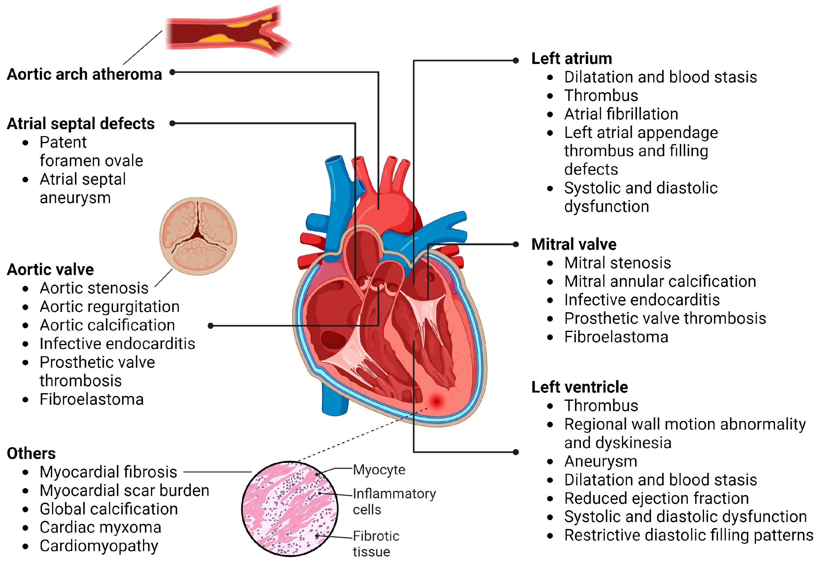
3.2. Cardiac Imaging Based on Individual Sources of Cardioembolic Stroke
3.2.1. Thrombus Formation
Atrial Fibrillation (AF), Left-atrial (LA) Dilatation, and LA Thrombus
Acute Myocardial Infarction (AMI) and LV Thrombus
Heart Failure (HF) and Cardiomyopathy
Aortic Arch Atheroma
Cardiac Tumours
3.2.2. Defects of the Atrial Septum
Patent Foramen Ovale (PFO)
Atrial Septal Aneurysm (ASA)
3.2.3. Valvulopathies
Infective Endocarditis (IE)
Prosthetic Valve Endocarditis and Thrombi
Mitral Valvulopathy
Mitral Annulus Calcification and Global Cardiac Calcification
Aortic Valvulopathy
| TTE | TOE | CT | CMR | Nuclear | Others | |
|---|---|---|---|---|---|---|
| LA dilation and LA thrombus | * + | ◊ | ++ | ++ | Strain imaging | |
| LV thrombus | * ++ | ++ | ++
| ◊
| ||
| HF/Cardiomyopathy | * ++ | ++
| ++ | ◊ | Speckle tracking/GLS
| |
| Aortic arch atheroma | +
| ◊ | ++ | ?
| ++ CT angiography | |
| Cardiac tumours | + | ++ | ++ | ◊ | ||
| PFO | * +
| ◊ | + | + | TCD ◊ | |
| ASA | *
| ◊
| ||||
| IE | * ++
| ◊ | ++
| ?
|
| |
| Prosthetic valve IE and thrombi | * ++ | ◊ | ++ | |||
| MS | * ++ | ◊ | ++ | ++/? | ||
| MAC/Cardiac calcification | * (for MAC) ++ | ++ | * (for global calcification) ◊ | |||
| AS/AR | *
| ◊
| ++ | ++ | - |
4. Conclusions
Author Contributions
Funding
Institutional Review Board Statement
Informed Consent Statement
Data Availability Statement
Conflicts of Interest
References
- Krishnamurthi, R.V.; Feigin, V.L.; Forouzanfar, M.H.; Mensah, G.A.; Connor, M.; Bennett, D.A.; Moran, A.E.; Sacco, R.L.; Anderson, L.M.; Truelsen, T.; et al. Global and regional burden of first-ever ischaemic and haemorrhagic stroke during 1990–2010: Findings from the Global Burden of Disease Study 2010. Lancet Glob. Health 2013, 1, e259–e281. [Google Scholar] [CrossRef] [PubMed]
- Palacio, S.; Hart, R.G. Neurologic Manifestations of Cardiogenic Embolism: An Update. Neurol. Clin. 2002, 20, 179–193. [Google Scholar] [CrossRef] [PubMed]
- Arboix, A.; Oliveres, M.; Massons, J.; Pujades, R.; Garcia-Eroles, L. Early differentiation of cardioembolic from atherothrombotic cerebral infarction: A multivariate analysis. Eur. J. Neurol. 1999, 6, 677–683. [Google Scholar] [CrossRef] [PubMed]
- Arboix, A.; Vericat, M.C.; Pujades, R.; Massons, J.; García-Eroles, L.; Oliveres, M. Cardioembolic infarction in the Sagrat Cor-Alianza Hospital of Barcelona Stroke Registry. Acta Neurol. Scand. 1997, 96, 407–412. [Google Scholar] [CrossRef]
- Adams, H.P., Jr.; Bendixen, B.H.; Kappelle, L.J.; Biller, J.; Love, B.B.; Gordon, D.L.; Marsh, E.E., 3rd. Classification of subtype of acute ischemic stroke. Definitions for use in a multicenter clinical trial. TOAST. Trial of Org 10172 in Acute Stroke Treatment. Stroke 1993, 24, 35–41. [Google Scholar] [CrossRef]
- Ay, H.; Benner, T.; Arsava, E.M.; Furie, K.L.; Singhal, A.B.; Jensen, M.B.; Ayata, C.; Towfighi, A.; Smith, E.E.; Chong, J.Y.; et al. A computerized algorithm for etiologic classification of ischemic stroke: The Causative Classification of Stroke System. Stroke 2007, 38, 2979–2984. [Google Scholar] [CrossRef] [PubMed]
- Amarenco, P.; Bogousslavsky, J.; Caplan, L.R.; Donnan, G.A.; Wolf, M.E.; Hennerici, M.G. The ASCOD phenotyping of ischemic stroke (Updated ASCO Phenotyping). Cerebrovasc. Dis. 2013, 36, 1–5. [Google Scholar] [CrossRef]
- De Paiva Bezerra, R.; de Miranda Alves, M.A.; Conforto, A.B.; Rodrigues, D.L.G.; Silva, G.S. Etiological Classification of Stroke in Patients with Chagas Disease Using TOAST, Causative Classification System TOAST, and ASCOD Phenotyping. J. Stroke Cerebrovasc. Dis. 2017, 26, 2864–2869. [Google Scholar] [CrossRef]
- Delewi, R.; Zijlstra, F.; Piek, J.J. Left ventricular thrombus formation after acute myocardial infarction. Heart 2012, 98, 1743–1749. [Google Scholar] [CrossRef]
- EAFT (European Atrial Fibrillation Trial) Study Group. Secondary prevention in non-rheumatic atrial fibrillation after transient ischaemic attack or minor stroke. Lancet 1993, 342, 1255–1262. [Google Scholar] [CrossRef]
- Adams, H.P.; del Zoppo, G.; Alberts, M.J.; Bhatt, D.L.; Brass, L.; Furlan, A.; Grubb, R.L.; Higashida, R.T.; Jauch, E.C.; Kidwell, C.; et al. Guidelines for the Early Management of Adults with Ischemic Stroke. Circulation 2007, 115, e478–e534. [Google Scholar] [CrossRef] [PubMed]
- Jauch, E.C.; Saver, J.L.; Adams, H.P.; Bruno, A.; Connors, J.J.; Demaerschalk, B.M.; Khatri, P.; McMullan, P.W.; Qureshi, A.I.; Rosenfield, K.; et al. Guidelines for the Early Management of Patients with Acute Ischemic Stroke. Stroke 2013, 44, 870–947. [Google Scholar] [CrossRef] [PubMed]
- Hindricks, G.; Potpara, T.; Dagres, N.; Arbelo, E.; Bax, J.J.; Blomström-Lundqvist, C.; Boriani, G.; Castella, M.; Dan, G.-A.; Dilaveris, P.E.; et al. 2020 ESC Guidelines for the diagnosis and management of atrial fibrillation developed in collaboration with the European Association for Cardio-Thoracic Surgery (EACTS): The Task Force for the diagnosis and management of atrial fibrillation of the Europea. Eur. Heart J. 2021, 42, 373–498. [Google Scholar] [CrossRef] [PubMed]
- Ringelstein, E.B.; Chamorro, A.; Kaste, M.; Langhorne, P.; Leys, D.; Lyrer, P.; Thijs, V.; Thomassen, L.; Toni, D. European Stroke Organisation Recommendations to Establish a Stroke Unit and Stroke Center. Stroke 2013, 44, 828–840. [Google Scholar] [CrossRef] [PubMed]
- Pepi, M.; Evangelista, A.; Nihoyannopoulos, P.; Flachskampf, F.A.; Athanassopoulos, G.; Colonna, P.; Habib, G.; Ringelstein, E.B.; Sicari, R.; Zamorano, J.L.; et al. Recommendations for echocardiography use in the diagnosis and management of cardiac sources of embolism: European Association of Echocardiography (EAE) (A registered branch of the ESC). Eur. J. Echocardiogr. 2010, 11, 461–476. [Google Scholar] [CrossRef] [PubMed]
- Ferro, J.M. Brain embolism—Answers to practical questions. J. Neurol. 2003, 250, 139–147. [Google Scholar] [CrossRef] [PubMed]
- Weir, N.U. An update on cardioembolic stroke. Postgrad. Med. J. 2008, 84, 133–140. [Google Scholar] [CrossRef]
- Wasser, K.; Weber-Krüger, M.; Jürries, F.; Liman, J.; Hamann, G.F.; Kermer, P.; Uphaus, T.; Protsenko, E.; Seegers, J.; Mende, M.; et al. The cardiac diagnostic work-up in stroke patients—A subanalysis of the Find-AFRANDOMISED trial. PLoS ONE 2019, 14, e0216530. [Google Scholar] [CrossRef]
- Arnautu, S.F.; Arnautu, D.A.; Lascu, A.; Hajevschi, A.A.; Rosca, C.I.I.; Sharma, A.; Jianu, D.C. A Review of the Role of Transthoracic and Transesophageal Echocardiography, Computed Tomography, and Magnetic Resonance Imaging in Cardioembolic Stroke. Med. Sci. Monit Int. Med. J. Exp. Clin. Res. 2022, 28, e936365. [Google Scholar] [CrossRef]
- Jugdutt, B.I.; Sivaram, C.A.; Wortman, C.; Trudell, C.; Penner, P. Prospective two-dimensional echocardiographic evaluation of left ventricular thrombus and embolism after acute myocardial infarction. J. Am. Coll. Cardiol. 1989, 13, 554–564. [Google Scholar] [CrossRef]
- Mustafa, K.; Shaikh, A.K.; Peterson, L.; Kurrelmeyer, K.M.; Shah, G.; Nagueh, S.F.; Fromm, R.; Quinones, M.A.; Zoghbi, W.A. Impact of Contrast Echocardiography on Evaluation of Ventricular Function and Clinical Management in a Large Prospective Cohort. J. Am. Coll. Cardiol. 2009, 53, 802–810. [Google Scholar] [CrossRef]
- Wehrum, T.; Dragonu, I.; Strecker, C.; Hennig, J.; Harloff, A. Multi-contrast and three-dimensional assessment of the aortic wall using 3T MRI. Eur. J. Radiol. 2017, 91, 148–154. [Google Scholar] [CrossRef] [PubMed]
- Saric, M.; Armour, A.C.; Arnaout, M.S.; Chaudhry, F.A.; Grimm, R.A.; Kronzon, I.; Landeck, B.F.; Maganti, K.; Michelena, H.I.; Tolstrup, K. Guidelines for the Use of Echocardiography in the Evaluation of a Cardiac Source of Embolism. J. Am. Soc. Echocardiogr. 2016, 29, 1–42. [Google Scholar] [CrossRef] [PubMed]
- Toufan Tabrizi, M.; Faraji Azad, H.; Khezerlouy-Aghdam, N.; Sakha, H. Measurement of mitral valve area by direct three dimensional planimetry compared to multiplanar reconstruction in patients with rheumatic mitral stenosis. Int. J. Cardiovasc. Imaging 2022, 38, 1341–1349. [Google Scholar] [CrossRef] [PubMed]
- Srichai, M.B.; Junor, C.; Rodriguez, L.L.; Stillman, A.E.; Grimm, R.A.; Lieber, M.L.; Weaver, J.A.; Smedira, N.G.; White, R.D. Clinical, imaging, and pathological characteristics of left ventricular thrombus: A comparison of contrast-enhanced magnetic resonance imaging, transthoracic echocardiography, and transesophageal echocardiography with surgical or pathological validation. Am. Heart J. 2006, 152, 75–84. [Google Scholar] [CrossRef]
- Malik, S.B.; Chen, N.; Parker, R.A.; Hsu, J.Y. Transthoracic Echocardiography: Pitfalls and Limitations as Delineated at Cardiac CT and MR Imaging. RadioGraphics 2017, 37, 383–406. [Google Scholar] [CrossRef]
- Pearson, A.C. Transthoracic echocardiography versus transesophageal echocardiography in detecting cardiac sources of embolism. Echocardiography 1993, 10, 397–403. [Google Scholar] [CrossRef]
- Harloff, A.; Handke, M.; Reinhard, M.; Geibel, A.; Hetzel, A. Therapeutic strategies after examination by transesophageal echocardiography in 503 patients with ischemic stroke. Stroke 2006, 37, 859–864. [Google Scholar] [CrossRef]
- Strandberg, M.; Marttila, R.J.; Helenius, H.; Hartiala, J. Transoesophageal echocardiography in selecting patients for anticoagulation after ischaemic stroke or transient ischaemic attack. J. Neurol. Neurosurg. Psychiatry 2002, 73, 29–33. [Google Scholar] [CrossRef]
- Cujec, B.; Polasek, P.; Voll, C.; Shuaib, A. Transesophageal echocardiography in the detection of potential cardiac source of embolism in stroke patients. Stroke 1991, 22, 727–733. [Google Scholar] [CrossRef]
- Murtagh, B.; Smalling, R.W. Cardioembolic stroke. Curr. Atheroscler. Rep. 2006, 8, 310–316. [Google Scholar] [CrossRef] [PubMed]
- Guidelines for management of ischaemic stroke and transient ischaemic attack 2008. Cerebrovasc. Dis. 2008, 25, 457–507. [CrossRef] [PubMed]
- Inzitari, D. The Italian Guidelines for stroke prevention. The Stroke Prevention and Educational Awareness Diffusion (SPREAD) Collaboration. Neurol. Sci. 2000, 21, 5–12. [Google Scholar] [CrossRef] [PubMed]
- De Castro, S.; Papetti, F.; Di Angelantonio, E.; Razmovska, B.; Truscelli, G.; Tuderti, U.; Puca, E.; Correnti, A.; Fiorelli, M.; Prencipe, M.; et al. Feasibility and Clinical Utility of Transesophageal Echocardiography in the Acute Phase of Cerebral Ischemia. Am. J. Cardiol. 2010, 106, 1339–1344. [Google Scholar] [CrossRef]
- Thomalla, G.; Upneja, M.; Camen, S.; Jensen, M.; Schröder, J.; Barow, E.; Boskamp, S.; Ostermeier, B.; Kissling, S.; Elke Leinisch, E.; et al. Treatment-Relevant Findings in Transesophageal Echocardiography After Stroke: A Prospective Multicenter Cohort Study. Stroke 2022, 53, 177–184. [Google Scholar] [CrossRef]
- Ulrich, J.N.; Hesse, B.; Schuele, S.; Vlassak, I.; Sila, C.A.; Jaber, W.A. Single-vessel versus multivessel territory acute ischemic stroke: Value of transesophageal echocardiography in the differentiation of embolic stroke. J. Am. Soc. Echocardiogr. Off. Publ. Am. Soc. Echocardiogr. 2006, 19, 1165–1169. [Google Scholar] [CrossRef]
- Schnabel, R.B.; Camen, S.; Knebel, F.; Hagendorff, A.; Bavendiek, U.; Böhm, M.; Doehner, W.; Endres, M.; Gröschel, K.; Goette, A.; et al. Expert opinion paper on cardiac imaging after ischemic stroke. Clin. Res. Cardiol. 2021, 110, 938–958. [Google Scholar] [CrossRef]
- Wolber, T.; Maeder, M.; Atefy, R.; Bluzaite, I.; Blank, R.; Rickli, H.; Ammann, P. Should routine echocardiography be performed in all patients with stroke? J. Stroke Cerebrovasc. Dis. 2007, 16, 1–7. [Google Scholar] [CrossRef]
- Leung, D.Y.; Black, I.W.; Cranney, G.B.; Walsh, W.F.; Grimm, R.A.; Stewart, W.J.; Thomas, J.D. Selection of patients for transesophageal echocardiography after stroke and systemic embolic events. Role of transthoracic echocardiography. Stroke 1995, 26, 1820–1824. [Google Scholar] [CrossRef]
- De Castro, S.; Rasura, M.; Di Angelantonio, E.; Beccia, M.; Passaseo, I.; Di Lisi, F.; Fieschi, C.; Pandian, N.; Fedele, F. Distribution of potential cardiac sources of embolism in young and older stroke patients: Implications for recurrent vascular events. J. Cardiovasc. Med. 2006, 7, 191–196. [Google Scholar] [CrossRef]
- de Bruijn, S.F.T.M.; Agema, W.R.P.; Lammers, G.J.; van der Wall, E.E.; Wolterbeek, R.; Holman, E.R.; Bollen, E.L.E.M.; Bax, J.J. Transesophageal echocardiography is superior to transthoracic echocardiography in management of patients of any age with transient ischemic attack or stroke. Stroke 2006, 37, 2531–2534. [Google Scholar] [CrossRef] [PubMed]
- Handke, M.; Harloff, A.; Olschewski, M.; Hetzel, A.; Geibel, A. Patent foramen ovale and cryptogenic stroke in older patients. N. Engl. J. Med. 2007, 357, 2262–2268. [Google Scholar] [CrossRef] [PubMed]
- Vitebskiy, S.; Fox, K.; Hoit, B.D. Routine transesophageal echocardiography for the evaluation of cerebral emboli in elderly patients. Echocardiography 2005, 22, 770–774. [Google Scholar] [CrossRef] [PubMed]
- Cho, H.J.; Choi, H.Y.; Kim, Y.D.; Nam, H.S.; Han, S.W.; Ha, J.W.; Chung, N.-S.; Heo, J.H. Transoesophageal echocardiography in patients with acute stroke with sinus rhythm and no cardiac disease history. J. Neurol. Neurosurg. Psychiatry 2010, 81, 412–415. [Google Scholar] [CrossRef] [PubMed]
- Viedma-Guiard, E.; Guidoux, C.; Amarenco, P.; Meseguer, E. Aortic Sources of Embolism. Front. Neurol. 2020, 11, 606663. [Google Scholar] [CrossRef] [PubMed]
- Palazzuoli, A.; Ricci, D.; Lenzi, C.; Lenzi, J.; Palazzuoli, V. Transesophageal echocardiography for identifying potential cardiac sources of embolism in patients with stroke. Neurol. Sci. 2000, 21, 195–202. [Google Scholar] [CrossRef] [PubMed]
- Senapati, A.; Faza, N.N.; Mahmarian, J.; Chang, S.M. Cardiac Computed Tomography for Structural Heart Disease Assessment and Therapeutic Planning: Focus on Prosthetic Valve Dysfunction. Methodist Debakey Cardiovasc. J. 2020, 16, 86–96. [Google Scholar] [CrossRef]
- Habib, G.; Badano, L.; Tribouilloy, C.; Vilacosta, I.; Zamorano, J.L.; Galderisi, M.; Voigt, J.-U.; Sicari, R.; Cosyns, B.; Fox, K.; et al. Recommendations for the practice of echocardiography in infective endocarditis. Eur. J. Echocardiogr. 2010, 11, 202–219. [Google Scholar] [CrossRef]
- Gomes, A.; Glaudemans, A.W.J.M.; Touw, D.J.; van Melle, J.P.; Willems, T.P.; Maass, A.H.; Natour, E.; Prakken, N.H.J.; Borra, R.J.H.; van Geel, P.P.; et al. Diagnostic value of imaging in infective endocarditis: A systematic review. Lancet Infect. Dis. 2017, 17, e1–e14. [Google Scholar] [CrossRef]
- De Oliveira, D.C.L.; Assunção, F.B.; Dos Santos, A.A.S.M.D.; Nacif, M.S. Cardiac Magnetic Resonance and Computed Tomography in Hypertrophic Cardiomyopathy: An Update. Arq. Bras. Cardiol. 2016, 107, 163–172. [Google Scholar] [CrossRef]
- Capmany, R.P.; Ibañez, M.O.; Pesquer, X.J. Complex atheromatosis of the aortic arch in cerebral infarction. Curr. Cardiol. Rev. 2010, 6, 184–193. [Google Scholar] [CrossRef] [PubMed]
- Vizzardi, E.; Gelsomino, S.; D’Aloia, A.; Lorusso, R. Aortic atheromas and stroke: Review of literature. J. Investig. Med. 2013, 61, 956–966. [Google Scholar] [CrossRef] [PubMed]
- Goyal, P.; Weinsaft, J.W. Cardiovascular magnetic resonance imaging for assessment of cardiac thrombus. Methodist Debakey Cardiovasc. J. 2013, 9, 132–136. [Google Scholar] [CrossRef] [PubMed]
- Mollet, N.R.; Dymarkowski, S.; Volders, W.; Wathiong, J.; Herbots, L.; Rademakers, F.E.; Bogaert, J. Visualization of ventricular thrombi with contrast-enhanced magnetic resonance imaging in patients with ischemic heart disease. Circulation 2002, 106, 2873–2876. [Google Scholar] [CrossRef] [PubMed]
- Weir, R.A.P.; Martin, T.N.; Petrie, C.J.; Murphy, A.; Clements, S.; Steedman, T.; Wagner, G.S.; McMurray, J.J.V.; Dargie, H.J. Cardiac and extracardiac abnormalities detected by cardiac magnetic resonance in a post-myocardial infarction cohort. Cardiology 2009, 113, 1–8. [Google Scholar] [CrossRef]
- Barkhausen, J.; Hunold, P.; Eggebrecht, H.; Schüler, W.O.; Sabin, G.V.; Erbel, R.; Debatin, J.F. Detection and characterization of intracardiac thrombi on MR imaging. Am. J. Roentgenol. 2002, 179, 1539–1544. [Google Scholar] [CrossRef]
- Chai, P.; Mohiaddin, R. How we perform cardiovascular magnetic resonance flow assessment using phase-contrast velocity mapping. J. Cardiovasc. Magn. Reson. 2005, 7, 705–716. [Google Scholar] [CrossRef]
- Inamdar, A.A.; Inamdar, A.C. Heart Failure: Diagnosis, Management and Utilization. J. Clin. Med. 2016, 5, 62. [Google Scholar] [CrossRef]
- Wehrum, T.; Dragonu, I.; Strecker, C.; Schuchardt, F.; Hennemuth, A.; Drexl, J.; Reinhard, T.; Böhringer, D.; Vach, W.; Hennig, J.; et al. Aortic atheroma as a source of stroke—Assessment of embolization risk using 3D CMR in stroke patients and controls. J. Cardiovasc. Magn. Reson. 2017, 19, 67. [Google Scholar] [CrossRef]
- Vilacosta, I.; Gómez, J. Complementary Role of MRI in Infectious Endocarditis. Echocardiography 1995, 12, 673–676. [Google Scholar] [CrossRef]
- Dursun, M.; Yılmaz, S.; Yılmaz, E.; Yılmaz, R.; Onur, İ.; Oflaz, H.; Dindar, A. The utility of cardiac MRI in diagnosis of infective endocarditis: Preliminary results. Diagn. Interv. Radiol. 2015, 21, 28–33. [Google Scholar] [CrossRef]
- Meinel, T.R.; Eggimann, A.; Brignoli, K.; Wustmann, K.; Buffle, E.; Meinel, F.G.; Scheitz, J.F.; Nolte, C.H.; Gräni, C.; Fischer, U.; et al. Cardiovascular MRI Compared to Echocardiography to Identify Cardioaortic Sources of Ischemic Stroke: A Systematic Review and Meta-Analysis. Front. Neurol. 2021, 12, 699838. [Google Scholar] [CrossRef] [PubMed]
- El-Koussy, M.; Schroth, G.; Brekenfeld, C.; Arnold, M. Imaging of Acute Ischemic Stroke. Eur. Neurol. 2014, 72, 309–316. [Google Scholar] [CrossRef] [PubMed]
- Picano, E. Economic and biological costs of cardiac imaging. Cardiovasc. Ultrasound 2005, 3, 13. [Google Scholar] [CrossRef] [PubMed]
- Andreucci, M.; Solomon, R.; Tasanarong, A. Side effects of radiographic contrast media: Pathogenesis, risk factors, and prevention. Biomed. Res. Int. 2014, 2014, 741018. [Google Scholar] [CrossRef] [PubMed]
- Boutagy, N.E.; Feher, A.; Alkhalil, I.; Umoh, N.; Sinusas, A.J. Molecular Imaging of the Heart. Compr. Physiol. 2019, 9, 477–533. [Google Scholar] [CrossRef] [PubMed]
- Kim, J.; Song, H.-C. Role of PET/CT in the evaluation of aortic disease. Chonnam Med. J. 2018, 54, 143–152. [Google Scholar] [CrossRef]
- Bruun, N.E.; Habib, G.; Thuny, F.; Sogaard, P. Cardiac imaging in infectious endocarditis. Eur. Heart J. 2014, 35, 624–632. [Google Scholar] [CrossRef]
- Rominger, A.; Saam, T.; Wolpers, S.; Cyran, C.C.; Schmidt, M.; Foerster, S.; Nikolaou, K.; Reiser, M.F.; Bartenstein, P.; Hacker, M. 18F-FDG PET/CT Identifies Patients at Risk for Future Vascular Events in an Otherwise Asymptomatic Cohort with Neoplastic Disease. J. Nucl. Med. 2009, 50, 1611–1620. [Google Scholar] [CrossRef]
- Xu, G.; Zhao, L.; He, Z. Performance of whole-body PET/CT for the detection of distant malignancies in various cancers: A systematic review and meta-analysis. J. Nucl. Med. 2012, 53, 1847–1854. [Google Scholar] [CrossRef]
- Sener, U.; Keser, Z. Ischemic Stroke in Patients with Malignancy. Mayo Clin. Proc. 2022, 97, 2139–2144. [Google Scholar] [CrossRef] [PubMed]
- Baron, J.C. Stroke: Imaging and differential diagnosis. In Stroke-Vascular Diseases; Springer: Vienna, Austria, 2002; pp. 19–36. [Google Scholar] [CrossRef]
- Amarenco, P.; Cohen, A.; Tzourio, C.; Bertrand, B.; Hommel, M.; Besson, G.; Chauvel, C.; Touboul, P.J.; Bousser, M.G. Atherosclerotic disease of the aortic arch and the risk of ischemic stroke. N. Engl. J. Med. 1994, 331, 1474–1479. [Google Scholar] [CrossRef]
- Otsuka, K.; Ishikawa, H.; Kono, Y.; Oku, S.; Yamaura, H.; Shirasawa, K.; Hirata, K.; Shimada, K.; Kasayuki, N.; Fukuda, D. Aortic arch plaque morphology in patients with coronary artery disease undergoing coronary computed tomography angiography with wide-volume scan. Coron. Artery Dis. 2022, 33, 531–539. [Google Scholar] [CrossRef] [PubMed]
- Chugh, S.S.; Havmoeller, R.; Narayanan, K.; Singh, D.; Rienstra, M.; Benjamin, E.J.; Gillum, R.F.; Kim, Y.-H.; McAnulty, J.H.J.; Zheng, Z.-J.; et al. Worldwide epidemiology of atrial fibrillation: A Global Burden of Disease 2010 Study. Circulation 2014, 129, 837–847. [Google Scholar] [CrossRef] [PubMed]
- Wolf, P.A.; Abbott, R.D.; Kannel, W.B. Atrial fibrillation as an independent risk factor for stroke: The Framingham Study. Stroke 1991, 22, 983–988. [Google Scholar] [CrossRef] [PubMed]
- Go, A.S.; Hylek, E.M.; Phillips, K.A.; Chang, Y.; Henault, L.E.; Selby, J.V.; Singer, D.E. Prevalence of diagnosed atrial fibrillation in adults: National implications for rhythm management and stroke prevention: The AnTicoagulation and Risk Factors in Atrial Fibrillation (ATRIA) Study. JAMA 2001, 285, 2370–2375. [Google Scholar] [CrossRef] [PubMed]
- Migdady, I.; Russman, A.; Buletko, A.B. Atrial Fibrillation and Ischemic Stroke: A Clinical Review. Semin. Neurol. 2021, 41, 348–364. [Google Scholar] [CrossRef] [PubMed]
- Adam, O.; Neuberger, H.-R.; Böhm, M.; Laufs, U. Prevention of atrial fibrillation with 3-hydroxy-3-methylglutaryl coenzyme A reductase inhibitors. Circulation 2008, 118, 1285–1293. [Google Scholar] [CrossRef]
- Corrado, G.; Klein, A.L.; Santarone, M. Echocardiography in atrial fibrillation. J. Cardiovasc. Med. 2006, 7, 498–504. [Google Scholar] [CrossRef]
- Benjamin, E.J.; D’Agostino, R.B.; Belanger, A.J.; Wolf, P.A.; Levy, D. Left atrial size and the risk of stroke and death. The Framingham Heart Study. Circulation 1995, 92, 835–841. [Google Scholar] [CrossRef]
- Atrial Fibrillation Investigators: Atrial Fibrillation, Aspirin, Anticoagulation Study; European Atrial Fibrillation Study; Stroke Prevention in Atrial Fibrillation Study; Boston Area Anticoagulation Trial for Atrial Fibrillation Study; Canadian Atrial Fibrillation Study; Veterans Affairs Prevention in Atrial Fibrillation Study. Echocardiographic Predictors of Stroke in Patients with Atrial Fibrillation: A Prospective Study of 1066 Patients from 3 Clinical Trials. Arch. Intern. Med. 1998, 158, 1316–1320. [Google Scholar] [CrossRef]
- Juey-Jen, H.; Jin-Jer, C.; Shen-Chang, L.; Yung-Zu, T.; Peiliang, K.; Wen-Pin, L.; Fang-Yue, L.; Shu-Hsun, C.; Chi-Ren, H.; Shu-Wen, H. Diagnostic accuracy of transesophageal echocardiography for detecting left atrial thrombi in patients with rheumatic heart disease having undergone mitral valve operations. Am. J. Cardiol. 1993, 72, 677–681. [Google Scholar] [CrossRef] [PubMed]
- Nakanishi, K.; Homma, S. Role of echocardiography in patients with stroke. J. Cardiol. 2016, 68, 91–99. [Google Scholar] [CrossRef] [PubMed]
- Fatkin, D.; Kelly, R.; Feneley, M.P. Left atrial appendage blood velocity and thromboembolic risk in patients with atrial fibrillation. J. Am. Coll. Cardiol. 1994, 24, 1429–1430. [Google Scholar] [CrossRef] [PubMed]
- Goldman, M.E.; Pearce, L.A.; Hart, R.G.; Zabalgoitia, M.; Asinger, R.W.; Safford, R.; Halperin, J.L.; for the Stroke Prevention in Atrial Fibrillation Investigators. Pathophysiologic correlates of thromboembolism in nonvalvular atrial fibrillation: I. Reduced flow velocity in the left atrial appendage (The Stroke Prevention in Atrial Fibrillation [SPAF-III] study). J. Am. Soc. Echocardiogr. 1999, 12, 1080–1087. [Google Scholar] [CrossRef]
- Donal, E.; Galli, E.; Lederlin, M.; Martins, R.; Schnell, F. Multimodality Imaging for Best Dealing with Patients in Atrial Arrhythmias. JACC Cardiovasc. Imaging 2019, 12, 2245–2261. [Google Scholar] [CrossRef] [PubMed]
- Iwataki, M.; Takeuchi, M.; Otani, K.; Kuwaki, H.; Haruki, N.; Yoshitani, H.; Tamura, M.; Abe, H.; Otsuji, Y. Measurement of left atrial volume from transthoracic three-dimensional echocardiographic datasets using the biplane Simpson’s technique. J. Am. Soc. Echocardiogr. 2012, 25, 1319–1326. [Google Scholar] [CrossRef] [PubMed]
- Ohyama, H.; Hosomi, N.; Takahashi, T.; Mizushige, K.; Osaka, K.; Kohno, M.; Koziol, J.A. Comparison of magnetic resonance imaging and transesophageal echocardiography in detection of thrombus in the left atrial appendage. Stroke 2003, 34, 2436–2439. [Google Scholar] [CrossRef]
- Tsang, T.S.; Abhayaratna, W.P.; Barnes, M.E.; Miyasaka, Y.; Gersh, B.J.; Bailey, K.R.; Cha, S.S.; Seward, J.B. Prediction of Cardiovascular Outcomes with Left Atrial Size. J. Am. Coll. Cardiol. 2006, 47, 1018–1023. [Google Scholar] [CrossRef]
- Vandenberg, B.F.; Weiss, R.M.; Kinzey, J.; Acker, M.; Stark, C.A.; Stanford, W.; Burns, T.L.; Marcus, M.L.; Kerber, R.E. Comparison off loft atrial volume by two-dimensional echocardiography and cine-computed tomography. Am. J. Cardiol. 1995, 75, 754–757. [Google Scholar] [CrossRef]
- Luigi, D.B.; Pasquale, S.; Matteo, A.; Prasant, M.; Ilaria, S.; Sebastiano, G.; Rodney, H.; Sanchez, J.E.; Rong, B.; Sanghamitra, M.; et al. Does the Left Atrial Appendage Morphology Correlate with the Risk of Stroke in Patients with Atrial Fibrillation? J. Am. Coll. Cardiol. 2012, 60, 531–538. [Google Scholar] [CrossRef]
- Shih, J.-Y.; Tsai, W.-C.; Huang, Y.-Y.; Liu, Y.-W.; Lin, C.-C.; Huang, Y.-S.; Tsai, L.-M.; Lin, L.-J. Association of decreased left atrial strain and strain rate with stroke in chronic atrial fibrillation. J. Am. Soc. Echocardiogr. 2011, 24, 513–519. [Google Scholar] [CrossRef]
- Obokata, M.; Negishi, K.; Kurosawa, K.; Tateno, R.; Tange, S.; Arai, M.; Amano, M.; Kurabayashi, M. Left Atrial Strain Provides Incremental Value for Embolism Risk Stratification over CHA2DS2-VASc Score and Indicates Prognostic Impact in Patients with Atrial Fibrillation. J. Am. Soc. Echocardiogr. 2014, 27, 709–716.e4. [Google Scholar] [CrossRef]
- Jankajova, M.; Kubikova, L.; Valocik, G.; Candik, P.; Mitro, P.; Kurecko, M.; Sabol, F.; Kolesar, A.; Kubikova, M.; Vachalcova, M.; et al. Left atrial appendage strain rate is associated with documented thromboembolism in nonvalvular atrial fibrillation. Wien. Klin. Wochenschr. 2019, 131, 156–164. [Google Scholar] [CrossRef] [PubMed]
- Park, J.-H.; Hwang, I.-C.; Park, J.J.; Park, J.-B.; Cho, G.-Y. Left Atrial Strain to Predict Stroke in Patients with Acute Heart Failure and Sinus Rhythm. J. Am. Heart Assoc. 2021, 10, e020414. [Google Scholar] [CrossRef] [PubMed]
- Saberniak, J.; Skrebelyte-Strøm, L.; Orstad, E.B.; Hilde, J.M.; Solberg, M.G.; Rønning, O.M.; Kjekshus, H.; Steine, K. Left atrial appendage strain predicts subclinical atrial fibrillation in embolic strokes of undetermined source. Eur. Heart J. Open 2023, 3, oead039. [Google Scholar] [CrossRef] [PubMed]
- Mannina, C.; Ito, K.; Jin, Z.; Yoshida, Y.; Matsumoto, K.; Shames, S.; Russo, C.; Elkind, M.S.V.; Rundek, T.; Yoshita, M.; et al. Association of Left Atrial Strain with Ischemic Stroke Risk in Older Adults. JAMA Cardiol. 2023, 8, 317–325. [Google Scholar] [CrossRef] [PubMed]
- Kamel, H.; Healey, J.S. Cardioembolic Stroke. Circ. Res. 2017, 120, 514–526. [Google Scholar] [CrossRef]
- Leow, A.S.-T.; Sia, C.-H.; Tan, B.Y.-Q.; Chan, M.Y.-Y.; Loh, J.P.-Y. Characterisation of patients with acute myocardial infarction complicated by left ventricular thrombus. Eur. J. Intern. Med. 2020, 74, 110–112. [Google Scholar] [CrossRef]
- Spencer, F.A.; Gore, J.M.; Yarzebski, J.; Lessard, D.; Jackson, E.A.; Goldberg, R.J. Trends (1986 to 1999) in the incidence and outcomes of in-hospital stroke complicating acute myocardial infarction (The Worcester Heart Attack Study). Am. J. Cardiol. 2003, 92, 383–388. [Google Scholar] [CrossRef]
- Saczynski, J.S.; Spencer, F.A.; Gore, J.M.; Gurwitz, J.H.; Yarzebski, J.; Lessard, D.; Goldberg, R.J. Twenty-Year Trends in the Incidence of Stroke Complicating Acute Myocardial Infarction: Worcester Heart Attack Study. Arch. Intern. Med. 2008, 168, 2104–2110. [Google Scholar] [CrossRef] [PubMed]
- Budaj, A.; Flasinska, K.; Gore, J.M.; Anderson, F.A.J.; Dabbous, O.H.; Spencer, F.A.; Goldberg, R.J.; Fox, K.A.A. Magnitude of and risk factors for in-hospital and postdischarge stroke in patients with acute coronary syndromes: Findings from a Global Registry of Acute Coronary Events. Circulation 2005, 111, 3242–3247. [Google Scholar] [CrossRef] [PubMed]
- Mahaffey, K.W.; Harrington, R.A.; Simoons, M.L.; Granger, C.B.; Graffagnino, C.; Alberts, M.J.; Laskowitz, D.T.; Miller, J.M.; Sloan, M.A.; Berdan, L.G.; et al. Stroke in patients with acute coronary syndromes: Incidence and outcomes in the platelet glycoprotein IIb/IIIa in unstable angina. Receptor suppression using integrilin therapy (PURSUIT) trial. The PURSUIT Investigators. Circulation 1999, 99, 2371–2377. [Google Scholar] [CrossRef]
- Witt, B.J.; Brown, R.D.; Jacobsen, S.J.; Weston, S.A.; Yawn, B.P.; Roger, V.L. A Community-Based Study of Stroke Incidence after Myocardial Infarction. Ann. Intern. Med. 2005, 143, 785–792. [Google Scholar] [CrossRef] [PubMed]
- Weinreich, D.J.; Burke, J.F.; Pauletto, F.J. Left ventricular mural thrombi complicating acute myocardial infarction. Long-term follow-up with serial echocardiography. Ann. Intern. Med. 1984, 100, 789–794. [Google Scholar] [CrossRef] [PubMed]
- Hornung, M.; Franke, J.; Gafoor, S.; Sievert, H. Cardioembolic Stroke and Postmyocardial Infarction Stroke. Cardiol. Clin. 2016, 34, 207–214. [Google Scholar] [CrossRef] [PubMed]
- Shacham, Y.; Leshem-Rubinow, E.; Ben Assa, E.; Rogowski, O.; Topilsky, Y.; Roth, A.; Steinvil, A. Frequency and correlates of early left ventricular thrombus formation following anterior wall acute myocardial infarction treated with primary percutaneous coronary intervention. Am. J. Cardiol. 2013, 111, 667–670. [Google Scholar] [CrossRef]
- Bhatia, G.S.; Lip, G.Y.H. Atrial fibrillation post-myocardial infarction: Frequency, consequences, and management. Curr. Heart Fail. Rep. 2004, 1, 149–155. [Google Scholar] [CrossRef]
- Merlini, P.A.; Bauer, K.A.; Oltrona, L.; Ardissino, D.; Cattaneo, M.; Belli, C.; Mannucci, P.M.; Rosenberg, R.D. Persistent activation of coagulation mechanism in unstable angina and myocardial infarction. Circulation 1994, 90, 61–68. [Google Scholar] [CrossRef]
- Kassem-Moussa, H.; Mahaffey, K.W.; Graffagnino, C.; Tasissa, G.; Sila, C.A.; Simes, R.J.; White, H.D.; Califf, R.M.; Bhapkar, M.V.; Newby, L.K. Incidence and characteristics of stroke during 90-day follow-up in patients stabilized after an acute coronary syndrome. Am. Heart J. 2004, 148, 439–446. [Google Scholar] [CrossRef]
- Loh, E.; Sutton, M.S.; Wun, C.C.; Rouleau, J.L.; Flaker, G.C.; Gottlieb, S.S.; Lamas, G.A.; Moyé, L.A.; Goldhaber, S.Z.; Pfeffer, M.A. Ventricular dysfunction and the risk of stroke after myocardial infarction. N. Engl. J. Med. 1997, 336, 251–257. [Google Scholar] [CrossRef] [PubMed]
- Neumann, F.J.; Ott, I.; Gawaz, M.; Richardt, G.; Holzapfel, H.; Jochum, M.; Schömig, A. Cardiac release of cytokines and inflammatory responses in acute myocardial infarction. Circulation 1995, 92, 748–755. [Google Scholar] [CrossRef] [PubMed]
- Anavekar, N.S.; Skali, H.; Bourgoun, M.; Ghali, J.K.; Kober, L.; Maggioni, A.P.; McMurray, J.J.V.; Velazquez, E.; Califf, R.; Pfeffer, M.A.; et al. Usefulness of Right Ventricular Fractional Area Change to Predict Death, Heart Failure, and Stroke Following Myocardial Infarction (from the VALIANT ECHO Study). Am. J. Cardiol. 2008, 101, 607–612. [Google Scholar] [CrossRef] [PubMed]
- Johnsen, S.H.; Mathiesen, E.B.; Joakimsen, O.; Stensland, E.; Wilsgaard, T.; Løchen, M.-L.; Njølstad, I.; Arnesen, E. Carotid Atherosclerosis Is a Stronger Predictor of Myocardial Infarction in Women Than in Men. Stroke 2007, 38, 2873–2880. [Google Scholar] [CrossRef]
- Thanigaraj, S.; Schechtman, K.B.; Pérez, J.E. Improved echocardiographic delineation of left ventricular thrombus with the use of intravenous second-generation contrast image enhancement. J. Am. Soc. Echocardiogr. 1999, 12, 1022–1026. [Google Scholar] [CrossRef]
- Shaw, L.J. Impact of contrast echocardiography on diagnostic algorithms: Pharmacoeconomic implications. Clin. Cardiol. 1997, 20 (Suppl. 1), I39–I48. [Google Scholar] [CrossRef]
- Mansencal, N.; Nasr, I.A.; Pillière, R.; Farcot, J.-C.; Joseph, T.; Lacombe, P.; Dubourg, O. Usefulness of contrast echocardiography for assessment of left ventricular thrombus after acute myocardial infarction. Am. J. Cardiol. 2007, 99, 1667–1670. [Google Scholar] [CrossRef]
- Manning, W.J.; Weintraub, R.M.; Waksmonski, C.A.; Haering, J.M.; Rooney, P.S.; Maslow, A.D.; Johnson, R.G.; Douglas, P.S. Accuracy of transesophageal echocardiography for identifying left atrial thrombi. A prospective, intraoperative study. Ann. Intern. Med. 1995, 123, 817–822. [Google Scholar] [CrossRef]
- Chen, C.; Koschyk, D.; Hamm, C.; Sievers, B.; Kupper, W.; Bleifeld, W. Usefulness of transesophageal echocardiography in identifying small left ventricular apical thrombus. J. Am. Coll. Cardiol. 1993, 21, 208–215. [Google Scholar] [CrossRef]
- Tomoda, H.; Hoshiai, M.; Furuya, H.; Shotsu, A.; Ootaki, M.; Matsuyama, S. Evaluation of left ventricular thrombus with computed tomography. Am. J. Cardiol. 1981, 48, 573–577. [Google Scholar] [CrossRef]
- Ambrosy, A.P.; Fonarow, G.C.; Butler, J.; Chioncel, O.; Greene, S.J.; Vaduganathan, M.; Nodari, S.; Lam, C.S.; Sato, N.; Shah, A.N.; et al. The Global Health and Economic Burden of Hospitalizations for Heart Failure. J. Am. Coll. Cardiol. 2014, 63, 1123–1133. [Google Scholar] [CrossRef] [PubMed]
- Witt, B.J.; Gami, A.S.; Ballman, K.V.; Brown, R.D.; Meverden, R.A.; Jacobsen, S.J.; Roger, V.L. The Incidence of Ischemic Stroke in Chronic Heart Failure: A Meta-Analysis. J. Card. Fail. 2007, 13, 489–496. [Google Scholar] [CrossRef] [PubMed]
- Witt, B.J.; Brown, R.D.J.; Jacobsen, S.J.; Weston, S.A.; Ballman, K.V.; Meverden, R.A.; Roger, V.L. Ischemic stroke after heart failure: A community-based study. Am. Heart J. 2006, 152, 102–109. [Google Scholar] [CrossRef] [PubMed]
- Tan, S.; Ho, C.E.S.M.; Teo, Y.N.; Teo, Y.H.; Chan, M.Y.-Y.; Lee, C.-H.; Evangelista, L.K.M.; Lin, W.; Chong, Y.-F.; Yeo, T.-C.; et al. Prevalence and incidence of stroke, white matter hyperintensities, and silent brain infarcts in patients with chronic heart failure: A systematic review, meta-analysis, and meta-regression. Front. Cardiovasc. Med. 2022, 9, 967197. [Google Scholar] [CrossRef]
- Siachos, T.; Vanbakel, A.; Feldman, D.S.; Uber, W.; Simpson, K.N.; Pereira, N.L. Silent strokes in patients with heart failure. J. Card. Fail. 2005, 11, 485–489. [Google Scholar] [CrossRef] [PubMed]
- Lip, G.Y.; Gibbs, C.R. Does heart failure confer a hypercoagulable state? Virchow’s triad revisited. J. Am. Coll. Cardiol. 1999, 33, 1424–1426. [Google Scholar] [CrossRef] [PubMed]
- Sosin, M.D.; Bhatia, G.; Davis, R.C.; Lip, G.Y. Congestive heart failure and Virchow’s triad: A neglected association. Wien. Med. Wochenschr. 2003, 153, 411–416. [Google Scholar] [CrossRef]
- Sharma, N.D.; McCullough, P.A.; Philbin, E.F.; Weaver, W.D. Left ventricular thrombus and subsequent thromboembolism in patients with severe systolic dysfunction. Chest 2000, 117, 314–320. [Google Scholar] [CrossRef]
- Shantsila, E.; Kozieł, M.; Lip, G.Y. Anticoagulation versus placebo for heart failure in sinus rhythm. Cochrane Database Syst. Rev. 2021, 5, CD003336. [Google Scholar] [CrossRef]
- Aktas, M.K.; Zareba, W.; Butler, J.; Younis, A.; McNitt, S.; Brown, M.W.; Rao, N.; Rao, N.; Steinberg, J.; Chen, L.; et al. Confirm Rx insertable cardiac monitor for primary atrial fibrillation detection in high-risk heart failure patients (Confirm-AF trial). Ann. Noninvasive Electrocardiol. 2023, 28, e13021. [Google Scholar] [CrossRef]
- Santoro, F.; Stiermaier, T.; Tarantino, N.; De Gennaro, L.; Moeller, C.; Guastafierro, F.; Marchetti, M.F.; Montisci, R.; Carapelle, E.; Graf, T.; et al. Left Ventricular Thrombi in Takotsubo Syndrome: Incidence, Predictors, and Management: Results From the GEIST (German Italian Stress Cardiomyopathy) Registry. J. Am. Heart Assoc. 2017, 6, e006990. [Google Scholar] [CrossRef] [PubMed]
- Ding, K.J.; Cammann, V.L.; Szawan, K.A.; Stähli, B.E.; Wischnewsky, M.; Di Vece, D.; Citro, R.; Jaguszewski, M.; Seifert, B.; Sarcon, A. Intraventricular thrombus formation and embolism in Takotsubo syndrome: Insights from the International Takotsubo Registry. Arterioscler. Thromb. Vasc. Biol. 2020, 40, 279–287. [Google Scholar] [CrossRef] [PubMed]
- Bozkurt, B.; Colvin, M.; Cook, J.; Cooper, L.T.; Deswal, A.; Fonarow, G.C.; Francis, G.S.; Lenihan, D.; Lewis, E.F.; McNamara, D.M.; et al. Current Diagnostic and Treatment Strategies for Specific Dilated Cardiomyopathies: A Scientific Statement From the American Heart Association. Circulation 2016, 134, e579–e646. [Google Scholar] [CrossRef] [PubMed]
- Haruki, S.; Minami, Y.; Hagiwara, N. Stroke and Embolic Events in Hypertrophic Cardiomyopathy: Risk Stratification in Patients without Atrial Fibrillation. Stroke 2016, 47, 936–942. [Google Scholar] [CrossRef] [PubMed]
- Guttmann, O.P.; Pavlou, M.; O’Mahony, C.; Monserrat, L.; Anastasakis, A.; Rapezzi, C.; Biagini, E.; Gimeno, J.R.; Limongelli, G.; Garcia-Pavia, P.; et al. Prediction of thrombo-embolic risk in patients with hypertrophic cardiomyopathy (HCM Risk-CVA). Eur. J. Heart Fail. 2015, 17, 837–845. [Google Scholar] [CrossRef] [PubMed]
- Higashikawa, M.; Nakamura, Y.; Yoshida, M.; Kinoshita, M. Incidence of ischemic strokes in hypertrophic cardiomyopathy is markedly increased if complicated by atrial fibrillation. Jpn. Circ. J. 1997, 61, 673–681. [Google Scholar] [CrossRef]
- Olivotto, I.; Cecchi, F.; Casey, S.A.; Dolara, A.; Traverse, J.H.; Maron, B.J. Impact of atrial fibrillation on the clinical course of hypertrophic cardiomyopathy. Circulation 2001, 104, 2517–2524. [Google Scholar] [CrossRef]
- Chawla, D.; Jahangir, A.; Cooley, R.; Sra, J.; Tajik, A.J. Isolated left atrial standstill in patients with hypertrophic cardiomyopathy and atrial fibrillation after restoration of sinus rhythm. J. Am. Soc. Echocardiogr. 2019, 32, 1369–1372. [Google Scholar] [CrossRef]
- Wlodarska, E.K.; Wozniak, O.; Konka, M.; Rydlewska-Sadowska, W.; Biederman, A.; Hoffman, P. Thromboembolic complications in patients with arrhythmogenic right ventricular dysplasia/cardiomyopathy. EP Eur. 2006, 8, 596–600. [Google Scholar] [CrossRef]
- Wu, L.; Yao, Y.; Chen, G.; Fan, X.; Zheng, L.; Ding, L.; Zhang, S. Intracardiac thrombosis in patients with arrhythmogenic right ventricular cardiomyopathy. J. Cardiovasc. Electrophysiol. 2014, 25, 1359–1362. [Google Scholar] [CrossRef]
- Akdis, D.; Chen, K.; Saguner, A.M.; Stämpfli, S.F.; Chen, X.; Chen, L.; Rao, M.; Haegeli, L.M.; Tanner, F.C.; Brunckhorst, C.; et al. Clinical Characteristics of Patients with a Right Ventricular Thrombus in Arrhythmogenic Right Ventricular Cardiomyopathy. Thromb. Haemost. 2019, 119, 1373–1378. [Google Scholar] [CrossRef] [PubMed]
- Mankad, R.; Bonnichsen, C.; Mankad, S. Hypereosinophilic syndrome: Cardiac diagnosis and management. Heart 2016, 102, 100–106. [Google Scholar] [CrossRef] [PubMed]
- Bauersachs, J.; König, T.; van der Meer, P.; Petrie, M.C.; Hilfiker-Kleiner, D.; Mbakwem, A.; Hamdan, R.; Jackson, A.M.; Forsyth, P.; de Boer, R.A.; et al. Pathophysiology, diagnosis and management of peripartum cardiomyopathy: A position statement from the Heart Failure Association of the European Society of Cardiology Study Group on peripartum cardiomyopathy. Eur. J. Heart Fail. 2019, 21, 827–843. [Google Scholar] [CrossRef] [PubMed]
- Hilfiker-Kleiner, D.; Haghikia, A.; Berliner, D.; Vogel-Claussen, J.; Schwab, J.; Franke, A.; Schwarzkopf, M.; Ehlermann, P.; Pfister, R.; Michels, G. Bromocriptine for the treatment of peripartum cardiomyopathy: A multicentre randomized study. Eur. Heart J. 2017, 38, 2671–2679. [Google Scholar] [CrossRef] [PubMed]
- Zhu, X.; Wang, Z.; Ferrari, M.W.; Ferrari-Kuehne, K.; Bulter, J.; Xu, X.; Zhou, Q.; Zhang, Y.; Zhang, J. Anticoagulation in cardiomyopathy: Unravelling the hidden threat and challenging the threat individually. ESC Heart Fail. 2021, 8, 4737–4750. [Google Scholar] [CrossRef] [PubMed]
- Gosling, R.C.; Al-Mohammad, A. The Role of Cardiac Imaging in Heart Failure with Reduced Ejection Fraction. Card. Fail. Rev. 2022, 8, e22. [Google Scholar] [CrossRef]
- Kim, W.; Kim, E.J. Heart Failure as a Risk Factor for Stroke. J. Stroke 2018, 20, 33–45. [Google Scholar] [CrossRef]
- Collins, S.P.; Lindsell, C.J.; Storrow, A.B.; Abraham, W.T. Prevalence of negative chest radiography results in the emergency department patient with decompensated heart failure. Ann. Emerg. Med. 2006, 47, 13–18. [Google Scholar] [CrossRef]
- Heidenreich, P.A.; Bozkurt, B.; Aguilar, D.; Allen, L.A.; Byun, J.J.; Colvin, M.M.; Deswal, A.; Drazner, M.H.; Dunlay, S.M.; Evers, L.R.; et al. 2022 AHA/ACC/HFSA Guideline for the Management of Heart Failure: A Report of the American College of Cardiology/American Heart Association Joint Committee on Clinical Practice Guidelines. Circulation 2022, 145, E895–E1032. [Google Scholar] [CrossRef]
- Kozdag, G.; Ciftci, E.; Ural, D.; Sahin, T.; Selekler, M.; Agacdiken, A.; Demirci, A.; Komsuoglu, S.; Komsuoglu, B. Silent cerebral infarction in chronic heart failure: Ischemic and nonischemic dilated cardiomyopathy. Vasc. Health Risk Manag. 2008, 4, 463–469. [Google Scholar] [CrossRef]
- Kozdag, G.; Ciftci, E.; Vural, A.; Selekler, M.; Sahin, T.; Ural, D.; Kahraman, G.; Agacdiken, A.; Demirci, A.; Komsuoglu, S.; et al. Silent cerebral infarction in patients with dilated cardiomyopathy: Echocardiographic correlates. Int. J. Cardiol. 2006, 107, 376–381. [Google Scholar] [CrossRef] [PubMed]
- Mitropoulou, P.; Georgiopoulos, G.; Figliozzi, S.; Klettas, D.; Nicoli, F.; Masci, P.G. Multi-Modality Imaging in Dilated Cardiomyopathy: With a Focus on the Role of Cardiac Magnetic Resonance. Front. Cardiovasc. Med. 2020, 7, 97. [Google Scholar] [CrossRef] [PubMed]
- Potter, E.; Marwick, T.H. Assessment of Left Ventricular Function by Echocardiography: The Case for Routinely Adding Global Longitudinal Strain to Ejection Fraction. JACC. Cardiovasc. Imaging 2018, 11, 260–274. [Google Scholar] [CrossRef] [PubMed]
- Sengeløv, M.; Jørgensen, P.G.; Jensen, J.S.; Bruun, N.E.; Olsen, F.J.; Fritz-Hansen, T.; Nochioka, K.; Biering-Sørensen, T. Global Longitudinal Strain Is a Superior Predictor of All-Cause Mortality in Heart Failure with Reduced Ejection Fraction. JACC Cardiovasc. Imaging 2015, 8, 1351–1359. [Google Scholar] [CrossRef] [PubMed]
- Haugaa, K.H.; Goebel, B.; Dahlslett, T.; Meyer, K.; Jung, C.; Lauten, A.; Figulla, H.R.; Poerner, T.C.; Edvardsen, T. Risk assessment of ventricular arrhythmias in patients with nonischemic dilated cardiomyopathy by strain echocardiography. J. Am. Soc. Echocardiogr. 2012, 25, 667–673. [Google Scholar] [CrossRef]
- Trivedi, S.J.; Altman, M.; Stanton, T.; Thomas, L. Echocardiographic Strain in Clinical Practice. Heart Lung Circ. 2019, 28, 1320–1330. [Google Scholar] [CrossRef]
- Galderisi, M.; Cosyns, B.; Edvardsen, T.; Cardim, N.; Delgado, V.; Di Salvo, G.; Donal, E.; Sade, L.E.; Ernande, L.; Garbi, M.; et al. Standardization of adult transthoracic echocardiography reporting in agreement with recent chamber quantification, diastolic function, and heart valve disease recommendations: An expert consensus document of the European Association of Cardiovascular Imag. Eur. Heart J. Cardiovasc. Imaging 2017, 18, 1301–1310. [Google Scholar] [CrossRef]
- Shiozaki, A.A.; Senra, T.; Arteaga, E.; Martinelli Filho, M.; Pita, C.G.; Ávila, L.F.R.; Parga Filho, J.R.; Mady, C.; Kalil-Filho, R.; Bluemke, D.A.; et al. Myocardial fibrosis detected by cardiac CT predicts ventricular fibrillation/ventricular tachycardia events in patients with hypertrophic cardiomyopathy. J. Cardiovasc. Comput. Tomogr. 2013, 7, 173–181. [Google Scholar] [CrossRef]
- Tunick, P.A.; Rosenzweig, B.P.; Katz, E.S.; Freedberg, R.S.; Perez, J.L.; Kronzon, I. High risk for vascular events in patients with protruding aortic atheromas: A prospective study. J. Am. Coll. Cardiol. 1994, 23, 1085–1090. [Google Scholar] [CrossRef]
- The French Study of Aortic Plaques in Stroke Group. Atherosclerotic disease of the aortic arch as a risk factor for recurrent ischemic stroke. N. Engl. J. Med. 1996, 334, 1216–1221. [Google Scholar] [CrossRef]
- The Stroke Prevention in Atrial Fibrillation Investigators. Transesophageal echocardiographic correlates of thromboembolism in high-risk patients with nonvalvular atrial fibrillation. Ann. Intern. Med. 1998, 128, 639–647. [Google Scholar] [CrossRef]
- Kronzon, I.; Tunick, P.A. Aortic Atherosclerotic Disease and Stroke. Circulation 2006, 114, 63–75. [Google Scholar] [CrossRef]
- Amarenco, P.; Duyckaerts, C.; Tzourio, C.; Hénin, D.; Bousser, M.G.; Hauw, J.J. The prevalence of ulcerated plaques in the aortic arch in patients with stroke. N. Engl. J. Med. 1992, 326, 221–225. [Google Scholar] [CrossRef]
- Karalis, D.G.; Chandrasekaran, K.; Victor, M.F.; Ross, J.J.J.; Mintz, G.S. Recognition and embolic potential of intraaortic atherosclerotic debris. J. Am. Coll. Cardiol. 1991, 17, 73–78. [Google Scholar] [CrossRef] [PubMed]
- Tunick, P.A.; Perez, J.L.; Kronzon, I. Protruding atheromas in the thoracic aorta and systemic embolization. Ann. Intern. Med. 1991, 115, 423–427. [Google Scholar] [CrossRef] [PubMed]
- Yang, T.-L.; Huang, C.-C.; Huang, S.-S.; Chiu, C.-C.; Leu, H.-B.; Lin, S.-J. Aortic Arch Calcification Associated with Cardiovascular Events and Death among Patients with Acute Coronary Syndrome. Acta Cardiol. Sin. 2017, 33, 241–249. [Google Scholar] [CrossRef] [PubMed]
- Ntaios, G.; Pearce, L.A.; Meseguer, E.; Endres, M.; Amarenco, P.; Ozturk, S.; Lang, W.; Bornstein, N.M.; Molina, C.A.; Pagola, J.; et al. Aortic Arch Atherosclerosis in Patients with Embolic Stroke of Undetermined Source: An Exploratory Analysis of the NAVIGATE ESUS Trial. Stroke 2019, 50, 3184–3190. [Google Scholar] [CrossRef]
- Cohen, A.; Tzourio, C.; Bertrand, B.; Chauvel, C.; Bousser, M.G.; Amarenco, P.; FAPS Investigators. Aortic plaque morphology and vascular events: A follow-up study in patients with ischemic stroke. French Study of Aortic Plaques in Stroke. Circulation 1997, 96, 3838–3841. [Google Scholar] [CrossRef]
- Krinsky, G.A. Diagnostic imaging of aortic atherosclerosis and its complications. Neuroimaging Clin. 2002, 12, 437–443. [Google Scholar] [CrossRef]
- Tenenbaum, A.; Garniek, A.; Shemesh, J.; Fisman, E.Z.; Stroh, C.I.; Itzchak, Y.; Vered, Z.; Motro, M. Dual-helical CT for detecting aortic atheromas as a source of stroke: Comparison with transesophageal echocardiography. Radiology 1998, 208, 153–158. [Google Scholar] [CrossRef]
- Barazangi, N.; Wintermark, M.; Lease, K.; Rao, R.; Smith, W.; Josephson, S.A. Comparison of Computed Tomography Angiography and Transesophageal Echocardiography for Evaluating Aortic Arch Disease. J. Stroke Cerebrovasc. Dis. 2011, 20, 436–442. [Google Scholar] [CrossRef] [PubMed]
- Amarenco, P.; Davis, S.; Jones, E.F.; Cohen, A.A.; Heiss, W.-D.; Kaste, M.; Laouénan, C.; Young, D.; Macleod, M.; Donnan, G.A.; et al. Clopidogrel Plus Aspirin Versus Warfarin in Patients with Stroke and Aortic Arch Plaques. Stroke 2014, 45, 1248–1257. [Google Scholar] [CrossRef] [PubMed]
- Reynen, K. Cardiac myxomas. N. Engl. J. Med. 1995, 333, 1610–1617. [Google Scholar] [CrossRef] [PubMed]
- Sun, J.P.; Asher, C.R.; Yang, X.S.; Cheng, G.G.; Scalia, G.M.; Massed, A.G.; Griffin, B.P.; Ratliff, N.B.; Stewart, W.J.; Thomas, J.D. Clinical and echocardiographic characteristics of papillary fibroelastomas: A retrospective and prospective study in 162 patients. Circulation 2001, 103, 2687–2693. [Google Scholar] [CrossRef] [PubMed]
- Sordelli, C.; Fele, N.; Mocerino, R.; Weisz, S.H.; Ascione, L.; Caso, P.; Carrozza, A.; Tascini, C.; De Vivo, S.; Severino, S. Infective Endocarditis: Echocardiographic Imaging and New Imaging Modalities. J. Cardiovasc. Echogr. 2019, 29, 149–155. [Google Scholar] [CrossRef] [PubMed]
- Bzikha, R.; Serradj, A.; Queron, S. Papillary fibroelastoma of aortic valve mimicking an infective endocarditis. Cirugía Cardiovasc. 2021, 28, 300–303. [Google Scholar] [CrossRef]
- Hagen, P.T.; Scholz, D.G.; Edwards, W.D. Incidence and size of patent foramen ovale during the first 10 decades of life: An autopsy study of 965 normal hearts. Mayo Clin. Proc. 1984, 59, 17–20. [Google Scholar] [CrossRef] [PubMed]
- Meissner, I.; Whisnant, J.P.; Khandheria, B.K.; Spittell, P.C.; O’Fallon, W.M.; Pascoe, R.D.; Enriquez-Sarano, M.; Seward, J.B.; Covalt, J.L.; Sicks, J.D.; et al. Prevalence of potential risk factors for stroke assessed by transesophageal echocardiography and carotid ultrasonography: The SPARC study. Stroke Prevention: Assessment of Risk in a Community. Mayo Clin. Proc. 1999, 74, 862–869. [Google Scholar] [CrossRef]
- Lamy, C.; Giannesini, C.; Zuber, M.; Arquizan, C.; Meder, J.F.; Trystram, D.; Coste, J.; Mas, J.L. Clinical and imaging findings in cryptogenic stroke patients with and without patent foramen ovale: The PFO-ASA Study. Atrial Septal Aneurysm. Stroke 2002, 33, 706–711. [Google Scholar] [CrossRef]
- Homma, S.; Sacco, R.L.; Di Tullio, M.R.; Sciacca, R.R.; Mohr, J.P. Effect of medical treatment in stroke patients with patent foramen ovale: Patent foramen ovale in Cryptogenic Stroke Study. Circulation 2002, 105, 2625–2631. [Google Scholar] [CrossRef]
- Pristipino, C.; Sievert, H.; D’Ascenzo, F.; Louis Mas, J.; Meier, B.; Scacciatella, P.; Hildick-Smith, D.; Gaita, F.; Toni, D.; Kyrle, P.; et al. European position paper on the management of patients with patent foramen ovale. General approach and left circulation thromboembolism. Eur. Heart J. 2019, 40, 3182–3195. [Google Scholar] [CrossRef] [PubMed]
- Rodrigues, A.C.; Picard, M.H.; Carbone, A.; Arruda, A.L.; Flores, T.; Klohn, J.; Furtado, M.; Lira-Filho, E.B.; Cerri, G.G.; Andrade, J.L. Importance of adequately performed Valsalva maneuver to detect patent foramen ovale during transesophageal echocardiography. J. Am. Soc. Echocardiogr. 2013, 26, 1337–1343. [Google Scholar] [CrossRef]
- Katsanos, A.H.; Psaltopoulou, T.; Sergentanis, T.N.; Frogoudaki, A.; Vrettou, A.-R.; Ikonomidis, I.; Paraskevaidis, I.; Parissis, J.; Bogiatzi, C.; Zompola, C.; et al. Transcranial Doppler versus transthoracic echocardiography for the detection of patent foramen ovale in patients with cryptogenic cerebral ischemia: A systematic review and diagnostic test accuracy meta-analysis. Ann. Neurol. 2016, 79, 625–635. [Google Scholar] [CrossRef]
- Souteyrand, G.; Motreff, P.; Lusson, J.-R.; Rodriguez, R.; Geoffroy, E.; Dauphin, C.; Boire, J.-Y.; Lamaison, D.; Cassagnes, J. Comparison of transthoracic echocardiography using second harmonic imaging, transcranial Doppler and transesophageal echocardiography for the detection of patent foramen ovale in stroke patients. Eur. J. Echocardiogr. 2006, 7, 147–154. [Google Scholar] [CrossRef] [PubMed]
- Tobe, J.; Bogiatzi, C.; Munoz, C.; Tamayo, A.; Spence, J.D. Transcranial Doppler is complementary to echocardiography for detection and risk stratification of patent foramen ovale. Can. J. Cardiol. 2016, 32, 986.e9–986.e16. [Google Scholar] [CrossRef] [PubMed]
- Caputi, L.; Carriero, M.R.; Falcone, C.; Parati, E.; Piotti, P.; Materazzo, C.; Anzola, G.P. Transcranial Doppler and transesophageal echocardiography: Comparison of both techniques and prospective clinical relevance of transcranial Doppler in patent foramen ovale detection. J. Stroke Cerebrovasc. Dis. 2009, 18, 343–348. [Google Scholar] [CrossRef] [PubMed]
- Lange, M.C.; Zétola, V.F.; deSouza, A.M.; Novak, F.M.; Piovesan, E.J.; Werneck, L.C. Intracranial embolism characteristics in PFO patients: A comparison between positive and negative PFO by transesophageal echocardiography: The rule of nine. J. Neurol. Sci. 2010, 293, 106–109. [Google Scholar] [CrossRef]
- Lee, P.H.; Song, J.K.; Kim, J.S.; Heo, R.; Lee, S.; Kim, D.H.; Song, J.M.; Kang, D.H.; Kwon, S.U.; Kang, D.W.; et al. Cryptogenic Stroke and High-Risk Patent Foramen Ovale. J. Am. Coll. Cardiol. 2018, 71, 2335–2342. [Google Scholar] [CrossRef]
- Mas, J.-L.; Derumeaux, G.; Guillon, B.; Massardier, E.; Hosseini, H.; Mechtouff, L.; Arquizan, C.; Béjot, Y.; Vuillier, F.; Detante, O.; et al. Patent Foramen Ovale Closure or Anticoagulation vs. Antiplatelets after Stroke. N. Engl. J. Med. 2017, 377, 1011–1021. [Google Scholar] [CrossRef]
- Søndergaard, L.; Kasner, S.E.; Rhodes, J.F.; Andersen, G.; Iversen, H.K.; Nielsen-Kudsk, J.E.; Settergren, M.; Sjöstrand, C.; Roine, R.O.; Hildick-Smith, D. Patent foramen ovale closure or antiplatelet therapy for cryptogenic stroke. N. Engl. J. Med. 2017, 377, 1033–1042. [Google Scholar] [CrossRef]
- Stafford, M.B.; Bagley, J.E.; DiGiacinto, D. Comparison of Transthoracic Echocardiography, Transesophageal Echocardiography, and Transcranial Doppler in the Detection of Patent Foramen Ovale as the Etiology for Cryptogenic Stroke. J. Diagn. Med. Sonogr. 2018, 35, 127–133. [Google Scholar] [CrossRef]
- Nemec, J.J.; Marwick, T.H.; Lorig, R.J.; Davison, M.B.; Chimowitz, M.I.; Litowitz, H.; Salcedo, E.E. Comparison of transcranial Doppler ultrasound and transesophageal contrast echocardiography in the detection of interatrial right-to-left shunts. Am. J. Cardiol. 1991, 68, 1498–1502. [Google Scholar] [CrossRef]
- Mangiafico, S.; Scandura, S.; Ussia, G.P.; Privitera, A.; Capodanno, D.; Petralia, A.; Tamburino, C. Transesophageal echocardiography and transcranial color Doppler: Independent or complementary diagnostic tests for cardiologists in the detection of patent foramen ovale? J. Cardiovasc. Med. 2009, 10, 143–148. [Google Scholar] [CrossRef]
- Devuyst, G.; Despland, P.-A.; Bogousslavsky, J.; Jeanrenaud, X. Complementarity of contrast transcranial Doppler and contrast transesophageal echocardiography for the detection of patent foramen ovale in stroke patients. Eur. Neurol. 1997, 38, 21–25. [Google Scholar] [CrossRef]
- Belvís, R.; Leta, R.G.; Martí-Fàbregas, J.; Cocho, D.; Carreras, F.; Pons-Lladó, G.; Martí-Vilalta, J.L. Almost perfect concordance between simultaneous transcranial Doppler and transesophageal echocardiography in the quantification of right-to-left shunts. J. Neuroimaging 2006, 16, 133–138. [Google Scholar] [CrossRef]
- Klötzsch, C.; Janßen, G.; Berlit, P. Transesophageal echocardiography and contrast-TCD in the detection of a patent foramen ovale: Experiences with 111 patients. Neurology 1994, 44, 1603. [Google Scholar] [CrossRef]
- Jauss, M.; Kaps, M.; Keberle, M.; Haberbosch, W.; Dorndorf, W. A comparison of transesophageal echocardiography and transcranial Doppler sonography with contrast medium for detection of patent foramen ovale. Stroke 1994, 25, 1265–1267. [Google Scholar] [CrossRef]
- Komar, M.; Olszowska, M.; Przewłocki, T.; Podolec, J.; Stępniewski, J.; Sobień, B.; Badacz, R.; Kabłak-Ziembicka, A.; Tomkiewicz-Pająk, L.; Podolec, P. Transcranial Doppler ultrasonography should it be the first choice for persistent foramen ovale screening? Cardiovasc. Ultrasound 2014, 12, 16. [Google Scholar] [CrossRef]
- Zito, C.; Dattilo, G.; Oreto, G.; Di Bella, G.; Lamari, A.; Iudicello, R.; Trio, O.; Caracciolo, G.; Coglitore, S.; Arrigo, F. Patent foramen ovale: Comparison among diagnostic strategies in cryptogenic stroke and migraine. Echocardiography 2009, 26, 495–503. [Google Scholar] [CrossRef]
- Martínez-Sánchez, P.; Medina-Báez, J.; Lara-Lara, M.; Oliva-Navarro, J.; Cazorla-García, R.; Ruiz-Ares, G.; Martínez-Martínez, M.; Fuentes, B.; Díez-Tejedor, E. Low sensitivity of the echocardiograph compared with contrast transcranial Doppler in right-to-left shunt. Neurología 2012, 27, 61–67. [Google Scholar] [CrossRef]
- Pristipino, C.; Sievert, H.; D’Ascenzo, F.; Mas, J.L.; Meier, B.; Scacciatella, P.; Hildick-Smith, D.; Gaita, F.; Toni, D.; Kyrle, P. European Association of Percutaneous Cardiovascular Interventions (EAPCI); European Stroke Organisation (ESO); European Heart Rhythm Association (EHRA); European Association for Cardiovascular Imaging (EACVI); Association for European Paediatric and Conge. European position paper on the management of patients with patent foramen ovale. General approach and left circulation thromboembolism. EuroIntervention 2019, 14, 1389–1402. [Google Scholar] [PubMed]
- Mojadidi, M.K.; Bogush, N.; Caceres, J.D.; Msaouel, P.; Tobis, J.M. Diagnostic Accuracy of Transesophageal Echocardiogram for the Detection of Patent Foramen Ovale: A Meta-Analysis. Echocardiography 2014, 31, 752–758. [Google Scholar] [CrossRef]
- Mojadidi, M.K.; Roberts, S.C.; Winoker, J.S.; Romero, J.; Goodman-Meza, D.; Gevorgyan, R.; Tobis, J.M. Accuracy of transcranial Doppler for the diagnosis of intracardiac right-to-left shunt: A bivariate meta-analysis of prospective studies. JACC Cardiovasc. Imaging 2014, 7, 236–250. [Google Scholar] [CrossRef] [PubMed]
- Silvestry, F.E.; Cohen, M.S.; Armsby, L.B.; Burkule, N.J.; Fleishman, C.E.; Hijazi, Z.M.; Lang, R.M.; Rome, J.J.; Wang, Y. Guidelines for the Echocardiographic Assessment of Atrial Septal Defect and Patent Foramen Ovale: From the American Society of Echocardiography and Society for Cardiac Angiography and Interventions. J. Am. Soc. Echocardiogr. 2015, 28, 910–958. [Google Scholar] [CrossRef]
- Chakravarty, T.; Søndergaard, L.; Friedman, J.; De Backer, O.; Berman, D.; Kofoed, K.F.; Jilaihawi, H.; Shiota, T.; Abramowitz, Y.; Jørgensen, T.H. Subclinical leaflet thrombosis in surgical and transcatheter bioprosthetic aortic valves: An observational study. Lancet 2017, 389, 2383–2392. [Google Scholar] [CrossRef] [PubMed]
- Mügge, A.; Daniel, W.G.; Angermann, C.; Spes, C.; Khandheria, B.K.; Kronzon, I.; Freedberg, R.S.; Keren, A.; Dennig, K.; Engberding, R.; et al. Atrial Septal Aneurysm in Adult Patients. Circulation 1995, 91, 2785–2792. [Google Scholar] [CrossRef] [PubMed]
- Pearson, A.C.; Nagelhout, D.; Castello, R.; Gomez, C.R.; Labovitz, A.J. Atrial septal aneurysm and stroke: A transesophageal echocardiographic study. J. Am. Coll. Cardiol. 1991, 18, 1223–1229. [Google Scholar] [CrossRef] [PubMed]
- Overell, J.R.; Bone, I.; Lees, K.R. Interatrial septal abnormalities and stroke: A meta-analysis of case-control studies. Neurology 2000, 55, 1172–1179. [Google Scholar] [CrossRef]
- Schneider, B.; Hanrath, P.; Vogel, P.; Meinertz, T. Improved morphologic characterization of atrial septal aneurysm by transesophageal echocardiography: Relation to cerebrovascular events. J. Am. Coll. Cardiol. 1990, 16, 1000–1009. [Google Scholar] [CrossRef]
- Thuny, F.; Di Salvo, G.; Belliard, O.; Avierinos, J.-F.; Pergola, V.; Rosenberg, V.; Casalta, J.-P.; Gouvernet, J.; Derumeaux, G.; Iarussi, D.; et al. Risk of embolism and death in infective endocarditis: Prognostic value of echocardiography: A prospective multicenter study. Circulation 2005, 112, 69–75. [Google Scholar] [CrossRef]
- Habib, G.; Lancellotti, P.; Antunes, M.J.; Bongiorni, M.G.; Casalta, J.-P.; Del Zotti, F.; Dulgheru, R.; El Khoury, G.; Erba, P.A.; Iung, B.; et al. 2015 ESC Guidelines for the management of infective endocarditis: The Task Force for the Management of Infective Endocarditis of the European Society of Cardiology (ESC). Endorsed by: European Association for Cardio-Thoracic Surgery (EACTS), the European. Eur. Heart J. 2015, 36, 3075–3128. [Google Scholar] [CrossRef]
- Daniel, W.G.; Mügge, A.; Martin, R.P.; Lindert, O.; Hausmann, D.; Nonnast-Daniel, B.; Laas, J.; Lichtlen, P.R. Improvement in the diagnosis of abscesses associated with endocarditis by transesophageal echocardiography. N. Engl. J. Med. 1991, 324, 795–800. [Google Scholar] [CrossRef]
- Vilacosta, I.; San Román, J.A.; Sarriá, C.; Iturralde, E.; Graupner, C.; Batlle, E.; Peral, V.; Aragoncillo, P.; Stoermann, W. Clinical, anatomic, and echocardiographic characteristics of aneurysms of the mitral valve. Am. J. Cardiol. 1999, 84, 110–113.A9. [Google Scholar] [CrossRef]
- Bhuta, S.; Patel, N.J.; Ciricillo, J.A.; Haddad, M.N.; Khokher, W.; Mhanna, M.; Patel, M.; Burmeister, C.; Malas, H.; Kammeyer, J.A. Cardiac Magnetic Resonance Imaging for the Diagnosis of Infective Endocarditis in the COVID-19 Era. Curr. Probl. Cardiol. 2023, 48, 101396. [Google Scholar] [CrossRef]
- Orvin, K.; Goldberg, E.; Bernstine, H.; Groshar, D.; Sagie, A.; Kornowski, R.; Bishara, J. The role of FDG-PET/CT imaging in early detection of extra-cardiac complications of infective endocarditis. Clin. Microbiol. Infect. 2015, 21, 69–76. [Google Scholar] [CrossRef]
- Erba, P.A.; Conti, U.; Lazzeri, E.; Sollini, M.; Doria, R.; De Tommasi, S.M.; Bandera, F.; Tascini, C.; Menichetti, F.; Dierckx, R.A.J.O.; et al. Added value of 99mTc-HMPAO-labeled leukocyte SPECT/CT in the characterization and management of patients with infectious endocarditis. J. Nucl. Med. 2012, 53, 1235–1243. [Google Scholar] [CrossRef]
- Hyafil, F.; Rouzet, F.; Lepage, L.; Benali, K.; Raffoul, R.; Duval, X.; Hvass, U.; Iung, B.; Nataf, P.; Lebtahi, R.; et al. Role of radiolabelled leucocyte scintigraphy in patients with a suspicion of prosthetic valve endocarditis and inconclusive echocardiography. Eur. Heart J. Cardiovasc. Imaging 2013, 14, 586–594. [Google Scholar] [CrossRef]
- Erba, P.A.; Sollini, M.; Conti, U.; Bandera, F.; Tascini, C.; De Tommasi, S.M.; Zucchelli, G.; Doria, R.; Menichetti, F.; Bongiorni, M.G.; et al. Radiolabeled WBC scintigraphy in the diagnostic workup of patients with suspected device-related infections. JACC Cardiovasc. Imaging 2013, 6, 1075–1086. [Google Scholar] [CrossRef]
- Litzler, P.-Y.; Manrique, A.; Etienne, M.; Salles, A.; Edet-Sanson, A.; Vera, P.; Bessou, J.-P.; Hitzel, A. Leukocyte SPECT/CT for detecting infection of left-ventricular-assist devices: Preliminary results. J. Nucl. Med. 2010, 51, 1044–1048. [Google Scholar] [CrossRef]
- Makkar, R.R.; Fontana, G.; Jilaihawi, H.; Chakravarty, T.; Kofoed, K.F.; De Backer, O.; Asch, F.M.; Ruiz, C.E.; Olsen, N.T.; Trento, A. Possible subclinical leaflet thrombosis in bioprosthetic aortic valves. N. Engl. J. Med. 2015, 373, 2015–2024. [Google Scholar] [CrossRef]
- Puvimanasinghe, J.P.; Steyerberg, E.W.; Takkenberg, J.J.; Eijkemans, M.J.; van Herwerden, L.A.; Bogers, A.J.; Habbema, J.D. Prognosis after aortic valve replacement with a bioprosthesis: Predictions based on meta-analysis and microsimulation. Circulation 2001, 103, 1535–1541. [Google Scholar] [CrossRef] [PubMed]
- Roudaut, R.; Serri, K.; Lafitte, S. Thrombosis of prosthetic heart valves: Diagnosis and therapeutic considerations. Heart 2007, 93, 137–142. [Google Scholar] [CrossRef] [PubMed]
- Lin, S.S.; Tiong, I.Y.H.; Asher, C.R.; Murphy, M.T.; Thomas, J.D.; Griffin, B.P. Prediction of thrombus-related mechanical prosthetic valve dysfunction using transesophageal echocardiography. Am. J. Cardiol. 2000, 86, 1097–1101. [Google Scholar] [CrossRef] [PubMed]
- Gurol, M.E.; Sposato, L.A. Advances in Neurocardiology: Focus on Anticoagulation for Valvular Heart Disease with and without Atrial Fibrillation. Stroke 2022, 53, 3763–3768. [Google Scholar] [CrossRef]
- Cannegieter, S.C.; Rosendaal, F.R.; Briet, E. Thromboembolic and bleeding complications in patients with mechanical heart valve prostheses. Circulation 1994, 89, 635–641. [Google Scholar] [CrossRef] [PubMed]
- Egbe, A.C.; Pislaru, S.V.; Pellikka, P.A.; Poterucha, J.T.; Schaff, H.V.; Maleszewski, J.J.; Connolly, H.M. Bioprosthetic valve thrombosis versus structural failure: Clinical and echocardiographic predictors. J. Am. Coll. Cardiol. 2015, 66, 2285–2294. [Google Scholar] [CrossRef]
- Egbe, A.C.; Connolly, H.M.; Pellikka, P.A.; Schaff, H.V.; Hanna, R.; Maleszewski, J.J.; Nkomo, V.T.; Pislaru, S.V. Outcomes of warfarin therapy for bioprosthetic valve thrombosis of surgically implanted valves: A prospective study. JACC Cardiovasc. Interv. 2017, 10, 379–387. [Google Scholar] [CrossRef]
- Egbe, A.; Pislaru, S.V.; Ali, M.A.; Khan, A.R.; Boler, A.N.; Schaff, H.V.; Akintoye, E.; Connolly, H.M.; Nkomo, V.T.; Pellikka, P.A. Early prosthetic valve dysfunction due to bioprosthetic valve thrombosis: The role of echocardiography. JACC Cardiovasc. Imaging 2018, 11, 951–958. [Google Scholar] [CrossRef]
- Daniel, W.G.; Mügge, A.; Grote, J.; Hausmann, D.; Nikutta, P.; Laas, J.; Lichtlen, P.R.; Martin, R.P. Comparison of transthoracic and transesophageal echocardiography for detection of abnormalities of prosthetic and bioprosthetic valves in the mitral and aortic positions. Am. J. Cardiol. 1993, 71, 210–215. [Google Scholar] [CrossRef]
- Khandheria, B.K.; Seward, J.B.; Oh, J.K.; Freeman, W.K.; Nichols, B.A.; Sinak, L.J.; Miller, F.A.J.; Tajik, A.J. Value and limitations of transesophageal echocardiography in assessment of mitral valve prostheses. Circulation 1991, 83, 1956–1968. [Google Scholar] [CrossRef]
- Grigg, L.; Fulop, J.; Daniel, L.; Weisel, R.; Rakowski, H. Doppler echocardiography assessment of prosthetic heart valves. Echocardiography 1990, 7, 97–114. [Google Scholar] [CrossRef] [PubMed]
- Nellessen, U.; Schnittger, I.; Appleton, C.P.; Masuyama, T.; Bolger, A.; Fischell, T.A.; Tye, T.; Popp, R.L. Transesophageal two-dimensional echocardiography and color Doppler flow velocity mapping in the evaluation of cardiac valve prostheses. Circulation 1988, 78, 848–855. [Google Scholar] [CrossRef] [PubMed]
- Kim, J.Y.; Suh, Y.J.; Han, K.; Kim, Y.J.; Choi, B.W. Diagnostic value of advanced imaging modalities for the detection and differentiation of prosthetic valve obstruction: A systematic review and meta-analysis. JACC Cardiovasc. Imaging 2019, 12, 2182–2192. [Google Scholar] [CrossRef]
- Chaikriangkrai, K.; Maragiannis, D.; Belousova, T.; Little, S.; Nabi, F.; Mahmarian, J.; Chang, S.M. Clinical utility of multidetector computed tomography in redo valve procedures. J. Card. Surg. 2016, 31, 139–146. [Google Scholar] [CrossRef] [PubMed]
- Dilsizian, V. Highlights from the Updated Joint ASNC/SNMMI PET Myocardial Perfusion and Metabolism Clinical Imaging Guidelines. J. Nucl. Med. 2016, 57, 1327–1328. [Google Scholar] [CrossRef]
- Mahmood, M.; Kendi, A.T.; Ajmal, S.; Farid, S.; O’Horo, J.C.; Chareonthaitawee, P.; Baddour, L.M.; Sohail, M.R. Meta-analysis of 18F-FDG PET/CT in the diagnosis of infective endocarditis. J. Nucl. Cardiol. 2019, 26, 922–935. [Google Scholar] [CrossRef]
- Bing, R.; Deutsch, M.-A.; Sellers, S.L.; Corral, C.A.; Andrews, J.P.M.; van Beek, E.J.R.; Bleiziffer, S.; Burchert, W.; Clark, T.; Dey, D.; et al. 18F-GP1 Positron Emission Tomography and Bioprosthetic Aortic Valve Thrombus. JACC Cardiovasc. Imaging 2022, 15, 1107–1120. [Google Scholar] [CrossRef]
- Lin, H.-J.; Wolf, P.A.; Kelly-Hayes, M.; Beiser, A.S.; Kase, C.S.; Benjamin, E.J.; D’Agostino, R.B. Stroke severity in atrial fibrillation: The Framingham Study. Stroke 1996, 27, 1760–1764. [Google Scholar] [CrossRef]
- Islam, H.; Puttagunta, S.M.; Islam, R.; Kundu, S.; Jha, S.B.; Rivera, A.P.; Flores Monar, G.V.; Sange, I. Risk of Stroke with Mitral Stenosis: The Underlying Mechanism, Treatment, and Prevention. Cureus 2022, 14, e23784. [Google Scholar] [CrossRef]
- Enriquez-Sarano, M.; Akins, C.W.; Vahanian, A. Mitral regurgitation. Lancet 2009, 373, 1382–1394. [Google Scholar] [CrossRef]
- Lip, G.Y.H.; Collet, J.P.; de Caterina, R.; Fauchier, L.; Lane, D.A.; Larsen, T.B.; Marin, F.; Morais, J.; Narasimhan, C.; Olshansky, B.; et al. Antithrombotic therapy in atrial fibrillation associated with valvular heart disease: A joint consensus document from the European Heart Rhythm Association (EHRA) and European Society of Cardiology Working Group on Thrombosis, endorsed by the ESC Working. EP Eur. 2017, 19, 1757–1758. [Google Scholar] [CrossRef]
- Handke, M.; Harloff, A.; Hetzel, A.; Olschewski, M.; Bode, C.; Geibel, A. Left atrial appendage flow velocity as a quantitative surrogate parameter for thromboembolic risk: Determinants and relationship to spontaneous echocontrast and thrombus formation—A transesophageal echocardiographic study in 500 patients with cerebral ischemia. J. Am. Soc. Echocardiogr. 2005, 18, 1366–1372. [Google Scholar] [CrossRef]
- Nakagami, H.; Yamamoto, K.; Ikeda, U.; Mitsuhashi, T.; Goto, T.; Shimada, K. Mitral regurgitation reduces the risk of stroke in patients with nonrheumatic atrial fibrillation. Am. Heart J. 1998, 136, 528–532. [Google Scholar] [CrossRef]
- Bisson, A.; Bernard, A.; Bodin, A.; Clementy, N.; Babuty, D.; Lip, G.Y.H.; Fauchier, L. Stroke and Thromboembolism in Patients with Atrial Fibrillation and Mitral Regurgitation. Circ. Arrhythmia Electrophysiol. 2019, 12, e006990. [Google Scholar] [CrossRef] [PubMed]
- Dominguez Erquicia, P.; Raposeiras-Roubin, S.; Abu-Assi, E.; Ledo-Pineiro, A.; Gonzalez-Garcia, A.; Iglesias-Otero, C.; Garcia-Campo, E.; Iniguez Romo, A. Effect of mitral regurgitation in stroke risk in patients with nonrheumatic atrial fibrillation. Eur. Heart J. 2022, 43, ehac544.1998. [Google Scholar] [CrossRef]
- Hieda, M.; Ono, Y.; Moriyama, S.; Kisanuki, M.; Ishiguro, C.; Sato, S.; Fukuda, H.; Akashi, K. The impact of mitral regurgitation on the incidence of stroke in patients with atrial fibrillation. Eur. Heart J. 2022, 43, ehac544.546. [Google Scholar] [CrossRef]
- Avierinos, J.-F.; Gersh, B.J.; Melton, L.J., 3rd; Bailey, K.R.; Shub, C.; Nishimura, R.A.; Tajik, A.J.; Enriquez-Sarano, M. Natural history of asymptomatic mitral valve prolapse in the community. Circulation 2002, 106, 1355–1361. [Google Scholar] [CrossRef]
- Calicchio, F.; Lim, L.J.; Cross, D.; Bibby, D.; Fang, Q.; Meisel, K.; Schiller, N.B.; Delling, F.N. Stroke in mitral valve prolapse: Risk factors and left atrial function in cryptogenic versus non-cryptogenic ischemic subtypes. Front. Neurol. 2023, 14, 1058697. [Google Scholar] [CrossRef]
- Avierinos, J.-F.; Brown, R.D.; Foley, D.A.; Nkomo, V.; Petty, G.W.; Scott, C.; Enriquez-Sarano, M. Cerebral Ischemic Events After Diagnosis of Mitral Valve Prolapse. Stroke 2003, 34, 1339–1344. [Google Scholar] [CrossRef]
- Orencia, A.J.; Petty, G.W.; Khandheria, B.K.; Annegers, J.F.; Ballard, D.J.; Sicks, J.D.; O’Fallon, W.M.; Whisnant, J.P. Risk of stroke with mitral valve prolapse in population-based cohort study. Stroke 1995, 26, 7–13. [Google Scholar] [CrossRef]
- Gilon, D.; Buonanno, F.S.; Joffe, M.M.; Leavitt, M.; Marshall, J.E.; Kistler, J.P.; Levine, R.A. Lack of evidence of an association between mitral-valve prolapse and stroke in young patients. N. Engl. J. Med. 1999, 341, 8–13. [Google Scholar] [CrossRef] [PubMed]
- Dhungana, S.P.; Nepal, R.; Ghimire, R. Prevalence and Factors Associated with Atrial Fibrillation Among Patients with Rheumatic Heart Disease. J. Atr. Fibrillation 2019, 12, 2143. [Google Scholar] [CrossRef] [PubMed]
- Nishimura, R.A.; Rihal, C.S.; Tajik, A.J.; Holmes, D.R., Jr. Accurate measurement of the transmitral gradient in patients with mitral stenosis: A simultaneous catheterization and Doppler echocardiographic study. J. Am. Coll. Cardiol. 1994, 24, 152–158. [Google Scholar] [CrossRef] [PubMed]
- Carabello, B.A. Modern Management of Mitral Stenosis. Circulation 2005, 112, 432–437. [Google Scholar] [CrossRef]
- Abdelaziz, H.M.; Tawfik, A.M.; Abd-Elsamad, A.A.; Sakr, S.A.; Algamal, A.M. Cardiac magnetic resonance imaging for assessment of mitral stenosis before and after percutaneous balloon valvuloplasty in comparison to two- and three-dimensional echocardiography. Acta Radiol. 2020, 61, 1176–1185. [Google Scholar] [CrossRef] [PubMed]
- Naoum, C.; Blanke, P.; Cavalcante, J.L.; Leipsic, J. Cardiac Computed Tomography and Magnetic Resonance Imaging in the Evaluation of Mitral and Tricuspid Valve Disease: Implications for Transcatheter Interventions. Circ. Cardiovasc. Imaging 2017, 10, e005331. [Google Scholar] [CrossRef] [PubMed]
- Silbiger, J.J. Mitral Annular Calcification and Calcific Mitral Stenosis: Role of Echocardiography in Hemodynamic Assessment and Management. J. Am. Soc. Echocardiogr. 2021, 34, 923–931. [Google Scholar] [CrossRef]
- Hart, R.G.; Pearce, L.A.; Rothbart, R.M.; McAnulty, J.H.; Asinger, R.W.; Halperin, J.L.; for the Stroke Prevention in Atrial Fibrillation Investigators. Stroke with intermittent atrial fibrillation: Incidence and predictors during aspirin therapy. J. Am. Coll. Cardiol. 2000, 35, 183–187. [Google Scholar] [CrossRef]
- Kim, D.; Shim, C.Y.; Hong, G.-R.; Jeong, H.; Ha, J.-W. Morphological and functional characteristics of mitral annular calcification and their relationship to stroke. PLoS ONE 2020, 15, e0227753. [Google Scholar] [CrossRef]
- Fox, C.S.; Vasan, R.S.; Parise, H.; Levy, D.; O’Donnell, C.J.; D’Agostino, R.B.; Benjamin, E.J. Mitral annular calcification predicts cardiovascular morbidity and mortality: The Framingham Heart Study. Circulation 2003, 107, 1492–1496. [Google Scholar] [CrossRef]
- O’Neal, W.T.; Efird, J.T.; Nazarian, S.; Alonso, A.; Heckbert, S.R.; Soliman, E.Z. Mitral annular calcification and incident atrial fibrillation in the Multi-Ethnic Study of Atherosclerosis. EP Eur. 2014, 17, 358–363. [Google Scholar] [CrossRef] [PubMed]
- Fox, C.S.; Parise, H.; Vasan, R.S.; Levy, D.; O’Donnell, C.J.; D’Agostino, R.B.; Plehn, J.F.; Benjamin, E.J. Mitral annular calcification is a predictor for incident atrial fibrillation. Atherosclerosis 2004, 173, 291–294. [Google Scholar] [CrossRef] [PubMed]
- Benjamin, E.J.; Plehn, J.F.; D’Agostino, R.B.; Belanger, A.J.; Comai, K.; Fuller, D.L.; Wolf, P.A.; Levy, D. Mitral annular calcification and the risk of stroke in an elderly cohort. N. Engl. J. Med. 1992, 327, 374–379. [Google Scholar] [CrossRef] [PubMed]
- Kizer, J.R.; Wiebers, D.O.; Whisnant, J.P.; Galloway, J.M.; Welty, T.K.; Lee, E.T.; Best, L.G.; Resnick, H.E.; Roman, M.J.; Devereux, R.B. Mitral annular calcification, aortic valve sclerosis, and incident stroke in adults free of clinical cardiovascular disease: The Strong Heart Study. Stroke 2005, 36, 2533–2537. [Google Scholar] [CrossRef] [PubMed]
- Rodriguez, C.J.; Bartz, T.M.; Longstreth, W.T.; Kizer, J.R.; Barasch, E.; Lloyd-Jones, D.M.; Gottdiener, J.S. Association of annular calcification and aortic valve sclerosis with brain findings on magnetic resonance imaging in community dwelling older adults: The Cardiovascular Health Study. J. Am. Coll. Cardiol. 2011, 57, 2172–2180. [Google Scholar] [CrossRef] [PubMed]
- Adler, Y.; Fink, N.; Spector, D.; Wiser, I.; Sagie, A. Mitral annulus calcification—A window to diffuse atherosclerosis of the vascular system. Atherosclerosis 2001, 155, 1–8. [Google Scholar] [CrossRef] [PubMed]
- Li, T.Y.W.; Yeo, L.L.L.; Ho, J.S.Y.; Leow, A.S.; Chan, M.Y.; Dalakoti, M.; Chan, B.P.L.; Seow, S.-C.; Kojodjojo, P.; Sharma, V.K.; et al. Association of Global Cardiac Calcification with Atrial Fibrillation and Recurrent Stroke in Patients with Embolic Stroke of Undetermined Source. J. Am. Soc. Echocardiogr. 2021, 34, 1056–1066. [Google Scholar] [CrossRef]
- Holtz, J.E.; Upadhyaya, D.S.; Cohen, B.E.; Na, B.; Schiller, N.B.; Whooley, M.A. Mitral Annular Calcium, Inducible Myocardial Ischemia, and Cardiovascular Events in Outpatients with Coronary Heart Disease (from the Heart and Soul Study). Am. J. Cardiol. 2012, 109, 1092–1096. [Google Scholar] [CrossRef]
- Pressman, G.S.; Crudu, V.; Parameswaran-Chandrika, A.; Romero-Corral, A.; Purushottam, B.; Figueredo, V.M. Can total cardiac calcium predict the coronary calcium score? Int. J. Cardiol. 2011, 146, 202–206. [Google Scholar] [CrossRef]
- Eleid, M.F.; Foley, T.A.; Said, S.M.; Pislaru, S.V.; Rihal, C.S. Severe Mitral Annular Calcification: Multimodality Imaging for Therapeutic Strategies and Interventions. JACC Cardiovasc. Imaging 2016, 9, 1318–1337. [Google Scholar] [CrossRef]
- Xu, B.; Kocyigit, D.; Wang, T.K.M.; Tan, C.D.; Rodriguez, E.R.; Pettersson, G.B.; Unai, S.; Griffin, B.P. Mitral annular calcification and valvular dysfunction: Multimodality imaging evaluation, grading, and management. Eur. Heart J. Cardiovasc. Imaging 2022, 23, e111–e122. [Google Scholar] [CrossRef] [PubMed]
- Van Ooij, P.; Markl, M.; Collins, J.D.; Carr, J.C.; Rigsby, C.; Bonow, R.O.; Malaisrie, S.C.; McCarthy, P.M.; Fedak, P.W.M.; Barker, A.J. Aortic Valve Stenosis Alters Expression of Regional Aortic Wall Shear Stress: New Insights From a 4-Dimensional Flow Magnetic Resonance Imaging Study of 571 Subjects. J. Am. Heart Assoc. 2023, 6, e005959. [Google Scholar] [CrossRef] [PubMed]
- Arjunon, S.; Rathan, S.; Jo, H.; Yoganathan, A.P. Aortic valve: Mechanical environment and mechanobiology. Ann. Biomed. Eng. 2013, 41, 1331–1346. [Google Scholar] [CrossRef] [PubMed]
- Kojima, K.; Hiro, T.; Koyama, Y.; Ohgaku, A.; Fujito, H.; Ebuchi, Y.; Arai, R.; Monden, M.; Migita, S.; Morikawa, T.; et al. High Wall Shear Stress Is Related to Atherosclerotic Plaque Rupture in the Aortic Arch of Patients with Cardiovascular Disease: A Study with Computational Fluid Dynamics Model and Non-Obstructive General Angioscopy. J. Atheroscler. Thromb. 2021, 28, 742–753. [Google Scholar] [CrossRef] [PubMed]
- Greve, A.M.; Dalsgaard, M.; Bang, C.N.; Egstrup, K.; Ray, S.; Boman, K.; Rossebø, A.B.; Gohlke-Baerwolf, C.; Devereux, R.B.; Køber, L.; et al. Stroke in patients with aortic stenosis: The Simvastatin and Ezetimibe in Aortic Stenosis study. Stroke 2014, 45, 1939–1946. [Google Scholar] [CrossRef] [PubMed]
- Minamino-Muta, E.; Kato, T.; Morimoto, T.; Taniguchi, T.; Shiomi, H.; Nakatsuma, K.; Shirai, S.; Ando, K.; Kanamori, N.; Murata, K.; et al. Causes of Death in Patients with Severe Aortic Stenosis: An Observational study. Sci. Rep. 2017, 7, 14723. [Google Scholar] [CrossRef] [PubMed]
- Petty, G.W.; Khandheria, B.K.; Whisnant, J.P.; Sicks, J.D.; O’Fallon, W.M.; Wiebers, D.O. Outcomes among valvular heart disease patients experiencing ischemic stroke or transient ischemic attack in Olmsted County, Minnesota. Mayo Clin. Proc. 2005, 80, 1001–1008. [Google Scholar] [CrossRef]
- Eveborn, G.W.; Schirmer, H.; Heggelund, G.; Lunde, P.; Rasmussen, K. The evolving epidemiology of valvular aortic stenosis. the Tromsø study. Heart 2013, 99, 396–400. [Google Scholar] [CrossRef]
- Rossebø, A.B.; Pedersen, T.R.; Boman, K.; Brudi, P.; Chambers, J.B.; Egstrup, K.; Gerdts, E.; Gohlke-Bärwolf, C.; Holme, I.; Kesäniemi, Y.A.; et al. Intensive Lipid Lowering with Simvastatin and Ezetimibe in Aortic Stenosis. N. Engl. J. Med. 2008, 359, 1343–1356. [Google Scholar] [CrossRef]
- Ngiam, N.J.; Tan, B.Y.; Sia, C.-H.; Chan, B.P.; Anil, G.; Cunli, Y.; Holmin, S.; Anderssen, T.; Poh, K.-K.; Yeo, L.L.; et al. Significant aortic stenosis associated with poorer functional outcomes in patients with acute ischaemic stroke undergoing endovascular therapy. Interv. Neuroradiol. 2020, 26, 793–799. [Google Scholar] [CrossRef]
- Li, G.; Li, T.; Chen, Y.; Guo, X.; Li, Z.; Zhou, Y.; Yang, H.; Yu, S.; Sun, G.; Zheng, L.; et al. Associations between aortic regurgitation severity and risk of incident myocardial infarction and stroke among patients with degenerative aortic valve disease: Insights from a large Chinese population-based cohort study. BMJ Open 2021, 11, e046824. [Google Scholar] [CrossRef] [PubMed]
- Kim, D.-W.; Cho, J.S.; Cho, J.Y.; Kim, K.H.; Sun, B.J.; Park, J.-H. The association between aortic regurgitation and undetermined embolic infarction with aortic complex plaque. Int. J. Stroke 2017, 13, 391–399. [Google Scholar] [CrossRef] [PubMed]
- Zoghbi, W.A.; Adams, D.; Bonow, R.O.; Enriquez-Sarano, M.; Foster, E.; Grayburn, P.A.; Hahn, R.T.; Han, Y.; Hung, J.; Lang, R.M.; et al. Recommendations for Noninvasive Evaluation of Native Valvular Regurgitation: A Report from the American Society of Echocardiography Developed in Collaboration with the Society for Cardiovascular Magnetic Resonance. J. Am. Soc. Echocardiogr. 2017, 30, 303–371. [Google Scholar] [CrossRef] [PubMed]
- Pawade, T.; Clavel, M.-A.; Tribouilloy, C.; Dreyfus, J.; Mathieu, T.; Tastet, L.; Renard, C.; Gun, M.; Jenkins, W.S.A.; Macron, L.; et al. Computed Tomography Aortic Valve Calcium Scoring in Patients with Aortic Stenosis. Circ. Cardiovasc. Imaging 2018, 11, e007146. [Google Scholar] [CrossRef]

Disclaimer/Publisher’s Note: The statements, opinions and data contained in all publications are solely those of the individual author(s) and contributor(s) and not of MDPI and/or the editor(s). MDPI and/or the editor(s) disclaim responsibility for any injury to people or property resulting from any ideas, methods, instructions or products referred to in the content. |
© 2023 by the authors. Licensee MDPI, Basel, Switzerland. This article is an open access article distributed under the terms and conditions of the Creative Commons Attribution (CC BY) license (https://creativecommons.org/licenses/by/4.0/).
Share and Cite
Thong, E.H.E.; Kong, W.K.F.; Poh, K.-K.; Wong, R.; Chai, P.; Sia, C.-H. Multimodal Cardiac Imaging in the Assessment of Patients Who Have Suffered a Cardioembolic Stroke: A Review. J. Cardiovasc. Dev. Dis. 2024, 11, 13. https://doi.org/10.3390/jcdd11010013
Thong EHE, Kong WKF, Poh K-K, Wong R, Chai P, Sia C-H. Multimodal Cardiac Imaging in the Assessment of Patients Who Have Suffered a Cardioembolic Stroke: A Review. Journal of Cardiovascular Development and Disease. 2024; 11(1):13. https://doi.org/10.3390/jcdd11010013
Chicago/Turabian StyleThong, Elizabeth Hui En, William K. F. Kong, Kian-Keong Poh, Raymond Wong, Ping Chai, and Ching-Hui Sia. 2024. "Multimodal Cardiac Imaging in the Assessment of Patients Who Have Suffered a Cardioembolic Stroke: A Review" Journal of Cardiovascular Development and Disease 11, no. 1: 13. https://doi.org/10.3390/jcdd11010013