Chemical and Biological Sensors for Food-Quality Monitoring and Smart Packaging


Department of Chemistry and Biomolecular Science, Clarkson University, Potsdam, NY 13699, USA
*
Author to whom correspondence should be addressed.
Foods 2018, 7(10), 168; https://doi.org/10.3390/foods7100168
Submission received: 25 August 2018
/
Revised: 6 October 2018
/
Accepted: 10 October 2018
/
Published: 16 October 2018
(This article belongs to the Special Issue Sensors Application in Food Analysis and Detection)
Abstract
:The growing interest in food quality and safety requires the development of sensitive and reliable methods of analysis as well as technology for freshness preservation and food quality. This review describes the status of chemical and biological sensors for food monitoring and smart packaging. Sensing designs and their analytical features for measuring freshness markers, allergens, pathogens, adulterants and toxicants are discussed with example of applications. Their potential implementation in smart packaging could facilitate food-status monitoring, reduce food waste, extend shelf-life, and improve overall food quality. However, most sensors are still in the development stage and need significant work before implementation in real-world applications. Issues like sensitivity, selectivity, robustness, and safety of the sensing materials due to potential contact or migration in food need to be established. The current development status of these technologies, along with a discussion of the challenges and opportunities for future research, are discussed.
1. Introduction
The growing food industry and increased demand for the long-term storage and preservation of food have created the need to develop methods that can easily track and preserve food freshness and safety throughout the shelf life of the product (production, storage, shipment, and consumption). Smart sensors and labels that can be attached to packaging represent next-generation technology that can help monitor the status of the product. These can be designed to measure markers of freshness and provide an “index of quality” of the product in real time, measure temperature changes, or identify the presence of harmful components. Additional capabilities can be added to provide protective functions, for example, packaging with coating that can act as an oxygen barrier to prevent spoilage [1]. These methods can replace costly techniques that are currently being used to monitor food products and increase sample throughput. Here, we describe the status of chemical and biological sensors for monitoring food quality and safety that can be used to improve shelf life and measurement capabilities of food products.
Traditionally, sensors are composed of chemical or biological receptors that are specifically designed to recognize a target analyte, and a physical transducer that converts the recognition process into a measurable signal, generating a quantitative and/or qualitative output [2]. Biological sensors or biosensors rely on the recognition properties of biomolecules such as enzymes, aptamers, and antibodies [3], which are integrated with the physical transducer to enhance selectivity for the target analyte. The binding process is monitored through a variety of methods, including colorimetric [4], electrochemical [5], optical [6], and mass-based detection [7]. While biosensors have greatly influenced the medical-diagnosis field, e.g., the widely used electrochemical biosensor for glucose monitoring, which accounts for 85% of the biosensors in the market [8], the use of biosensors in other fields such as food industry is still in its infancy, with few examples available.
Sensors have been described for raw-materials testing, authenticity evaluation, and identification of genetically modified materials [7], allergens [9], and pathogenic [10] or chemical contamination. Incorporation of sensors in smart packaging is increasing [11] and has found some commercial success for food-freshness assessment [12], with notable examples for chemical rather than biological sensors. This review provides an overview of the development and implementation of chemical and biological sensors used for food-monitoring applications and their potential use as labels in smart packaging. Some commercial examples are discussed along with emerging technologies from the scientific research. Moreover, future trends in research and development of smart packaging are discussed.
2. Current Status of Active and Functional Packaging
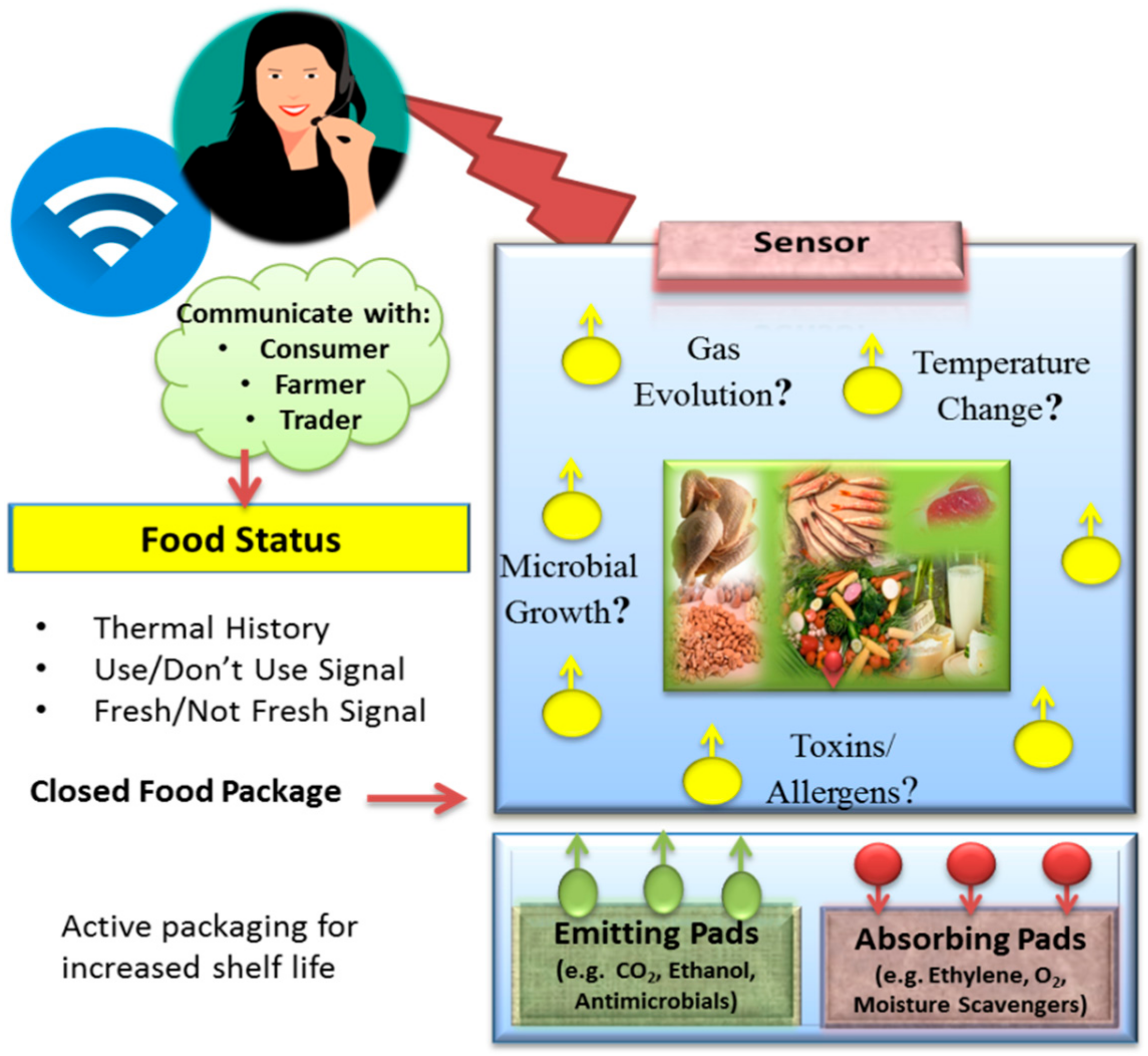
Conventional packaging methods have been used not only to facilitate product handling, but also to preserve nutrition value, extend their shelf life, and reduce spoilage. Recent efforts are designed to develop smart and active packaging technologies [13] that are able to provide additional functions, such as detection and communication to inform consumers when spoilage occurs, in addition to preserving the product [1]. Examples of active packaging include the use of absorbing or emitting sachets/pads [14], as shown in Figure 1. Absorbing sachets could contain O2 scavengers [15,16] to decrease fat oxidation, ethylene scavengers to minimize fruit and vegetable ripening [17,18,19], humidity absorbers, odor absorbers, and antimicrobial growth inhibitors [20]. Emitting pads include CO2 emitters inhibiting microbial growth in meat [21], antimicrobial preservative releasers to minimize spoilage due to bacterial growth [22], and antioxidant releasers to reduce oil and fat oxidation [23].
Smart packaging can incorporate sensing platforms that provide information about the quality of food through the entire food chain such as composition, storage conditions, and bacterial growth [24]. The concept is depicted in Figure 1. Existing systems include time temperature indicators (TTI), which provide a thermal history of the product during time of storage, and distribution, enabling the consumer or the manufacturer to assess the product status [25]. For instance, MonitorMark™ a TTI sensor developed by 3M™ (3M™, Maplewood, MN, USA) was designed to monitor thermal exposure for meat, fish, and dairy products during storage and transportation below 20 °C [26]. Another example is the CoolVu indicator [27] developed in Freshpoint-Switzerland, which includes a metal and a transparent label. The label is composed of an etchant material and it is designed to provide a visual “use”/“do not use” change to inform customers of product-quality changes.
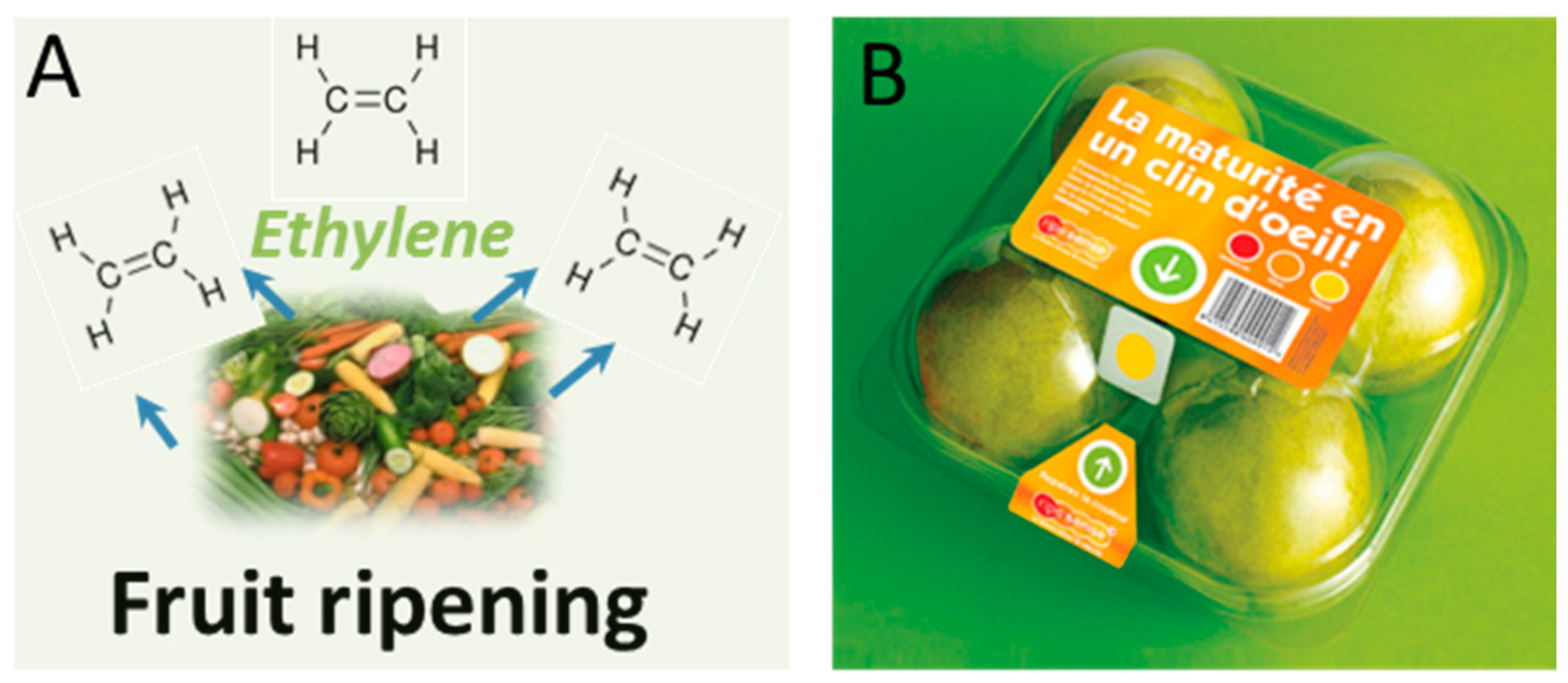
Other platforms have been developed to respond to the generation of gas during ripening processes in the packaging headspace [28]. Some systems enable remote monitoring via radio-frequency identification (RFID) [29]. An RFID sensor for the detection of Escherichia coli (E. coli) and Salmonella in packaged foods was designed by Flex Alert [30]. This technology is based on antitoxins immobilized on flexible RFID tags incorporated within packaging. Ideally, the system is connected to a wireless network and produces a visual alert for real-time monitoring by farmers and producers. Another system, RipeSense® (RipeSense, Auckland, New Zealand) [31] was designed as a smart ripeness-indicator label, developed in New Zealand. The system is able to communicate the ripeness degree of fruits without the need to open the package, but only by observing the change in the label color reacting with the gases evolved from the fruit, placed on the top of package. Among evolved gases, ethylene is the most widely used ripening indicator that is released during the ripening process [28] (Figure 2).
3. Food Freshness/Quality Monitoring
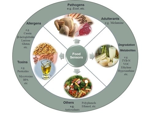
Several changes can take place in packaged food as a result of metabolism or microbial growth over time. For example, changes in gas evolution or microbial accumulation can be used to obtain information about the status of food, e.g., freshness or degradation [32]. Sensors that can measure such changes could provide an overall estimation of food quality. Examples include “on-package” pH indicators that change color when food decays as a result of pH changes associated with the release of volatile amines generated during meat or fish spoilage [33,34]. In this section, several developed and commercially available freshness indicators are described for different types of food including fish, meat, and poultry, cereal grains, fruits, and vegetables.
3.1. Fish, Meat, and Poultry
When meat, fish, or poultry undergo degradation, different spoilage indicators can be found indicating lipid decay, protein breakdown, and adenosine triphosphate (ATP) decay. The speed of degradation is dependent on the type of product, storage temperature, feeding habits, and harvesting methods. Traditional methods to assess freshness rely on human senses; although they are essential, they provide no quantitative data of spoiled food. Methods that can quantitatively measure markers of degradation through chemical or biological reactions can provide the means to more precisely assess the status and quality of food. In fish products, for example, one of the main freshness indicators is hypoxanthine, which is produced by the metabolic degradation of ATP [35]. Karube et al. (1984) [36] developed an equation for fish freshness assessment based on the content of inosine 5-phosphate, inosine, and hypoxanthine. Several enzymatic biosensors with colorimetric [37,38,39] or electrochemical detection have been developed to quantify the level of hypoxanthine [40] using the enzyme xanthine oxidase for biorecognition of hypoxanthine or xanthine [41,42]. An electrochemical biosensor prepared by immobilizing xanthine oxidase on a carbon-paste electrode modified with gold nanoparticles was reported and tested on chicken and meat samples, with a limit of detection of 2.2 × 10⁻⁷ M hypoxanthine [43]. To develop the sensor, xanthine oxidase (XOD) was immobilized by cross-linking with glutaraldehyde and bovine serum albumin (BSA) on different types of electrodes: carbon-paste electrodes and electrodeposited gold-over-gold disks. The highest sensitivity was obtained in the case of XOD immobilization on a carbon paste electrode modified with gold nanoparticles (AuNPs). The sensor was tested at operating potentials between 0.00 and 0.6 V and showed the possibility of working at 0.00 V, which enables the elimination of interfering compounds such as ascorbic acid. Detection of hypoxanthine by XOD involves the following steps in which hypoxanthine is first oxidized to xanthine, and then to uric acid:
Yan et al. (2017) [44] reported a colorimetric sensor for xanthine detection using a copper nanocluster with peroxidase-like activity. The use of copper nanoclusters showed to enhance the oxidation of 3,3′,5,5′-tetramethylbenzidine dye (TMB) in the presence of H2O2 formed from the oxidation of xanthine. The sensor exhibited a detection limit of 3.8 × 10⁻⁷ M, and a linear range from 5.0 × 10⁻⁷ to 1.0 × 10⁻⁴ M. Chen et al. (2017) [37] developed a multicolor sensor for hypoxanthine detection by using gold nanorods (GNRs). The H2O2 produced from hypoxanthine oxidation by XOD undergoes a Fenton reaction to produce hydroxyl radicals in the presence of Fe2+. In the presence of hydroxyl radicals, the GNRs are etched forming a vivid color change. Different colors were generated, such as reddish brown, gray, green, blue, purple, pink, and yellow, depending on the concentration of hypoxanthine in the range of 0–1.13 mM. The sensor showed the ability to semiquantitatively assess the levels of hypoxanthine in fish extract by the naked eye (Figure 3).
Other biomarkers for the identification of fish degradation are biogenic amines such as putrescine, cadaverine, histamine, tyramine, spermidine, spermine, and tryptamine that accumulate as a result of microbial decarboxylation of amino acids [45]. The most common measurement systems for the detection of biogenic amines are those based on the use of the diamineoxidase (DAO) enzyme, which catalyzes the oxidation of biogenic amines to their corresponding aldehyde, hydrogen peroxide, and ammonia. This can be detected by colorimetric or electrochemical means. Enzymatic biosensors for biogenic amines were demonstrated for analysis of cheese and anchovies degradation [46]. The DAO enzyme was immobilized on Pt or Au electrodes and entrapped between electrosynthesized polypyrrole (PPY) and poly-ortho-phenylenediamine (PPD) layers. The sensor showed high sensitivity, with a limit of detection (LOD) in the range of 6–12 μM of histamine, putrescine, and cadaverine and a stability of about three weeks with 87% retention of initial sensitivity. The sensor showed potential as a screening tool for the presence of biogenic amines.
Trimethylamine oxide is another marker that can be used to assess fish decomposition. Trimethylamine oxide degrades to trimethylamine increasing the fishy odor [47]. A colorimetric system for the detection of trimethylamine oxide has been developed by using pH indicator dyes immobilized on cellulose microparticles [48] that change color from green to red when the food is spoiled. The particles were embedded into food-grade silicone and were safely integrated into food packaging. The system showed no leaching, and cytotoxicity tests confirmed compatibility. Another example was described by using dyes loaded within porous TiO2 nanoparticles [49]. In other examples, total volatile basic nitrogen (TVB-N) was quantified by an organic semiconductor gas sensor with a porous top metal electrode sensor with a nanostructured surface for enhanced gas adsorption [50]. The sensor was able to detect 100 ppb where the accepted level for ammonia is 200–300 ppb. A colorimetric sensor to detect TVB-N such as ammonia and dimethylamine was fabricated with the pH-sensitive dye bromocresol green [51] and tested in fish packaging. Although the sensor exhibited a color change from yellow to blue when exposed to the evolved TVB-N from fish spoilage, a false-negative signal may occur. Quantification of volatile organic compounds has also been used to evaluate meat, fish, and poultry freshness, for example, the system used to build the Swiss sensor FOOD sniffer [52]. Distell developed a Fish Freshness Meter and Torrymeter to measure fish freshness [53]. The sensing system is based on measuring the dielectric properties of fish-flesh skin when it starts to spoil, providing a numerical read out that reflects the degree of freshness. Portable meat fish fat meters based on water-content measurements, which is proportional to fat content, were also developed and are available.
Microbial activity in meat leads to the generation of compounds such as NH3, CO2, and H2S due to decarboxylation, deamination, and desulfurization of amino acids. Visual freshness monitoring of skinless chicken breasts was performed by detecting CO2 metabolites with mixtures of pH-sensitive dyes (bromothymol blue, bromophenol blue, bromocresol purple, methyl red, bromocresol green, methyl orange, methyl yellow, phenol red) [54]. The most sensitive mixture of dyes was composed of bromothymol blue and methyl red, which turned from green to yellow when the meat starts to deteriorate. Although several studies have been reported in the literature to develop sensors for meat and fish packaging, there is a general lack of toxicity investigations and the long-term effects of the sensing materials proposed for food packaging.
3.2. Cereal Grains
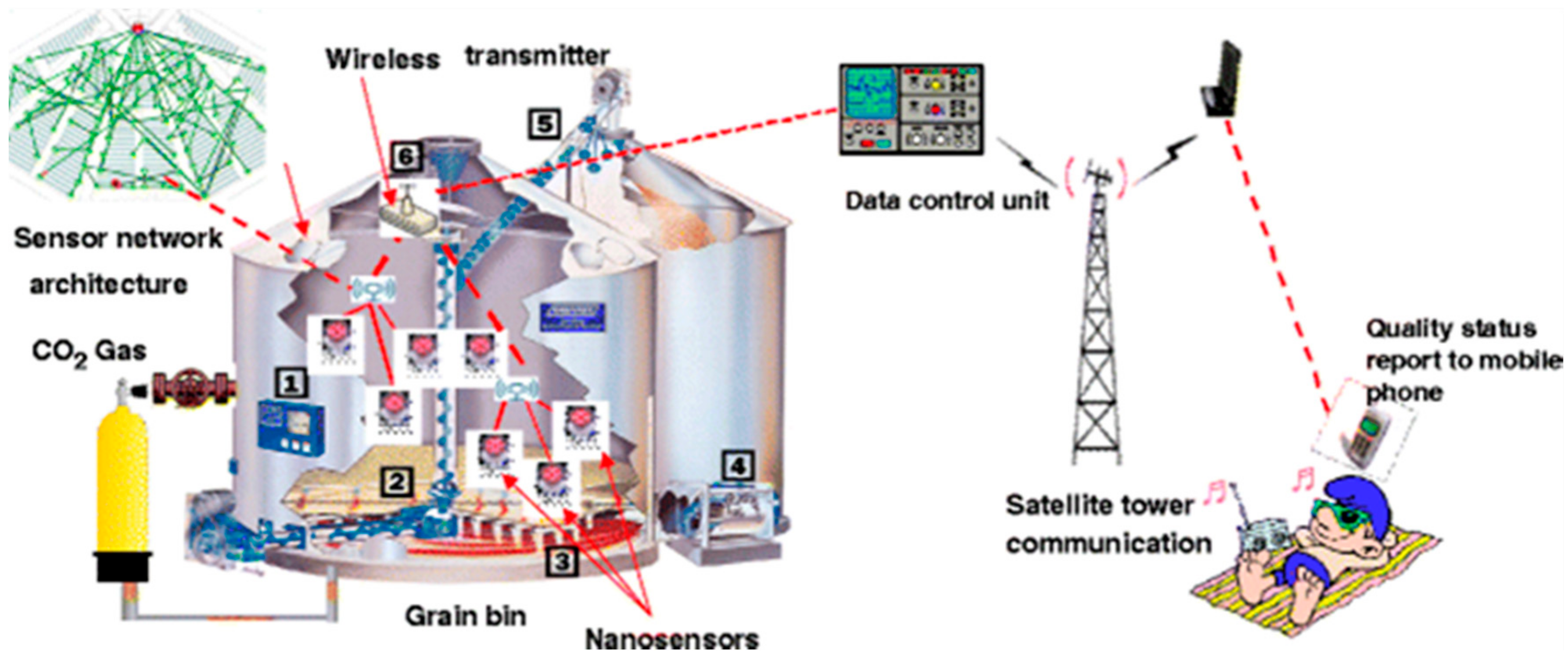
One of the indicators of grain spoilage during storage is the emission of CO2 as a result of insect infestations, and mold spoilage [55] causing grain deterioration or the production of harmful mycotoxins [56,57,58]. Developing CO2 sensors for early spoilage detection has been reported [59,60]. Neethirajan et al. (2010) [61] developed a sensor based on polyaniline boronic acid (PABA) conducting polymer for measuring CO2 levels in the range of 380–2400 ppm in simulated grain bulk. The sensing mechanism is based on the conversion between the emeraldine salt form and the insulating emeraldine base form of polyaniline and PABA through protonation and deprotonation. When gaseous CO2 reacts with water, it creates carbonic acid that protonates the polyaniline and further increases conductivity as CO2 partial pressure increases. However, the sensor performance was found to be dependent on the relative humidity, and further work should be done to enhance sensitivity and response time. An example of an advanced sensor concept with wireless transmission for large-scale monitoring of this type of spoilage, as well as the source in the grain bin, are shown in Figure 4 [1].
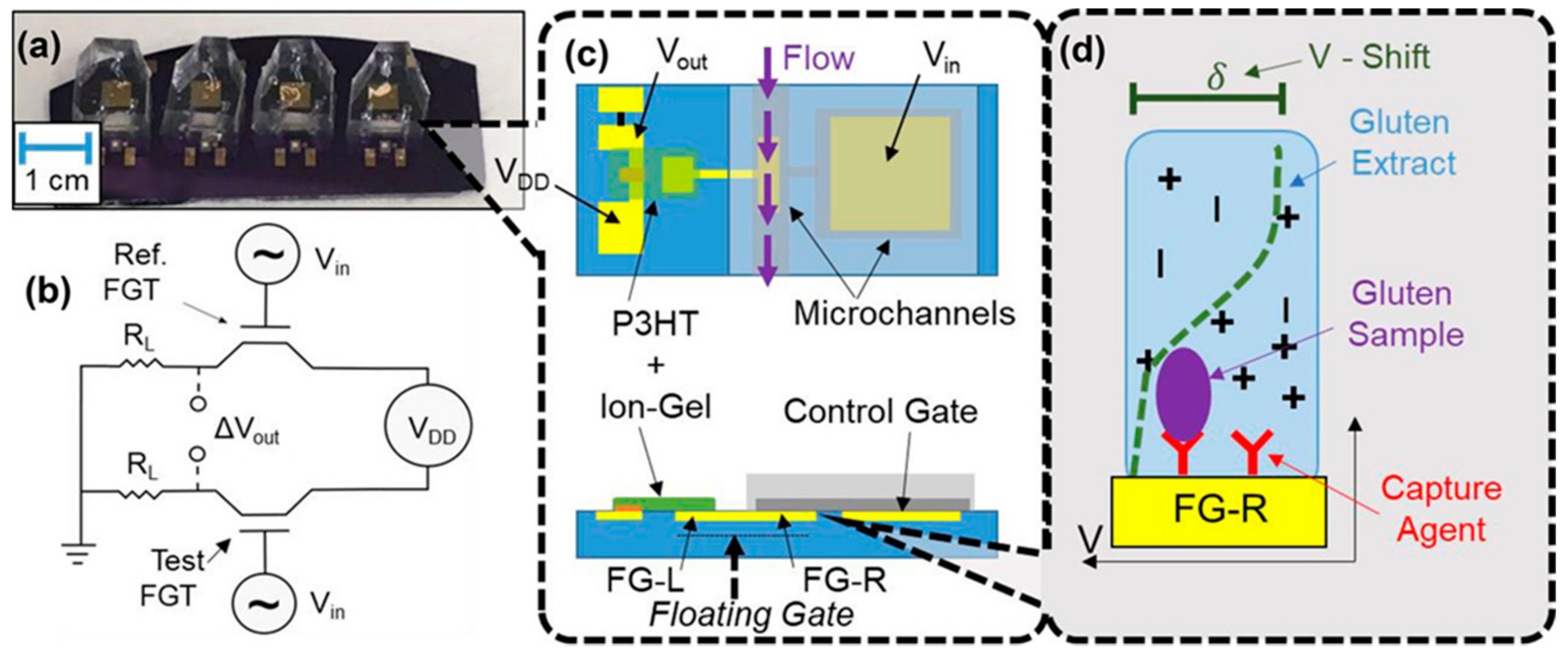
Gluten is another component of interest for grain analysis, as certain individuals can develop gluten intolerance that can cause serious disorders of the digestive system [62]. The most common method for gluten analysis is by the conventional enzyme-linked immunosorbent assay (ELISA) [63]. Recently, an antibody-based device has been reported to analyze the gluten content in a variety of food samples in only 3 min [64]. Gliadin, a fraction of gluten was also detected using an impedimetric aptasensor with an LOD of 5 ppm, lower than the maximum allowed level for gluten-free products of 20 ppm [63]. The biosensor was tested for gluten content and gluten-free food, and was comparable with the ELISA technique. White et al. (2018) [65] developed an electronic sensor based on a floating-gate transistor (FGT). Both antibodies and aptamers were used as selective receptors for binding different types of gluten sources, such as wheat and barley. The sensor principle and its surface modification are shown in Figure 5. The antibody or aptamer-modified surface on floating-gate electrode (FG-R) is susceptible to potential change (green dotted line), producing a voltage shift.
Analysis of toxicity caused by mycotoxin contamination in cereals is also of special interest. Mycotoxins, such as ochratoxin A (OTA), aflatoxins, trichothecenes, fumonisins, zearalenone, and ergot alkaloids, are produced by fungi [66]. Several biosensor types for mycotoxin detection have been reported and reviewed extensively [56,67,68,69]. Electrochemical biosensors were developed for the detection of OTA using a competitive mechanism using OTA-specific aptamers and horseradish peroxidase (HRP) enzyme [70]. An OTA-specific aptamer was immobilized on paramagnetic microparticles, which compete with the HRP–OTA conjugate and free OTA in the developed assay. The beads were drop-casted over a screen-printed electrode, and the product of the enzymatic HRP reaction was detected using differential-pulse voltammetry. The sensor was applied for OTA detection in wheat in the linear range of 0.78–8.74 ng/mL and LOD of 0.07 ± 0.01 ng/mL. In another work, ceria nanoparticles functionalized with OTA-specific aptamers were used as enzyme-mimetic probes for colorimetric OTA detection [71]. OTA binding caused changes in ceria-particle reactivity, which was assessed by measuring TMB oxidation by ceria. The assay was able to detect down to 0.15 nM OTA. Overall, estimation of allergens and mycotoxins in cereals requires sample pretreatment and extraction of the targeted component; thus, direct application of these sensing mechanisms in food packaging is challenging.
3.3. Fruits and Vegetables
Fresh fruit and vegetable production is drastically increasing globally as a result of increased demand [72]. In the United States, about 40% of food is wasted, causing a yearly loss of about $165 billion [73]. Fruits and vegetables are highly perishable and can therefore easily degrade before reaching the consumer. Technology for monitoring and preserving fruits and vegetables is necessary to decrease food loss during transportation and storage [74]. Many fruits and vegetables produce ethylene due to environmental stress after being harvested. Ethylene can enhance ripening even at extremely low concentrations [75,76]. The presence of aging fruits and vegetables close to fresh ones can also cause aging and ripening as ethylene is emitted. Ethylene can be removed by using ethylene absorbers or oxidizers (scavengers). Scavenging systems facilitate removal, thus lowering the loss of other products due to overproduced ethylene. The most available ethylene scavenger is potassium permanganate (KMnO4), which oxides ethylene to ethylene glycol and can be further oxidized to CO2 and H2O, producing dark brown MnO2. Several commercial scavengers have been developed based on ethylene chemisorption by KMnO4 granules over clays or activated carbon [77]. Jiang et al. (2013) [78] suggested an ethylene-removal method by oxidation at low temperatures over a platinum catalyst on mesoporous silica. The catalyst was able to remove 50 ppm ethylene even at 0 °C.
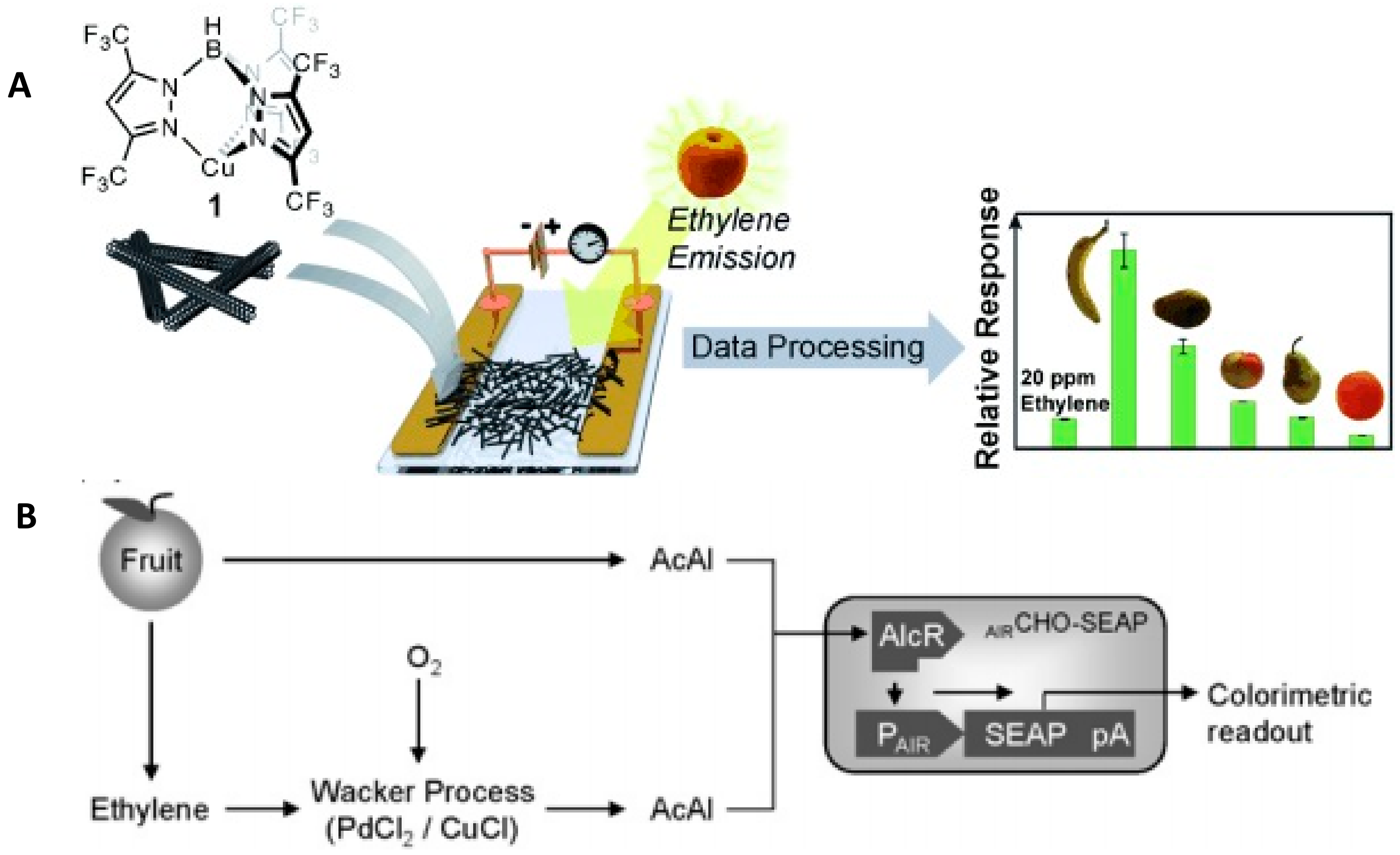
To control produce freshness, ethylene sensors could be useful for detecting rapid ripening and prevent fruit degradation. Several sensors for ethylene detection have been reported. Esser et al. (2012) [28] developed a chemoresistive sensor made of single-walled carbon nanotubes (SWNTs) mixed with a Cu (I) complex placed between gold electrodes. Upon binding to ethylene, a resistance change occurred (Figure 6A); however, the sensor generated a significant signal of tested solvents such as acetonitrile and tetrahydrofuran, but the low concentrations of these compounds had little effect. Weber et al. (2009) [78] developed a biosensor for the detection of ethylene and acetaldehyde produced by plants. In their system, ethylene was oxidized to acetaldehyde on PdCl2 with Cu (I) based on the Wacker process, and the generated acetaldehyde was detected by a CHO-K1-derived sensor cell line AIRCHO-SEAP (Figure 6B).
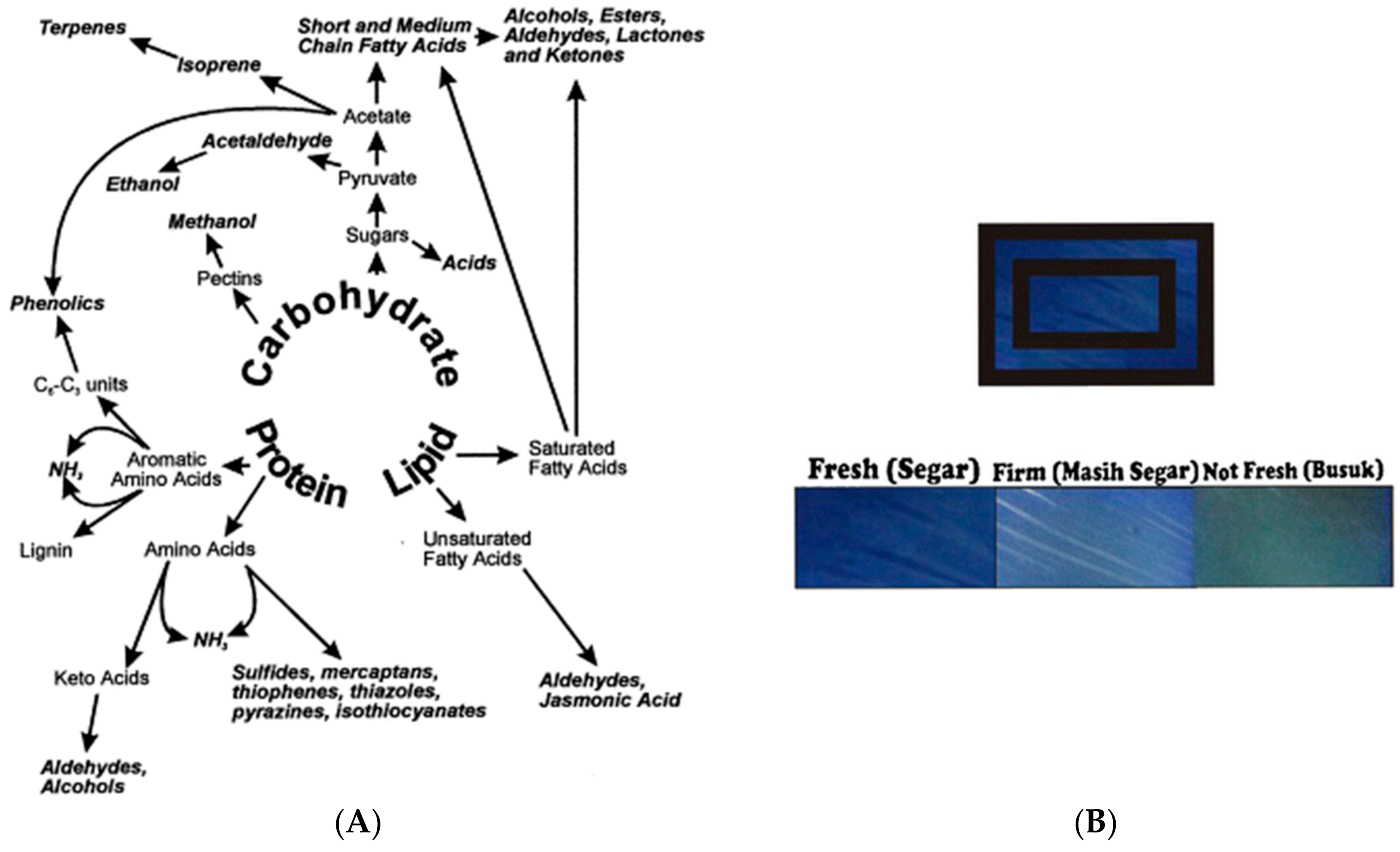
Volatile organic compounds (VOC) accumulate in the presence of fruits and vegetables in closed containers or packages. Indicators for the detection of these compounds, such as terpenes, carboxylic acids, alcohols, aldehydes, sulfur compounds, ammonia, and jasmonates [79], have been reported (Figure 7A). A color-based pH indicator, developed using bromophenol blue immobilized on a cellulose membrane, enabled the detection of VOC (e.g., acetic acid) evolution in the headspace of guava packaging (Figure 7B) [80]. This label provides the consumer with the freshness status of guava; however, more investigations are needed to ensure the safety of using chemical dyes and limit the probability of migration into the food.
4. Biosensors in Food Analysis
The need for simple, rapid, and field-portable analytical methods has boosted development of biosensors for food analysis. The integration of biomolecules, such as enzymes, immunosystems, tissues, organelles, or whole cells, with a variety of transduction methods, such as electrical, thermal, or optical signals, has enabled the development of a wide array of biosensing devices [81]. Their selectivity and relative ease of analysis make them advantageous for use in food analysis. The development of biosensors in this field is described with biosensor examples for the detection of pathogens [82], allergens [83], and other toxicants, such as pesticides [84] and mycotoxins. In this section, we briefly highlight the application of biosensors for allergens, toxicants, and pathogen detection.
4.1. Biosensors for Food-Allergen Detection
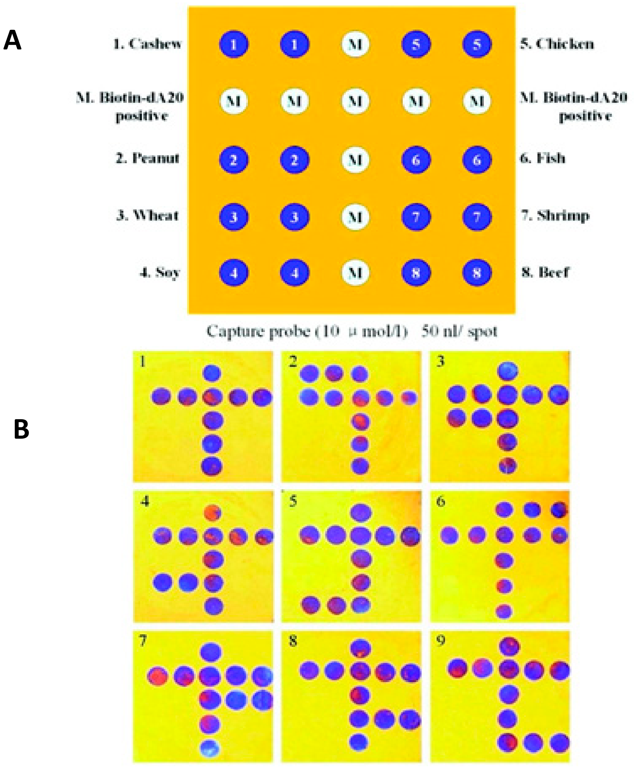
The presence of allergens in food products such as milk, soybeans, crustaceans, eggs, gluten-containing cereals, peanuts, and nuts (e.g., almonds, Brazil nuts, cashews, walnuts) is an increased safety concern, as prevalence of food allergies due to even trace amounts of allergens is increasing. About 10% of preschool children in industrial countries suffer from clinical food allergies [85]. A variety of DNA or immune-based biosensors have been developed for allergen detection [86], but in many cases sample preparation and purification are laborious and time-consuming [9]. Employing antibody-based detection and magnetite beads, NIMA Company (San Francisco, CA, USA) has developed a sensor to detect peanut allergens in ppm [87]. Archin (Ara h1) peanut allergens were detected by surface plasmon resonance (SPR)-immune-based biosensor in chocolate candy bars [88] with an LOD of 0.09 μg/mL for the optimized system. Three systems were tested and compared for the detection of the Ara h1 allergen: a label-free assay, a secondary antibody sandwich assay, and an NPs-based signal-enhanced SPR biosensor consisting of functionalized magnetic nanobeads. The SPR biosensor with NP enhancement provided the best sensitivity among the three assays, with a detection limit of 0.09 μg/mL, a linearity range from 0.1 to 2 μg/mL, and good correlation with a commercially available ELISA kit. Wang et al. (2011) [89] developed a colorimetric, silicon-based, optical thin-film biosensor chip with PCR amplification and demonstrated the ability to simultaneously identify eight food allergens found in soybeans, wheat, peanuts, cashews, shrimp, fish, beef, and chicken (Figure 8), with an LOD of 0.5 pg. The presence of gluten in food such as wheat, barley, and rye causes celiac disease for individuals who are unable to digest gluten [90]. A label-free electrochemical immunological sensor for β-lactoglobulin, an allergen usually found in milk, was developed with an LOD of 0.85 pg/mL [91]. The sensor was fabricated on a graphene-modified screen-printed electrode with immobilized β-lactoglobulin antibodies. Binding of β-lactoglobulin was quantified by measuring the reduction peak of [Fe(CN)6]3−/4−, which linearly decreased with the increase of β-lactoglobulin concentration. The sensor was applied to screen several samples such as cheese snacks, cake, and sweet biscuits, and the results were comparable with the commercial ELISA assay.
In another assay, allergy-causing proteins, such as casein and β-lactoglobulin (β-LG), in milk have also been detected based on aptamer recognition with the aptamer immobilized on a graphene-modified screen-printed electrode with voltammetric detection [92]. The method was tested on spiked milk samples and showed no significant matrix effect. Another common allergen in milk is casein. A casein immunosensor with Localized Surface Plasmon Resonance (LSPR) detection and immobilized casein antibodies was reported to detect casein in raw milk with an LOD of 10 ng/mL [93].
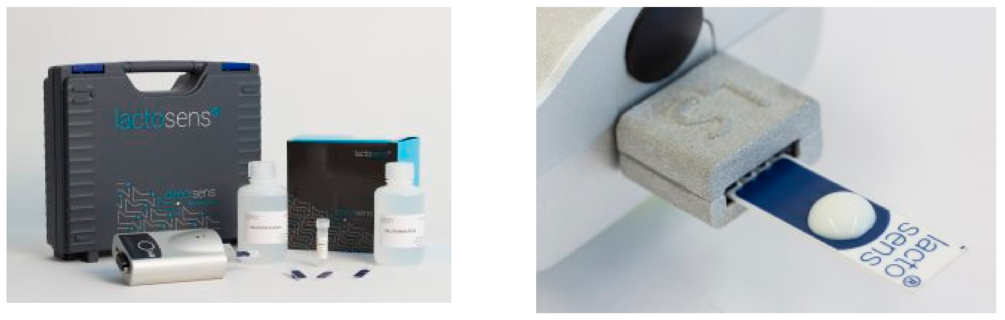
Lactose intolerance is found in all parts of the world forming about 15% of Northern Europeans, 80% of black people and Latinos, and about 100% American Indians and Asians [94]. It occurs in individuals lacking the enzyme (β-galactosidase) responsible for digesting lactose sugar into galactose and glucose [95]. Several types of electrochemical biosensors have been designed for lactose quantification utilizing coimmobilized β-galactosidase and glucose oxidase enzymes [96,97,98,99]. Several enzymatic lactose sensors are also available for the detection of trace amounts of lactose in lactose-free milk products, for example, LactoSens® (Directsens, Klosterneuburg, Austria) biosensors. The enzymes are immobilized on disposable test strips, and lactose content is directly quantified by a reader (Figure 9) [100].
4.2. Biosensors for Bacterial-Pathogen Detection
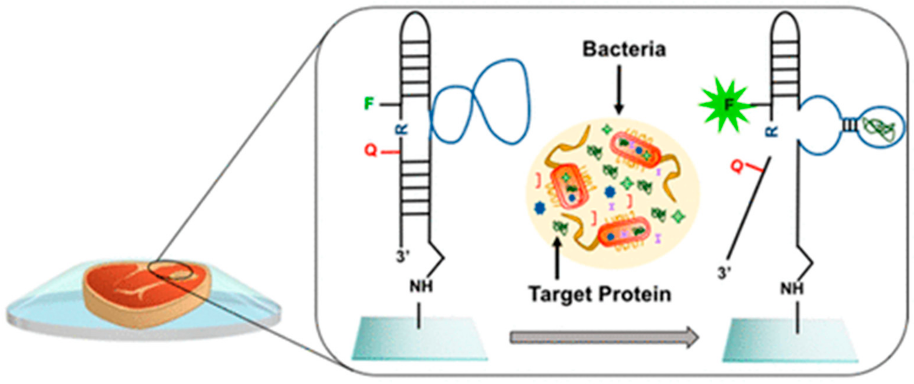
According to the Centers for Disease Control and Prevention (CDC) in the United States, the estimated number of people infected by bacteria-, virus-, and parasite-caused diseases is approximately 48 million [101]. The World Health Organization (WHO) Foodborne Disease Burden Epidemiology Reference Group (FERG) estimated 600 million foodborne illnesses with 420,000 deaths in 2010 globally [102]. The main causes of these diseases are pathogenic bacteria Escherichia coli and Salmonella causing most foodborne outbreaks in the United States [103]. Rapid detection of pathogenic bacteria plays an important role in food analysis. The main methods for pathogen detection are based on Polymerase Chain Reaction (PCR) or plate counting, which require sample enrichment and long analysis time [104]. Biosensors represent a possible alternative for pathogens detection due to their portability and potential for onsite detection. Most biosensors for bacterial-pathogen detection are those based on immune and DNA recognition, but these require extensive preparation procedures, involve labeling, multiple washing steps [105,106], and specialized facilities [107]. Alternatively, synthetic antimicrobial peptides have been proposed as recognition agents, enabling detection and quantification of four bacterial strains, Escherichia coli, Pseudomonas aeruginosa, Staphylococcus aureus, and Staphylococcus epidermidis [108]. Synthetic antimicrobial peptides have advantages over immunological and DNA-based receptors that include low cost, large-scale production, and high stability. Moreover, they could also be used to inactivate pathogens [109,110]. Colorimetric biosensor strips fabricated with peptides immobilized on a gold chip were also reported for the detection of Listeria monocytogenes in milk and meat samples with an LOD of 2.17 × 10² CFU/mL [111]. Recently, a fluorescent DNAzyme probe that specifically binds E. coli was developed and printed on a cyclo-olefin polymer transparent package (Figure 10) [112]. The study reported detection of E. coli in meat and apple-juice samples with a low LOD of 103 CFU/mL.
Biosensors could be designed to incorporate materials with antimicrobial activity to create smart packaging [113]. Silver NPs (AgNPs), one of the most extensively studied antimicrobial agents, have been considered [114,115]. For example, AgNPs were imbedded into a hydroxypropyl methylcellulose polymer showing antimicrobial inhibition of Escherichia coli and Staphylococcus aureus [116]. In another study, acrylamide was copolymerized on paper with AgNPs entrapment, showing activity against Escherichia coli [117]. The use of AgNPs and other nanomaterials in active packaging applications is an active field of research. However the possible migration of NPs from the package to the foodstuff is a concern and should be considered in future research [118]. Natural antimicrobial materials, such as ethanol and essential-oil extracts, were suggested for possible use as emitters with protective functions in packaging [119]. Moreover, the potential to use the essential oils of oregano, rosemary, and garlic in edible films with antimicrobial activity has been demonstrated [120]. The idea of integrating biosensing platforms with systems for bacteria inactivation that can both detect and deactivate pathogens is emerging [10,121,122]. A 3D-ZnO nanorod-based electrochemical sensor was developed to both detect and kill bacteria showing the inactivation of 50% of bacteria [122]. Another example was reported using a surface-enhanced Raman scattering (SERS) silicon-wafer multifunctional chip [121]. The chip was modified with AgNPs to deactivate bacteria, and 4-mercaptophenylboronic acid to detect the pathogens. The assay measured E. coli and S. aureus down to 102 cell/mL, with 60% deactivation efficiency at a bacterial concentration of 0.5–2 × 10² CFU/mL. Most sensors were developed to demonstrate proof of concept. Issues regarding the safety and hazardous effect of antimicrobials (e.g., the release of Ag) need to be clarified before considering such platforms for food packaging. Moreover, reported sensors should be designed to meet regulatory limits and extensive validation should be provided.
4.3. Biosensors for Food Adulteration, Authenticity, and Toxicicity Assessment
Food adulteration is based on the change of food composition for the purpose of financial profit regardless safety consideration. Some examples including using illegal Sudan dyes in foods, bovine addition to chicken fillets [123], and the counterfeiting of olive oil [124]. Food adulteration is a growing concern, which creates the necessity for reliable analysis methods. A serious safety issue is adulteration of milk products with melamine due to its property to increase the apparent level of protein content as determined by protein quantification assay (Kjeldahl method). The incident resulted in about 300,000 sick children and six deaths [125]. An antibody-based optical biosensor was developed for melamine detection [126]. A polyclonal antibody was immobilized on the surface of an SPR biosensor, resulting in a sensitive platform measuring IC50 of 67.9 ng/mL. The sensor was selective for melamine but it showed cross reactivity with cyromazine, an insecticide that decays to form melamine. A label-free AgNP colorimetric sensor was developed to detect melamine in milk [127]. Yellow-red color was produced by AgNPs in presence of melamine as a result of aggregation. The method was able to detect down to 2.32 μM, lower than the Food and Drug Administration (FDA)-allowed limit of 20 µM [122]. Ni et al. (2014) [128] reported that melamine enhances the peroxidase-like activity of AuNPs in presence of 3,3′,5,5′-tetramethlybenzidine (TMB) and H2O2, generating a blue color formation. The method showed high sensitivity with an LOD of 0.2 nM.
Moreover, biosensors have been described to confirm the claimed composition and concentration of food products. Ethanol content has been estimated in alcoholic beverages by electrochemical biosensors based on immobilized alcohol oxidase enzyme [129]. An alcohol dehydrogenase-based biosensor was developed in presence of Meldola’s Blue on multiwall carbon nanotubes [130]. The linear range was 0.05–10 mM ethanol. Glucose evaluation in food products was also determined by an amperometric biosensor based on electropolymerized thin films [131] or based on carbon-nanotube osmium polymers [132].
The determination of polyphenol content in vegetable oils enables the estimation of the product’s antioxidant capacity. An HPR-based electrochemical biosensor was designed to evaluate chlorogenic acid content in vegetable oil. The sensor showed a sensitivity of 0.7 µM comparable with conventional detection methods [133]. An electrochemical biosensor was developed to discriminate between different types of vegetable oils (e.g., olive oil, sunflower oil, and corn oil), as well as the ability to differentiate between different types of olive oil (e.g., extra virgin, virgin, etc.) [134]. The working principle is based on the distinct polyphenol content, resulting in different electrochemical signals. Several types of sensors that utilize nanomaterials as a means to increase detection sensitivity and portability have been developed [135]. A portable paper-based assay was developed to determine the antioxidant capacity using ceria NP-modified paper. NPs interact with antioxidant compounds by means of surface and redox reaction, producing a colorimetric response in the range of 20–400 μM for several tested antioxidants, such as ascorbic acid, gallic acid, vanillic acid, quercetin, caffeic acid, and epigallocatechin gallate [136]. In other works, AgNPs were entrapped in poly (vinyl alcohol) (PVA) to develop a colorimetric sensor for quantifying antioxidant capacity, with an example demonstrated for the detection of gallic acid. The detection principle was based on seed-mediated NP growth. Ag+ was reduced to Ag0 that accumulated on the sensor surface, showing an increase on the particle surface. The PVA in the sensor acts as a catalyst for the reduction of Ag+ by gallic acid by providing AgNPs as nucleation seeds. Once the seed is formed, it accumulates on the PVA–AgNP surface leading to a red shift. The method provided a linear range from 25 to 200 μM and an LOD of 22.1 μM [137].
Other adulterants, such as monosodium glutamate (MGM) in food are also of interest. L-glutamate is a natural amino acid, and it is usually added to enhance food flavor and to add the umami taste to food. It has been reported that L-glutamate enhances food intake with no health risks [138]. However; it has been shown that L-glutamate has neuroexcitatory action [139]. Therefore, developing reliable methods to evaluate glutamate content in food products is important. An amperometric biosensor was developed by immobilizing glutamate oxidase on a Prussian Blue-modified electrode [140]. The biosensor was able to detect L-glutamate with an LOD of 0.7 nM. An electrochemical biosensor constructed by functionalization of a platinum anode with a thin film of Nafion and glutamate oxidase was able to selectively measure glutamate concentration as low as 0.3 µM [141].
The widespread use of pesticides in agriculture leads to their accumulation in soil, ground water, and crops. Due to their inherent toxicity, a control of pesticide levels in food products is necessary. Biosensors based on acetylcholinesterase (AChE) inhibition have been reported for the detection of carbamates and organophosphate used in insecticides [142,143,144,145]. Andreescu et al. (2002) [146] reported a comparative study of three AChE immobilization approaches on screen-printed electrodes by bioincapsulation in sol-gel composites, metal-chelate affinity, and entrapment in a photopolymerizable polymer. The enzyme entrapped in a polymer matrix showed stability for more than six months. The LOD for organophosphate-insecticide detection was in the range of 1–10 nM. As an alternative, biosensors based on organophosphorus hydrolase, which catalyzes the hydrolysis of organophosphorous pesticides, were also developed. Carbon black and mesoporous carbon-modified electrodes exhibited high sensitivity for the detection of p-nitrophenol resulted from hydrolase reaction of organophosphate [147]. The method was able to detect concentrations as low as 0.12 µM.
Another analyte of interest in the food sector is bisphenol A (BPA). BPA is a monomer used in the manufacture of polycarbonate and epoxy resin employed in the fabrication of a variety of food packaging such as water bottles, feeding bottles and coating material for processed food cans [148,149]. Exposure to BPA is associated with health risks and a ban on the use of BPA in children products is under debate since 2008 [150]. The FDA has banned the use of polycarbonates in baby bottles and spill-proof cups and the use of BPA-based coatings in packaging for infant milk in 2012 [151,152]. As a result, BPA detection is an emerging research topic in the sensing field. Alkasir et al. (2012) [153] developed an enzyme-based NP-functionalized electrochemical biosensor for quick and sensitive BPA detection. The biosensor modified with Ni NPs exhibited higher sensitivity and a lower LOD of 7.1 nM, in comparison with the biosensors modified with Fe2O3 (LOD, 8.3 nM) and AuNPs (LOD, 10 nM). Moreover, a paper-based colorimetric biosensor was developed for the detection of phenolic compounds, including BPA. The paper sensors showed an LOD of 0.86 µM and demonstrated high stability during 260 days, preserving 92% of its activity. BPA was estimated in drinking plastic water bottles and beverages cans using the method developed by Deng et al. (2014) [154]. The electrochemical biosensor employed a molecularly imprinted chitosan-graphene composite to selectively detect BPA with a reported LOD of 6.0 nM.
5. Conclusions and Future Trends
The implementation of chemical sensors, biological sensors, and indicator labels in smart and active packaging, as well as the development of methods enabling freshness investigation of food products and crops, is a growing field of study. While significant progress has been achieved, there is still a need to demonstrate the functionality of biosensing devices in realistic settings for evaluating the quality of packaged food. One of the main challenges in this field is the complexity of food samples and the difficulty to measure markers of degradation directly in closed packaging without prior sample treatment. Most successful examples of sensors to date are for the detection of volatile compounds, such as amines and ethylene. In the current development status, most food biosensors still need food-sample pretreatment. Future developments should be directed towards decreasing the detection limit and increasing the possibility to measure markers upon simple contact with the sample. Validation of measurements on large amounts of samples is also needed before any implementation in the field. In the biosensing field, there is a need to further simplify detection systems to avoid the use of multiple steps or reagents, decrease the cost, and miniaturize the sensor. Increasing the stability of the biological element in biosensing designs and ensuring long operability time during storage in packaged food is another challenge that needs to be addressed, especially for systems that incorporate sensitive biorecognition elements such as enzymes or antibodies. The effect of environmental parameters should also be established. Most sensors that have been reported in the scientific literature are not commercialized. These are in their vast majority in the initial design/proof of concept stage and require significant efforts to further develop into a marketable product to meet health concerns and current regulation. Additionally, integration of materials in smart packaging and food sensors should be done with full consideration of safety regulations due to the potential migration and contact with food.
Achieving multifunctionality through the combined use of detection and food-preservation methods is another growing field of research. Integration of these technologies is a challenge, but the rapid development of new antimicrobial agents and their assembly in functional coatings is expected to positively impact development. The use of nanomaterials in both sensing and packaging technologies is also growing and has demonstrated promising potential. However, toxicity concerns and safety need to be evaluated for promoting further use of nanotechnologies in the food industry. Moreover, ensuring connectivity of sensing devices and developing wireless, independently operated sensors are needed to facilitate the rapid monitoring of a large number of samples and to provide real-time status during shipping and long-term storage.
Author Contributions
F.M. reviewed the literature and wrote the manuscript. S.A. contributed with review and editing. Both the authors read and approved the final manuscript.
Funding
This material is based on work supported by the National Science Foundation under Grants No. 0954919 and 1561491. Any opinions, findings, and conclusions or recommendations expressed in this material are those of the authors and do not necessarily reflect the views of the National Science Foundation.
Conflicts of Interest
The authors declare no conflict of interest.
References
- Neethirajan, S.; Jayas, D.S. Nanotechnology for the food and bioprocessing industries. Food Bioprocess Technol. 2011, 4, 39–47. [Google Scholar] [CrossRef]
- Kerry, J.; O’grady, M.; Hogan, S. Past, current and potential utilisation of active and intelligent packaging systems for meat and muscle-based products: A review. Meat Sci. 2006, 74, 113–130. [Google Scholar] [CrossRef] [PubMed]
- Turner, A.; Karube, I.; Wilson, G.S. Biosensors: Fundamentals Applications; Oxford University Press: New York, NY, USA, 1987. [Google Scholar]
- Yang, C.; Wang, Y.; Marty, J.-L.; Yang, X. Aptamer-based colorimetric biosensing of Ochratoxin A using unmodified gold nanoparticles indicator. Biosens. Bioelectron. 2011, 26, 2724–2727. [Google Scholar] [CrossRef] [PubMed]
- Viswanathan, S.; Radecka, H.; Radecki, J. Electrochemical biosensors for food analysis. Monatsh. Chem. Chem. Mon. 2009, 140, 891. [Google Scholar] [CrossRef]
- Narsaiah, K.; Jha, S.N.; Bhardwaj, R.; Sharma, R.; Kumar, R. Optical biosensors for food quality and safety assurance—A review. J. Food Sci. Technol. 2012, 49, 383–406. [Google Scholar] [CrossRef] [PubMed]
- Mannelli, I.; Minunni, M.; Tombelli, S.; Mascini, M. Quartz crystal microbalance (QCM) affinity biosensor for genetically modified organisms (GMOs) detection. Biosens. Bioelectron. 2003, 18, 129–140. [Google Scholar] [CrossRef]
- Wang, J. Electrochemical glucose biosensors. Chem. Rev. 2008, 108, 814–825. [Google Scholar] [CrossRef] [PubMed]
- Alves, R.C.; Barroso, M.F.; González-García, M.B.; Oliveira, M.B.P.; Delerue-Matos, C. New trends in food allergens detection: Toward biosensing strategies. Crit. Rev. Food Sci. Nutr. 2016, 56, 2304–2319. [Google Scholar] [CrossRef] [PubMed]
- Mustafa, F.; Hassan, R.Y.; Andreescu, S. Multifunctional nanotechnology-enabled sensors for rapid capture and detection of pathogens. Sensors 2017, 17, 2121. [Google Scholar] [CrossRef] [PubMed]
- Kuswandi, B.; Wicaksono, Y.; Abdullah, A.; Heng, L.Y.; Ahmad, M. Smart packaging: Sensors for monitoring of food quality and safety. Sens. Instrum. Food Qual. Saf. 2011, 5, 137–146. [Google Scholar] [CrossRef]
- Bahadır, E.B.; Sezgintürk, M.K. Applications of commercial biosensors in clinical, food, environmental, and biothreat/biowarfare analyses. Anal. Biochem. 2015, 478, 107–120. [Google Scholar] [CrossRef] [PubMed]
- Rodriguez-Aguilera, R.; Oliveira, J.C. Review of design engineering methods and applications of active and modified atmosphere packaging systems. Food Eng. Rev. 2009, 1, 66–83. [Google Scholar] [CrossRef]
- Otoni, C.G.; Espitia, P.J.; Avena-Bustillos, R.J.; McHugh, T.H. Trends in antimicrobial food packaging systems: Emitting sachets and absorbent pads. Food Res. Int. 2016, 83, 60–73. [Google Scholar] [CrossRef]
- Busolo, M.A.; Lagaron, J.M. Oxygen scavenging polyolefin nanocomposite films containing an iron modified kaolinite of interest in active food packaging applications. Innov. Food Sci. Emerg. Technol. 2012, 16, 211–217. [Google Scholar] [CrossRef]
- Tewari, G.; Jayas, D.S.; Jeremiah, L.E.; Holley, R.A. Absorption kinetics of oxygen scavengers. Int. J. Food Sci. Technol. 2002, 37, 209–217. [Google Scholar] [CrossRef]
- Terry, L.A.; Ilkenhans, T.; Poulston, S.; Rowsell, L.; Smith, A.W. Development of new palladium-promoted ethylene scavenger. Postharvest Biol. Technol. 2007, 45, 214–220. [Google Scholar] [CrossRef]
- Smith, A.W.; Poulston, S.; Rowsell, L.; Terry, L.A.; Anderson, J.A. A new palladium-based ethylene scavenger to control ethylene-induced ripening of climacteric fruit. Platin. Met. Rev. 2009, 53, 112–122. [Google Scholar] [CrossRef]
- Wills, R.; Warton, M. Efficacy of potassium permanganate impregnated into alumina beads to reduce atmospheric ethylene. J. Am. Soc. Hortic. Sci. 2004, 129, 433–438. [Google Scholar]
- Contreras, C.B.; Charles, G.; Toselli, R.; Strumia, M.C. Antimicrobial active packaging. Biopackaging 2017, 36–54. [Google Scholar]
- Hansen, A.; Mørkøre, T.; Rudi, K.; Olsen, E.; Eie, T. Quality changes during refrigerated storage of MA-packaged pre-rigor fillets of farmed Atlantic cod (Gadus morhua L.) using traditional MAP, CO2 emitter, and vacuum. J. Food Sci. 2007, 72, 423–430. [Google Scholar] [CrossRef] [PubMed]
- Cagri, A.; Ustunol, Z.; Ryser, E.T. Antimicrobial edible films and coatings. J. Food Prot. 2004, 67, 833–848. [Google Scholar] [CrossRef] [PubMed]
- Gómez-Estaca, J.; López-de-Dicastillo, C.; Hernández-Muñoz, P.; Catalá, R.; Gavara, R. Advances in antioxidant active food packaging. Trends Food Sci. Technol. 2014, 35, 42–51. [Google Scholar] [CrossRef]
- Yam, K.L.; Takhistov, P.T.; Miltz, J. Intelligent packaging: Concepts and applications. J. Food Sci. 2005, 70. [Google Scholar] [CrossRef]
- Wang, S.; Liu, X.; Yang, M.; Zhang, Y.; Xiang, K.; Tang, R. Review of time temperature indicators as quality monitors in food packaging. Packag. Technol. Sci. 2015, 28, 839–867. [Google Scholar] [CrossRef]
- 3M™ MonitorMark™ Time Temperature Indicators. Available online: https://www.3m.com/3M/en_US/company-us/all-3m-products/~/MONMARK-3M-MonitorMark-Time-Temperature-Indicators/?N=5002385+3293785721&rt=rud (accessed on 21 August 2018).
- Time Temperature Indicators. Available online: http://freshpoint-tti.com/time-temperature-indicators/ (accessed on 21 August 2018).
- Esser, B.; Schnorr, J.M.; Swager, T.M. Selective detection of ethylene gas using carbon nanotube-based devices: Utility in determination of fruit ripeness. Angew. Chem. Int. Ed. 2012, 51, 5752–5756. [Google Scholar] [CrossRef] [PubMed]
- Jones, P.; Clarke-Hill, C.; Hillier, D.; Comfort, D. The benefits, challenges and impacts of radio frequency identification technology (RFID) for retailers in the UK. Mark. Intell. Plan. 2005, 23, 395–402. [Google Scholar] [CrossRef]
- Biosensors. Available online: http://www2.Flex-alert.Com/flexalert/applications/biosensors (accessed on 21 August 2018).
- How Ripe Do You Like It. Available online: http://www.ripesense.co.nz/ (accessed on 21 August 2018).
- Smolander, M. Freshness indicators for food packaging. In Smart Packaging Technologies for Fast Moving Consumer Goods; John Wiley & Sons, Inc.: Hoboken, NJ, USA, 2008; pp. 111–127. [Google Scholar]
- Kuswandi, B.; Restyana, A.; Abdullah, A.; Heng, L.Y.; Ahmad, M. A novel colorimetric food package label for fish spoilage based on polyaniline film. Food Control 2012, 25, 184–189. [Google Scholar] [CrossRef]
- Kuswandi, B.; Nurfawaidi, A. On-package dual sensors label based on pH indicators for real-time monitoring of beef. Food Control 2017, 82, 91–100. [Google Scholar] [CrossRef]
- Ashie, I.; Smith, J.; Simpson, B.; Haard, N.F. Spoilage and shelf-life extension of fresh fish and shellfish. Crit. Rev. Food Sci. Nutr. 1996, 36, 87–121. [Google Scholar] [CrossRef] [PubMed]
- Karube, I.; Matsuoka, H.; Suzuki, S.; Watanabe, E.; Toyama, K. Determination of fish freshness with an enzyme sensor system. J. Agric. Food Chem. 1984, 32, 314–319. [Google Scholar] [CrossRef]
- Cunningham, S.; Keaveny, T. A two-stage enzymatic method for determination of uric acid and hypoxanthine/xanthine. Clin. Chim. Acta 1978, 86, 217–221. [Google Scholar] [CrossRef]
- Chen, Z.; Lin, Y.; Ma, X.; Guo, L.; Qiu, B.; Chen, G.; Lin, Z. Multicolor biosensor for fish freshness assessment with the naked eye. Sens. Actuators B Chem. 2017, 252, 201–208. [Google Scholar] [CrossRef]
- Berti, G.; Fossati, P.; Tarenghi, G.; Musitelli, C.; Melzi d’Eril, G. Enzymatic colorimetric method for the determination of inorganic phosphorus in serum and urine. Clin. Chem. Lab. Med. 1988, 26, 399–404. [Google Scholar] [CrossRef]
- Lawal, A.T.; Adeloju, S.B. Progress and recent advances in fabrication and utilization of hypoxanthine biosensors for meat and fish quality assessment: A review. Talanta 2012, 100, 217–228. [Google Scholar] [CrossRef] [PubMed]
- Lin, Z.; Sun, J.; Chen, J.; Guo, L.; Chen, Y.; Chen, G. Electrochemiluminescent biosensor for hypoxanthine based on the electrically heated carbon paste electrode modified with xanthine oxidase. Anal. Chem. 2008, 80, 2826–2831. [Google Scholar] [CrossRef] [PubMed]
- Nakatani, H.S.; dos Santos, L.V.; Pelegrine, C.P.; Gomes, S.; Matsushita, M.; de Souza, N.E.; Visentainer, J.V. Biosensor based on xanthine oxidase for monitoring hypoxanthine in fish meat. Am. J. Biochem. Biotechnol. 2005, 1, 85–89. [Google Scholar] [CrossRef]
- Agüí, L.; Manso, J.; Yáñez-Sedeño, P.; Pingarrón, J.M. Amperometric biosensor for hypoxanthine based on immobilized xanthine oxidase on nanocrystal gold-carbon paste electrodes. Sens. Actuators B Chem. 2006, 113, 272–280. [Google Scholar] [CrossRef]
- Yan, Z.; Niu, Q.; Mou, M.; Wu, Y.; Liu, X.; Liao, S. A novel colorimetric method based on copper nanoclusters with intrinsic peroxidase-like for detecting xanthine in serum samples. J. Nanopart. Res. 2017, 19, 235. [Google Scholar] [CrossRef]
- Draisci, R.; Volpe, G.; Lucentini, L.; Cecilia, A.; Federico, R.; Palleschi, G. Determination of biogenic amines with an electrochemical biosensor and its application to salted anchovies. Food Chem. 1998, 62, 225–232. [Google Scholar] [CrossRef]
- Carelli, D.; Centonze, D.; Palermo, C.; Quinto, M.; Rotunno, T. An interference free amperometric biosensor for the detection of biogenic amines in food products. Biosens. Bioelectron. 2007, 23, 640–647. [Google Scholar] [CrossRef] [PubMed]
- Park, Y.-H.; Choi, K.-J.; Bae, J.-Y.; Yoon, S.-K.; Jang, H.-I.; Lee, C.-S. Development of a detection sensor for mixed trimethylamine and ammonia gas. J. Ind. Eng. Chem. 2013, 19, 1703–1707. [Google Scholar] [CrossRef]
- Schaude, C.; Meindl, C.; Fröhlich, E.; Attard, J.; Mohr, G.J. Developing a sensor layer for the optical detection of amines during food spoilage. Talanta 2017, 170, 481–487. [Google Scholar] [CrossRef] [PubMed]
- Huang, X.; Li, Z.; Zou, X.; Shi, J.; Mao, H.; Zhao, J.; Hao, L.; Holmes, M. Detection of meat-borne trimethylamine based on nanoporous colorimetric sensor arrays. Food Chem. 2016, 197, 930–936. [Google Scholar]
- Chang, L.-Y.; Chuang, M.-Y.; Zan, H.-W.; Meng, H.-F.; Lu, C.-J.; Yeh, P.-H.; Chen, J.-N. One-minute fish freshness evaluation by testing the volatile amine gas with an ultrasensitive porous-electrode-capped organic gas sensor system. ACS Sens. 2017, 2, 531–539. [Google Scholar] [CrossRef] [PubMed]
- Pacquit, A.; Lau, K.T.; McLaughlin, H.; Frisby, J.; Quilty, B.; Diamond, D. Development of a volatile amine sensor for the monitoring of fish spoilage. Talanta 2006, 69, 515–520. [Google Scholar] [CrossRef] [PubMed]
- Food Sniffer. Available online: http://www.myfoodsniffer.com/foodsniffer.html (accessed on 21 August 2018).
- Distell Fish Freshness Meter. Available online: https://www.distell.com/wp-content/uploads/2014/04/Freshness-Meter-User-Manual-v2.9.pdf (accessed on 21 August 2018).
- Rukchon, C.; Nopwinyuwong, A.; Trevanich, S.; Jinkarn, T.; Suppakul, P. Development of a food spoilage indicator for monitoring freshness of skinless chicken breast. Talanta 2014, 130, 547–554. [Google Scholar] [CrossRef] [PubMed]
- Maier, D.; Channaiah, L.; Martinez-Kawas, A.; Lawrence, J.; Chaves, E.; Coradi, P.; Fromme, G. Monitoring carbon dioxide concentration for early detection of spoilage in stored grain. Julius-Kühn-Archiv 2010, 425, 505. [Google Scholar]
- Vidal, J.C.; Bonel, L.; Ezquerra, A.; Hernández, S.; Bertolín, J.R.; Cubel, C.; Castillo, J.R. Electrochemical affinity biosensors for detection of mycotoxins: A review. Biosens. Bioelectron. 2013, 49, 146–158. [Google Scholar] [CrossRef] [PubMed]
- Pohanka, M.; Jun, D.; Kuca, K. Mycotoxin assays using biosensor technology: A review. Drug Chem. Toxicol. 2007, 30, 253–261. [Google Scholar] [CrossRef] [PubMed]
- Neethirajan, S.; Karunakaran, C.; Jayas, D.; White, N. Detection techniques for stored-product insects in grain. Food Control 2007, 18, 157–162. [Google Scholar] [CrossRef]
- Neethirajan, S.; Jayas, D.; Sadistap, S. Carbon dioxide (CO2) sensors for the agri-food industry—A review. Food Bioprocess Technol. 2009, 2, 115–121. [Google Scholar] [CrossRef]
- Puligundla, P.; Jung, J.; Ko, S. Carbon dioxide sensors for intelligent food packaging applications. Food Control 2012, 25, 328–333. [Google Scholar] [CrossRef]
- Neethirajan, S.; Freund, M.; Jayas, D.; Shafai, C.; Thomson, D.; White, N. Development of carbon dioxide (CO2) sensor for grain quality monitoring. Biosyst. Eng. 2010, 106, 395–404. [Google Scholar] [CrossRef]
- Malvano, F.; Albanese, D.; Pilloton, R.; Di Matteo, M. A new label-free impedimetric aptasensor for gluten detection. Food Control 2017, 79, 200–206. [Google Scholar] [CrossRef]
- Nassef, H.M.; Bermudo Redondo, M.C.; Ciclitira, P.J.; Ellis, H.J.; Fragoso, A.; O’Sullivan, C.K. Electrochemical immunosensor for detection of celiac disease toxic gliadin in foodstuff. Anal. Chem. 2008, 80, 9265–9271. [Google Scholar] [CrossRef] [PubMed]
- Test Your Food Gluten: Anytime, Anywhere. Available online: https://nimasensor.com/gluten/ (accessed on 21 August 2018).
- White, S.P.; Frisbie, C.D.; Dorfman, K.D. Detection and sourcing of gluten in grain with multiple floating-gate transistor biosensors. ACS Sens. 2018, 3, 395–402. [Google Scholar] [CrossRef] [PubMed]
- Zain, M.E. Impact of mycotoxins on humans and animals. J. Saudi Chem. Soc. 2011, 15, 129–144. [Google Scholar] [CrossRef]
- Van der Gaag, B.; Spath, S.; Dietrich, H.; Stigter, E.; Boonzaaijer, G.; van Osenbruggen, T.; Koopal, K. Biosensors and multiple mycotoxin analysis. Food Control 2003, 14, 251–254. [Google Scholar] [CrossRef]
- Chen, J.; Fang, Z.; Liu, J.; Zeng, L. A simple and rapid biosensor for ochratoxin a based on a structure-switching signaling aptamer. Food Control 2012, 25, 555–560. [Google Scholar] [CrossRef]
- Bulbul, G.; Hayat, A.; Andreescu, S. A generic amplification strategy for electrochemical aptasensors using a non-enzymatic nanoceria tag. Nanoscale 2015, 7, 13230–13238. [Google Scholar] [CrossRef] [PubMed]
- Bonel, L.; Vidal, J.C.; Duato, P.; Castillo, J.R. An electrochemical competitive biosensor for ochratoxin a based on a DNA biotinylated aptamer. Biosens. Bioelectron. 2011, 26, 3254–3259. [Google Scholar] [CrossRef] [PubMed]
- Bülbül, G.; Hayat, A.; Andreescu, S. Ssdna-functionalized nanoceria: A redox-active aptaswitch for biomolecular recognition. Adv. Healthc. Mater. 2016, 5, 822–828. [Google Scholar] [CrossRef] [PubMed]
- FAO. The Future of Food and Agriculture—Trends and Challenges; FAO: Rome, Italy, 2017. [Google Scholar]
- Gunders, D. Wasted: How America is Losing up to 40 Percent of its Food from Farm to Fork to Landfill; Natural Resources Defense Council: New York, NY, USA, 2012. [Google Scholar]
- Buzby, J.C.; Wells, H.F.; Axtman, B.; Mickey, J. Supermarket loss estimates for fresh fruit, vegetables, meat, poultry, and seafood and their use in the ERS loss-adjusted food availability data. Econ. Inf. Bull.-USDA Econ. Res. Serv. 2009, 44, 26. [Google Scholar]
- Adams, D.; Yang, S. Ethylene biosynthesis: Identification of 1-aminocyclopropane-1-carboxylic acid as an intermediate in the conversion of methionine to ethylene. Proc. Natl. Acad. Sci. USA 1979, 76, 170–174. [Google Scholar] [CrossRef] [PubMed] [Green Version]
- Jiang, C.; Hara, K.; Fukuoka, A. Low-temperature oxidation of ethylene over platinum nanoparticles supported on mesoporous silica. Angew. Chem. Int. Ed. 2013, 52, 6265–6268. [Google Scholar] [CrossRef] [PubMed]
- Vermeiren, L.; Devlieghere, F.; van Beest, M.; de Kruijf, N.; Debevere, J. Developments in the active packaging of foods. Trends Food Sci. Technol. 1999, 10, 77–86. [Google Scholar] [CrossRef]
- Weber, W.; Luzi, S.; Karlsson, M.; Fussenegger, M. A novel hybrid dual-channel catalytic-biological sensor system for assessment of fruit quality. J. Biotechnol. 2009, 139, 314–317. [Google Scholar] [CrossRef] [PubMed]
- Toivonen, P.M. Non-ethylene, non-respiratory volatiles in harvested fruits and vegetables: Their occurrence, biological activity and control. Postharvest Biol. Technol. 1997, 12, 109–125. [Google Scholar] [CrossRef]
- Kuswandi, B.; Maryska, C.; Abdullah, A.; Heng, L.Y. Real time on-package freshness indicator for guavas packaging. J. Food Meas. Charact. 2013, 7, 29–39. [Google Scholar] [CrossRef]
- IUPAC. Compendium of Chemical Terminology, 2nd ed.; IUPAC: Zurich, Switzerland, 1997. [Google Scholar]
- Silva, N.F.; Magalhães, J.M.; Freire, C.; Delerue-Matos, C. Electrochemical biosensors for salmonella: State of the art and challenges in food safety assessment. Biosens. Bioelectron. 2018, 99, 667–682. [Google Scholar] [CrossRef] [PubMed]
- Neethirajan, S.; Weng, X.; Tah, A.; Cordero, J.; Ragavan, K. Nano-biosensor platforms for detecting food allergens-new trends. Sens. Bio-Sens. Res. 2018, 18, 13–30. [Google Scholar] [CrossRef]
- Nikoleli, G.-P.; Nikolelis, D.P.; Siontorou, C.G.; Karapetis, S.; Varzakas, T. Novel biosensors for the rapid detection of toxicants in foods. Adv. Food Nutr. Res. 2018, 84, 57–102. [Google Scholar] [PubMed]
- Prescott, S.L.; Pawankar, R.; Allen, K.J.; Campbell, D.E.; Sinn, J.K.; Fiocchi, A.; Ebisawa, M.; Sampson, H.A.; Beyer, K.; Lee, B.-W. A global survey of changing patterns of food allergy burden in children. World Allergy Organ. J. 2013, 6, 1. [Google Scholar] [CrossRef] [PubMed]
- Yman, I.M.; Eriksson, A.; Johansson, M.A.; Hellens, K.-E. Food allergen detection with biosensor immunoassays. J. AOAC Int. 2006, 89, 856–861. [Google Scholar] [PubMed]
- Test Your Food for Peanuts: anytime, Anywhere. Available online: https://nimasensor.Com/peanut/ (accessed on 21 August 2018).
- Pollet, J.; Delport, F.; Janssen, K.; Tran, D.; Wouters, J.; Verbiest, T.; Lammertyn, J. Fast and accurate peanut allergen detection with nanobead enhanced optical fiber SPR biosensor. Talanta 2011, 83, 1436–1441. [Google Scholar] [CrossRef] [PubMed]
- Wang, W.; Han, J.; Wu, Y.; Yuan, F.; Chen, Y.; Ge, Y. Simultaneous detection of eight food allergens using optical thin-film biosensor chips. J. Agric. Food Chem. 2011, 59, 6889–6894. [Google Scholar] [CrossRef] [PubMed]
- Denham, J.M.; Hill, I.D. Celiac disease and autoimmunity: Review and controversies. Curr. Allergy Asthma Rep. 2013, 13, 347–353. [Google Scholar] [CrossRef] [PubMed]
- Eissa, S.; Tlili, C.; L’Hocine, L.; Zourob, M. Electrochemical immunosensor for the milk allergen β-lactoglobulin based on electrografting of organic film on graphene modified screen-printed carbon electrodes. Biosens. Bioelectron. 2012, 38, 308–313. [Google Scholar] [CrossRef] [PubMed]
- Eissa, S.; Zourob, M. In vitro selection of DNA aptamers targeting β-lactoglobulin and their integration in graphene-based biosensor for the detection of milk allergen. Biosens. Bioelectron. 2017, 91, 169–174. [Google Scholar] [CrossRef] [PubMed]
- Hiep, H.M.; Endo, T.; Kerman, K.; Chikae, M.; Kim, D.-K.; Yamamura, S.; Takamura, Y.; Tamiya, E. A localized surface plasmon resonance based immunosensor for the detection of casein in milk. Sci. Technol. Adv. Mater. 2007, 8, 331–338. [Google Scholar] [CrossRef] [Green Version]
- Swagerty, D.L.; Walling, A.D.; Klein, R.M. Lactose intolerance. Am. Fam. Physician 2002, 65, 1845–1860. [Google Scholar] [PubMed]
- Paige, D.M.; Bayless, T.M.; Huang, S.-S.; Wexler, R. Lactose intolerance and lactose hydrolyzed milk. ACS Symp. Ser. Am. Chem. Soc. 1975, 15, 191–206. [Google Scholar]
- Ammam, M.; Fransaer, J. Two-enzyme lactose biosensor based on β-galactosidase and glucose oxidase deposited by ac-electrophoresis: Characteristics and performance for lactose determination in milk. Sens. Actuators B Chem. 2010, 148, 583–589. [Google Scholar] [CrossRef]
- Watanabe, E.; Takagi, M.; Takei, S.; Hoshi, M.; Cao, S. Development of biosensors for the simultaneous determination of sucrose and glucose, lactose and glucose, and starch and glucose. Biotechnol. Bioeng. 1991, 38, 99–103. [Google Scholar] [CrossRef] [PubMed]
- Garcia, J.L.; López-Munguia, A.; Galindo, E. Modeling the non-steady-state response of an enzyme electrode for lactose. Enzym. Microb. Technol. 1991, 13, 672–675. [Google Scholar] [CrossRef]
- Marrakchi, M.; Dzyadevych, S.V.; Lagarde, F.; Martelet, C.; Jaffrezic-Renault, N. Conductometric biosensor based on glucose oxidase and beta-galactosidase for specific lactose determination in milk. Mater. Sci. Eng. C 2008, 28, 872–875. [Google Scholar] [CrossRef]
- Lactose Biosensor Test System. Available online: http://www.directsens.com/lactosens/ (accessed on 21 August 2018).
- Centers for Disease Control and Prevention (CDC). Foodborne Illness: Frequently Asked Questions; CDC: Atlanta, GA, USA, 2018. [Google Scholar]
- WHO. Global Burden of Foodborne Diseases; WHO Press: Geneva, Switzerland, 2015. [Google Scholar]
- Centers for Disease Control and Prevention. Surveillance for foodborne disease outbreaks-united states, 2009–2010. MMWR Morb. Mortal. Wkly. Rep. 2013, 62, 41. [Google Scholar]
- De Boer, E.; Beumer, R.R. Methodology for detection and typing of foodborne microorganisms. Int. J. Food Microbiol. 1999, 50, 119–130. [Google Scholar] [CrossRef]
- Beumer, R.R.; Brinkman, E. Detection of Listeria spp. With a monoclonal antibody-based enzyme-linked immunosorbent assay (ELISA). Food Microbiol. 1989, 6, 171–177. [Google Scholar] [CrossRef]
- Palumbo, J.D.; Borucki, M.K.; Mandrell, R.E.; Gorski, L. Serotyping of listeria monocytogenes by enzyme-linked immunosorbent assay and identification of mixed-serotype cultures by colony immunoblotting. J. Clin. Microbiol. 2003, 41, 564–571. [Google Scholar] [CrossRef] [PubMed]
- Cai, H.Y.; Caswell, J.L.; Prescott, J.F. Nonculture molecular techniques for diagnosis of bacterial disease in animals: A diagnostic laboratory perspective. Vet. Pathol. Online 2014, 51, 341–350. [Google Scholar] [CrossRef] [PubMed]
- Liu, X.; Marrakchi, M.; Xu, D.; Dong, H.; Andreescu, S. Biosensors based on modularly designed synthetic peptides for recognition, detection and live/dead differentiation of pathogenic bacteria. Biosens. Bioelectron. 2016, 80, 9–16. [Google Scholar] [CrossRef] [PubMed]
- Friedrich, C.; Scott, M.G.; Karunaratne, N.; Yan, H.; Hancock, R.E.W. Salt-resistant alpha-helical cationic antimicrobial peptides. Antimicrob. Agents Chemother. 1999, 43, 1542–1548. [Google Scholar] [CrossRef] [PubMed]
- Rydlo, T.; Rotem, S.; Mor, A. Antibacterial properties of dermaseptin S4 derivatives under extreme incubation conditions. Antimicrob. Agents Chemother. 2006, 50, 490–497. [Google Scholar] [CrossRef] [PubMed]
- Alhogail, S.; Suaifan, G.A.; Zourob, M. Rapid colorimetric sensing platform for the detection of Listeria monocytogenes foodborne pathogen. Biosens. Bioelectron. 2016, 86, 1061–1066. [Google Scholar] [CrossRef] [PubMed]
- Yousefi, H.; Ali, M.M.; Su, H.-M.; Filipe, C.D.; Didar, T.F. Sentinel wraps: Real-time monitoring of food contamination by printing dnazyme probes on food packaging. ACS Nano 2018, 12, 3287–3294. [Google Scholar] [CrossRef] [PubMed]
- Duncan, T.V. Applications of nanotechnology in food packaging and food safety: Barrier materials, antimicrobials and sensors. J. Colloid Interface Sci. 2011, 363, 1–24. [Google Scholar] [CrossRef] [PubMed]
- Wang, L.-F.; Rhim, J.-W. Preparation and application of agar/alginate/collagen ternary blend functional food packaging films. Int. J. Biol. Macromol. 2015, 80, 460–468. [Google Scholar] [CrossRef] [PubMed]
- Gallocchio, F.; Cibin, V.; Biancotto, G.; Roccato, A.; Muzzolon, O.; Carmen, L.; Simone, B.; Manodori, L.; Fabrizi, A.; Patuzzi, I. Testing nano-silver food packaging to evaluate silver migration and food spoilage bacteria on chicken meat. Food Addit. Contam. Part A 2016, 33, 1063–1071. [Google Scholar] [CrossRef] [PubMed]
- De Moura, M.R.; Mattoso, L.H.; Zucolotto, V. Development of cellulose-based bactericidal nanocomposites containing silver nanoparticles and their use as active food packaging. J. Food Eng. 2012, 109, 520–524. [Google Scholar] [CrossRef]
- Tankhiwale, R.; Bajpai, S. Graft copolymerization onto cellulose-based filter paper and its further development as silver nanoparticles loaded antibacterial food-packaging material. Colloids Surf. B Biointerfaces 2009, 69, 164–168. [Google Scholar] [CrossRef] [PubMed]
- Kuorwel, K.K.; Cran, M.J.; Orbell, J.D.; Buddhadasa, S.; Bigger, S.W. Review of mechanical properties, migration, and potential applications in active food packaging systems containing nanoclays and nanosilver. Compr. Rev. Food Sci. Food Saf. 2015, 14, 411–430. [Google Scholar] [CrossRef]
- Kapetanakou, A.; Agathaggelou, E.; Skandamis, P. Storage of pork meat under modified atmospheres containing vapors from commercial alcoholic beverages. Int. J. Food Microbiol. 2014, 178, 65–75. [Google Scholar] [CrossRef] [PubMed]
- Seydim, A.; Sarikus, G. Antimicrobial activity of whey protein based edible films incorporated with oregano, rosemary and garlic essential oils. Food Res. Int. 2006, 39, 639–644. [Google Scholar] [CrossRef]
- Wang, H.; Zhou, Y.; Jiang, X.; Sun, B.; Zhu, Y.; Wang, H.; Su, Y.; He, Y. Simultaneous capture, cetection, and inactivation of bacteria as enabled by a surface-enhanced raman scattering multifunctional chip. Angew. Chem. Int. Ed. 2015, 54, 5132–5136. [Google Scholar] [CrossRef] [PubMed]
- Yang, Z.; Wang, Y.; Zhang, D. A novel multifunctional electrochemical platform for simultaneous detection, elimination, and inactivation of pathogenic bacteria based on the Vancomycin-functionalised AgNPs/3d-ZnO nanorod arrays. Biosens. Bioelectron. 2017, 98, 248–253. [Google Scholar] [CrossRef] [PubMed]
- Grundy, H.; Kelly, S.; Charlton, A.; Donarski, J.; Hird, S.; Collins, M. Food authenticity and food fraud research: Achievements and emerging issues. J. Assoc. Public Analy. (Online) 2012, 40, 65–68. [Google Scholar]
- Poiana, M.-A.; Alexa, E.; Munteanu, M.-F.; Gligor, R.; Moigradean, D.; Mateescu, C. Use of ATR-FTIR spectroscopy to detect the changes in extra virgin olive oil by adulteration with soybean oil and high temperature heat treatment. Open Chem. 2015, 13, 1. [Google Scholar] [CrossRef]
- Gossner, C.M.-E.; Schlundt, J.; Embarek, P.B.; Hird, S.; Lo-Fo-Wong, D.; Beltran, J.J.O.; Teoh, K.N.; Tritscher, A. The melamine incident: Implications for international food and feed safety. Environ. Health Perspect. 2009, 117, 1803. [Google Scholar] [CrossRef] [PubMed]
- Fodey, T.L.; Thompson, C.S.; Traynor, I.M.; Haughey, S.A.; Kennedy, D.G.; Crooks, S.R. Development of an optical biosensor based immunoassay to screen infant formula milk samples for adulteration with melamine. Anal. Chem. 2011, 83, 5012–5016. [Google Scholar] [CrossRef] [PubMed]
- Ping, H.; Zhang, M.; Li, H.; Li, S.; Chen, Q.; Sun, C.; Zhang, T. Visual detection of melamine in raw milk by label-free silver nanoparticles. Food Control 2012, 23, 191–197. [Google Scholar] [CrossRef]
- Ni, P.; Dai, H.; Wang, Y.; Sun, Y.; Shi, Y.; Hu, J.; Li, Z. Visual detection of melamine based on the peroxidase-like activity enhancement of bare gold nanoparticles. Biosens. Bioelectron. 2014, 60, 286–291. [Google Scholar] [CrossRef] [PubMed]
- Boujtita, M.; Hart, J.P.; Pittson, R. Development of a disposable ethanol biosensor based on a chemically modified screen-printed electrode coated with alcohol oxidase for the analysis of beer. Biosens. Bioelectron. 2000, 15, 257–263. [Google Scholar] [CrossRef]
- Santos, A.S.; Pereira, A.C.; Durán, N.; Kubota, L.T. Amperometric biosensor for ethanol based on co-immobilization of alcohol dehydrogenase and meldola’s blue on multi-wall carbon nanotube. Electrochim. Acta 2006, 52, 215–220. [Google Scholar] [CrossRef]
- Palmisano, F.; Rizzi, R.; Centonze, D.; Zambonin, P. Simultaneous monitoring of glucose and lactate by an interference and cross-talk free dual electrode amperometric biosensor based on electropolymerized thin films. Biosens. Bioelectron. 2000, 15, 531–539. [Google Scholar] [CrossRef]
- Antiochia, R.; Gorton, L. Development of a carbon nanotube paste electrode osmium polymer-mediated biosensor for determination of glucose in alcoholic beverages. Biosens. Bioelectron. 2007, 22, 2611–2617. [Google Scholar] [CrossRef] [PubMed]
- Mello, L.D.; Sotomayor, M.D.P.T.; Kubota, L.T. HRP-based amperometric biosensor for the polyphenols determination in vegetables extract. Sens. Actuators B Chem. 2003, 96, 636–645. [Google Scholar] [CrossRef]
- Apetrei, C.; Rodriguez-Mendez, M.; De Saja, J. Modified carbon paste electrodes for discrimination of vegetable oils. Sens. Actuators B Chem. 2005, 111, 403–409. [Google Scholar] [CrossRef]
- Della Pelle, F.; Compagnone, D. Nanomaterial-based sensing and biosensing of phenolic compounds and related antioxidant capacity in food. Sensors 2018, 18, 462. [Google Scholar] [CrossRef] [PubMed]
- Sharpe, E.; Frasco, T.; Andreescu, D.; Andreescu, S. Portable ceria nanoparticle-based assay for rapid detection of food antioxidants (NanoCerac). Analyst 2013, 138, 249–262. [Google Scholar] [CrossRef] [PubMed]
- Teerasong, S.; Jinnarak, A.; Chaneam, S.; Wilairat, P.; Nacapricha, D. Poly (vinyl alcohol) capped silver nanoparticles for antioxidant assay based on seed-mediated nanoparticle growth. Talanta 2017, 170, 193–198. [Google Scholar] [CrossRef] [PubMed]
- Jinap, S.; Hajeb, P. Glutamate. Its applications in food and contribution to health. Appetite 2010, 55, 1–10. [Google Scholar] [CrossRef] [PubMed]
- Choi, D.W. Glutamate neurotoxicity and diseases of the nervous system. Neuron 1988, 1, 623–634. [Google Scholar] [CrossRef]
- Karyakin, A.A.; Karyakina, E.E.; Gorton, L. Amperometric biosensor for glutamate using prussian blue-based “artificial peroxidase” as a transducer for hydrogen peroxide. Anal. Chem. 2000, 72, 1720–1723. [Google Scholar] [CrossRef] [PubMed]
- Pan, S.; Arnold, M.A. Selectivity enhancement for glutamate with a Nafion/glutamate oxidase biosensor. Talanta 1996, 43, 1157–1162. [Google Scholar] [CrossRef]
- Skládal, P.; Nunes, G.S.; Yamanaka, H.; Ribeiro, M.L. Detection of carbamate pesticides in vegetable samples using cholinesterase-based biosensors. Electroanalysis 1997, 9, 1083–1087. [Google Scholar] [CrossRef]
- Cesarino, I.; Moraes, F.C.; Lanza, M.R.; Machado, S.A. Electrochemical detection of carbamate pesticides in fruit and vegetables with a biosensor based on acetylcholinesterase immobilised on a composite of polyaniline-carbon nanotubes. Food Chem. 2012, 135, 873–879. [Google Scholar] [CrossRef] [PubMed]
- Hildebrandt, A.; Bragos, R.; Lacorte, S.; Marty, J. Performance of a portable biosensor for the analysis of organophosphorus and carbamate insecticides in water and food. Sens. Actuators B Chem. 2008, 133, 195–201. [Google Scholar] [CrossRef]
- Del Carlo, M.; Mascini, M.; Pepe, A.; Diletti, G.; Compagnone, D. Screening of food samples for carbamate and organophosphate pesticides using an electrochemical bioassay. Food Chem. 2004, 84, 651–656. [Google Scholar] [CrossRef]
- Andreescu, S.; Barthelmebs, L.; Marty, J.-L. Immobilization of acetylcholinesterase on screen-printed electrodes: Comparative study between three immobilization methods and applications to the detection of organophosphorus insecticides. Anal. Chim. Acta 2002, 464, 171–180. [Google Scholar] [CrossRef]
- Lee, J.H.; Park, J.Y.; Min, K.; Cha, H.J.; Choi, S.S.; Yoo, Y.J. A novel organophosphorus hydrolase-based biosensor using mesoporous carbons and carbon black for the detection of organophosphate nerve agents. Biosens. Bioelectron. 2010, 25, 1566–1570. [Google Scholar] [CrossRef] [PubMed]
- Lim, D.S.; Kwack, S.J.; Kim, K.-B.; Kim, H.S.; Lee, B.M. Potential risk of bisphenol a migration from polycarbonate containers after heating, boiling, and microwaving. J. Toxicol. Environ. Health Part A 2009, 72, 1285–1291. [Google Scholar] [CrossRef] [PubMed]
- Goodson, A.; Robin, H.; Summerfield, W.; Cooper, I. Migration of bisphenol a from can coatings—Effects of damage, storage conditions and heating. Food Addit. Contam. 2004, 21, 1015–1026. [Google Scholar] [CrossRef] [PubMed]
- Erler, C.; Novak, J. Bisphenol A exposure: Human risk and health policy. J. Pediatr. Nurs. 2010, 25, 400–407. [Google Scholar] [CrossRef] [PubMed]
- Indirect Food Additives: Polymers. Available online: https://www.federalregister.gov/documents/2012/07/17/2012-17366/indirect-food-additives-polymers (accessed on 24 August 2018).
- Indirect Food Additives: Adhesives and Components of Coatings. Available online: https://www.federalregister.gov/documents/2013/07/12/2013-16684/indirect-food-additives-adhesives-and-components-of-coatings (accessed on 24 August 2018).
- Alkasir, R.S.; Ornatska, M.; Andreescu, S. Colorimetric paper bioassay for the detection of phenolic compounds. Anal. Chem. 2012, 84, 9729–9737. [Google Scholar] [CrossRef] [PubMed]
- Deng, P.; Xu, Z.; Kuang, Y. Electrochemical determination of bisphenol a in plastic bottled drinking water and canned beverages using a molecularly imprinted chitosan-graphene composite film modified electrode. Food Chem. 2014, 157, 490–497. [Google Scholar] [CrossRef] [PubMed]
Figure 1.
Representation of the concept of smart and active packaging technology.

Figure 2.
(A) Schematic illustrating the release of ethylene during fruit ripening; (B) example of a ripening label by RipeSense® (RipeSense, Auckland, New Zealand) placed on the top of the package, where color changes from red to yellow according to the ripeness degree. Color development originates from the reaction of evolved gases with the label. Reproduced with permission from Reference [31].

Figure 3.
Example of colorimetric freshness sensor for fish via hypoxanthine detection (reproduced with permission from Reference [38]).
Figure 3.
Example of colorimetric freshness sensor for fish via hypoxanthine detection (reproduced with permission from Reference [38]).

Figure 4.
Futuristic nanosensor based on wireless network for grain-spoilage detection. (1) Control panel, (2) grain auger, (3) air plenum, (4) fan, (5) auger to transfer grain, if needed, (6) wireless transmitter (reproduced with permission from Reference [1]).
Figure 4.
Futuristic nanosensor based on wireless network for grain-spoilage detection. (1) Control panel, (2) grain auger, (3) air plenum, (4) fan, (5) auger to transfer grain, if needed, (6) wireless transmitter (reproduced with permission from Reference [1]).

Figure 5.
Floating-gate transistor (FGT)-based electronic biosensor structure. (a) Image of FGTs. (b) Circuit for a pair of FGTs. (c) Single FGT schematic showing electrodes, materials, and sample flow. (d) Sensor surface modified with antibodies or aptamers on floating-gate electrode (FG-R), upon binding to gluten, a change in the potential occurs (green line) leading to a shift in voltage value (reprinted with permission from Reference [65]. Copyright 2018, American Chemical Society).

Figure 6.
(A) Schematic representation of an ethylene chemoresistive sensor. Mixture of a Cu (I) complex and single-walled carbon nanotubes (SWNTs) were drop-cast between gold electrodes. When ethylene binds to the mixture, resistance changes. (Reproduced with permission from Reference [28]). (B) Dual-channel catalytic-biosensor. Acetaldehyde (AcAl) generated by fruit diffuses through the gas phase to biosensor cells (AIRCHO-SEAP) genetically engineered to express the Aspergillus nidulans-derived transactivator AlcR that, in the presence of acetaldehyde, activates its cognate promoter PAIR, driving expression of the reporter gene SEAP (AIRCHO-SEAP cells). Ethylene is oxidized to acetaldehyde on PdCl2 with Cu+ based on the Wacker process. The generated acetaldehyde is captured by AIRCHO-SEAP and converted into SEAP expression, measured as a colorimetric signal (reproduced with permission from Reference [78]).

Figure 7.
(A) Volatile organic compounds (VOC) that could accumulate in the presence of fruits and vegetables (reproduced with permission from Reference [79]); (B) VOC freshness indicator in guava packaging (reproduced with permission from Reference [80]).

Figure 8.
Allergen-detection chip. (A) Food probes were spotted in addition to positive control and marker (M); (B) detection of corresponding allergens: 1, H2O; 2, cashews; 3, peanuts; 4, wheat; 5, soybeans; 6, chicken; 7, fish; 8, shrimp; 9, beef (reprinted with permission from Reference [89]. Copyright 2011, American Chemical Society).

Figure 9.
LactoSens® enzymatic biosensor for lactose detection in milk. Reproduced with permission from DirectSens® (Directsens, Klosterneuburg, Ausria) [100].
Figure 9.
LactoSens® enzymatic biosensor for lactose detection in milk. Reproduced with permission from DirectSens® (Directsens, Klosterneuburg, Ausria) [100].

Figure 10.
Fluorescent DNAzyme probe with specific binding characteristics for E. coli was printed on cyclo-olefin polymer transparent package (reprinted with permission from Reference [112]. Copyright 2018, American Chemical Society).
Figure 10.
Fluorescent DNAzyme probe with specific binding characteristics for E. coli was printed on cyclo-olefin polymer transparent package (reprinted with permission from Reference [112]. Copyright 2018, American Chemical Society).

© 2018 by the authors. Licensee MDPI, Basel, Switzerland. This article is an open access article distributed under the terms and conditions of the Creative Commons Attribution (CC BY) license (http://creativecommons.org/licenses/by/4.0/).
Share and Cite
MDPI and ACS Style
Mustafa, F.; Andreescu, S. Chemical and Biological Sensors for Food-Quality Monitoring and Smart Packaging. Foods 2018, 7, 168. https://doi.org/10.3390/foods7100168
AMA Style
Mustafa F, Andreescu S. Chemical and Biological Sensors for Food-Quality Monitoring and Smart Packaging. Foods. 2018; 7(10):168. https://doi.org/10.3390/foods7100168
Chicago/Turabian StyleMustafa, Fatima, and Silvana Andreescu. 2018. "Chemical and Biological Sensors for Food-Quality Monitoring and Smart Packaging" Foods 7, no. 10: 168. https://doi.org/10.3390/foods7100168
Note that from the first issue of 2016, this journal uses article numbers instead of page numbers. See further details here.