Colorimetric Detection of Multiple Metal Ions Using Schiff Base 1-(2-Thiophenylimino)-4-(N-dimethyl)benzene


1
Chemistry Department, Faculty of Science, Albaha University, Albaha (P.O. Box 1988), Saudi Arabia
2
Chemistry Department, Faculty of Science, Al Azhar University, Assiut Branch, Assiut 71524, Egypt
*
Author to whom correspondence should be addressed.
Chemosensors 2020, 8(1), 1; https://doi.org/10.3390/chemosensors8010001
Submission received: 1 November 2019
/
Revised: 11 December 2019
/
Accepted: 16 December 2019
/
Published: 18 December 2019
(This article belongs to the Collection Optical Chemosensors and Biosensors)
Abstract
:In this paper, a Schiff base ligand 1-(2-thiophenylimino)-4-(N-dimethyl)benzene (SL1) bearing azomethine (>C=N-) and thiol (-SH) moieties capable of coordinating to metals and forming colored metal complexes was synthesized and examined as a colorimetric chemosensor. The sensing ability toward the metal ions of Cu2+, Cr3+, Fe2+ Ni2+, Co2+, Mg2+, Zn2+, Fe2+, Fe3+, NH4VO3 (V5+), Mn2+, Hg2+, Pb2+, and Al3+ was investigated in a mixture of H2O and dimethylformamide (DMF) solvent using the UV–Visible spectra monitoring method. The synthesized Schiff base ligand showed colorimetric properties with Cr3+, Fe2+, Fe3+, and Hg2+ ions, resulting in a different color change for each metal that could be identified easily with the naked eye. The UV–Vis spectra indicated a significant red shift (~69–288 nm) from the origin after the addition of the ligand to these metal ions, which may be due to ligand-to-metal charge-transfer (LMCT). On applying Job’s plot, it was indicated that the ligand binds to the metal ions in a 2:1 ligand-to-metal molar ratio. SL1 behaves as a bidentate ligand and binds through the N atom of the imine group and the S atom of the thiol group. The results indicate that the SL1 ligand is an appropriate coordination entity and can be developed for use as a chemosensor for the detection of Cr3+, Fe2+, Fe3+, and Hg2+ ions.
1. Introduction
The detection of transition metal ions as pollutants has gained extreme importance in chemical, biological, and environmental sciences because of the toxic impact of such ions on the environment and human health. Numerous analytical techniques have been employed for the determination of toxic transition metal elements including atomic absorption spectrometry (AAS), inductively coupled plasma mass spectrometry, inductively coupled plasma (ICP), atomic emission spectrometry, and fluorescence spectroscopy. The results obtained from analyses with these modern instruments are very sensitive, leading to trace analysis at ppt. or even at the femto mole level. However, these techniques are very expensive to purchase and maintain, and the analysis cost per sample is typically high. In addition, they need a complicated procedure for ion analysis, which makes them inconvenient for routine analysis [1,2]. Therefore, recent analytical interest has focused on exploring different organic molecules with optical properties and employing them as chemosensors for the determination of metal ions [3,4,5,6] so that the detection of metal ions can be observed directly with the naked eye and without the need for expensive and complicated equipment. This visualization is based on the coordination between organic molecules, having lone pair electrons (acting as donors), and the metals (acting as acceptors). Much effort has been made to design and synthesize organic molecules with colorimetric and fluorescence properties for metal ion sensing with high sensitivity and selectivity [6,7]. Various molecular structures of sensors comprising a crown ether, pyridines, and quinolines have been synthesized for binding with metal ions [7,8,9,10].
Among all the identified organic sensors, Schiff base ligands are one of the chemosensors that have been extensively studied for sensing both cations and anions [6]. Schiff bases are able to coordinate with nearly all metal ions through the nitrogen atom of the azomethine group and any other donating atoms adjacent to the azomethine group such as oxygen, nitrogen, or sulfur atoms. Many Schiff base ligands have been used as chemosensors for various metal ions such as Zn2+ [11,12], Hg2+ [13,14,15,16], Cu2+ [17,18,19], Fe+3 [19,20,21], and Cr3+ [19,22].
Keeping the above facts in mind, this present work aimed to synthesize a Schiff base ligand with a nitrogen of the azomethine group as the donor atom, supported with a soft donor sulfur atom adjacent to it at the ortho position to enhance the binding ability toward the metal ions. The research attempted to examine this Schiff base ligand as a chemosensor for the detection of selected heavy metals through colorimetric and visible color changes.
2. Materials and Methods
2.1. Materials and Instrumentation
All of the precursor chemicals, namely 2-aminothiophenol, 4-(dimethylamino)benzaldehyde, metal nitrate salts, and solvents, used in this study were of analytical grade purchased from Sigma-Aldrich (St. Louis, MO, USA) and BDH (UK) and used directly without purification. Dimethylformamide (DMF) solvent used in the experiments was of UV HPLC spectroscopic grade (99.7%). UV–Vis absorption spectra were recorded with a Thermo Scientific Evolution 300 UV–Visible double beam spectrophotometer. An FT-IR spectrum for the ligand was recorded on a Thermo Scientific Nicolet iS50 FT-IR spectrophotometer using the attenuated total reflection (ATR) method for solid powder. Elemental analysis was performed using a Thermo Fisher Scientific CHN/S/O analyzer instrument (Leco Model VTF-900 CHN-S-O 932 version 1.3x, Waltham, MA, USA). The mass spectrum was recorded on a Thermo Scientific-LCQ fleet ion trap mass spectrometer with high resolution using the electrospray ionization (ESI) method.
2.2. Preparation of 1-(2-Thiophenylimino)-4-(N-dimethyl)benzene
The Schiff base ligand 1-(2-thiophenylimino)-4-(N-dimethyl)benzene (SL1, Scheme 1) was prepared by following the reported procedure [23]. It was prepared by mixing the solution of 2-aminothiophenol (0.02 mol, 2.08 mL) in 25 mL ethanol and the solution of 4-(dimethylamino)benzaldehyde (0.02 mol, 2.98 gm) in 25 mL ethanol in a round-bottomed flask. The mixture was refluxed with continuous stirring for nearly one hour. The yellow precipitate was filtered, washed with hot ethanol several times and finally, with ether, and then dried in the open air. The product compound was purified by recrystallization using an ethanol and DMF mixture. A yellow-colored, stable solid was obtained with a yield of 93.40%, and the melting point was measured at 177 °C. The molecular formula was C15H16N2S; m/z: 255.08 [L–H]+ (Mol. Wt. = 256 g mol−1). The elemental analysis was as follows: %C 70.31 calculated (70.84 found), %H 6.25 (5.96), %N 10.93 (11.13), and %S 12.50 (12.22), and the important IR spectra bands were 1606 cm−1ν (C = N) and 2560 cm−1ν (SH).
2.3. UV–Visible Absorption Spectra for SL1
UV–Visible maximum absorption spectra (λmax) for the free ligand SL1 were recorded in DMF at room temperature with 0.001 M concentration over the wavelength range of 200–800 nm.
2.4. Detection of Metal Ions with SL1—Competition Experiments
Stock solutions for transition metal ions (Cu2+, Cr3+, Fe2+, Ni2+, Co2+, Mg2+, Zn2+, Fe2+, Fe3+, NH4VO3, (V5+), Mn2+, Hg2+, Pb2+, and Al3+) were prepared with a 1 × 10⁻² M concentration using metal nitrates in deionized water, and the pH was 6.45. The solution of the Schiff base ligand SL1 sensor was prepared with a concentration of 1 × 10⁻² M in DMF, and the pH of the solution was 7.84. The advantage of SL1 is that it is soluble in DMF solvent, which is miscible with water.
For the color change analysis, UV–Visible spectra were recorded in the range of 200–800 nm using an Evolution 300 UV–Visible double beam spectrophotometer instrument using a quartz cell with a 1 cm path length. The colorimetric detection of various transition metal ions was carried out at room temperature using 1 mL volume of each metal ion solution of a 1.0 × 10⁻² M concentration and 2 mL of the Schiff base ligand (1.0 × 10⁻² M), and the solution mixture was diluted to 10 mL by adding the DMF solvent. The pH of the solution mixture was 7.37. After mixing properly, the UV–Visible spectra of the mixtures were recorded.
2.4.1. UV–Visible Titration Experiment
To perform the titration experiment for each of the Cr3+, Fe2+, Fe3+, and Hg2+ metal ions, a solution (fixed volume = 1 mL) of the ligand SL1 of concentration 0.01 M in DMF was taken in a test tube and increasing volumes (0.05–1.5 mL) of 0.01 M of each metal ion in deionized water were added. After mixing well, the UV–Visible spectra were recorded at room temperature in the range of 200–800 nm.
2.4.2. Job Plot Quantification
Using the solutions of the metal ions Cr3+, Fe2+, Fe3+, and Hg2+ (1 × 10⁻² M) and the solution of the ligand (1 × 10⁻² M), a fixed volume of the metal ions (1 mL) was placed into the cuvette and then an incremental amount of the ligand SL1 was added. The molar ratio of the Schiff base ligand ([M]/[M] + [L]) was changed from 0.1 to 0.9 M. The maximum absorbance intensity for each metal at its particular wavelength was observed and recorded.
3. Results and Discussion
During the past few years, several chemosensors have evolved for the selective qualitative analysis of different metal ions based on metal–ligand coordination as the host–guest interaction principle [7,24]. Our aim in this research work was to develop ligand bearing coordination sites that had a binding ability and selectivity toward specific transition metal ions in aqueous systems. The ligand chosen in this investigation was the Schiff base compound named 1-(2-thiophenylimino)-4-(N-dimethyl)benzene containing the functional moieties of the azomethine (CH=N-) group and the thiol (-SH) group at the ortho position to the azomethine group. This compound is considered a bidentate ligand capable of coordinating to the metal ions through the nitrogen atom of the azomethine group and the soft sulfur atom of the thiol group [25]. It is reported that compounds containing amine, mercapto, hydroxyl, or carboxyl groups adjacent to the azomethine group have high affinity and coordination capability to the heavy metal ions [24]. This selected Schiff base ligand, as a chromogenic system that has binding and signaling components, acts as a signal transduction bond with the analyte (metal ion), forms a metal complex, and generates electronic modulation, causing a color change that can be detected easily by the naked eye [26].
3.1. Preparation of the Schiff Base Sensor Compound
The Schiff base ligand (SL1) was synthesized using the precursor 2-aminothiophenol as the primary amine and condensed with the aldehydic compound 4-(dimethylamino)benzaldehyde in a 1:1 molar ratio according to the reported procedure [23]. The synthesized SL1 compound was purified by recrystallization, and the yield was in good quantity. It was a solid, stable, yellow compound and soluble in hot ethanol, DMF, and DMSO. The purity of the compound was checked using thin layer chromatography (TLC), and the structure of the compound was characterized using UV–Vis, IR, and mass spectra. The mass spectra of the prepared Schiff base showed a peak at 255.08, which is in agreement with the theoretically calculated molecular weight (256 g mole−1 ) of the Schiff base ligand. The elemental analysis was in good agreement with the theoretically calculated percentage values for the proposed molecular formulae and, hence, confirmed and proved the expected structures of the synthesized Schiff base ligand SL1. Moreover, the IR spectra showed the characteristic bands for the azomethine group (1606 cm−1) and for the thiol group (2560 cm−1). Therefore, the SL1 ligand structure was confirmed before proceeding to the sensing experimentation.
It is well known that Schiff base ligands are usually suitable sensors for both cations and anions [6]. Thus, a synthesized Schiff base (Scheme 1) ligand was investigated for its ability to sense multiple metal ions. Here, the cations could coordinate with the prepared ligand through the nitrogen atom of the azomethine group and the adjacent soft sulfur atom of the mercapto group, forming a colored solution due to the formation of metal complexes that can be recognized by the naked eye.
3.2. Transition Metal Ions Sensing with Colorimetry Analysis
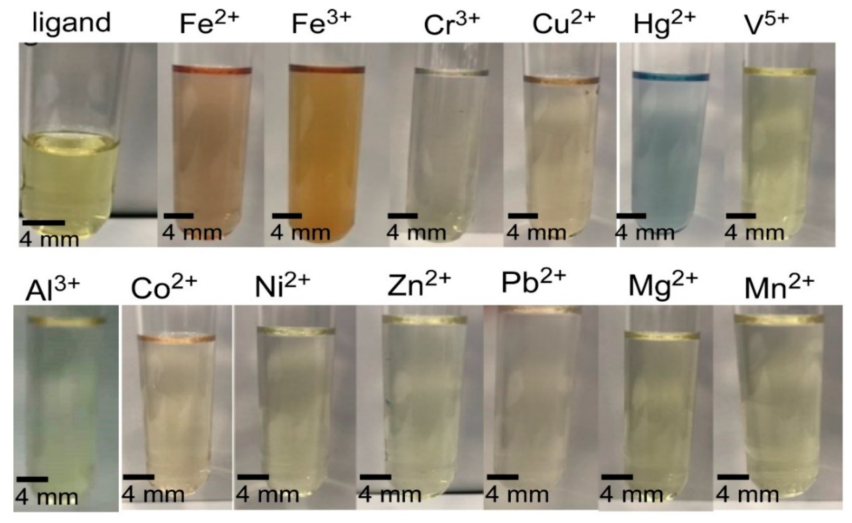
The sensing ability of SL1 (1 × 10⁻² M, prepared in DMF) toward various metal ions such as Cu2+, Cr3+, Fe2+, Ni2+, Co2+, Mg2+, Zn2+, Fe2+, Fe3+, NH4VO3 (V5+), Mn2+, Hg2+, Pb2+, and Al3 was monitored by the naked eye experiment. When the metal ion solution in H2O (1 × 10⁻² M) was added to the solution of the SL1 compound, the color changed from light yellow to light brown for Fe2+ and Fe3+, to purple for Cr3+, and to indigo blue for Hg+2, as shown in Figure 1. This observation indicates that these four metal ions successfully formed metal complexes with the Schiff base ligand, which may be due to the high affinity of the azomethine moiety and the adjacent mercapto group to these metal ions. The SL1 solution did not show any significant color change upon the addition of the other metal solutions of Cu2+, Co2+, Ni2+, Zn2+, Mg2+, Mn2+, V5+, and Pb2+. This could be explained by the Gibb’s free energy of the reaction for the formation of the metal–ligand (SL1) complex for these metals might not be sufficient for the randomness of the reaction and, therefore, has no influence on the formation of the metal–SL1-ligand complex and no colors were seen [24]. Therefore, SL1 was only able to sense Fe2+, Fe3+, Cr3+, and Hg+2 ions in aqueous solutions by giving a different color for each metal ion.
3.3. UV–Visible Monitoring
The Schiff base’s (SL1) behavior as a chemosensor was examined by monitoring the UV–Visible absorption spectra upon the addition of the different metal ions under investigation in a distilled water–DMF (1:1) solvent mixture at room temperature (Figure 2). First, we recorded the UV–Visible spectra of the free Schiff base ligand in DMF, which exhibited a broad band absorption centered at 365 nm (Figure 2a), assigned to n→π* transition due to the azomethine moiety [27]. Upon the addition of the examined metal ion solutions to the SL1 ligand, the absorbance intensity of SL1 at 365 nm decreased, a red shift (~69–288 nm) was observed, and new bands appeared at 434, 447, 570, and 653 nm after the addition of Fe+3, Fe+2, Cr+3, and Hg+2 solutions to the SL1 solution, respectively (Figure 2b). This bathochromic shift may be due to the ligand-to-metal charge-transfer (LMCT). The appearance of new peaks was due to the coordination between the Schiff base ligand and the metal ions that may take place [16]. The azomethine moiety increases the withdrawing character of the ligand, causing a stronger intramolecular charge transfer from the electron-donating group (-SH) to the metal ions in the complex [27]. This is the charge transfer from the ligand molecular orbitals to the partially filled metal d-orbitals, and this results in a reduction of the metal ions. The difference in the red shift for different metals may be due to the difference in metal ion size and the charge densities [27,28]. For other ions, the UV–Vis absorption spectra of SL1 exhibited no change because no complexes were formed in these cases. These observations suggest that SL1 can only act as a sensor for Fe+3, Fe+2, Cr+3, and Hg2+ metal ions.
3.4. Job’s Plot and Detection Limit
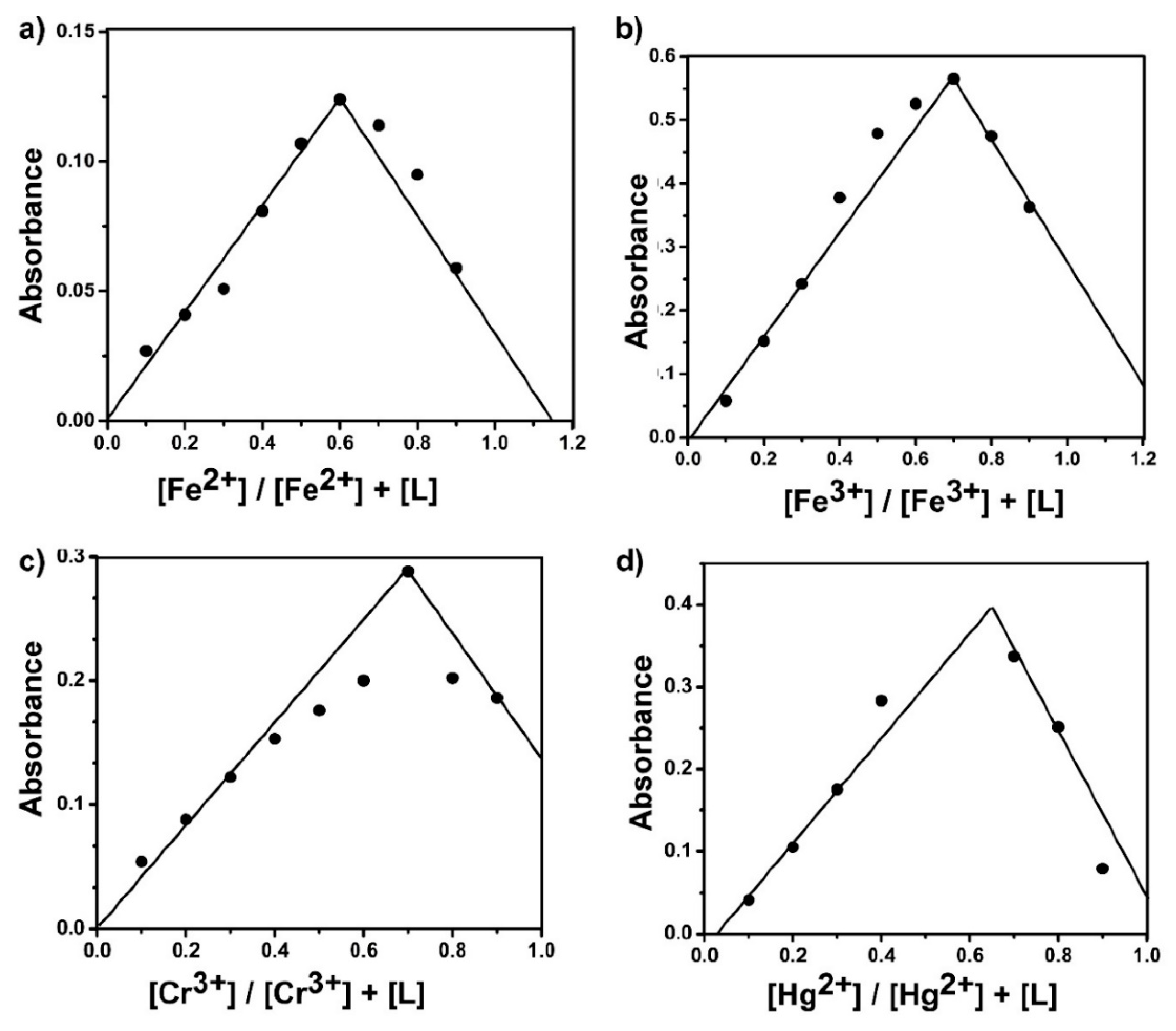
In order to study the sensitivity and the binding mechanism of Fe2+, Fe3+, Cr3+, and Hg2+ metal ions to the SL1 ligand, UV–Vis titration experiments were performed in DMF solution. For the stoichiometric ratio between the ligand SL1 and Fe2+, Fe3+, Cr3+, and Hg2+ metal ions for the formation of metal complexes, the Job’s plot continuous variation method was utilized (Figure 3) [29,30,31]. The method kept the total concentration of SL1 and metal ions at 0.01 M and changed the molar ratio of the metal ions from 0.1 to 1.0 M.
It can be seen from Figure 3 that the maximum absorbance was observed when the molar ratio of the ligand to metal was 0.66, indicating the formation of a 1:2 (Metal:Ligand) complex [6]. The possible binding mode of the ligand with the metal ions is through the coordination of the nitrogen atom (N) of the azomethine moiety and the sulfur atom (S) of the thiol group. Therefore, SL1 is a bidentate ligand and forms metal complexes with a possible structure as shown in Scheme 2.
The Job’s plots (Figure 3) show some significant deviation from linearity, which may be due to the formation of weak complexes and, hence, the stability constants of the complexes are affected [32]. Moreover, some mixtures of complex species cause displacement of maximum plots.
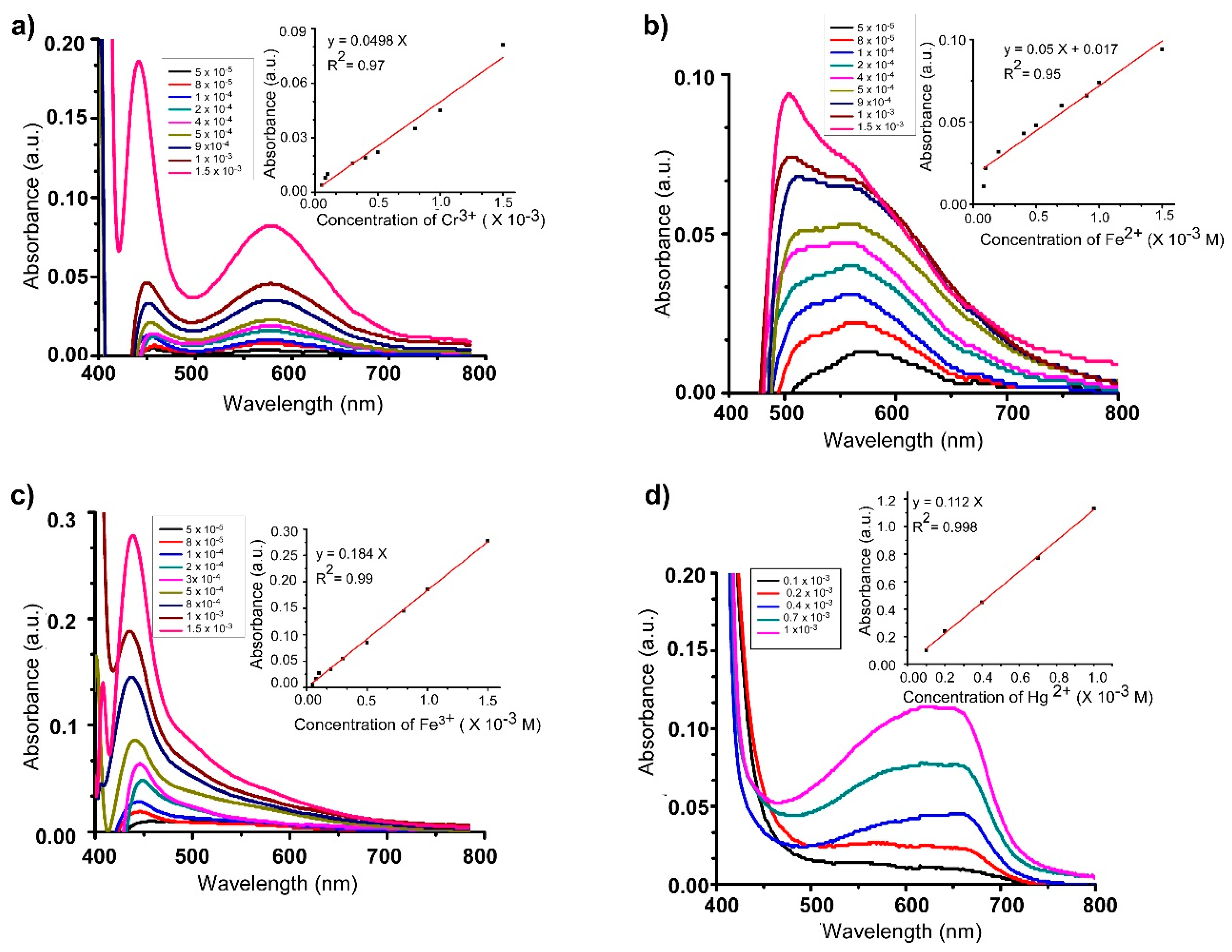
The UV–Vis spectrum of SL1 upon the addition of Fe2+, Fe3+, Cr+3, and Hg2+ is shown in Figure 4a–d, respectively It can be seen that increasing the concentration of the metal ions that were added to SL1 resulted in an increased absorbance intensity at the isosbestic points (i.e., specific wavelength) of 434 nm (Fe+3), 447 nm (Fe+2), 570 nm (Cr+3), and 653 nm (Hg+2). The detection limit of metal ions (Fe+3, Fe+2, Cr+3, and Hg2+) by SL1 was calculated from the plot of absorbance intensity versus the ion concentrations. Therefore, the detection limit for the metal ions was at 10−5 M. According to the World Health Organization (WHO-1984), 0.1 mg/L of iron, 0.05 mg/L of chromium (III), and 0.001 mg/L of mercury are present in drinking water [19]. Consequently, our ligand can be employed to recognize the tested metal ions of Fe+3, Fe+2, Cr+3, and Hg+2 at 10−5 M.
3.5. Comparison with Other Studies
Alizadeh et al. (2011) reported the successful use of the Schiff base 2-[(2-sulfanylphenyl)ethanimidoyl] phenol, a tridentate ligand with N, S, and O donor atoms, as a sensor for the selective monitoring of the Hg2+ ion by the chemical immobilization of the ligand on an agarose film membrane. The study revealed the successful application of a selective sensor for Hg2+ ions in an amalgam alloy and spiked water samples without any significant interference from other metal ions.
A novel Schiff base containing a pyrene ring with the thiol group adjacent to the azomethine group has been reported and used as an effective fluorescent probe for detecting Hg2+ in living cells via a chelation mechanism [33].
New rhodamine Schiff base sensors carrying the dithiocarbonate group have been shown to respond selectively to Hg2+ by showing a strong colorimetric change and intense fluorescence [29]. This was explained in part to be due to the preferential metal bonding of two sulfur atoms of the dithiocarbonate groups regardless of the main structure of rhodamine.
The reported Schiff base ligands previously mentioned have similar binding sites to our Schiff base ligand. Based on hard–soft trends, they are similar in having the soft sulfur atom of the thiol group and the intermediate hardness nitrogen atom of the azomethine group as binding sites to the metal ions. It seems to be that this system with a soft sulfur atom adjacent to the azomethine group demonstrated a high binding affinity to metal ions via a strong S–Mn+ interaction, resulting in unstable complexes and, hence, a color change [33].
4. Conclusions
In conclusion, a Schiff base ligand named 1-(2-thiophenylimino)-4-(N-dimethyl)benzene was prepared and used for the detection of multiple metal ions in solution. The synthesized Schiff base ligand exhibited a remarkable selectivity and sensitivity response toward four metal cations: Fe2+, Fe3+, Cr3+, and Hg2+. The investigation method used was UV–Visible spectra. A new peak was observed at 447 nm with a red shift of 82 nm in the case of Fe2+, a peak at 434 nm in the case of Fe3+ with a red-shift value of 69 nm, a peak at 570 nm with a red shift of 205 nm in the case of Cr3+, and a peak at 653 nm with a red shift of 288 nm in the case of Hg2+, accompanied by a color change that could be distinguished by the naked eye. The investigation of the Schiff base ligand binding to the metal ions was found to be a 2:1 ligand-to-metal ratio, according to Job’s plot and using a UV–Visible method as a direct approach to monitoring ligand–metal interactions.
Author Contributions
The three authors worked as a team and contributed equally throughout the research work including the design of the study; the experimentation, analysis and interpretation of data; preparation of the manuscript; writing, editing, and revising. The corresponding author A.Q.A. obtained funding from the Research Deanship at Al Baha University, and is the project administrator. All authors have read and agreed the published version of the manuscript.
Funding
The Research Deanship at Al Baha University funded this research (grant number 1439/20).
Acknowledgments
We gratefully acknowledged the financial support by Al Baha University (Project No: 1439/20), and are grateful to the Scientific Research Deanship and the Dean of the Faculty of Science at Al Baha University for their encouragement in our research and for the use of the laboratory facilities.
Conflicts of Interest
The funder, Al Baha University, had no role in the design of the study, in the execution of the research, in the interpretation or analysis of data, in the writing of the manuscript, or in the decision to publish the results.
References
- Bansod, B.; Kumar, T.; Thakur, R.; Rana, S.; Singh, I. A review on various electrochemical techniques for heavy metal ions detection with different sensing platforms. Biosens. Bioelectron. 2017, 94, 443–455. [Google Scholar] [CrossRef] [PubMed]
- Awual, M.R.; Hasan, M.M. Colorimetric detection and removal of copper (II) ions from wastewater samples using tailor-made composite adsorbent. Sens. Actuators B Chem. 2015, 206, 692–700. [Google Scholar] [CrossRef]
- Peralta-Domínguez, D.; Rodriguez, M.; Ramos-Ortiz, G.; Maldonado, J.; Luna-Moreno, D.; Ortiz-Gutierrez, M.; Barba, V. A Schiff base derivative used as sensor of copper through colorimetric and surface plasmon resonance techniques. Sens. Actuators B Chem. 2016, 225, 221–227. [Google Scholar] [CrossRef]
- Kundu, A.; Hariharan, P.; Prabakaran, K.; Anthony, S.P. Developing new Schiff base molecules for selective colorimetric sensing of Fe3+ and Cu2+ metal ions: Substituent dependent selectivity and colour change. Sens. Actuators B Chem. 2015, 206, 524–530. [Google Scholar] [CrossRef]
- Özdemir, Ö. Novel symmetric diimine-Schiff bases and asymmetric triimine-Schiff bases as chemosensors for the detection of various metal ions. J. Mol. Struct. 2016, 1125, 260–271. [Google Scholar] [CrossRef]
- Berhanu, A.L.; Mohiuddin, I.; Malik, A.K.; Aulakh, J.S.; Kumar, V.; Kim, K.-H. A review of the applications of Schiff bases as optical chemical sensors. TrAC Trends Anal. Chem. 2019, 116, 74–91. [Google Scholar] [CrossRef]
- Patil, A.; Salunke-Gawali, S. Overview of the chemosensor ligands used for selective detection of anions and metal ions (Zn2+, Cu2+, Ni2+, Co2+, Fe2+, Hg2+). Inorg. Chim. Acta 2018, 482, 99–112. [Google Scholar] [CrossRef]
- Nolan, E.M.; Lippard, S.J. Small-molecule fluorescent sensors for investigating zinc metalloneurochemistry. Acc. Chem. Res. 2008, 42, 193–203. [Google Scholar] [CrossRef] [Green Version]
- Wang, H.-H.; Xue, L.; Qian, Y.-Y.; Jiang, H. Novel ratiometric fluorescent sensor for silver ions. Org. Lett. 2009, 12, 292–295. [Google Scholar] [CrossRef]
- Wu, J.; Liu, W.; Ge, J.; Zhang, H.; Wang, P. New sensing mechanisms for design of fluorescent chemosensors emerging in recent years. Chem. Soc. Rev. 2011, 40, 3483–3495. [Google Scholar] [CrossRef]
- Azadbakht, R.; Keypour, H. A new Schiff base system bearing two naphthalene groups as fluorescent chemodosimeter for Zn2+ ion and its logic gate behavior. Spectrochim. Acta Part A Mol. Biomol. Spectrosc. 2012, 85, 293–297. [Google Scholar] [CrossRef] [PubMed]
- Yin, S.; Zhang, J.; Feng, H.; Zhao, Z.; Xu, L.; Qiu, H.; Tang, B. Zn2+-selective fluorescent turn-on chemosensor based on terpyridine-substituted siloles. Dyes Pigment. 2012, 95, 174–179. [Google Scholar] [CrossRef]
- Li, M.; Lu, H.-Y.; Liu, R.-L.; Chen, J.-D.; Chen, C.-F. Turn-on fluorescent sensor for selective detection of Zn2+, Cd2+, and Hg2+ in water. J. Org. Chem. 2012, 77, 3670–3673. [Google Scholar] [CrossRef] [PubMed]
- Yang, M.-H.; Thirupathi, P.; Lee, K.-H. Selective and sensitive ratiometric detection of Hg (II) ions using a simple amino acid based sensor. Org. Lett. 2011, 13, 5028–5031. [Google Scholar] [CrossRef]
- Bhalla, V.; Kumar, M.; Sharma, P.R.; Kaur, T. New fluorogenic sensors for Hg2+ ions: Through-bond energy transfer from pentaquinone to rhodamine. Inorg. Chem. 2012, 51, 2150–2156. [Google Scholar] [CrossRef]
- Alizadeh, K.; Parooi, R.; Hashemi, P.; Rezaei, B.; Ganjali, M.R. A new Schiff’s base ligand immobilized agarose membrane optical sensor for selective monitoring of mercury ion. J. Hazard. Mater. 2011, 186, 1794–1800. [Google Scholar] [CrossRef]
- Mergu, N.; Gupta, V.K. A novel colorimetric detection probe for copper (II) ions based on a Schiff base. Sens. Actuators B Chem. 2015, 210, 408–417. [Google Scholar] [CrossRef]
- Chang, I.J.; Choi, M.G.; Jeong, Y.A.; Lee, S.H.; Chang, S.-K. Colorimetric determination of Cu2+ in simulated wastewater using naphthalimide-based Schiff base. Tetrahedron Lett. 2017, 58, 474–477. [Google Scholar] [CrossRef]
- Udhayakumari, D.; Velmathi, S. Colorimetric chemosensor for multi-signaling detection of metal ions using pyrrole based Schiff bases. Spectrochim. Acta Part A Mol. Biomol. Spectrosc. 2014, 122, 428–435. [Google Scholar] [CrossRef]
- Narayanaswamy, N.; Govindaraju, T. Aldazine-based colorimetric sensors for Cu2+ and Fe3+. Sens. Actuators B Chem. 2012, 161, 304–310. [Google Scholar] [CrossRef]
- Sivaraman, G.; Sathiyaraja, V.; Chellappa, D. Turn-on fluorogenic and chromogenic detection of Fe (III) and its application in living cell imaging. J. Lumin. 2014, 145, 480–485. [Google Scholar] [CrossRef]
- Saluja, P.; Sharma, H.; Kaur, N.; Singh, N.; Jang, D.O. Benzimidazole-based imine-linked chemosensor: Chromogenic sensor for Mg2+ and fluorescent sensor for Cr3+. Tetrahedron 2012, 68, 2289–2293. [Google Scholar] [CrossRef]
- Zamfir, G.; Stanica, N.; Draghici, C.; Kriza, C.A.A. Some Transitional Metal Complexes of 4-dimethylaminobenzilidene-2 mercaptoaniline. Rev. Chim. 2012, 63, 1176–1180. [Google Scholar]
- Jeong, U.; Kim, Y. Colorimetric detection of heavy metal ions using aminosilane. J. Ind. Eng. Chem. 2015, 31, 393–396. [Google Scholar] [CrossRef]
- Arunachalam, S.; Priya, N.P.; Jayabalakrishnan, C.; Chinnusamy, V. Ruthenium (II) Schiff base: complexes, physico-chemical, spectrometric, microbial and DNA binding and cleaving studies. Int. J. Appl. Biol. Pharm. Technol. 2011, 2, 110–122. [Google Scholar]
- Santos-Figueroa, L.E.; Moragues, M.E.; Climent, E.; Agostini, A.; Martínez-Máñez, R.; Sancenon, F. Chromogenic and fluorogenic chemosensors and reagents for anions. A comprehensive review of the years 2010–2011. Chem. Soc. Rev. 2013, 42, 3489–3613. [Google Scholar] [CrossRef] [PubMed]
- Gupta, V.K.; Singh, A.; Ganjali, M.; Norouzi, P.; Faridbod, F.; Mergu, N. Comparative study of colorimetric sensors based on newly synthesized Schiff bases. Sens. Actuators B Chem. 2013, 182, 642–651. [Google Scholar] [CrossRef]
- Bourson, J.; Pouget, J.; Valeur, B. Ion-responsive fluorescent compounds. 4. Effect of cation binding on the photophysical properties of a coumarin linked to monoaza-and diaza-crown ethers. J. Phys. Chem. 1993, 97, 4552–4557. [Google Scholar] [CrossRef]
- Heo, G.; Lee, D.; Kim, C.G.; Do, J.Y. Rhodamine spirolactam sensors operated by sulfur-cooperated metal complexation. Spectrochim. Acta Part A Mol. Biomol. Spectrosc. 2018, 188, 285–290. [Google Scholar] [CrossRef]
- Luthern, J.; Peredes, A. Determination of the stoichiometry of a thermochromic color complex via Job’s method. J. Mater. Sci. Lett. 2000, 19, 185–188. [Google Scholar] [CrossRef]
- Feng, G.; Geng, L.; Wang, T.; Li, J.; Yu, X.; Wang, Y.; Li, Y.; Xie, D. Fluorogenic and chromogenic detection of biologically important fluoride anion with schiff-bases containing 4-amino-1, 8-naphthalimide unit. J. Lumin. 2015, 167, 65–70. [Google Scholar] [CrossRef]
- Bosque-Sendra, J.M.; Almansa-López, E.; García-Campaña, M.; Cuadros-Rodríguez, L. Data analysis in the determination of stoichiometries and stability constants of complexes. Anal. Sci. 2003, 19, 1431–1439. [Google Scholar] [CrossRef] [PubMed] [Green Version]
- Shellaiah, M.; Rajan, Y.C.; Balu, P.; Murugan, A. A pyrene based Schiff base probe for selective fluorescence turn-on detection of Hg2+ ions with live cell application. New J. Chem. 2015, 39, 2523–2531. [Google Scholar] [CrossRef]
Scheme 1.
Synthesis of Schiff base ligand (SL1).
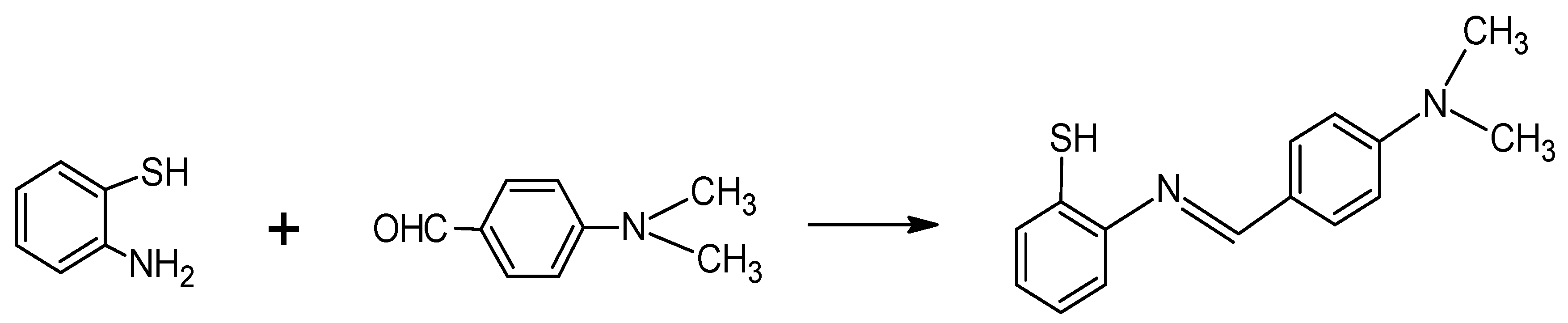
Figure 1.
Color change of SL1 (1 × 10⁻² M in DMF) before and after the addition of the metal ions of Cu2+, Cr3+, Fe2+, Ni2+, Co2+, Mg2+, Zn2+, Fe2+, Fe3+, NH4VO3 (V5+), Mn2+, Hg2+, Pb2+, and Al3 (1 × 10⁻² M solution in H2O).
Figure 1.
Color change of SL1 (1 × 10⁻² M in DMF) before and after the addition of the metal ions of Cu2+, Cr3+, Fe2+, Ni2+, Co2+, Mg2+, Zn2+, Fe2+, Fe3+, NH4VO3 (V5+), Mn2+, Hg2+, Pb2+, and Al3 (1 × 10⁻² M solution in H2O).

Figure 2.
(a) UV–Vis spectra of SL1 (1 × 10⁻³ M, in DMF) and (b) UV–Vis spectra of SL1 upon titration with aqueous solution of cations (SL1 + Al3+, SL1 + Cr3+, SL1 + Cu2+, SL1 + Fe2+, SL1 + Fe3+, SL1 + Hg2+, SL1 + Mg2+, SL1 + Mn2+, R + Ni2+, SL1 + Pb2+, SL1 + Zn2+, and R + V5).
Figure 2.
(a) UV–Vis spectra of SL1 (1 × 10⁻³ M, in DMF) and (b) UV–Vis spectra of SL1 upon titration with aqueous solution of cations (SL1 + Al3+, SL1 + Cr3+, SL1 + Cu2+, SL1 + Fe2+, SL1 + Fe3+, SL1 + Hg2+, SL1 + Mg2+, SL1 + Mn2+, R + Ni2+, SL1 + Pb2+, SL1 + Zn2+, and R + V5).
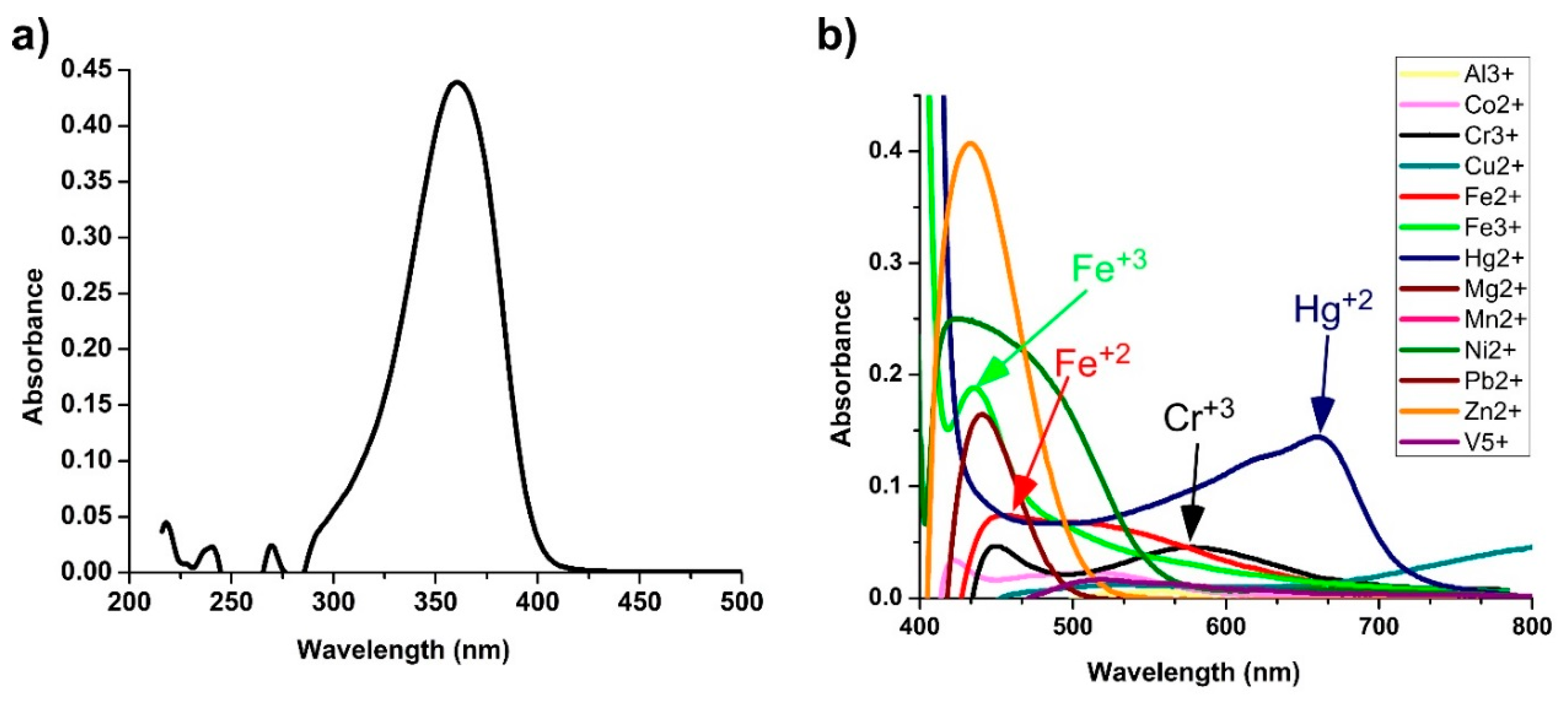
Figure 3.
Job’s plot of (a) SL1 and Fe2+ complex, (b) SL1 and Fe3+ complex, (c) SL1 and Cr3+ complex, and (d) SL1 and Hg2+ complex.
Figure 3.
Job’s plot of (a) SL1 and Fe2+ complex, (b) SL1 and Fe3+ complex, (c) SL1 and Cr3+ complex, and (d) SL1 and Hg2+ complex.

Scheme 2.
Possible metal complex structure.
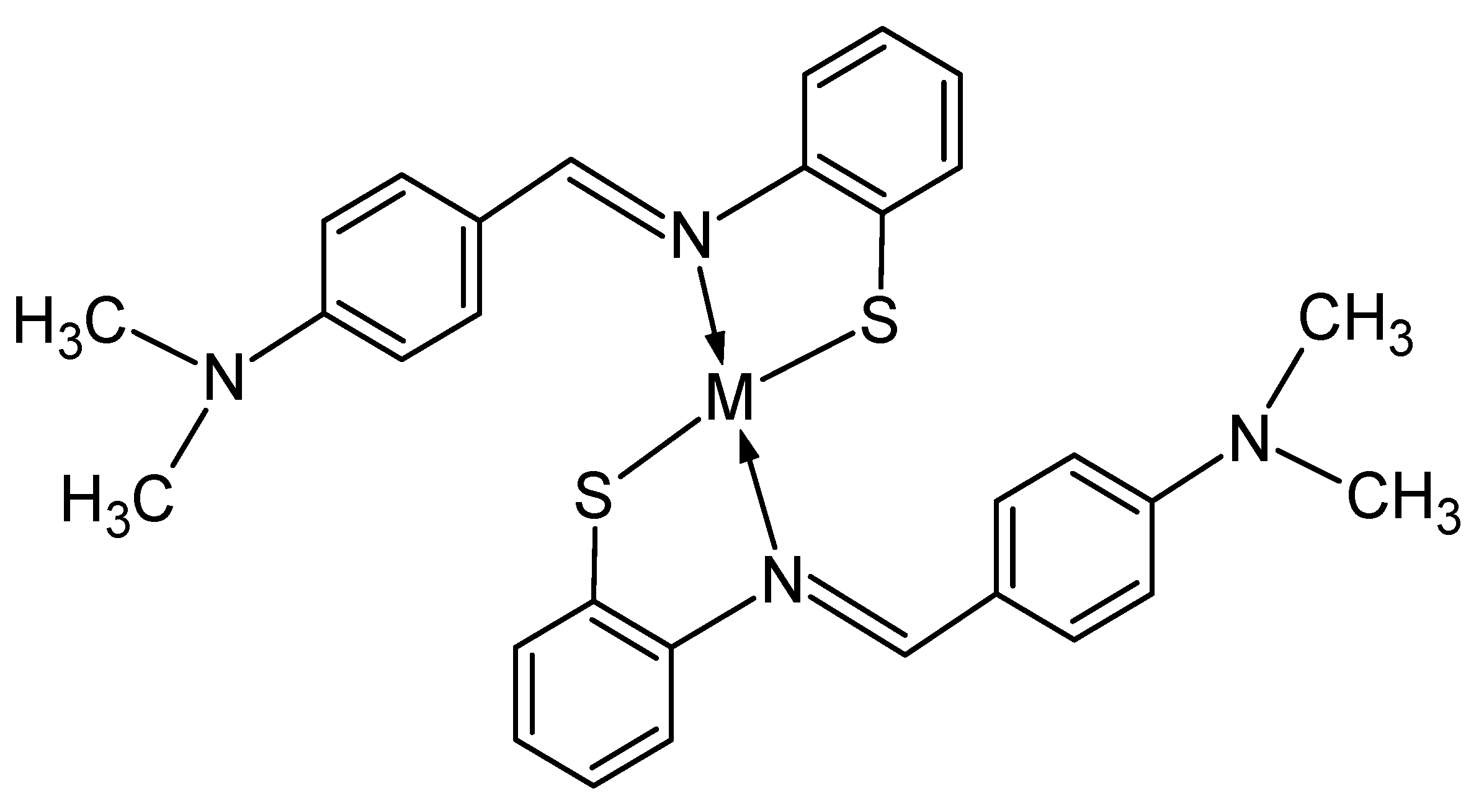
Figure 4.
(a) UV–Vis absorption spectroscopic titration of SL1 (1 × 10⁻² M) with various concentrations of Cr3+ in DMF solution (λmax = 570 nm)—inset: changes of UV–Vis absorption intensity upon the addition of Cr3+ at 570 nm; (b) UV–Vis absorption spectroscopic titration of SL1 (1 × 10⁻² M) with various concentrations of Fe2+ in DMF solution (λmax = 447 nm)—inset: changes of UV–Vis absorption intensity upon the addition of Fe2+ at 447 nm; (c) UV–Vis absorption spectroscopic titration of SL1 (1 × 10⁻² M) with various concentrations of Fe3+ in DMF solution (λmax = 434 nm)—inset: changes of UV–Vis absorption intensity upon the addition of Fe3+ at 434 nm; and (d) UV–Vis absorption spectroscopic titration of SL1 (1 × 10⁻² M) with various concentrations of Hg2+ in DMF solution (λmax = 653 nm)—inset: changes of UV–Vis absorption intensity upon the addition of Cr3+ at 653 nm.

© 2019 by the authors. Licensee MDPI, Basel, Switzerland. This article is an open access article distributed under the terms and conditions of the Creative Commons Attribution (CC BY) license (http://creativecommons.org/licenses/by/4.0/).
Share and Cite
MDPI and ACS Style
Alorabi, A.Q.; Abdelbaset, M.; Zabin, S.A. Colorimetric Detection of Multiple Metal Ions Using Schiff Base 1-(2-Thiophenylimino)-4-(N-dimethyl)benzene. Chemosensors 2020, 8, 1. https://doi.org/10.3390/chemosensors8010001
AMA Style
Alorabi AQ, Abdelbaset M, Zabin SA. Colorimetric Detection of Multiple Metal Ions Using Schiff Base 1-(2-Thiophenylimino)-4-(N-dimethyl)benzene. Chemosensors. 2020; 8(1):1. https://doi.org/10.3390/chemosensors8010001
Chicago/Turabian StyleAlorabi, Ali Q., Mohamed Abdelbaset, and Sami A. Zabin. 2020. "Colorimetric Detection of Multiple Metal Ions Using Schiff Base 1-(2-Thiophenylimino)-4-(N-dimethyl)benzene" Chemosensors 8, no. 1: 1. https://doi.org/10.3390/chemosensors8010001
Note that from the first issue of 2016, this journal uses article numbers instead of page numbers. See further details here.