Article

Microstructure and Photoluminescence of ZrTiO4:Eu3+ Phosphors: Host-Sensitized Energy Transfer and Optical Thermometry

1
State Key Laboratory of Luminescence and Applications, Changchun Institute of Optics, Fine Mechanics and Physics, Chinese Academy of Sciences, 3888 Dong Nanhu Road, Changchun 130033, China
2
Center of Materials Science and Optoelectronics Engineering, University of Chinese Academy of Sciences, Beijing 100049, China
*
Authors to whom correspondence should be addressed.
Chemosensors 2022, 10(12), 527; https://doi.org/10.3390/chemosensors10120527
Submission received: 19 October 2022 / Revised: 12 November 2022 / Accepted: 16 November 2022 / Published: 12 December 2022
(This article belongs to the Special Issue Advances in Fluorescence Sensing)

Abstract

:
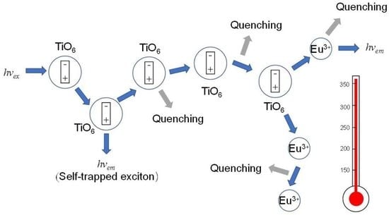
Orthorhombic ZrTiO4 is an attractive dielectric material; its optical properties are, however, less known. In this paper, we reported on the microstructure and luminescence studies of pristine ZrTiO4 and Eu3+-doped ZrTiO4 phosphors. The results indicated that two types of TiO6 octahedra, the isolated/ localized and coupled/delocalized, coexisted in host matrix. Eu3+ doping could induce oxygen vacancy defect states located below the bottom of the conduction band. Pristine ZrTiO4 showed bright yellow luminescence via STEs recombination at defects sites at low temperatures, but significant thermal quenching occurred due to STEs migration to quenching centers at elevated temperatures. Effective host sensitized energy transfer to Eu3+ was observed in ZrTiO4:Eu3+ phosphors and yielded the red characteristic emissions of Eu3+. Anomalous STEs luminescence enhancement and spectral blue-shift in the excitation spectra with higher Eu3 + concentration appeared and were explained by considering three factors: competitive absorption between electron transitions from the top of the valence band to the defect states and host conduction band, Eu3+ doping driving the production of more isolated TiO6 octahedra, and energy back-transfer from Eu3+ activators to other titanate groups. On the basis of the dual-emitting combination strategy involving host STEs and Eu3+ luminescence, ZrTiO4:Eu3+ phosphors were demonstrated to be ratiometric self-referencing optical thermometric materials, with a working range of 153–313 K and a maxima of relative sensitivity to ~1.1% K−1 at 243 K.

Graphical Abstract

1. Introduction

Solid state luminescent materials or phosphors have a wide range of applications in laser, display, lighting, detection, and sensing technologies [1,2,3,4,5]. Such materials are typically comprised of a host that is doped with a small quantity of activators or dopants (typically including transition metal (TM) ions and lanthanide ions). For inorganic crystalline materials, the host lattice could provide a certain crystallographic symmetry of the surroundings for the activators and then modulate their luminescence properties. Achieving new types of phosphors and finding potential applications for them remains a challenge and is of great scientific and technical significance. In this connection, the searching for a novel host and the following effective doping are both essential.
Bulk zirconium titanate (ZrTiO4), one ternary compound in the ZrO2−TiO2 system, is of technological importance in microwave telecommunications (as a capacitor, dielectric resonator in filters, and oscillator), memory devices (ReRAM), humidity/gas sensors, and structural ceramic due to its high dielectric constant at microwave frequencies, high corrosion resistance, and excellent thermal stability [6,7,8,9]. Great interest has been generated in studying its microstructure, phase transformation, thermal expansivity, and dielectricity [9,10,11,12,13]. However, in terms of its use as a luminescent material or particularly as a host of accommodating activators, only minimal research has been conducted to date. A weak blue-white broadband photoluminescence was observed on the nanocrystalline products of ZrTiO4 after heating treatment of its chemical precursor (dry-gel), but no photoluminescence was elucidated in the crystalline phase [14,15]. In this paper, we reported on the optical properties of ZrTiO4 powders prepared by the solid-state reaction method, including the self-activated and doping-induced photoluminescence upon hosting trivalent lanthanide (Ln3+) ions of Eu3+. It was found that the pristine ZrTiO4 shows bright yellow host luminescence at a low temperature due to the recombination of self-trapped excitons (STEs) at defect sites. Efficient host-mediated resonance energy transfer (Förster type) from STEs to Eu3+ was observed upon further substitution of Eu3+, yielding the characteristic red luminescence. STEs luminescence and STEs-mediated energy transfer to Ln3+ were investigated extensively in lanthanide-doped vanadates, tungstates, borates, and niobates [16,17,18,19]. It is interesting that in these crystals, the STEs were usually identified with a localized excitation of the oxy-anionic complexes (for instance, VO43−, BO33−) [17,18,19]. Herein, the emission of STEs was considered to be located in the TiO68− complexes but with a large amount of delocalization, which is in accordance with previous reports on other titanates (SrTiO3, BaTiO3, La2TiO5) [19,20,21].
Temperature is one of the fundamental thermodynamic properties and an important parameter in industry and science communities [22]. Appling luminescent materials to temperature sensing (i.e., optical thermometry) has been the subject of world-wide interest due to their fast response and noninvasive features as well as their potential in harsh environments in comparison to traditional techniques derived from variations in thermal expansion, electrical resistance, capacitance, and thermoelectricity [2,3,4,5,23,24]. Based on the temperature-dependent optical spectroscopic characteristics of phosphors, including fluorescence intensity or intensity ratio (FIR), bandwidth, band shape, polarization, spectral shift and lifetime, and many others, a variety of luminescent sensing materials have been developed in recent years [2,3,4,5,25,26,27]. Of particular interest is the FIR technique, a ratiometric luminescence thermometry involving the intensity of two luminescence bands, which is fundamentally more resistant to the external undulations (e.g., pumping power, sample size) than those based on the single band intensity, peak position, and the bandwidth, thus being capable of probing the temperature more accurately [2,22]. Obviously, high performance FIR based optical thermometry relies on two temperature-sensitivity- distinct luminescent bands, which could originate from one and the same luminescent centers or respectively from two different centers. The luminescence from TM ions (e.g., V5+,4+,3+, Ti3+,4+, Cr3+,4+, Mn2+,3+,4+, Fe3+, Cu2+ with higher and larger activation energies) via d-d crystal-field transitions or TM(d0)→O2− charge transfer (CT) transitions is characterized by strong absorption and emission bands, and is highly susceptible to temperature variations in many cases due to bared d-orbitals and strong electron-lattice coupling [2]. They have been widely used in luminescence thermometry. In contrast, Ln3+ (e.g., Eu3+, Er3+, Nd3+ and Dy3+) could show more temperature-inert narrow emissions, via 4f-4f intraconfigurational electronic transitions, due to the weak electron–phonon coupling as result of the good shielding of a partly-filled 4fn shell by the outer 5s and 5p orbitals [2,22]. In addition, narrow emission band/peaks could facilitate spectral separation of bands during spectral measurements [2]. It is thus expected that the combination of the luminescence of TM and Ln3+ ions in one material is particularly well-suited for luminescence thermometry, and could even yield highly sensitive FIR-based optical thermometry by using Ln3+ luminescence as a self-referencing standard. An overview of the state of the art of advance was reviewed recently by L. Marciniak [2]. In particular, this was exampled by LaGaO3:V5+,4+,3+, Nd3+, and YVO4:Er3+ phosphors, where the temperature-sensitive broad band luminescence of CT transition (V5+→O2−), and d-d transitions of 2E→2T2 (V4+), and 3T1g3T1g (V3+) were utilized as temperature indicators, while the line-like emissions of 4F3/24I9/2 transitions of Nd3+ or 2H11/2/4S3/24I15/2 transitions of Er3+ were utilized as luminescent references [28,29]; other examples included Cr3+, Nd3+ co-doped LiLaP4O12, Cr3 +, Ln3+ (Ln3+ = Nd3+, Dy3+, Eu3+) co-doped garnet phosphors (Gd3Ga5O12, Y3Al5O12). Likewise, the broad band emissions of d-d transitions of 4T24A2 of Cr3+ served as temperature signals, the corresponding internal reference signals were from Ln3+ (Nd3+, 4F3/24I11/2; Dy3+, 4F9/26H9/2,11/2,13/2; Eu3+, 5D07F1) [30,31,32]. A high relative sensitivity of Sr = 4.89%/K at 323 K and sensitivities Sr > 15%/K for temperatures in the 423–473 K range was achieved in LiLaP4O12: Cr3+, Nd3+ due to the efficient thermal quenching of the broad band luminescence of Cr3+ [32]. It is worth noting that in many cases the two luminescent bands for the FIR were obtained upon single wavelength excitation into the TM ions followed by energy transfer to Ln3+.
The TMs of titanium (Ti), with two oxide states of Ti3+ (3d1) and Ti4+ (3d0), were considerably less-used in luminescence thermometry, and there only exist a few reports on this when examining the literature. An earlier study in 2007 was presented by Katsumata et al. [33] on the luminescence of Ti3+ ions in sapphire (Al2O3) crystal; they found a sublinear dependence of luminescence intensity against temperature and further demonstrated them as luminescent temperature sensors on the basis of a single emission band of crystal field transition 2Eg2T2g of Ti3+. Recently, there has been increasing interest in exploiting Ti ions-doped powder materials toward optical thermometry, and especially in developing the FIR-based thermometry by combining Ti3+/4+ ions with the luminescent centers of Ln3+. Examples are Y3Al5O12:Ti4+, Ln3+ (Ln3+ = Nd3+, Eu3+, Dy3+) by Drabik et al. [34], LiTaO3:Ti4+, Eu3+ by Wang et al. [35], and ZrO2:Ti4+, Eu3+ by us [22]. Note that in these studies, Ti plays a role of the impurity ions (dopant) at low concentrations rather than that of the host constituent. In contrast, a host of titanate (100% concentration of Ti4+) of Gd2Ti2O7 was developed by M. D. Dramićanin et al. [36] for luminescence thermometry upon introducing Eu3+ into the lattice, where the temperature indicator of the host luminescence was considered to be the trap emission induced by the oxygen vacancies–a lower relative sensitivity of Sr = 0.46%K−1 at 423 K was obtained. In this study, we demonstrated the new type of titanate phosphors of Eu3+-doped ZrTiO4 as FIR optical thermometry by combining the temperature-sensitive STEs broad emissions (Ti4+→O2− CT type) of the host lattice and somewhat temperature-inert sharp emissions of the Eu3+ activator. The temperature sensing was achieved upon excitation at the absorption edge followed by host-sensitized energy transfer to Eu3+, and covered a working range of 153–313 K with a maximum relative sensitivity of ~1.1%K−1.

2. Experimental

2.1. Chemicals and Materials Preparation

All chemicals used in this study were of analytical grade and were used without further purification. High purity starting oxides ZrO2 (99.99%, metals basis, without Hf or HfO2), TiO2 (99.8%, anatase, metals basis), and Eu2O3 (99.999%) were supplied by Aladdin Industrial Inc., China. The samples of Zr1−xEuxTiO4 (x = 0, 0.0001, 0.0002, 0.0005, 0.001, 0.002, 0.005, 0.01, 0.05) were prepared by the high-temperature solid phase method. The starting materials according to the stoichiometric proportion were weighed, mixed, and were then ball-milled for 12 h by using a high-energy ball milling machine (Fritsch pulverisette 4, Germany) with ZrO2 grinding balls and methyl alcohol as the dispersant. The resulting slurry was then dried in an oven at 60 ℃ in air, followed by calcining in a high-temperature muffle furnace at 1300 ℃ for 4 h in air.

2.2. Characterization and Measurements

The phase purity and crystalline structure were checked by X-ray power diffraction (XRD) techniques on a powder diffractometer (D8 Advance, Bruker, Billerica, MA, USA), which was operated at 40 kV and 30 mA in the range from 20° to 65° with Cu-Kα radiation. The morphology and energy dispersive X-ray spectroscopy (EDS) were characterized by scanning electron microscopy (SEM, S-4800, Hitachi, Tokyo, Japan). The diffuse reflectance (DR) spectra in the 200–800 nm region were recorded by using a dual beam UV-vis-NIR spectrophotometer (UV-3600 plus, Shimadzu, Kyoto, Japan) equipped with an integrating sphere assembly (150 nm), with BaSO4 as a reference standard. Raman spectra were acquired on an Infinity Raman spectrometer (Horiba Jobin Yvon, Paris, France) with a 488 nm laser excitation source (He-Cd laser, Wavicle, Beijing, China). Steady state photoluminescence (PL) spectra were recorded on a FLS 920 spectrofluorometer (Edinburgh Instruments, Edinburgh, UK) with a 450 W xenon lamp as the excitation source. The emission spectra were corrected to the instrument responses. The fluorescence decays in the microsecond and millisecond scales were conducted by using an optical parametric oscillator (OPO) as the pulse excitation source, and the signals were analyzed by using a spectrometer (Triax550, Horiba, Paris, France) and a digital oscilloscope (TDS3052, Tektronix, Beaverton, OR, USA), while those in the nanosecond scale were obtained by using the time-correlated single-photon counting method on a spectrofluorometer (FLS 920, Edinburgh Instruments, UK) equipped with a photon counting detector of photomultiplier tube (PMT) R928 and a hydrogen flash lamp as the excitation source. For temperature-dependent (153–313 K) PL measurements, the powdered samples were placed on a heating and cooling stage (THMS-600, Linkam, Salfords, UK) mounted on the objective table of an upright fluorescence microscope (BX53M, Olympus, Tokyo, Japan) jointed sideways with a fiber optic spectrometer (QEPro, Ocean Optics, Largo, FL, USA). An excitation irradiation in the range of 340–370 nm from a mercury burner lamp (130 W) was selected by a band-pass filter. Fluorescence signals were collected with an objective lens and focused onto the entrance of the distal end of the combined fiber of the spectrometer. Both illuminations of the surface and detection of the fluorescence were performed along an axis coincident with the surface normal. A cooled digital color camera (DP74, Olympus, Tokyo, Japan) equipped on the microscope was used for fluorescence microscopy and photograph capture. All the measurements were carried out at room temperature unless otherwise noted.

3. Results and Discussion

3.1. Composition, Crystalline Structure and Morphology

Zirconium titanate may exist in a number of modifications in the ZrO2−TiO2 phase diagram. Zirconium orthotitanate of ZrTiO4, with equivalent amounts of Ti4+ and Zr4+ ions in the composition, is one of the stable ternary compounds [11]. It has the orthorhombic α-PbO2-type structure with Pbcn space group, where Ti4+ and Zr4+ ions are both randomly distributed on an octahedral site of 4c2 (Wyckoff notation)–which are quite disordered [7,11]. This structure is extremely similar to those of TiO2 polymorphs consisting of edge-shearing TiO6 octahedra [7,13]. The phase purity of Zr1−xEuxTiO4 powders under investigation was checked by XRD analysis, as was shown in Figure 1a together with the diffraction lines of a reference ZrTiO4 (PDF#74-1504). It was found that at the lower doping concentration (x ≤ 0.01) of Eu3+, all the peaks of the diffraction pattern of the samples correspond the ZrTiO4 phase. No evidence of the presence of any peaks of impurities was observed. A small quantity of impurities, however, appeared in the samples at higher doping concentration, as was indicated by asterisks in the figure for a typical sample of Zr1−xEuxTiO4 (x = 0.05). This was tentatively assigned to the Zr5Ti7O24 phase. Semi-quantitative EDS analysis (see Figure S1) confirmed the presence of Eu3+ in the sample when its doping concentration was higher (x = 0.05). The presence of such impurities was thus attributed to the aliovalent substitution, where the trivalent Eu3+ located on a site normally occupied by a tetravalent host ion of Zr4+, leaving an effective negative charge at that site and creating 0.5 positively charged extrinsic oxygen vacancy for charge compensation. The incorporation reaction can be expressed as:
Eu 2 O 3 ZrTiO 4 2 Eu Zr + V O + 3 O O ×
where an oxygen vacancy can compensate two Eu3+ ions either locally or distantly. It was considered that a larger mismatch of ionic radius of between Eu3+ (0.95 Å, CN = 6) and Zr4+ (0.72 Å, CN = 6) and a higher concentration of oxygen vacancy induced by the incorporation of more Eu3+ caused the great distortion of lattices and then the occurrence of phase change. Additionally, when increasing the doping level (x) of Eu3+ ions, the (111) diffraction peaks gradually shift toward larger 2θ angles (the right part of Figure 1a), suggesting a contraction of the lattice cell upon Eu3+ substitution due to the presence of oxygen vacancy defects.
Raman spectroscopy is a powerful tool to determine the phase compositions of many transition metal oxides; herein, this technique was employed to distinguish the phase purity of the samples as prepared. In term of the space group of Pbcn- D 2 h 14 , 18 Raman-active could be predicted for ZrTiO4 [14,35]. However, only 12 Raman bands are generally observed. As shown in Figure 1b, in our samples, only five bands were significantly distinguished in the range of 200–1000 cm−1 due to their occurrence at lower wavenumbers, their overlapping of the band, or due to them being too weak to be observed for the unobserved modes/bands. The three bands centered at 640, 404, and 267 cm−1 are attributed to vibrational modes A1g, Eg, and F2g, respectively, of Ti(Zr)O6 octahedron, and the two bands at 327 and 799 cm−1 are considered to arise from vibration modes E and A1, respectively, of Ti(Zr)O4 [14,35]. A clear shift of the Raman band with the higher Eu3+ concentration was found upon taking a closer look–particularly, the F2g mode shifted to lower wavenumbers while A1g and Eg moved to a higher frequency region when increasing the x value. In addition, an extra band appeared at 602 cm−1 for the Zr0.95Eu0.05TiO4. Such spectral evolutions could be interpreted as being due to the presence of lattice distortion and impurity constituents caused by Eu3+ doping, which results in the variation of bond length and/or force constant of bond. These results coincided with the above analysis by XRD.
Figure 2 depicts the SEM images of the obtained samples. For the pristine ZrTiO4, a single particle is characteristic of a quasi-sphere shape and has a size in the range of ~0.6—1 μm. Some neighboring particles are connected through necks and exhibit dendritical growth from the nucleation centers. It was found that the starting materials of Eu2O3 played another role of sintering flux and favored the growth of ZrTiO4 crystallites. Upon increasing the Eu3+ concentration, the size of the resulting phosphors constantly increased and approached to ~2 μm or more at x ≥ 0.01; serious agglomeration and more irregular morphology were clearly observed at higher Eu3+ concentrations. Surface diffusion and grain boundary diffusion of the various elements in the samples, which mainly controlled the formation and growth of the sintering necks, is expected to cause significant changes in the size and morphology during high-temperature calcinations [37,38]. It is apparent that additions of a small amount of Eu2O3 enhanced such microscopic diffusion processes, and then drove the solid-state reaction of different starting materials; further aggregation/coalescence and growth of crystallites thus arose.

3.2. Electronic Band Structure of Zr1−xEuxTiO4

In the systems of oxides typically containing closed-shell TM ions (TM4+/5+/6+, nd0) and oxygen anions (O2−, 2p6), including simple binary compounds (e.g., TiO2, ZrO2, Nb2O5, Ta2O5, WO3) and ternary compounds (various titanates, niobates, tantalates, tungstates and molybdates), their band gaps are commonly of ligand-metal CT type with the character of the upper part of the valence band (VB) mainly constituted with the oxygen 2p states, while the bottom of the conduction band (CB) is predominantly formed by the empty d states of the TM ions [39,40,41]. The fundamental optical absorption across the band gap occurs in the TMOx complex polyhedron, involving an optical excitation of electrons from one of the highest filled molecular orbitals, which are localized on the O2− ions (2p orbitals), to one of the lowest empty molecular orbitals, which are mainly localized on the TM4+/5+/6+ ion (t2g orbital) [39,40,41]. For ZrTiO4, as demonstrated above, it comprises two types of closed-shell TM ions (3d0/Ti4+, 4d0/Zr4+) and then two kinds of TMO6 octahedra (TiO6/ZrO6), so the band gap was also considered to be the charge transfer type of O2−→Ti4+ or O2−→Zr4+. It is worthwhile to note that the fourth ionization potential (coulomb binding energy) of Ti4+ (43.27 eV) is ~8.93 eV higher in energy than that of Zr4+ (34.34 eV) [42]. According to the model suggested by Blasses et al. [39,40,41], the band gap should be mainly of the O2−→Ti4+ CT type. Such an inference was confirmed by the DFT calculation on the electronic structure of ZrTiO4 recently made by E. Giamello et al. [7], which further indicated that ZrTiO4 has the nature of the direct band gap.
The band gap of ZrTiO4 was calculated according to Tuac’s method [43]; the absorption coefficient α for the direct band gap materials is given by:
α · h υ = B h υ E g m
where h is the Planck constant, ν is the frequency of photon, Eg is the band gap energy, B is an energy independent constant, and m is an index that depends on the nature of electronic transition responsible for the optical absorption. The value of m for the allowed direct transition is 0.5. For powdered samples, usually only the diffuse reflectance spectra could be measured; an inverse relation between the reflectance and absorption enables the α to be approximately given using the Kubelka–Munk function [44]:
α S F R = 1 R 2 2 R
where R is the reflectance of an infinitely thick specimen; S is defined as the scattering factor, which becomes constant when the thickness of the sample is much larger than the size of the individual particles. The absorption spectra (F(R)) of Zr1−xEuxTiO4 against wavelengths were shown in Figure 3, where a steep optical absorption edge followed by a peak at ~320 nm was observed for ZrTiO4. The optical band gap energy for ZrTiO4 was evaluated to be ~3.65 eV (~340 nm) with Tauc’s plot (the inset of Figure 3), which is analogous to previous reports [7,14,45].
In addition, an extra absorption band lying well in the transparent region of crystal (i.e., below the absorption edge, see Figure 3) could be distinguished for the samples doped with Eu3+ at concentration above x = 0.01, which dramatically intensified with the increase in Eu3+ concentration. At x = 0.05, the onset of the absorption has shifted from ~350 nm for pure ZrTiO4 to ~404 nm in the visible spectral region. This band should arise from the impurities or defects in the lattice matrix. As is well known, the introduction of Eu3+ impurities in one host lattice could give rise to additional absorption bands originating from the O2−→Eu3+ CT process and electronic transitions within the 4f6 shell. However, the former was commonly characterized by a strong broad band in the higher energy of the ultraviolet (UV) region whereas the latter by weak and sharp peaks. The extra absorption band observed, therefore, arose from the defects. As demonstrated above, Eu3+ incorporation yielded extrinsic oxygen vacancies for charge compensation. Such defects were thus identified as oxygen vacancies; the absorption originated from the transition of electron from the top of the valence band to the defect levels lying below the bottom of the conduction band.
It is known that optical excitation across the band gap of the crystal lattice could yield free charge carriers (electron, hole) or electron-hole pairs (exciton), which is subject to the competition of two opposing interactions: delocalization and localization [46]. The delocalization of the excited state, i.e., energy band formation, would necessarily result in the lower energy position of the optical absorption edge [19,21,47]. This is well illustrated by the titanates following the decreased coupling strength between the titanate octahedra; some values of absorption edge have been reported and compared: TiO2 (rutile), 3.0 eV; SrTiO3, 3.2 eV; MgTiO3, 3.7 eV; BaTi(PO4)2, 3.8 eV, where the TiO2 is constituted with strongly coupled TiO6 octahedra and corresponds to one extreme of the formation of free exciton while the latter compound of BaTi(PO4)2 contains completely isolated TiO6 octahedra and corresponds to another extreme of the formation of self-trapped exciton [47]. For ZrTiO4, the onset of the absorption edge is located at ~3.54 eV (350 nm), which lies between these two extremes. We can thus conclude that the excited electrons upon excitation into the absorption edge show a large amount of delocalization. This could be understood from the structural features of ZrTiO4. As demonstrated above, the TiO6/ZrO6 octahedra (50%/50%) in the ZrTiO4 lattice are of random distribution and of edge-shearing. There must be a somewhat high probability to form some spatial areas comprising of only TiO6 octahedra or of a larger percentage of TiO6 octahedra inside, that is, some TiO6 may aggregate together and favor the strong three-dimensional coupling between them. As a consequence, the delocalization of excited electrons occurred.

3.3. Luminescence Properties of ZrTiO4

Following the characteristics of optical absorption, the PL spectra of ZrTiO4 at room temperature were recorded upon excitation into the optical absorption edge (at 340 nm), above (at 300 nm) and below (at 430 nm) the band gap, respectively, as shown in Figure 4a. It was found that upon excitation into the optical absorption edge, ZrTiO4 shows a very weak yellow luminescence (see Figure 4b) characterized by a superbroad band (with the full width at the half maximum of 191 and 197 nm for 300 and 340 nm excitations, respectively) in the range of ~450–800 nm and a peak at ~570 nm; while an analogous luminescence band in addition to a slight blue shift (peaking at ~550 nm) was observed after excitation above the band gap. For the excitation below the band gap, a much weaker red emission (with a peak at ~650 nm) was observed. Since this excitation corresponds to the extrinsic absorption of the exciting radiation by the defect states, such emissions were thus attributed to the defect-mediated luminescence. In contrast, the difference in spectral profiles enabled us to preliminarily assign the luminescence under intrinsic excitation to host emissions.
The identity of such defects was considered to be oxygen vacancies, arising from the nonstoichiometry of ZrTiO4 due to the reducibility of metal cations, especially the Ti4+ ions. The compound should undergo oxygen depletion under high temperature annealing, which more favorably occurred under low oxygen partial pressure or reducing atmospheres. In fact, the starting materials of TiO2 and ZrO2 are both typical anion-vacancy-typed nonstoichiometric compounds. The reaction is represented by:
O O 1 2 O 2 + V O + 2 e
an oxygen ion in the lattice dissociates into a neutral oxygen atom, which leaves the crystal, and two electrons that are left behind. The presence of oxygen vacancies (neutral, singly, or doubly charged) enables the formation of donor levels under the bottom of the conduction band, which is positioned over a wider range in energy caused by the difference in charge and local environment.
Figure 4c depicts the excitation spectra of ZrTiO4 emissions at different wavelengths. By monitoring the host-dominated emissions (550 and 570 nm), the excitation spectra almost overlap each other, where a broad asymmetric band with a sharp edge at long wavelengths side peaking at ~340 nm was observed, which was followed by a tail extending into the visible region. When monitoring the defects-dominated long-wave emissions (650 and 800 nm), the ratio of the tail to the main peak increased; an apparent peak at ~380 nm then appeared for 800 nm emission. It further indicated that undoped ZrTiO4 could be excited by irradiation with energy below and above the band gap. The former mainly corresponds to the defects-mediated luminescence, while the latter yields the intrinsic host luminescence. The asymmetric nature of excitation spectra in the short-wave region was also observed in other titanate systems [21]. Upon Gaussian fitting, the excitation spectrum of ZrTiO4 host emissions could be well decomposed into three sub-bands peaking at 316, 342, and 437 nm (denoted as G316, G342, and G437, respectively), as shown in Figure 4d. The two UV bands coincided with the host absorption, where free electrons in the conduction band could be created, which could bind with holes to form exciton states after fast lattice relaxation. Herein, the following yellow luminescence was ascribed to STEs recombination at defects sites as deep center emissions in view of the characteristics of the effective excitation not located in the transparent region of the crystals, superbroad band, and large Stokes shift (1.64 eV for 316 nm excitation and 1.45 eV for 342 nm excitation) [47]. The formation of STEs from free excitons was accompanied by a distortion of the lattice surroundings due to the strong electron-lattice coupling. This could be supported by the aforementioned microstructure features, including the random distribution of Ti(Zr)O6 octahedra and intrinsic oxygen vacancies due to nonstoichiometry. In accordance with the electronic band structure, herein the STEs were suggested to occur in the TiO68− octahedra, of which the emissions were of the charge-transfer type. Alternatively, titanate luminescence in several other types of TiOx groups was previously reported [19,20,21].
As regard to two Gaussian sub-bands (G316 and G342 nm) in the short-wave region, they could be tentatively distinguished as two types of TiO68−–the isolated/ localized and coupled/delocalized ones, respectively. Note that the peak energy of shorter-wave constituent (316 nm, 3.90 eV) is in good agreement with the absorption of titanate groups in BaTi(PO4)2, where completely isolated TiO68− octahedra have been demonstrated [21]. In addition, it cannot be excluded that some intermediary situations occurred in the lattices by considering the coupling extent between the titanate groups or their amounts in the spatial regions of aggregation, of which the exact excitation peaks were situated between 316 and 342 nm.
In Figure S2, it is shown that the yellow luminescence was significantly intensified by a factor of 12 under the band gap excitation as the temperature decreased to 77 K. This could be interpreted as being due to the reduced nonradiative transitions in the titanate group which competes with the radiative transitions and the suppression of luminescence quenching via migration of STEs to the energy sinks present in the lattice. The mobility of STEs at lower temperature would be constrained due to the smaller thermal energy.
The fluorescence kinetics of ZrTiO4 was studied at room temperature and 77 K. As shown in Figure 5a, the decay traces are nonexponential and closely follow each other upon excitation at 300 and 340 nm. The decay curves could be well fitted by double exponential function ( I = A 1 e t τ 1 + A 2 e t τ 2 , with the intensity I, different lifetime constants ( τ 1 , a fast one; τ 2 , a slow one) and percentages of the initial intensity of each lifetime component (A1, A2)), yielding shorter average lifetimes (   τ ¯ ) of ~20.9 ns by using the equation   τ ¯ = A 1 τ 1 2 + A 2 τ 2 2 A 1 τ 1 + A 2 τ 2 (see Table S1); At 77 K, however, the decays remarkably prolonged, as shown in Figure 5b and Table S1. The average lifetimes increased by three orders of magnitude and reached the microsecond scale (~15.6 µs for isolated titanate groups at λex = 300 nm, λem = 550 nm and ~37.2 µs for coupled titanate groups at λex = 340 nm, λem = 550 nm), which coincided with the observation of the considerable increase in fluorescence intensity due to the reduced nonradiative transitions. The slightly faster fluorescence decay for isolated titanate groups was due to the larger Stokes shift than that of the coupled ones, which resulted in the smaller activation energy of thermal quenching according to the configuration coordinate model and then the increased nonradiative return from the excited to the ground state via the parabola crossover. In addition, the long lifetimes in the microsecond scale indicate that STEs in the studied TiO68− groups have a triplet nature. The radiative recombination became (partly) forbidden by the spin selection rule, which slows down the optical transition rate. In contrast, decays of defect luminescence (see Figure 5b, Figure S3 and Table S1) are somewhat allowed with a shorter lifetime component in the the nanosecond scale, even at 77 K. The significant difference in fluorescence kinetics further supported the above assignment of the luminescence to the defect and intrinsic STEs states. A host-referred binding energy scheme for defect and intrinsic STEs states, as well as the related absorption/excitation and luminescent process in ZrTiO4, was shown in Figure 6.

3.4. Luminescence Properties of Zr1−xEuxTiO4

Figure 7a,b show the emission spectra of Zr1−xEuxTiO4 under band gap excitation. The emission of these phosphors exhibits the well-known Eu3+ emission lines from the 5D0 level to the 7F manifold, which do not need a further assignment. The availability of a low symmetry of a site as C2 without a reversion center for Eu3+ in ZrTiO4 leads to the more intense red emissions arising from 5D07F2 forced electronic dipole transitions than the orange emissions originating from 5D07F1 magnetic dipole transitions due to the breakdown of parity prohibition by the odd crystal-field term [48,49]. Noteworthy are the Eu3+ sharp emissions that are much stronger than the broadband STEs emissions, a continuous increase rather than decrease in the luminescence intensity of STEs with higher Eu3+ concentration for excitation at 300 nm, and the first increase and then decrease in STEs emissions for excitation at 340 nm (see the insets in Figure 7a,b and Figure S4). The excitation spectra of the Eu3+ fluorescence of phosphors Zr1−xEuxTiO4 are given in Figure 7c. The sharp lines are the intraconfigurational transitions of the 4f6 configuration of the Eu3+ ion; the broad bands in the UV region correspond well with the optical absorption observed in the reflection spectrum and show an asymmetric profile analogous to that of the STEs excitation bands shown in Figure 4c–it is therefore due to host excitation (titanate groups). This shows that effective host sensitized energy transfer takes place from both isolated and coupled titanates (corresponding to the G316 and G342 sub-bands in Figure 4d, respectively) to europium ions. Although the STEs luminesced weakly at room temperature and most of the excitation energy dissipated via exciton migration to quenching centers (as illustrated in preceding section), they still could serve as donors and effectively sensitize the Eu3+ luminescence. At room temperature, the principal luminescence is the red emissions for sample Zr0.99Eu0.01TiO4 under 340–370 nm excitation (see Figure S5); it appeared that Eu3+ ions were much more effective in trapping the mobile excitons than the other quenchers. In addition, the transfer time to Eu3+ must be shorter than the hopping time of the exciton, that is, the average energy transfer rate to Eu3+ is much higher. An important suggested factor is that the Eu3+ ions are prone to situate in the vicinity of isolated titanate groups or to enter in the spatial area of coupled titanates, whereas the quenchers are not, since the energy transfer/migration is of distance dependence. Host-sensitized energy transfer to Eu3+ was schematically shown in Figure 6.
In the presence of the acceptor of Eu3+, the continuous increase in the luminescence intensity of STEs as donors was anomalous, because the nonradiative energy transfer from the donor to acceptor commonly degrades the donor luminescence. Such an anomalous phenomenon was further confirmed by the excitation spectra of STEs emissions of samples doped with different concentration of Eu3+. As shown in Figure 7d, upon monitoring the 520 nm emission at which the interference of luminescence from Eu3+ was significantly reduced, it was found that the broad band of host excitation intensified with a higher Eu3+ concentration in the range x = 0~0.002 while keeping the spectral profile constant. At much higher concentrations, the band became narrow, accompanied by a peak shift to the short-wave side. This actually arose from the evolution of the relative intensity of such two spectral constituents, where the G316 constituent enhanced with the higher Eu3+ concentration while the G342 band weakened continuously. At x = 0.05, the latter almost disappeared and only the former was left behind. Returning to the excitation spectra of Eu3+ emissions in Figure 7c, a similar evolution of the spectral profile was also observed in this part of the broad band. In the Eu3+concentration range under investigation, the fraction of G316 constituent increased gradually, while from x = 0.01 to 0.05, the fraction of G342 sub-band significantly decreased.
Three factors were considered to account for the above results. Given the fact that Eu3+ doping produced an amount of defect states below the conduction bottom, and their optical absorption shared the same electrons from O 2p levels at the top of valence band with fundamental absorption, the first factor is thus the competitive absorption occurrence between the defect states and host conduction band in the spectral region of the absorption edge. The electron transitions from the top of the valence band to the defect states were considered to be more competitive, and the fraction of exciting irradiation being absorbed via fundamental process was then reduced; thus, it suppressed/eliminated the effective band gap excitations belong to the G342 constituent. This factor favors the observation on the significant degradation of the G342 constituent present in the excitation spectra of both STEs and Eu3+ emissions when doping with higher concentration of Eu3+ (see inset of Figure 7c,d). The competitive absorption also occurred in the near UV region (375–410 nm) between the defect-mediated process and electronic transitions within the 4f6 shell of Eu3+. As shown in Figure 7c, the excitation intensity of 7F05D3, 5L6, 5GJ, 5D4 transitions dramatically decreased when the doping concentration of Eu3+ increased from x=0.01 to 0.05, while the excitation line of 7F05D2 transition (~465 nm), located outside the defect-mediated absorption region, increased in succession (see Figure S6). Another factor is the increased amounts of isolated TiO6 octahedra driven by Eu3+ doping. As suggested above, the Eu3+dopants may enter the spatial area comprising aggregated TiO6 octahedra, which would break the interactions between them and produce more isolated TiO6 octahedra. This factor especially favors the continuous enhancement, with increased Eu3+ concentration of the G316 constituent present in the excitation of STEs emissions (see inset of Figure 7a,d). The third is the energy back-transfer from Eu3+ activators to other titanate groups, which could emit more efficiently than such donors actioned in the first step of sensitized process. It favors the observation on the continuous enhancement, in the low Eu3+ concentration range, of the whole broad band present in the excitation spectra of STEs emissions (see Figure 7d). Depending on the Eu3+ concentration, the above factors are each expected to function dominantly in different samples.
The fluorescence decays of 5D0 level of Eu3+ were measured at room temperature upon excitation into the 5D1 level (λex = 534 nm, λem = 613 nm) (see Figure 8 and Figure S7). Following the increase in Eu3+ concentration, the decay could be distinguished by two regions with an abrupt lifetime extension between x=0.001 and 0.002. In each region, the decays are almost independent of Eu3+ concentration. Another feature is that the decay slightly deviated from single exponential at a lower concentration (x ≤ 0.001), but more closely followed the single exponential at a higher concentration (0.002 ≤ x ≤ 0.05); particularly at x = 0.05, the decay became pure exponential with a 1/e value of ~0.73 ms. This behavior is anomalous for trivalent Ln3+ ions, since a nonexponential decay was commonly observed at higher concentrations due to concentration quenching. Herein, it is considered that energy back-transfers from the lowest excited state of 5D0 level of Eu3+ to host lattice (titanate groups) are responsible for the faster and nonexponential decay at lower Eu3+ concentrations, at which concentration isolated Eu3+ are preferentially formed in close connection with the surrounding titanate groups. Such an energy back-transfer was possible by considering the superbroad character of STEs luminescence and large spectral overlapping between the luminescence of Eu3+ and STEs. It should occur from 5D0 level to the spin-forbidden emitting level of STEs. Nevertheless, the Eu3+ ions tend to aggregate into clusters in the lattice at higher concentration due to the aliovalent substitution on the tetravalent sites, where the interaction between Eu3+ ions and the titanate groups of hosts weakened but enhanced with themselves. Under this circumstance, the energy back-transfer from the guest of Eu3+ to the host was then suppressed/ reduced, and an extended lifetime of the emitting level thus arose. These results coincided with the above observation on the anomalous increase in STEs luminescence with a higher doping concentration.

3.5. Optical Thermometry

As demonstrated in the preceding section, energy transfer-derived dual-emissions were observed in Zr1−xEuxTiO4 phosphors upon host excitation–the yellow STEs and red Eu3+ emissions. There is delocalization for STEs at elevated temperatures and quenching canters are easily reached, so that the quantum efficiency becomes low at room temperature. For Eu3+, given that fact that it could be effectively sensitized by the host lattice, including nearby and migrated STEs and the well-shielded character of the 4f electrons, the resulting luminescence from 4f6–4f6 electronic transitions is expected to be somewhat stable against temperature. Zr1−xEuxTiO4 phosphors thus have promising potential for developing high performance ratiometric self-referencing optical thermometry upon single wavelength excitation, with STEs luminescence as temperature signals and Eu3+ luminescence as internal reference. Towards the high signal discriminability required for both working and reference signals, a lower concentration of Eu3+ doping was chosen to attain the temperature sensing by considering the effective luminescence of Eu3+ itself and luminescence quenching of STEs induced by defects stated as being produced upon heavy doping of Eu3+. In the following, the temperature dependence (153–313 K) of luminescence of typical Zr1−xEuxTiO4 (x = 0.0001) phosphor was examined upon 340–370 nm excitation for temperature sensing, as shown in Figure 9a. In view of the feature of spectral overlapping between STEs and Eu3+ and the necessity of avoiding the interference from Eu3+ luminescence, the luminescence in a spectral region of 500–570 nm was extracted as signals for STEs (ISTEs). The luminescence in the range of 609–612 nm was detected for Eu3+(IEu), which actually involved emissions from both STEs and5D0–7F2 transitions of Eu3+. Temperature-dependent integrated intensity in such spectral regions is calculated according to:
I STEs = 500 570 I λ , T d λ
I Eu = 609 612 I λ , T d λ
where λ is the wavelength, and T is the corresponding temperature. A histogram of the integrated intensity was shown in Figure 9b. Impressively, the signals from STEs weakened quickly with an increase in temperature, whereas the signals from Eu3+ decreased much more slowly. At 153 K, the STEs signals were much higher than those of Eu3+, whereas at 313 K, the STEs signals were almost quenched and those of Eu3+ remained. A remarkable change in FIR (ISTEs/IEu) was thus attained against temperature, which follows the equation:
FIR = I STEs I Eu = 500 570 I λ , T d λ 609 612 I λ , T d λ = A 1 + B × exp C T + D
where A, B, C and D are constants. As shown in Figure 10a, the calculated FIR (ISTEs/IEu) values could be approximated well by the fit using Equation (7), which could be specifically written as FIR = 9.47 1 + 3520 × e 1973 T + 2.51 . Two parameters for evaluating and comparing the performance of optical thermometric materials are absolute sensitivity (Sa) and relative sensitivity (Sr), which can be expressed as [22]:
S a = d FIR dT = 6.58 × 10 7 × e 1973 T T × 1 + 3520 × e 1973 T 2
S r = 100 % × 1 FIR d FIR dT = 100 % × 6.58 × 10 7 × e 1973 T 11.98 × e 1973 T + 8835 × e 1973 T + 3520 × T 2
Note that the Sr involves the variations of FIR value and absolute sensitivity Sa with respect to the temperature, which is independent of the nature of the thermometer, and was alternatively adopted in an effort to compare broad categories of temperature sensors [22]. Using Equations (8) and (9), the Sa and Sr parameters were determined and plotted in Figure 10b. The maximal Sa and Sr of the Zr1−xEuxTiO4 (x = 0.0001) sensing materials were 0.08 K−1 (at 233 K) and 1.11% K−1 (at 243 K), respectively.
Table 1 compiled the related optical parameters of several representative optical thermometric materials on the basis of FIR strategy of dual-emitting centers. It can be seen that the maximal Sa and Sr values in this work are comparable with those of some materials.
Temperature uncertainty (or resolution) δT is an important parameter to evaluate the accuracy of thermometers. It can be valued by the following Equation (10) [19,21]:
δ T = 1 S r δ FIR FIR
As shown in Figure S8, the present optical thermometric material of Zr1−xEuxTiO4 (x = 0.0001) has a high resolution within 0.0016–0.2182 K in the temperature range of 153–313 K, indicating a high precision towards temperature sensing. The repeatability and reversibility of this thermometry was checked through six cycling experiments. The FIR at specific temperature is plotted in Figure 10c. It can be seen that the data points only show a small deviation from the average values (dashed lines) for schemes, indicating a high durability and reproducibility for optical thermometry.

4. Conclusions

In summary, orthorhombic ZrTiO4 and ZrTiO4:Eu3+ phosphors were prepared by a solid-state reaction method, and their composition, structure, morphology, and optical properties were characterized by various techniques. The results indicated that the fundamental optical absorption edge of ZrTiO4 is of O2−→Ti4+ CT type and shifts to the long-wave side due to a large amount of delocalization of the excited state. Two types of TiO6 octahedra, the isolated/localized and coupled/delocalized, coexisted in the ZrTiO4 host matrix. Some spatial areas comprising only or a larger percentage of TiO6 octahedra were suggested to form towards the strong three-dimensional coupling between them, which favor the delocalization. It was found that Eu3+ doping induced the distortion of the lattice and produced oxygen vacancy defect states below the bottom of the conduction band.
In undoped ZrTiO4, bright yellow luminescence of STEs recombination at defect sites was observed at low temperature under conditions of band gap excitation, but this was greatly decreased at room temperature due to the thermally activated STEs migration to quenching centers. Weak red luminescence from oxygen defects appeared when exciting into the transparent region of crystals. In ZrTiO4:Eu3+ phosphors, effective host sensitized energy transfer to Eu3+ was observed and yielded the red characteristic emissions of Eu3+. Anomalous STEs luminescence enhancement and spectral blue-shift in the excitation spectra with higher Eu3+ doping concentration were found and explained upon considering three factors: competitive absorption between electron transitions from the top of the valence band to the defect states and host conduction band, Eu3+ doping-driven production of more isolated TiO6 octahedra, and energy back-transfer from Eu3+ activators to other titanate groups emitting more efficiently, which was supported by the studies on decay kinetics of Eu3+.
Due to the dramatic thermal quenching of STEs luminescence ZrTiO4:Eu3+ phosphors were demonstrated to be ratiometric self-referencing optical temperature sensing materials on the basis of a dual-emitting combination strategy. Zr1−xEuxTiO4 (x = 0.0001) phosphor was selected as a proof of concept for the optical thermometry. The FIR(ISTEs/IEu) was extracted by using the luminescence in a selective spectral region of 500–570 nm for STEs signals (ISTEs) and that in spectral region of 609–612 nm for Eu3+ (IEu), which closely follows the Arrhenius-type Mott equation in the working range of 153–313 K. The maximum relative sensitivity amounts to ~1.1%K−1 at 243 K.

Supplementary Materials

The following supporting information can be downloaded at: https://www.mdpi.com/article/10.3390/chemosensors10120527/s1, Figure S1: EDS spectra of Zr1−xEuxTiO4 (x = 0(a), 0.01 (b), 0.05(c)) and the relative content of different elements in the samples derived from EDS analysis. The signals of Eu3+ was not detected at lower doping concentration (x ≤ 0.01); The Si signals detected are from the silicon substrate used to support the powdered samples; Figure S2: PL spectra of the ZrTiO4 recorded at 77 K and room temperature (λex = 340 nm). The luminescence peaking at ~725 nm and the band above 800 nm are probably from crystal- field transitions of traces of transition metal impurities (e.g., Cr3+, Mn4+), which were commonly contained in the commercially available TiO2 starting materials; Figure S3: Fluorescence decay trace of 650 nm emission in ZrTiO4 upon excitation at 430 nm at 77 K; Figure S4: The luminescence intensity of 612 (a) 520 nm (b) emissions in Zr1−xEuxTiO4 (x = 0.0001, 0.0005, 0.002, 0.005, 0.01, 0.05) phosphors upon host excitation at 300 and 340 nm; Figure S5: Fluorescence microscopy image (200×) of Zr0.99Eu0.01TiO4 under excitation of 340–370 nm; Figure S6: Excitation spectra of Zr1−xEuxTiO4 (x= 0, 0.0001, 0.0005, 0.002, 0.005, 0.01, 0.05) phosphors of Eu3+ emissions (λem = 613 nm); Figure S7: Fluorescence decay traces of 5D0 level Eu3+ ions in Zr1−xEuxTiO4 (x = 0.0001, 0.05) (λex = 534 nm λem = 613 nm) at 300 K and fitting curves; Figure S8: Temperature resolution δT of Zr1−xEuxTiO4 (x = 0.0001) temperature sensing material in the temperature range from 153 K to 313 K; Table S1: The excitation and emission wavelengths used in decay kinetics measurements, lifetime constants and percentage of two components, reduced chi-squared value for exponential and biexponential fitting and average lifetimes in different samples (Zr1−xEuxTiO4).

Author Contributions

Conceptualization, G.-H.P.; Methodology, G.-H.P.; Software, A.G.; Formal analysis, A.G., H.W. (Huajun Wu), L.Z. (Liangliang Zhang), L.Z. (Ligong Zhang) and H.W. (Hao Wu); Investigation, A.G., G.-H.P. and J.Z.; Resources, H.W. (Huajun Wu), L.Z. (Liangliang Zhang), H.W. (Hao Wu) and J.Z.; Data curation, A.G., L.Z. (Ligong Zhang) and H.W. (Hao Wu); Writing—original draft, A.G.; Writing—review & editing, G.-H.P. and J.Z.; Visualization, A.G. and H.W. (Huajun Wu); Supervision, G.-H.P.; Project administration, G.-H.P.; Funding acquisition, G.-H.P. and J.Z. All authors have read and agreed to the published version of the manuscript.

Funding

This work was partially supported by National Natural Science Foundation of China (Grant No. 12074374, 12074373, 11874055, 11974346, 11904361, 52072361 and 52102192), Youth Innovation Promotion Association CAS No. 2020222, Key Research and Development Program of Jilin province (20200401050GX, 20200401004GX and 20210201024GX), Changchun science and technology planning project (Grant No. 21ZGY05), the Opening Project Key Laboratory of Transparent Opto-functional Inorganic Material, Chinese Academy of Sciences.

Conflicts of Interest

The authors declare no conflict of interest.

References

  1. Diaz, A.L. Progress in understanding host-sensitized excitation processes in luminescent materials. ECS J. Solid State Sci. Technol. 2019, 8, R14–R26. [Google Scholar] [CrossRef]
  2. Marciniak, L.; Kniec, K.; Elżbieciak-Piecka, K.; Trejgis, K.; Stefanska, J.; Dramićanin, M. Luminescence thermometry with transition metal ions. A review. Coordin. Chem. Rev. 2022, 469, 214671–214702. [Google Scholar] [CrossRef]
  3. Bednarkiewicz, A.; Drabik, J.; Trejgis, K.; Jaque, D.; Ximendes, E.; Marciniak, L. Luminescence based temperature bio-imaging: Status, challenges, and perspectives. Appl. Phys. Rev. 2021, 8, 011317–011371. [Google Scholar] [CrossRef]
  4. Jaque, D.; Vetrone, F. Luminescence nanothermometry. Nanoscale 2012, 4, 4301–4326. [Google Scholar] [CrossRef]
  5. Zheng, B.; Fan, J.; Chen, B.; Qin, X.; Wang, J.; Wang, F.; Deng, R.; Liu, X. Rare-earth doping in nanostructured inorganic materials. Chem. Rev. 2022, 122, 5519–5603. [Google Scholar] [CrossRef] [PubMed]
  6. Bianco, A.; Gusmano, G.; Freer, R.; Smith, P. Zirconium titanate microwave dielectrics prepared via polymeric precursor route. J. Eur. Ceram. Soc. 1999, 19, 959–963. [Google Scholar] [CrossRef]
  7. Polliotto, V.; Albanese, E.; Livraghi, S.; Indyka, P.; Sojka, Z.; Pacchioni, G.; Giamello, E. Fifty–Fifty Zr–Ti solid solution with a TiO2-Type structure: Electronic structure and photochemical properties of zirconium titanate ZrTiO4. J. Phys. Chem. C 2017, 121, 5487–5497. [Google Scholar] [CrossRef]
  8. Cosentino, I.C.; Muccillo, E.N.S.; Muccillo, R. Development of zirconia-titania porous ceramics for humidity sensors. Sens. Actuat. B-Chem. 2003, 96, 677–683. [Google Scholar] [CrossRef]
  9. Park, Y. Zr0.98Sn0.02TiO4 single crystal in a low electric field: Birefringence, dielectric, and synchrotron X-ray studies. Phys. Rev. B 2000, 62, 8794–8801. [Google Scholar] [CrossRef]
  10. Lynch, R.W.; Morosin, B. Thermal expansion, compressibility, and polymorphism in hafnium and zirconium titanates. J. Am. Ceram. Soc. 1972, 55, 409–413. [Google Scholar] [CrossRef]
  11. Bordet, P.; Mchale, A.; Santoro, A.; Roth, R. Powder neutron diffraction study of ZrTiO4, Zr5Ti7O24, and FeNb2O6. J. Solid State Chem. 1986, 64, 30–46. [Google Scholar] [CrossRef]
  12. Bayer, G.; Hofmann, M.; Gauckler, L.J. Effect of ionic substitution on the thermal expansion of ZrTiO4. J. Am. Ceram. Soc. 1991, 74, 2205–2208. [Google Scholar] [CrossRef]
  13. Mchale, A.; Roth, R. Investigation of the phase-transition in ZrTiO4 and ZrTiO4-SnO2 solid-solutions. J. Am. Ceram. Soc. 1983, 66, C18–C20. [Google Scholar] [CrossRef]
  14. Oanh, L.M.; Do, D.B.; Hung, N.M.; Thang, D.V.; Phuong, D.T.; Ha, D.T.; Van Minh, N. Formation of Crystal structure of zirconium titanate ZrTiO4 powders prepared by Sol–Gel method. J. Electron. Mater. 2016, 45, 2553–2558. [Google Scholar] [CrossRef]
  15. de Lucena, P.R.; Roberto, L.E.; Pontes, F.M.; Longo, E.; Pizani, P.S.; Arana, V.J. Photoluminescence: A probe for short, medium and long-range self-organization order in oxide. J. Solid State Chem. 2006, 179, 3997–4002. [Google Scholar] [CrossRef]
  16. Shi, Q.; You, F.; Huang, S.; Cui, J.; Huang, Y.; Tao, Y. Host sensitization of Tb3+ through Gd3+ in Na3Gd(BO3):Tb3+. J. Alloys Compd. 2016, 654, 441–444. [Google Scholar] [CrossRef]
  17. Eempicki, A. The physics of inorganic scintillators. J. Appl. Spectrosc. 1995, 62, 787–802. [Google Scholar] [CrossRef]
  18. Richard, C.P.; Blasse, G. Energy transfer in concentrated systems. In Luminescence and Energy Transfer; Springer: Berlin/Heidelberg, Germany, 1980; Volume 42, pp. 43–96. [Google Scholar]
  19. Blasse, G. Luminescence of inorganic solids: From isolated centres to concentrated systems. Prog. Solid State Chem. 1988, 18, 79–171. [Google Scholar] [CrossRef]
  20. Alarcon, J.; Blasse, G. Luminescence of Eu3+-doped lanthanum titanate (La2TiO5), a system with one-dimensional energy migration. J. Phys. Chem. Solids 1992, 53, 677–680. [Google Scholar] [CrossRef]
  21. Dehaart, L.; Devries, A.; Blasse, G. On the photoluminescence of semiconducting titanates applied in photoelectrochemical cells. J. Solid State Chem. 1985, 59, 291–300. [Google Scholar] [CrossRef]
  22. Pan, G.; Zhang, L.; Wu, H.; Qu, X.; Wu, H.; Hao, Z.; Zhang, L.; Zhang, X.; Zhang, J. On the luminescence of Ti4+ and Eu3+ in monoclinic ZrO2: High performance optical thermometry derived from energy transfer. J. Mater. Chem. C 2020, 8, 4518–4533. [Google Scholar] [CrossRef]
  23. Yu, D.; Li, H.; Zhang, D.; Zhang, Q.; Meijerink, A.; Suta, M. One ion to catch them all: Targeted high-precision Boltzmann thermometry over a wide temperature range with Gd3+. Light-Sci. Appl. 2021, 10, 236–247. [Google Scholar] [CrossRef] [PubMed]
  24. Liao, C.; Chen, F.; Wu, H.; Wu, H.; Zhang, L.; Pan, G.; Liu, F.; Wang, X.; Zhang, J. Afterglow-intensity-ratio-based temperature sensing using a persistent phosphor. J. Mater. Chem. C 2022, 10, 11884–11890. [Google Scholar] [CrossRef]
  25. Marciniak, L.; Bednarkiewicz, A.; Strek, W. Tuning of the up-conversion emission and sensitivity of luminescent thermometer in LiLaP4O12: Tm, Yb nanocrystals via Eu3+ dopants. J. Lumin. 2017, 184, 179–184. [Google Scholar] [CrossRef]
  26. Yu, H.; Su, W.; Chen, L.; Deng, D.; Xu, S. Excellent temperature sensing characteristics of europium ions self-reduction Sr3P4O13 phosphors for ratiometric luminescence thermometer. J. Alloys Compd. 2019, 806, 833–840. [Google Scholar] [CrossRef]
  27. Maciejewska, K.; Pozniak, B.P.; Tikhomirov, M.; Kobylin´ska, A.; Marciniak, L. Synthesis, cytotoxicity assessment and optical properties characterization of colloidal GdPO4: Mn2+, Eu3+ for high sensitivity luminescent nanothermometers operating in the physiological temperature range. Nanomaterials 2020, 10, 421. [Google Scholar] [CrossRef] [Green Version]
  28. Kniec, K.; Marciniak, L. Spectroscopic properties of LaGaO3: V, Nd3+ nanocrystals as a potential luminescent thermometer. Phys. Chem. Chem. Phys. 2018, 20, 21598–21606. [Google Scholar] [CrossRef]
  29. Kolesnikov, I.E.; Mamonova, D.V.; Kurochkin, M.A.; Kolesnikov, E.Y.; Lähderanta, E. Multimode luminescence thermometry based on emission and excitation spectra. J. Lumin. 2021, 231, 117828–117831. [Google Scholar] [CrossRef]
  30. Elzbieciak, K.; Bednarkiewicz, A.; Marciniak, L. Temperature sensitivity modulation through crystal field engineering in Ga3+ co-doped Gd3Al5-xGaxO12:Cr3+, Nd3+ nanothermometers. Sens. Actuat. B-Chem. 2018, 269, 96–102. [Google Scholar] [CrossRef]
  31. Mykhaylyk, V.; Kraus, H.; Zhydachevskyy, Y.; Tsiumra, V.; Luchechko, A.; Wagner, A.; Suchocki, A. Multimodal non-contact luminescence thermometry with Cr-doped oxides. Sensors 2020, 20, 5259. [Google Scholar] [CrossRef]
  32. Marciniak, L.; Bednarkiewicz, A.; Kowalska, D.; Strek, W. A new generation of highly sensitive luminescent thermometers operating in the optical window of biological tissues. J. Mater. Chem. C 2016, 4, 5559–5563. [Google Scholar] [CrossRef]
  33. Katsumata, T.; Nakayama, A.; Kano, Y.; Aizawa, H.; Komuro, S. IEEE Characteristics of Ti-doped sapphire for fluorescence thermo-sensor. In Proceedings of the 2007 International Conference on Control, Automation and Systems, Seoul, Republic of Korea, 17–20 October 2007; pp. 1025–1028. [Google Scholar]
  34. Drabik, J.; Cichy, B.; Marciniak, L. New type of nanocrystalline luminescent thermometers based on Ti3+/Ti4+ and Ti4+/Ln3+ (Ln3+ = Nd3+, Eu3+, Dy3+) luminescence intensity ratio. J. Phys. Chem. C 2018, 122, 14928–14936. [Google Scholar] [CrossRef]
  35. Wang, C.; Jin, Y.; Yuan, L.; Wu, H.; Ju, G.; Li, Z.; Liu, D.; Lv, Y.; Chen, L.; Hu, Y. A spatial/temporal dual-mode optical thermometry platform based on synergetic luminescence of Ti4+-Eu3+ embedded flexible 3D micro-rod arrays: High-sensitive temperature sensing and multi-dimensional high-level secure anti-counterfeiting. Chem. Eng. J. 2019, 374, 992–1004. [Google Scholar] [CrossRef]
  36. Dramićanin, M.D. Sensing temperature via downshifting emissions of lanthanide-doped metal oxides and salts. A review. Methods Appl. Fluoresc. 2016, 4, 042001–042024. [Google Scholar] [CrossRef] [PubMed] [Green Version]
  37. Michael, N.G.; Per-Anders, H.; Helmer, F.; Ola, N. Luminescent properties of europium titanium phosphate thin films deposited by atomic layer deposition. RSC Adv. 2017, 7, 8051–8059. [Google Scholar] [CrossRef] [Green Version]
  38. Veronica, P.; Marco, B.; Rosanna, R.; Asmaa El, K.; Manuela, R.; Giancarlo, D.V.; Francesco, C. Characterization and Luminescence of Eu3+- and Gd3+-Doped Hydroxyapatite Ca10(PO4)6(OH)2. Crystals 2020, 10, 806. [Google Scholar] [CrossRef]
  39. de Haart, L.G.J.; Boessenkoo, H.J.; Blasse, G. Photoelectrochemical properties of titanium niobate (TiNb2O7) and titanium tantalate (TiTa2O7). Mater. Chem. Phys. 1985, 13, 85–90. [Google Scholar] [CrossRef] [Green Version]
  40. Blasse, G.; Bril, A. The influence of crystal structure on the fluorescence of oxidic niobates and related compounds. Z. Phys. Chem. 1968, 57, 187–202. [Google Scholar] [CrossRef]
  41. Blasse, G. Optical electron-transfer between metal-ions and its consequences. Struct. Bond. 1991, 76, 153–187. [Google Scholar]
  42. Haynes, W.M.; Bruno, T.J.; David, R. CRC Handbook of Chemistry and Physics, 96th ed.; CRC Press: Boca Raton, FL, USA, 2015. [Google Scholar]
  43. Krebs, M.A.; Condrate, R.A. A Raman spectral characterization of various crystalline mixtures in the ZrO2-TiO2 and HfO2-TiO2 systems. J. Mater. Sci. Lett. 1988, 7, 1327–1330. [Google Scholar] [CrossRef]
  44. Nowak, M.; Kauch, B.; Szperlich, P. Determination of energy band gap of nanocrystalline SbSI using diffuse reflectance spectroscopy. Rev. Sci. Instrum. 2009, 80, 046107–046110. [Google Scholar] [CrossRef] [PubMed]
  45. Chang, D.; Lin, P.; Tseng, T. Optical properties of ZrTiO4 films grown by radio-frequency magnetron sputtering. J. Appl. Phys. 1995, 77, 4445–4451. [Google Scholar] [CrossRef]
  46. Toyozawa, Y. Electrons, Holes and Excitons in Deformable Lattice. In Relaxation of Elementary Excitations; Ryogo, K., Eiichi, H., Eds.; Springer: Berlin/Heidelberg, Germany, 1980; pp. 3–18. [Google Scholar]
  47. Blasse, G. The influence of crystal structure on the luminescence of tantalates and niobates. J. Solid State Chem. 1988, 72, 72–79. [Google Scholar] [CrossRef]
  48. Blasse, G.; Bril, A. On the Eu3+ Fluorescence in mixed metal oxides. III. Energy transfer in Eu3+-activated tungstates and molybdates of the type Ln2WO6 and Ln2MoO6. J. Chem. Phys. 1966, 45, 2350–2355. [Google Scholar] [CrossRef]
  49. Blasse, G.; Bril, A. Fluorescence of Eu3+-activated sodium lanthanide titanates (NaLn1–xEuxTiO4). J. Chem. Phys. 1968, 48, 3652–3656. [Google Scholar] [CrossRef]
  50. Ran, W.; Noh, H.; Park, S.; Lee, B.; Kim, J.; Jeong, J.; Shi, J. Er3+-activated NaLaMgWO6 double perovskite phosphors and their bifunctional application in solid-state lighting and non-contact optical thermometry. Dalton Trans. 2019, 48, 4405–4412. [Google Scholar] [CrossRef]
  51. Shi, R.; Lin, L.; Dorenbos, P.; Liang, H. Development of a potential optical thermometric material through photoluminescence of Pr3+ in La2MgTiO6. J. Mater. Chem. C 2017, 5, 10737–10745. [Google Scholar] [CrossRef]
  52. Glais, E.; Pellerin, M.; Castaing, V.; Alloyeau, D.; Touati, N.; Viana, B.; Chaneac, C. Luminescence properties of ZnGa2O4:Cr3+, Bi3+ nanophosphors for thermometry applications. RSC Adv. 2018, 8, 41767–41774. [Google Scholar] [CrossRef] [Green Version]
  53. Zhu, Y.; Li, C.; Deng, D.; Yu, H.; Li, H.; Wang, L.; Shen, C.; Jing, X.; Xu, S. High-sensitivity based on Eu2+/Cr3+ co-doped BaAl12O19 phosphors for dual-mode optical thermometry. J. Lumin. 2021, 237, 118142–118150. [Google Scholar] [CrossRef]
  54. Li, L.; Tong, Y.; Chen, J.; Chen, Y.; Ghulam, A.A.; Chen, L.; Pang, T.; Guo, H. Up-conversion and temperature sensing properties of Na2GdMg2(VO4)3: Yb3+, Er3+ phosphors. J. Am. Ceram. Soc. 2021, 105, 384–391. [Google Scholar] [CrossRef]
  55. Piotrowski, W.M.; Ristic, Z.; Dramićanin, M.D.; Marciniak, Ł. Modification of the thermometric performance of the lifetime-based luminescent thermometer exploiting Ti3+ emission in SrTiO3 and CaTiO3 by doping with lanthanide ions. J. Alloys Compd. 2022, 906, 164398–164406. [Google Scholar] [CrossRef]
Figure 1. (a) XRD patterns of Zr1−xEuxTiO4 (x = 0, 0.001, 0.01, 0.05) samples. (b) Raman spectra of Zr1−xEuxTiO4 (x = 0, 0.001, 0.002, 0.005, 0.01, 0.05) samples. The dashed lines are drawn to guide the eye.
Chemosensors 10 00527 g001
Figure 2. SEM images of the as-prepared Zr1−xEuxTiO4 (x = 0, 0.0005, 0.001, 0.005, 0.01, 0.05) phosphors.
Chemosensors 10 00527 g002
Figure 3. Absorption spectra (F(R)) of Zr1−xEuxTiO4 (x = 0, 0.0005, 0.001, 0.01, 0.05). In the inset is the Tuac’s plot of ZrTiO4, a linear extrapolation of the steep slope in the curve to the X-axis was used to determine the band gap energy.
Chemosensors 10 00527 g003
Figure 4. (a) Emission and (c,d) excitation spectra of the luminescence of ZrTiO4 at room temperature. (b) Fluorescence microscopy image (200×) of ZrTiO4 under excitation of 340–370 nm.
Chemosensors 10 00527 g004
Figure 5. Fluorescence decays of ZrTiO4 at room temperature (a) and 77 K (b); The excitation and emission wavelengths for each decay measurement were indicated.
Chemosensors 10 00527 g005
Figure 6. The host-referred binding energy scheme for the STE state, the Eu3+ ions and defect state together with their absorption/excitation, related energy transfer, and luminescence processes in ZrTiO4.
Chemosensors 10 00527 g006
Figure 7. (a,b) Emission spectra of Zr1−xEuxTiO4 (x = 0, 0.0001, 0.0005, 0.002, 0.005, 0.01, 0.05) phosphors upon host excitation (λex= 300 and 340 nm); (c,d) Excitation spectra of Zr1−xEuxTiO4 (x = 0, 0.0001, 0.0005, 0.002, 0.005, 0.01, 0.05) phosphors of Eu3+ emissions (λem = 613 nm) and STEs emissions (λem = 520 nm). The inset is a zoomed view of the area shown by a rectangle in the figure.
Chemosensors 10 00527 g007
Figure 8. Fluorescence decay curve of 5D0 level Eu3+ ions in Zr1−xEuxTiO4 (x = 0.0001, 0.0002, 0.0005, 0.001, 0.002, 0.005, 0.01, 0.05) (λex = 534 nm λem = 613 nm).
Chemosensors 10 00527 g008
Figure 9. (a) Temperature-dependent PL spectra (λex = 340–370 nm) of the Zr1−xEuxTiO4 (x = 0.0001) phosphors recorded from 153 to 313 K (b) Temperature-dependent integrated spectral intensity for the STEs signals (ISTEs) (500–570 nm) and Eu3+signals (609–612 nm) (IEu).
Chemosensors 10 00527 g009
Figure 10. (a) Temperature dependence of the experimental FIR (ISTEs/IEu) values and the fitting curves using Equation (7) (b) Absolute sensitivity value Sa and relativity sensitivity value Sr at different temperatures. (c) Temperature-induced switching of the FIR between the STEs signals (ISTEs) (500–570 nm) and Eu3+ signals (609–610 nm) (IEu) (alternating between 153 K and 313 K).
Chemosensors 10 00527 g010
Table 1. Optical parameters of several typical temperature sensing materials.
MaterialsLuminescent Ions for SensingExcitation Wavelength (nm)Maximum Sa (K−1)Maximum Sr (%K−1)Temperature Range (K)Ref.
LiLaP4O12Tm3+, Eu3+975-1.8123–473[25]
Sr3P4O13Eu2+, Eu3+3600.0081.06293–573[26]
GdPO4: Mn2+, Eu3+Mn2+, Eu3+375-8.88303–323[27]
NaLaMgWO6:Er3+Er3+3780.02231.04303–483[50]
La2MgTiO6: Pr3+Pr3+3500.0721.2880–500[51]
ZnGa2O4:Cr3+, Bi3+Cr3+, Bi3+430-1.93293–473[52]
BaAl12O19:Eu2+, Cr3+Eu2+, Cr3+3250.01430.466290–480[53]
Na2GdMg2(VO4)3Yb3+, Er3+9800.7490.976303–573[54]
CaTiO3:Ti3+, Yb3+Ti3+, Yb3+2660.3113.5577–480[55]
GaTi2O7:Eu3+Traps Eu3+--0.46313–423[36]
LiTaO3:Ti4+, Eu3+Ti4+, Eu3+2700.671 3.395303–443[35]
ZrO2: Ti4+, Eu3+Ti4+, Eu3+2800.4143.84303–413[22]
ZrTiO4: Eu3+Eu3+, Ti4+3400.0841.11153–313This work
Publisher’s Note: MDPI stays neutral with regard to jurisdictional claims in published maps and institutional affiliations.