

Chronic Low Dose Rate Ionizing Radiation Exposure Induces Premature Senescence in Human Fibroblasts that Correlates with Up Regulation of Proteins Involved in Protection against Oxidative Stress

 and
and
Abstract
1. Introduction
2. Experimental
2.1. Radiation Source
2.2. Cell Culture Condition and Cell Growth Kinetics
2.3. Senescence-Associated β-Galactosidase (SA-βgal) Assay
2.4. Western Blot Analysis
2.5. Two-Dimensional Polyacrylamide Gel Electrophoresis (2DE)
2.6. MALDI-TOF Mass Spectrometry Analysis and Protein Identification
2.7. Heatmap and Hierarchical Clustering
3. Results and Discussion
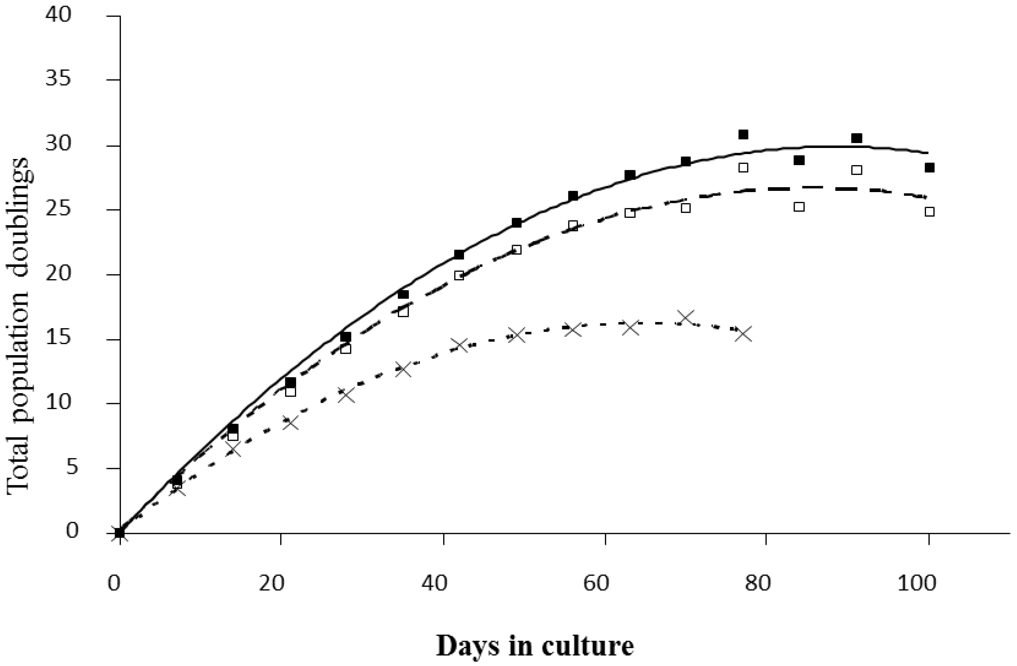
3.1. Radiation-Induced Senescence in Normal Human Fibroblasts
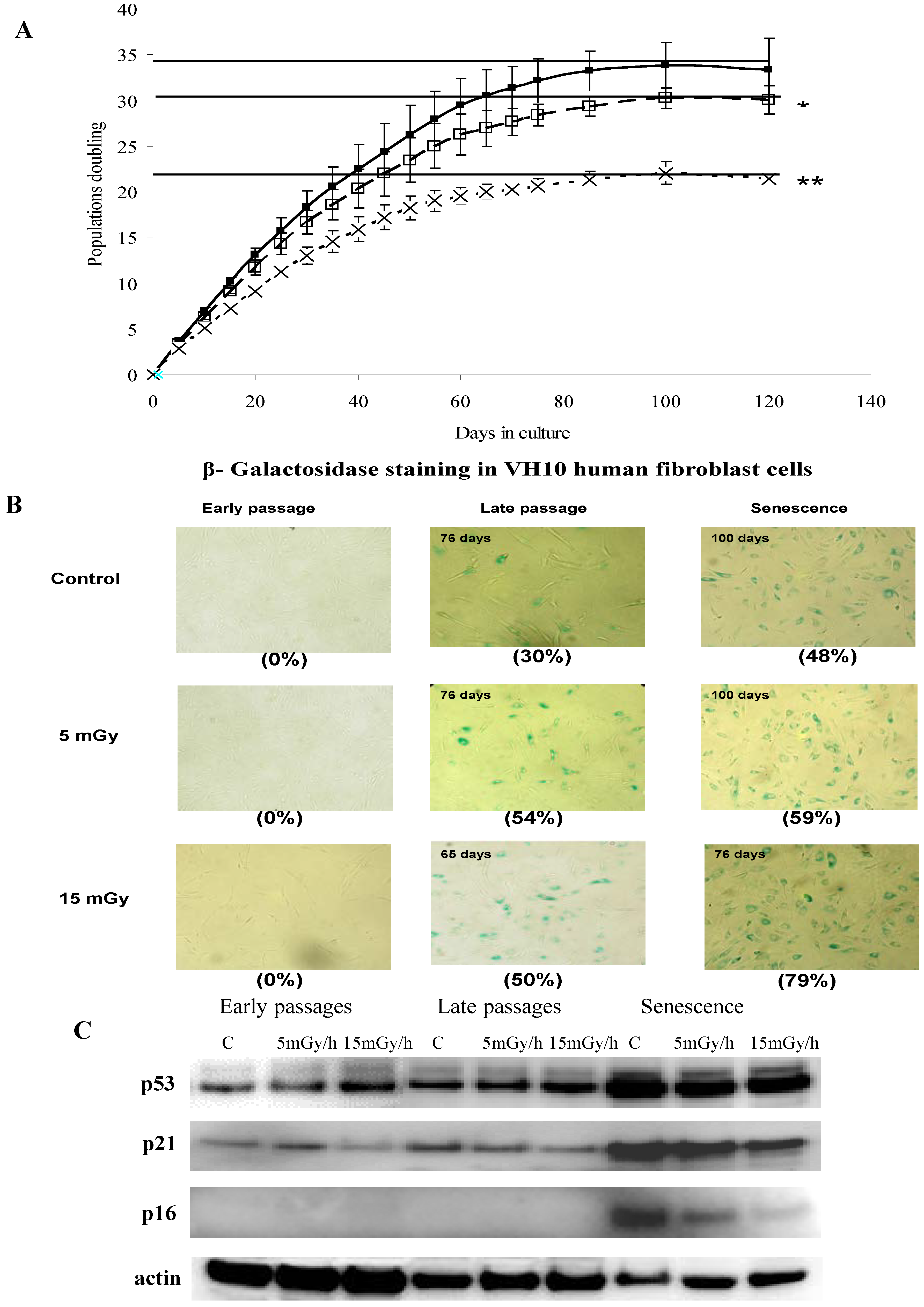
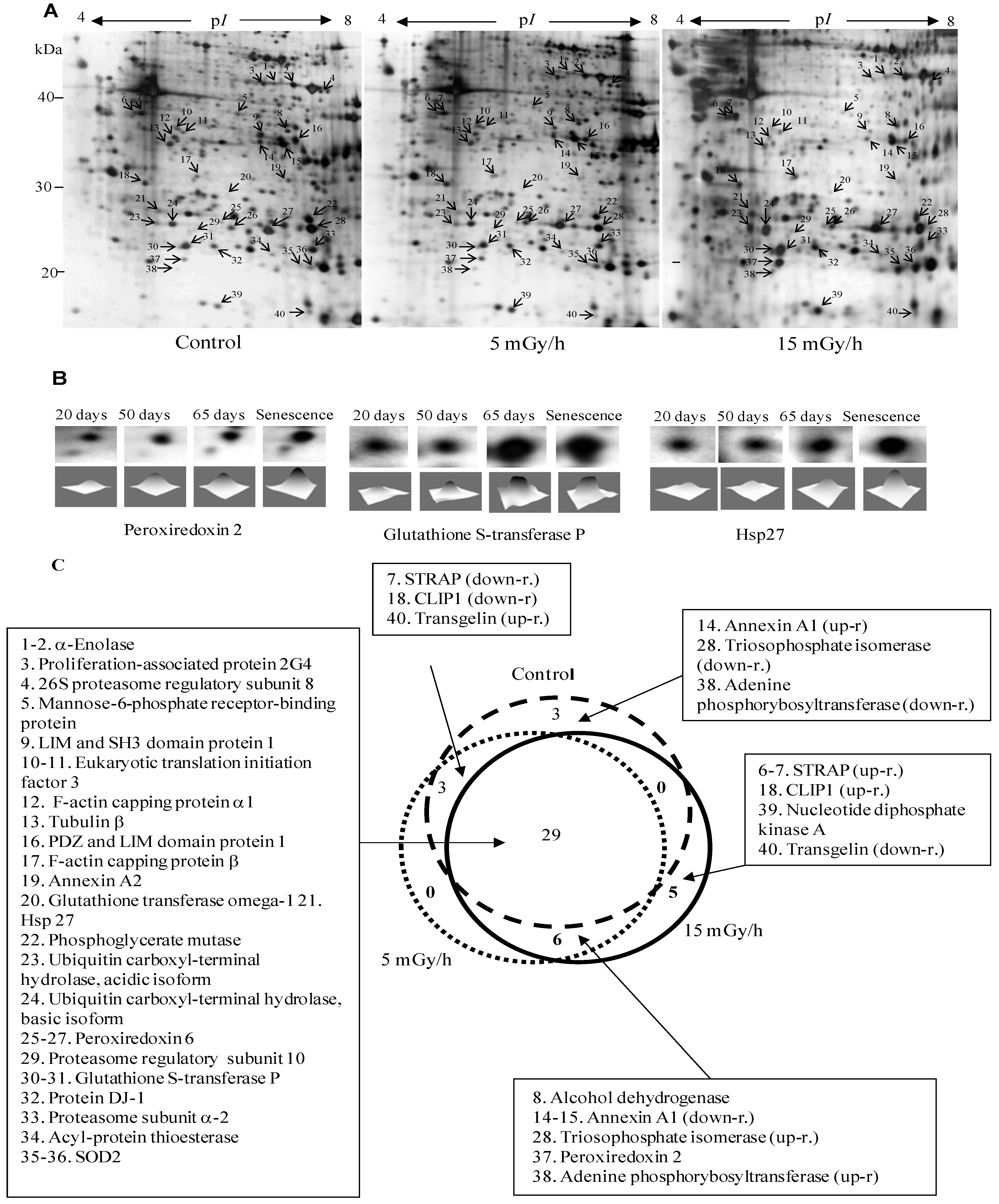
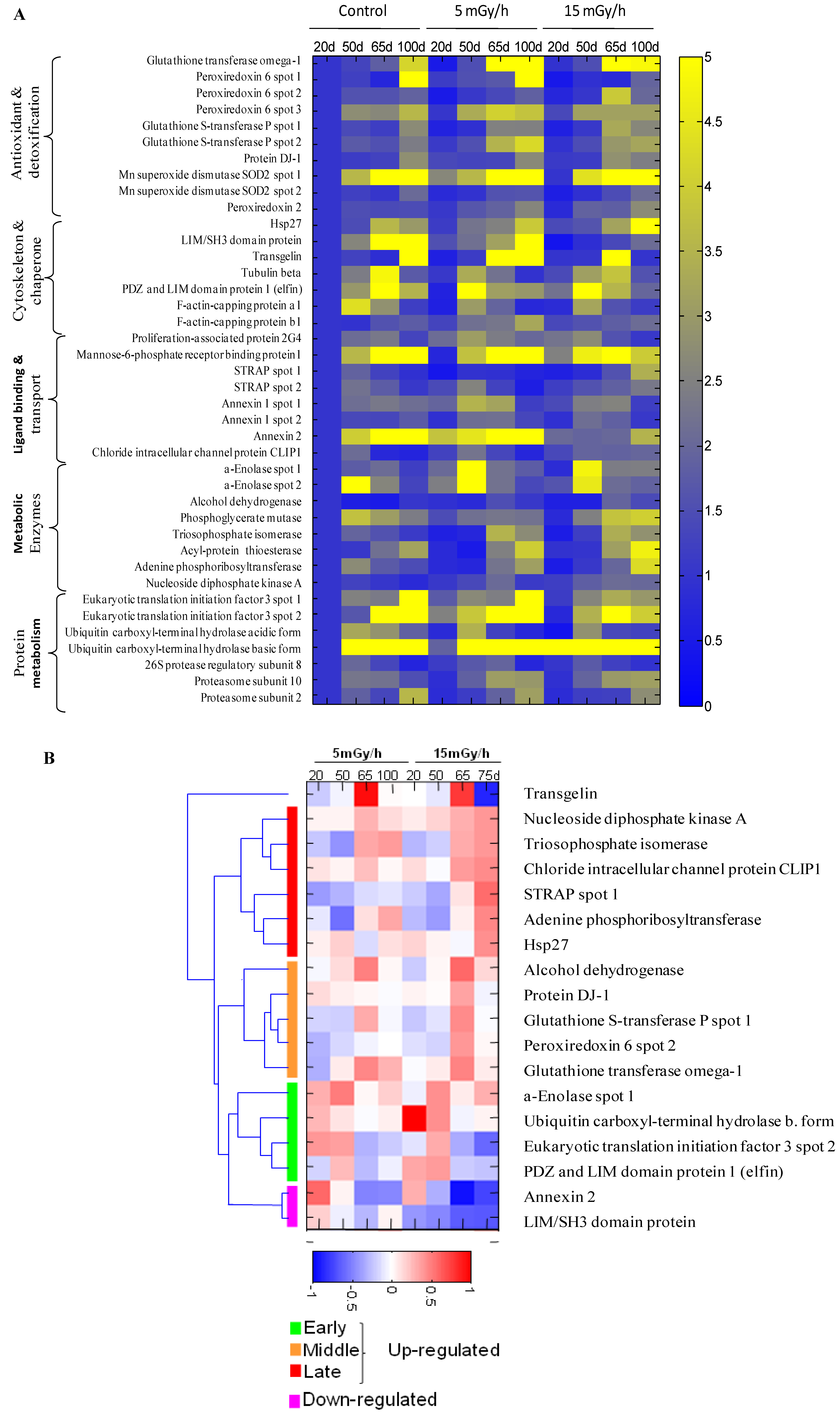
3.2. Effect of Ionizing Radiation on Fibroblast Proteome

| Spot Number | Protein Name | Protein ID | Theoretical pI | Theoretical Mr, kDa | Peptide Matches | Sequence Coverage % | Mascot Score | Protein Function |
|---|---|---|---|---|---|---|---|---|
| 1 | α-Enolase | P06733 | 7.0 | 47.2 | 15 | 36 | 120 | Glycolysis |
| 2 | α-Enolase | P06733 | 7.0 | 47.2 | 20 | 33 | 197 | Glycolysis |
| 3 | Proliferation-associated protein 2G4 | Q9UQ80 | 6.1 | 43.8 | 14 | 27 | 66 | Involved in cell cycle arrest/cell proliferation |
| 4 | 26S protease regulatory subunit 8 | P62195 | 7.1 | 45.6 | 12 | 36 | 85 | Proteasome complex |
| 5 | Mannose-6-phosphate receptor-binding protein 1 | O60664 | 5.8 | 28.1 | 15 | 48 | 141 | Vesicle-mediated transport |
| 6 | Serine-threonine kinase receptor-associated protein | Q9Y3F4 | 5.0 | 38.4 | 9 | 41 | 70 | mRNA processing, regulator of TGFβ pathway, cofactor of p53 |
| 7 | Serine-threonine kinase receptor-associated protein | Q9Y3F4 | 5.0 | 38.4 | 8 | 32 | 65 | mRNA processing, regulator of TGFβ pathway, cofactor of p53 |
| 8 | Alcohol dehydrogenase | P14550 | 6.3 | 36.6 | 13 | 55 | 110 | Glucose metabolic process |
| 9 | LIM and SH3 domain protein 1 | Q14847 | 6.4 | 30.1 | 10 | 31 | 75 | Actin-binding protein |
| 10 | Eukaryotic translation initiation factor 3 | Q13347 | 5.4 | 36.5 | 9 | 30 | 71 | Protein biosynthesis |
| 11 | Eukaryotic translation initiation factor 3 | Q13347 | 5.4 | 36.5 | 10 | 32 | 73 | Protein biosynthesis |
| 12 | F-actin-capping protein α-1 | P52907 | 5.4 | 32.9 | 14 | 67 | 172 | Regulation of cell motility |
| 13 | Tubulin beta | P07437 | 4.8 | 48.7 | 14 | 45 | 132 | Cytoskeleton |
| 14 | Annexin A1 | P04083 | 6.6 | 38.7 | 18 | 52 | 179 | Regulation of apoptosis |
| 15 | Annexin A1 | P04083 | 6.6 | 38.7 | 19 | 62 | 202 | Regulation of apoptosis |
| 16 | PDZ and LIM domain protein 1 | O00151 | 6.6 | 36.5 | 9 | 23 | 78 | Cytoskeleton protein required for actin stress fiber formation |
| 17 | F-actin-capping protein subunit β | P47756 | 5.4 | 31.5 | 9 | 44 | 67 | Actin-binding protein |
| 18 | Chloride intracellular channel protein CLIP1 | O00299 | 5.1 | 27.4 | 16 | 70 | 183 | Chloride ion channel, anti-apoptotic |
| 19 | Annexin A2 | P07355 | 7.6 | 38.6 | 20 | 51 | 210 | Stress response, regulation of apoptosis |
| 20 | Glutathione transferase omega-1 | P78417 | 6.2 | 27.8 | 10 | 29 | 100 | Metabolism of xenobiotics, antioxidant |
| 21 | Heat shock protein β-1 (Hsp27) | P04792 | 6.0 | 22.8 | 8 | 38 | 65 | Involved in stress resistance and actin organization |
| 22 | Phosphoglycerate mutase | P18669 | 6.4 | 26.7 | 18 | 73 | 200 | Glycolysis |
| 23 | Ubiquitin thiolesterase L1, acidic isoforms | P09936 | 5.3 | 24.8 | 14 | 72 | 131 | Processing of ubiquitinated proteins; anti-apoptotic |
| 24 | Ubiquitin thiolesterase L1, basic isoforms | P09936 | 5.3 | 24.8 | 14 | 75 | 171 | Processing of ubiquitinated proteins; anti-apoptotic |
| 25 | Peroxiredoxin 6 | P30041 | 6.0 | 25.0 | 7 | 36 | 57 | Antioxidant |
| 26 | Peroxiredoxin 6 | P30041 | 6.0 | 25.0 | 13 | 64 | 144 | Antioxidant |
| 27 | Peroxiredoxin 6 | P30041 | 6.0 | 25.0 | 14 | 70 | 155 | Antioxidant |
| 28 | Triosephosphate isomerase | P60174 | 6.4 | 26.7 | 15 | 58 | 177 | Carbohydrate metabolism |
| 29 | 26S proteasome subunit 10 | O75832 | 5.4 | 20.4 | 8 | 49 | 58 | Acts as a regulatory subunit of the 26S proteasome |
| 30 | Glutathione S-transferase P | P09211 | 5.4 | 23.6 | 9 | 48 | 94 | Antioxidant, anti-apoptotic |
| 31 | Glutathione S-transferase P | P09211 | 5.4 | 23.6 | 10 | 56 | 146 | Antioxidant, anti-apoptotic |
| 32 | Protein DJ-1 | Q99497 | 6.3 | 19.9 | 8 | 43 | 60 | Redox-sensitive chaperone and a sensor for oxidative stress |
| 33 | Proteasome subunit α type-2 | P25787 | 6.9 | 26.0 | 8 | 44 | 70 | Proteasome complex |
| 34 | Acyl-protein thioesterase 1 | O75608 | 6.3 | 26.7 | 5 | 41 | 56 | De-palmitoylation of signaling proteins |
| 35 | Superoxide dismutase Mn SOD2 | P04179 | 8.3 | 24.7 | 7 | 36 | 69 | Antioxidant, age-dependent response to ROS |
| 36 | Superoxide dismutase Mn SOD2 | P04179 | 8.3 | 24.7 | 8 | 40 | 78 | Antioxidant, age-dependent response to ROS |
| 37 | Peroxiredoxin-2 | P32119 | 5.7 | 21.9 | 9 | 35 | 79 | Antioxidant, anti-apoptotic |
| 38 | Adenine phosphoribosyltransferase | P07741 | 5.8 | 19.6 | 8 | 68 | 91 | Nucleotide metabolism |
| 39 | Nucleoside diphosphate kinase A | P15531 | 5.8 | 17.3 | 8 | 61 | 90 | Synthesis of nucleoside triphosphates other than ATP, tumor suppressor, cofactor of p53 |
| 40 | Transgelin | Q01995 | 8.9 | 22.6 | 13 | 56 | 121 | Actin-binding protein, senescence marker |
3.3. Radiation Induced Stress Response
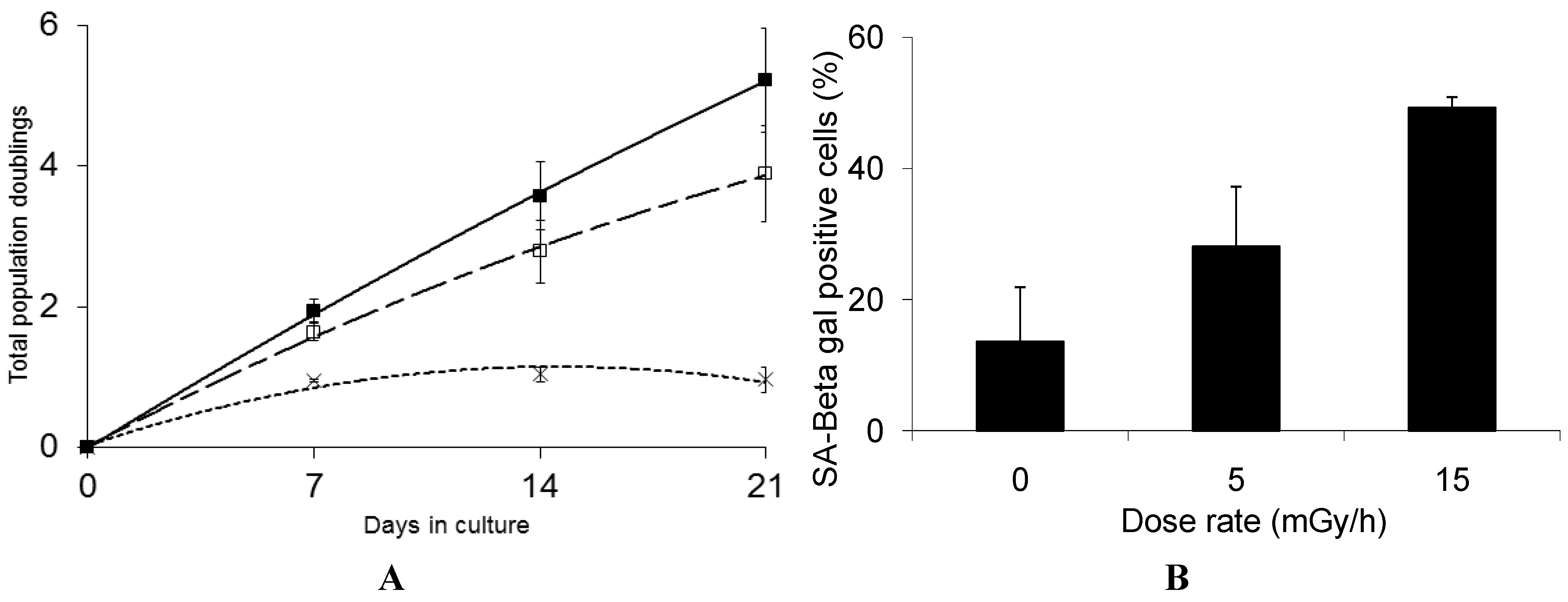
4. Conclusions
Acknowledgments
Author Contributions
Conflicts of Interest
References
- Little, M.; Tawn, E.J.; Tzoulaki, I.; Wakeford, R.; Hildebrandt, G.; Paris, F.; Tapio, S.; Elliott, P. A systematic review of epidemiological associations between low and moderate doses of ionizing radiation and late cardiovascular effects and their possible mechanisms. Radiat. Res. 2008, 169, 99–109. [Google Scholar] [CrossRef]
- Chodick, G.; Bekiroglu, N.; Hauptmann, M.; Alexander, B.H.; Freedman, D.M.; Doody, M.M.; Cheung, L.C.; Simon, S.L.; Weinstock, R.M.; Bouville, A.; et al. Risk of cataract after exposure to low doses of ionizing radiation: A 20-year prospective cohort study among us radiologic technologists. Am. J. Epidemiol. 2008, 168, 620–631. [Google Scholar] [CrossRef]
- Hall, P.; Adami, H.O.; Trichopoulos, D.; Pedersen, N.L.; Lagiou, P.; Ekbom, A.; Ingvar, M.; Lundell, M.; Granath, F. Effect of low doses of ionising radiation in infancy on cognitive function in adulthood: Swedish population based cohort study. Br. Med. J. 2004, 328, e19. [Google Scholar]
- Suzuki, K.; Mori, I.; Nakayama, Y.; Miyakoda, M.; Kodama, S.; Watanabe, M. Radiation-induced senescence-like growth arrest requires tp53 function but not telomere shortening. Radiat. Res. 2001, 155, 248–253. [Google Scholar] [CrossRef]
- Yentrapalli, R.; Azimzadeh, O.; Barjaktarovic, Z.; Sarioglu, H.; Wojcik, A.; Harms-Ringdahl, M.; Atkinson, M.J.; Haghdoost, S.; Tapio, S. Quantitative proteomic analysis reveals induction of premature senescence in human umbilical vein endothelial cells exposed to chronic low-dose rate gamma radiation. Proteomics 2013, 13, 1096–1107. [Google Scholar] [CrossRef]
- Yentrapalli, R.; Azimzadeh, O.; Sriharshan, A.; Malinowsky, K.; Merl, J.; Wojcik, A.; Harms-Ringdahl, M.; Atkinson, M.J.; Becker, K.F.; Haghdoost, S.; et al. The pi3k/akt/mtor pathway is implicated in the premature senescence of primary human endothelial cells exposed to chronic radiation. PLoS One 2013, 8, e70024. [Google Scholar] [CrossRef]
- Rombouts, C.; Aerts, A.; Quintens, R.; Baselet, B.; El-Saghire, H.; Harms-Ringdahl, M.; Haghdoost, S.; Janssen, A.; Michaux, A.; Yentrapalli, R.; et al. Transcriptomic profiling suggests a role for igfbp5 in premature senescence of endothelial cells after chronic low dose rate irradiation. Int. J. Radiat. Biol. 2014. [Google Scholar] [CrossRef]
- Ben-Porath, I.; Weinberg, R.A. The signals and pathways activating cellular senescence. Int. J. Biochem. Cell. Biol. 2005, 37, 961–976. [Google Scholar] [CrossRef]
- Bartkova, J.; Rezaei, N.; Liontos, M.; Karakaidos, P.; Kletsas, D.; Issaeva, N.; Vassiliou, L.V.; Kolettas, E.; Niforou, K.; Zoumpourlis, V.C.; et al. Oncogene-induced senescence is part of the tumorigenesis barrier imposed by DNA damage checkpoints. Nature 2006, 444, 633–637. [Google Scholar] [CrossRef]
- Campisi, J. Aging, cellular senescence, and cancer. Ann. Rev. Physiol. 2013, 75, 685–705. [Google Scholar] [CrossRef]
- Lu, T.; Finkel, T. Free radicals and senescence. Exp. Cell. Res. 2008, 314, 1918–1922. [Google Scholar] [CrossRef]
- Rai, P.; Onder, T.T.; Young, J.J.; McFaline, J.L.; Pang, B.; Dedon, P.C.; Weinberg, R.A. Continuous elimination of oxidized nucleotides is necessary to prevent rapid onset of cellular senescence. Proc. Natl. Acad. Sci. USA 2009, 106, 169–174. [Google Scholar] [CrossRef]
- Haghdoost, S.; Czene, S.; Näslund, I.; Skog, S.; Harms-Ringdahl, M. Extracellular 8-oxo-dg as a sensitive parameter for oxidative stress in vivo and in vitro. Free Radic. Res. 2005, 39, 153–162. [Google Scholar] [CrossRef]
- Haghdoost, S.; Sjölander, L.; Czene, S.; Harms-Ringdahl, M. The nucleotide pool is a significant target for oxidative stress. Free Radic. Biol. Med. 2006, 41, 620–626. [Google Scholar] [CrossRef]
- Sangsuwan, T.; Haghdoost, S. The nucleotide pool, a target for low-dose gamma-ray-induced oxidative stress. Radiat. Res. 2008, 170, 776–783. [Google Scholar] [CrossRef]
- Njålsson, R. Glutathione synthetase deficiency. Cell. Mol. Life Sci. 2005, 62, 1938–1945. [Google Scholar] [CrossRef]
- Dahl, N.; Pigg, M.; Ristoff, E.; Gali, R.; Carlsson, B.; Mannervik, B.; et al. Missense mutations in the human glutathione synthetase gene result in severe metabolic acidosis, 5-oxoprolinuria, hemolytic anemia and neurological dysfunction. Hum. Mol. Genet. 1997, 6, 1147–1152. [Google Scholar] [CrossRef]
- Laemmli, U. Cleavage of structural proteins during the assembly of the head of bacteriophage t4. Nat. Med. 1970, 227, 680–685. [Google Scholar] [CrossRef]
- Yan, J.; Wait, R.; Berkelman, T.; Harry, R.A.; Westbrook, J.A.; Wheeler, C.H.; Dunn, M.J. A modified silver staining protocol for visualization of proteins compatible with matrix-assisted laser desorption/ionization and electrospray ionization-mass spectrometry. Electrophoresis 2000, 21, 3666–3672. [Google Scholar] [CrossRef]
- Gharahdaghi, F.; Weinberg, C.R.; Meagher, D.A.; Imai, B.S.; Mische, S.M. Mass spectrometric identification of proteins from silver-stained polyacrylamide gel: A method for the removal of silver ions to enhance sensitivity. Electrophoresis 1999, 20, 601–605. [Google Scholar] [CrossRef]
- Mass Spectrometry Facility of University of California at San-Francisco. Available online: http://donatello.ucsf.edu (assessed on 1 December 2008).
- Matrix science. Available online: http://www.matrixscience.com (assessed on 8 December 2008).
- Proteinprospector. Available online: http://prospector.ucsf.edu (assessed on 8 December 2008).
- Rockeffeler University. Available online: http://prowl.rockeffeler.edu (assessed on 8 December 2008).
- ExPASy. Available online: http://www.expasy.org (assessed on 12 December 2008).
- HUPO Proteomics Standards Initiative. Available online: http://www.psidev.info (assessed on 15 December 2008).
- Campisi, J.; d’Adda di Fagagna, F. Cellular senescence: When bad things happen to good cells. Nat. Rev. Mol. Cell Biol. 2007, 8, 729–740. [Google Scholar] [CrossRef]
- d’Adda di Fagagna, F. Living on a break: Cellular senescence as a DNA-damage response. Nat. Rev. Cancer 2008, 8, 512–522. [Google Scholar] [CrossRef]
- Dimri, G.; Lee, X.; Basile, G.; Acosta, M.; Scott, G.; Roskelley, C.; Medrano, E.E.; Linskens, M.; Rubelj, I.; Pereira-Smith, O.; et al. A biomarker that identifies senescent human cells in culture and in aging skin in vivo. Proc. Natl. Acad. Sci. USA 1995, 92, 9363–9367. [Google Scholar] [CrossRef]
- Suzuki, M.; Boothman, D.A. Stress-induced premature senescence (sips)—Influence of sips on radiotherapy. J. Radiat. Res. (Tokyo) 2008, 49, 105–112. [Google Scholar] [CrossRef]
- Alcorta, D.; Xiong, Y.; Phelps, D.; Hannon, G.; Beach, D.; Barrett, J.C. Involvement of the cyclin-dependent kinase inhibitor p16 (ink4a) in replicative senescence of normal human fibroblasts. Proc. Natl. Acad. Sci. USA 1996, 93, 13742–13747. [Google Scholar] [CrossRef]
- Brugarolas, J.; Chandrasekaran, C.; Gordon, J.I.; Beach, D.; Jacks, T.; Hannon, G.J. Radiation-induced cell cycle arrest compromised by p21 deficiency. Nature 1995, 377, 552–557. [Google Scholar] [CrossRef]
- Dulić, V.; Kaufmann, W.K.; Wilson, S.J.; Tlsty, T.D.; Lees, E.; Harper, J.W.; Elledge, S.J.; Reed, S.I. P53-dependent inhibition of cyclin-dependent kinase activities in human fibroblasts during radiation-induced g1 arrest. Cell 1994, 76, 1013–1023. [Google Scholar] [CrossRef]
- Radomski, N.; Jost, E. Molecular cloning of a murine cdna encoding a novel protein, p38–2g4, which varies with the cell cycle. Exp. Cell Res. 1995, 220, 434–445. [Google Scholar] [CrossRef]
- Zhang, Y.; Lu, Y.; Zhou, H.; Lee, M.; Liu, Z.; Hassel, B.A.; Hamburger, A.W. Alterations in cell growth and signaling in erbb3 binding protein -1 (ebp1) deficient mice. BMC Cell Biol. 2008, 9, e69. [Google Scholar] [CrossRef]
- Dierick, J.; Eliaers, F.; Remacle, J.; Raes, M.; Fey, S.J.; Larsen, P.M. Toussaint O, Stress-induced premature senescence and replicative senescence are different phenotypes, proteomic evidence. Biochem. Pharmacol. 2002, 64, 1011–1017. [Google Scholar] [CrossRef]
- Beckman, K.; Ames, B.N. The free radical theory of aging matures. Physiol. Rev. 1998, 78, 547–581. [Google Scholar]
- Culotta, V.; Yang, M.; O’Halloran, T.V. Activation of superoxide dismutases: Putting the metal to the pedal. Biochim. Biophys. Acta 2006, 1763, 747–758. [Google Scholar] [CrossRef]
- Asikainen, T.; Huang, T.T.; Taskinen, E.; Levonen, A.L.; Carlson, E.; Lapatto, R.; Epstein, C.J.; Raivio, K.O. Increased sensitivity of homozygous SOD2 mutant mice to oxygen toxicity. Free Radic. Biol. Med. 2002, 32, 175–186. [Google Scholar] [CrossRef]
- Landis, G.; Tower, J. Superoxide dismutase evolution and life span regulation. Mech. Ageing Dev. 2005, 126, 365–379. [Google Scholar] [CrossRef]
- Wood, Z.; Schröder, E.; Robin Harris, J.; Poole, L.B. Structure, mechanism and regulation of peroxiredoxins. Trends Biochem. Sci. 2003, 28, 32–40. [Google Scholar] [CrossRef]
- Neumann, C.; Krause, D.S.; Carman, C.V.; Das, S.; Dubey, D.P.; Abraham, J.L.; Bronson, R.T.; Fujiwara, Y.; Orkin, S.H.; van Etten, R.A. Essential role for the peroxiredoxin prdx1 in erythrocyte antioxidant defence and tumour suppression. Nat. Rev. Mol. Cell Biol. 2003, 424, 561–565. [Google Scholar]
- Wang, Y.; Feinstein, S.I.; Fisher, A.B. Peroxiredoxin 6 as an antioxidant enzyme: Protection of lung alveolar epithelial type II cells from H2O2-induced oxidative stress. J. Cell. Biochem. 2008, 104, 1274–1285. [Google Scholar] [CrossRef]
- Zhou, W.; Freed, C.R. Dj-1 up-regulates glutathione synthesis during oxidative stress and inhibits a53t-synuclein toxicity. J. Biol. Chem. 2005, 280, 43150–43158. [Google Scholar] [CrossRef]
- Andres-Mateos, E.; Perier, C.; Zhang, L.; Blanchard-Fillion, B.; Greco, T.M.; Thomas, B.; Ko, H.S.; Sasaki, M.; Ischiropoulos, H.; Przedborski, S.; et al. Dj-1 gene deletion reveals that dj-1 is an atypical peroxiredoxin-like peroxidase. Proc. Natl. Acad. Sci. USA 2007, 104, 14807–14812. [Google Scholar] [CrossRef]
- Arrigo, A.; Simon, S.; Gibert, B.; Kretz-Remy, C.; Nivon, M.; Czekalla, A.; Guillet, D.; Moulin, M.; Diaz-Latoud, C.; Vicart, P. Hsp27 (hspb1) and b-crystallin (hspb5) as therapeutic targets. FEBS Lett. 2007, 581, 3665–3674. [Google Scholar] [CrossRef]
- Arrigo, A.; Virot, S.; Chaufour, S.; Firdaus, W.; Kretz-Remy, C.; Diaz-Latoud, C. Hsp27 consolidates intracellular redox homeostasis by upholding glutathione in its reduced form and by decreasing iron intracellular levels. Antioxid. Redox Signal. 2005, 7, 414–422. [Google Scholar] [CrossRef]
- Ahmed, E.K.P.C.; Bulteau, A.L.; Friguet, B. Protein oxidative modifications and replicative senescence of wi-38 human embryonic fibroblasts. Ann. N Y Acad. Sci. 2007, 1119, 88–96. [Google Scholar] [CrossRef]
- Jung, H.; Seong, H.A.; Ha, H. Nm23-h1 tumor suppressor and its interacting partner strap activate p53 function. J. Biol. Chem. 2007, 282, 35293–35307. [Google Scholar] [CrossRef]
- Gourlay, C.; Carpp, L.N.; Timpson, P.; Winder, S.J.; Ayscough, K.R. A role for the actin cytoskeleton in cell death and aging in yeast. J. Cell. Biol. 2004, 164, 803–809. [Google Scholar] [CrossRef]
- Gonos, E.; Derventzi, A.; Kveiborg, M.; Agiostratidou, G.; Kassem, M.; Clark, B.F.; Jat, P.S.; Rattan, S.I. Cloning and identification of genes that associate with mammalian replicative senescence. Exp. Cell. Res. 1998, 240, 66–74. [Google Scholar] [CrossRef]
- Assinder, S.; Stanton, J.A.; Prasad, P.D. Transgelin: An actin-binding protein and tumour suppressor. Int. J. Biochem. Cell Biol. 2009, 41, 482–486. [Google Scholar] [CrossRef]
- Littler, D.; Harrop, S.J.; Fairlie, W.D.; Brown, L.J.; Pankhurst, G.J.; Pankhurst, S.; DeMaere, M.Z.; Campbell, T.J.; Bauskin, A.R.; Tonini, R.; et al. The intracellular chloride ion channel protein clic1 undergoes a redox-controlled structural transition. J. Biol. Chem. 2004, 279, 9298–9305. [Google Scholar] [CrossRef]
- Zimniak, P. Detoxification reactions: Relevance to aging. Ageing Res. Rev. 2008, 7, 281–300. [Google Scholar] [CrossRef]
- Sheehan, D.; Meade, G.; Foley, V.M.; Dowd, C.A. Structure, function and evolution of glutathione transferases: Implications for classification of non-mammalian members of an ancient enzyme superfamily. Biochem. J. 2001, 360, 1–16. [Google Scholar] [CrossRef]
- Dalton, T.; Chen, Y.; Schneider, S.N.; Nebert, D.W.; Shertzer, H.G. Genetically altered mice to evaluate glutathione homeostasis in health and disease. Free Radic. Biol. Med. 2004, 37, 1511–1526. [Google Scholar] [CrossRef]
- Meister, A. Glutathione metabolism and its selective modification. J. Biol. Chem. 1988, 263, 17205–17208. [Google Scholar]
- Ristoff, E.; Larsson, A. Patients with genetic defects in the gamma-glutamyl cycle. Chem. Biol. Interact. 1998, 111–112, 113–121. [Google Scholar] [CrossRef]
- Njålsson, R.; Ristoff, E.; Carlsson, K.; Winkler, A.; Larsson, A.; Norgren, S. Genotype, enzyme activity, glutathione level, and clinical phenotype in patients with glutathione synthetase deficiency. Hum. Genet. 2005, 116, 384–389. [Google Scholar] [CrossRef]
- Deschavanne, P.; Debieu, D.; Fertil, B.; Malaise, E.P. Re-evaluation of in vitro radiosensitivity of human fibroblasts of different genetic origins. Int. J. Radiat. Biol. Relat. Stud. Phys. Chem. Med. 1986, 50, 279–293. [Google Scholar] [CrossRef]
- Edgren, M.; Révész, L.; Larsson, A. Induction and repair of single-strand DNA breaks after x-irradiation of human fibroblasts deficient in glutathione. Int. J. Radiat. Biol. Relat. Stud. Phys. Chem. Med. 1981, 40, 355–363. [Google Scholar] [CrossRef]
- Nygren, J.; Ristoff, E.; Carlsson, K.; Moller, L.; Larsson, A. Oxidative DNA damage in cultured fibroblasts from patients with hereditary glutathione synthetase deficiency. Free Radic. Res. 2005, 39, 595–601. [Google Scholar] [CrossRef]
- Ristoff, E.; Hebert, C.; Njålsson, R.; Norgren, S.; Rooyackers, O.; Larsson, A. Glutathione synthetase deficiency: Is gamma-glutamylcysteine accumulation a way to cope with oxidative stress in cells with insufficient levels of glutathione? J. Inherit. Metab. Dis. 2002, 25, 577–584. [Google Scholar] [CrossRef]
- Ayyadevara, S.; Dandapat, A.; Singh, S.P.; Siegel, E.R.; Shmookler Reis, R.J.; Zimniak, L.; Zimniak, P. Life span and stress resistance of caenorhabditis elegans are differentially affected by glutathione transferases metabolizing 4-hydroxynon-2-enal. Mech. Ageing Dev. 2007, 128, 196–205. [Google Scholar] [CrossRef]
- Ayyadevara, S.; Engle, M.R.; Singh, S.P.; Dandapat, A.; Lichti, C.F.; Benes, H.; Shmookler Reis, R.J.; Liebau, E.; Zimniak, P. Lifespan and stress resistance of caenorhabditis elegans are increased by expression of glutathione transferases capable of metabolizing the lipid peroxidation product 4-hydroxynonenal. Aging Cell. 2005, 4, 257–271. [Google Scholar] [CrossRef]
Appendix
© 2014 by the authors; licensee MDPI, Basel, Switzerland. This article is an open access article distributed under the terms and conditions of the Creative Commons Attribution license (http://creativecommons.org/licenses/by/3.0/).
Share and Cite
Loseva, O.; Shubbar, E.; Haghdoost, S.; Evers, B.; Helleday, T.; Harms-Ringdahl, M. Chronic Low Dose Rate Ionizing Radiation Exposure Induces Premature Senescence in Human Fibroblasts that Correlates with Up Regulation of Proteins Involved in Protection against Oxidative Stress. Proteomes 2014, 2, 341-362. https://doi.org/10.3390/proteomes2030341
Loseva O, Shubbar E, Haghdoost S, Evers B, Helleday T, Harms-Ringdahl M. Chronic Low Dose Rate Ionizing Radiation Exposure Induces Premature Senescence in Human Fibroblasts that Correlates with Up Regulation of Proteins Involved in Protection against Oxidative Stress. Proteomes. 2014; 2(3):341-362. https://doi.org/10.3390/proteomes2030341
Chicago/Turabian StyleLoseva, Olga, Emman Shubbar, Siamak Haghdoost, Bastiaan Evers, Thomas Helleday, and Mats Harms-Ringdahl. 2014. "Chronic Low Dose Rate Ionizing Radiation Exposure Induces Premature Senescence in Human Fibroblasts that Correlates with Up Regulation of Proteins Involved in Protection against Oxidative Stress" Proteomes 2, no. 3: 341-362. https://doi.org/10.3390/proteomes2030341
APA StyleLoseva, O., Shubbar, E., Haghdoost, S., Evers, B., Helleday, T., & Harms-Ringdahl, M. (2014). Chronic Low Dose Rate Ionizing Radiation Exposure Induces Premature Senescence in Human Fibroblasts that Correlates with Up Regulation of Proteins Involved in Protection against Oxidative Stress. Proteomes, 2(3), 341-362. https://doi.org/10.3390/proteomes2030341

