Review

Recent Advances in the Phytochemistry of Bryophytes: Distribution, Structures and Biological Activity of Bibenzyl and Bisbibenzyl Compounds

1
Department of Botany, University of Kalyani, Kalyani 741245, India
2
Department of Biotechnology, Gauhati University, Guwahati 781014, India
3
Institute of Pharmacognosy, Tokushima Bunri University, Tokushima 770-8514, Japan
*
Author to whom correspondence should be addressed.
Plants 2023, 12(24), 4173; https://doi.org/10.3390/plants12244173 (registering DOI)
Submission received: 25 September 2023 / Revised: 21 November 2023 / Accepted: 21 November 2023 / Published: 15 December 2023
(This article belongs to the Special Issue Chemical Characteristics and Bioactivity of Plant Natural Products)

Abstract

:
Research on bryophyte phytochemistry has revealed the presence of different phytochemicals like fatty acids, terpenoids, small phenolic molecules, etc. Small phenolic molecules, i.e., bibenzyls (of two aromatic rings) and bisbibenzyls (four aromatic rings), are unique signature molecules of liverworts. The first bisbibenzyls marchantin A and riccardin A were discovered in two consecutive years, i.e., 1982 and 1983, respectively, by Asakawa and coworkers. Since then, about 70 bisbibenzyls have been reported. These molecules are characterized and identified using different spectroscopic techniques and surveyed for different bioactivity and structure–activity relations. Biochemistry is determined by the season, geography, and environment. In this review, quantitative and qualitative information on bibenzyls and bisbibenzyl compounds and their distribution in different liverworts across, geographies along withtraditional to advanced extraction methods, and characterization techniques are summarized. Also, a comprehensive account of characteristic spectra of different bisbibenzyl compounds, their subtypes, and their basic skeleton patterns are compared. A comprehensive table is provided here for the first time presenting the quantity of bibenzyls, bisbenzyls, and their derivatives found in bryophytes, mentioning the spectroscopic data and mass profiles of the compounds. The significance of these compounds in different bioactivities like antibiotic, antioxidative, antitumor, antivenomous, anti-influenza, insect antifeedant, cytotoxic, and anticancerous activities are surveyed and critically enumerated.

Graphical Abstract

1. Introduction

Bryophytes are non-vascular land plants belonging to the non-tracheophyte group that do not produce bona fide lignin polymers even though many of the genes for its biosynthesis are present [1,2,3]. There are three bryophyte lineages: Anthocerotophyta (hornworts, consisting of ~250 species), Marchantiophyta (liverworts, ~9000 species), and Bryophyta (mosses, ~12,700 species) [4]. Bryophytes, particularly Marchantiophyta/liverworts, emerged ~500 million years ago on Earth [5], and they are the ancestors of all land plants [1,6]. Liverworts and mosses form a monophyletic clade [7], whereas hornworts are sisters to them [3].
Bryophytes started blooming under challenging environmental conditions of high-intensity solar radiation and high CO2 level even though they were biochemically constrained for photosynthesis because of lower Rubisco expression and anatomical limitations of CO2 diffusion [8]. Another challenge they faced was adapting their life in a water-deficient and UV-abundant terrestrial environment; see the review by [9,10]. Bryophytes are morphologically inconspicuous, meaning they lack well-developed tissue structures to protect them from stress factors. However, to make up for their morphological shortcomings, bryophytes have evolved to synthesize specialized phytochemicals to contribute to their ecometabolics [10], which is the metabolic response to ecological challenges through the production of specific compounds [9]. Among the array of metabolites produced by bryophytes, flavonoids protect from light [11], sesquiterpenoids and flavonoids act counter biotic stress [11,12], most glycosides act as shields against low temperature and desiccation [13], and methoxyphenols and cinnamic acid derivatives constitute anatomical structures that support growth and development [14]. The presence of bibenzyls (compounds with two aromatic rings) and bisbibenzyls (compounds with four aromatic rings), a unique group of phenolic molecules of liverworts present in oil bodies, are abundant in bryophytes, with a structural diversity. Mosses are generally devoid of these molecules, as oil bodies are absent in them [9], and the role of these oil bodies is known to prevent desiccation.
Metabolites of these minuscule organisms perform the role of overcoming the challenges they face in nature [9,14,15,16,17]. Therefore, when they are extracted separately and tested, purified andtested, they show many bioactive properties, including antibacterial, antifungal, antioxidative, antivenomous, anticancerous, anti-inflammatory, antiulcer and others [18,19,20] of human interest. As a diverse array of metabolites and their function has been reported, for convenience, we restricted our major focus to bibenzyl and bisbibenzyl compounds of liverworts and other bryophytic taxa as reported in the literature. A number of reviews have covered the bioactivity and structure–activity relation of bisbibenzyls [21,22,23,24], the ethnobryological uses of bryophytes [25,26,27], and the synthesis and structural diversity of said compounds [15,28,29]. The gap in the previous reviews was the absence of comprehensive quantitative information on specialized bryophyte metabolites for ready reference and the mechanistic explanation. Also, the difference in extraction protocols vis-a-vis the corresponding yield has not been critically appraised. As such, in this review, we complement the literature with recent updates of the liverwort taxa surveyed for the extraction and characterization of signature molecules of liverworts, i.e., bibenzyls and bisbibenzyls, and their quantities in different taxa, the conventional extraction method vis-à-vis recent micromanipulation techniques for cell-based characterization, and the structure–activity relationship. We critically analyze the knowledge gap in the above-mentioned areas to lay the foundation for future research questions.

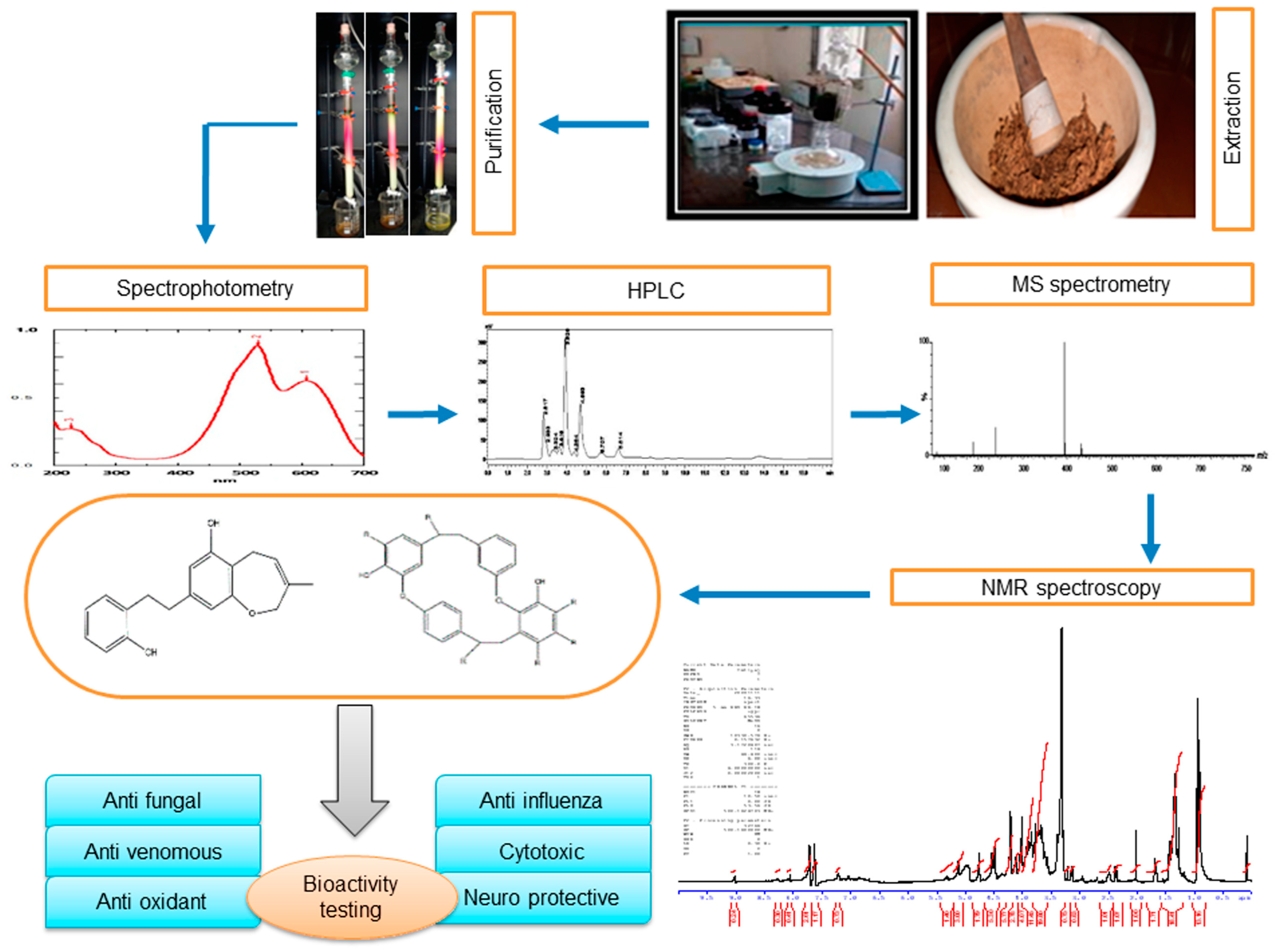
2. Extraction and Purification of Major Bioactive Compounds

Different extraction methods involving different solvent systems have been used for the extraction of bryophyte phytoconstituents, like macrocyclic phenolic compounds, such as bibenzyls and bisbibenzyls. To increase the yield, the extraction process is complemented by treatments like sonication or an extended period of extraction, for example, a week. Depending upon the chemical nature/polarity, methanol (MeOH), ethanol (EtOH), diethylether (Et2O), and other solvents have been used for preparing crude extracts. The fractionation of the extract or the chromatographic separation of various compounds in the extract through adsorption or gel permeation is carried out using different mixtures of solvents, mostly low-polarity ones, with the dominant one being ethyl acetate (EtOAc), whereas MeOH is the most polar organic solvents used in the extraction/purification schemes. A general scheme of phytoconstituent extraction, purification, and structure identification is presented in Figure 1. A few selected extraction protocols are given below to highlight the similarity and differences in the yield when different solvents were used to extract bisbibenzyls only or alongside terpenes from different bryophytes. For example, air-dried bryophyte samples were extracted with Et2O-MeOH [30,31]. This was followed by silica gel or Sephadex LH-20 chromatography using n-hexane-EtOAc, C6H6-EtOAc, or CHCl3-MeOH (1:1) as eluents. Then, the eluates were purified with preparative TLC to obtain pure cyclic bisbibenzyls, viz., marchantin A (1), B (2), and C (3), from Marchantia polymorpha L., M. paleacea var. diptera (Nees & Mont.) Inoue, and M. tosana Stephani (Figure 2a,b, Figure 3b,d and Figure 4). The yield of the pure compounds ranged from 0.7% to 2.6% of the MeOH-Et2O extract [31]. The structures of these isolated compounds were identified using a combination of spectral data, X-ray crystallography and chemical degradation studies [30,31]. In a somewhat similar manner, Huang et al. (2010) extracted a macrocyclic bisbibenzyl from the air-dried powder of M. tosana with MeOH, followed by sonication for 1 h and incubation for 2 weeks at room temperature. Then, the extract was filtered, evaporated to dryness, resuspended in water, and partitioned successively with n-hexane and EtOAc. The EtOAc fraction exhibited significant anticancer activity against MCF-7 cells. Therefore, it was chromatographed in a silica gel column and purified on a preparative reversed-phase high-performance liquid chromatography (RP-HPLC) with a UV detector. A white amorphous solid was obtained, with a yield of 6.6% of the MeOH extract, which, on subsequent analysis using high-resolution electron ionization mass spectrometry (HR-EI-MS), 1H NMR and 13C NMR, including 2D NMR, was confirmed to be marchantin A (1) [32]. On the other hand, Niu et al., 2006, took the air-dried powder of M. polymorpha, and an Et2O extract was prepared at room temperature and then concentrated in vacuo. The extract, which showed antifungal activity against Candida albicans, was chromatographed on SiO2, a stepwise gradient system of petroleum ether (PE)/acetone (Me2CO)), followed by rechromatographed on Sephadex LH-20 with mobile phase CHCl3/MeOH (1:1) to obtain subfractions, which were further purified with the gradient of PE/Me2CO. The purified fractions were confirmed to be marchantin A (1), marchantin B (2), marchantin E (4), neomarchantin A (5), 13,13′-O-isopropylidenericcardin D (6), plagiochin E (7) and riccardin H (8) through 1H NMR and 13C NMR [33]. Lu et al., 2006, successfully extracted mixtures of terpenes and macrocyclic bisbibenzyls from air-dried powders of Conocephalum conicum (L.) Dumort. (Figure 3a) and Dumortiera hirsuta (Sw.) Nees (Figure 3c) following sequential ultrasonic extractions with Et2O and MeOH. The Et2O extract of C. conicum was subjected to adsorption chromatography in silica gel (mobile phase: PE–EtOAc, gradient) to yield seven fractions. These fractions on further chromatographic separation on Sephadex LH-20 and silica gel gave five pure compounds (yield ranging from 0.03% to 0.16% of the Et2O extract). The Et2O extract of D. hirsuta on subsequent chromatography separated into four compounds, including riccardin D (9) (yield 0.04% of the Et2O extract), whereas the MeOH extract was processed to obtain only two compounds (the yield was 0.04–0.5% of the MeOH extract) [34]. The mixtures of phenolic compounds, cyclic bisbibenzyls, and terpenes were extracted exhaustively by Liu et al., 2011, from the air-dried plant material of C. japonicum (Thunb.) Grolle with 95% EtOH under reflux. After resuspending the crude extract in H2O and successive partitioning of the extract with Et2O and n-butanol (BuOH), the Et2O extract was subjected to adsorption chromatography on a silica gel column, and eluted with a PE−Me2CO gradient system to yield seven fractions. These fractions were separated into eleven pure compounds, with yields ranging from 0.09% to 3.6% of the Et2O extract, after further purification using gel permeation, HPLC and other techniques [35].
Owing to the unavailability of bulk samples and the low yield of macrocyclic phenol compounds, it will be useful to have a method that can detect such compounds from a crude sample even if the concentration is very low. In this direction, Xing et al., 2007, developed a rapid screening protocol employing LC-DAD/MS analysis, which could detect even very low concentrations. Xing et al., 2007, extracted the dried powders of M. polymorpha, Asterella angusta (Stephani) Pandé, K.P. Srivast & Sultan Khan, and Plagiochasma intermedium Lindenb.& Gottsche with EtOH, followed by sonication for 30 min and vigorous shaking for complete extraction. The ethanol extracts were partitioned into Et2O and water. The Et2O fraction was dissolved in acetonitrile (ACN), passed through a microporous membrane (0.45 µm) and subjected to LC-DAD/MS analysis. The method was capable of detecting very low concentrations of bisbibenzyls in a 50 min run. Because of high sensitivity and a relatively short run time, this method could be routinely used for screening macrocyclic bisbibenzyl compounds in bryophytes [36].
Direct molecular analysis from single-cell content was reported by using a nanoelectrospray tip following the analysis of the mass profile of the compound using nanoelectrospray ionization [37]. Isomers can be separated by combining online MS with ion-mobility separation which enables the rapid sampling at sub-attomolar-level sensitivity [38]. Following the same technique, oil body contents were analyzed using a micromanipulator, and direct evidence of two sesquiterpenes and marchantin A (1) was detected. The number of oil body cells correlated with the quantified amount of the metabolites, and low-mineral condition induced the increase in oil-body cells in the thallus [39].
Quantities of bibenzyls, bisbibenzyls, and their derivatives in liverworts, along with their spectral characteristics, have been compiled for the first time, as presented in Table 1. Compiled data regarding the quantity of phytochemicals is calculated in 100 g of dry weight for the convenience of presentation. A brief discussion of the tabulated data is presented below to show the quantitative variations of phytochemicals in inter- and intraspecific taxa.
Asakawa et al., 1982, extracted 6.11 g of perrottetin D (10) from Radula perrottetii Gottsche ex Stephani with diethyl ether following methanol extraction [40], and 0.149 g of perrottetin D was obtained directly in methanol [41], though both the samples were collected from the same geographical region (Tokushima, Japan). Riccardin A (11) and B (12) production varies from 0.13 g to 4.45 g and 0.11 g to 4.25 g, respectively, in the same species, i.e., Riccardia multifida (L.) Gray of the same geographical region and extracted in the same solvent system, as reported by Asakawa et al., 1983, and Nagashima et al., 1996, respectively [42,43]. Perrottetin E (13) was extracted at 83.57 mg, 36.82 mg, and 218.4 mg, respectively, from R. perrottetii, R. laxiramea Stephani, and Pellia epiphylla (L.) Corda [41,44,45]. The quantity of perrottetinene (14) also varies from 18.57 to 562.5 mg in R. laxiramea and R. marginata Gottsche, Lindenb. & Nees [45,46].
Marchantin A (1)was obtained from Hungarian M. polymorpha in quantities of 0.14 g and 1.9 g to 5/6 g when extracted M. paleacea subsp. diptera [47,48], and the quantity of marchantin B (2) was 0.13 mg to 12.87 mg from M. polymorpha [21,33], 13.48 mg from M. paleacea subsp. diptera [21], and 42.85 mg from Schistochila glaucescens (Hook.) A. Evans [49]. Marchantin C (3) was obtained in 6.5 mg from M. polymorpha [21] and 16.69 mg from M. paleacea subsp. diptera [21] showing that the quantity of the same phytochemical varies with species or genus or may be influenced by geography and season as well. Bisbibenzyl contents of P. appendiculatum Lehm. & Lindenb. vary in soil-grown thallus, and hence the induced callus in the laboratory may be utilized further for the mass production of bisbibenzyl in optimum growth conditions [50].
Table 1. Total content (in mg/100 mg dry weight) and chemical characteristics of bibenzyl and bisbibenzyl compound reported from bryophytes.
Type of ChemicalName of the ChemicalName of the TaxaContent (mg/100 g dw)λmax (nm)Mass Spectra (m/z)References
PrebibenzylDumhirone A (20)Dumortiera hirsuta0.4274 in MeOH230.0963 [M]+[51]
Prelunularin(22)Ricciocarpos natans1.2272na[52]
BibenzylDihydroresveratol (65)Blasia pusilla2.82nana[53]
Brittonin A (39)Frullaniabrittoniae
subsp. truncatifolia
50217362
[M]+
[54]
Brittonin A (39)Frullania inouei2.55nana[55]
Brittonin B (40)Frullania brittoniae subsp. truncatifolia25276346 [M]+[54]
Brittonin B (40)Frullania inouei0.83nana[55]
3,4′-Dimethoxyl-4-hydroxylbibenzyl (141)Radula complanata11.76–14.11na258 [M]+,[56]
2-(3-Methyl-2-butenyl)-3,5-dihydroxybibenzyl (32)Radula voluta1034.38284 in MeOH282 [M]+[57]
2-Geranyl-3,5-dihydroxybienzyl (33)Radula voluta381.25284 in MeOH350 [M]+[57]
3,5-Dihydroxy-4-(3-hydroxy-3-methylbutyl) bibenzyl (34)Radula laxiramea117.30 300 [M]+[45]
Radulanolide (44)Radula complanata42.82216322 [M]+[40]
Radulanin H (48)Radula complanata999.14220324 [M][40]
Radulanin L (51)Radula complanata147.54282296.1446 [M]+[56]
Lunularin (21)Blasia pusilla3.77nana[53]
Lunularin (21)Dumortiera hirsuta0.85nana[51]
Lunularic acid (23)Blasia pusilla0.48nana[53]
Prenyl bibenzylPerrottetin A (52)Radula perrottetii11,162.08211298 [M-91][40]
Perrottetin D (10)Radula perrottetii6116.20216296 [M][40]
Perrottetin D (10)Radula perrottetii149.85na296.1425 [M]+[56]
2-Carbomethoxy-3,5,4″-trihydroxy-
4,6-(3-methyl-2-butenyl)-Bibenzyl (27)
Lethocolea glossophyla72313 in MeOH424 [M]+[57]
2,4-di-(3-methyl-2-butenyl)-3,5,4′-Trihydroxy-bibenzyl (28)Lethocolea glossophyla44313 in MeOH366 [M]+[57]
2,2-Dimethyl-5-hydroxy-6-(3-methyl-2-butenyl)-7-[2-(4′-hydroxyphenyl)-ethyl]-chromene (29) Lethocolea glossophyla52315 in MeOH364 [M]+[57]
2,2-Dimethyl-5-hydroxyl-7-[2-(4′-hydroxyphenyl)-ethyl]-8-(3-methyl-2-butenyl)-chromene (30)Lethocolea glossophyla232320 in MeOH364 [M]+[57]
Bibenzyl cannabinoidPerrottetinene (14)Radula marginata562.5nana[46]
Perrottetinene (14)Radula laxiramea18.57nana[45]
Perrottetinenic acid (58)Radula marginata87.5301.4 in EtOH392.1985, 392 [M]+[46]
Cinnamoyl bibenzylPallidisetin A (35)Polytrichum pallidisetum0.50318342.1277 [M]+[58]
Pallidisetin B (36)Polytrichum pallidisetum0.21294342.1236 [M]+[58]
Bibenzyl derivativePellepiphyllin (37)Pellia epiphylla6.8na258 [M]+[44]
7-Hydroxy-pellepiphyllin (38) Pellia epiphylla0.24na273 [M-H][44]
2,5,4′-Trihydroxy–bibenzyl (24)Ricciocarpus natans0.8284na[52]
3-Methoxybibenzyl (26)Radula complanata11.76212na[59]
3,3′,4,4′-Tetramethoxy-bibenzyl (41)Frullania inouei0.34nana[55]
Chrysotobibenzyl (42)Frullania inouei9.02nana[55]
Bisbibenzyl and its derivativeRiccardin A (11)Riccardia multifida130.64213438.1831[42]
Riccardin A (11)Riccardia multifida4458.33283na[43]
Riccardin B (12)Riccardia multifida115.64233424.1674[42]
Riccardin B (12)Riccardia multifida4250280424.1700[43]
Riccardin C (15)Plagiochasma intermedium0.22 anana[60]
Riccardin C (15)Marchantia palmata9nana[61]
Riccardin C (15)Blasia pusilla53.35
3.74
nana[53]
Riccardin C (15)Dumortiera hirsuta0.23na425.30 [M][51]
Riccardin D (9)Monoclea forsteri141na424.1674[62]
Isoriccardin C (83)Plagiochasma intermedium0.05 anana[60]
Isoriccardin C (83)Marchantia palmata12.5nana[61]
Isoriccardin C (83)Marchantia paleacea4.31nana[63]
Isoriccardin D (87)Marchantia polymorpha0.45na424.16 [M]+[64]
Riccardin F (79)Plagiochasma intermedium0.07 anana[60]
Riccardin F (79)Blasia pusilla14.64215438 [M]+[53]
Riccardin H (8)Marchantia polymorpha0.06na456 [M]+[33]
Riccardin I (82)Asterella angusta0.32na454.1440 [M]+[65]
Paleatin A (95)Marchantia paleacea subsp. diptera5.75nana[21]
Paleatin B (96)Marchantia paleacea subsp. diptera16.51nana[21]
Polymorphatin A (136)Marchantia polymorpha0.40na424 [M]+[64]
Marchantin A (1)Marchantia polymorpha0.13na439 [M]+[33]
Marchantin A (1)Marchantia polymorpha377.5nana[21]
Marchantin A (1)Marchantia paleacea subsp. diptera1204.54nana[21]
Marchantin A (1)Marchantia paleacea subsp. diptera5000–6000nana[47]
Marchantin A (1)Marchantia paleacea subsp. diptera1191.90nana[48]
Marchantin A (1)Marchantia tosananana440.1632[32]
Marchantin B (2)Marchantia polymorpha0.13na455 [M]+[33]
Marchantin B (2)Marchantia polymorpha12.87nana[21]
Marchantin B (2)Marchantia paleacea subsp. diptera13.48nana[21]
Marchantin C (3)Marchantia paleacea subsp. diptera16.79nana[21]
Marchantin C (3)Marchantia polymorpha6.5nana[21]
Marchantin C (3)Marchantia polymorpha40208.5na[61]
Marchantin C (3)Marchantia palmata10nana[61]
Marchantin C (3)Dumortiera hirsuta0.3nana[60]
Marchantin C (3)Schistochila glaucescens61.22nana[49]
Marchantin C (3)Reboulia hemisphaerica120na424.1680 [M]+[66]
Marchantin C (3)Marchantia paleacea38.79nana[63]
Isomarchantin C (67)Marchantia polymorpha8.63211.5424.1707[61]
Isomarchantin C (67)Marchantia palmata6nana[21]
Marchantin D (147)Marchantia paleacea subsp. diptera5.30nana[21]
Marchantin E (4)Marchantia paleacea subsp. diptera126.36nana[21]
Marchantin E (4)Marchantia polymorpha0.11na469[33]
Marchantin E (4)Marchantia paleacea subsp. diptera125.03nana[48]
Marchantin H (144)Plagiochasma intermedium0.04 anana[60]
Marchantin M (72)Reboulia hemisphaerica100283454.1774 [M]+[66]
Marchantin N (74)Reboulia hemisphaerica120276.5471 [M]+[66]
Marchantin O (75)Reboulia hemisphaerica300274438.1817 [M]+[66]
Marchantin J (68)Marchantia polymorpha30.30na424 [M]+[64]
Neomarchantin A (5)Plagiochasma intermedium0.05nana[60]
Neomarchantin A (5)Marchantia polymorpha0.04na423 [M]+[33]
Neomarchantin A (5)Schistochila glaucescens51.02272 in EtOH424 [M]+[49]
Neomarchantin B (77)Schistochila glaucescens42.85272 in EtOH440 [M+[49]
Pakyonol A (145)Plagiochasma intermedium0.40 anana[60]
Angustatin A (146)Asterella angusta0.28na436.1330 [M]+[65]
Isoplagiochin A (62)Plagiochila sp.2.85288–250 Sh423 [M+H]+[67]
Isoplagiochin A (102a)Plagiochila fruticosa33.48 anana[68]
Isoplagiochin A (102a)Heteroscyphus planus0.29292422.1542 [M]+[69]
Isoplagiochin B (103a)Plagiochila fruticosa17.79 anana[68]
Isoplagiochin C (104)Herbertus sakerraii4.36nana[70]
Isoplagiochin C (104)Plagiochila fruticosa8.34 a287422.1485 [M]+[68]
Isoplagiochin D (105)Herbertus sakerraii35.43nana[70]
Isoplagiochin D (105)Plagiochila fruticosa5.32 ana424.1654 [M]+[68]
Isoplagiochin F (107)Plagiochila sp.2.85283–251 Sh441 [M+H]+[67]
Dihydroisoplagiochin (108)Plagiochila sp.5288–282 Sh425 [M+H]+[67]
Monochlorinated isoplagiochin D (109)Plagiochila sp.0.71288–282 460[67]
2,12-Dichloroisoplagiochin D (110)Herbertus sakuraii6.48288492.0904[70]
12,7′-Dichloroisoplagiochin D (111)Herbertus sakuraii3.04288492.0923[70]
12, 10′-Dichloroisoplagiochin C (112)Herbertus sakuraii4.36294490.0714[70]
12-Chloroisoplagiochin D (113)Mastigophora diclados0.94nana[70]
2,12-Dichloroisoplagiochin D (110)Mastigophora diclados0.68nana[70]
Plagiochin E (7)Marchantia polymorpha13.40na424.1685[33]
Planusin A (106)Heteroscyphus planus3.23297422.1485 [M]+[69]
Pusillatin A (90)Blasia pusilla0.65212 in EtOH869 [M+Na]+,
846 [M]+
[53]
Pusillatin B (91)Blasia pusilla5.18227 in EtOH869 [M+Na]+,
846 [M]+
[53]
Pusillatin C (92)Blasia pusilla19.69, 3.05215 in EtOH869 [M+Na]+,
846 [M]+
[53]
Perrottetin E (13)Radula perrottetii9.62nana[71]
Perrottetin E (13)Radula perrottetii83.57nana[56]
Perrottetin E (13)Plagiochila sp.1.42nana[67]
Perrottetin E (13)Pellia epiphylla218.4nana[44]
Perrottetin E (13)Marchantia polymorpha2.02na424.16 [M]+[64]
Perrottetin E (13)Radula laxiramea36.82nana[45]
Perrottetin F (55)Radula perrottetii63.55nana[71]
Perrottetin F (55)Radula perrottetii86.45nana[56]
Isoperrottetin A (56)Radula perrottetii11.96213426 [M]+[71]
Perrottetin G (57)Radula perrottetii12.79nana[71]
Perrottetin G (57)Radula perrottetii4.61nana[56]
10′-Hydroxyperrottetin E (59)Pellia epiphylla139.44na442 [M]+[44]
10′-Hydroxyperrottetin E-11-methylether (60)Pellia epiphylla11.38na456 [M]+[44]
14′-Hydroxyperrottetin E (61)Pellia epiphylla17.47na442 [M]+[44]
10,10′-Dihydroxy-perrottetin E (62)Pellia epiphylla81.73na458 [M]+[44]
13′,13″-Bis(I0′-hydroxyperrottetin E (63)Pellia epiphylla2.04na882 [M]+[44]
Perrottetin E-11-methyl
ether (64)
Pellia epiphylla27.44na440 [M]+[44]
Marchantin G methyl ether (78)Marchantia palmata1.5nana[61]
6′,6″-Bis-riccardin C (25)Ricciocarpus natans4847na[52]
Isoriccardinoquinone A (84)Marchantia paleacea10.34na468 [M]+[63]
Isoriccardinoquinone B (85)Marchantia paleacea7.75na452 [M]+[63]
2-Hydro-3,7-dimethoxyphenanthrene (86)Marchantia paleacea19.82nana[63]
13, 13′-O-Isopropylidenericcardin D (6)Marchantia polymorpha0.06na464[33]
11-O-Demethyl-marchantin I (143)Asterella angusta0.4210 in MeOH423 [M-H],
424.1712 [M]
[72]
Dihydroptychantol A (135a)Asterella angusta0.32210 in MeOH 423 [M-H],
424.1720 [M]
[72]
Ptychantol A (132)Ptychanthus strictus39.67342HRMS m/z (422.15)[73]
Ptychantol B (133) Ptychanthus strictus21.08341HRMS m/z (438.14)[73]
Ptychantol C (134)Ptychanthus strictus1.95342HRMS m/z (438.14)[73]
Dibenzofuran bisbibenzylAsterellin A (88)Asterella angusta0.4212 in MeOH421 [M-H],
422.1514 [M]+
[72]
Asterellin B (89)Asterella angusta0.6na435 [M-H],
436.1655 [M]+
[72]
Chlorinated bisbibenzylBazzanin A (114)Bazzania trilobata0.38250 456 [M]+[74]
Bazzanin B (115)Bazzania trilobata2.50255490.0728 [M]+[74]
Bazzanin B (115)Bazzania trilobata0.44nana[75]
Bazzanin C (116)Bazzania trilobata1.13252524.0345 [M]+[74]
Bazzanin D (117)Bazzania trilobata0.93250524.0344 [M]+[74]
Bazzaninin E (118)Bazzania trilobata1.07252557.9968 [M]+[74]
Bazzanin F (119)Bazzania trilobata0.21255558 [M]+[74]
Bazzanin G (120)Bazzania trilobata0.65255592 [M]+[74]
Bazzanin H (121)Bazzania trilobata0.21252592 [M]+[74]
Bazzanin I (122)Bazzania trilobata0.26252626 [M]+[74]
Bazzanin J (123)Bazzania trilobata0.51252492.0898 [M]+[74]
Bazzanin K (124)Bazzania trilobata0.67246502 [M]+[74]
Bazzanin S (131)Bazzania trilobata2.33nana[75]
Bisprenylated bisbibenzylGlossophylin (31)Lethocolea glossophyla20320729 [M+H]+[57]
Derivative of bisbibenzyl with sesquiterpene moietyGlaucescenolide (137) Schistochila glaucescens44.89217 in EtOH250.1576 [M]+[49]
GBBA b (138)Schistochila glaucescens12.24273 in EtOH679.3044 [M]+[49]
GBBB b (139)Schistochila glaucescens10.20271 in EtOH679.3022 [M]+[49]
Foot notes: a fresh weight b glaucescenolide, na—data not available, EtOH—ethanol, MeOH—methanol.

3. The Occurrence and Diversity of Bibenzyls and Bisbibenzyls in Marchantiophyta

The natural product chemistry of bryophytes has been popularized by Asakawa and his collaborators in the last four decades through the discovery of significant molecules of bibenzyl and bisbibenzyl classes. These are small phenolic molecules with two to four aromatic rings, where hydroxyl or alkoxy substituent groups are present, and these compounds are further elaborated via dimerization or additional ring formation through halogenations or oxidation [18,19,21]. The first discovered bisbibenzyl compounds were marchantin A (1), by Asakawa, 1982, and riccardin A (11), by Asakawa et al., 1983 [42]. A series of bisbibenzyls have been reported since then, and about 70 bisbibenzyl compounds have been characterized [15,19,20,21]; macrocyclic bisbibenzyls are exclusive chemical compounds of liverworts but are exceptionally reported from moss, i.e., marchantin C, M, N, O from Reboulia hemispherica (Figure 5) [66], and riccardin C (15) was recently identified in the acetone extract of Primula macrocalyx Bunge when fractionated through column chromatography. This is the first bisbibenzyl from flowering tracheophytes [76], and the acyclic bisbibenzyl, perrottetin H (16), was also reported earlier from a fern Hymenophyllum barbatum (Bosch) Baker [77]. Bisbibenzyls have different structural moieties, and it is absolutely necessary that the conformations of the molecule are involved in the reactions—this structure–activity relation is discussed below. The structure of bibenzyls, prebibenzyls, and stilbenoid compounds is covered in Figure 6, while the structure of some important macrocyclic and acyclic bisbibenzyls are presented in the structure–activity section in Figure 7, Figure 8, Figure 9, Figure 10, Figure 11, Figure 12, Figure 13 and Figure 14.

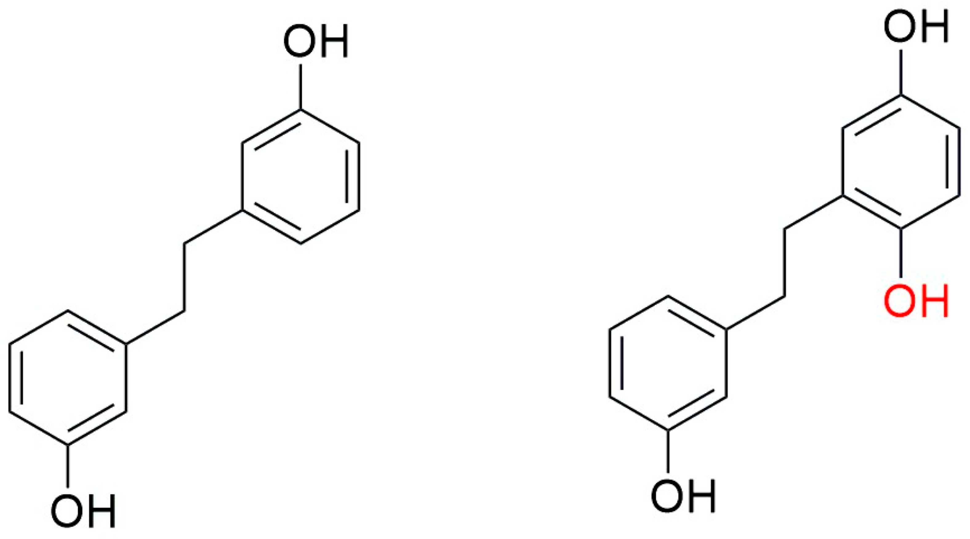
3.1. Diversity of Bibenzyls and Prebibenzyls

Bibenzyls are compounds with the dihydro-stilbenoidF type skeleton, and they are abundantly distributed in liverworts and serve as chemotaxonomic markers. Macrocylic bisbibenzyls and acyclic bisbibenzyls are derived from bibenzyls (see the next section). The following two consecutive sections describe the critical analysis of different bibenzyl and bisbibenzyls with their source taxa and structure elucidation through derivative formation following the spectral characteristics and mass profile most extensively used by researchers, as shown in Table 1.
Prebibenzyls, bibenzyls, prenylated bibenzyls, cavicularin and cannabinoid bibenzyls have varying carbon skeletons and are distributed in liverworts, as well as some angiosperm families. Plagiochila buchtiniana Stephani, P. diversifolia Lindenb. & Gottsche and P. longispina Lindenb. & Gottsche produce three prebibenzyls, which were also later reported from the New Zealand liverwort Balantiopsis rosea Berggr. The structures of these three prebibenzyls (longispinone A (17), longispinone B (18), and longispinol (19), Figure 7)) were confirmed using different spectroscopic techniques as (5S)-p-(methoxyphenylethyl) cyclohexan-1-one, (5R)-p-(methoxyphenylethyl) cyclohexan-1-ol and (5R)-p-(methoxyphenylethyl) cyclohex-2-en-1-one. These three compounds are probable precursors of the dicarboxylic bibenzyls or some macrocyclic bisbibenzyls [78]. An unusual phenylethyl cyclohexadienone was identified from D. hirsuta as dumhirone A (20) (Figure 7), the only reported natural product containing a 4-substituted-cyclohexa-2,5-dienone skeleton, and it can be synthesized from lunularin (21). The occurrence of dumhirone in D. hirsuta belonging to the family Marchantiaceae indicates the possible relationship of the biosynthetic pathways with genera like P. longispina (Plagiochilaceae) and B. rosea (Balantiopsidaceae) accumulating hydrogenated bibenzyl derivatives, though experimental evidence is required [51].
Lunularin (21, Figure 7) is a prebibenzyl obtained from M. polymorpha, Ricciocarposnatans and C. conicum [79]. Prebibenzyl prelunularin (22) (Figure 4) gives rise to lunularin, which, along with lunularic acid (23) acts as a precursor for bisbibenzyl synthesis, and it has been identified in many liverworts, including M. polymorpha, M. tosana, B. pusilla, Frullania convoluta Lindenb. & Hampe, and Monoclea forsteri Hook. [79] (see Figure 7). Bibenzyl 2,5,4′-trihydroxybibenzyl (24), a dimeric bisbibenzyl (6′,6″-bis-riccardin C, 25) and a phenylethylcyclohexenone (prelunularin, 22) were obtained from an axenic culture of Ricciocarpos natans [52]. The contents of Radula complanata (L.) Dumort. oil bodies were analyzed using GC-MS, and 3-methoxybibenzyl (26) was found as the main constituent [59].
Prenylated bibenzyl derivatives were obtained from the liverwort Lethocolea glossophylla (Spruce) Grolle, and their structures were elucidated through spectral analysis as 2-carbomethoxy-3,5,4″-trihydroxy-4,6-(3-methyl-2-butenyl)-bibenzyl (27); 2,4-di-(3-methyl-2-butenyl)-3,5,4′-trihydroxybibenzyl (28); 2,2-dimethyl-5-hydroxy-6-(3-methyl-2-butenyl)-7-[2-(4′-hydroxyphenyl)-ethyl]-chromene (29); 2,2-dimethyl-5-hydroxy-7-[2-(4′-hydroxyphenyl)-ethyl]-8-(3-methyl-2-butenyl)-chromene (30); and, with these, one bisprenylated bisbibenzyl named glossophylin (31). Two bibenzyls, 2-(3-methyl-2-butenyl)-3,5-dihydroxy-bibenzyl (32) and 2-geranyl-3,5-dihydroxy-bibenzyl (33), were identified from Radula voluta Taylor [57]. A new prenyl bibenzyl named 3, 5-dihydroxy-4-(3-hydroxy-3-methylbutyl) bibenzyl (34) was reported from Radula laxiramea [45]. Two cinnamoyls, bibenzyl pallidisetin A (35) and pallidisetin B (36), were reported from a moss Polytrichum pallidisetum Funck [58]. Two bibenzyl derivatives pelliepiphyllin (37) and 7-hydroxypelliepiphyllin (38) were obtained from P. epiphylla [44]. Two bibenzyls, brittonin A (39) and brittonin B (40), and two bibenzyl derivatives, 3, 3′-4,4′-tetramethoxybibenzyl (41) and chrysotobibenzyl (42), were obtained from Frullania inouei S. Hatt. [55], though the bibenzyls were earlier reported from Frullania brittoniae subsp. truncatifolia (Stephani) R. M. Schust. & S.Hatt. [54].
Cavicularia densa Stephani (family Blasiaceae) produces a novel optically active cyclic compound (+) cavicularin (43) (Figure 7), which is one of the unusual natural products with a bibenzyl and dihydrophenanthrene unit conjugated via a biaryl bond and an ether linkage. This compound lacks a stereogenic carbon center but, possessing conformational chirality, is one of the major attractions of synthetic chemists. This compound might be formed with intramolecular phenolic oxidative coupling between C-3′ and C-10′ from riccardin C (15) [18].
Radulanolide (44), a bibenzyl with a chemical formula of C20H18O4, was identified from Radula complanata [40]. Radulanin A (45) and radulanin C (46) are seven-membered heterocyclic rings and are richly present in R. buccinifera (Hook.f. & Taylor) Gottsche, Lindenb. & Nees, R. complanata, R. japonica Gottsche ex Stephani, R. tokiensis Stephanii and R. variabilis S. Hatt., among which the last four species also produce radulanin E (46) [40]. Radulanin H (48) is extracted from R. complanata and R. perrottetii [40]. Two bibenzyls with a dihydrooxepin skeleton, radulanin A-5-one (49) and β-hydroxyradulanin 5-one (50), were reported in Marsupidium epiphytum Colenso [19]. The morphological separation of different sections of the genus Radula is supported by chemical evidence; i.e., prenylated bibenzyls or bibenzyl derivatives with dihydro-oxepin skeleton have only been noted in Radula species. So, these bibenzyls are significant chemosystematic markers of the family Radulaceae. Angiosperm families Compositae and Leguminosae also possess such bibenzyls [40,79]. Radulanin L (51) was reported from Radula complanata [56].
Perrottetins A-D (5254, 10), perrottetin E (13) and perrotetin F (13) were first reported from R. perrottetii [40]. Perrottetin H (16) is an acyclic bisbibenzyl obtained from Marchantiophyta and used as a chemosystematic marker [79]. Isoperrottetin A (56), a novel bisbibenzyl obtained from R. perrottetii [71]. R. perrottetii and Radula marginata produce (-) perrottetinene (14) (Figure 7), a cannabinoid derivative with the cis moiety structurally analogous to tetrahydrocannabinol from Cannabis sativa L. PerrottetinG (57) and perrottetinenic acid (58) were also obtained from Radula perrottettii and Radula marginata [46,56,71]. Bibenzyl tetrahydrocannabinoids, which were previously reported from higher plants, have trans geometric isomerism at the cyclohexane ring. Apart from the structural analogy, some shared features have been observed in C. sativa L. and Radula marginata; that is, the cannabinoids in C. sativa are stored in specialized trichome, whereas in R. marginata, oil bodies are the depository sites of these secondary metabolites [80]. 10′-Hydroxyperrottetin E (59), 10′-hydroxyperrottetin E-11-methylether (60), 14′-hydroxyperrottetin E (61), 10, 10′-dihydroxyperrottetin E (62), 13′, 13″-bis(O′-hydroxyperrottetin E) (63) and perrottetin E-11-methyl ether (64) were isolated from P. epiphylla [44].
Dihydroresveratrol (65) (resveratrol, a well-known stilbenoid compound of red wine with antineoplastic properties; Figure 7), previously isolated from Dioscorea dumentorum, was also reported from Blasia pusilla of Metzgeriales [19].

3.2. Diversity of Macrocyclic and Acyclic Bisbibenzyls

Bisbibenzyls are the dimeric form of bibenzyls present in cyclic or acyclic forms in different orders of Marchantiophyta, and their derivatives are also naturally present in some taxa of liverworts, as seen in Table 1. Naturally produced from two molecules of lunularin (21), each has a core structure consisting of four aromatic rings and two ethano bridges. Marchantin type bisbibenzyls are more diverse and predominantly reported from different species of Marchantia and also from some other genera, and they act as chemotaxonomic markers [15,19].

3.2.1. Marchantins, Isomarchantins and Their Derivatives

Marchantins are a class of bisbibenzyl compounds with two diaryl ether subunits. The biosynthesis of the diaryl ether natural products is evolving continuously. Their structural analogs are given much importance due to their pharmacological implications [18,21,32,81,82].
The oxygen atom in diaryl ether is a favorable flexible linker that can allow molecules to form multi-dimensional conformation, and due to the binding affinity to a potential target, its importance in molecular drug design is increasing [83]. Marchantins were the first reported bisbibenzyls [40] among all the macrocyclic bisbibenzyls, and they represent the largest class of bisbibenzyls reported from different species of the genus Marchantia (M. polymorpha, M. palmata, M. diptera subsp. paleacea and others) and also from the genera P. intermedium, Reboulia hemisphaerica, etc. (Figure 5) [30,61,66]. The structures of the marchantin series compounds were determined through 1H and 13C NMR, and COSY. The structure of marchantin A (1) was determined through chemical degradation and X-ray crystallographic analysis of its trimethyl ether (66). Protonated aromatic carbons were identified using the 13C-1H correlation spectrum. The remaining aromatic and benzylic carbons are determined using long-range selective proton decoupling (LSPD) experiments. As the other compounds of the marchantin series are non-crystaline and available in very small quantities, the total assignment of 1H-13C would facilitate the detection of the other members of this class [21,31,60,61]. The 1H NMR spectrum showing the characteristic signal appearing at δ 5.13 (J = 2 Hz) is due to H-3′ strongly shielded by ring A [31], whereas the proton signal for marchantin C (3) and isomarchantin C (67) appears at δ 5.52 and δ 6.08, respectively [61]. Marchantin A (1) and its trimethyl ether (66) are difficult to crystallize as they are tightly protected from water/solvents. To eliminate the crystallization difficulty, a small column packed with silica gel and dried magnesium sulfate (1:1) was prepared, and each sample passed through this column gives a beautiful single crystal [21]. The total synthesis of marchnatin A (1) was accomplished after its discovery from the natural product [84]. The Indian and French races of M. polymorpha produce marchantin E (4) as major components, whereas Japanese M. polymorpha produces marchantin A (1) as a major component [61,85]. Marchantin B (2) and C (3) were also discovered [30] with two ether links from M. polymorpha, M. paleacea subsp. diptera and M. tosana. A few years later, Asakawa and coworkers reported Marchnatin J-L (6870) from the German race of M. polymorpha. Marchantin J trimethyl ether (71) possessed an ethoxyl group resembling marchantin E. 1H and 13C NMR spectroscopy, including NOE difference spectra, confirmed the structure of the derived compound, and this is the first record of the ethoxylated compound from liverworts [86]. The 1H and 13C NMR signals of marchantin M (72) were very close to those of marchantinquinone (73) except 13C absorption on the B-ring and two protons (δ 6.62, d and 6.70, d, J = 8.6 Hz) on ring B, and the two hydroxyl and methoxy substituents must be located on either C-10 or C-13, as well as a strong molecular ion m/z at 454. Final assignments were based on the hydroxyl group at C-10 based on the results of deuterium-induced 13C chemical shifts, and the structure was defined as marchantin M (72). Another similar compound giving the singlet proton peak at δ 5.73 on the B-ring suggested the methoxy group at either C-11 or C-12. The functional group at C-10 and C-13 was either a ketone (benzoquinone) or a hydroxyl (hydroquinone). 13C shifts of the compound were assigned to marchantin N (74), as the placement of the methoxy group at C-11 was mainly based on the shifts of C-10 and C-13 [66]. Marchantin O (75) was purified from Taiwanese liverwort R. hemisphaerica and characterized using NMR spectra and earlier isolated and identified from Japanese R. hemisphaerica [66]. Marchantin P (76) was characterized based on M. chenopoda L. of Venezuela [87]. Isomarchantins have a similar structure to marchantins, though the connectivity between arenes B and D is opposite to that found in the marchantins. Isomarchantin C (67) was isolated from Indian M. polymorpha and M. palmata Reinw., Nees & Blume, followed by structural characterization by the 1H and 13C NMR spectra [61]. Neomarchantin A (5) and B (77), with diaryl ether subunits, were first isolated from a leafy liverwort S. glaucescens and a thalloid liverwort Preissia quadrata (Scop.) Nees [49]. In these compounds, two para- and meta-substituted arenes form the macrocycle and impart some unusual bond angles to the natural products in he solid state. The structure elucidation was carried out using COSY, HMQC, HMBC and NOE data [49].
Air-dried M. palmata was extracted with diethyl ether, and the ether-soluble portion was treated with alkali and neutralized with diluted HCl and further separated with column chromatography. Bibenzyl mixtures of the same Rf value were obtained from the fraction further methylated to form marchantin G methyl ether (78), which was confirmed through 1H NMR [61].

3.2.2. Bisbibenzyls of Riccardin Family and Derivatives

The Riccardin family of compounds has 18 membered rings, and its members are characterized by one diaryl ether, one biaryl linkage, and a para-disustituted A ring. Benzene units are connected via ether oxygen between rings A and B and a biphenyl bond between the benzene rings C and D, which has been reported since the 1980s. Riccardia multifida subsp. decrescens produces riccardins A and B (1112) [30]. Riccardin C (15) was isolated from B. pusilla, D. hirsuta, Jungermannia infusca (Mitt.) Stephani and M. paleacea subsp. diptera, Mastigophora diclados, Monoclea forsteri, Plagiochasma pterospermum C. Massal, P. rupestre (G.Frost) Stephani, R. hemisphaerica and Ricciocarpus natans, riccardin F (79) from M. tosana. Riccardin C was the first macrocyclic bisbibenzyls reported from the liverwort [88] and a higher plant [76]. The structure of riccardin A (11) was confirmed based on the formation of its diacetate and trimethyl ether derivatives [30]. The 1H NMR spectrum of riccardin A (11), a strongly shielded one-proton doublet at δ 5.33 (1H). has been assigned to an Inner proton H-3′ on benzene ring C, which lies over the plane of benzene ring B [30]. The structure of riccardin B (12) was determined based on a comparison of the 1H and 13C NMR spectra with those of riccardin A (11) and its derivatives. The methylation of riccardin C (15) gave trimethyl ether, whose spectral data were identical to the dimethyl ether of riccardin A [88]. The structure elucidation of riccardin D (9), riccardin E (80), riccardin F (79) and riccardin G (81) was performed through extensive decoupling and NOE spectrometry on the naturally occurring compounds and their premethylated products [30,87]. Riccardins A and B may be biosynthesized from lunularic acid (23) or lunularin (21), which are widespread in thalloid liverworts [30]. Riccardin family members were synthesized through a convergent scheme [89]. Another riccardin family member, riccardin H (8), and 13,13′-O-isopropylidenericcardin D (6) were reported from Chinese M. polymorpha [33]. Another novel variant, benzyloxidized bis (bibenzyl), belonging to riccardin and named riccardin I (82), was isolated from A. angusta [65]. Macrocyclic bisbibenzyl mono ether was reported for the first time from M. palaeacea [63]. Phenolic protons are ascribed based on the broad singlet at δ 5.37 and 5.84 and the quinone carbonyl carbons at δ 187.4 and 187.5 from 13C NMR spectra. The presence of the bisbibenzyl moiety was indicated by the appearance of four characteristic carbon peaks at δ 31.0, 33.9, 33.9 and 38.1 in 13C NMR spectra, and the ether oxygen linked to two benzene rings is further confirmed by DEPT and HMBC correlation.
Isoriccardin C (83) was reported from Indian M. polymorpha and M. palmata [61]. Hong Kong liverwort M. paleacea thallus in 95% ethanol gives rise to two novel isoriccardinquinones, A (84) and B (85), along with marchantin C (3), isoriccardin C (83) and a phenanthrene derivative, 2-hydroxy-3,7-dimethoxyphenanthrene (86), whose structures were confirmed using 2D NMR [63]. Isoriccardin D (87), along with other bisbibenzyl derivatives, was identified from Chinese M. polymorpha in ethanol extract [64].
Asterelins A and B (8889), the first dibenzofuran bisbibenzyls obtained from A. angusta, and the elucidation of their structure were established using 1D and 2D NMR, MS and XRD. The coupling patterns of proton resonances in the range from δH 5.33 to 7.47 established the presence of four independent aromatic rings. The four signals at δH 6.61, 7.47, 5.98, and 6.29, could be assigned to 1,4-disubstituted benzene ring (ring A) from their coupling patterns using the 1H-1H COSY, while the two resonances at δH 6.83 and 6.87 suggest the presence of a 1,2,3,5-tetrasubstituted benzene ring (ring B). The signals at δH 5.33, 6.84 and 6.84, together with resonances at δH 6.28, 7.14, and 7.26, indicated the presence of 1,2,4-trisubstituted benzene (ring C and D, respectively). Both the compounds contained a novel dibenzofuran linkage, and the stereochemical structure of asterelin B (89) suggested the presence of a magnetically anisotropic effect in ring A; the compounds are present as a natural product, as confirmed via the HPLC-UV analysis of cold diethyl ether extract, not as an artifact [72].

3.2.3. Pusilatins-Derivatives of Riccardin

B. pusilla L., another genus of the family Blasiaceae, produces riccardin C (15) dimer, i.e., pusilatins A, B, C and D (9093), which are also produced through oxidative coupling reactions [53]. Another novel structure was elucidated in the consecutive year, and the monomethyl ether of pusilatin B (91) was named pusilatin E (94) isolated from Riccardia multifida subsp. decrescens [53]. In pusilatin A (90), four benzyl methylene signals (δ 35.7, 37.9, 38.6 and 38.8) were detected in the 13C NMR. The 1H NMR spectrum indicated the presence of 12 protons on benzene rings at δ 5.37–7.14 ppm and four benzylic methylenes at δ 2.61–2.92 ppm (8H). Spectral data show that pusilatin A is a dimer of riccardin C with a C12-C-12″ phenyl linkage. Pusilatin A (90) shows a similar coupling pattern with riccardin C, and the structure was established with a 2D NMR spectra, including 1H-1H COSY, HMQC and HMBC. The stereostructure of pusilatin A was established using the X-ray crystallographic analysis of its hexaacetate derivatives [53]. Also, the spectral data of pusilatin B were almost the same as those of pusilatin A. Pusilatin C (92) is an asymmetrical dimer of riccardin C (15). Pusilatin D (93) is regarded as a dimeric riccardin C linked by an ether C-12-O-C-1‴ bond. The 13C NMR spectrum exhibited 56 signals, including 8 benzyl methylene signals (δ 37.8, 37.89, 37.92 (two carbons), 38.3, 38.5, and 38.7 (two carbons)). Methylated and acetylated derivatives further confirm the location of functional groups and linkage patterns [53].

3.2.4. Paleatins

Two acyclic bisbibenzyls were obtained from M. paleacea subsp. diptera, i.e., paleatin A and paleatin B (9596), where rings A and C, and B and D, are connected through oxygen bonds. Paleatin B (96) differs from A due toan additional methyl group [21].

3.2.5. Plagiochin Type Bisbibenzyl

Plagiochins A, B, C and D (97100), first isolated from Plagiochila sciophila Nees (=Plagiochila acanthophylla subsp. japonica), possess two ortho-biphenyl linkages between two benzyl groups. 1H, 13 C NMR and NOE studies, as well as XRD analysis on its tetramethyl ether, showed that ring A is perpendicular to ring C and parallel with ring D. The proton at −3′ is strongly shielded by both rings A and D, causing a high field shift to δ 4.84 in the tetramethyl ether of plagiochin A (97) [90], and the same phenomenon has also been observed in marchantins and riccardins [30,31], as mentioned earlier. Plagiochins E (7) and F (101) were C7′-C8′ dihydroisoplagiochin A and 10-hydroxy C7′-C8′ dihydroisoplagiochin A, respectively [33]. Plagiochins E (7) and F (101) were synthesized through intramolecular Wittig reactions [91] and are supposed to exist in nature based on energetic and biosynthetic considerations [92], but the second one was never been isolated [91]. Isoplagiochins A and B (102a103a) were first isolated from the liverwort Plagiochila fruticosa Mitt. [68]. Further fractionation of the crude extract of the same species resulted in the isolation of isoplagiochins C and D (104105), and their structures have been elucidated [68]. Furthermore, isoplagiochin A was isolated from culture gametophytes of Heteroscyphus planus (Mitt.) Schiffner [69]. The hydrogenation of this compound gives a dihydro derivative. The 1H and 13C spectra of of isoplagiachin C resemble those of isoplagiochin A (102a), except for the signal patterns of a D ring; an additional phenolic hydroxyl group is at the D ring in place of the ethereal oxygen in isoplagiachin A. Isoplagiochin C (104) forms a tetraacetate, and the location of the hydroxyl group at C-13′ and the whole structure was determined using HMBC, HMQC and NOESY [68]. Isoplagiochin A (102a) and planusin A (106) were obtained from culture cells of H. planus, and their structures were determined using spectral analysis [69].
Isoplagiochins C and D (104105) were obtained in an enantiomeric mixture at 85:15 and 48:52 ratios, in favor of the enantiomer possessing the p–configuration (p-64 and p-65) [21]. Three novel bisbibenzyls, namely isoplagiochoin F (107), dihydroisoplagiochin (108) and a chlorinated derivative monochlorinated isoplagiochin D (109), were isolated from a Plagiochila sp. [67].
2,12-Dichloroisoplagiochin D (110), 12,7′-dichloroisoplagiochin D (111) and 12, 10′-dichloroisoplagiochin C (112) were isolated from Herbertus sakurii of Herbertaceae along with isoplagiochin C (104) and D (105), and 2,12-dichloroisoplagiochin D (110) and 12-chloroisoplagiochion D (113) were isolated from Mastigophora diclados (Brid. ex F.Weber) Nees, suggesting the close chemical relation of these two genera [70].

3.2.6. Halogenated Bisbibenzyls

Bazzanins A, B, C, D, E, F, G, H, I and J (114123) are isoplagiochin derivatives with two biphenyl linkages and are substituted with 1–6 chlorine atoms, whereas bazzanin K (124) is a dichlorinated macrocycle consisting of a phenanthrene and a bibenzyl moiety connected with two biphenyl linkages, which are isolated from Bazzania trilobata (L.) Gray [74]. The linkages of the bibenzyl moiety are detected through HMBC and NOESY [74]. Bazzanins M-S (125131) were obtained from Lepidozia incurvata [19].

3.2.7. Ptychantols

Three novel macrocyclic bisbibenzyls, named ptychantols A, B and C (132134), possess a trans-stilbene moiety isolated from Ptychantus striatus. Ptychantol B was the first reported macrocyclic bisbibenzyls possessing two biphenyl linkages between rings A and C and between rings B and D. The methylation of ptychantol A gave a dimethyl ether, whose IR spectrum indicated neither hydroxyl nor carbonyl absorption bands, indicating the presence of two ether oxygen in this compound. Hydrogenation gave a dihydroderivative, showing that this compound contains one olefinic group. The substitution pattern was suggested based on the 1H, 13C NMR, HMBC and NOESY spectra [73]. Another compound named dihyrdroptychantol A (i.e., 1′, 13′-dihydroxyisomarchantin, 135a) was isolated from the natural source A. angusta [72], asproposed earlier via a computational method [92].

3.2.8. Polymorphatins

This is a bisbibenzyl derivate with a novel skeleton named Polymorphatin A (136) [64] with a 1,4 disubstituted aromatic ring (ring A) and three 1,2,4-trisubstituted aromatic ring (rings B–D); in addition, the 13C NMR spectra shown δ (C) at 36.2, 37.3, 38.7 and 38.1 though the spectral discrepancies were reported in natural and synthetic materials [28].

3.2.9. Glaucescenolide and Bisbibenzyl Glaucescenolides (GBB)

Glaucescenolide (137) is a compound with a sesquiterpenoid skeleton reported first from S. glaucescens, a plant source, earlier known only from marine mollusks and sponges [93]. Glaucescenolide shows an IR spectrum at 3378 cm−1 for hydroxyl absorption and a carbonyl absorption at 1740 cm−1, indicating the presence of a α, β –unsaturated γ-lactone. Scher et al. (2002) first proposed that furan, a natural product, can be an intermediate linking the biosynthetic pathways for glaucescenolide, GBB A and GBB B (138139) (Figure 6) [49].

4. Structure–Activity Relationship of Bisbibenzyls and Bibenzyls

Bisbibenzyl compounds show a number of bioactivities like antibacterial [94], antiviral [47,95,96], antifungal [94,97,98], cytotoxic [18,23,42,99], antioxidant [18,19,100], neuroprotective [81], antivenomous [101], HAT treatment [102], muscle relaxing [103] and others. Bibenzyl and bisbibenzyl compounds involved in diverse types of bioactivities, as established through in vitro, in vivo and in silico studies, are the main contents of the discussion below.
The pusilatin and riccardin classes of compounds from liverwort were tested for their inhibitory activities against HIV-1 reverse transcriptase (RT), HIV-2 RT, a mutant RT, avian myeloblastosis virus RT, DNA polymerase β and RNA polymerase [53]. None of the viruses or enzymes were inhibited except DNA polymerase β, which was inhibited at IC50 ranging from 5 μM to 38 μM [53]. However, studies on the influenza virus revealed the efficacy of bryophyte phytoconstituents possessing a particular substructure. The RNA-dependent RNA polymerase of influenza virus has an acidic protein (PA) subunit with endonuclease activity, which carries out the decapping of host pre-mRNAs, resulting in the initiation of the viral transcription process [104]. Hence, the endonuclease activity has become a target for studies on anti-influenza activities. Iwai et al. (2011) screened 33 phytochemicals using assays, like PA endonuclease inhibition in vitro and anti-influenza A virus. Marchantins A (1), B (2) and E (4); plagiochin A (97); and perrottetin F (55), isolated from liverworts, were found to inhibit influenza PA endonuclease activity in vitro [47]. The authos found that the aforementioned compounds are effective because they have a 3,4–dihydroxyphenethyl substructure in them, apparently contributing to the inhibition of PA endonuclease, as was already reported [105] from a study of thalidomide derivatives containing the same substructure of 3,4-dihydroxyphenethyl. Among the effective compounds, the authors found marchantin E to inhibit the growth of both influenza A (H3N2 and H1N1) and B viruses, whereas marchantin A (1) and perrottetin F (55) were inhibitory to the growth of influenza B only. In addition, all five of the above compounds also reduce viral infectivity, with marchantin E (4) showing the highest activity, probably because of its fitting and chelating model of the inhibition of PA endonuclease activity revealed in a docking simulation study [47]; see Figure 7.
Figure 7. The structures of marchantin E (4), epicatechin gallate and phenethyphenylpthalimide adapted from [105].
Plants 12 04173 g007
The PA endonuclease activity of these compounds and other structurally similar compounds has been ascribed to the chelation of the manganese ions present in the active site of the enzyme through X-ray crystallography [106,107] and also interactions with different active site amino acids, as revealed by mutational studies [108]. The macrocyclic structure of marchantin-type compounds in combination with oseltamivir would be effective against the influenza virus.
Marchantin A (1) was first reported for its antibacterial activities [40,86], i.e., growth inhibition of several Gram-positive and Gram-negative bacteria with varying inhibition concentrations, and the findings were later corroborated [85], also the analogs and derivatives of this compound was tested for comparison. This macrocyclic compound possesses antibacterial activity, as it has diaryl ether or biphenyl bonds [109]. Marchantin A (1) was isolated from M. emarginata subsp. tosana (Stephani) Bischl., and it has been established that it induces cell growth inhibition in human MCF-7 breast cancer cells at IC50 4.0 µg/mL. Marchantin A (1) induced a reduction in the cell viability of breast cancer cell lines A256, MCF7 and T47D (IC50 5.5, 11.5 and 15.3 µM, respectively). The effect was dramatically increased in all cell lines in a synergistic manner. Marchantin A (1) possesses antimelanoma activity (IC50 7.45–11.97 µg/mL) against human malignant melanoma cell lines (A375) [32,110,111].
4-Hydroxy-3′methoxybibenzyl (140) from a liverwort Plagiochila stephensoniana Mitt. exhibited antifungal activities due to the free hydroxyl group [112]. Two bibenzyls, viz., 3,4′-dimethoxyl-4-hydroxybibenzyl (141) and 3-hydroxy-4′-methoxybibenzyl (142), isolated from Frullania muscicola Stephani showed potent inhibitory effect against fungi [113]. Bisbibenzyls 6′,8′-dichloroisoplagiochin C (bazzanin B, 115), isoplagiochin D (105) and 6′-chloroisoplagiochin D (bazzanin S, 131) showed strong antifungal activities and large inhibition zones. Furthermore, bazzanin S (131) was compared with its permethylated derivatives against the pathogenic fungi, wherein bazzanin S showed a larger inhibition zone than the latter [75]. Asterelin A (88), asterelin B (89), 11-O-demethyl marchantin I (143), dihydroptychantol A (135a), marchantin H (144), marchantin M (72), marchantin P (76), perrottetin E (13), plagiochin E (7) and riccardin B (12) show moderate antifungal activity against common clinical pathogenic fungus C. albicans, with MIC value ranges from 16 μg/mL to 512 μg/mL. MIQ and MIC values of asterelin A are lower than asterelin B, indicating that the methylation of the OH groups decreases their antifungal activity [72]; see Figure 8.
Figure 8. The structures of Asterelin A [R=OH] 88 and Asterelin B [R=OMe] 89 adapted from [72].
Plants 12 04173 g008
From the structure–activity point of view, previous studies also support the idea that the free hydroxyl group plays an important role in antifungal activity [33,75]. Riccardin D (9), another macrocyclic bisbibenzyl, inhibited biofilm formation by C. albicans, and it downregulated the hyphae-specific gene expression to inhibit hyphae formation [12]. Riccardin F (79) and riccardin C (15) showed antifungal activities against the fluconazole-sensitive and -resistant strains of C. albicans, with MIC values ranging from 32 μg/mL to 256 μg/mL, whereas pakyonol (145), neomarchantin A (5), isoriccardin C (83) and marchantin H (144) were inactive, wth MIC values >512 μg/mL. The presence of an additional aromatic O-methyl group may reduce the in vitro antifungal efficacy [60]. Macrocyclic bisbibenzyl may exert direct antifungal action by forming pores in the cell membranes, leading to fungal cell lysis [114]. Riccardin C (15), in combination with fluconazole with synergistic/additive activity, reduced the MIC from 256 μg/mL to <8 μg/mL. The possible reason for this drastic reduction seems to be the increased permeability of fluconazole and the inhibition of the drug efflux pump on the membrane [115]. Plagiochin E (7) acts as an antifungal agent and acts through the metacaspase-dependent apoptotic pathway in C. albicans. It induces G2/M cell cycle arrest, as evidenced by other cellular changes, contributing to the inhibition of cell cycle regulators CDC28, CLB2 and CLB 4 in C. albicans [116]. The diethyl ether extract of the liverwort A. angusta contained two minor bisbibenzyls, riccardin I (82) and angustatin A (146). Th latter one is the first non-heteroatom-containing variant possessing a bibenzylphenanthrene skeleton. Both variants possess moderate antifungal activities against the strains of fluconazole-sensitive and -resistant C. albicans [65].
Riccardin G (81), isolated from the Italian Lunularia cruciata (L.) Dumort. ex Lindb., showed cytotoxic activity against two human lung cancer cell lines, A549 and MRC5 [117]. Riccardin D (9) also demonstrates the induction of apoptosis against human non-small-cell lung cancer (NSCLCH460) and A549 (human non-small-cell lung carcinoma cells) and H460 (lung carcinoma epithelial cells) due to DNA topoisomerase-II inhibition [118].
The same bisbibenzyl (9) inhibited the proliferation of HUVECs (human umbilical vascular endothelial cells), decreased mortality and migration of HEVECs and vascular endothelial growth factor (VEGF) against human lung cancer H460 cell lines [119]. Riccardin D (9) also exhibited the prevention of intestinal adenoma (polyp) formation in APCMin/+ mice, a decrease in beta-catenin and cyclin D1 expression, the prevention of proliferation of intestinal polyps, and the triggeroing of apoptosis via the caspase-dependent pathway and te decrease in angiogenesis in intestinal polyps [120].
Riccardin D (9) may inhibit cell proliferation and induce apoptosis in HT-29 cells. This may be associated with the blocking of the NF-ΚB signaling pathway [121]. Hu et al., 2014, reported that the same bisbibenzyl (9) exhibited cytotoxic activity through the induction of apoptosis and the inhibition of angiogenesis and toposimerase-2. They also confirmed that apoptosis was not the sole mechanism by which riccardin D inhibits tumor cell growth, because a low concentration of riccardin caused cellular senescence in prostate cancer PCa cells, one of the most common malignant prostate tumors [122]. Riccardin D-26 derived from riccardin D (9) significantly inhibited cancer growth in both KB and KB/VCR xenografts without significant toxicity. It also inhibited cancer growth by inducing apoptosis in the activation of the mitochondria-mediated intrinsic apoptosis pathway. The above activity is stronger than that of the original riccardin D (9) [123].
Cyclic bisbibenzyls (marchantin A (1), B (2), D (147), E (4), riccardin C (15), isoriccardin C (83), acyclic bisbibenzyls (paleatin B, 95), bibenzyls (perrottetin D, 10 and radulanin H, 14) and NDGA were tested to see the inhibition efficacy in the key enzymes cyclooxygenase and 5-lipooxygenase 5-(LOX) of the arachidonic acid cascade. Acyclic paleatin B (95) showed the highest activity due to its best fit into the active site of the enzyme and is additionally stabilized by its lipophilic chain. Perrottetin D (10) also possessed the same activity, but it functions as a radical trap, existing as a free radical itself under the elevated temperature of the assay. Of marchantin A (1) and B (2), the latter showed two-fold more activity than the former, as it possesses two catechol moieties. Similarly, marchantin A (1) also showed inhibition on 5-lipooxygenase, as it possesses one catechol moiety in ring C and a phenolic hydroxyl group in ring A. Another compound of this series, i.e., marchantin D (147), reduced the activity five-fold due to the presence of a hydroxyethyl bridge, but the blocking of this hydroxyl group via methoxylation restores the activity almost to the level of marchantin A. Monophenols isoriccardin C (83), riccardin C (15) and radulanin H (48) exhibited the lowest reactivity among the compounds [124]; see Figure 9.
Figure 9. The structures of different bibenzyl and bisbibenzyls compared with another phenolic compound NGDA adapted from [124].
Plants 12 04173 g009
However, marchantin H (144) showed an IC50 value of 0.51 ± 0.03 μmol/L against the non-enzymatic iron-induced lipid peroxidation in rat brain homogenates. In the same study, marchantin H (144) also suppressed NADPH-dependent microsomal lipid peroxidation with an IC50 value of 0.32 ± 0.01 μmol/L without affecting the microsomal electron transport of NADPH-cytochrome P450 reductase. Marchantin H (144) can scavenge free radicals in the aqueous phase and act in a concentration-dependent manner. Marchantin H is a potentially effective and versatile antioxidant, and hence, it can be used as a chaperon protecting biomacromolecules against peroxidative damage [125].
Marchantin A trimethyl ether (66) was reported for having skeletal-muscle-relaxation properties compared to d-tubocurarine possessing the bisbibenzyl ether moiety, which differs from MATE in terms of the connection of the B and D rings only. d-TC combines with the nicotinic cholinergic receptor at the post-junctional membrane in the motor end-plate and thereby shows competitive inhibition with the transmitter action of acetylcholine. XRD data of MATE show the convex and concave surfaces surrounded by four benzene rings, and the central hole at the concave surface is responsible for the bioactivity [103]. Biological activity was explained with conformational analysis and computational chemistry, and the underlying mechanism of action was explained based on the minimum energy conformers [92,126]. Some bibenzyls with cyclic and acyclic ring structures were investigated for calmodulin inhibitory activities, and compounds like marchantin D and marchantin E showed a moderate effect due to the presence and nature of a nucleophilic atom at the ethano bridges, while the perrotettin E and perrotettin A showed increasing inhibitory concentrations due to the former being a linear bisbibenzyl and the latter being a monomeric stilbene where the macrocyclic ring influences the calmodulin-inhibitory activity [126]; see Figure 10.
Figure 10. The structures of perrottetin A 52 and perrottetin E 13.
Plants 12 04173 g010
Isoplagiochins A and B (102a–103a) show the inhibition of tubulin polymerization at IC50 50 μM and 25 μM, respectively, but their dihydro derivatives (102b, 103b) are less potent, with an IC50 > 100 μM, indicating that the restricted biaryl ring system of bisbibenzyls may be favorable for tubulin binding [127]; see Figure 11.
Figure 11. The structures of isoplagiochin A and B and their dihydroderivatives adapted from [127].
Plants 12 04173 g011
Genes regulating bile acid and cholesterol homeostasis are activated by the farnesoid X receptor (FXR), which has endogenous ligands, as well as some other non-steroidal and steroidal ligands. Suzuki et al. (2008) first reported marchantin A (1) and marchantin E (4), along with five bile acid derivatives that activated FXR in a reporter assay. The gene expression elevated by the screened compounds was much different for Cos-7, HepG2, HuH-7 and Caco-2 cells, and the genes regulated by FXR act in a cell-type-specific or gene-selective fashion, indicating the FXR target gene expression can be regulated by the molecular design of the compounds. Also, the screened compounds can be useful for studying FXR modulation, leading to selective FXR modulation for therapeutic use [48].
Glaucescenolide (137) is the most cytotoxic compound, with an IC50 of 2.3 µg/mL against P388 leukemia cells, and the mechanism of action shows similarity with other α, β-unsaturated sesquiterpene lactones, i.e., the alkylating activity, through the Michael addition of biological nucleophiles. Bisbibenzyl neomarchantin A (5) and B (77) showed weak to moderate cytotoxicity against P388 cells and antimicrobial activity against the Gram-positive bacterium Bacillus subtilis [49]. The inhibition potency of lipopolysaccharide-induced nitric oxide synthase (NOS) in RAW 264.7 macrophases was tested with marchantin A (1) and derivatives. Marchantin A (1) showed the strongest inhibition, with an IC50 of 1.44 μmol/L and the introduction of the hydroxyl group and methylation, and the positions of ether linkages in the ring regulate the inhibition [109]. Macrocyclic bisbibenzyls are also tested to see their role in apoptosis and cell cycle regulation, i.e., the major controlling mechanism of cancer cells. Marchantin A-treated MCF-7 cells show suppressed cyclin B1 gene expression, whereas marchantin C-treated human A172 glioma cells show increased cyclin B1 levels, indicating the different methods of cell cycle regulations of the two compounds [128]. Marchantin A (1) induces cytotoxicity and suppresses the proliferation of MCF-7 breast cancer cells via apoptosis through a caspase-dependent pathway and also influences the cell cycle [32]. Marchantin A (1) possesses two hydroxyl groups at C1′ and C6′ positions, which are very important parameters for scavenging free radicals like DPPH. The same two adjacent hydroxyl groups have been implicated in cytotoxicity induction, and, on the contrary, marchantin C has no hydroxyl group at the C6′ position, showing less antioxidant activity [128]. Later on, it was established that marchantin A and marchantin C both have the potential to develop candidate drugs for chemotherapy. Marchantin C induces the mitochondria-dependent intrinsic apoptosis pathway by arresting the G2/M phase of the cell cycle, as observed in vitro and in vivo. It upregulates Bax expression but downregulates Bcl-2 expression [128]. The study with marchantin C (3) was further extended by the same group of workers to see anti-invasiveness and antiangiogenic activity in glioma cells, and it was observed that marchantin C (3) inhibited the progression in a dose-dependent manner by affecting the MMP-2 activity via the MAPK pathway. However, further confirmation is required for a complete understanding of the mechanism of marchantin C (3) in brain cancer invasion and the migration of cancer cells [99].
A nitrogen-containing marchantin C and brominated products (148–150) prepared from marchantin C (3), as shown in Figure 12, possess cytotoxicity against KB, MCF-7 and PC3 (IC50 6.3–27.2 µM). 10-Bromo-(148), 11-bromo-(149) and 12-bromomacrchantin C (150) are slightly more active than marchantin C (3). The dimer (151) and 12-N,N-dimethylaminomarchantin C (152) are less active than the brominated products [129]. Marchantin C (3) strongly inhibited the growth of human cervical tumor xenografts in a nude mouse model and decreased the quantity of microtubules in a time- and dose-dependent manner at the G2/M phase in human glioma tumor cells and HeLa (human cervical adenocarcinoma cell line) cells at 8–16 µM [99,128].
Zhang et al., 2015, reported that marchantin M (72) circumvented the growth of prostate cancer PC-3 tumors and upregulated expressions of CHOP and GRP78. The same authors indicated that marchantin M limited the proliferation and favored the apoptosis of DU145 cells in a time- and dose-dependent manner. Jian et al., 2013, found that marchantin M (72) induced autophagy-dependent cell death, which was accompanied by the induction of endoplasmic reticulum (ER) stress and the inhibition of proteasome activity in PCa cells [129,130].
Figure 12. Preparation of bromomarchantin C (148150) and dimeric marchantin C (151) from marchantin C (3).
Plants 12 04173 g012
Highly methoxylated bibenzyls, viz., 3,3′,4,4′-tetramethoxybibenzyl (41), chrysotobibenzyl (42), brittonin A (39) and brittonin B (40), were investigated to see the potentiality of inhibition in vincristine-resistant KB/VCR and adriamycin-resistant human myelogenous leukemia cells (K562)/A02 cells. Moderate cytotoxicity, with an LD50 value ranging from 11.3 μmol/L to 49.6 μmol/L, and the reversion of multi-drug resistance (MDR) with the reversal fold ranging from 3.19 μmol/L to 10.91 μmol/L at 5 μmol/L for vincristin resistant KB/VCR cells and 4.40 to 8.26 at 5 μmol/L for adriamycin-resistant K562/A02 cells were reported [55]. Plagiochin E (7) also has a reversal effect on MDR of K562/A02 cells [128]. DHA (135a), which are phenolic compounds, are the most promising MDR reversal agents. DHA and its structural analogs were tested on p-gp-mediated MDR in K562 and its multi-drug-resistant counterpart cells (K562/A02). DHA showed the largest potency to enhance adriamycin cytotoxicity in drug resistance cells with a reversal fold of 3.84–8.18, while its structural analogs were less effective. Phenolic groups binding to aromatic rings are necessary to increase the sensitivity of K562/A02 cells to adriamycin. When the phenolic groups are methylated, this activity is attenuated. On the other hand, the stilbenoid double bond in the structure has little effect on the reversal of MDR cells such as the hydrogenated structure in DHA. DHA (135a) acts as an antifungal product and has more potent MDR reversal activity by increasing the adriamycin cytotoxicity toward K562/A02 cells and vincristine cytotoxicity toward KB/VCR cells [131]. As mentioned above, DHA (135a) showed MDR reversal activity through the inhibition of the p-gp function and its expression, which prevents the efflux of drugs. However, some derived DHA compounds, i.e., with thiazole ring, such as the cyclic hexapeptide dendromide A, exhibited MDR reversal activity. Similarly, heterocyclic derivatives of combrestatins A, B, C and D, containing the bibenzyl moiety, were also cytotoxic to MDR cell lines [131]. In light of these results, researchers took an interest in the effect of thiazole moieties, and derivatives were synthesized to assess their biological activities, particularly MDR reversal activities, toward the cancer cell lines K56/A02 and KB/VCR and also molecular docking analyses to elucidate the binding modes of the analogs to p-gp [132]. DHA analogs were shown to be more potent in MDR reversal activities and increased vincristine cytotoxicity in KB/VCR cells, with the reversal fold ranges from 10.54 to 13.81 at 10 µmol/L, which is 3.2–4.3-fold stronger than DHA [132]. The exposure of U2OS cells to DHA treatment led to a remarkable growth inhibition in dose- and time-dependent manners, and the IC50 values of DHA for 24 and 48 hr to U2OS and U87 were 29.6, 24.7 and 21.2, 23.7 µM, respectively. DHA-induced autophagy is followed by apoptotic cell death accompanied by G2/M-phase cell cycle arrest in U2OS cells [133]. Pang et al., 2014, also evaluated dihydroptychantol A (135a) and its synthetic DHC derivative, DHA-2 (135b), against ovarian cancer cells. The exposure of ovarian cancer SKOv3 cell lines to DHA-2 resulted in the downregulation of the antiapoptotic X-linked inhibition of apoptosis protein (XIAP) and Bcl-2 and led to caspase-independent cell death [134]; see Figure 13.
Figure 13. The structures of dihydroptychantol A 135a and dihydroptychantol A2 135b.
Plants 12 04173 g013
Cytotoxicity against chemoresistant prostate cancer PC3 cells was treated with four cyclic bisbibenzyls, viz., riccardin C (9), pakyonol (145), marchantin M (72) and plagiochin E (7), and the underlying mechanism was observed to be the inhibition of proliferation and subsequent cell death through the induction of the apoptotic pathway via the downregulation of Bcl-2 expression and the upregulation of Bax expression by all four compounds, with IC50 values ranging from 3.22 μmol/L to 7.98 μmol/L. The effect of pakyonol was weaker than that of the other three compounds tested because pakyonol has one hydroxyl group in its structure and is methylated. The level of activity shown by the other three compounds at 10 μmol/L was achieved by pakyonol at only 20 μmol/L. Whether the -OH group and the adjacent oxygen bridge are essential features of the inhibitory effects of bisbibenzyls is not yet clear [135]. Forty-two selected bryophytes were screened in vitro to see the antiproliferative activity on human gynecological cancer cell lines (HeLa, A2780, and T47D), and potential antiproliferative activities were found [136].
A bibenzyl derivative 14-hydroxylunularin (153) was suggested for leishmanicidal therapy, as its bioactivity was established in in vitro conditions, with the activity depending upon the hybridization at the carbon–carbon bridge, the position, and a number of the free hydroxyl groups on the aromatic ring. In silico prediction using non-stochastic quadratic fingerprint-based algorithms also corroborated the in vitro results [137]; see Figure 14.
Figure 14. The structures of lunularin and 14-hydroxylunularin adapted from [137].
Plants 12 04173 g014
Almost all Radula species belonging to the Radulaceae are chemically very isolated from the other genera of liverworts, since they produce bibenzyls, prenyl bibenzyls and/or bisbibenzyls, along with their related compounds as major components, many of which possess cytotoxic effects against various cancer cell lines [79]. The Chinese Radula apiculata Sande Lac. ex Stephani elaborated radulapins A-H (154161), which showed potent cytotoxic activity against PC-3, A549, MCF-7 and NCI-H12199 cancer cell lines at IC50 1.4–9.8 µM [138].
Zhang et al., 2019, isolated nine prenyl bibenzyls from the Chinese R. constricta Stephani, six of which were racemate or scalemic mixtures, together with eleven related compounds. Among all of the prenyl bibenzyls, 2-carbomethoxy-3,5-dihydroxy-4(3-methyl-2-butenyl)-bibenzyl (162) indicated strong cytotoxicity against A549, NCI-H1299, HepG-2, HT-29 and KB at IC50 6.0, 5.1, 6.3 and 6.7 µM, respectively. Cell death is triggered via mitochondria-derived paraptosis. Benzyl/o-cannabicyclol hybrid (163) also had the same activity against only NCI-H1299 and A549 cancer cell lines at IC50 7.5 and 9.8 µM, respectively [139].
The Chinese R. sumatrana Stephani elaborated four new pairs of bibenzyl/monoterpene hybrid enantiomers, named rasumatranins A-D (164167), two new pairs of prenyl bibenzyl enantiomers, radulanins M (168) and N (169), known bibenzyl/o-cannabicyclol hybrid (163), 5-oxoradulanin A (170), radulanin A (45), 2-isopropenyl-6(β-phenylethyl)-4-hydroxy-2,3-dihydrobenzofuran (=tylimanthin B) (171) [18], radulanin I (172) and radulanin J (173), among which compound 170 showed the most potent cytotoxic activity against MCF-7, PC-3 and SMMC-7721 at IC50 3.86, 6.60, and 3.58 µM. Compounds 164, 165 and 172 also indicated the same moderate activity, as mentioned above [140].
Perrottetin E (13), 10′-hydroxyperrottetin E (59) and 10,10′-dihydroxyperrottetin E (62) showed modest activity on human leukemia cell lines HL-60, U-937 and K-562 and significant activity in human embryonal teratocarcinoma cell line (NT2/D1) and human glioblastoma cell line A-172 [141]. Twenty-eight compounds belonging to various classes, viz., phenol, quinolones, phenylpropanoid, terpenoid, and fatty acids, were detected via UHPLC-QTOF-MS metabolic profiling of the chloroform extract of M. polymorpha.
The main attributes, i.e., hepatoprotective activity, of these compounds are reported against hepatocellular injury caused by paracetamol-treated mice in dose-dependent manner, though preclinical studies are required to strengthen this conclusion [142].
The bibenzyls and bisbibenzyls discussed in the aforementioned section show a greater number of studies dealing with the screening of bioactivities and a lesser number of them focusing on the structure–activity relation. The moiety-based molecular docking study for future drug design is very low, which clearly shows that the last two phases of the clinical trial are required in a more structured way to commercialize these natural products for clinical uses.

5. Conclusions and Future Perspectives

Bryophytes, the minuscule creatures of nature, have great potential to produce bioactive secondary metabolites from which life-saving drugs can be obtained. The Chinese traditional medicine system has mentioned the use of about 50 bryophytes. As the primary colonizers of terrestrial ecosystem function, this group of plants is enriching and regulating the Earth’s biodiversity. Macrocyclic and acyclic bisbibenzyls quantified from different liverworts are now ongoing work platforms of several pharmacists and biochemists for establishing structure–activity relationships. Synthetic chemists are also interested in natural compounds with conformational chirality with no stereogenic carbon centers, as cavicularin-like unusual compounds are reported from natural products. In the forthcoming days, we hope to see a surge in liverwort-omics and the scaling up of the production of specific metabolites like bibenzyls and bisbibenzyls at the industrial level using recent knowledge of genome-editing and gene-expression tools.

Author Contributions

Conceptualization: K.S.; Methodology: K.S. and M.I.K.; Software: R.P.; Validation: K.S.; Formal analysis: K.S., R.P., U.G. and M.I.K.; Investigation: K.S., U.G., R.P., Y.A. and M.I.K.; Resources: K.S., U.G., M.I.K. and Y.A.; Data Curation: K.S., R.P. and Y.A.; Writing: K.S. and M.I.K.; Review and Editing: K.S. and Y.A.; Supervision: K.S.; Project administration: K.S.; Funding acquisition: K.S. and Y.A. All authors have read and agreed to the published version of the manuscript.

Funding

The first and corresponding author of this manuscript acknowledges the grant received from DST-PURSE (Phase-II) and RUSA C 10 (Ref. No IP/RUSA(C-10)/04/2021), University of Kalyani, to conduct this review work. A part of this work was supported by a Grant-in-Aid for Scientific Research (A) (No. 11309012) from the Ministry of Education, Culture, Sports, Science and Technology, Tokyo, Japan.

Data Availability Statement

Not applicable.

Conflicts of Interest

The authors declare no conflict of interest.

Abbreviations

A549human non-small-cell lung carcinoma cells
CHOPCHOP is the acronym for achemotherapy regimen used in the treatment of non-Hodgkin lymphoma.
DPPH-22-diphenylpicrylhydrazyl assay
GRP78 Glucose-regulated protein
HATHuman African trypanosomiasis
JAJasmonic acid
LALunularic acid
MATEmarchantin A trimethyl ether.
MDRmulti drug resistance
MICminimal inhibitory concentration
MRC5human lung fibroblast cells
MpMarchantia polymorpha
NDGANordihydroguaiaretic acid

References

  1. Bowman, J.L.; Kohchi, T.; Yamato, K.T.; Jenkins, J.; Shu, S.; Ishizaki, K.; Yamaoka, S.; Nishihama, R.; Nakamura, Y.; Berger, F.; et al. Insights into Land Plant Evolution Garnered from the Marchantia Polymorpha Genome. Cell 2017, 171, 287–304.e15. [Google Scholar] [CrossRef] [PubMed]
  2. Renault, H.; Alber, A.; Horst, N.A.; Basilio Lopes, A.; Fich, E.A.; Kriegshauser, L.; Wiedemann, G.; Ullmann, P.; Herrgott, L.; Erhardt, M.; et al. A Phenol-Enriched Cuticle Is Ancestral to Lignin Evolution in Land Plants. Nat. Commun. 2017, 8, 14713. [Google Scholar] [CrossRef] [PubMed]
  3. Zhang, J.; Fu, X.-X.; Li, R.-Q.; Zhao, X.; Liu, Y.; Li, M.-H.; Zwaenepoel, A.; Ma, H.; Goffinet, B.; Guan, Y.-L. The Hornwort Genome and Early Land Plant Evolution. Nat. Plants 2020, 6, 107–118. [Google Scholar] [CrossRef]
  4. Christenhusz, M.J.M.; Byng, J.W. The Number of Known Plants Species in the World and Its Annual Increase. Phytotaxa 2016, 261, 201–217. [Google Scholar] [CrossRef]
  5. Barros, J.; Dixon, R.A. Plant Phenylalanine/Tyrosine Ammonia-Lyases. Trends Plant Sci. 2020, 25, 66–79. [Google Scholar] [CrossRef] [PubMed]
  6. Wellman, C.H.; Osterloff, P.L.; Mohiuddin, U. Fragments of the Earliest Land Plants. Nature 2003, 425, 282–285. [Google Scholar] [CrossRef] [PubMed]
  7. Renzaglia, K.; Aguilar, J.C.V.; Garbary, D. Morphology Supports the Setaphyte Hypothesis: Mosses plus Liverworts Form a Natural Group. Bryophyt. Divers. Evol. 2018, 40, 11–17. [Google Scholar] [CrossRef]
  8. Perera-Castro, A.V.; Waterman, M.J.; Robinson, S.A.; Flexas, J. Limitations to Photosynthesis in Bryophytes: Certainties and Uncertainties Regarding Methodology. J. Exp. Bot. 2022, 73, 4592–4604. [Google Scholar] [CrossRef]
  9. Kulshrestha, S.; Jibran, R.; van Klink, J.W.; Zhou, Y.; Brummell, D.A.; Albert, N.W.; Schwinn, K.E.; Chagné, D.; Landi, M.; Bowman, J.L.; et al. Stress, Senescence, and Specialized Metabolites in Bryophytes. J. Exp. Bot. 2022, 73, 4396–4411. [Google Scholar] [CrossRef]
  10. Xie, C.-F.; Lou, H.-X. Secondary Metabolites in Bryophytes: An Ecological Aspect. Chem. Biodivers. 2009, 6, 303–312. [Google Scholar] [CrossRef]
  11. Davies, K.M.; Jibran, R.; Zhou, Y.; Albert, N.W.; Brummell, D.A.; Jordan, B.R.; Bowman, J.L.; Schwinn, K.E. The Evolution of Flavonoid Biosynthesis: A Bryophyte Perspective. Front. Plant Sci. 2020, 11, 7. [Google Scholar] [CrossRef] [PubMed]
  12. Cheng, A.; Sun, L.; Wu, X.; Lou, H. The Inhibitory Effect of a Macrocyclic Bisbibenzyl Riccardin D on the Biofilms of Candida albicans. Biol. Pharm. Bull. 2009, 32, 1417–1421. [Google Scholar] [CrossRef] [PubMed]
  13. Takezawa, D. Mechanisms Underlying Freezing and Desiccation Tolerance in Bryophytes. In Survival Strategies in Extreme Cold and Desiccation: Adaptation Mechanisms and Their Applications; Springer: Singapore, 2018; pp. 167–187. [Google Scholar] [CrossRef]
  14. Peters, K.; Treutler, H.; Döll, S.; Kindt, A.S.D.; Hankemeier, T.; Neumann, S. Chemical Diversity and Classification of Secondary Metabolites in Nine Bryophyte Species. Metabolites 2019, 9, 222. [Google Scholar] [CrossRef]
  15. Asakawa, Y.; Ludwiczuk, A.; Novakovic, M.; Bukvicki, D.; Anchang, K.Y. Bis-Bibenzyls, Bibenzyls, and Terpenoids in 33 Genera of the Marchantiophyta (Liverworts): Structures, Synthesis, and Bioactivity. J. Nat. Prod. 2022, 85, 729–762. [Google Scholar] [CrossRef] [PubMed]
  16. Dziwak, M.; Wróblewska, K.; Szumny, A.; Galek, R. Modern Use of Bryophytes as a Source of Secondary Metabolites. Agronomy 2022, 12, 1456. [Google Scholar] [CrossRef]
  17. Horn, A.; Pascal, A.; Lončarević, I.; Volpatto Marques, R.; Lu, Y.; Miguel, S.; Bourgaud, F.; Thorsteinsdóttir, M.; Cronberg, N.; Becker, J.D.; et al. Natural Products from Bryophytes: From Basic Biology to Biotechnological Applications. Crit. Rev. Plant Sci. 2021, 40, 191–217. [Google Scholar] [CrossRef]
  18. Asakawa, Y. Chemical Constituents of the Bryophytes. In Progress in the Chemistry of Organic Natural Products; Asakawa, Y., Ed.; Fortschritte der Chemie Organischer Naturstoffe; Springer: Vienna, Austria, 1995; pp. 1–562. [Google Scholar] [CrossRef]
  19. Asakawa, Y.; Ludwiczuk, A.; Nagashima, F. Chemical Constituents of Bryophyta. In Chemical Constituents of Bryophytes: Bio- and Chemical Diversity, Biological Activity, and Chemosystematics; Asakawa, Y., Ludwiczuk, A., Nagashima, F., Eds.; Springer: Vienna, Austria, 2013; pp. 563–605. [Google Scholar] [CrossRef]
  20. Asakawa, Y.; Ludwiczuk, A. Chemical Constituents of Bryophytes: Structures and Biological Activity. J. Nat. Prod. 2018, 81, 641–660. [Google Scholar] [CrossRef] [PubMed]
  21. Asakawa, Y. The Isolation, Structure Elucidation, and Bio- and Total Synthesis of Bis-Bibenzyls, from Liverworts and Their Biological Activity. Nat. Prod. Commun. 2017, 12, 1934578X1701200. [Google Scholar] [CrossRef]
  22. Dey, A.; De, J.N. Antifungal Bryophytes: A Possible Role against Human Pathogens and in Plant Protection. Res. J. Bot. 2011, 6, 129–140. [Google Scholar] [CrossRef]
  23. Dey, A.; Mukherjee, A. Therapeutic Potential of Bryophytes and Derived Compounds against Cancer. J. Acute Dis. 2015, 4, 236–248. [Google Scholar] [CrossRef]
  24. Nandy, S.; Dey, A. Bibenzyls and Bisbybenzyls of Bryophytic Origin as Promising Source of Novel Therapeutics: Pharmacology, Synthesis and Structure-Activity. DARU J. Pharm. Sci. 2020, 28, 701–734. [Google Scholar] [CrossRef] [PubMed]
  25. Bandyopadhyay, A.; Dey, A. The Ethno-Medicinal and Pharmaceutical Attributes of Bryophytes: A Review. Phytomed. Plus 2022, 2, 100255. [Google Scholar] [CrossRef]
  26. Chandra, S.; Chandra, D.; Barh, A.; Pankaj; Pandey, R.K.; Sharma, I.P. Bryophytes: Hoard of Remedies, an Ethno-Medicinal Review. J. Tradit. Complement. Med. 2017, 7, 94–98. [Google Scholar] [CrossRef] [PubMed]
  27. Motti, R.; Palma, A.D.; de Falco, B. Bryophytes Used in Folk Medicine: An Ethnobotanical Overview. Horticulturae 2023, 9, 137. [Google Scholar] [CrossRef]
  28. Harrowven, D.C.; Kostiuk, S.L. Macrocylic Bisbibenzyl Natural Products and Their Chemical Synthesis. Nat. Prod. Rep. 2012, 29, 223–242. [Google Scholar] [CrossRef] [PubMed]
  29. Zhao, P.; Song, C. Macrocyclic Bisbibenzyls: Properties and Synthesis. In Studies in Natural Products Chemistry; Atta-ur-Rahman, Ed.; Elsevier: Amsterdam, The Netherlands, 2018; Volume 55, pp. 73–110. [Google Scholar] [CrossRef]
  30. Asakawa, Y.; Toyota, M.; Matsuda, R.; Takikawa, K.; Takemoto, T. Distribution of Novel Cyclic Bisbibenzyls in Marchantia and Riccardia Species. Phytochemistry 1983, 22, 1413–1415. [Google Scholar] [CrossRef]
  31. Tori, M.; Toyota, M.; Harrison, L.J.; Takikawa, K.; Asakawa, Y. Total Assignment of 1H and 13C NMR Spectra of Marchantins Isolated from Liverworts and Its Application to Structure Determination of Two New Macrocyclic Bis(Bibenzyls) from Plagiochasma intermedium and Riccardia multifida. Tetrahedron Lett. 1985, 26, 4735–4738. [Google Scholar] [CrossRef]
  32. Huang, W.-J.; Wu, C.-L.; Lin, C.-W.; Chi, L.-L.; Chen, P.-Y.; Chiu, C.-J.; Huang, C.-Y.; Chen, C.-N. Marchantin A, a Cyclic Bis(Bibenzyl Ether), Isolated from the Liverwort Marchantia emarginata Subsp. tosana Induces Apoptosis in Human MCF-7 Breast Cancer Cells. Cancer Lett. 2010, 291, 108–119. [Google Scholar] [CrossRef]
  33. Niu, C.; Qu, J.-B.; Lou, H.-X. Antifungal Bis [Bibenzyls] from the Chinese Liverwort Marchantia polymorpha L. Chem. Biodivers. 2006, 3, 34–40. [Google Scholar] [CrossRef]
  34. Lu, Z.-Q.; Fan, P.-H.; Ji, M.; Lou, H.-X. Terpenoids and Bisbibenzyls from Chinese Liverworts Conocephalum conicum and Dumortiera hirsuta. J. Asian Nat. Prod. Res. 2006, 8, 187–192. [Google Scholar] [CrossRef]
  35. Liu, N.; Guo, D.-X.; Wang, Y.-Y.; Wang, L.-N.; Ji, M.; Lou, H.-X. Aromatic Compounds from the Liverwort Conocephalum japonicum. Nat. Prod. Commun. 2011, 6, 49–52. [Google Scholar] [CrossRef]
  36. Xing, J.; Xie, C.; Qu, J.; Guo, H.; Lv, B.; Lou, H. Rapid Screening for Bisbibenzyls in Bryophyte Crude Extracts Using Liquid Chromatography/Tandem Mass Spectrometry. Rapid Commun. Mass Spectrom. 2007, 21, 2467–2476. [Google Scholar] [CrossRef]
  37. Lorenzo Tejedor, M.; Mizuno, H.; Tsuyama, N.; Harada, T.; Masujima, T. In Situ Molecular Analysis of Plant Tissues by Live Single-Cell Mass Spectrometry. Anal. Chem. 2012, 84, 5221–5228. [Google Scholar] [CrossRef]
  38. Fujii, T.; Matsuda, S.; Tejedor, M.L.; Esaki, T.; Sakane, I.; Mizuno, H.; Tsuyama, N.; Masujima, T. Direct Metabolomics for Plant Cells by Live Single-Cell Mass Spectrometry. Nat. Protoc. 2015, 10, 1445–1456. [Google Scholar] [CrossRef] [PubMed]
  39. Tanaka, M.; Esaki, T.; Kenmoku, H.; Koeduka, T.; Kiyoyama, Y.; Masujima, T.; Asakawa, Y.; Matsui, K. Direct Evidence of Specific Localization of Sesquiterpenes and Marchantin A in Oil Body Cells of Marchantia polymorpha L. Phytochemistry 2016, 130, 77–84. [Google Scholar] [CrossRef]
  40. Asakawa, Y.; Takikawa, K.; Toyota, M.; Takemoto, T. Novel Bibenzyl Derivatives and Ent-Cuparene-Type Sesquiterpenoids from Radula Species. Phytochemistry 1982, 21, 2481–2490. [Google Scholar] [CrossRef]
  41. Asakawa, Y.; Kondo, K.; Takikawa, N.K.; Tori, M.; Hashimoto, T.; Ogawa, S. Prenyl Bibenzyls from the Liverwort Radula kojana. Phytochemistry 1991, 30, 219–234. [Google Scholar] [CrossRef]
  42. Asakawa, Y.; Toyota, M.; Taira, Z.; Takemoto, T.; Kido, M. Riccardin A and Riccardin B, Two Novel Cyclic Bis (Bibenzyls) Possessing Cytotoxicity from the Liverwort Riccardia multifida (L.) S. Gray. J. Org. Chem. 1983, 48, 2164–2167. [Google Scholar] [CrossRef]
  43. Nagashima, F.; Momosaki, S.; Watanabe, Y.; Toyota, M.; Huneck, S.; Asakawa, Y. Terpenoids and Aromatic Compounds from Six Liverworts. Phytochemistry 1996, 41, 207–211. [Google Scholar] [CrossRef]
  44. Cullmann, F.; Becker, H.; Pandolfi, E.; Roeckner, E.; Eicher, T. Bibenzyl Derivatives from Pellia epiphylla. Phytochemistry 1997, 45, 1235–1247. [Google Scholar] [CrossRef]
  45. Cullmann, F.; Becker, H. Prenylated Bibenzyls from the Liverwort Radula laxiramea. Z. Für Naturforschung C 1999, 54, 147–150. [Google Scholar] [CrossRef]
  46. Toyota, M.; Shimamura, T.; Ishii, H.; Renner, M.; Braggins, J.; Asakawa, Y. New Bibenzyl Cannabinoid from the New Zealand Liverwort Radula marginata. Chem. Pharm. Bull. 2002, 50, 1390–1392. [Google Scholar] [CrossRef] [PubMed]
  47. Iwai, Y.; Murakami, K.; Gomi, Y.; Hashimoto, T.; Asakawa, Y.; Okuno, Y.; Ishikawa, T.; Hatakeyama, D.; Echigo, N.; Kuzuhara, T. Anti-Influenza Activity of Marchantins, Macrocyclic Bisbibenzyls Contained in Liverworts. PLoS ONE 2011, 6, e19825. [Google Scholar] [CrossRef] [PubMed]
  48. Suzuki, T.; Tamehiro, N.; Sato, Y.; Kobayashi, T.; Ishii-Watabe, A.; Shinozaki, Y.; Nishimaki-Mogami, T.; Hashimoto, T.; Asakawa, Y.; Inoue, K.; et al. The Novel Compounds That Activate Farnesoid X Receptor: The Diversity of Their Effects on Gene Expression. J. Pharmacol. Sci. 2008, 107, 285–294. [Google Scholar] [CrossRef]
  49. Scher, J.M.; Burgess, E.J.; Lorimer, S.D.; Perry, N.B. A Cytotoxic Sesquiterpene and Unprecedented Sesquiterpene-Bisbibenzyl Compounds from the Liverwort Schistochila glaucescens. Tetrahedron 2002, 58, 7875–7882. [Google Scholar] [CrossRef]
  50. Zhao, Y.; Wen, X.; Ni, R.; Cheng, A.; Hongxiang, L. Tissue Culture of Plagiochasma appendiculatum and the Effect of Callus Differentiation on Types and Content of Bisbibenzyls. Nat. Prod. Commun. 2022, 17, 1934578X2211062. [Google Scholar] [CrossRef]
  51. Xie, C.-F.; Qu, J.-B.; Sun, B.; Guo, H.-F.; Lou, H.-X. Dumhirone A, an Unusual Phenylethyl Cyclohexadienone from the Chinese Liverwort Dumortiera hirsuta. Biochem. Syst. Ecol. 2007, 35, 162–165. [Google Scholar] [CrossRef]
  52. Kunz, S.; Becker, H. Bibenzyl Derivatives from the Liverwort Ricciocarpos natans. Phytochemistry 1994, 36, 675–677. [Google Scholar] [CrossRef]
  53. Yoshida, T.; Hashimoto, T.; Takaoka, S.; Kan, Y.; Tori, M.; Asakawa, Y.; Pezzuto, J.M.; Pengsuparp, T.; Cordell, G.A. Phenolic Constituents of the Liverwort: Four Novel Cyclic Bisbibenzyl Dimers from Blasia pusilla L. Tetrahedron 1996, 52, 14487–14500. [Google Scholar] [CrossRef]
  54. Asakawa, Y.; Tanikawa, K.; Aratani, T. New Substituted Bibenzyls of Frullania brittoniae Subsp. truncatifolia. Phytochemistry 1976, 15, 1057–1059. [Google Scholar] [CrossRef]
  55. Guo, D.-X.; Xiang, F.; Wang, X.-N.; Yuan, H.-Q.; Xi, G.-M.; Wang, Y.-Y.; Yu, W.-T.; Lou, H.-X. Labdane Diterpenoids and Highly Methoxylated Bibenzyls from the Liverwort Frullania inouei. Phytochemistry 2010, 71, 1573–1578. [Google Scholar] [CrossRef] [PubMed]
  56. Asakawa, Y.; Hashimoto, T.; Takikawa, K.; Tori, M.; Ogawa, S. Prenyl Bibenzyls from the Liverworts Radula perrottetii and Radula complanata. Phytochemistry 1991, 30, 235–251. [Google Scholar] [CrossRef]
  57. Kraut, L.; Mues, R.; Zinsmeister, H.D. Prenylated Bibenzyl Derivatives from Lethocolea glossophylla and Radula voluta. Phytochemistry 1997, 45, 1249–1255, Erratum in Phytochemistry 2006, 12, 1297. [Google Scholar] [CrossRef]
  58. Zheng, G.-Q.; Ho, D.K.; Elder, P.J.; Stephens, R.E.; Cottrell, C.E.; Cassady, J.M. Ohioensins and Pallidisetins: Novel Cytotoxic Agents from the Moss Polytrichum pallidisetum. J. Nat. Prod. 1994, 57, 32–41. [Google Scholar] [CrossRef] [PubMed]
  59. Flegel, M.; Becker, H. Characterization of the Contents of Oil Bodies from the Liverwort Radula complanata1. Plant Biol. 2000, 2, 208–210. [Google Scholar] [CrossRef]
  60. Xie, C.-F.; Qu, J.-B.; Wu, X.-Z.; Liu, N.; Ji, M.; Lou, H.-X. Antifungal Macrocyclic Bis(Bibenzyls) from the Chinese Liverwort Plagiochasma intermedium L. Nat. Prod. Res. 2010, 24, 515–520. [Google Scholar] [CrossRef] [PubMed]
  61. Asakawa, Y.; Tori, M.; Takikawa, K.; Krishnamurty, H.; Kar, S.K. Cyclic Bis (Bibenzyls) and Related Compounds from the Liverworts Marchantia polymorpha and Marchantia palmata. Phytochemistry 1987, 26, 1811–1816. [Google Scholar] [CrossRef]
  62. Toyota, M.; Nagashima, F.; Asakawa, Y. Fatty Acids and Cyclic Bis(Bibenzyls) from the New Zealand Liverwort Monoclea forsteri. Phytochemistry 1988, 27, 2603–2608. [Google Scholar] [CrossRef]
  63. So, M.-L.; Chan, W.-H.; Xia, P.-F.; Cui, Y. Two New Cyclic Bis (Bibenzyl) s, Isoriccardinquinone A and B from the Liverwort Marchantia paleacea. Nat. Prod. Lett. 2002, 16, 167–171. [Google Scholar] [CrossRef]
  64. Fang, L.; Guo, H.-F.; Lou, H.-X. Three New Bibenzyl Derivatives from the Chinese Liverwort Marchantia Polymorpha L. Helv. Chim. Acta 2007, 90, 748–752. [Google Scholar] [CrossRef]
  65. Qu, J.-B.; Sun, L.-M.; Lou, H.-X. Antifungal Variant Bis(Bibenzyl)s from the Liverwort Asterella angusta. Chin. Chem. Lett. 2013, 24, 801–803. [Google Scholar] [CrossRef]
  66. Wei, H.-C.; Ma, S.-J.; Wu, C.-L. Sesquiterpenoids and Cyclic Bisbibenzyls from the Liverwort Reboulia hemisphaerica. Phytochemistry 1995, 39, 91–97. [Google Scholar] [CrossRef]
  67. Anton, H.; Kraut, L.; Mues, R.; Morales Maria, I.Z. Phenanthrenes and Bibenzyls from a Plagiochila Species. Phytochemistry 1997, 46, 1069–1075. [Google Scholar] [CrossRef]
  68. Hashimoto, T.; Kanayama, S.; Kan, Y.; Tori, M.; Asakawa, Y. Isoplagiochins C and D, New Type of Macrocyclic Bis (Bibenzyls), Having Two Biphenyl Linkages from the Liverwort Plagiochila fruticosa. Chem. Lett. 1996, 25, 741–742. [Google Scholar] [CrossRef]
  69. Nabeta, K.; Ohkubo, S.; Hozumi, R.; Katoh, K. Macrocyclic Bisbibenzyls in Cultured Cells of the Liverwort, Heteroscyphus planus. Phytochemistry 1998, 49, 1941–1943. [Google Scholar] [CrossRef]
  70. Hashimoto, T.; Irita, H.; Takaoka, S.; Tanaka, M.; Asakawa, Y. New Chlorinated Cyclic Bis(Bibenzyls) from the Liverworts Herbertus sakuraii and Mastigophora diclados. Tetrahedron 2000, 56, 3153–3159. [Google Scholar] [CrossRef]
  71. Toyota, M.; Kinugawa, T.; Asakawa, Y. Bibenzyl Cannabinoid and Bisbibenzyl Derivative from the Liverwort Radula perrottetii. Phytochemistry 1994, 37, 859–862. [Google Scholar] [CrossRef]
  72. Qu, J.; Xie, C.; Guo, H.; Yu, W.; Lou, H. Antifungal Dibenzofuran Bis(Bibenzyl)s from the Liverwort Asterella angusta. Phytochemistry 2007, 68, 1767–1774. [Google Scholar] [CrossRef]
  73. Hashimoto, T.; Ikeda, H.; Takaoka, S.; Tanaka, M.; Asakawa, Y. Ptychantols A–C, Macrocyclic Bis(Bibenzyls), Possessing a Trans-Stilbene Structure from the Liverwort Ptychanthus striatus. Phytochemistry 1999, 52, 501–509. [Google Scholar] [CrossRef]
  74. Martini, U.; Zapp, J.; Becker, H. Chlorinated Macrocyclic Bisbibenzyls from the Liverwort Bazzania trilobata. Phytochemistry 1998, 47, 89–96. [Google Scholar] [CrossRef]
  75. Scher, J.; Speakman, J.; Zapp, J.; Becker, H. Bioactivity Guided Isolation of Antifungal Compounds from the Liverwort Bazzania trilobata (L.) S.F. Gray. Phytochemistry 2004, 65, 2583–2588. [Google Scholar] [CrossRef]
  76. Kosenkova, Y.S.; Polovinka, M.P.; Komarova, N.I.; Korchagina, D.V.; Kurochkina, N.Y.; Cheremushkina, V.A.; Salakhutdinov, N.F. Riccardin C, a Bisbibenzyl Compound from Primula macrocalyx. Chem. Nat. Compd. 2007, 43, 712–713. [Google Scholar] [CrossRef]
  77. Oiso, Y.; Toyota, M.; Asakawa, Y. Occurrence of a Bis-Bibenzyl Derivative in the Japanese Fern Hymenophyllum barbatum: First Isolation and Identification of Perrottetin H from the Pteridophytes. ChemInform 2010, 30. [Google Scholar] [CrossRef]
  78. Heinrichs, J.; Anton, H.; Gradstein, S.R.; Mues, R. Systematics of Plagiochila Sect. glaucescentes Carl (Hepaticae) from Tropical America: A Morphological and Chemotaxonomical Approach. Plant Syst. Evol. 2000, 220, 115–138. [Google Scholar] [CrossRef]
  79. Asakawa, Y.; Nagashima, F.; Ludwiczuk, A. Distribution of Bibenzyls, Prenyl Bibenzyls, Bis-Bibenzyls, and Terpenoids in the Liverwort Genus Radula. J. Nat. Prod. 2020, 83, 756–769. [Google Scholar] [CrossRef] [PubMed]
  80. Hussain, T.; Espley, R.; Gertsch, J.; Whare, T.; Stehle, F.; Kayser, O. Demystifying the Liverwort Radula marginata, a Critical Review on Its Taxonomy, Genetics, Cannabinoid Phytochemistry and Pharmacology. Phytochem. Rev. 2019, 18, 953–965. [Google Scholar] [CrossRef]
  81. Asakawa, Y. Liverworts-Potential Source of Medicinal Compounds. Curr. Pharm. Des. 2008, 14, 3067–3088. [Google Scholar] [CrossRef]
  82. Sawyer, J.S.; Schmittling, E.A.; Palkowitz, J.A.; Smith, W.J. Synthesis of Diaryl Ethers, Diaryl Thioethers, and Diarylamines Mediated by Potassium Fluoride−Alumina and 18-Crown-6:  Expansion of Scope and Utility. J. Org. Chem. 1998, 63, 6338–6343. [Google Scholar] [CrossRef]
  83. Chen, N.; Zhou, M.; Dong, X.; Qu, J.; Gong, F.; Han, Y.; Qiu, Y.; Wang, J.; Liu, Y.; Wei, Y.; et al. Epidemiological and Clinical Characteristics of 99 Cases of 2019 Novel Coronavirus Pneumonia in Wuhan, China: A Descriptive Study. Lancet 2020, 395, 507–513. [Google Scholar] [CrossRef]
  84. Kodama, M.; Shiobara, Y.; Sumitomo, H.; Matsumura, K.; Tsukamoto, M.; Harada, C. Total Syntheses of Marchantin A and Riccardin B, Cytotoxic Bis(Bibenzyls) from Liverworts. J. Org. Chem. 1988, 53, 72–77. [Google Scholar] [CrossRef]
  85. Kámory, E.; Keserü, G.M.; Papp, B. Isolation and Antibacterial Activity of Marchantin A, a Cyclic Bis(Bibenzyl) Constituent of Hungarian Marchantia polymorpha. Planta Med. 1995, 61, 387–388. [Google Scholar] [CrossRef]
  86. Asakawa, Y.; Tori, M.; Masuya, T.; Frahm, J.P. Ent Sesquiterpenoids and Cyclic Bisbibenzyls from the German Liverwort Marchantia polymorpha. Phytochemistry 1990, 29, 1577–1584. [Google Scholar] [CrossRef]
  87. Tori, M.; Aoki, M.; Asakawa, Y. Chenopodene, Marchantin P and Riccardin G from the Liverwort Marchantia chenopoda. Phytochemistry 1994, 36, 73–76. [Google Scholar] [CrossRef]
  88. Asakawa, Y.; Matsuda, R. Riccardin C, a Novel Cyclic Bibenzyl Derivative from Reboulia hemisphaerica. Phytochemistry 1982, 21, 2143–2144. [Google Scholar] [CrossRef]
  89. Gottsegen, Á.; Nógrádi, M.; Vermes, B.; Kajtár-Peredy, M.; Bihátsi-Karsai, É. Total Syntheses of Riccardins A, B, and C, Cytotoxic Macrocyclic Bis (Bibenzyls) from Liverworts. J. Chem. Soc. Perkin Trans. 1990, 1, 315–320. [Google Scholar] [CrossRef]
  90. Hashimoto, T.; Tori, M.; Asakawa, Y.; Fukazawa, Y. Plagiochins A, B, C, and D, New Type of Macrocyclic Bis(Bibenzyls) Having a Biphenyl Linkage between the Ortho Positions to the Benzyl Methylenes, from the Liverwort Plagiochila acanthophylla Subsp. Japonica. Tetrahedron Lett. 1987, 28, 6295–6298. [Google Scholar] [CrossRef]
  91. Speicher, A.; Groh, M.; Zapp, J.; Schaumlöffel, A.; Knauer, M.; Bringmann, G. A Synthesis-Driven Structure Revision of ‘Plagiochin E’, a Highly Bioactive Bisbibenzyl. Synlett 2009, 2009, 1852–1858. [Google Scholar] [CrossRef]
  92. Keserü, G.M.; Nógrádi, M. Biosynthesis and Molecular Strain. A Computational Study on the Conformation of Cyclic Bis(Bibenzyl) Constituents of Liverwort Species. Phytochemistry 1992, 31, 1573–1576. [Google Scholar] [CrossRef]
  93. Musman, M.; Tanaka, J.; Higa, T. New Sesquiterpene Carbonimidic Dichlorides and Related Compounds from the Sponge Stylotella aurantium. J. Nat. Prod. 2001, 64, 111–113. [Google Scholar] [CrossRef]
  94. Banerjee, R.D.; Sen, S.P. Antibiotic Activity of Bryophytes. Bryophytes 1979, 82, 141–153. [Google Scholar] [CrossRef]
  95. Abdel-Shafi, S.; Hussein, Y.; Abou Sabaa, G.; Abdel-Monaem, A.-S. An Evaluation of the Antibacterial and Antiviral Activities of Some Bryophytes. Egypt. J. Microbiol. 2017, 52, 63–86. [Google Scholar] [CrossRef]
  96. Joshi, S.; Singh, S.; Sharma, R.; Vats, S.; Alam, A. Gas Chromatography-Mass Spectrometry (GC–MS) Profiling of Aqueous Methanol Fraction of Plagiochasma appendiculatum Lehm. & Lindenb. and Sphagnum fimbriatum Wilson for Probable Antiviral Potential. Vegetos 2023, 36, 87–92. [Google Scholar] [CrossRef]
  97. Commisso, M.; Guarino, F.; Marchi, L.; Muto, A.; Piro, A.; Degola, F. Bryo-Activities: A Review on How Bryophytes Are Contributing to the Arsenal of Natural Bioactive Compounds against Fungi. Plants 2021, 10, 203. [Google Scholar] [CrossRef]
  98. Wu, X.; Cheng, A.; Sun, L.; Lou, H. Effect of Plagiochin E, an Antifungal Macrocyclic Bis(Bibenzyl), on Cell Wall Chitin Synthesis in Candida albicans. Acta Pharmacol. Sin. 2009, 29, 1478–1485. [Google Scholar] [CrossRef]
  99. Shi, Y.; Zhu, C.; Yuan, H.; Li, B.; Gao, J.; Qu, X.; Sun, B.; Cheng, Y.; Li, S.; Li, X.; et al. Marchantin C, a Novel Microtubule Inhibitor from Liverwort with Anti-Tumor Activity Both In Vivo and In Vitro. Cancer Lett. 2009, 276, 160–170. [Google Scholar] [CrossRef]
  100. Dey, A.; De, J. Antioxidative Potential of Bryophytes: Stress Tolerance and Commercial Perspectives: A Review. Pharmacologia 2012, 3, 151–159. [Google Scholar] [CrossRef]
  101. Pereañez, J.A.; Lobo-Echeverri, T.; Rojano, B.; Vargas, L.; Fernandez, M.; Gaviria, C.A.; Núñez, V. Correlation of the Inhibitory Activity of Phospholipase A2 Snake Venom and the Antioxidant Activity of Colombian Plant Extracts. Rev. Bras. Farmacogn. 2010, 20, 910–916. [Google Scholar] [CrossRef]
  102. Otoguro, K.; Ishiyama, A.; Iwatsuki, M.; Namatame, M.; Nishihara-Tukashima, A.; Kiyohara, H.; Hashimoto, T.; Asakawa, Y.; Ōmura, S.; Yamada, H. In Vitro Antitrypanosomal Activity of Bis(Bibenzyls)s and Bibenzyls from Liverworts against Trypanosoma brucei. J. Nat. Med. 2012, 66, 377–382. [Google Scholar] [CrossRef]
  103. Taira, Z.; Takei, M.; Endo, K.; Hashimoto, T.; Sakiya, Y.; Asakawa, Y. Marchantin A Trimethyl Ether: Its Molecular Structure and Tubocurarine-like Skeletal Muscle Relaxation Activity. Chem. Pharm. Bull. 1994, 42, 52–56. [Google Scholar] [CrossRef]
  104. Hara, K.; Schmidt, F.I.; Crow, M.; Brownlee, G.G. Amino Acid Residues in the N-Terminal Region of the PA Subunit of Influenza A Virus RNA Polymerase Play a Critical Role in Protein Stability, Endonuclease Activity, Cap Binding, and Virion RNA Promoter Binding. J. Virol. 2006, 80, 7789–7798. [Google Scholar] [CrossRef]
  105. Iwai, Y.; Takahashi, H.; Hatakeyama, D.; Motoshima, K.; Ishikawa, M.; Sugita, K.; Hashimoto, Y.; Harada, Y.; Itamura, S.; Odagiri, T. Anti-Influenza Activity of Phenethylphenylphthalimide Analogs Derived from Thalidomide. Bioorg. Med. Chem. 2010, 18, 5379–5390. [Google Scholar] [CrossRef]
  106. DuBois, R.M.; Slavish, P.J.; Baughman, B.M.; Yun, M.-K.; Bao, J.; Webby, R.J.; Webb, T.R.; White, S.W. Structural and Biochemical Basis for Development of Influenza Virus Inhibitors Targeting the PA Endonuclease. PLoS Pathog. 2012, 8, e1002830. [Google Scholar] [CrossRef]
  107. Kowalinski, E.; Zubieta, C.; Wolkerstorfer, A.; Szolar, O.H.J.; Ruigrok, R.W.H.; Cusack, S. Structural Analysis of Specific Metal Chelating Inhibitor Binding to the Endonuclease Domain of Influenza pH1N1 Polymerase. PLoS Pathog. 2012, 8, e1002831. [Google Scholar] [CrossRef]
  108. Stevaert, A.; Dallocchio, R.; Dessì, A.; Pala, N.; Rogolino, D.; Sechi, M.; Naesens, L. Mutational Analysis of the Binding Pockets of the Diketo Acid Inhibitor L-742,001 in the Influenza Virus PA Endonuclease. J. Virol. 2013, 87, 10524–10538. [Google Scholar] [CrossRef]
  109. Harinantenaina, L.; Quang, D.N.; Takeshi, N.; Hashimoto, T.; Kohchi, C.; Soma, G.-I.; Asakawa, Y. Bis (Bibenzyls) from Liverworts Inhibit Lipopolysaccharide-Induced Inducible NOS in RAW 264.7 Cells: A Study of Structure−Activity Relationships and Molecular Mechanism. J. Nat. Prod. 2005, 68, 1779–1781. [Google Scholar] [CrossRef]
  110. Gaweł-Bęben, K.; Osika, P.; Asakawa, Y.; Antosiewicz, B.; Głowniak, K.; Ludwiczuk, A. Evaluation of Anti-Melanoma and Tyrosinase Inhibitory Properties of Marchantin A, a Natural Macrocyclic Bisbibenzyl Isolated from Marchantia Species. Phytochem. Lett. 2019, 31, 192–195. [Google Scholar] [CrossRef]
  111. Jensen, S.; Omarsdottir, S.; Þorsteinsdóttir, J.; Ogmundsdottir, H.; Olafsdottir, E. Synergistic Cytotoxic Effect of the Microtubule Inhibitor Marchantin A from Marchantia polymorpha and the Aurora Kinase Inhibitor MLN8237 on Breast Cancer Cells In Vitro. Planta Medica 2012, 78, 448–454. [Google Scholar] [CrossRef]
  112. Lorimer, S.D.; Perry, N.B.; Tangney, R.S. An Antifungal Bibenzyl from the New Zealand Liverwort, Plagiochila stephensoniana. Bioactivity-Directed Isolation, Synthesis, and Analysis. J. Nat. Prod. 1993, 56, 1444–1450. [Google Scholar] [CrossRef]
  113. Lou, H.-X.; Li, G.-Y.; Wang, F.-Q. A Cytotoxic Diterpenoid and Antifungal Phenolic Compounds from Frullania muscicola Steph. J. Asian Nat. Prod. Res. 2002, 4, 87–94. [Google Scholar] [CrossRef]
  114. Gibbons, S. Anti-Staphylococcal Plant Natural Products. Nat. Prod. Rep. 2004, 21, 263–277. [Google Scholar] [CrossRef]
  115. Guo, X.-L.; Leng, P.; Yang, Y.; Yu, L.-G.; Lou, H.-X. Plagiochin E, a Botanic-Derived Phenolic Compound, Reverses Fungal Resistance to Fluconazole Relating to the Efflux Pump. J. Appl. Microbiol. 2008, 104, 831–838. [Google Scholar] [CrossRef]
  116. Wu, X.-Z.; Chang, W.-Q.; Cheng, A.-X.; Sun, L.-M.; Lou, H.-X. Plagiochin E, an Antifungal Active Macrocyclic Bis(Bibenzyl), Induced Apoptosis in Candida albicans through a Metacaspase-Dependent Apoptotic Pathway. Biochim. Biophys. Acta 2010, 1800, 439–447. [Google Scholar] [CrossRef]
  117. Novakovic, M.; Bukvicki, D.; Andjelkovic, B.; Ilic-Tomic, T.; Veljic, M.; Tesevic, V.; Asakawa, Y. Cytotoxic Activity of Riccardin and Perrottetin Derivatives from the Liverwort Lunularia cruciata. J. Nat. Prod. 2019, 82, 694–701. [Google Scholar] [CrossRef]
  118. Xue, X.; Qu, X.-J.; Gao, Z.-H.; Sun, C.-C.; Liu, H.; Zhao, C.-R.; Cheng, Y.-N.; Lou, H.-X. Riccardin D, a Novel Macrocyclic Bisbibenzyl, Induces Apoptosis of Human Leukemia Cells by Targeting DNA Topoisomerase II. Investig. New Drugs 2012, 30, 212–222. [Google Scholar] [CrossRef]
  119. Sun, C.; Zhang, Y.; Xue, X.; Cheng, Y.; Liu, H.; Zhao, C.; Lou, H.; Qu, X. Inhibition of Angiogenesis Involves in Anticancer Activity of Riccardin D, a Macrocyclic Bisbibenzyl, in Human Lung Carcinoma. Eur. J. Pharmacol. 2011, 667, 136–143. [Google Scholar] [CrossRef]
  120. Liu, H.-P.; Gao, Z.-H.; Cui, S.-X.; Sun, D.-F.; Wang, Y.; Zhao, C.-R.; Lou, H.-X.; Qu, X.-J. Inhibition of Intestinal Adenoma Formation in APCMin/+ Mice by Riccardin D, a Natural Product Derived from Liverwort Plant Dumortiera hirsuta. PLoS ONE 2012, 7, e33243. [Google Scholar] [CrossRef]
  121. Liu, H.; Li, G.; Zhang, B.; Sun, D.; Wu, J.; Chen, F.; Kong, F.; Luan, Y.; Jiang, W.; Wang, R.; et al. Suppression of the NF-κB Signaling Pathway in Colon Cancer Cells by the Natural Compound Riccardin D from Dumortiera hirsuta. Mol. Med. Rep. 2018, 17, 5837–5843. [Google Scholar] [CrossRef]
  122. Hu, Z.; Zhang, D.; Hao, J.; Tian, K.; Wang, W.; Lou, H.; Yuan, H. Induction of DNA Damage and P21-Dependent Senescence by Riccardin D Is a Novel Mechanism Contributing to Its Growth Suppression in Prostate Cancer Cells In Vitro and In Vivo. Cancer Chemother. Pharmacol. 2014, 73, 397–407. [Google Scholar] [CrossRef]
  123. Yue, B.; Zhao, C.-R.; Xu, H.-M.; Li, Y.-Y.; Cheng, Y.-N.; Ke, H.-N.; Yuan, Y.; Wang, R.-Q.; Shi, Y.-Q.; Lou, H.-X.; et al. Riccardin D-26, a Synthesized Macrocyclic Bisbibenzyl Compound, Inhibits Human Oral Squamous Carcinoma Cells KB and KB/VCR: In Vitro and in Vivo Studies. Biochim. Biophys. Acta 2013, 1830, 2194–2203. [Google Scholar] [CrossRef]
  124. Schwartner, C.; Bors, W.; Michel, C.; Franck, U.; Müller-Jakic, B.; Nenninger, A.; Asakawa, Y.; Wagner, H. Effect of Marchantins and Related Compounds on 5-Lipoxygenase and Cyclooxygenase and Their Antioxidant Properties: A Structure Activity Relationship Study. Phytomedicine 1995, 2, 113–117. [Google Scholar] [CrossRef]
  125. Hsiao, G.; Teng, C.-M.; Wu, C.-L.; Ko, F.-N. Marchantin H as a Natural Antioxidant and Free Radical Scavenger. Arch. Biochem. Biophys. 1996, 334, 18–26. [Google Scholar] [CrossRef]
  126. Keseru, G.M.; Nógrádi, M. The Biological Activity of Cyclic Bis(Bibenzyls): A Rational Approach. Bioorg. Med. Chem. 1995, 3, 1511–1517. [Google Scholar] [CrossRef]
  127. Morita, H.; Tomizawa, Y.; Tsuchiya, T.; Hirasawa, Y.; Hashimoto, T.; Asakawa, Y. Antimitotic Activity of Two Macrocyclic Bis (Bibenzyls), Isoplagiochins A and B from the Liverwort Plagiochila fruticosa. Bioorg. Med. Chem. Lett. 2009, 19, 493–496. [Google Scholar] [CrossRef]
  128. Shi, Y.-Q.; Liao, Y.-X.; Qu, X.-J.; Yuan, H.-Q.; Li, S.; Qu, J.-B.; Lou, H.-X. Marchantin C, a Macrocyclic Bisbibenzyl, Induces Apoptosis of Human Glioma A172 Cells. Cancer Lett. 2008, 262, 173–182. [Google Scholar] [CrossRef]
  129. Jiang, J.; Sun, B.; Wang, Y.; Cui, M.; Zhang, L.; Cui, C.; Wang, Y.; Liu, X.; Lou, H. Synthesis of Macrocyclic Bisbibenzyl Derivatives and Their Anticancer Effects as Anti-Tubulin Agents. Bioorg. Med. Chem. 2012, 20, 2382–2391. [Google Scholar] [CrossRef]
  130. Zhang, T.-W.; Xing, L.; Tang, J.-L.; Lu, J.-X.; Liu, C.-X. Marchantin M Induces Apoptosis of Prostate Cancer Cells through Endoplasmic Reticulum Stress. Med. Sci. Monit. Int. Med. J. Exp. Clin. Res. 2015, 21, 3570. [Google Scholar] [CrossRef]
  131. Li, X.; Sun, B.; Zhu, C.-J.; Yuan, H.-Q.; Shi, Y.-Q.; Gao, J.; Li, S.-J.; Lou, H.-X. Reversal of P-Glycoprotein-Mediated Multidrug Resistance by Macrocyclic Bisbibenzyl Derivatives in Adriamycin-Resistant Human Myelogenous Leukemia (K562/A02) Cells. Toxicol. Vitr. 2009, 23, 29–36. [Google Scholar] [CrossRef]
  132. Sun, B.; Yuan, H.; Xi, G.; Ma, Y.; Lou, H. Synthesis and Multidrug Resistance Reversal Activity of Dihydroptychantol A and Its Novel Derivatives. Bioorg. Med. Chem. 2009, 17, 4981–4989. [Google Scholar] [CrossRef]
  133. Li, X.; Wu, W.K.; Sun, B.; Cui, M.; Liu, S.; Gao, J.; Lou, H. Dihydroptychantol A, a Macrocyclic Bisbibenzyl Derivative, Induces Autophagy and Following Apoptosis Associated with P53 Pathway in Human Osteosarcoma U2OS Cells. Toxicol. Appl. Pharmacol. 2011, 251, 146–154. [Google Scholar] [CrossRef]
  134. Pang, Y.; Si, M.; Sun, B.; Niu, L.; Xu, X.; Lu, T.; Yuan, H.; Lou, H. DHA2, a Synthesized Derivative of Bisbibenzyl, Exerts Antitumor Activity against Ovarian Cancer through Inhibition of XIAP and Akt/mTOR Pathway. Food Chem. Toxicol. 2014, 69, 163–174. [Google Scholar] [CrossRef]
  135. Xu, A.; Hu, Z.; Qu, J.; Liu, S.; Syed, A.K.A.; Yuan, H.; Lou, H. Cyclic Bisbibenzyls Induce Growth Arrest and Apoptosis of Human Prostate Cancer PC3 Cells. Acta Pharmacol. Sin. 2010, 31, 609–615. [Google Scholar] [CrossRef]
  136. Vollár, M.; Gyovai, A.; Szűcs, P.; Zupkó, I.; Marschall, M.; Csupor-Löffler, B.; Bérdi, P.; Vecsernyés, A.; Csorba, A.; Liktor-Busa, E.; et al. Antiproliferative and Antimicrobial Activities of Selected Bryophytes. Molecules 2018, 23, 1520. [Google Scholar] [CrossRef]
  137. Roldos, V.; Nakayama, H.; Rolón, M.; Montero-Torres, A.; Trucco, F.; Torres, S.; Vega, C.; Marrero-Ponce, Y.; Heguaburu, V.; Yaluff, G.; et al. Activity of a Hydroxybibenzyl Bryophyte Constituent against Leishmania spp. and Trypanosoma cruzi: In Silico, in Vitro and in Vivo Activity Studies. Eur. J. Med. Chem. 2008, 43, 1797–1807. [Google Scholar] [CrossRef]
  138. Zhang, C.-Y.; Gao, Y.; Zhou, J.-C.; Xu, Z.-J.; Qiao, Y.-N.; Zhang, J.-Z.; Lou, H.-X. Diverse Prenylated Bibenzyl Enantiomers from the Chinese Liverwort Radula apiculata and Their Cytotoxic Activities. J. Nat. Prod. 2021, 84, 1459–1468. [Google Scholar] [CrossRef]
  139. Zhang, C.-Y.; Gao, Y.; Zhu, R.-X.; Qiao, Y.-N.; Zhou, J.-C.; Zhang, J.-Z.; Li, Y.; Li, S.-W.; Fan, S.-H.; Lou, H.-X. Prenylated Bibenzyls from the Chinese Liverwort Radula constricta and Their Mitochondria-Derived Paraptotic Cytotoxic Activities. J. Nat. Prod. 2019, 82, 1741–1751. [Google Scholar] [CrossRef]
  140. Wang, X.; Li, L.; Zhu, R.; Zhang, J.; Zhou, J.; Hongxiang, L. Bibenzyl-Based Meroterpenoid Enantiomers from the Chinese Liverwort Radula sumatrana. J. Nat. Prod. 2017, 80, 3143–3150. [Google Scholar] [CrossRef]
  141. Ivković, I.; Novaković, M.; Veljić, M.; Mojsin, M.; Stevanović, M.; Marin, P.D.; Bukvički, D. Bis-Bibenzyls from the Liverwort Pellia endiviifolia and Their Biological Activity. Plants 2021, 10, 1063. [Google Scholar] [CrossRef]
  142. Asif, A.; Ishtiaq, S.; Kamran, S.H.; Youssef, F.S.; Lashkar, M.O.; Ahmed, S.A.; Ashour, M.L. UHPLC–QTOF–MS Metabolic Profiling of Marchantia polymorpha and Evaluation of Its Hepatoprotective Activity Using Paracetamol-Induced Liver Injury in Mice. ACS Omega 2023, 8, 19037–19046. [Google Scholar] [CrossRef]
Figure 1. Schematic workflow of extraction, purification, characterization, structure elucidation and bioactivity testing.
Plants 12 04173 g001
Figure 2. (a,b): Field photographs of Marchantia sp.
Plants 12 04173 g002
Figure 3. Photographs showing (a) Conocephalum conicum, (b) Marchantia paleacea subsp. diptera, (c) Dumortiera hirsuta, and (d) M. polymorpha growing in the research station (Tokushima Bunri University, Tokushima, Japan).
Plants 12 04173 g003
Figure 4. Culture room of liverworts showing M. paleacea subsp. diptera under LED light.
Plants 12 04173 g004
Figure 5. Photographs showing sporophytes of Reboulia hemisphaerica in the research station (Tokushima Bunri University, Tokushima, Japan).
Plants 12 04173 g005
Figure 6. The structures of prebibenzyl, bisbibenzyl and bisbibenzyl–sesquiterpene ethers.
Plants 12 04173 g006
Disclaimer/Publisher’s Note: The statements, opinions and data contained in all publications are solely those of the individual author(s) and contributor(s) and not of MDPI and/or the editor(s). MDPI and/or the editor(s) disclaim responsibility for any injury to people or property resulting from any ideas, methods, instructions or products referred to in the content.

