Review

Applications of Microwaves in Medicine Leveraging Artificial Intelligence: Future Perspectives

1
Microwave Engineering and Imaging Laboratory (MEIL), Division of Gastroenterology & Hepatology, Department of Medicine, Mayo Clinic, Rochester, MN 55905, USA
2
GIH Artificial Intelligence Laboratory (GAIL), Division of Gastroenterology and Hepatology, Department of Medicine, Mayo Clinic, Rochester, MN 55905, USA
3
Department of Radiology, Mayo Clinic, Rochester, MN 55905, USA
4
Department of Electrical Engineering and Computer Science, South Dakota Mines, Rapid City, SD 57701, USA
5
Department of Medicine, Mayo Clinic, Rochester, MN 55905, USA
6
Department of Cardiovascular Medicine, Mayo Clinic, Rochester, MN 55905, USA
7
Department of Internal Medicine, Texas Tech University Health Science Center, El Paso, TX 79995, USA
8
Department of Laboratory Medicine and Pathology, Mayo Clinic, Rochester, MN 55905, USA
9
Department of Electrical and Computer Engineering, North Dakota State University, Fargo, ND 58105, USA
10
Department of Computer Science, University of Wisconsin-La Crosse, La Crosse, WI 54601, USA
*
Author to whom correspondence should be addressed.
Electronics 2023, 12(5), 1101; https://doi.org/10.3390/electronics12051101
Submission received: 29 December 2022 / Revised: 31 January 2023 / Accepted: 20 February 2023 / Published: 23 February 2023

Abstract

:
Microwaves are non-ionizing electromagnetic radiation with waves of electrical and magnetic energy transmitted at different frequencies. They are widely used in various industries, including the food industry, telecommunications, weather forecasting, and in the field of medicine. Microwave applications in medicine are relatively a new field of growing interest, with a significant trend in healthcare research and development. The first application of microwaves in medicine dates to the 1980s in the treatment of cancer via ablation therapy; since then, their applications have been expanded. Significant advances have been made in reconstructing microwave data for imaging and sensing applications in the field of healthcare. Artificial intelligence (AI)-enabled microwave systems can be developed to augment healthcare, including clinical decision making, guiding treatment, and increasing resource-efficient facilities. An overview of recent developments in several areas of microwave applications in medicine, namely microwave imaging, dielectric spectroscopy for tissue classification, molecular diagnostics, telemetry, biohazard waste management, diagnostic pathology, biomedical sensor design, drug delivery, ablation treatment, and radiometry, are summarized. In this contribution, we outline the current literature regarding microwave applications and trends across the medical industry and how it sets a platform for creating AI-based microwave solutions for future advancements from both clinical and technical aspects to enhance patient care.

1. Introduction

The application of microwave energy in medicine has been a field of high interest since the early 1980s. Microwave energy is a form of electromagnetic radiation with frequencies ranging from 300 MHz to 300 GHz. The most common frequency is approximately 2.45 GHz, within the industrial, scientific, and medical radio band. In recent years, microwave energy has been exploited in healthcare industries for various purposes, especially with frequencies other than 2.45 GHz [1]. The interaction of microwaves with polar molecules within substances sets a base for various developments. As microwave energy is a form of non-ionizing radiation, it does not alter the molecular structure of biological tissue and has significant biomedical applications. The future use of microwave energy in the healthcare system is promising.
There are thermal and nonthermal interactions of microwave energy. Mumtaz et al. summarized the biological effects of microwave energy, which shows that continuous emission of microwave energy causes vibration of electrons and ions to oscillate in an altering electric field which increases the temperature in the biological tissue [2]. This thermal interaction is widely used in the ablation of tissues to treat various diseases. Other uses of thermal interactions are sterilization and biohazard waste disposal [3]. Astani et al. compared the cost of various percutaneous ablation techniques and found that microwave ablation was cost-effective and helpful in various cancer treatments [4]. Similarly, according to Goel et al., microwave energy used in sterilization has significant environmental impacts compared to chemical sterilization. This suggests that using microwave energy in the healthcare system has high economic and ecological benefits [5]. Microwave ablation has significant advantages over other ablation methods; however, more research is needed to improve ablation accuracy for personalized treatment.
The nonthermal interactions of microwave energy can be used primarily in diagnosing diseases via imaging modalities and detecting the permittivity of tissues. Other effects are used in molecular diagnostics to lyse and extract DNA incorporated in diagnostic laboratories. In recent years, there has been significant interest in measuring the dielectric properties of biological tissues and developing new therapeutic and diagnostic devices. The physiological state of tissue influences the dielectric properties, which is a determining factor for the dissipation of electromagnetic waves in biological tissue. Knowledge of the complex permittivity of normal and diseased tissues is important in diagnosing various diseases. According to Farrugia et al., the dielectric properties are mainly driven by the water content of these tissues. In this study, he found a significant decrease in dielectric properties in dehydrated tissues. Several studies have shown that tissue water content changes in normal and pathological tissues; this changes the permittivity and conductivity that can be measured using microwave-based methods [6]. This difference in dielectric properties can be used in diagnosing various diseases, especially in the early detection of cancer. Figure 1 depicts the principle of sensing dielectric contrast between normal and pathological tissues based on water content using microwaves.
However, there can be errors and limitations in measuring tissues’ dielectric properties and using microwave technologies in the healthcare industry. One of the studies by Porter et al. suggested that sensing depth while measuring dielectric properties can significantly vary, and rectifying this can improve medical technologies’ development. Though microwave imaging modalities have significant advantages over other imaging methods, there are some pitfalls, such as phase distortion within the biological tissues, a high attenuation of EM waves, and depth of penetration in the tissues. Choosing the right operating frequency range is challenging [7]. We believe these limitations can be addressed using machine-learning models.
AI is a growing field in modern healthcare and has been widely explored in various fields of medicine for clinical decision making and accelerating diagnosis. It helps improve health outcomes and overall patient and physician experience. The use of AI is data-driven medicine and has the potential to improve the speed of providing healthcare. Data collected using microwave energy in the healthcare industry can be used to feed and develop machine-learning (ML) models to improve their efficacy and for accurate prediction. Various studies have reported dielectric data measured using different protocols that can be collected and merged as a single database. This data can be used to develop machine-learning models to augment microwave applications in diagnostics and treatment methods. Recently, various machine-learning techniques have been developed to improve the efficacy and accuracy of microwave thermal ablation techniques. Similarly, ML methods can be used to strengthen microwave hardware and diagnostic tools.
The purpose of this review is to provide a comprehensive overview of recent developments in several areas of microwave applications in medicine, namely microwave imaging, dielectric spectroscopy for tissue classification, molecular diagnostics, telemetry, biohazard waste management, diagnostic pathology, biomedical sensor design, drug delivery, ablation treatment, and radiometry. Additionally, current challenges in each application will be summarized, identifying potential areas where AI can enhance healthcare practice and patient care.
Figure 1. Principle of dielectric properties contrast sensing using microwaves [8].
Electronics 12 01101 g001

2. Search Strategy

Multiple databases, including Google Scholar, PubMed, and IEEE Xplore, were comprehensively searched without language and time restrictions. For prediction techniques, the literature search was focused on microwave energy in healthcare as well as novel techniques incorporating ML models. The following keywords were used as search criteria: “(Neural networks OR deep learning OR artificial intelligence OR machine learning OR clinical diagnosis OR prediction) AND tumor detection and treatment.” For uses of microwave energy, the literature search was focused on diagnostics and therapeutics using microwave energy. The following keywords were used as search criteria: “(tumor detection OR tumor ablation OR microwave imaging OR dielectric properties of tissues OR tissue classification OR microwave ablation OR microwave radiometry OR molecular diagnostics OR diagnostic pathology OR waste management OR microwave telemetry) AND microwave energy”.

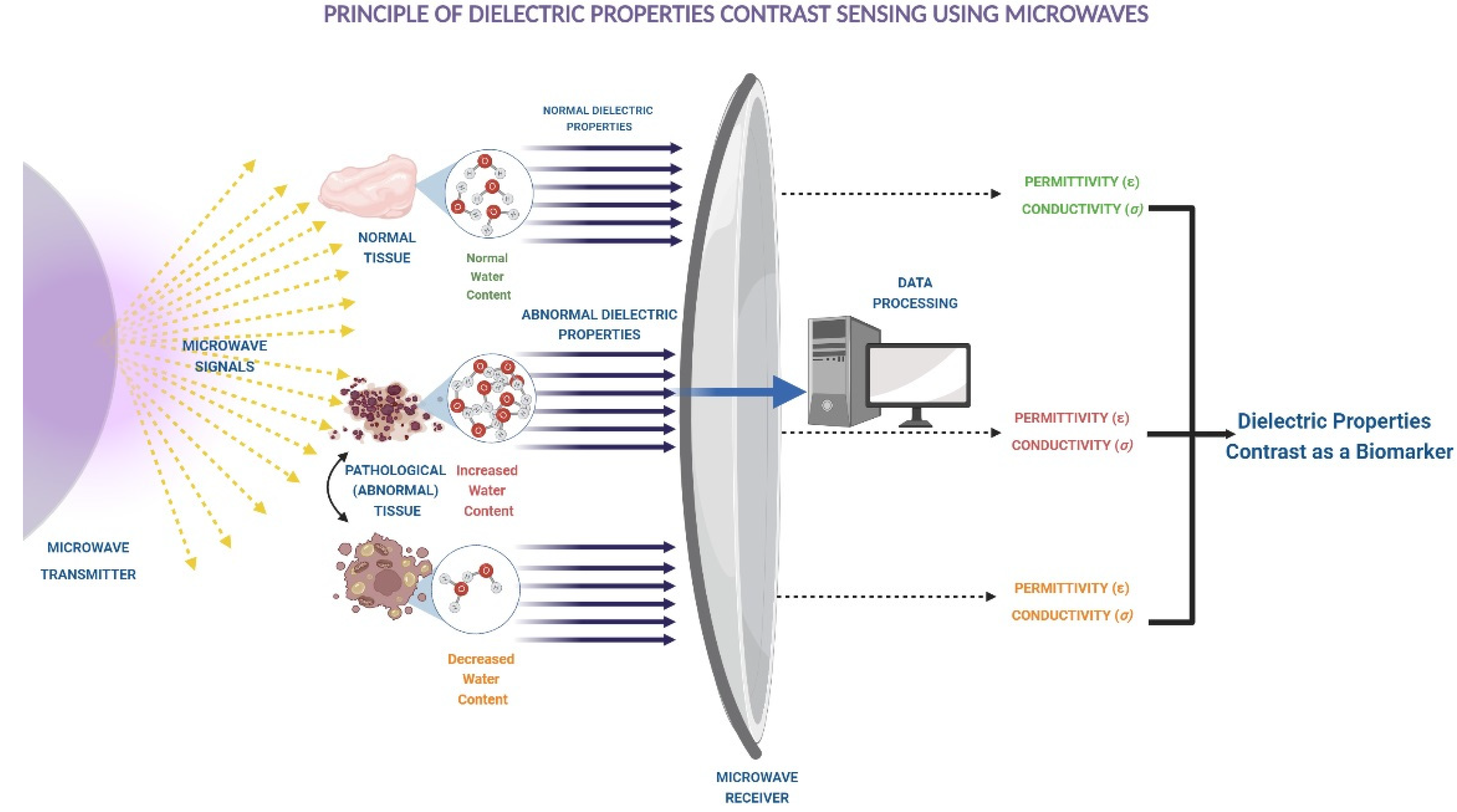
3. Diagnostic Applications of Microwaves

3.1. Microwave Imaging (MWI) Techniques

Various imaging methods have evolved over the course of time, such as X-ray, computed tomography (CT), positron emission tomography (PET), magnetic resonance imaging (MRI), single-photon emission computed tomography (SPECT), digital mammography, and diagnostic sonography, among others, which provide information about tissues [9]. However, these methods have varying limitations, such as complexity, high cost, low spatial resolution, low sensitivity to tissue changes, or exposure to harmful radiation and nephrotoxic contrast agents. Microwave imaging (MWI) is an emerging method that has the potential to overcome some of these limitations. MWI is feasible because dielectric properties vary from one tissue type to another and between healthy and abnormal tissues. It can be easily mapped using a system consisting of antenna pairs or arrays, a vector network analyzer (VNA), and a radio frequency switch to control the antennas. Measured tissue properties can be expressed as a visual image for medical personnel to interpret, assess, and make evidence-based decisions.
Based on holographic domains, this technology can be broadly classified as follows:
  • Qualitative: It uses confocal microwave imaging and radar imaging algorithms where every single antenna is used to transmit and receive its own scattered signal. This technique has shown promising results so far. In oncology, its utility to detect malignant breast tissue was elicited by Oloumi et al. using the time-domain UWB circular-SAR technique [10]. Another study conducted by Grzegorczyk TM et al. in 2012 revealed the first 3D reconstructed image of breast tissue using microwaves within a timeframe of twenty minutes [11]. Surprisingly, attempts have also been made to use it in acute care settings to identify the site of brain stroke. The associated edema or hemorrhage causes up to 20% alteration in dielectric properties of the brain tissue [12]. Another common pathology we encounter very frequently in clinical practice is osteoporosis, which is very prevalent in post-menopausal females and elderly populations. The existing gold standard investigation used for its diagnosis, i.e., dual X-ray absorptiometry (DXA), exposes the patient to radiation and fails to assess bone quality, which is dictated by microarchitecture, composition, and the degree of microdamage. These problems can be easily circumvented by MWI, as shown in a study by Amin et al. on weight-bearing trabecular calcaneal bone [13]. However, even with the immense potential to evolve into a prominent diagnostic tool, using microwave energy for imaging needs extensive research as not many studies are available to properly evaluate dielectric properties of all the body tissues, facilitate clinical translation of these measurements, and address its potential limitations.
  • Quantitative: also known as microwave tomography (MT): It relies on the tissue dielectric properties (relative permittivity and conductivity) to create an image of the tissue using a set of antennas where one of them is used to illuminate the tissue and others gather the scattered waves. On a technical aspect, MWI is plagued by the inverse electromagnetic (EM) scattering problem during the processing of the data for image reconstruction. Typically, iterative inversion methods such as the Born iterative method (BIM), distorted Born iterative method (DBIM), contrast source inversion, etc., are used, but even with advances in numerical methods, solving the inverse problem is still challenging due to slow convergence, non-linearities, and ill-posedness leading to false solutions and unstable outcomes. This difficulty is further complicated by the 3D nature of the imaging domain, increasing the computational demand and processing times [14,15]. This is where deep learning (DL), a subset of artificial intelligence (AI), comes to the rescue, as it can quickly reconstruct the images within a few seconds or minutes, making the overall process suitable for real-time applications. According to L Ahmadi et al., DL approaches have been proven to be twice as fast for similar accuracy thresholds compared to conventional iterative methods [16]. AI is a rapidly evolving field with new architectures and approaches demonstrated by researchers in different areas. Many studies reveal a variety of DL architectures for MWI. Xudong Chen et al. authored a comprehensive review of different types of DL approaches explicitly used for solving inverse EM problems [17]. However, solving this inverse scattering problem in a 3D domain, at high resolution and dynamic range, is still a big challenge where AI can play a crucial role.

3.2. Microwaves in Diagnostic Pathology

Diagnostic pathology is a specialized area of medical science focusing on disease identification. Suspected tissues such as tumors are excised from patients and are processed in the laboratory for examination. Tissue preparation typically includes two essential techniques where microwaves are commonly used, i.e., fixation and staining of the sample tissues:
  • Fixation is a chemical preservation process to maintain the in vivo cellular and extracellular morphological state for analysis [18].
  • Staining is used to highlight important features and enhance the contrast for microscopy [19].
Conventional fixation and staining methods are time-consuming and result in artifacts such as shrinkage that can affect the quality of the analysis. Immediate fixation is required to halt autolysis and preserve the tissue. Proper staining is needed to minimize any biasing effects in visual contrast. To reduce the tissue processing times, microwave (MW) heating at 2.45 GHz is used to accelerate the fixation and staining techniques. Some studies have shown that MW-based techniques reduced the tissue processing time from 1/3 to 1/10 of the conventional procedures [20].
Decreased tissue processing times have been reported with MW-based methods, with an approximate processing duration of 113 min, as opposed to conventional methods that can be as high as 515 min.
AI-based solutions to optimize tissue heating at different frequencies are possible to enhance staining approaches. A wide range of data of different tissue heating profiles or fingerprints can be generated and optimized using AI to subsequently improve staining that will improve disease diagnostics using microwaves.
Microwave irradiation can be applied to expedite the process of routine, special, metallic, as well as immunofluorescent staining, as it helps with diffusing the dye into the tissue and binding to the substrate, which is a key step in tissue staining [20,21]. Furthermore, microwave-assisted rapid tissue processing technology has a higher safety profile, as it eliminates the usage of harmful chemicals such as formalin and xylene as opposed to conventional methods [21,22]. There is increased clinical use of this technology in neuropathology when using stereotactic brain biopsy techniques, which require rapid intraoperative techniques such as the smear technique and cryostat section [23]. Microwave technology can be used to immediately fix the cryostat sections, lay out a superior tissue morphology, and further promote the usage of special stains and immunochemistry for diagnostic purposes. In addition, this technique does not provide undesirable effects while using special stains and causes only minor shrinkage artifacts as opposed to conventional formalin fixation [24]. In addition to faster processing, MW techniques improve the uniformity of the temperature in the tissue and allow precise temperature control when used in conjunction with fiber-optic temperature sensors.
One of the challenges of pathology analysis is the need for skilled specialists with years of experience. Repetitive and tedious tasks make it challenging for human interpreters to maintain a high level of performance, leading to errors. Digital pathology and AI over the last two decades have proven to be important alternatives to reduce this burden on specialists. A study published in 2019 introduced the DAPPER framework that included a deep-learning pipeline and histological imaging [25]. The study compared the classification performance of the framework against an expert pathologist using tiles (histological images). The results demonstrated that the AI framework could classify the tiles with higher accuracy. Whole slide imaging (WSI) are rich datasets that have enabled the use of AI in pathology and surpassed the capabilities of human interpreters [26]. MW-based tissue preparations, a rich set of WSI data, and continuously evolving AI algorithms have transformed diagnostic pathology, improving the efficiency and accuracy of the outcomes.

3.3. Microwave-Based Molecular Diagnostics

Molecular diagnostics (MDx) is another area of laboratory science specialized in genomic investigations, where microwaves are being used. Microwave-accelerated metal-enhanced fluorescence (MAMEF) explains the platform and application combining microwave heating and metal-enhanced fluorescence (MEF) [27,28,29]. Microwave energy is used for DNA extraction. Lyse-It is a rapid and low-cost method to prepare sample cells and fragment the DNA and RNA, protein release, and degradation in a single step [30]. The technology uses a gold bow-tie vapor deposited on a glass slide which is then used for cellular lysis. The rapid creation of thermal and convection gradients exceeds the mechanical strength of the cell membranes and results in biomolecule degradation. The level of fragmentation is dependent on MW power and irradiation time. An increase in either MW power or exposure time results in an increase in fragmentation [31].
The challenge of COVID-19 raised expectations for better diagnostics, isolation, treatment, and tracking. Rapid molecular diagnostics such as real-time reverse transcriptase polymerase chain reaction (RT-PCR) are indispensable for COVID-19 diagnosis. Although this test was widely used across the world, it requires a long duration of ~6 h from the time of sampling before conclusions can be drawn. The study used raw data consisting of fluorescence values measured over 40 cycles from RT-PCR on nasopharyngeal swab specimens from each patient. A deep-learning (DL) model was developed and trained on the time-series fluorescence dataset from the RT-PCR. A DL model approach achieved a sensitivity of 93.33%, while the RT-PCR test had a sensitivity of 89%. The study confirms that the diagnostic speed can be greatly improved without sacrificing sensitivity [32]. The use of microwaves along with artificial intelligence together are great amplifiers of speed and sensitivity in the field of molecular diagnostics.

3.4. Dielectric Spectroscopy Applications

Dielectric spectroscopy is an effective method that can measure the dielectric property or permittivity of a material under test (MUT). The dielectric properties of different biological tissues at microwave frequencies have been investigated by many researchers in the last two decades to build tissue dielectric property databases [33,34,35,36]. This section discusses the developments and possible applications of machine learning using dielectric spectroscopy data.

3.4.1. Breast

A study reported by Helwan et al. used machine-learning techniques such as feedforward neural networks using the backpropagation learning algorithm (BPNN) and radial basis function network (RBFN) for breast tissue classification [37]. They used a dielectric dataset from the UCI repository under the breast tissue database (classification category). The RBFN showed the best performance at around 94.33% classification accuracy.

3.4.2. Liver

Microwave ablation demands accurate knowledge of the dielectric properties of the liver. Researchers over the years have measured and documented the in vivo and ex vivo dielectric characteristics of different animal as well as human liver tissues. In addition, few animal studies show immense scope for dielectric spectroscopy paired with artificial intelligence (AI) in the diagnosis of liver diseases. In a study conducted by Yilmaz on rats having a malignant liver, the author classified the liver tissue using support vector machine (SVM) learning algorithms and experimented on a total of 771 in vivo samples from 30 adult female albino rats with an open-ended coaxial probe technique. Out of these, they were able to identify cancerous and normal tissue correctly and reported that SVM algorithms with radial basis had 98.3% accuracy in identifying cancerous tissue vs. normal tissue [38].

3.4.3. Kidney

Banu Sacli et al. [39] used dielectric properties of naturally formed renal calculi (calcium oxalate, cystine, struvite) measured using an open-ended contact probe technique between 500 MHz and 6 GHz. He used a machine-learning model k-nearest neighbors (KNN) to classify the types of renal calculi. The algorithm had 98.17% accuracy. H. Rahmani et al. [40] used a machine-learning algorithm to classify the permittivity of normal and wounded skin created by scratch, punch, and UVB burn using principal component analysis for data dimensionality reduction on the measured loss tangent (ε″/ε′) data. Furthermore, they used the gaussian mixture model (GMM), an unsupervised learning method, support vector classifier (SVC), a supervised learning method, naïve Bayes, and neural net to classify between normal skin and punch wound and reported 97% to 100% accuracy of using these models.

3.4.4. Lungs

In China, Lu et al. [41] developed an ML model (XG Boost) combining dielectric properties to assess whether thoracic lymph nodes were benign or malignant in patients with non-small cell lung cancer. Traditionally, surgeons use frozen sections to diagnose metastatic lymph nodes, which is time-consuming and expensive. They used XG Boost, where the model accuracy was 87.8% with a sensitivity of 58.33% and specificity of 100%. Therefore, they combined this with the SMOTE algorithm and found that the sensitivity was 83.33% and specificity was 96.55%. These machine-learning models can be helpful to surgeons intraoperatively to rapidly diagnose with high efficacy.

3.4.5. Machine Learning to Solve Analytical Problems

The use of machine-learning models has been largely used to resolve analytical problems or to classify the permittivity associated with the usage of dielectric spectroscopy. Dielectric properties are expressed using parametric models (mathematical models) such as multi-pole Debye or Cole–Cole expression for better understanding or to represent the tissue performances over a wide frequency range [42]. Historically, numerical statistical nonlinear regression or similar techniques are mostly used to fit these measured complex permittivity results with Debye or Cole–Cole parameters [42,43].
Recent literature described the usage of machine-learning models to improve this data-fitting process. Yilmaz et al. used a particle swarm optimization algorithm to fit Cole–Cole parameters to the average of multiple measurements from the same animal liver sample [38]. Salahuddin et al. used six different optimization algorithms, i.e., least squares method, particle swarm optimization, weighted least squares algorithm, hybrid particle swarm–least squares algorithm, one-stage genetic algorithm, and two-stage genetic algorithm, to fit measured data to the multi-pole Debye model. According to the reported work novel, a two-stage genetic algorithm proved to be the most effective and efficient method. Recently Bai et al. published a study that presented an extensive analysis of recent accomplishments of deep-learning methods for solving linear inverse problems [44].

3.5. Microwave Radiometry in Medicine

Microwave radiometry (MWR) is a safe and patient-friendly technology to measure and visualize the temperatures of human tissues. In this technology, a signal is received from tissues’ intrinsic radiation, wherein the power of the tissues in the microwave range is proportional to the average temperature of tissues in the volume under the antenna. MWR is a novel technique currently being explored as a potential way to detect at-risk vascular lesions.
Despite recent developments in vascular imaging technologies that allow risk categorization of asymptomatic patients based on the degree of carotid artery stenosis and how prone the plaque is to rupture, risk stratification remains a challenging task [45,46]. Intravascular thermography is a method that detects vulnerable plaques, but its invasive nature excludes it from being used as a screening tool. Hence, the hunt for non-invasive indicators for assessing vascular inflammation in patients at high cardiovascular risk is still an area of active research. It was reported that there is appreciable temperature variation in normal and atherosclerotic vessels, and MR gives a reliable measurement of the internal tissue temperature by converting electromagnetic radiation to microwave frequencies [47]. Non-invasive sensors have also been developed to detect these changes [48]. MWR has also been applied in oncology, where the temperature variation of malignant breast tissue compared to the normal surrounding tissue due to their more rapid growth and replication was detected using MR sensors.
However, the major roadblock in its clinical use is that—being a newer technique—healthcare professionals are not adapting to its interpretation and use. This can be addressed using artificial intelligence models to interpret data from these sensors to extract useful information, translate it to prognosticate patients, and provide the best diagnostic capacity in a timely manner [49]. The use of MWR and artificial intelligence together was attempted by Levshinskii et al. [50], wherein deep-learning models were employed to identify chronic venous insufficiency due to temperature alteration in the inflamed vessel walls.

4. Applications of Microwaves in Treatment

4.1. Microwave Ablation

Microwave ablation is a form of thermal ablative technique that causes coagulative necrosis and cell death via two mechanisms: dipole and ionic. Water molecules are dipoles, and when energy is applied, these molecules in the tissue agitate continuously, generating heat that leads to cell death [51,52]. This technique is used to treat various benign and malignant tumors, including tumors of the liver, pancreas, kidney, breast, and adrenal. As in other ablation techniques, microwave ablation allows for different approaches, including percutaneous, laparoscopic, and open surgical access. Other than tumors, MWA is also used in treating biliary strictures and cholecystocutaneous fistulas, particularly as a minimally invasive procedure in poor surgical candidates. It was also used in the ablation of focal nodular hyperplasia in children without necrotizing the adjacent structures such as the gallbladder, highlighting the precision and effectiveness of the procedure [53]. Following are various treatments using microwave ablation techniques and how AI has been used thus far.

4.1.1. Liver

Many patients with small hepatocellular carcinomas may benefit from ablation therapy as their first-line therapy or as an alternative for those who are not candidates for surgery. Thermal ablation involves heating cancerous tissues to high enough temperatures (usually over 60 °C) to cause prompt coagulative necrosis. MWA for hepatocellular carcinoma (HCC) was first used in Japan by Saitsu et al. [54] and has been used worldwide for the last two decades. MWA is a safe technique for the treatment of unresectable liver tumors with a mortality that ranges from 0% to 0.36%. Liang P et al. created practice guidelines for ultrasound-guided MWA for liver cancer which aim to standardize MWA treatment and criteria for treatment candidates. A better convection profile, greater consistent intratumoral temperatures, quicker ablation periods, and the capacity to employ numerous probes to treat multiple tumors at once are all advantages of MWA. According to Izzo et al. [55], MWA is considered superior to radiofrequency ablation (RFA) for liver tumors with perivascular lesions, suggesting that candidates for MWA should be chosen according to patient characteristics for ablation. In an attempt to identify the differences between microwave ablation (MWA) systems using different frequencies, Kerri A Simo et al. [56], found that both 915 MHz and 2.45 GHz MWA systems achieve reproducible hepatic tumor ablation.

4.1.2. Bone

  • Tumors
The primary composition of bone is collagen and inorganic salts, which makes it relatively hard, making it able to withstand high heat. For this reason, microwaves can be used in the treatment of primary bone tumors as well as bony metastasis [57]. RFA and cryoablation (CA) are considered well-established, safe, effective, and durable methods of treatment for painful bone tumors and bone metastasis [58,59]. Microwave ablation has been added to the arsenal of treatment for the ablation of bone tumors in recent times [60]. Direct MW-induced thermocoagulation was used in a procedure by Fan et al. to treat 62 patients with diverse bone cancers. The goal of this surgical procedure was to isolate the tumor from nearby healthy structures. Out of 62 patients, 57 had local tumor control, but 5 had local fracture problems. However, the effectiveness of these minimally invasive treatments for malignant bone metastases must be established through extensive randomized multicenter trials [57,61]. Research findings and clinical experience concerning the use of microwave ablation to treat bone cancers in the limbs have been compiled, and a clinical guideline has been created to standardize the application of this treatment of bone tumors in the limbs. Through the use of evidence-based medicine, this guideline aims to provide a solid clinical foundation for indications, preoperative evaluation, and decision making, perioperative management, and complications. The goal of the recommendation is to standardize care and enhance the therapeutic effectiveness of MWA of bone cancers in the extremities [62].
  • Osteomyelitis
Osteomyelitis is an inflammatory disorder of the bone caused by infection. It is still challenging for orthopedic surgeons because of its protracted treatment process. The reported infection control rate ranges from 67 to 95%, and 5% to 33% of patients suffer from a recurrence of infection [63]. Patients with this condition have impaired local blood supply to the bone. Therefore, improving blood supply and tissue perfusion will help promote microbial clearance in infected areas and reduce recurrence in susceptible areas. By raising the temperature of deep tissues, MW diathermy can cause hyperthermia in tissue, which can speed up the healing process, boost drug activity, provide more effective pain relief, aid in the clearance of toxic waste, promote tendon flexibility, and lessen muscle and joint stiffness. Additionally, hyperthermia causes changes in the cell membrane, hyperemia, enhanced local tissue drainage, and elevated metabolic rate [64]. In a study, 40 rats infected with methicillin-sensitive Staphylococcus aureus in the medullary cavity of the right tibia were randomly assigned to one of four treatment groups: saline (control); saline + MW therapy; systemic cefuroxime; or systemic cefuroxime + MW therapy. This experimental model revealed that MW therapy significantly enhanced the effectiveness of the systemic antibiotic medication. However, more clinical research is necessary before this therapeutic option may be used on patients [65].

4.1.3. Uterus

  • Menorrhagia
Menorrhagia is a type of abnormal uterine bleeding that occurs in 53 out of 1000 women. Symptoms may include bleeding for 7 or more days and soaking through one or more sanitary pads or tampons every hour for several consecutive hours [66]. Although the definitive treatment is hysterectomy, it has physical and emotional complications along with high social and economic costs. To remove the complete thickness of the endometrium while maintaining the structural integrity of the reproductive system, several less invasive surgical procedures have been devised. The first-generation endometrial ablation techniques were introduced in the mid-1980s and included loop resection, rollerball, or laser ablation. The second-generation ablation techniques include thermal balloon (Thermachoice), hot fluid circulation (Hydro ThermAblator), cryotherapy (Her Option), microwave energy (MEA), and radiofrequency electrosurgery (NovaSure) [67]. Indeed, second-generation endometrial ablation is becoming the new gold standard for dysfunctional uterine bleeding [68].
Thermal endometrial ablation using microwaves, a recent treatment, is a minimally invasive technique that thermally eliminates the endometrial lining of the uterine cavity to reduce bleeding. A comparison of microwave endometrial ablation (MEA) with transcervical resection of the endometrium (TRCE) was performed and was followed up for a minimum of five years. Published operational results for this trial and clinical outcome at one year and two years showed that MEA produced equivalent clinical results to TCRE but with some operational advantages. In addition to being as effective at reducing menstruation symptoms, MEA also produces higher levels of acceptance, is easy to learn, is performed more quickly, is cost-effective, and is tolerable under local anesthesia compared to TCRE [69].
  • Fibroids
The most frequent benign pelvic tumors in women of reproductive age are uterine fibroids or leiomyomas. Any thermal ablative treatment for uterine fibroids aims to safely remove as much fibroid tissue as possible while leaving the surrounding uterine tissue unharmed [70]. In 2005, Goldberg described fibroid degeneration following microwave ablation of the endometrium [71]. A transvaginal ultrasound probe coupled to a 14-gauge needle set in an adapter has been used for transcervical microwave ablation of fibroids. A study of nine women undergoing transcervical microwave ablation of their fibroids found a decrease in volume between 37 and 69% at 6 months with no significant complications [72]. A similar study used a 20 mm long antenna that can operate in both continuous and pulsed microwave modes with a 15-gauge microwave needle. Following this, the mean fibroid volume reduction following the treatment is 90 and 94%, and no major adverse events were described and concluded that percutaneous microwave thermal ablation is a quick, painless, and minimally invasive technique that can be used to treat fibroid [73].

4.1.4. Prostate

In elderly men, benign prostatic hyperplasia (BPH) is a prevalent illness that may cause uncomfortable symptoms in the lower urinary tract (LUTS). More than 30% of men aged 65 and older possess irritable (frequency, nocturia, urgency) and/or obstructive urinary symptoms associated with BPH, including weak stream, hesitation, intermittency, and incomplete emptying [74]. Transurethral resection of the prostate (TURP) has been the gold-standard treatment for reducing urinary symptoms and enhancing urine flow in symptomatic BPH. However, the morbidity of TURP is increasing; subsequently, less invasive techniques have been developed for treating BPH [75]. Transurethral microwave thermotherapy (TUMT), in which microwaves are used to induce coagulation necrosis in prostatic tissue, appears to be a safe and efficient treatment for BPH [76]. It has also been proposed that this heating also destroys the alpha-adrenergic receptors, thereby increasing the urinary flow after the TUMT [77]. It has also been shown that TUMT increases sensory frequency, which also helps in relieving the irritative symptoms of the bladder [78]. Compared to TURP, microwave thermotherapy can be performed as an outpatient procedure and has fewer, milder adverse effects. However, the improvement in urinary symptoms and urine flow was better after TURP; nevertheless, fewer men required retreatment. Additional research is required to assess the long-term effects of microwave thermotherapy and develop the most efficient microwave thermotherapy equipment and energy levels [75].

4.1.5. Kidney

Whenever renal cell carcinoma is diagnosed, we usually opt for radical nephrectomy, which is associated with significant morbidity and mortality. With standard nephrectomy posing a high risk for older or comorbid patients, localized kidney tumors are being treated with percutaneous ablation. The most challenging issue with partial nephrectomy is the inability to control bleeding while the treatment is being performed [79]. Controlling parenchymal bleeding in solid vascular organs has been possible because Tabuse invented the microwave tissue coagulator in 1979 [80]. This device, according to Kagebayashi, was also helpful in partial nephrectomy [81]. Six patients underwent laparoscopic partial nephrectomy with a microwave tissue coagulator for small renal tumors. In terms of cancer control, five out of six patients had their tumors completely removed, while the sixth patient had the tumor capsule damaged. Laparoscopic partial nephrectomy with a microwave tissue coagulator is useful for small renal tumors, providing short operating time, minimal blood loss, and rapid recovery. However, it is necessary to compare the method to surgical treatments or other percutaneous ablation approaches in a comparative randomized mode [79].

4.1.6. Adrenal

Local tumor ablation in the adrenal gland presents unique challenges secondary to the adrenal gland’s unique anatomic and physiological features. Technically successful microwave ablation of primary adrenocortical cancer and adrenal metastases has been described by Simon and colleagues. Following adrenal microwave ablation, short-term follow-up outcomes are similarly positive. Given the cystic structure of adrenal metastases and the failure of radiofrequency to sufficiently heat cystic tumors, microwave ablation is thought to be preferable over RF ablation [82]. However, applying slowly in increments may be trickier with microwave ablation since it raises temperature more quickly than RFA. Even if it is theoretical, microwaves might present different difficulties for a titrated ablation in the case of more excessive catecholamine release than RFA [83].

4.1.7. Thyroid

Non-invasive procedures such as microwave, radiofrequency, high-intensity focused ultrasound, percutaneous ethanol injection (PEI), sclerotherapy, and laser photocoagulation (HIFU) ablation have been suggested as alternatives to surgery, particularly for the compression of nearby structures and cosmetic issues [84,85]. A clinical study was conducted on eleven patients with benign thyroid nodules who underwent MWA. In this small, non-randomized feasibility study, nodule volume was reduced by more than 50%, which resolved the cosmetic complaints of people with compressive neck symptoms. Nine participants with nodular goiter-related pain and two participants with Hashimoto’s thyroiditis improved upon treatment. The study concluded that it is possible to treat thyroid nodules that are cytologically benign with ultrasound-guided percutaneous MWA. However, only trained medical professionals with knowledge of neck anatomy, ultrasound imaging, and MWA should carry out this surgery. Due to this study’s small sample size and brief follow-up time, additional research using larger samples and prolonged follow-up will be required [85].

4.1.8. Lung

In the past two decades, there has been a significant diversity in the curative management of lung cancers, both primary and metastatic. MWA is a relatively recent thermal ablation technology that is being used more frequently to treat incurable lung cancers. Wolf et al. published the first substantial patient series study on it in 2008 [86]. MWA typically creates wider, more spherical, and predictable ablation zones because the tissue is evenly penetrated by the microwave field and is less affected by its characteristics. The extension of the ablation zone is particularly dependent on heat conduction, especially at its periphery, due to the high electrical resistivity of a ventilated lung and the tissue inhomogeneity. MWA causes thrombosis in vessels less than 6 mm in diameter and is less vulnerable to the heat sink effect [87]. Initial animal experiments have demonstrated definite advantages of MWA over RFA. MWA produces ablation zones that are larger, more spherical, and less time-consuming. However, the scant and inconsistent research on MWA does not offer enough proof to suggest a benefit over RFA in terms of local control or overall survival [88].

4.1.9. Heart

The role of microwave energy also extends to therapeutics, as it is used to treat various cardiac arrhythmias, i.e., idiopathic sinus node tachycardia and atrial fibrillation, using a minimally invasive transthoracic approach [89,90]. Strides have been made by using microwave-based irrigated ablation of deep myocardial ectopic foci of ventricular arrhythmias without damaging the superficial epicardial tissue and surrounding coronaries and thus preventing complications of conventional radiofrequency ablation [91]. The utilization of microwave energy has also sprawled into treating various peripheral vascular lesions, as was observed in studies by Zhang et al. and Sun et al., where microwave-based ablation was successfully used to coagulate and stop the bleeding from the renal and hepatic arteries, respectively, by using a percutaneous approach. This was found to be superior to the currently used intraluminal drug injections [92,93]. The ability of MR to detect temperature changes can also be used for targeted therapy, as was attempted by Tarakanov et al. [94], to treat patients with lower back pain. In this study, thermal asymmetry was assessed for back muscles and focused self-controlled energy-neuroadaptive regulation (SCENAR) therapy was used for muscles with more spasms and inflammation. However, extensive in vivo and human studies are needed to further validate the use of this technology in current clinical practice.

4.2. Microwave Ablation with AI

In the past decade, there has been a continuous development of machine-learning and computational models for thermal ablative procedures. Most of these models have been applied to RFA with very scarce application in MWA. There is less information and technique to automate treatment detection of thermal ablation using MWA. Some researchers have incorporated AI and ML models to optimize and predict MWA outcomes. Brunese et al. [95], conducted a study to test machine-learning models to automatically predict the thermal ablation treatment and reduce tissue damage for patients under analysis using features from the patient medical health records. The model reached an F1 score of 0.91 using the NBTree classification algorithm to 0.92 with the LibSVM and Neural Network classification algorithm. Chao An et al. developed an ML model to predict the early recurrence of cancer in patients treated with MWA in the early stages of hepatic cell carcinoma (HCC) based on the clinical text data. After using SHapley Additive exPlanations (SHAP) and local interpretable model-agnostic explanations (LIME) algorithms as interpretation algorithms, the authors finally concluded that the ML method using the XGBoost model helps physicians with decision making before MWA for HCC in clinical practice and trials [96]. Although these studies have incorporated AI for thermal ablative techniques, further research in the development of ML models is necessary for more accurate diagnosis and treatment outcomes.

5. Microwave Energy in Drug Delivery

Advancement in technology has opened various doors to increase the efficacy and decrease the side-effect profile of various drugs, especially those used in cancer treatments. Various methods, including therapeutic drug monitoring, tailoring the dose, and selective use of drugs, have been used conventionally. However, a relatively new approach to delivering the drugs to their targeted action sites and circumventing any systemic interaction is currently being explored [97]. These drug delivery systems are a formulation or a device involved in transporting a pharmaceutical compound to its target site for a desired therapeutic effect. The working principle behind this is the development of a carrier molecule that transports the drug to the required site and releases it in a controlled manner triggered by changes in the surrounding environment (change in pH, temperature, chemical milieu, etc.) or by application of external magnetic field, ultrasound, or microwaves. Among these approaches, drug release controlled by temperature changes has attracted much attention because it does not rely on changes in specific chemical properties of the environment, which can be problematic in intracellular or in vivo applications [98]. However, it has limited application in vivo because of the damage caused to the non-target tissues and poor penetration into the deeper tissues.
Emerging evidence with the development of nanoparticles that have microwave-absorbing properties can address most of the limitations of these delivery systems. It allows for a non-invasive approach, allowing the carrier to heat faster and providing better thermal efficiency while allowing the stimulus to penetrate deep (10–15 cm) into the body [99,100]. A study conducted by Peng et al. in 2014 provided tangible evidence that particles coated by specific microwave-absorbing materials such as ZnO: Er(3+), Yb(3+) can successfully deliver chemotherapeutic drug etoposide. The use of microwave-based systems also helps to tackle the crystallization of drug compounds. This occurs due to the low water solubility of most compounds leading to their poor bioavailability. Converting the crystal structure of drugs to amorphous forms using microwave irradiation can form a supersaturated formulation with better bioavailability. Although the amorphous structure is unstable and can reconvert into a crystal form, various studies have been conducted to explore the possibility of making stable amorphous compounds with better potency than conventional drug forms [100,101]. Furthermore, the remarkable advancements in nanotechnology have resulted in the development of implantable robots which can perform controlled and targeted drug delivery [102]. These can be used along with artificial intelligence and deep-learning methods to develop novel astute delivery systems which can assess the dose, drug selectivity, and their targeted release depending upon the disease burden and pathology [103].

6. Microwaves in Telemetry

Telemetry can be defined as the automatic measurement and wireless transmission of data from remote sources. In medicine, it is widely used to monitor heart rate and rhythm (EKG), respiratory rate, partial pressure of oxygen in the blood, serum glucose, neural recordings, stimulation devices, and cochlear and retinal implants while the patient is ambulatory and not restricted to bedside monitors. Most of the conventional monitoring systems use inductive transmission for both data transfer and device recharge, with issues of high power requirement and biocompatibility. However, newer options in the form of high-frequency (~400 MHz) microwave devices with small implantable antennas can serve the same purpose with better battery life and compatibility [104,105,106,107].
Over the decades, various engineering techniques such as AI and machine learning have been used in the field of medicine. They have served as a catalyst, whether it be increasing the clinical and treatment outcome or early diagnosis of a disease. One such interesting application is in the analysis of telemetry data, as enlightened by a study that used AI analysis of continuous EEG data to rapidly determine a good 6-month functional outcome for patients [105]. Similarly, machine learning has been used to analyze the telemetry data to detect sepsis in its early stages and determine the optimal timing for antibiotics and additional fluids. This can have great positive impacts on patient outcomes [108]. Despite these promising results, clinical implementation is still a barrier, and future studies should be focused on sorting out the evidence that can be incorporated widely into AI. This can be achieved by the collaboration of experts in their respective fields, such as clinicians and data analysts [109].

7. Microwaves in Hospital Waste Management

The majority of waste that requires inactivation processes comes from research facilities and commercial companies, yet most of it is generated from hospitals. About 10% of hospital waste is biohazardous, requiring proper inactivation [110]. There are four basic domains for the treatment and safe disposal of such waste, namely chemical, thermal, biological, and irradiative. Incineration and autoclaving are the most widely used of all these. However, this can be achieved by microwave irradiation as well. Ideally, the waste generated should be inactivated at the site of production, but this can be cumbersome and resource-intensive, making it less suitable for low-resource settings.
Microwave irradiation is an emerging method to achieve sterilization of waste as it has been commercially used, for example, in Sanitec systems and documented to destroy pathogens such as E. coli, Bacillus subtilis, and Salmonella. These were exposed to a conventional microwave at around 2500 MHz and had a 6-log cycle reduction in viability [111]. Various studies conducted on food articles replicated the same results. In a study carried out by Song et al., it was observed that microwaves reduced salmonella in peanut butter without deteriorating the quality of the food [112]. There was also complete inactivation of E. coli and microflora when microwaves were used on mechanically tenderized beef at temperatures above 70 degrees Celsius for more than 1 min [113].
The mechanism of its action is yet to be fully defined, but it is postulated to be a combination of thermal effects on the dipolar components in the waste and nonthermal alteration in protein structures of the infectious waste constituents [114,115]. However, it is more suitable than conventional methods in terms of cost-effectiveness, transportation, and eco-friendliness [116].

8. Microwave Hardware Design

Any microwave imaging system has mainly two key components:
i.
hardware—the antenna system that collects microwave signals reflected from tissues
ii.
software techniques that recreate an image of the object [117].
Moreover, an antenna system plays a vital role in implantable and wearable medical devices (IMDs). These IMDs are medical devices that are implanted inside the body of the patient during surgery and can be utilized for a range of diagnostic, monitoring, and therapeutic purposes. IMDs include intracranial pressure monitors, neurostimulators, glucose meters, cochlear implants, implanted pacemakers, defibrillators, and glucose meters [118]. In the healthcare system, AI can be incorporated with smart wearable sensors and give trustworthy, secure, and valuable information that can help medical professionals make clinical judgments [119]. For example, for evaluating correct diagnosis, artificial intelligence can provide several points of view by combining medical data, text, and other information [120].
In a study, authors have presented a smart antenna design using machine learning. Here, the authors forecast the antenna’s behavior and reference point by implementing an extra-tree-regression-based machine-learning model over an extensive range of frequencies [121]. While designing an antenna, a significant amount of time and resources is required due to the complexity. For an antenna design, frequency is an independent parameter, while reflectance is a dependent parameter. Therefore, predicting the reflectance parameter can reduce the high cost and workforce. Another study proposed an efficient long short-term memory artificial neural network to model the antennas more efficiently and precisely. This work validated the algorithm by modeling two implantable antennas as 2.4 and 2.5 GHz [122]. In another research study, the authors used the artificial neural network to minimize the antenna size for biomedical applications (medical device radiocommunication service, 401–406 MHz, and industrial, scientific, and medical bands, 902–928 MHz) [123]. A size minimization of 21.48% has been achieved, maintaining the antenna’s performance for both bands. To achieve better miniaturization for biomedical applications, an innovative approach has been proposed where an artificial neural network and particle swarm optimization techniques have been adopted to optimize the feed position of Minkowski, Giuseppe Peano, and Koch curves-based hybrid fractal antenna [124].
Recently, AI has shown prominent involvement in developing sensors for disease diagnosis and other medical applications. A machine-learning approach has been proposed to detect Parkinsonian tremors [125]. Decision tree (DT), support vector machine (SVM), discriminant analysis (DA), random forest (RF), and k-nearest-neighbor (kNN) algorithms have been presented for the prediction. A total of eighty-five patients with this disease were taken, and by using a wristwatch-type wearable device, the authors performed the analysis. An AI-assisted urine multi-marker sensing platform based on the dual-gate field effect transistor (DGFET) was created to avoid repeated biopsies when treating prostate cancer [126]. Random forest and neural network, these two machine-learning algorithms, have been used with these DGFET biosensors by analyzing 76 urine samples.
One of the most crucial properties of materials is relative permittivity, or dielectric constant, whose precise determination is essential in medicine and healthcare. For the characterization of dielectric materials, RF and microwave measurement-based sensing techniques have gained a lot of attention due to their accuracy. Table 1 summarizes an overview of various microwave sensors in medicine. Both sensor design and performance could be enhanced with AI in the various applications discussed.

9. Discussion

Microwaves are versatile electromagnetic waves with various uses in the field of medicine. The idea of using MW energy for imaging began in the early 1980s and was first tested on an isolated well, perfused canine kidney [139]. The results were surprising, with good spatial resolution using the immersion technique. However, their use had limitations, making them difficult for clinical application. Nevertheless, microwaves were considered promising because of their non-ionizing way of visualizing tissues via dielectric properties. It gained special attention for biomedical imaging, especially in breast cancer detection. Despite this, their applications are still being explored in diagnosing various diseases of the brain, lungs, neck, heart, bone, etc. The contrast in dielectric properties within normal and abnormal tissues serves as the basis for such diagnostic studies. Microwave energy has been used not only in diagnostics but also in therapeutics to treat various tumors and other diseases through ablation techniques, as discussed above. Despite these uses, significant research and development within medicine are imperative to address the challenges faced by using microwave energy, improve their use in the healthcare industry, and make it patient- and physician-friendly. With microwaves gaining popularity and attention, so is the opportunity to combine them with the power of AI and deep learning to improve performance and make it cost-effective.

9.1. Microwave Imaging Hardware Design with AI

In the past decade, using deep-learning models has gained attention to achieve success in many microwave applications. W. shao et al. developed a neural network to reduce the training difficulty in microwave imaging for image segmentation and classification. An autoencoder was used to find compact representations for high-resolution images, and then these were used as labels to train another neural network converting EM signals. Better results were shown with DLN in signal-to-image mapping [140]. The difficulties of the inverse scattering problem of microwave imaging have been addressed by various studies using DL models. These methodologies, however, require demanding algorithms with computation time and memory. Therefore, online inverse scattering methods based on AI were introduced to reduce computation time and computer resources. Multilayer perception neural networks were also studied to address the inverse scattering problem in MWI. Ioannis T. Rekanos introduced radial basis function neural networks and tested them to estimate the size and position of proliferating bone marrow in the limb. The construction of the network was performed by applying the orthogonal least-squares algorithm (OLSA). The model performance was excellent, and it proposed that this model can be used for rapid estimation [141].
As an initial experiment, C. Dachena et al. tested artificial neural networks for quantitative MWI of the human neck for diagnosing cervical myelopathy. The measured scattered electrical fields were used to reconstruct two-dimensional images solving the inverse scattering problem. The model detected significant changes in the size of the spinal cord and suggested that further studies on the human neck should be considered in the future to validate the capabilities of similar approaches using the DL model [142]. Similarly, Costanzo. S. et al. combined the born iterative method with convolutional neural networks to solve ill-framed inverse problems in MWI on phantom breast models. This model was believed to reduce the reconstruction time for clinicians with more than 90% accuracy. They proposed that this ML model can be applied to the relative permittivity reconstruction and can be tested on brain tumor phantoms for further development [143].
MWI systems entail complex hardware designs with customized antenna and switching networks to obtain 3D data for reconstructing the images. Human body organs and structures are quite heterogenous in terms of dielectric properties, and with disease pathology, the dynamic range of the tissue property changes makes microwave antenna designs very challenging. The future of microwave imaging will rely on the AI-driven design of microwave antennas specific to medical applications. The parallel surrogate model-assisted hybrid differential evolution for antenna optimization (PSADEA) method has shown good performance for AI-assisted ultra-wide band (UWB) antennas suitable for medical applications [144]. Machine-learning approaches offer promise for accelerated antenna designs for the medical applications of interest. These AI-assisted designs minimize errors, maintain high accuracy, save design and optimization time in terms of the reduced number of electromagnetic simulations required, and improved computational efficiency and, more importantly, AI-assisted prediction of the antenna behavior for these medical applications [145]. Therefore, researchers should use realistic models that best represent human organs and tissues in their antenna designs, allowing seamless clinical translation. For example, MWI application for imaging thin-walled myocardium is extremely challenging, and translational efforts will fail without realistic designs of antennas that acquire scattered data from thin-walled myocardium in addition to its dynamic nature. The potential of AI-assisted antenna designs will be integral for any future design of MWI systems for clinical applications.
The next immediate challenge with MWI is the complex RF switching needed across several transmitter/receiver pairs to acquire the volumetric data to reconstruct the image. While a fully functional MWI system is yet to be demonstrated, several researchers are performing active research using conventional RF switching systems. AI-assisted RF switch will be the future for MWI for medical applications, wherein a machine-learning-based RF switch model can be highly reliable and accurate, provide better prediction, and allows generalization and extrapolation capability over broad frequency range [146,147]. Researchers also have explored phase change materials (PCM) for designing microwave switching systems which can be optimized using AI [148]. For example, in the case of cardiac applications, high temporal resolution is necessary to accurately reconstruct microwave images throughout the cardiac cycle, which can demand switching speeds as high as 30 GHz or more. These novel approaches can create a paradigm shift in designing robust AI-assisted switching systems for MWI applications.
Finally, accurate reconstruction of 3D microwave medical images with heterogeneous structures from the scattered microwave data is important for fully leveraging the diagnostic potential for various medical applications. A fully functional 3D MWI system requires several challenges, namely the spatial EM-field polarization, data redundancy from multiple antenna array systems, dynamicity of tissue contrast, tissue motion, flow artifacts, etc., which need to be effectively resolved [140]. Deep-learning models capable of capturing these various features specific to medical applications are necessary for robust reconstruction for a reliable diagnosis. This also demands larger compressing space to accommodate several features for a 3D deep-learning network that can potentially resolve translation efforts into medicine.
As evident from the above summary, MWI is gaining attention in medicine with its significant advantages in providing structural and functional information of various organ systems, thereby opening novel avenues of research to enhance patient care. Robust 3D reconstruction of microwave images using deep-learning methodologies is currently underway, offering huge promise toward clinical translation. Therefore, the future of MWI in medicine relies on developing powerful deep-learning networks to reconstruct more practical and complicated heterogeneous human structures to complement existing imaging modalities such as MRI, CT, and ultrasound [149].

9.2. AI-Assisted Dielectric Spectroscopy

Several researchers have highlighted the use of dielectric property databases for potential diagnostics. Recently, many have also developed machine-learning models using dielectric measurements in various tissues with good accuracy. One of our recent studies used multi-classification models to differentiate healthy liver, non-alcoholic steatohepatitis, and fibrosis liver using dielectric properties measured with a coaxial probe technique. Samaddar et al. used four classifiers, including logistic regression, random forests, k-nearest neighbors, and support vector machines. The logistic regression and random forest classifier performed well, with an accuracy of 0.80. This was the first study of its kind to differentiate between non-alcoholic steatohepatitis (NASH) and fibrotic liver disease [150]. Deep-learning models were developed using dielectric permittivity to classify normal vs. fibrotic liver as well as to classify normal vs. NASH in mice models. The dataset was classified using six machine-learning models. Among all k-nearest neighbors and random forests, the classifier performed the best [151,152]. These studies must be expanded in the future on human subjects, as this can potentially eliminate the highly invasive diagnostic method such as biopsy for NASH and fibrosis. Most dielectric property measurements are carried out on ex vivo tissues, whereas in vivo properties are preferred to create realistic results. It is important to investigate whether there are changes in the dielectric properties of tissues after excision from the host. Gerazov et al. [153] used machine-learning models to predict in vivo dielectric properties using ex vivo property measurements for different levels of hydration. The model showed that it is possible to predict in vivo permittivity based on ex vivo permittivity measurements with promising results.
Currently used, dielectric spectroscopy (DS) instrumentation is not suited for biological tissue characterization. However, researchers have adapted these existing instruments for testing biological tissues of varying dimensions. This has limited the testing capabilities of various tissues over wide frequency ranges with high accuracy. With machine-learning models becoming integral for analyzing dielectric spectroscopy data to classify tissues accurately, novel probe designs suitable for biological tissues of varying dimensions ranging from resected tissues to biopsies are warranted. Novel AI-assisted dielectric spectroscopy sensor designs are necessary beyond the conventionally used coaxial probe to provide a wide range of data acquisition for biological samples. Deep-learning-based models on large, simulated datasets to assist DS sensors for varying thicknesses and dielectric contrast can help resolve this challenge to characterize biological tissues. DS has also emerged as a powerful tool to characterize organic systems containing molecules such as enzymes, proteins, and antibodies using machine-learning approaches that will disrupt the sensing of biological tissues in the future [154]. However, these are characterized at much lower frequencies (20–1 MHz); therefore, research efforts should focus on developing advanced DS systems over a wide frequency range for various medical applications. DS sensor designs should focus on measuring such properties in small volumes of liquids or small tissue samples to propel widespread characterization of all tissue types aiding machine-learning-based diagnostics based on DS data. For example, a forward move from the material under test (MUT) to liquid under test (LUT) towards versatile biological tissue classification is necessary to propel the potential of DS-based diagnostics using AI [155].
With a growing application of dielectric tissue testing across the entire body system, the big data that are generated offer a challenge to accurately classify the tissues based on the measured properties. ML methods offer the advantage of providing tissue contrast even with subtle changes in dielectric properties, which could be diagnostic. Therefore, AI-assisted tissue classification based on dielectric properties using dielectric spectroscopy will dictate the future of microwaves in medicine in tissue characterization and novel microwave-based medical sensors suitable for these specific applications.

9.3. AI-Assisted Molecular Diagnostic Using Microwaves

Microwave-based molecular diagnostics using AI can disrupt the field in both high speed and accuracy of the diagnostics procedure. With our recent experience with COVID-19 and the rising prevalence of other infectious diseases worldwide, there is an undisputable need for point-of-care molecular diagnostic solutions. This warrants novel AI-assisted approaches for the various molecular diagnostics segments such as in situ hybridization, mass spectrometry, PCR, sequencing, isothermal amplification, and chips and microarrays. Microwaves have already proven their potential with genomic sequencing with machine-learning models. Novel microwave-based, AI-assisted assays derived from advanced sequencing solutions can leverage the genomic power in diagnosing more complex diseases. With anticipated market growth for genomic sequencing, researchers should focus on the effective use of microwave energies for nucleic acid extraction by using machine-learning models to optimize microwave irradiation. Microwave irradiation is key to accelerating a variety of chemical reactions that aid in faster molecular diagnostics [156]. Since heating effects are incurred with microwave irradiation [157], AI tools to optimize microwave irradiation based on experimental and numerical studies specific to the molecular targets will be the key to the future of microwave-based molecular diagnostics. It is expected that microwave-based molecular diagnostic assays can provide high-throughput diagnosis delivery at high speed and accuracy for both screening and testing applications to face the growing challenges of new variants for infectious diseases.

9.4. AI-Assisted Telemetry Using Microwaves

Microwave telemetry systems are employed in medicine to improve clinical workflow process efficiency and safety. Microwave telemetry performance measured in terms of throughput should demonstrate a reduced bit error ratio and packet loss ratio of the system [158]. Microwave telemetry performance is in constant need of improvements for better diagnostic potential. A microwave telemetry system is complex hardware with microwave antennas and associated electronics to transmit data wirelessly. The future of microwave telemetry systems in medicine will see a paradigm shift in a wearable system such as a microwave telemetry patch where individual patients in a remote setting can transmit data to the cloud for real-time monitoring. This scalable design will face significant system design and implementation challenges, with strict requirements on specific absorption rates (SAR) toward clinical translation efforts. Machine-learning approaches offer promise for accelerated telemetry antenna designs with permissible SARs and calibration with improved performance.
With the growing demand for continuous multiparameter physiological monitoring, multi-lead electrocardiography (ECG), pulse oximetry (SpO2), core and peripheral thermometry, invasive arterial and central venous blood pressure monitoring, and electroencephalography (EEG) measurements are increasingly becoming common to optimize patient care through the real-time microwave-based wireless data [109]. The biggest challenge with telemetry data is the presence of various artifacts that makes pattern recognition challenging [109,159]. AI-assisted mining of microwave telemetry data will be the future in solving multiple problems in telemetry data, such as artifact removal, error detection, prediction, summarization, and visualization of large quantities [160]. These AI tools will aid in better understanding the pattern recognition from the telemetry data towards medical diagnostics in a reliable fashion. Therefore, machine-learning approaches for microwave telemetry system design and extensive data analysis are integral for their effective use in the healthcare system.

9.5. AI-Assisted Hospital Waste Management Using MW

Managing hospital wastes is a crucial aspect of everyday logistics to allow hospital administration and supply chain management to operate effectively. Medical waste is inevitable in hospitals and can be categorized into many groups discussed below [161,162]. Infectious wastes include lab cultures including bacterial, viral, and other agents, different types of human tissues, used dressings from patients, and various wastes from isolation wards. Pathological waste includes human tissues, fetuses, placentas, blood, and other body fluids. Pharmaceutical waste includes unwanted drugs and expired drugs. Chemical waste includes biochemicals from diagnostic or therapeutic work chemicals used for cleaning various instruments and materials in a day-to-day setting—biohazard sharps materials such as disposable surgical instruments, needles, blades, etc. Radioactive waste comes from radioactive substances from radiotherapy and lab work in a hospital setting. Pressurized container wastes include gas cylinders, cartridges, and aerosol cans. Wastes come from medical instrumentation containing heavy metals such as batteries, broken thermometers, blood pressure gauges, etc. [161,162]. As evident, medical wastes comprise a wide range of materials that need effective irradiation strategies to sterilize and eliminate medical waste. Each of these medical wastes requires varying levels of microwave irradiation energies, and effective dose planning is integral for medical waste management. The future of hospital waste management using microwave irradiation will leverage AI tools for optimizing irradiation energy delivery. This approach will require good a priori knowledge of the dielectric properties of the various materials in each waste category to plan irradiation strategies effectively.
Depending on the type of medical waste, including radioactive wastes, hybrid microwave heating strategies must be employed, aided by machine-learning models, which will rely on big dielectric data that will prove to be efficient in how each of these materials is sterilized and eliminated [163]. AI-assisted microwave power titration strategies for microwave irradiation are crucial for managing the heavy medical wastes in the near future.

9.6. Microwaves in the Field of Pathology Leveraging AI

Tissue fixation and staining are primary steps in pathology toward diagnosis. Microwave heating through effective irradiation strategies has already shown promise for improved efficacy for microwave pathology. Since different tissue types have varying contrast for dielectric properties, power titration of microwave heating is crucial to optimize both tissue fixation and staining. Machine-learning models can leverage the temperature dependency of dielectric properties toward adequate tissue fixation and staining plans for the varieties of tissue types. For instance, a bioheat transfer model of the target tissue and microwave-sensitive radionics features can be employed in a machine-learning model to customize irradiation dose specific to each tissue type that can optimize fixation and staining for high throughput and efficacy [164]. Large amounts of empirical and simulated electromagnetic data are needed for such model development leveraging AI, without which data processing for dose prediction can be challenging.
Multimodal digital pathology is already disrupting this space by integrating and analyzing multimodal data (images from other modalities such as MRI, CT, molecular imaging, etc., laboratory data, electronic health data, etc.) to identify patterns and relationships between various morphological features and gene expression [165,166]. While improved performance is reported using a variety of multimodal deep-learning models, the potential of dielectric properties of the tissue under investigation is not utilized to enhance diagnostic efficacy. Dielectric spectroscopy can be integrated into the upstream processing of tissues before fixing and staining to characterize their complex permittivity. This can create a big database of dielectric properties from varieties of tissues that can be fused to the deep-learning models in a multimodal fashion to provide tissue-specific diagnostics. In addition, DS should be integrated with the hospital’s core resource facility and seamlessly integrated with the digital pathology suite as routine testing.

9.7. Microwave-Based Medical Sensors with AI

Non-invasive and patient-friendly medical sensors are the future of digital health with remote monitoring capabilities. Existing challenges in sensor designs limit patient-friendliness and remote monitoring for continuity of care. Microwave-based medical sensor designs have offered huge promise in providing non-invasive sensing in real-time with long-term capabilities.
Microwave biomedical sensors for glucose monitoring, pressure sensing, nucleic acids sensing, and various other biomolecules have demonstrated their potential. Additionally, microwave acoustic sensors are capable of effectively sensing audio data such as heart sounds with the potential to sense other biomedical body sounds [167,168,169]. However, the clinical utility of these sensors is limited by their robustness in accurate diagnosis. Therefore, AI-assisted novel antenna and sensor designs are explicitly warranted for these applications that can be translated for clinical applications. Several researchers have used deep-learning models to identify novel frequency-selective surfaces for sensor designs [170,171]. Moreover, novel metamaterials can be designed using machine-learning methods to customize sensors with high sensitivity and specificity [121,172,173,174,175]. Characterizing the dielectric properties of target tissues using dielectric spectroscopy of the target tissues will provide the contrast needed for the specific sensor design. Therefore, AI will become inevitable in designing novel microwave medical sensors for implementation and data analysis to enhance patient care.

9.8. AI-Assisted Drug Delivery Using Microwaves

Microwave heating has proven potential to identify target molecules effectively and deliver drugs for various diseases [176]. Different drugs’ biochemical properties vary significantly, providing specificity for target molecules that potentially can be related to their dielectric properties. Temperature dependency on dielectric properties allows effective microwave heating titration specific for the target molecules suitable for drug delivery planning. AI-assisted microwave heating can be developed from experimental and simulated temperature profiles based on dielectric properties, which can be optimized for identifying target drug-delivery molecules. Machine-learning models have already disrupted the interpretation and decoding of genetic information, accelerated drug discovery by identifying novel compounds, and allowed behavior prediction in a complex molecular setting. Similar approaches can be used for identifying biological targets for drug delivery. Machine-learning models have enhanced target fishing (TF) for biological molecules by rapid prediction to link targets to novel compounds [103]. In addition, AI-assisted microwave theranostic strategies will be the future for drug delivery to enhance patient-specific therapies.

9.9. AI-Assisted Microwave Ablation

Microwave thermal energy has been used in various ablation techniques, especially in cancers, which reduce the cost and duration of the treatment. MWA has been gaining increasing popularity and is being utilized more so than RFA due to its higher heating efficiency as well as to deliver of energy consistently to the target tissues. The machine-learning model is a powerful tool to study the effect of electromagnetic interactions and predict treatment outcomes during thermal ablative techniques. Over the past decade, various computational models’ refinements have been performed, including thermo-electric and biophysical parameters. Considering the biophysical parameters are crucial to creating high-accuracy prediction models, some of the studies have mentioned the importance of considering temperature-dependent biophysical parameters during deep-learning modeling of thermal ablation [177,178,179]. It was found that blood perfusion significantly affects the ML model’s results regarding the ablation zone dimensions. Therefore, most of the studies used a thermal damage-dependent piecewise model of blood perfusion to address this issue [180,181,182]. All these studies have shown that computational modeling has become a significant tool to assist and estimate treatment outcomes providing support to physicians [183].
MWA is known to adapt well to automated and reliable treatment planning due to the relatively consistent ablation zone since the microwave is less susceptible to tissue-specific and heat-sink effects [184]. However, the ablation zone’s safety margins remain vague and do not guarantee optimal ablation treatment of the target areas and protect neighboring tissues. This ongoing challenge stems from the fact that there are no real-time lesion monitoring tools that accurately reflect the temperature profiles of the ablation zone for real-time intraprocedural decision making. It is well known that dielectric properties of tissues correlate with the temperature that could be used for thermal imaging for ablation zone monitoring for safety margins [185].
AI can disrupt MWA treatment planning and execution effectively in the future. First, from the biopsies of the target tissue, dielectric tissue property characterization can be performed that can inspire advanced electromagnetic simulations using AI to profile the target tissue on its temperature dependency. Second, AI-assisted power titration of the microwave energy is possible based on this a priori knowledge that precisely allows dose delivery on the target tissue. Third, based on the heating, AI-based temperature maps can be created in real-time, enabling real-time interrogation of the safety margins to make real-time clinical decisions about the procedure. This will create a paradigm shift in AI-assisted MWA to optimize the treatment that will reduce recurrence, reduce patient discomfort, and subsequently lower the cost, making a significant practice impact.

9.10. AI-Assisted Microwave Radiometry

Internal body temperature can be measured using microwave radiometry. These thermal patterns can be used for disease diagnosis and treatment monitoring [186]. This measures the intrinsic radiation of tissues in the microwave range, which is proportional to the average temperature of the tissues under the antenna. With new developments in antennas, MWR has become more accessible for clinical applications. Recently, researchers have used the help of machine-learning models along with MWR for better diagnostic prediction and accuracy in cancer detection. The dataset of the temperature readings during regular monitoring of diseases was used to develop ML models by V. Levshinskii. They developed non-neural network models such as random forest, XGBoost, k-nearest neighbors, support vector machine, and neural network models. The results of this experiment showed that machine-learning models could be used as a part of the diagnostic system [48,49]. The performances of these models can be improved by taking other factors into account from clinical professionals, as these models were created only using temperature data [187].
MWR in medical applications relies on its potential to sense temperature manifestations of tissue pathology before any structural changes, which offers a huge promise for the early-stage detection of diseases [188]. While MWR offers huge diagnostic potential for medical applications, translational efforts into clinical practice as a diagnostic tool face several challenges. MWR instrumentation currently explored in medicine uses the single channel and single frequency devices and is extremely bulky. This limits the wide-range application of MWR to evaluate temperatures at varying depths over different body habitus on a wide-range patient population. Therefore, multichannel systems with multi-frequency capabilities are essential for real-time assessment of changes in temperature with depth and evaluation of large areas of the body, as well as for long-term monitoring of the temperatures of the internal tissues [189]. AI-assisted methods can help achieve the technical difficulty of integrating multichannel and multi-frequency into a single user system for medical applications. Novel AI-assisted radiometer designs driven by data from empirical and experimental simulations are warranted to accelerate MWR applications in medicine for non-invasive diagnosis of various diseases using intrinsic tissue temperatures. Deep-learning models from multichannel and multi-frequency acquisition can provide novel insights into the temperature profiles at varying depths that otherwise are not possible with existing systems. An AI-assisted clinical decision support system can be developed using various thermal profiles of tissues in effectively mining and interpreting the data to provide accurate diagnostics. Therefore, the future of MWR applications in medicine will rely on effective use of AI in MWR system design and calibration [3] and big data analysis for clinical decision support systems for medical diagnosis [190,191].
Figure 2 provides an overall view of AI-assisted applications of microwaves in medicine.
AI intersection with various applications of microwaves in medicine has become inevitable in both system design and care delivery to optimize patient care. In the growing era of the digital health revolution, real-time solutions in remote monitoring setting are often preferred, wherein healthcare providers can have bi-directional communication to provide prompt care. As evident from this review, microwaves have already disrupted medical applications in various domains. Opportunities to optimize each of these applications to enhance the performance using AI have been identified and discussed, which opens new avenues of research, which with deeper investigations can provide realistic pathways toward clinical translation. Researchers should focus on both microwave technology design as well as big data analytics using AI to address the current challenges in applying these methods to clinical practice. With the cost of electronics and computational capabilities going down, the focus of this AI-assisted microwave technology should be steered towards specific healthcare applications in continuously improving its capabilities in impacting patient care.

10. Conclusions

Microwave application in medicine has the potential to provide non-invasive and patient-friendly solutions for healthcare management and patient care delivery. Though the applications of microwave in healthcare have been explored, the use of AI-assisted microwave applications is limited. There is very minimal development of AI solutions that can enhance clinical practice. Therefore, this review identified various opportunities for AI-assisted solutions for microwave imaging, radiometry, telemetry, molecular diagnostics, ablation, spectroscopy, digital pathology, waste management, drug delivery, and sensor designs that can revolutionize microwave-based medical applications in the near future. Active research is needed in both microwave technology advancement as well as AI-based microwave data analysis to leverage its benefits for clinical translation.

Author Contributions

K.G. and S.P.A. defined the review scope, context, and purpose of this study. K.G., M.S., A.R., A.S.S.C., D.N.D. and S.S. provided clinical perspective and expertise for this study. K.G., A.A., N.P., M.S., T.N., A.S.S.C., S.G., P.S., A.Y. and S.S.M. conducted a literature review and drafted the manuscript. S.P.A., K.G., and A.R. conceived and crafted the illustrative figures. D.N.D., S.S., S.D., S.R., D.M. and S.P.A. provided consulting and performed a critical review of the manuscript. K.G., A.R. and S.P.A. performed the cleaning and organization of the manuscript. S.P.A. provided conceptualization, supervision, and project administration. All authors have read and agreed to the published version of the manuscript.

Funding

S.P.A. received the 2021 Gastroenterology and Hepatology (GIH) innovation grant from the GIH Division, Mayo Clinic, Rochester, MN, USA. This study was supported by the GIH Division’s internal funding for Microwave Engineering and Imaging Laboratory (MEIL) and GIH Artificial Intelligence Laboratory (GAIL).

Data Availability Statement

The review was based on publicly available academic literature databases.

Conflicts of Interest

The authors declare no conflict of interest. The authors declare that this review was conducted in the absence of any commercial or financial relationships that could be construed as a potential conflict of interest.

References

  1. Gartshore, A.; Kidd, M.; Joshi, L.T. Applications of microwave energy in medicine. Biosensors 2021, 11, 96. [Google Scholar] [CrossRef] [PubMed]
  2. Mumtaz, S.; Rana, J.N.; Choi, E.H.; Han, I. Microwave radiation and the brain: Mechanisms, current status, and future prospects. Int. J. Mol. Sci. 2022, 23, 9288. [Google Scholar] [CrossRef] [PubMed]
  3. Jeng, D.; Kaczmarek, K.A.; Woodworth, A.; Balasky, G. Mechanism of microwave sterilization in the dry state. Appl. Environ. Microbiol. 1987, 53, 2133–2137. [Google Scholar] [CrossRef] [PubMed] [Green Version]
  4. Astani, S.A.; Brown, M.L.; Steusloff, K. Comparison of procedure costs of various percutaneous tumor ablation modalities. Radiol. Manag. 2014, 36, 12–17. [Google Scholar]
  5. Goel, K.; Gupta, R.; Solanki, J.; Nayak, M. A comparative study between microwave irradiation and sodium hypochlorite chemical disinfection: A prosthodontic view. J. Clin. Diagn. Res. JCDR 2014, 8, ZC42. [Google Scholar]
  6. Kiricuta, I.-C., Jr.; Simplăceanu, V. Tissue water content and nuclear magnetic resonance in normal and tumor tissues. Cancer Res. 1975, 35, 1164–1167. [Google Scholar] [PubMed]
  7. Hinrikus, H.; Riipulk, J. Microwave imaging. Wiley Encycl. Biomed. Eng. 2006, 4, 2329–2340. [Google Scholar] [CrossRef]
  8. BioRender.com. BioRender. Available online: https://biorender.com/ (accessed on 1 December 2022).
  9. Hussain, S.; Mubeen, I.; Ullah, N.; Shah, S.S.U.D.; Khan, B.A.; Zahoor, M.; Ullah, R.; Khan, F.A.; Sultan, M.A. Modern Diagnostic Imaging Technique Applications and Risk Factors in the Medical Field: A Review. BioMed Res. Int. 2022, 2022, 5164970. [Google Scholar] [CrossRef]
  10. Oloumi, D.; Winter, R.S.; Kordzadeh, A.; Boulanger, P.; Rambabu, K. Microwave imaging of breast tumor using time-domain UWB circular-SAR technique. IEEE Trans. Med. Imaging 2019, 39, 934–943. [Google Scholar] [CrossRef]
  11. Grzegorczyk, T.M.; Meaney, P.M.; Kaufman, P.A.; Paulsen, K.D. Fast 3-D tomographic microwave imaging for breast cancer detection. IEEE Trans. Med. Imaging 2012, 31, 1584–1592. [Google Scholar] [CrossRef] [Green Version]
  12. Semenov, S. Microwave tomography: Review of the progress towards clinical applications. Philos. Trans. R. Soc. A Math. Phys. Eng. Sci. 2009, 367, 3021–3042. [Google Scholar] [CrossRef] [Green Version]
  13. Amin, B.; Shahzad, A.; Farina, L.; Parle, E.; McNamara, L.; O’Halloran, M.; Elahi, M.A. Investigating human bone microarchitecture and dielectric properties in microwave frequency range. In Proceedings of the 13th European Conference on Antennas and Propagation (EuCAP), Krakow, Poland, 31 March–5 April 2019; pp. 1–5. [Google Scholar]
  14. Benny, R.; Anjit, T.A.; Mythili, P. Deep Learning Based Non-Iterative Solution to the Inverse Problem in Microwave Imaging. Prog. Electromagn. Res. M 2022, 109, 231–240. [Google Scholar] [CrossRef]
  15. Bertero, M.; Boccacci, P.; De Mol, C. Introduction to Inverse Problems in Imaging; CRC Press: Boca Raton, FL, USA, 2021. [Google Scholar]
  16. Ahmadi, L.; Hosseini, S.M.; Shishegar, A.A. Solving Inverse Electromagnetic Problems Using Deep Learning. In Proceedings of the 28th Iranian Conference on Electrical Engineering (ICEE), Tabriz, Iran, 4–6 August 2020; pp. 1–4. [Google Scholar]
  17. Chen, X.; Wei, Z.; Li, M.; Rocca, P. A review of deep learning approaches for inverse scattering problems (invited review). Prog. Electromagn. Res. 2020, 167, 67–81. [Google Scholar] [CrossRef]
  18. Katoh, K. Microwave-assisted tissue preparation for rapid fixation, decalcification, antigen retrieval, cryosectioning, and immunostaining. Int. J. Cell Biol. 2016, 2016, 7076910. [Google Scholar] [CrossRef] [Green Version]
  19. Alturkistani, H.A.; Tashkandi, F.M.; Mohammedsaleh, Z.M. Histological stains: A literature review and case study. Glob. J. Health Sci. 2016, 8, 72. [Google Scholar] [CrossRef]
  20. Rao, M.; Pai, S.M.; Khanagar, S.B.; Siddeeqh, S.; Devang, D.D.; Naik, S. Microwave-assisted tissue processing, fixation and staining in tissues of different thicknesses: A comparative study. J. Oral Maxillofac. Pathol. 2020, 24, 186. [Google Scholar] [CrossRef] [PubMed]
  21. Shruthi, B.S.; Vinodhkumar, P.; Kashyap, B.; Reddy, P.S. Use of microwave in diagnostic pathology. J. Cancer Res. Ther. 2013, 9, 351. [Google Scholar] [CrossRef]
  22. Morales, A.R.; Nassiri, M.; Kanhoush, R.; Vincek, V.; Nadji, M. Experience with an automated microwave-assisted rapid tissue processing method: Validation of histologic quality and impact on the timeliness of diagnostic surgical pathology. Am. J. Clin. Pathol. 2004, 121, 528–536. [Google Scholar] [CrossRef]
  23. Ainley, C.; Ironside, J. Microwave technology in diagnostic neuropathology. J. Neurosci. Methods 1994, 55, 183–190. [Google Scholar] [CrossRef]
  24. Leong, A.S.Y.; Daymon, M.E.; Milios, J. Microwave irradiation as a form of fixation for light and electron microscopy. J. Pathol. 1985, 146, 313–321. [Google Scholar] [CrossRef]
  25. Bizzego, A.; Bussola, N.; Chierici, M.; Maggio, V.; Francescatto, M.; Cima, L.; Cristoforetti, M.; Jurman, G.; Furlanello, C. Evaluating reproducibility of AI algorithms in digital pathology with DAPPER. PLoS Comput. Biol. 2019, 15, e1006269. [Google Scholar] [CrossRef] [PubMed] [Green Version]
  26. Niazi, M.K.K.; Parwani, A.V.; Gurcan, M.N. Digital pathology and artificial intelligence. Lancet Oncol. 2019, 20, e253–e261. [Google Scholar] [CrossRef] [PubMed]
  27. Aslan, K. Rapid whole blood bioassays using microwave-accelerated metal-enhanced fluorescence. Nano Biomed. Eng. 2010, 2, 1. [Google Scholar] [CrossRef] [PubMed] [Green Version]
  28. Aslan, K.; Geddes, C.D. Microwave-accelerated metal-enhanced fluorescence: Platform technology for ultrafast and ultrabright assays. Anal. Chem. 2005, 77, 8057–8067. [Google Scholar] [CrossRef]
  29. Aslan, K.; Geddes, C.D. Microwave-accelerated metal-enhanced fluorescence (MAMEF): Application to ultra fast and sensitive clinical assays. J. Fluoresc. 2006, 16, 3–8. [Google Scholar] [CrossRef]
  30. Santaus, T.M.; Li, S.; Ladd, P.; Harvey, A.; Cole, S.; Stine, O.C.; Geddes, C.D. Rapid sample preparation with Lyse-It® for Listeria monocytogenes and Vibrio cholerae. PLoS ONE 2018, 13, e0201070. [Google Scholar] [CrossRef]
  31. Santaus, T.M.; Zhang, F.; Li, S.; Stine, O.C.; Geddes, C.D. Effects of Lyse-It on endonuclease fragmentation, function and activity. PLoS ONE 2019, 14, e0223008. [Google Scholar] [CrossRef]
  32. Lee, Y.; Kim, Y.-S.; Lee, D.-i.; Jeong, S.; Kang, G.-H.; Jang, Y.S.; Kim, W.; Choi, H.Y.; Kim, J.G.; Choi, S.-h. The application of a deep learning system developed to reduce the time for RT-PCR in COVID-19 detection. Sci. Rep. 2022, 12, 1234. [Google Scholar] [CrossRef]
  33. Gabriel, C.; Gabriel, S.; Corthout, Y. The dielectric properties of biological tissues: I. Literature survey. Phys. Med. Biol. 1996, 41, 2231. [Google Scholar] [CrossRef] [Green Version]
  34. Gabriel, S.; Lau, R.; Gabriel, C. The dielectric properties of biological tissues: II. Measurements in the frequency range 10 Hz to 20 GHz. Phys. Med. Biol. 1996, 41, 2251. [Google Scholar] [CrossRef] [Green Version]
  35. Andreuccetti, D. An Internet Resource for the Calculation of the Dielectric Properties of Body Tissues in the Frequency Range 10 Hz–100 GHz. 2012. Available online: http://niremf.ifac.cnr.it/tissprop/ (accessed on 1 December 2022).
  36. Farrugia, L.; Wismayer, P.S.; Mangion, L.Z.; Sammut, C.V. Accurate in vivo dielectric properties of liver from 500 MHz to 40 GHz and their correlation to ex vivo measurements. Electromagn. Biol. Med. 2016, 35, 365–373. [Google Scholar] [CrossRef]
  37. Helwan, A.; Idoko, J.B.; Abiyev, R.H. Machine learning techniques for classification of breast tissue. Procedia Comput. Sci. 2017, 120, 402–410. [Google Scholar] [CrossRef]
  38. Yilmaz, T.; Kılıç, M.A.; Erdoğan, M.; Çayören, M.; Tunaoğlu, D.; Kurtoğlu, İ.; Yaslan, Y.; Çayören, H.; Arıkan, A.E.; Teksöz, S. Machine learning aided diagnosis of hepatic malignancies through in vivo dielectric measurements with microwaves. Phys. Med. Biol. 2016, 61, 5089. [Google Scholar] [CrossRef] [PubMed]
  39. Saçlı, B.; Aydınalp, C.; Cansız, G.; Joof, S.; Yilmaz, T.; Çayören, M.; Önal, B.; Akduman, I. Microwave dielectric property based classification of renal calculi: Application of a kNN algorithm. Comput. Biol. Med. 2019, 112, 103366. [Google Scholar] [CrossRef] [PubMed] [Green Version]
  40. Rahmani, H.; Archang, M.M.; Jamali, B.; Forghani, M.; Ambrus, A.M.; Ramalingam, D.; Sun, Z.; Scumpia, P.O.; Coller, H.A.; Babakhani, A. Towards a machine-learning-assisted dielectric sensing platform for point-of-care wound monitoring. IEEE Sens. Lett. 2020, 4, 1–4. [Google Scholar] [CrossRef]
  41. Lu, D.; Yu, H.; Wang, Z.; Chen, Z.; Fan, J.; Liu, X.; Zhai, J.; Wu, H.; Yu, X.; Cai, K. Classification of Metastatic and Non-Metastatic Thoracic Lymph Nodes in Lung Cancer Patients Based on Dielectric Properties Using Adaptive Probabilistic Neural Networks. Front. Oncol. 2021, 11, 640804. [Google Scholar] [CrossRef]
  42. Salahuddin, S.; Porter, E.; Krewer, F.; O’Halloran, M. Optimised analytical models of the dielectric properties of biological tissue. Med. Eng. Phys. 2017, 43, 103–111. [Google Scholar] [CrossRef]
  43. Gabriel, S.; Lau, R.; Gabriel, C. The dielectric properties of biological tissues: III. Parametric models for the dielectric spectrum of tissues. Phys. Med. Biol. 1996, 41, 2271. [Google Scholar] [CrossRef] [Green Version]
  44. Bai, Y.; Chen, W.; Chen, J.; Guo, W. Deep learning methods for solving linear inverse problems: Research directions and paradigms. Signal Process. 2020, 177, 107729. [Google Scholar] [CrossRef]
  45. Shah, A.S.; Lee, K.K.; Pérez, J.A.R.; Campbell, D.; Astengo, F.; Logue, J.; Gallacher, P.J.; Katikireddi, S.V.; Bing, R.; Alam, S.R.; et al. Clinical burden, risk factor impact and outcomes following myocardial infarction and stroke: A 25-year individual patient level linkage study. Lancet Reg. Health-Eur. 2021, 7, 100141. [Google Scholar] [CrossRef]
  46. Schiele, F.; Navarese, E.P.; Visona, A.; Ray, K. What imaging techniques should be used in primary versus secondary prevention for further risk stratification? Atheroscler. Suppl. 2017, 26, 36–44. [Google Scholar] [CrossRef] [PubMed]
  47. Casscells, W.; Vaughn, W.; Mcallister, H.; Willerson, J.; Hathorn, B.; David, M.; Vaughn, W.; McAllister, H.; Krabach, T.; Bearman, G. Thermal detection of cellular infiltrates in living atherosclerotic plaques: Possible implications for plaque rupture and thrombosis. Lancet 1996, 347, 1447–1449. [Google Scholar] [CrossRef] [PubMed]
  48. Wagner, D.; Vogt, S.; Jamal, F.I.; Guha, S.; Wenger, C.; Wessel, J.; Kissinger, D.; Pitschmann, K.; Schumann, U.; Schmidt, B.; et al. Application of microwave sensor technology in cardiovascular disease for plaque detection. Curr. Dir. Biomed. Eng. 2016, 2, 273–277. [Google Scholar] [CrossRef]
  49. Levshinskii, V.; Galazis, C.; Ovchinnikov, L.; Vesnin, S.; Losev, A.; Goryanin, I. Application of data mining and machine learning in microwave radiometry (MWR). In Proceedings of the Biomedical Engineering Systems and Technologies: 12th International Joint Conference, BIOSTEC 2019, Prague, Czech Republic, 22–24 February 2019; Revised Selected Papers 12. pp. 265–288. [Google Scholar]
  50. Levshinskii, V.; Galazis, C.; Losev, A.; Zamechnik, T.; Kharybina, T.; Vesnin, S.; Goryanin, I. Using AI and passive medical radiometry for diagnostics (MWR) of venous diseases. Comput. Methods Programs Biomed. 2022, 215, 106611. [Google Scholar] [CrossRef] [PubMed]
  51. Gala, K.B.; Shetty, N.S.; Patel, P.; Kulkarni, S.S. Microwave ablation: How we do it? Indian J. Radiol. Imaging 2020, 30, 206–213. [Google Scholar] [CrossRef] [PubMed]
  52. Ho, J.S.; Li, Z. Microwave Metamaterials for Biomedical Sensing; Elsevier Inc.: Amsterdam, The Netherlands, 2015. [Google Scholar]
  53. Meaney, P.M. Microwave imaging and emerging applications. Int. J. Biomed. Imaging 2012, 2012, 252093. [Google Scholar] [CrossRef]
  54. Liang, P.; Yu, J.; Lu, M.-D.; Dong, B.-W.; Yu, X.-L.; Zhou, X.-D.; Hu, B.; Xie, M.-X.; Cheng, W.; He, W. Practice guidelines for ultrasound-guided percutaneous microwave ablation for hepatic malignancy. World J. Gastroenterol. WJG 2013, 19, 5430. [Google Scholar] [CrossRef] [PubMed]
  55. Izzo, F.; Granata, V.; Grassi, R.; Fusco, R.; Palaia, R.; Delrio, P.; Carrafiello, G.; Azoulay, D.; Petrillo, A.; Curley, S.A. Radiofrequency ablation and microwave ablation in liver tumors: An update. Oncologist 2019, 24, e990–e1005. [Google Scholar] [CrossRef] [PubMed] [Green Version]
  56. Simo, K.A.; Tsirline, V.B.; Sindram, D.; McMillan, M.T.; Thompson, K.J.; Swan, R.Z.; McKillop, I.H.; Martinie, J.B.; Iannitti, D.A. Microwave ablation using 915-MHz and 2.45-GHz systems: What are the differences? Hpb 2013, 15, 991–996. [Google Scholar] [CrossRef] [Green Version]
  57. Fan, Q.; Ma, B.; Guo, A.; Li, Y.; Ye, J.; Zhou, Y.; Qiu, X. Surgical treatment of bone tumors in conjunction with microwave-induced hyperthermia and adjuvant immunotherapy: A preliminary report. Chin. Med. J. 1996, 109, 425–431. [Google Scholar]
  58. Rehnitz, C.; Sprengel, S.D.; Lehner, B.; Ludwig, K.; Omlor, G.; Merle, C.; Kauczor, H.-U.; Ewerbeck, V.; Weber, M.-A. CT-guided radiofrequency ablation of osteoid osteoma and osteoblastoma: Clinical success and long-term follow up in 77 patients. Eur. J. Radiol. 2012, 81, 3426–3434. [Google Scholar] [CrossRef] [PubMed]
  59. Callstrom, M.R.; Dupuy, D.E.; Solomon, S.B.; Beres, R.A.; Littrup, P.J.; Davis, K.W.; Paz-Fumagalli, R.; Hoffman, C.; Atwell, T.D.; Charboneau, J.W. Percutaneous image-guided cryoablation of painful metastases involving bone: Multicenter trial. Cancer 2013, 119, 1033–1041. [Google Scholar] [CrossRef] [PubMed] [Green Version]
  60. Brace, C.L. Microwave ablation technology: What every user should know. Curr. Probl. Diagn. Radiol. 2009, 38, 61–67. [Google Scholar] [CrossRef] [PubMed] [Green Version]
  61. Simon, C.J.; Dupuy, D.E. Percutaneous minimally invasive therapies in the treatment of bone tumors: Thermal ablation. Semin. Musculoskelet. Radiol. 2006, 10, 137–144. [Google Scholar] [CrossRef]
  62. Zheng, K.; Yu, X.; Hu, Y.; Zhang, Y.; Wang, Z.; Wu, S.; Shen, J.; Ye, Z.; Tu, C.; Zhang, Y.; et al. Clinical guideline for microwave ablation of bone tumors in extremities. Orthop. Surg. 2020, 12, 1036–1044. [Google Scholar] [CrossRef] [PubMed]
  63. Patzakis, M.J.; Zalavras, C.G. Chronic posttraumatic osteomyelitis and infected nonunion of the tibia: Current management concepts. JAAOS-J. Am. Acad. Orthop. Surg. 2005, 13, 417–427. [Google Scholar] [CrossRef]
  64. Giombini, A.; Giovannini, V.; Cesare, A.D.; Pacetti, P.; Ichinoseki-Sekine, N.; Shiraishi, M.; Naito, H.; Maffulli, N. Hyperthermia induced by microwave diathermy in the management of muscle and tendon injuries. Br. Med. Bull. 2007, 83, 379–396. [Google Scholar] [CrossRef] [Green Version]
  65. Qi, X.-Y.; Qiu, X.-S.; Jiang, J.-Y.; Chen, Y.-X.; Tang, L.-M.; Shi, H.-F. Microwaves increase the effectiveness of systemic antibiotic treatment in acute bone infection: Experimental study in a rat model. J. Orthop. Surg. Res. 2019, 14, 286. [Google Scholar] [CrossRef] [Green Version]
  66. Hallberg, L.; Hogdahl, A.; Nilsson, L.; Rybo, G. Variation at different ages and attempts to define normality and Menstrual blood loss-a population study. Acta Obstet. Gynecol. Scand 1966, 45, 320–351. [Google Scholar] [CrossRef]
  67. van den Brink, J.; Kluivers, K.; Nieboer, T. Second-generation thermal endometrial ablation: Beware of metal clips in the lower abdomen. Gynecol. Surg. 2013, 10, 291–294. [Google Scholar] [CrossRef] [Green Version]
  68. Fernandez, H. Update on the management of menometrorrhagia: New surgical approaches. Gynecol. Endocrinol. 2011, 27, 1131–1136. [Google Scholar] [CrossRef] [PubMed]
  69. Cooper, K.G.; Bain, C.; Lawrie, L.; Parkin, D.E. A randomised comparison of microwave endometrial ablation with transcervical resection of the endometrium; follow up at a minimum of five years. BJOG Int. J. Obstet. Gynaecol. 2005, 112, 470–475. [Google Scholar] [CrossRef] [PubMed]
  70. Quinn, S.D.; Gedroyc, W.M. Thermal ablative treatment of uterine fibroids. Int. J. Hyperth. 2015, 31, 272–279. [Google Scholar] [CrossRef] [PubMed]
  71. Goldberg, J.; McCrosson, S.; Kaulback, K.R. Delayed leiomyoma degeneration after microwave endometrial ablation. Obstet. Gynecol. 2005, 106, 1176–1178. [Google Scholar] [CrossRef] [Green Version]
  72. Kanaoka, Y.; Yoshida, C.; Fukuda, T.; Kajitani, K.; Ishiko, O. Transcervical microwave myolysis for uterine myomas assisted by transvaginal ultrasonic guidance. J. Obstet. Gynaecol. Res. 2009, 35, 145–151. [Google Scholar] [CrossRef]
  73. Zhang, J.; Feng, L.; Zhang, B.; Ren, J.; Li, Z.; Hu, D.; Jiang, X. Ultrasound-guided percutaneous microwave ablation for symptomatic uterine fibroid treatment–a clinical study. Int. J. Hyperth. 2011, 27, 510–516. [Google Scholar] [CrossRef]
  74. Chapple, C.R. Lower urinary tract symptoms suggestive of benign prostatic obstruction–Triumph: Design and implementation. Eur. Urol. 2001, 39, 31–36. [Google Scholar] [CrossRef]
  75. Hoffman, R.M.; Monga, M.; Elliott, S.P.; MacDonald, R.; Langsjoen, J.; Tacklind, J.; Wilt, T.J. Microwave thermotherapy for benign prostatic hyperplasia. Cochrane Database Syst. Rev. 2012, 48, 160–172. [Google Scholar] [CrossRef]
  76. Gravas, S.; Laguna, M.P.; De La Rosette, J.J. Application of external microwave thermotherapy in urology: Past, present, and future. J. Endourol. 2003, 17, 659–666. [Google Scholar] [CrossRef]
  77. Brehmer, M.; Hilliges, M.; Kinn, A.-C. Denervation of periurethral prostatic tissue by transurethral microwave thermotherapy. Scand. J. Urol. Nephrol. 2000, 34, 42–45. [Google Scholar] [CrossRef]
  78. Brehmer, M.; Nilsson, B. Elevation of sensory thresholds in the prostatic urethra after microwave thermotherapy. BJU Int. 2000, 86, 427–431. [Google Scholar] [CrossRef] [PubMed] [Green Version]
  79. Yoshimura, K.; Okubo, K.; Ichioka, K.; Terada, N.; Matsuta, Y.; Arai, Y. Laparoscopic partial nephrectomy with a microwave tissue coagulator for small renal tumor. J. Urol. 2001, 165, 1893–1896. [Google Scholar] [CrossRef] [PubMed]
  80. Tabuse, K. A new operative procedure of hepatic surgery using a microwave tissue coagulator. Nihon Geka Hokan. Arch. Jpn. Chir. 1979, 48, 160–172. [Google Scholar]
  81. Kagebayashi, Y.; Hirao, Y.; Samma, S.; Fukui, Y.; Hirohashi, R. In situ non-ischemic enucleation of multilocular cystic renal cell carcinoma using a microwave coagulator. Int. J. Urol. 1995, 2, 339–343. [Google Scholar] [CrossRef]
  82. Simon, C.J.; Dupuy, D.E.; Mayo-Smith, W.W. Microwave ablation: Principles and applications. Radiographics 2005, 25, S69–S83. [Google Scholar] [CrossRef] [PubMed]
  83. Venkatesan, A.M.; Locklin, J.; Dupuy, D.E.; Wood, B.J. Percutaneous ablation of adrenal tumors. Tech. Vasc. Interv. Radiol. 2010, 13, 89–99. [Google Scholar] [CrossRef] [Green Version]
  84. Bandeira-Echtler, E.; Bergerhoff, K.; Richter, B. Levothyroxine or minimally invasive therapies for benign thyroid nodules. Cochrane Database Syst. Rev. 2014, 2014, CD004098. [Google Scholar] [CrossRef]
  85. Feng, B.; Liang, P.; Cheng, Z.; Yu, X.; Yu, J.; Han, Z.; Liu, F. Ultrasound-guided percutaneous microwave ablation of benign thyroid nodules: Experimental and clinical studies. Eur. J. Endocrinol. 2012, 166, 1031–1037. [Google Scholar] [CrossRef] [Green Version]
  86. Wolf, F.J.; Grand, D.J.; Machan, J.T.; DiPetrillo, T.A.; Mayo-Smith, W.W.; Dupuy, D.E. Microwave ablation of lung malignancies: Effectiveness, CT findings, and safety in 50 patients. Radiology 2008, 247, 871–879. [Google Scholar] [CrossRef]
  87. Planché, O.; Teriitehau, C.; Boudabous, S.; Robinson, J.M.; Rao, P.; Deschamps, F.; Farouil, G.; de Baere, T. In vivo evaluation of lung microwave ablation in a porcine tumor mimic model. Cardiovasc. Interv. Radiol. 2013, 36, 221–228. [Google Scholar] [CrossRef]
  88. Vogl, T.J.; Nour-Eldin, N.-E.A.; Albrecht, M.H.; Kaltenbach, B.; Hohenforst-Schmidt, W.; Lin, H.; Panahi, B.; Eichler, K.; Gruber-Rouh, T.; Roman, A. Thermal ablation of lung tumors: Focus on microwave ablation. RöFo—Fortschr. Auf Dem Geb. Der Röntgenstrahlen Bildgeb. Verfahr. 2017, 189, 828–843. [Google Scholar] [CrossRef] [PubMed] [Green Version]
  89. Straka, Z.; Budera, P.; Osmančík, P.; Malý, M.; Vaněk, T. Treatment of stand-alone atrial fibrillation with a right thoracoscopic approach employing a microwave or monopolar radiofrequency energy source: Long-term results. Interact. CardioVascular Thorac. Surg. 2016, 22, 762–768. [Google Scholar] [CrossRef] [PubMed] [Green Version]
  90. Shandling, A.H.; Rieders, D.; Bethencourt, D.M. Thoracoscopic microwave epicardial ablation: Feasibility for the treatment of idiopathic sinus node tachycardia. Ann. Thorac. Surg. 2007, 83, 300–302. [Google Scholar] [CrossRef] [PubMed]
  91. Qian, P.C.; Barry, M.A.; Tran, V.T.; Lu, J.; McEwan, A.; Thiagalingam, A.; Thomas, S.P. Irrigated microwave catheter ablation can create deep ventricular lesions through epicardial fat with relative sparing of adjacent coronary arteries. Am. Heart Assoc. 2020, 13, e008251. [Google Scholar] [CrossRef]
  92. Sun, Y.; Zhang, G.; Yu, J.; Dong, L.; Liu, W.; Liang, P. Evaluation of percutaneous microwave coagulation therapy for hepatic artery injury. Heliyon 2015, 1, e00030. [Google Scholar] [CrossRef] [PubMed] [Green Version]
  93. Zhang, G.; Dong, L.; Tai, Y.; Sun, Y.; Liang, P.; Liu, X.; Wang, H.; Zhang, Y.; Shen, H.; Sun, N. Contrast-Enhanced Sonographically Guided Percutaneous 915-MHz Microwave Ablation Therapy Compared to Local Hemostatic Drug Injection in a Renal Artery Injury Model. J. Ultrasound Med. 2014, 33, 611–621. [Google Scholar] [CrossRef] [PubMed]
  94. Tarakanov, A.V.; Tarakanov, A.A.; Kharybina, T.; Goryanin, I. Treatment and Companion Diagnostics of Lower Back Pain Using Self-Controlled Energo-Neuroadaptive Regulator (SCENAR) and Passive Microwave Radiometry (MWR). Diagnostics 2022, 12, 1220. [Google Scholar] [CrossRef]
  95. Brunese, L.; Mercaldo, F.; Santone, A.; Vanoli, G.P. Thermal Ablation Treatment Detection by means of Machine Learning. In Proceedings of the International Joint Conference on Neural Networks (IJCNN), Shenzhen, China, 18–22 July 2021; pp. 1–6. [Google Scholar]
  96. An, C.; Jiang, Y.; Huang, Z.; Gu, Y.; Zhang, T.; Ma, L.; Huang, J. Assessment of Ablative Margin After Microwave Ablation for Hepatocellular Carcinoma Using Deep Learning-Based Deformable Image Registration. Front. Oncol. 2020, 10, 573316. [Google Scholar] [CrossRef]
  97. Tiwari, G.; Tiwari, R.; Sriwastawa, B.; Bhati, L.; Pandey, S.; Pandey, P.; Bannerjee, S.K. Drug delivery systems: An updated review. Int. J. Pharm. Investig. 2012, 2, 2–11. [Google Scholar] [CrossRef] [Green Version]
  98. Riedinger, A.; Guardia, P.; Curcio, A.; Garcia, M.A.; Cingolani, R.; Manna, L.; Pellegrino, T. Subnanometer local temperature probing and remotely controlled drug release based on azo-functionalized iron oxide nanoparticles. Nano Lett. 2013, 13, 2399–2406. [Google Scholar] [CrossRef]
  99. Saito, K.; Tsubouchi, K.; Takahashi, M.; Ito, K. Practical evaluations on heating characteristics of thin microwave antenna for intracavitary thermal therapy. In Proceedings of the Annual International Conference of the IEEE Engineering in Medicine and Biology, Buenos Aires, Argentina, 31 August–4 September 2010; pp. 2755–2758. [Google Scholar]
  100. Peng, H.; Cui, B.; Wang, Y. Microwave-triggered drug release from a multifunctional β-CD-modified core-shell Fe3O4@ ZnO: Er3+, Yb3+ nanocarrier. Mat. Sci. Eng. C 2015, 46, 253–260. [Google Scholar] [CrossRef] [PubMed]
  101. Edinger, M.; Knopp, M.M.; Kerdoncuff, H.; Rantanen, J.; Rades, T.; Löbmann, K. Quantification of microwave-induced amorphization of celecoxib in PVP tablets using transmission Raman spectroscopy. Eur. J. Pharm. Sci. 2018, 117, 62–67. [Google Scholar] [CrossRef] [PubMed]
  102. Doreth, M.; Hussein, M.A.; Priemel, P.A.; Grohganz, H.; Holm, R.; de Diego, H.L.; Rades, T.; Löbmann, K. Amorphization within the tablet: Using microwave irradiation to form a glass solution in situ. Int. J. Pharm. 2017, 519, 343–351. [Google Scholar] [CrossRef] [PubMed]
  103. Hassanzadeh, P.; Atyabi, F.; Dinarvand, R. The significance of artificial intelligence in drug delivery system design. Adv. Drug Deliv. Rev. 2019, 151, 169–190. [Google Scholar] [CrossRef] [PubMed]
  104. Johansson, A.J. Simulation and verification of pacemaker antennas. In Proceedings of the 25th Annual International Conference of the IEEE Engineering in Medicine and Biology Society (IEEE Cat. No. 03CH37439), Cancun, Mexico, 17–21 September 2003; pp. 3279–3281. [Google Scholar]
  105. Guillory, K.; Normann, R. A 100-channel system for real time detection and storage of extracellular spike waveforms. J. Neurosci. Methods 1999, 91, 21–29. [Google Scholar] [CrossRef]
  106. Buchegger, T.; Ossberger, G.; Hochmair, E.; Folger, U.; Reisenzahn, A.; Springer, A. An ultra low power transcutaneous impulse radio link for cochlea implants. In Proceedings of the International Workshop on Ultra Wideband Systems Joint with Conference on Ultra Wideband Systems and Technologies. Joint UWBST & IWUWBS 2004 (IEEE Cat. No. 04EX812), Kyoto, Japan, 18–21 May 2004; pp. 356–360. [Google Scholar]
  107. Gosalia, K.; Lazzi, G.; Humayun, M. Investigation of a microwave data telemetry link for a retinal prosthesis. IEEE Trans. Microw. Theory Tech. 2004, 52, 1925–1933. [Google Scholar] [CrossRef]
  108. Giannini, H.M.; Ginestra, J.C.; Chivers, C.; Draugelis, M.; Hanish, A.; Schweickert, W.D.; Fuchs, B.D.; Meadows, L.; Lynch, M.; Donnelly, P.J. A machine learning algorithm to predict severe sepsis and septic shock: Development, implementation and impact on clinical practice. Crit. Care Med. 2019, 47, 1485. [Google Scholar] [CrossRef]
  109. Maslove, D.M.; Elbers, P.W.; Clermont, G. Artificial intelligence in telemetry: What clinicians should know. Intensive Care Med. 2021, 47, 150–153. [Google Scholar] [CrossRef]
  110. Chartier, Y. Safe Management of Wastes from Health-Care Activities; World Health Organization: Geneva, Switzerland, 2014. [Google Scholar]
  111. Woo, I.-S.; Rhee, I.-K.; Park, H.-D. Differential damage in bacterial cells by microwave radiation on the basis of cell wall structure. Appl. Environ. Microbiol. 2000, 66, 2243–2247. [Google Scholar] [CrossRef] [Green Version]
  112. Song, W.-J.; Kang, D.-H. Inactivation of Salmonella Senftenberg, Salmonella Typhimurium and Salmonella Tennessee in peanut butter by 915 MHz microwave heating. Food Microbiol. 2016, 53, 48–52. [Google Scholar] [CrossRef]
  113. Huang, L.; Sites, J. New automated microwave heating process for cooking and pasteurization of microwaveable foods containing raw meats. J. Food Sci. 2010, 75, E110–E115. [Google Scholar] [CrossRef] [PubMed]
  114. de Pomerai, D.I.; Smith, B.; Dawe, A.; North, K.; Smith, T.; Archer, D.B.; Duce, I.R.; Jones, D.; Candido, E.P.M. Microwave radiation can alter protein conformation without bulk heating. FEBS Lett. 2003, 543, 93–97. [Google Scholar] [CrossRef] [Green Version]
  115. Celandroni, F.; Longo, I.; Tosoratti, N.; Giannessi, F.; Ghelardi, E.; Salvetti, S.; Baggiani, A.; Senesi, S. Effect of microwave radiation on Bacillus subtilis spores. J. Appl. Microbiol. 2004, 97, 1220–1227. [Google Scholar] [CrossRef] [PubMed]
  116. Lanza, P.A. Healthcare Waste Treatment by Microwave: Critical Parameters and Future Perspectives. Appl. Microbiol. 2019, 97, 1220–1227. [Google Scholar]
  117. Kwon, S.; Lee, S. Recent advances in microwave imaging for breast cancer detection. Int. J. Biomed. Imaging 2016, 2016, 5054912. [Google Scholar] [CrossRef] [PubMed] [Green Version]
  118. Kiourti, A.; Psathas, K.A.; Nikita, K.S. Implantable and ingestible medical devices with wireless telemetry functionalities: A review of current status and challenges. Bioelectromagnetics 2014, 35, 1–15. [Google Scholar] [CrossRef] [PubMed]
  119. Dunn, J.; Runge, R.; Snyder, M. Wearables and the medical revolution. Pers. Med. 2018, 15, 429–448. [Google Scholar] [CrossRef] [Green Version]
  120. Haleem, A.; Javaid, M.; Khan, I.H. Current status and applications of artificial intelligence (AI) in medical field: An overview. Curr. Med. Res. Pract. 2019, 9, 231–237. [Google Scholar] [CrossRef]
  121. Patel, S.K.; Surve, J.; Katkar, V.; Parmar, J. Machine learning assisted metamaterial-based reconfigurable antenna for low-cost portable electronic devices. Sci. Rep. 2022, 12, 12354. [Google Scholar] [CrossRef]
  122. Kouhalvandi, L.; Matekovits, L.; Peter, I. Key generation of biomedical implanted antennas through artificial neural networks. In Proceedings of the IEEE/ACM Conference on Connected Health: Applications, Systems and Engineering Technologies (CHASE), Washington, DC, USA, 16–17 December 2021; pp. 161–165. [Google Scholar]
  123. Djellid, A.; Pichon, L.; Stavros, K.; Bouttout, F. Miniaturization of a PIFA antenna for biomedical applications using artificial neural networks. Prog. Electromagn. Res. M 2018, 70, 1–10. [Google Scholar] [CrossRef]
  124. Kaur, M.; Sivia, J.S. Minkowski, Giuseppe Peano and Koch curves based design of compact hybrid fractal antenna for biomedical applications using ANN and PSO. AEU-Int. J. Electron. Commun. 2019, 99, 14–24. [Google Scholar] [CrossRef]
  125. Jeon, H.; Lee, W.; Park, H.; Lee, H.J.; Kim, S.K.; Kim, H.B.; Jeon, B.; Park, K.S. Automatic classification of tremor severity in Parkinson’s disease using a wearable device. Sensors 2017, 17, 2067. [Google Scholar] [CrossRef] [Green Version]
  126. Kim, H.; Park, S.; Jeong, I.G.; Song, S.H.; Jeong, Y.; Kim, C.-S.; Lee, K.H. Noninvasive precision screening of prostate cancer by urinary multimarker sensor and artificial intelligence analysis. ACS Nano 2020, 15, 4054–4065. [Google Scholar] [CrossRef]
  127. Tseng, T.-J.; Tseng, C.-H. Noncontact wrist pulse waveform detection using 24-GHz continuous-wave radar sensor for blood pressure estimation. In Proceedings of the IEEE/MTT-S International Microwave Symposium (IMS), Los Angeles, CA, USA, 4–6 August 2020; pp. 647–650. [Google Scholar]
  128. Tseng, C.-H.; Tseng, T.-J.; Wu, C.-Z. Cuffless blood pressure measurement using a microwave near-field self-injection-locked wrist pulse sensor. IEEE Trans. Microw. Theory Tech. 2020, 68, 4865–4874. [Google Scholar] [CrossRef]
  129. Mohd Bahar, A.A.; Zakaria, Z.; Isa, A.; Dasril, Y.; Alahnomi, R.A. Real time microwave biochemical sensor based on circular SIW approach for aqueous dielectric detection. Sci. Rep. 2019, 9, 5467. [Google Scholar] [CrossRef] [PubMed] [Green Version]
  130. Saadat-Safa, M.; Nayyeri, V.; Ghadimi, A.; Soleimani, M.; Ramahi, O.M. A pixelated microwave near-field sensor for precise characterization of dielectric materials. Sci. Rep. 2019, 9, 13310. [Google Scholar] [CrossRef] [PubMed] [Green Version]
  131. Wang, X.; Guo, H.; Zhou, C.; Bai, J. High-resolution probe design for measuring the dielectric properties of human tissues. BioMedical Eng. OnLine 2021, 20, 86. [Google Scholar] [CrossRef] [PubMed]
  132. Lee, C.-S.; Bai, B.; Song, Q.-R.; Wang, Z.-Q.; Li, G.-F. Open complementary split-ring resonator sensor for dropping-based liquid dielectric characterization. IEEE Sens. J. 2019, 19, 11880–11890. [Google Scholar] [CrossRef]
  133. Chuma, E.L.; Iano, Y.; Fontgalland, G.; Roger, L.L.B. Microwave sensor for liquid dielectric characterization based on metamaterial complementary split ring resonator. IEEE Sens. J. 2018, 18, 9978–9983. [Google Scholar] [CrossRef]
  134. Omer, A.E.; Safavi-Naeini, S.; Hughson, R.; Shaker, G. Blood glucose level monitoring using an FMCW millimeter-wave radar sensor. Remote Sens. 2020, 12, 385. [Google Scholar] [CrossRef] [Green Version]
  135. Islam, M.; Ali, M.S.; Shoumy, N.J.; Khatun, S.; Karim, M.S.A.; Bari, B.S. Non-invasive blood glucose concentration level estimation accuracy using ultra-wide band and artificial intelligence. SN Appl. Sci. 2020, 2, 278. [Google Scholar] [CrossRef] [Green Version]
  136. Turgul, V.; Kale, I. Permittivity extraction of glucose solutions through artificial neural networks and non-invasive microwave glucose sensing. Sens. Actuators A Phys. 2018, 277, 65–72. [Google Scholar] [CrossRef] [Green Version]
  137. Yilmaz, T.; Foster, R.; Hao, Y. Towards accurate dielectric property retrieval of biological tissues for blood glucose monitoring. IEEE Trans. Microw. Theory Tech. 2014, 62, 3193–3204. [Google Scholar] [CrossRef]
  138. Keshavarz, A.; Vafapour, Z. Sensing avian influenza viruses using terahertz metamaterial reflector. IEEE Sens. J. 2019, 19, 5161–5166. [Google Scholar] [CrossRef]
  139. Jacobi, J.H.; Larsen, L.E.; Hast, C.T. Water-immersed microwave antennas and their application to microwave interrogation of biological targets. IEEE Trans. Microw. Theory Tech. 1979, 27, 70–78. [Google Scholar] [CrossRef]
  140. Shao, W.; Du, Y. Microwave imaging by deep learning network: Feasibility and training method. IEEE Trans. Antennas Propag. 2020, 68, 5626–5635. [Google Scholar] [CrossRef] [PubMed]
  141. Rekanos, I.T. Neural-network-based inverse-scattering technique for online microwave medical imaging. IEEE Trans. Magn. 2002, 38, 1061–1064. [Google Scholar] [CrossRef]
  142. Dachena, C.; Fedeli, A.; Fanti, A.; Lodi, M.B.; Fumera, G.; Pastorino, M.; Randazzo, A. Initial Experimental Tests of an ANN-Based Microwave Imaging Technique for Neck Diagnostics. IEEE Microw. Wirel. Compon. Lett. 2022, 32, 1495–1498. [Google Scholar] [CrossRef]
  143. Costanzo, S.; Flores, A.; Buonanno, G. Machine Learning Approach to Quadratic Programming-Based Microwave Imaging for Breast Cancer Detection. Sensors 2022, 22, 4122. [Google Scholar] [CrossRef]
  144. Akinsolu, M.O.; Danjuma, I.M.; Mistry, K.K.; Liu, B.; Abd-Alhameed, R.A.; Lazaridis, P.I.; Zaharis, Z.D.; Excell, P. Efficient AI-driven design of microwave antennas using PSADEA. In Proceedings of the 2nd IEEE Middle East and North Africa COMMunications Conference (MENACOMM), Manama, Bahrain, 19–21 November 2019; pp. 1–5. [Google Scholar]
  145. El Misilmani, H.M.; Naous, T. Machine learning in antenna design: An overview on machine learning concept and algorithms. In Proceedings of the International Conference on High Performance Computing & Simulation (HPCS), Dublin, Ireland, 15–19 July 2019; pp. 600–607. [Google Scholar]
  146. Khusro, A.; Yang, S.; Vaseem, M.; Hashmi, M.S.; Shamim, A. Role of Machine Learning in Rapid Modeling of RF Devices: VO2 RF Switch Modeling as a Case Study. In Proceedings of the IEEE Conference on Antenna Measurements & Applications (CAMA), Antibes Juan-les-Pins, France, 15–17 November 2021; pp. 448–453. [Google Scholar]
  147. Yang, S.; Khusro, A.; Li, W.; Vaseem, M.; Hashmi, M.; Shamim, A. A machine learning-based microwave device model for fully printed VO 2 RF switches. In Proceedings of the 50th European Microwave Conference (EuMC), Utrecht, The Netherlands, 12–14 January 2021; pp. 662–665. [Google Scholar]
  148. Ramer, R.; Chan, K.Y. Developing PCM-Based Microwave and Millimetre-Wave Switching Networks by Optimised Building Blocks. Electronics 2022, 11, 3683. [Google Scholar] [CrossRef]
  149. Yago Ruiz, Á.; Cavagnaro, M.; Crocco, L. An Effective Framework for Deep-Learning-Enhanced Quantitative Microwave Imaging and Its Potential for Medical Applications. Sensors 2023, 23, 643. [Google Scholar] [CrossRef] [PubMed]
  150. Samaddar, P.; Mishra, A.K.; Gaddam, S.; Singh, M.; Modi, V.K.; Gopalakrishnan, K.; Bayer, R.L.; Igreja Sa, I.C.; Khanal, S.; Hirsova, P.; et al. Machine Learning-Based Classification of Abnormal Liver Tissues Using Relative Permittivity. Sensors 2022, 22, 9919. [Google Scholar] [CrossRef] [PubMed]
  151. Samaddar, P.; Gopalakrishnan, K.; Anvekar, P.; Samadder, P.; Sa, I.C.I.E.; Bayer, R.; Gaddam, S.; Mitra, D.; Roy, S.; Hirsova, P.; et al. Multiclass Classification of Nonalcoholic Steatohepatitis Mouse Models Using Dielectric Properties as Disease Biomarker. In Proceedings of the IEEE International Conference on Bioinformatics and Biomedicine (BIBM), Las Vegas, NV, USA, 6–8 December 2022; pp. 3137–3143. [Google Scholar]
  152. Samaddar, P.; Samadder, P.; Baraskar, B.; Anvekar, P.; Khanal, S.; Gaddam, S.; Roy, S.; Mitra, D.; Kostallari, E.; Arunachalam, S.P. Machine Learning Models to Classify Normal and Fibrotic Mouse Liver Model using Dielectric Properties. In Proceedings of the IEEE International Conference on Bioinformatics and Biomedicine (BIBM), Las Vegas, NV, USA, 6–8 December 2022; pp. 2696–2703. [Google Scholar]
  153. Gerazov, B.; Caligari Conti, D.A.; Farina, L.; Farrugia, L.; Sammut, C.V.; Schembri Wismayer, P.; Conceição, R.C. Application of Machine Learning to Predict Dielectric Properties of In Vivo Biological Tissue. Sensors 2021, 21, 6935. [Google Scholar] [CrossRef] [PubMed]
  154. Wei, Y.; Chin, K.; Barge, L.M.; Perl, S.; Hermis, N.; Wei, T. Machine learning analysis of the thermodynamic responses of in situ dielectric spectroscopy data in amino acids and inorganic electrolytes. J. Phys. Chem. B 2020, 124, 11491–11500. [Google Scholar] [CrossRef]
  155. Liu, W.-N. A novel technology for measurements of dielectric properties of extremely small volumes of liquids. Int. J. Antennas Propag. 2016, 2016, 1436798. [Google Scholar] [CrossRef] [Green Version]
  156. Zhong, Y.; Peng, F.; Bao, F.; Wang, S.; Ji, X.; Yang, L.; Su, Y.; Lee, S.-T.; He, Y. Large-scale aqueous synthesis of fluorescent and biocompatible silicon nanoparticles and their use as highly photostable biological probes. J. Am. Chem. Soc. 2013, 135, 8350–8356. [Google Scholar] [CrossRef]
  157. Li, J.; Zhou, W.; Su, Y.; Wei, S.; Zhao, Y.; Zhang, L.; Ding, Y.; Xie, L.; Sun, F.; Gao, J.; et al. Experimental and numerical studies on the heating mechanism of millimeter multi-particle system under microwave irradiation. J. Energy Inst. 2022, 102, 216–228. [Google Scholar] [CrossRef]
  158. Almeida Costa e Silva, I. Medium-assisted Microwave Telemetry for Directional Drilling Applications. Master’s Thesis, Schulich School of Engineering, Calgary, AB, Canada, 2020. [Google Scholar]
  159. Chen, B.; Javadi, G.; Jamzad, A.; Hamilton, A.; Sibley, S.; Abolmaesumi, P.; Maslove, D.; Mousavi, P. Detecting atrial fibrillation in ICU telemetry data with weak labels. In Proceedings of the Machine Learning for Healthcare Conference, Virtual, 21 October 2021; pp. 176–195. [Google Scholar]
  160. Hassanien, A.E.; Darwish, A.; Abdelghafar, S. Machine learning in telemetry data mining of space mission: Basics, challenging and future directions. Artif. Intell. Rev. 2020, 53, 3201–3230. [Google Scholar] [CrossRef]
  161. Singh, N.; Ogunseitan, O.A.; Tang, Y. Medical waste: Current challenges and future opportunities for sustainable management. Crit. Rev. Environ. Sci. Technol. 2022, 52, 2000–2022. [Google Scholar] [CrossRef]
  162. Mazzei, H.G.; Specchia, S. Latest insights on technologies for the treatment of solid medical waste: A review. J. Environ. Chem. Eng. 2023, 11, 109309. [Google Scholar] [CrossRef]
  163. Corradi, A.; Lusvarghi, L.; Rivasi, M.R.; Siligardi, C.; Veronesi, P.; Marucci, G.; Annibali, M.; Ragazzo, G. Waste treatment under microwave irradiation. In Proceedings of the Advances in Microwave and Radio Frequency Processing: Report from the 8th International Conference on Microwave and High Frequency Heating, Bayreuth, Germany, 3–7 September 2001; pp. 341–348. [Google Scholar]
  164. Taeprasartsit, P.; Pathompatai, C.; Jusomjai, K.; Wibowo, H.; Sebek, J.; Prakash, P. A personalized approach for microwave ablation treatment planning fusing radiomics and bioheat transfer modeling. In Proceedings of the Medical Imaging 2020: Image-Guided Procedures, Robotic Interventions, and Modeling, Houston, TX, USA, 15–20 February 2020; pp. 780–795. [Google Scholar]
  165. Qiao, Y.; Zhao, L.; Luo, C.; Luo, Y.; Wu, Y.; Li, S.; Bu, D.; Zhao, Y. Multi-modality artificial intelligence in digital pathology. Brief. Bioinform. 2022, 23, bbac367. [Google Scholar] [CrossRef] [PubMed]
  166. Dwivedi, C.; Nofallah, S.; Pouryahya, M.; Iyer, J.; Leidal, K.; Chung, C.; Watkins, T.; Billin, A.; Myers, R.; Abel, J. Multi stain graph fusion for multimodal integration in pathology. In Proceedings of the IEEE/CVF Conference on Computer Vision and Pattern Recognition, New Orleans, LA, USA, 18–24 June 2022; pp. 1835–1845. [Google Scholar]
  167. Verona, E. Microwave Acoustic Sensors. In Proceedings of the Wave Electronics and its Application in Information and Telecommunication Systems (WECONF), Saint Petersburg, Russia, 3–7 June 2019; pp. 1–6. [Google Scholar]
  168. Hui, X.; Sharma, P.; Kan, E.C. Microwave stethoscope for heart sound by near-field coherent sensing. In Proceedings of the IEEE MTT-S International Microwave Symposium (IMS), Boston, MA, USA, 2–7 June 2019; pp. 365–368. [Google Scholar]
  169. Trinchero, D.; Galardini, A.; Stefanelli, R.; Fiorelli, B. Microwave acoustic sensors as an efficient means to monitor water infrastructures. In Proceedings of the IEEE MTT-S International Microwave Symposium Digest, Boston, MA, USA, 7–12 June 2019; pp. 1169–1172. [Google Scholar]
  170. Cong, R.; Liu, N.; Gao, X.; Zhang, C.; Yang, K.; Sheng, X. A Novel Method for Frequency Selective Surface Design Using Deep Learning with Improved Particle Swarm Algorithm. In Proceedings of the IEEE 9th International Symposium on Microwave, Antenna, Propagation and EMC Technologies for Wireless Communications (MAPE), Chengdu, China, 26–29 August 2022; pp. 374–379. [Google Scholar]
  171. Zhu, E.; Li, E.; Wei, Z.; Yin, W.-Y. Adversarial-Network Regularized Inverse Design of Frequency-Selective Surface With Frequency-Temporal Deep Learning. IEEE Trans. Antennas Propag. 2022, 70, 9460–9469. [Google Scholar] [CrossRef]
  172. Undrakonda, J.; Upadhyayula, R.K. Isolation Analysis of Miniaturized Metamaterial-Based MIMO Antenna for X-band Radar Applications Using Machine Learning Model. Prog. Electromagn. Res. C 2022, 124, 135–153. [Google Scholar] [CrossRef]
  173. Sağık, M.; Karaaslan, M.; Ünal, E.; Akgöl, O.; Bakır, M.; Akdogan, V.; Özdemir, E.; Abdulkarim, Y.I. C-shaped split ring resonator type metamaterial antenna design using neural network. Opt. Eng. 2021, 60, 047106. [Google Scholar] [CrossRef]
  174. Kıymık, E.; Ercelebi, E. Metamaterial Design with Nested-CNN and Prediction Improvement with Imputation. Appl. Sci. 2022, 12, 3436. [Google Scholar] [CrossRef]
  175. Khan, M.M.; Hossain, S.; Majumder, P.; Akter, S.; Ashique, R.H. A review on machine learning and deep learning for various antenna design applications. Heliyon 2022, 8, e09317. [Google Scholar] [CrossRef]
  176. Xu, J.; Cheng, X.; Tan, L.; Fu, C.; Ahmed, M.; Tian, J.; Dou, J.; Zhou, Q.; Ren, X.; Wu, Q.; et al. Microwave responsive nanoplatform via P-selectin mediated drug delivery for treatment of hepatocellular carcinoma with distant metastasis. Nano Lett. 2019, 19, 2914–2927. [Google Scholar] [CrossRef]
  177. Ahmed, M.; Liu, Z.; Humphries, S.; Nahum Goldberg, S. Computer modeling of the combined effects of perfusion, electrical conductivity, and thermal conductivity on tissue heating patterns in radiofrequency tumor ablation. Int. J. Hyperth. 2008, 24, 577–588. [Google Scholar] [CrossRef]
  178. Hall, S.K.; Ooi, E.H.; Payne, S.J. Cell death, perfusion and electrical parameters are critical in models of hepatic radiofrequency ablation. Int. J. Hyperth. 2015, 31, 538–550. [Google Scholar] [CrossRef] [Green Version]
  179. Lopresto, V.; Pinto, R.; Farina, L.; Cavagnaro, M. Microwave thermal ablation: Effects of tissue properties variations on predictive models for treatment planning. Med. Eng. Phys. 2017, 46, 63–70. [Google Scholar] [CrossRef]
  180. Ewertowska, E.; Mercadal, B.; Muñoz, V.; Ivorra, A.; Trujillo, M.; Berjano, E. Effect of applied voltage, duration and repetition frequency of RF pulses for pain relief on temperature spikes and electrical field: A computer modelling study. Int. J. Hyperth. 2018, 34, 112–121. [Google Scholar] [CrossRef] [PubMed] [Green Version]
  181. Ooi, E.H.; Lee, K.W.; Yap, S.; Khattab, M.A.; Liao, I.Y.; Ooi, E.T.; Foo, J.J.; Nair, S.R.; Ali, A.F.M. The effects of electrical and thermal boundary condition on the simulation of radiofrequency ablation of liver cancer for tumours located near to the liver boundary. Comput. Biol. Med. 2019, 106, 12–23. [Google Scholar] [CrossRef] [PubMed]
  182. Zhang, J.; Chauhan, S. Neural network methodology for real-time modelling of bio-heat transfer during thermo-therapeutic applications. Artif. Intell. Med. 2019, 101, 101728. [Google Scholar] [CrossRef] [PubMed]
  183. Singh, S.; Melnik, R. Thermal ablation of biological tissues in disease treatment: A review of computational models and future directions. Electromagn. Biol. Med. 2020, 39, 49–88. [Google Scholar] [CrossRef] [PubMed]
  184. Lyons, G.R.; Pua, B.B. Ablation planning software for optimizing treatment: Challenges, techniques, and applications. Tech. Vasc. Interv. Radiol. 2019, 22, 21–25. [Google Scholar] [CrossRef] [PubMed] [Green Version]
  185. Shi, Y.; Witte, R.; Milas, S.; Neiss, J.; Chen, X.; Cain, C.; O’Donnell, M. Microwave-induced thermal imaging of tissue dielectric properties. Ultrason. Imaging 2003, 25, 109–121. [Google Scholar] [CrossRef]
  186. Galazis, C.; Vesnin, S.; Goryanin, I. Application of Artificial Intelligence in Microwave Radiometry (MWR). In Proceedings of the Bioinformatics, Prague, Czech Republic, 22–24 February 2019; pp. 112–122. [Google Scholar]
  187. Land, D. Medical microwave radiometry and its clinical applications. In Proceedings of the IEE Colloquium on the Application of Microwaves in Medicine, London, UK, 28 February 1995. [Google Scholar]
  188. Osmonov, B.; Ovchinnikov, L.; Galazis, C.; Emilov, B.; Karaibragimov, M.; Seitov, M.; Vesnin, S.; Losev, A.; Levshinskii, V.; Popov, I.; et al. Passive microwave radiometry for the diagnosis of coronavirus disease 2019 lung complications in Kyrgyzstan. Diagnostics 2021, 11, 259. [Google Scholar] [CrossRef]
  189. Gudkov, A.; Leushin, V.Y.; Vesnin, S.; Sidorov, I.; Sedankin, M.; Solov’ev, Y.V.; Agasieva, S.; Chizhikov, S.; Gorbachev, D.; Vidyakin, S. Studies of a microwave radiometer based on integrated circuits. Biomed. Eng. 2020, 53, 413–416. [Google Scholar] [CrossRef]
  190. Ogut, M.; Bosch-Lluis, X.; Reising, S.C. A deep learning approach for microwave and millimeter-wave radiometer calibration. IEEE Trans. Geosci. Remote Sens. 2019, 57, 5344–5355. [Google Scholar] [CrossRef]
  191. Levshinskii, V.V. Mathematical models for analyzing and interpreting microwave radiometry data in medical diagnosis. J. Comput. Eng. Math. 2021, 8, 3–14. [Google Scholar] [CrossRef]
Figure 2. AI applications of microwaves in medicine to enhance patient care [8].
Electronics 12 01101 g002
Table 1. Overview of microwave sensor design and applications in medicine.
ApplicationSensor DesignFrequencyReference
Blood Pressure EstimationContinuous-wave radar sensorThe analysis is conducted by reflective pulse transit time (R-PTT) using the BP computation algorithm.24 GHz[127]
Wrist Pulse SensorThe sensor creates a focused electric field to detect wrist pulse waveforms in the near-field region. Then, the reflective pulse transit time is taken from this measured wrist pulse waveform and uses the blood pressure computational algorithm.5.7 GHz[128]
Dielectric CharacterizationMicrowave biochemical sensorCircular substrate integrated waveguide (CSIW) topology.1 to 6 GHz[129]
Microwave near-field sensorThe sensor is based on a small planar resonator and developed in a complete-cycle topology optimization where a binary particle swarm algorithm is applied.5.63 GHz[130]
High-resolution probeThe probe is designed based on a small loop antenna which is loaded by spiral resonator.915 MHz[131]
Liquid Dielectric CharacterizationSplit-Ring Resonator SensorA small volume of liquid is considered to conduct complex permittivity (ε′ + jε ″) characterization.Up to frequencies of approximately 200 MHz[132]
MW sensor with Metamaterial Complementary Split Ring ResonatorA contactless sensor is proposed by using liquid samples placed normally on the sensor surface. The sample is placed inside capillary glass tubes to determine the dielectric properties of liquids. The samples that were placed inside the tubes changed the resonant frequency of the CSRR sensor.2.4 GHz[133]
Blood Glucose Level MonitoringMillimeter-Wave Radar SensorThe radar’s several channels are used to gather the reflected mm waves, which serve as distinctive signatures for the internal synthesis and composition of the examined blood samples. Signal-processing techniques are used to distinguish between various glucose concentrations and link them to the reflected mm-wave data.60 GHz[134]
Ultra-wide band transceiverNon-invasive estimation is achieved by using UWB planar antenna as hardware and ANN with the signal acquisition as a software module.4.7 GHz[135]
Non-invasive microwave sensorAn in house open-ended coaxial cable is used, and the complex permittivity values are determined with the help of ANN from the value of complex reflection coefficients. Debye complex permittivity model is used.0.3 to 15 GHz[136]
Spiral microstrip resonatorAn analytical new equation is constructed with the help of Newton–Raphson iterative method.300 MHz to 2 GHz[137]
Avian Influenza VirusBiosensing metamaterial reflectorDifferent complex refractive indexes (CRIs) are detected1.71464 THz[138]
Kidney stones (renal calculi)Open-ended contact probeNewton–Raphson method is used to fit Cole–Cole parameters to the dielectric properties and k-nearest-neighbors (kNN) machine-learning algorithm is used for the classification.500 MHz to 6 GHz[39]
Disclaimer/Publisher’s Note: The statements, opinions and data contained in all publications are solely those of the individual author(s) and contributor(s) and not of MDPI and/or the editor(s). MDPI and/or the editor(s) disclaim responsibility for any injury to people or property resulting from any ideas, methods, instructions or products referred to in the content.

