Photodarkening of Infrared Irradiated Yb3+-Doped Alumino-Silicate Glasses: Effect on UV Absorption Bands and Fluorescence Spectra


 ,
,  ,
,  and
and 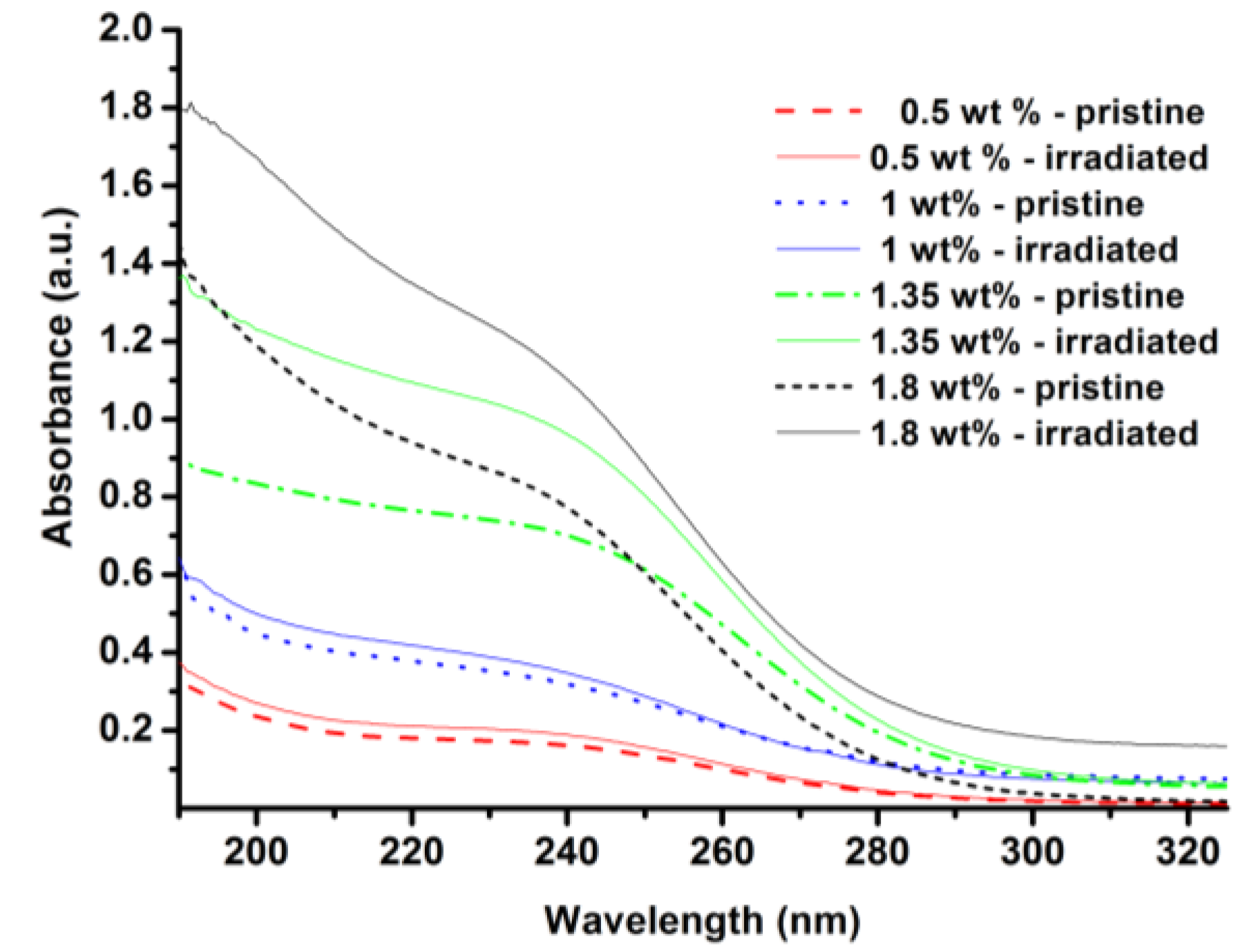
Abstract
:1. Introduction
2. Experimental Section
3. Results and Discussion
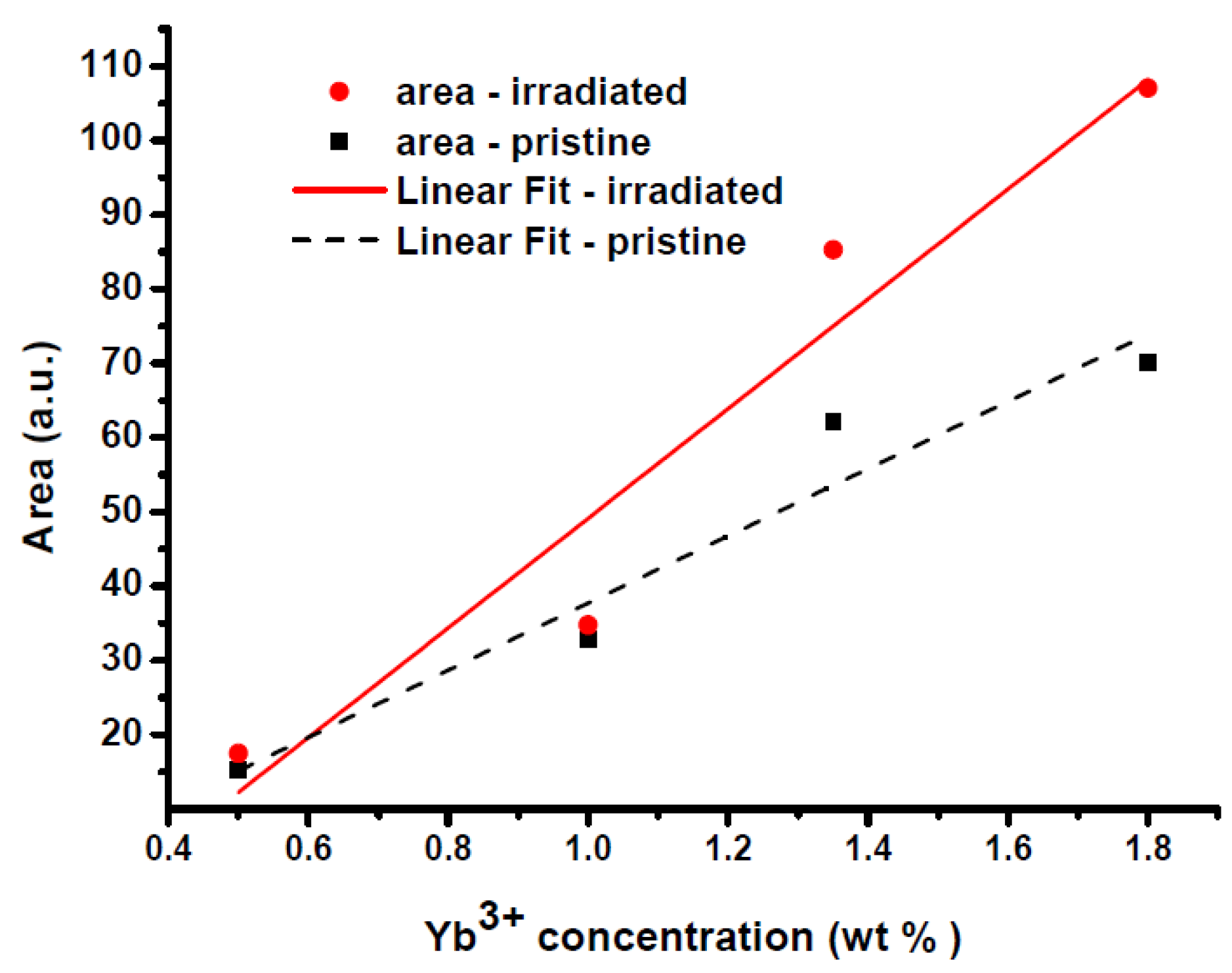
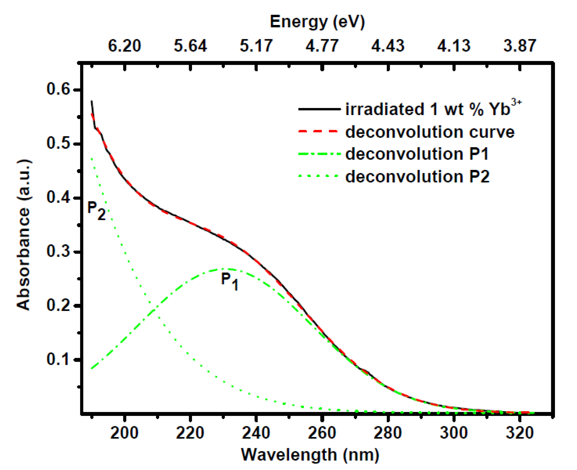
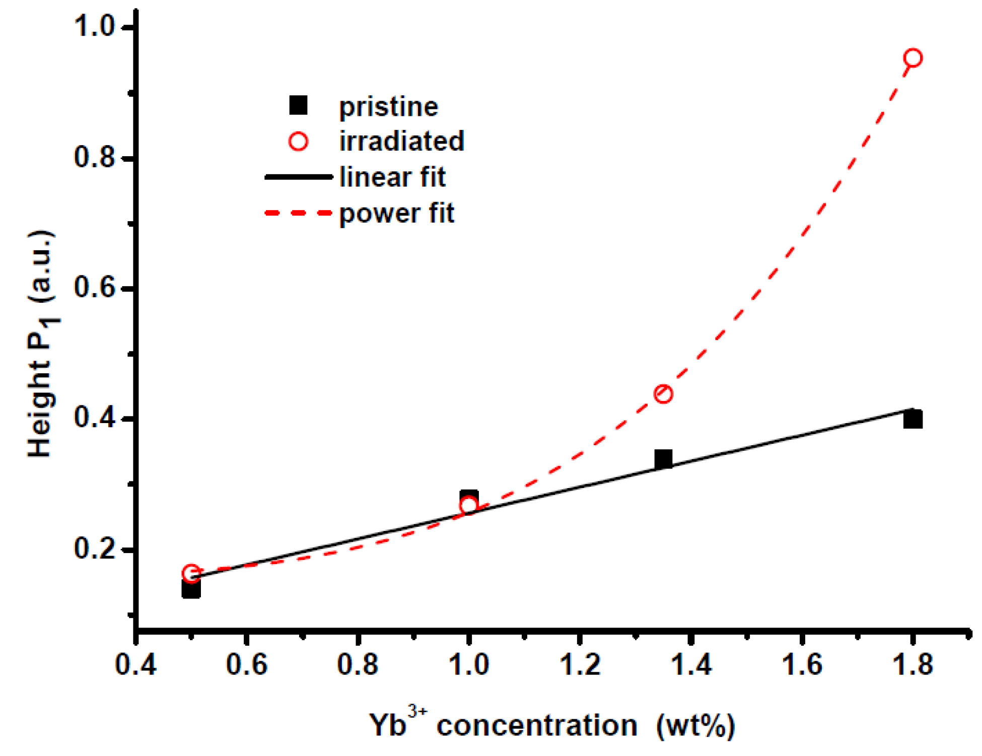
3.1. UV-VIS Absorption Spectra


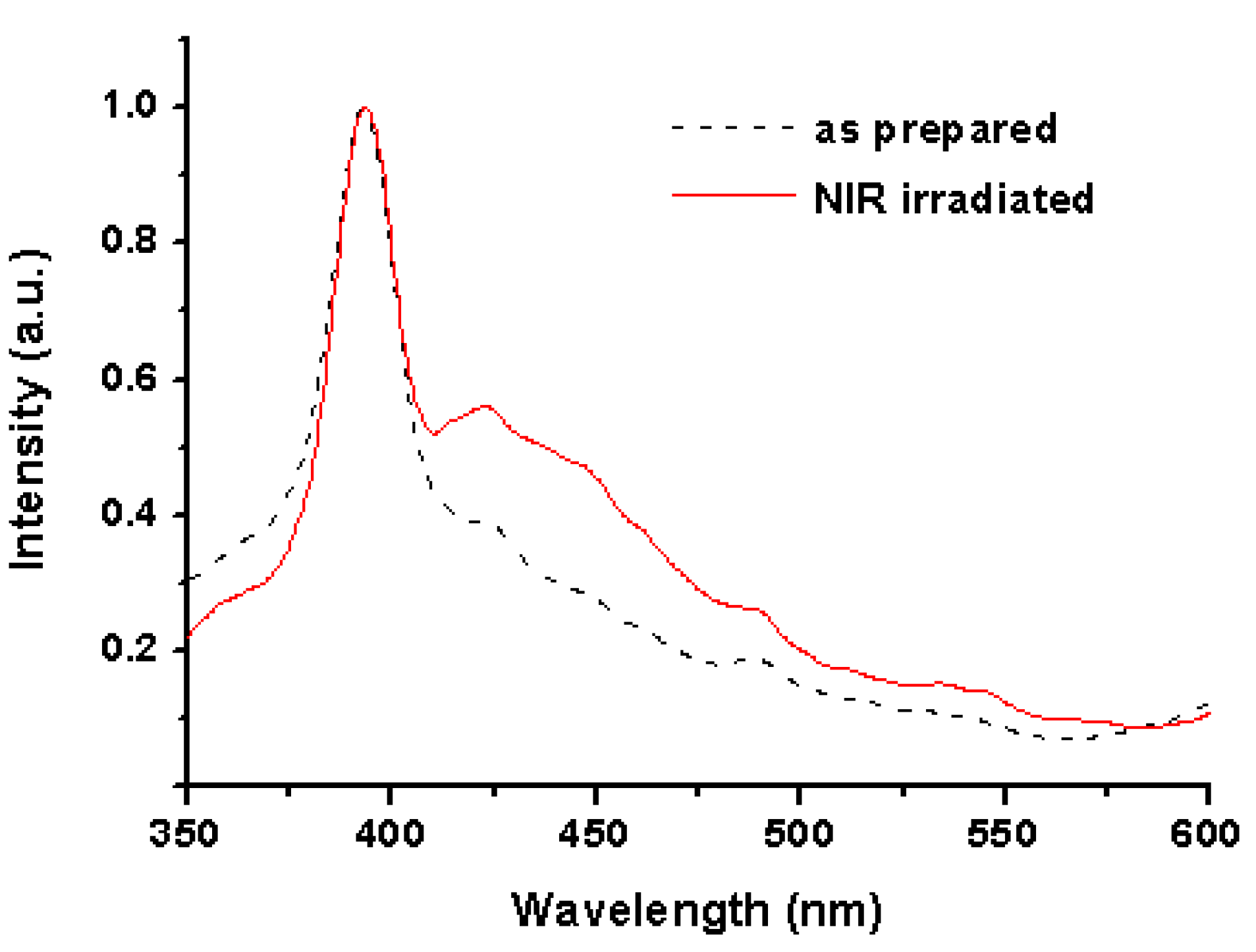
3.2. Photoluminescence Spectroscopy upon Excitation at 230 nm
4. Conclusions
Acknowledgments
Conflicts of Interest
References
- Richardson, D.J.; Nilsson, J.; Clarkson, W.A. High power fiber lasers: Current status and future perspective. J. Opt. Soc. Am. B 2010, 27, B63–B92. [Google Scholar] [CrossRef]
- Paschotta, R.; Nilsson, J.; Barber, P.R.; Caplen, J.E.; Tropper, A.C.; Hanna, D.C. Lifetime quenching in Yb doped fibres. Opt. Commun. 1997, 136, 375–378. [Google Scholar] [CrossRef]
- Koponen, J.; Söderlund, M.; Hoffman, H.J.; Kliner, D.A.V.; Koplow, J.P.; Hotoleanu, M. Photodarkening rate in Yb-doped silica fibers. Appl. Opt. 2008, 47, 1247–1256. [Google Scholar] [CrossRef]
- Likhachev, M.; Aleshkina, S.; Shubin, A.; Bubnov, M.; Dianov, E.; Lipatov, D.; Guryanov, A. Large-Mode-Area Highly Yb-doped Photodarkening-Free Al2O3-P2O5-SiO2-Based Fiber. In Proceedings of the European Conference on Lasers and Electro-Optics (CLEO Europe), Munich, Germany, 22–26 May 2011.
- Mattsson, K.E. Photo darkening of rare earth doped silica. Opt. Express 2011, 19, 19797–19812. [Google Scholar]
- Engholm, M.; Norin, L.; Aberg, D. Strong UV absorption and visible luminescence in ytterbium-doped aluminosilicate glass under UV excitation. Opt. Lett. 2007, 32, 3352–3354. [Google Scholar]
- Taccheo, S.; Gebavi, H.; Monteville, A.; Le Goffic, O.; Landais, D.; Mechin, D.; Tregoat, D.; Cadier, B.; Robin, T.; Milanese, D.; et al. Concentration dependence and self-similarity of photodarkening losses induced in Yb-doped fibers by comparable excitation. Opt. Express 2011, 19, 19340–19345. [Google Scholar]
- Engholm, M.; Norin, L. Comment on “Photodarkening in Yb-doped aluminosilicate fibers induced by 488 nm irradiation”. Opt. Lett. 2008, 33, 1216. [Google Scholar]
- Engholm, M.; Norin, L. Preventing photodarkening in ytterbium-doped high power fiber lasers; correlation to the UV-transparency of the core glass. Opt. Express 2008, 16, 1260–1268. [Google Scholar] [CrossRef]
- Rybaltovsky, A.A.; Aleshkina, S.S.; Likhachev, M.E.; Bubnov, M.M.; Umnikov, A.A.; Yashkov, M.V.; Gur’yanov, A.N.; Dianov, E.M. Luminescence and photoinduced absorption in ytterbium-doped optical fibres. Quant. Electron. 2011, 41, 1073–1079. [Google Scholar] [CrossRef]
- Yoo, S.; Basu, C.; Boyland, A.J.; Sones, C.; Nilsson, J.; Sahu, J.K.; Payne, D. Photodarkening in Yb-doped aluminosilicate fibers induced by 488 nm irradiation. Opt. Lett. 2007, 32, 1626–1628. [Google Scholar] [CrossRef]
- Carlson, C.G.; Keister, K.E.; Dragic, P.D.; Croteau, A.; Eden, J.G. Photoexcitation of Yb-doped aluminosilicate fibers at 250 nm: Evidence for excitation transfer from oxygen deficiency centers to Yb3+. J. Opt. Soc. Am. B 2010, 27, 2087–2094. [Google Scholar]
- Liu, Y.S.; Galvin, T.C.; Hawkins, T.; Ballato, J.; Dong, L.; Foy, P.R.; Dragic, P.D.; Eden, J.G. Linkage of oxygen deficiency defects and rare earth concentrations in silica glass optical fiber probed by ultraviolet absorption and laser excitation spectroscopy. Opt. Express 2012, 20, 14494–14507. [Google Scholar] [CrossRef]
- Gebavi, H.; Taccheo, S.; Tregoat, D.; Monteville, A.; Robin, T. Photobleaching of photodarkening in ytterbium doped aluminosilicate fibers with 633 nm irradiation. Opt. Mater. Express 2012, 2, 1286–1291. [Google Scholar]
- Auzel, F.; Pellk, F. Concentration and excitation effects in multiphonon non-radiative transitions of rare-earth ions. J. Lumin. 1996, 69, 249–255. [Google Scholar] [CrossRef]
- Gebavi, H.; Taccheo, S.; Milanese, D.; Monteville, A.; Le Goffic, O.; Landais, D.; Mechin, D.; Tregoat, D.; Cadier, B.; Robin, T. Temporal evolution and correlation between cooperative luminescence and photodarkening in ytterbium doped silica fibers. Opt. Express 2011, 19, 25077–25083. [Google Scholar] [CrossRef]
- Henke, M.; Persson, J.; Kuck, S. Preparation and spectroscopy of Yb2+-doped Y3Al5O12, YAlO3, and LiBaF3. J. Lumin. 2000, 87–89, 1049–1051. [Google Scholar] [CrossRef]
- Rydberg, S.; Engholm, M. Experimental evidence for the formation of divalent ytterbium in the photodarkening process of Yb-doped fiber lasers. Opt. Express 2013, 21, 6681–6688. [Google Scholar] [CrossRef]
- Arai, T.; Ichii, K.; Okada, K.; Kitabayashi, T.; Tanigawa, S.; Fujimaki, M. Photodarkening phenomenon in Yb-doped fibers. Fujikura Tech. Rev. 2009, 38, 6–11. [Google Scholar]
- Amossov, A.V.; Rybaltovsky, A.O. Oxygen-deficient centers in silica glasses: A review of their properties and structure. J. Non-Cryst. Solids 1994, 179, 75–83. [Google Scholar]
- Van Pieterson, L.; Heeroma, M.; de Heer, E.; Meijerink, A. Charge transfer luminescence of Yb3+. J. Lumin. 2000, 91, 177–193. [Google Scholar]
- Krasikov, D.N.; Scherbinin, A.V.; Vasil’ev, A.N.; Kamenskikh, I.A.; Mikhailin, V.V. Model of Y2O3–Yb charge-transfer luminescence based on ab initio cluster calculations. J. Lumin. 2008, 128, 1748–1752. [Google Scholar]
- Trukhin, A.N.; Golant, K.M. Absorption and luminescence in amorphous silica synthesized by low-pressure plasmachemical technology. J. Non-Cryst. Solids 2007, 353, 530–536. [Google Scholar]
- Kirchhof, J.; Unger, S.; Schwuchow, A.; Jetschke, S.; Reichel, V.; Leich, M.; Scheffel, A. The Influence of Yb2+ Ions on Optical Properties and Power Stability of Ytterbium Doped Laser Fibers. In Proceedings of SPIE 2010, San Francisco, CA, USA, 23 January 2010; Volume 7598, pp. 75980B:1–75980B:11.
© 2013 by the authors; licensee MDPI, Basel, Switzerland. This article is an open access article distributed under the terms and conditions of the Creative Commons Attribution license (http://creativecommons.org/licenses/by/3.0/).
Share and Cite
Gebavi, H.; Milanese, D.; Taccheo, S.; Mechin, D.; Monteville, A.; Freyria, F.S.; Bonelli, B.; Robin, T. Photodarkening of Infrared Irradiated Yb3+-Doped Alumino-Silicate Glasses: Effect on UV Absorption Bands and Fluorescence Spectra. Fibers 2013, 1, 101-109. https://doi.org/10.3390/fib1030101
Gebavi H, Milanese D, Taccheo S, Mechin D, Monteville A, Freyria FS, Bonelli B, Robin T. Photodarkening of Infrared Irradiated Yb3+-Doped Alumino-Silicate Glasses: Effect on UV Absorption Bands and Fluorescence Spectra. Fibers. 2013; 1(3):101-109. https://doi.org/10.3390/fib1030101
Chicago/Turabian StyleGebavi, Hrvoje, Daniel Milanese, Stefano Taccheo, David Mechin, Achille Monteville, Francesca S. Freyria, Barbara Bonelli, and Thierry Robin. 2013. "Photodarkening of Infrared Irradiated Yb3+-Doped Alumino-Silicate Glasses: Effect on UV Absorption Bands and Fluorescence Spectra" Fibers 1, no. 3: 101-109. https://doi.org/10.3390/fib1030101
APA StyleGebavi, H., Milanese, D., Taccheo, S., Mechin, D., Monteville, A., Freyria, F. S., Bonelli, B., & Robin, T. (2013). Photodarkening of Infrared Irradiated Yb3+-Doped Alumino-Silicate Glasses: Effect on UV Absorption Bands and Fluorescence Spectra. Fibers, 1(3), 101-109. https://doi.org/10.3390/fib1030101