Article

Efficient Antifungal and Flame-Retardant Properties of ZnO-TiO2-Layered Double-Nanostructures Coated on Bamboo Substrate

Key Laboratory of High Efficient Processing of Bamboo of Zhejiang Province, China National Bamboo Research Center, Chinese Academy of Forestry, Hangzhou 310012, China
*
Authors to whom correspondence should be addressed.
Coatings 2018, 8(10), 341; https://doi.org/10.3390/coatings8100341
Submission received: 20 August 2018 / Revised: 14 September 2018 / Accepted: 19 September 2018 / Published: 26 September 2018

Abstract

:
A facile method to synthesize ZnO-TiO2-layered double-nanostructures with the average thickness of 20 μm on a bamboo substrate was proposed to improve the antifungal and flame-retardant properties. The cross-linked wurtzite ZnO nanostructures with an average thickness of approximately 0.14 μm were uniformly distributed on the anatase TiO2 surface. The energy-dispersive X-ray spectroscopy (EDS) confirmed that the ZnO-TiO2 coating on bamboo was a layered double nanostructure. During a two-month antifungal test conducted in an outdoor environment, the fungi began to grow after one week on pristine bamboo and three weeks on ZnO-bamboo and TiO2-bamboo. Furthermore, there was an infected area of 100% after four weeks for pristine bamboo and six weeks for ZnO-bamboo, while there was an infected area of 43% after eight weeks for TiO2-bamboo. By comparison, there was no visible fungal growth on ZnO-TiO2-bamboo until the end of the test. The electron spin resonance (ESR) technique has demonstrated that the reactive oxygen species (ROS) of •O2 and •OH were produced from the ZnO-TiO2 surface under visible light irradiation (λ > 420 nm). This large quantity of •O2 compared to •OH is considered to be mainly responsible for the inactivation of fungi. Additionally, the limiting oxygen index has increased from 25.6% to 30.2% after being covered with a ZnO-TiO2 coating, which revealed a significant enhancement of its flame-retardant property.

Graphical Abstract

1. Introduction

Bamboo is an abundant natural resource in China. It has excellent physical and mechanical properties, such as low weight, easy machining, high intensity, and flexibility [1]. As a rapidly renewable sustainable resource, bamboo has been extensively applied in the production of bamboo-based composites, such as laminated bamboo board, bamboo particle board, bamboo plywood, and bamboo scrimber [2,3,4]. Due to its diverse usage and fast growth rate, bamboo has become an ideal material to satisfy the demands for wood. Bamboo is increasingly being utilized worldwide as an essential raw material for construction industry [5]. However, bamboo is easily infected with fungi because of its hygroscopicity and organic nature. With the increasing application of bamboo in an outdoor environment, it is necessary to develop an effective antifungal technique to protect bamboo in a natural environment. Meanwhile, the flammability of bamboo also restricts its utilization, with fire safety being a major concern for the application of bamboo as structural materials, especially in densely populated spaces. To overcome the inherent deficiencies and use of bamboo in a safe manner, the antifungal and flame-retardant properties of bamboo needs attention.
In recent years, the photocatalytic semiconducting nanomaterials (such as TiO2 [6,7,8], ZnO [9,10,11,12], CuO [13] and CeO2 [14]) have been used in various applications in antibacterial and antifungal fields. However, the energy ranges of photoexcitation for many photocatalytic materials are relatively narrow. Furthermore, the photogenerated electron-hole pairs are easily recombined, which results in a low quantum efficiency. To improve the photocatalytic activity and take full advantage of solar light, the combination of nanomaterials with different band gaps has been considered as the most promising technology. It could effectively increase the separation efficiency of electron-hole pairs and extend the energy range of photoexcitation [15,16]. Therefore, the increased reactive oxygen species (ROS, such as •O2, •OH, and H2O2) would be produced.
Among the various photocatalytic nanomaterials, titanium dioxide (TiO2) and zinc oxide (ZnO) have received increasing attention because of their high chemical stability, nontoxicity, relatively low cost and high photocatalytic efficiency [17]. Several reports have demonstrated that ZnO-TiO2 composites were quite effective when used as a photocatalyst [18,19,20,21,22], while the antifungal properties of bamboo modified with ZnO-TiO2 nanocomposites have not been explored under solar light irradiation. Inspired by this, using the two nanomaterials to cover the bamboo substrate may improve the antifungal property in a natural environment and provide a flame-retardant property.
In this study, a facile method to synthesize the ZnO-TiO2-layered double-nanostructures on the bamboo substrate was proposed to modify the properties of bamboo in terms of the antifungal property, thermal stability and flame-retardant property. The morphology, crystallinity and chemical structure of the ZnO-TiO2 coating were investigated. The ROS generated from ZnO-TiO2 coating has been verified by electron spin resonance (ESR) tests under visible light irradiation. The mechanism of the improvement in its antifungal property has been proposed. Furthermore, the thermal stability and flame-retardant property were also evaluated.

2. Materials and Methods

2.1. Materials

Moso bamboo with a size of 50 mm (length) × 20 mm (width) × 6 mm (thickness) were first ultrasonically washed with deionized (DI) water and dried at 60 °C about 24 h before use. All the chemicals were of analytical reagent grade. The DI water was used throughout the experiment.

2.2. Synthesis of TiO2 Thin Film on Bamboo Substrate

Firstly, a group of bamboo samples (12 pieces) was added to 400 mL of an aqueous solution containing 8.0 g of (NH4)2TiF6 and 7.4 g of H3BO3. Using the liquid phase deposition method, the temperature of the solution was maintained at 80 °C for 5 h in a beaker. Subsequently, the samples were washed with DI water, before being dried at 60 °C for 24 h. The bamboo with a TiO2 thin film were abbreviated as TiO2-B.

2.3. Synthesis of ZnO-TiO2-Layered Double-Nanostructures on Bamboo Substrate

Different concentrations of zinc nitrate solutions (0.05, 0.1, 0.2 and 0.3 M) were prepared to synthesize ZnO. The ammonia solution with a concentration of 28% was dropwise added to 400 mL of different concentration of zinc nitrate solutions under constant stirring until the solutions became clear. One group of TiO2-B was placed into the above mixture and heated at 80 °C for 3 h. Subsequently, the samples were washed with DI water, before being dried at 60 °C more than 24 h. The ZnO/TiO2-coated bamboo were abbreviated as ZnO/TiO2-B. For comparison, the pure ZnO coated bamboo samples were also prepared under the same conditions without the presence of TiO2 and were abbreviated as ZnO-B.

2.4. Characterization

The surface morphologies were observed by scanning electron microscopy (SEM, Hitachi SU8010, Tokyo, Japan) equipped with an energy-dispersive X-ray spectroscopy (EDS, OXFORD X-MAX80, Oxford, UK) system. The crystalline structures were identified by X-ray diffraction (XRD) using a Rigaku D/Max 2500 X-ray diffractometer (Rigaku, Tokyo, Japan) with Cu Kα (λ = 1.5418 Å) radiation at a scan speed of 5° min−1 from 10° to 80°. The electron spin resonance (ESR) signals of the radicals trapped by 5,5-dimethyl-1-pyrroline N-oxide (DMPO) were obtained at room temperature with a Bruker (E580, Karlsruhe, Germany) spectrometer under visible light irradiation (λ > 420 nm). Thermogravimetric and Differential Thermal Analysis (TG-DTA) were measured by a NETZSCH TG-209F3 (Selb, Germany) thermogravimetric analyzer at a temperature of 40–800 °C and a heating speed of 10 °C min−1 under N2 atmosphere. The limiting oxygen index (LOI) was evaluated by a JF-5 oxygen index instrument (Nanjing Jiangning Company, Jiangning, China).

2.5. Antifungal Test

The antifungal test was carried out in Hangzhou, China. Four groups of different samples (each group contains 12 pieces) were laid horizontally on a grid. The grid was 1 m above the ground and placed outdoors. Therefore, the samples were directly exposed to the natural environment. The temperature and weather during the test period were recorded. The antifungal test was performed from 24 April to 24 June 2018. It should be noted that the rainfall was sufficient in Hangzhou during this period, while the temperature and humidity were suitable for the growth of fungi. The antifungal properties of the samples were evaluated according to the Chinese Standard GB/T 18261-2013 [23]. The evaluation standards for the infection values are listed in Table 1.

3. Results and Discussion

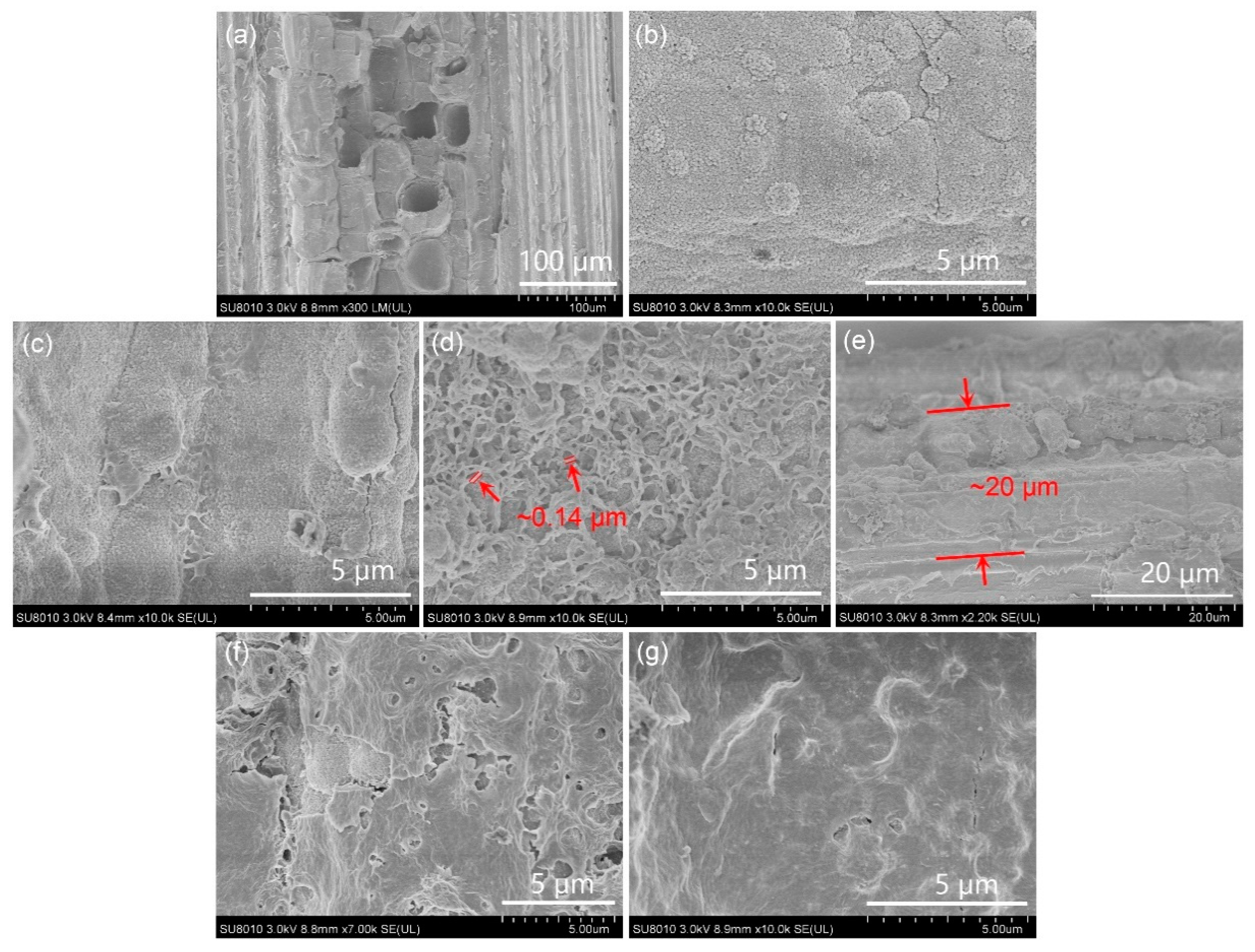
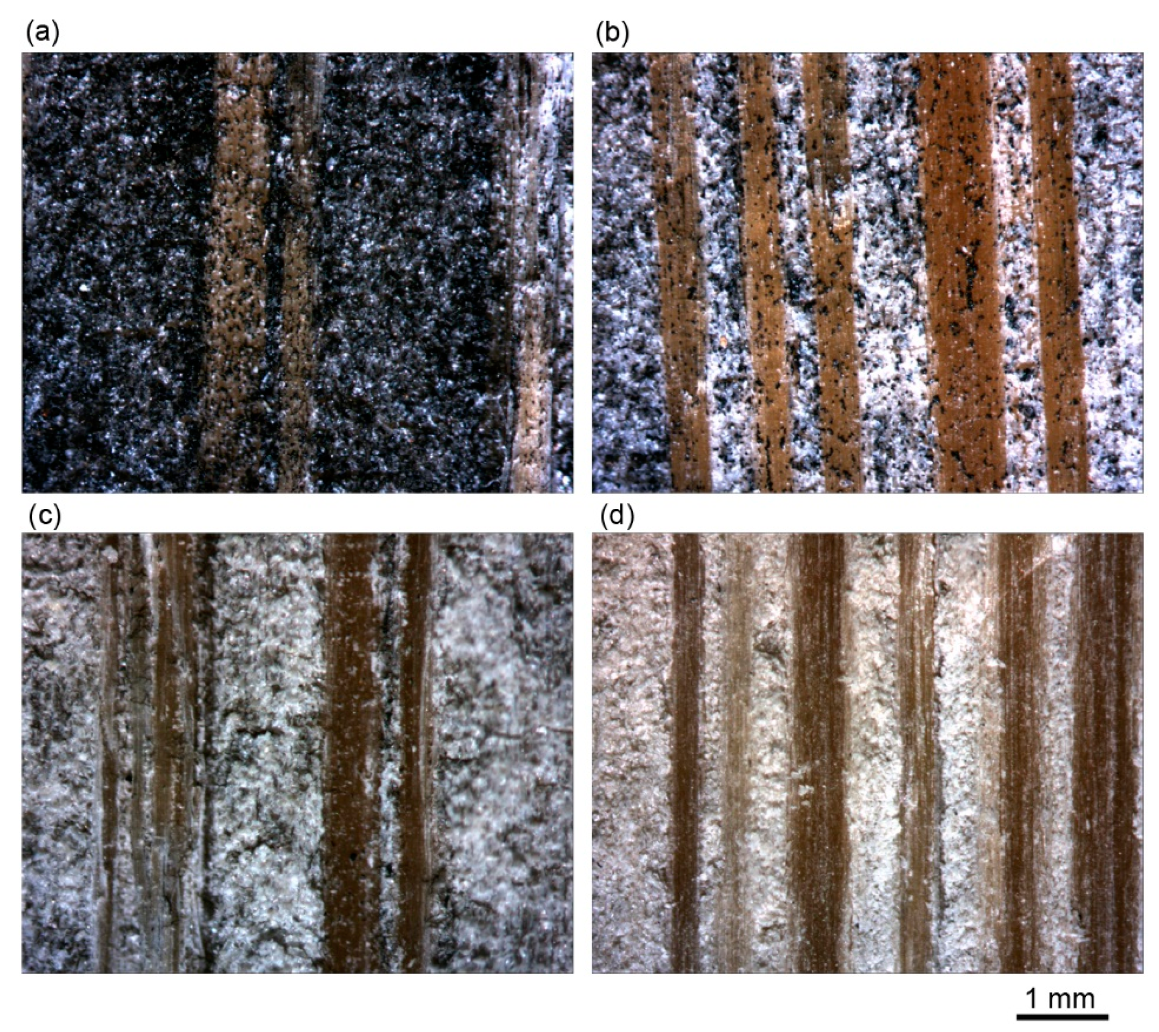
3.1. Morphological Observation

The microstructures of the pristine and functionalized bamboo were observed by SEM. It can be clearly seen that vascular bundles were embedded in the matrix of ground parenchyma from the SEM image of pristine bamboo (Figure 1a). After being coated with TiO2, TiO2 nanoparticles were uniformly distributed across the bamboo surface and formed a thin film (Figure 1b). The influence of the concentrations of ZnO nanostructures in the growth solutions were investigated, while other parameters remain unchanged (Figure 1c,d,f,g). Obviously, the content of the ZnO nanostructures on the TiO2 thin film increases with increasing ZnO concentration in the growth solution. At a relatively low concentration of 0.05 M, only a small amount of ZnO nanostructures formed and adhered to the TiO2 thin film. When the concentration increased to 0.1 M, the ZnO nanostructures were sufficiently continuous and uniformly distributed on the surface of TiO2 thin film. The cross-linked ZnO structures with an average thickness of approximately 0.14 μm effectively improved the specific surface area of the ZnO-TiO2 double-nanostructures coating. The average thickness of the ZnO-TiO2 coating was approximately 20 μm (Figure 1e). By contrast, the cross-linked structures disappeared when the growth solution concentration was at a high level (≥0.2 M), with the high-density ZnO nanostructures becoming a denser thin film and even covering the entire surface of bamboo.

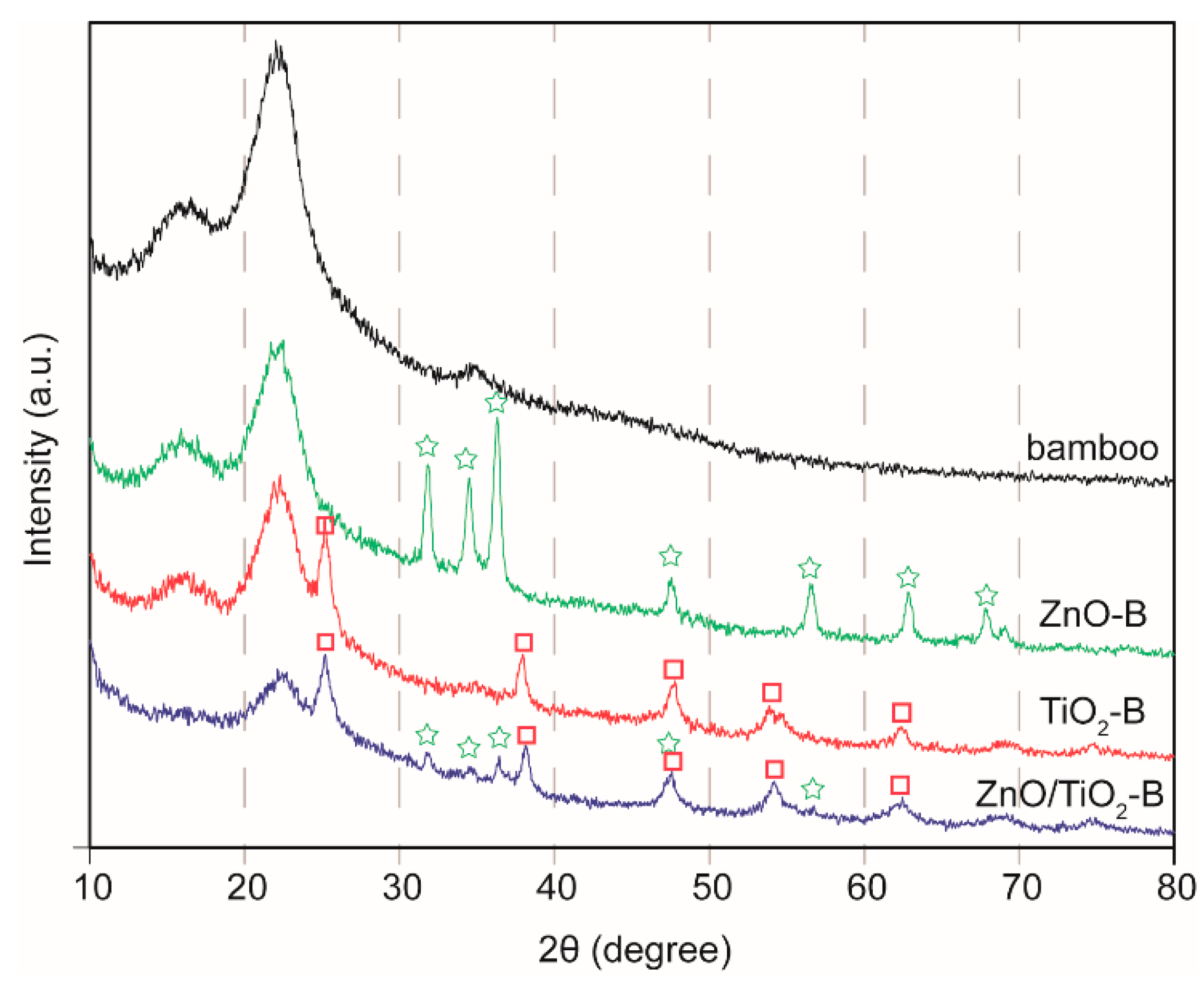
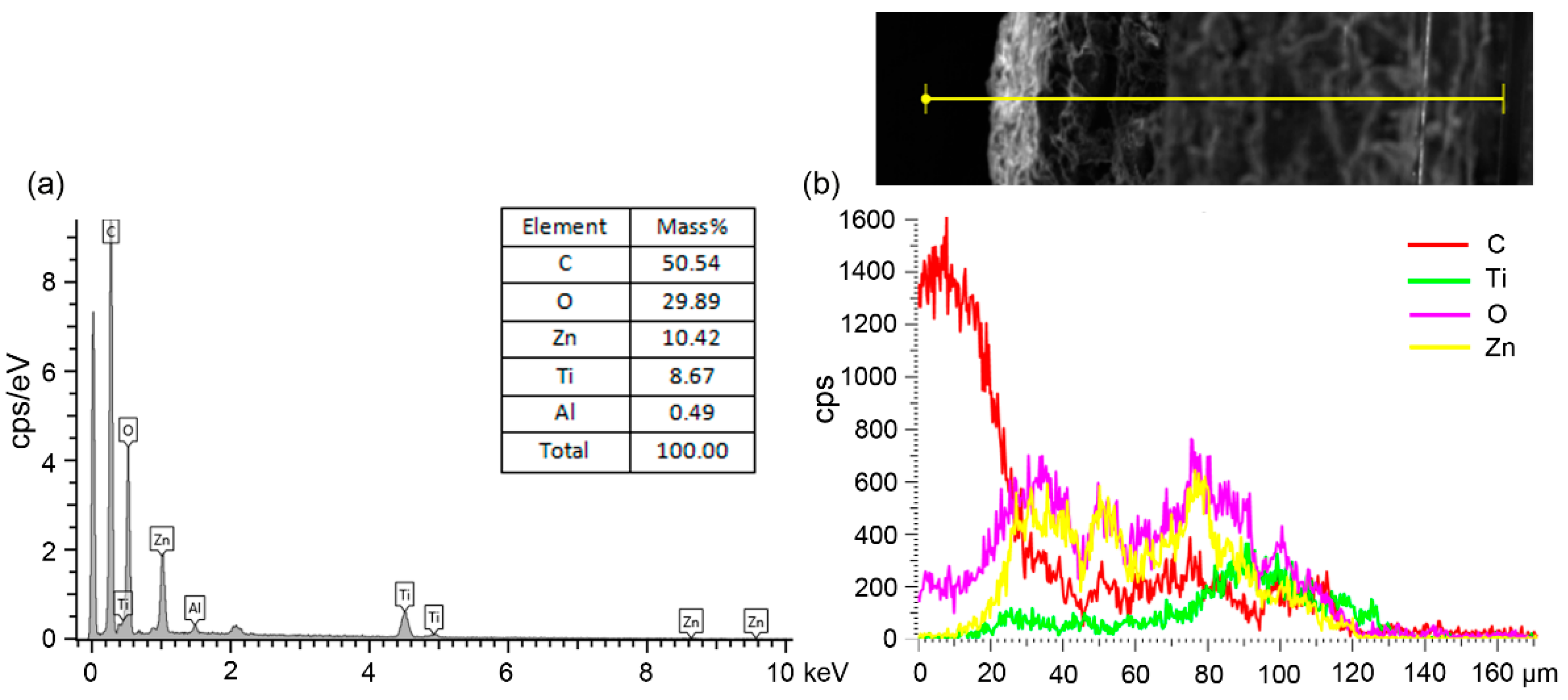
3.2. Elemental Composition and Crystalline Structure Analysis

Figure 2 presents the XRD patterns of pristine bamboo, TiO2-B, ZnO-B and ZnO/TiO2-B (0.1 M of ZnO concentration in growth solutions). The peaks at 16° and 22°, which belong to the cellulose of bamboo, were detected in all the samples. The peaks assigned to the wurtzite ZnO (JCPDS cards: 36-1451) and anatase TiO2 (JCPDS cards: 21-1272) were respectively observed in ZnO-B and TiO2-B [24,25]. Both peaks of ZnO and TiO2 appeared on ZnO/TiO2-B and no other characteristic peaks were discovered. These observations demonstrated that the coating consisted of the wurtzite ZnO and anatase TiO2. The EDS spectrum and SEM in the line scanning mode have provided evidence in the form of the above results and further confirmed the presence of the layered double nanostructures.
As shown in the EDS spectrum of the cross-section of ZnO/TiO2-B (Figure 3), Ti, Zn, O, C and Al elements were observed. The element of C originated from the bamboo substrate or the conducting resin during SEM measurements, while the Al element could be expected from the sample stage. Figure 3b shows the relative intensity of each element measured along the coating (yellow line). The signal of oxygen (pink) implies that a large quantity of O element was distributed across the coating thickness. The signal of titanium (green) and the signal of zinc (yellow) indicated that the Ti and Zn elements were located on different layers of the ZnO-TiO2 coating, which revealed the layered double nanostructures. In short, the above analysis further confirmed that the ZnO-TiO2-layered double-nanostructures were obtained and coated on the bamboo substrate.

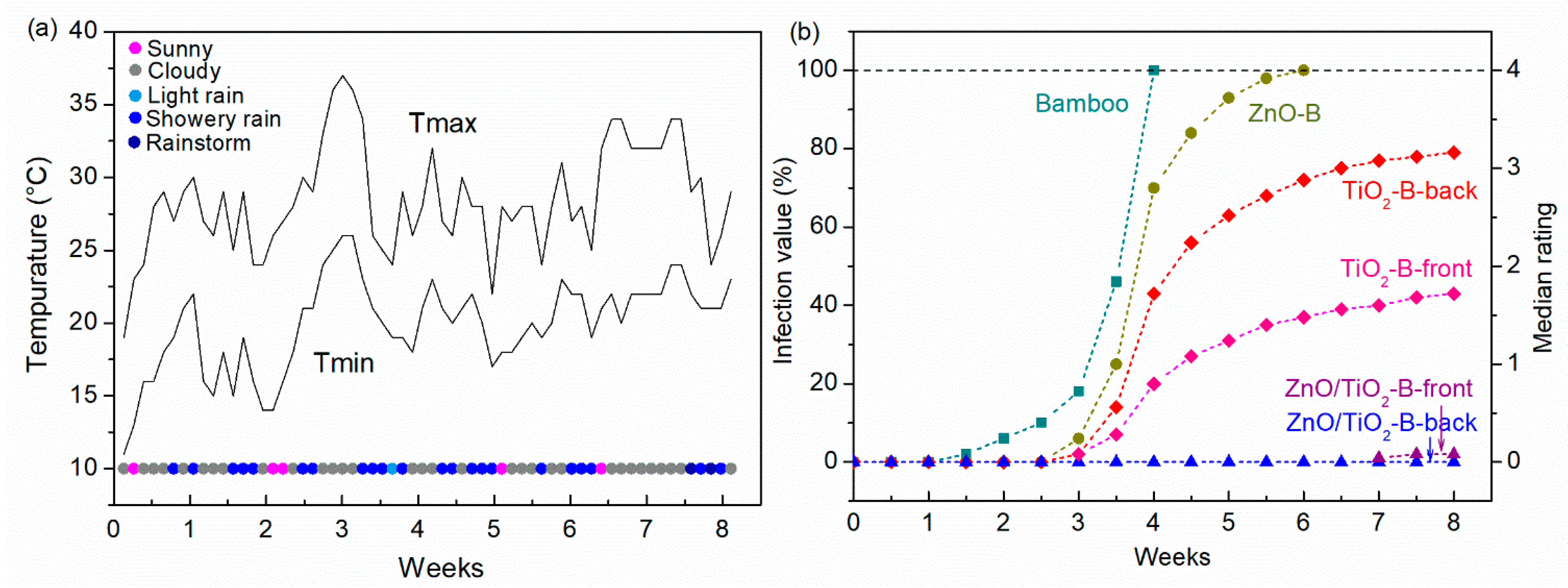
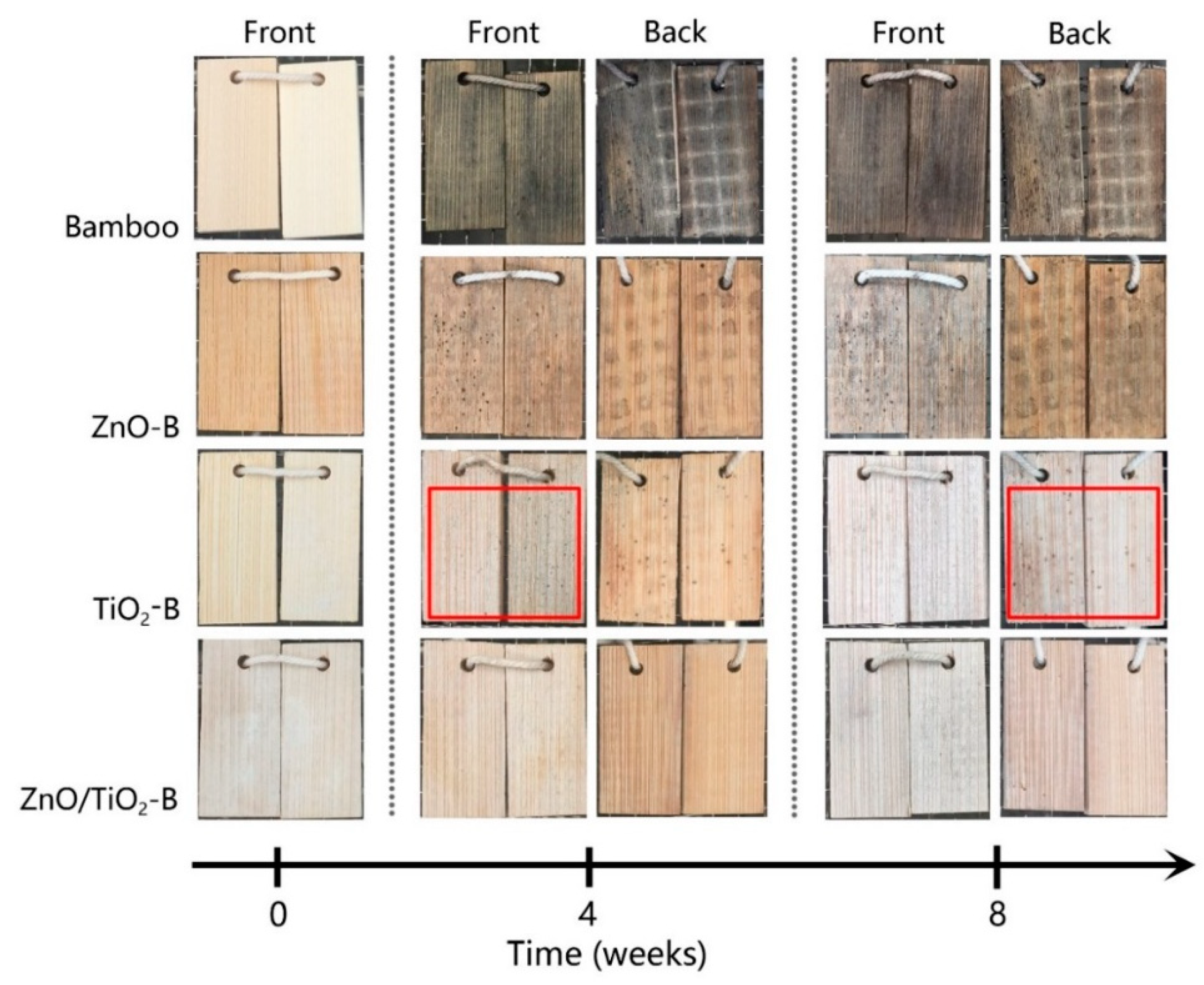
3.3. Antifungal Property

Bamboo is easily degraded when directly exposed outdoors because of the organic nature of bamboo and the complex environmental conditions. The relevant research demonstrated that the optimum temperature for fungal growth is 5–30 °C, while the optimal humidity is above 63% [26]. The temperature and weather during the test period are presented in Figure 4a. As shown, the weather was mainly cloudy or rainy because of the upcoming rainy season in Hangzhou. The rainy season is very suitable for the growth of fungi. The results of the antifungal evaluation are summarized in Figure 4b. The photographs (front and back) of the samples after four weeks and eight weeks of the test are exhibited in Figure 5. Obviously, the pristine bamboo exhibited poor resistance to fungal growth, with fungi having grown after one week of being exposed outdoors. Furthermore, the infected area reached 100% after four weeks of the test. As shown in the photographs, the pristine bamboo surface was only severely degraded after four weeks of exposure. By contrast, ZnO-B and TiO2-B showed weak resistance to fungal growth. Fungi began to grow on ZnO-B during the third week and covered the entire surface after six weeks. For TiO2-B, there were significant differences between growth on the front and the back as the fungi grew more easily on the back than on the front of the samples. After the two-month test, the face infection value of the front of TiO2-B was 43%, but the back reached 79%. The possible reason is that the back could not receive direct sunlight and the front of TiO2-B produced more ROS in the presence of light. As expected, ZnO/TiO2-B had an improved antifungal property as there were no visible fungi growth on both sides of the samples until the end of test.
To obtain more information of the fungal colonies, the samples were observed with an optical microscope after the two-month test period. As shown in Figure 6, fungi densely covered the entire surface of pristine bamboo. In contrast, the antifungal ability of TiO2-B was better than that of ZnO-B, and the ZnO-B was infected more severely. However, there was no visible fungal growth on the ZnO/TiO2-B surface, which indicated that ZnO-TiO2 double-nanostructures coating effectively inhibited fungal growth on bamboo surface in an outdoor environment.

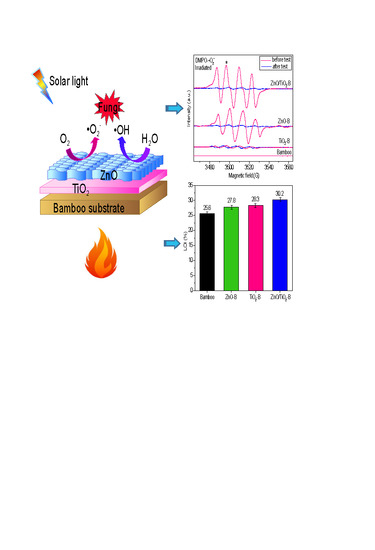
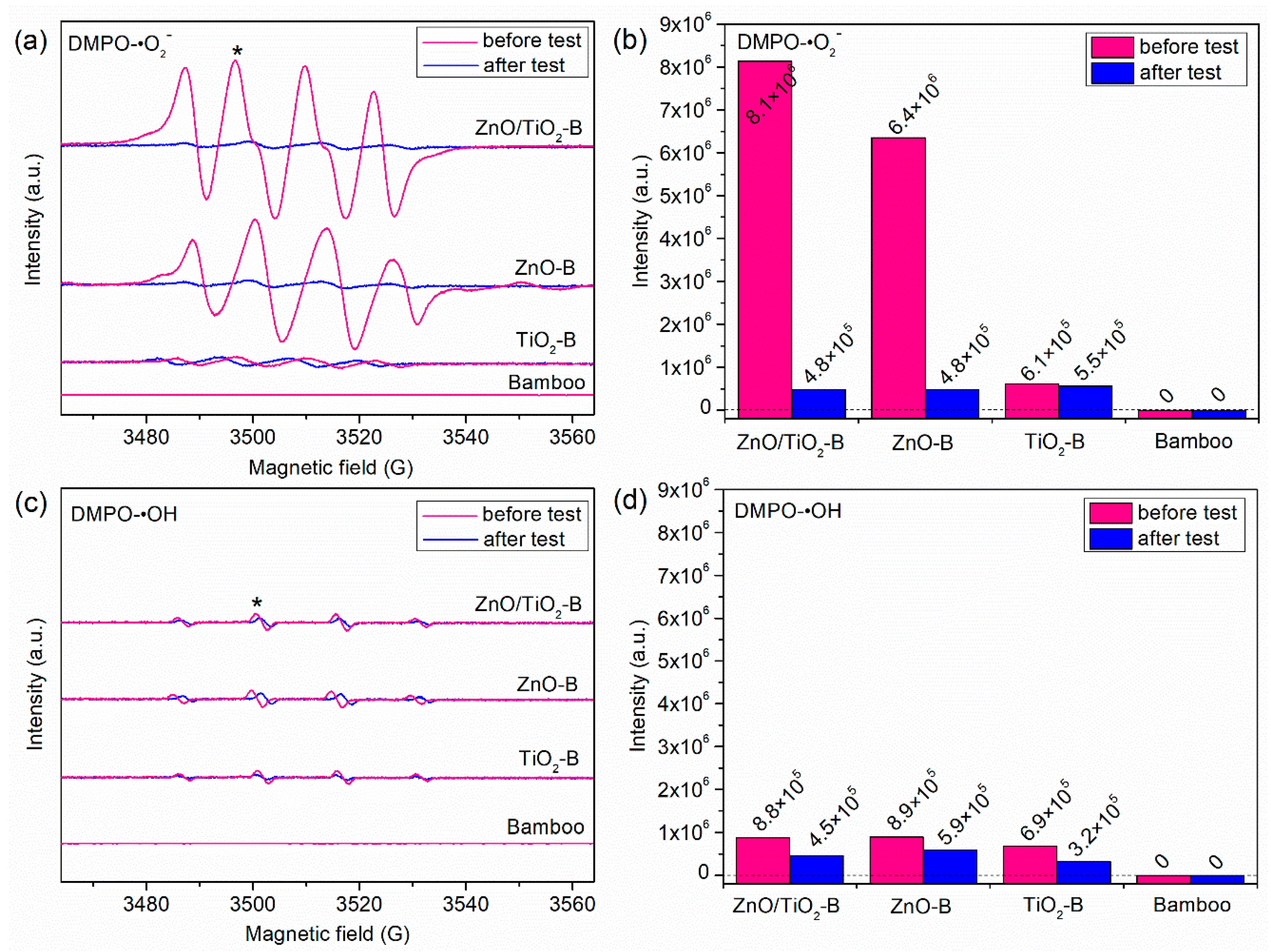
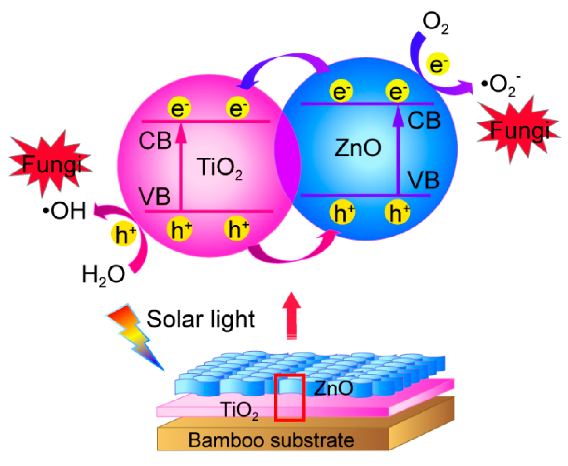
3.4. Mechanism of the Enhanced Antifungal Properties

It is known that ROS have the ability to attack the crucial organic components of fungi and cause their death [27]. To identify the mechanism behind the improved antifungal property, the ESR spin-trap technique (with DMPO) was carried out to determine the photogenerated ROS from the surface of samples under visible light irradiation (λ > 420 nm). The ESR spectra of DMPO-•O2 and DMPO-•OH are shown in Figure 7a,c. The characteristic peak with an intensity ratio of 1:2:2:1 were clearly observed. Due to the overall poor signal/noise ratio in certain samples, the quantitative analysis was performed by the highest signal/noise ratio in the 1:2:2:1 quartet profile, the quantified intensity of the second peak of the radical species (marked by the “*”) are summarized in Figure 7b,d. Before the test, the characteristic signals intensity of the DMPO-•O2 for the samples were ZnO/TiO2-B > ZnO-B > TiO2-B. This revealed that ZnO/TiO2-B produced a greater amount of •O2 than ZnO-B and TiO2-B under visible light irradiation [28,29,30]. There were no noticeable signals detected in pristine bamboo, which indicated that •O2 could not be produced from pristine bamboo. For the ESR spectra of DMPO-•OH, under visible light irradiation, the characteristic signals intensity for ZnO/TiO2-B, ZnO-B and TiO2-B were basically the same. It demonstrated that this large quantity of •O2 compared to •OH is considered to be mainly responsible for the inactivation of fungi. Therefore, ZnO/TiO2-B had an improved antifungal property than ZnO-B and TiO2-B because more •O2 was produced by ZnO/TiO2-B.
After the two-month antifungal test, the ROS signals of the samples were tested again. As shown, the intensity of ZnO/TiO2-B and ZnO-B for DMPO-•O2 was significantly reduced (Figure 7b). This might be due to the destruction of ZnO nanostructures by the rain wash in a natural environment, resulting in the gradual leaching of ZnO nanoparticles from the surface of the samples. As a consequence, the antifungal property of ZnO-B was weakened gradually. In contrast, the intensity of TiO2-B showed no significant decrease, which indicated that TiO2 nanoparticles have a better resistance leachability and it combined strongly with the bamboo substrate. Meanwhile, it is known that the band gap of TiO2 is narrower than that of ZnO and thus, TiO2 has a higher utilization rate of light. As a result, the antifungal activity of TiO2-B was better than that of ZnO-B in an outdoor environment.
Based on the results of the antifungal properties of the samples and the characterization, the schematic of the mechanism behind the enhanced antifungal property for ZnO/TiO2-B under solar light irradiation was proposed, which is shown in Figure 8 [18,31,32]. The ZnO-TiO2-layered double-nanostructures coating formed a heterojunction photocatalyst. Under solar light irradiation, the electrons were photoexcited from the valence band (VB) to the conduction band (CB) of ZnO, creating holes in the VB. After this, the photogenerated electrons (e) in the CB of ZnO were transferred into CB of TiO2 because of potential difference between them, while the holes (h+) in the VB of TiO2 were transferred into the VB of ZnO. Simultaneously, because of the existence of electrons and holes, the dissolved molecular oxygen was reduced to •O2 (O2 + e → •O2) and the H2O molecules were oxidized to •OH (h+ + H2O → •OH + H+). Therefore, the electrons and holes were effectively separated and the generation of ROS was increased. According to the above analysis, the ZnO-TiO2-layered double-nanostructures improved the separation efficiency of electron-hole pairs, increased the generation of ROS and resulted in effective antifungal activity.

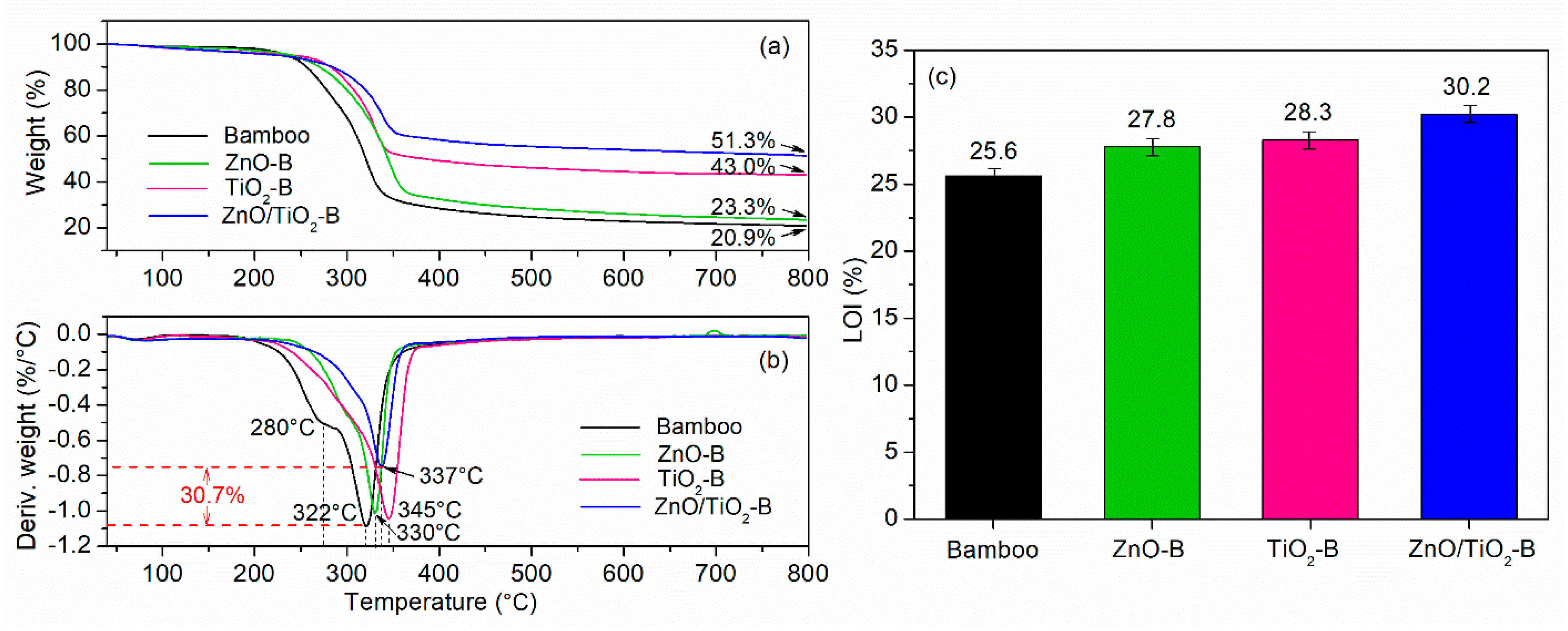
3.5. Thermal Stability and Flame-Retardant Property

The TG and DTA curves of the samples are presented in Figure 9. As shown, the bamboo components were thermally degraded in three stages. The first decomposition process (190–250 °C) was assigned to the thermal depolymerization of hemicelluloses. The second decomposition process (250–380 °C) were ascribed to the cellulose decomposition and continuous degradation of lignin. During the third process (380–800 °C), the residual bamboo components continued to degrade, resulting in aromatization and carbonization [33,34,35]. After the calcining process, the weight of mass residues for pristine bamboo, ZnO-B, TiO2-B and ZnO/TiO2-B were 20.9%, 23.3%, 43.0% and 51.3%, respectively. In the DTA curve of the pristine bamboo, the two shoulder peaks located at 280 °C and 322 °C were assigned to the decomposition of hemicelluloses and cellulose, respectively. Compared to the pristine bamboo, the sharp endothermic peaks of ZnO-B, TiO2-B and ZnO/TiO2-B had a right-shift to 330, 345 and 337 °C, respectively. Meanwhile, the maximal pyrolysis rates were decreased. The maximal pyrolysis rate of ZnO/TiO2-B was decreased by 30.7%. This might be due to the decomposition of the ZnO-TiO2 nanostructures coating, the strong interaction between ZnO and TiO2 and the interaction between bamboo substrate and the coating. Comparing the four samples, ZnO/TiO2-B exhibited the lowest maximal pyrolysis rate and the highest weight of residues, which indicated that ZnO-TiO2 nanostructures coating could act as a barrier and efficiently protect bamboo from the calcining process.
The flame retardancy property of the samples was determined by LOI, which is a common parameter used for assessing the flammability (Figure 9c). LOI refers to the minimum concentration of oxygen in an oxygen-nitrogen mixture [36]. It is determined as follows. A sample is ignited in an oxygen-nitrogen mixture. The oxygen concentration is adjusted until the minimum required to sustain steady burning is found. A higher LOI value indicates better flame-retardant capability. As shown, the pristine bamboo has a low LOI value of 25.6%. The LOI value of ZnO-B and TiO2-B increased to 27.8% and 28.3% because of the protective effects of ZnO and TiO2 nanomaterials, respectively. After covering the bamboo substrate with ZnO-TiO2 nanostructures, the LOI value increases dramatically to 30.2%, demonstrating the significant improvement in its flame-retardant property. Combined with the analysis of TG and DTA, ZnO-TiO2-layered double-nanostructures coating effectively improved the thermal stability and flame resistance of the samples, which provided a promising candidate for functional and building materials.

4. Conclusions

In this study, ZnO-TiO2-layered double-nanostructures were successfully coated on a bamboo substrate via a facile method to improve the antifungal and flame-retardant properties of bamboo. SEM, XRD and EDS confirmed that the ZnO-TiO2 coating was a layered double nanostructure, which was successfully coated on the bamboo surface. During a two-month antifungal test in an outdoor environment, the ZnO-TiO2-layered double-nanostructures coated bamboo exhibited improved antifungal activity, which was due to the double nanostructures effectively increasing the separation efficiency of electron-hole pairs and extending the energy range of photoexcitation. ESR showed that the ROS of •O2 and •OH were generated on the ZnO-TiO2 double-nanostructures surface under visible light irradiation (λ > 420 nm). This large quantity of •O2 compared to •OH is considered to be mainly responsible for the inactivation of fungi. Furthermore, the ZnO-TiO2-layered double-nanostructures coating improved the thermal stability and flame-retardant property of bamboo, with the LOI value having increased to 30.2%. Therefore, we conclude that ZnO-TiO2 double-nanostructures coating has a potential to inhibit fungal growth in an outdoor environment and to improve the flame-retardant property for protecting bamboo products.

Author Contributions

Conceptualization, J.L. and Y.C.; Formal Analysis, D.R. and J.X.; Investigation, D.R., Z.W., Y.B. and N.L.; Methodology, J.L.; Resources, Y.C.; Writing-Original Draft, D.R.

Funding

The research was funded by the Fundamental Research Funds for the Central Non-profit Research Institution of CAF (CAFYBB2017MA023), National Key Research and Development Program of China (2016YFD0600904) and Key Laboratory of High Efficient Processing of Bamboo of Zhejiang Province (2014F10047).

Acknowledgments

The authors wish to gratefully acknowledge the Zhejiang University for services related to SEM and EDS analysis of the samples.

Conflicts of Interest

The authors declare no conflicts of interest.

References

  1. Sharma, B.; Gatóo, A.; Bock, M.; Ramage, M. Engineered bamboo for structural applications. Constr. Build. Mater. 2015, 81, 66–73. [Google Scholar] [CrossRef]
  2. Verma, C.S.; Chariar, V.M. Development of layered laminate bamboo composite and their mechanical properties. Compos. Part B Eng. 2012, 43, 1063–1069. [Google Scholar] [CrossRef]
  3. Yu, Y.; Huang, X.; Yu, W. A novel process to improve yield and mechanical performance of bamboo fiber reinforced composite via mechanical treatments. Compos. Part B Eng. 2014, 56, 48–53. [Google Scholar] [CrossRef]
  4. Yu, Y.; Liu, R.; Huang, Y.; Meng, F.; Yu, W. Preparation, physical, mechanical, and interfacial morphological properties of engineered bamboo scrimber. Constr. Build. Mater. 2017, 157, 1032–1039. [Google Scholar] [CrossRef]
  5. Flynn, A.; Chan, K.W.; Zhu, Z.H.; Yu, L. Sustainability, space and supply chains: The role of bamboo in Anji County, China. J. Rural Stud. 2017, 49, 128–139. [Google Scholar] [CrossRef] [Green Version]
  6. Darbari, S.; Abdi, Y.; Haghighi, F.; Mohajerzadeh, S.; Haghighi, N. Investigating the antifungal activity of TiO2 nanoparticles deposited on branched carbon nanotube arrays. J. Phys. D Appl. Phys. 2011, 44, 245401–245410. [Google Scholar] [CrossRef]
  7. Hou, X.; Ma, H.; Liu, F.; Deng, J.; Ai, Y.; Zhao, X.; Mao, D.; Li, D.; Liao, B. Synthesis of Ag ion-implanted TiO2 thin films for antibacterial application and photocatalytic performance. J. Hazard. Mater. 2015, 299, 59–66. [Google Scholar] [CrossRef] [PubMed]
  8. Akhavan, O. Lasting antibacterial activities of Ag-TiO2/Ag/a-TiO2 nanocomposite thin film photocatalysts under solar light irradiation. J. Colloid Interface Sci. 2009, 336, 117–124. [Google Scholar] [CrossRef] [PubMed]
  9. Chauhan, I.; Aggrawal, S.; Mohanty, P. ZnO nanowire-immobilized paper matrices for visible light-induced antibacterial activity against Escherichia coli. Environ. Sci. Nano 2015, 2, 273–279. [Google Scholar] [CrossRef]
  10. Lipovsky, A.; Nitzan, Y.; Gedanken, A.; Lubart, R. Antifungal activity of ZnO nanoparticles–the role of ROS mediated cell injury. Nanotechnology 2011, 22, 105101–105105. [Google Scholar] [CrossRef] [PubMed]
  11. Kairyte, K.; Kadys, A.; Luksiene, Z. Antibacterial and antifungal activity of photoactivated ZnO nanoparticles in suspension. J. Photochem. Photobiol. B 2013, 128, 78–84. [Google Scholar] [CrossRef] [PubMed]
  12. Sharma, D.; Rajput, J.; Kaith, B.S.; Kaur, M.; Sharma, S. Synthesis of ZnO nanoparticles and study of their antibacterial and antifungal properties. Thin Solid Films 2010, 519, 1224–1229. [Google Scholar] [CrossRef]
  13. Chen, X.; Ku, S.; Weibel, J.A.; Ximenes, E.; Liu, X.; Ladisch, M.R.; Garimella, S.V. Enhanced antimicrobial efficacy of bimetallic porous CuO microspheres decorated with Ag nanoparticles. ACS Appl. Mater. Interfaces 2017, 9, 39165–39173. [Google Scholar] [CrossRef] [PubMed]
  14. Lu, Z.; Mao, C.; Meng, M.; Liu, S.; Tian, Y.; Yu, L.; Sun, B.; Li, C.M. Fabrication of CeO2 nanoparticle-modified silk for UV protection and antibacterial applications. J. Colloid Interface Sci. 2014, 435, 8–14. [Google Scholar] [CrossRef] [PubMed]
  15. Jing, L.; Xu, Y.; Huang, S.; Xie, M.; He, M.; Xu, H.; Li, H.; Zhang, Q. Novel magnetic CoFe2O4/Ag/Ag3VO4 composites: Highly efficient visible light photocatalytic and antibacterial activity. Appl. Catal. B Environ. 2016, 199, 11–22. [Google Scholar] [CrossRef]
  16. Xu, Y.; Liu, Q.; Liu, C.; Zhai, Y.; Xie, M.; Huang, L.; Xu, H.; Li, H.; Jing, J. Visible-light-driven Ag/AgBr/ZnFe2O4 composites with excellent photocatalytic activity for E. coli disinfection and organic pollutant degradation. J. Colloid Interface Sci. 2018, 512, 555–566. [Google Scholar] [CrossRef] [PubMed]
  17. Yin, Z.; Li, C.; Liu, X.; Feng, G.; Du, H.L.; Shi, L. Zn-doped TiO2 nanoparticles with high photocatalytic activity synthesized by hydrogen–oxygen diffusion flame. Appl. Catal. B Environ. 2008, 79, 208–215. [Google Scholar] [CrossRef]
  18. Lin, L.; Yang, Y.; Men, L.; Wang, X.; He, D.; Chai, Y.; Zhao, B.; Ghoshroy, S.; Tang, Q. A highly efficient TiO2@ZnO n-p-n heterojunction nanorod photocatalyst. Nanoscale 2012, 5, 588–593. [Google Scholar] [CrossRef] [PubMed]
  19. Wang, Y.; Zheng, Y.Z.; Lu, S.; Tao, X.; Che, Y.; Chen, J.F. Visible-light-responsive TiO2-coated ZnO:I nanorod array films with enhanced photoelectrochemical and photocatalytic performance. ACS Appl. Mater. Interfaces 2015, 7, 6093–6101. [Google Scholar] [CrossRef] [PubMed]
  20. Yuan, S.; Mu, J.; Mao, R.; Li, Y.; Zhang, Q.; Wang, H. All-nanoparticle self-assembly ZnO/TiO2; heterojunction thin films with remarkably enhanced photoelectrochemical activity. ACS Appl. Mater. Interfaces 2014, 6, 5719–5725. [Google Scholar] [CrossRef] [PubMed]
  21. Sun, W.; Meng, S.; Zhang, S.; Zheng, X.; Ye, X.; Fu, X.; Chen, S. Insight into the transfer mechanisms of photogenerated carriers for heterojunction photocatalysts with analogous positions of valence band and conduction band: A case study of ZnO/TiO2. J. Phys. Chem. C 2018, 122, 15409–15420. [Google Scholar] [CrossRef]
  22. Cheng, P.; Wang, Y.; Xu, L.; Sun, P.; Su, Z.; Jin, F.; Liu, F.; Sun, Y.; Lu, G. High specific surface area urchin-like hierarchical ZnO-TiO2 architectures: Hydrothermal synthesis and photocatalytic properties. Mater. Lett. 2016, 175, 52–55. [Google Scholar] [CrossRef]
  23. GB/T 18261-2013 Test Method for Anti-Mildew Agents in Controlling Wood Mould and Stain Fungi; Standardization Administration of China: Beijing, China, 2013. (In Chinese)
  24. Liu, B.; Sun, Y.; Wang, D.; Wang, L.; Zhang, L.; Zhang, X.; Lin, Y.; Xie, T. Construction of a branched ZnO-TiO2 nanorod array heterostructure for enhancing the photovoltaic properties in quantum dot-sensitized solar cells. RSC Adv. 2014, 4, 32773–32780. [Google Scholar] [CrossRef]
  25. Lachom, V.; Poolcharuansin, P.; Laokul, P. Preparation, characterizations and photocatalytic activity of a ZnO/TiO2 nanocomposite. Mater. Res. Express 2017, 4, 035006. [Google Scholar] [CrossRef]
  26. Li, J.; Yu, H.; Wu, Z.; Wang, J.; He, S.; Ji, J.; Li, N.; Bao, Y.; Huang, C.; Chen, Z. Room temperature synthesis of crystalline anatase TiO2 on bamboo timber surface and their short-term antifungal capability under natural weather conditions. Colloids Surf. A 2016, 508, 117–123. [Google Scholar] [CrossRef]
  27. Goffredo, G.B.; Citterio, B.; Biavasco, F.; Stazi, F.; Barcelli, S.; Munafò, P. Nanotechnology on wood: The effect of photocatalytic nanocoatings against Aspergillus niger. J. Cult. Herit. 2017, 27, 125–136. [Google Scholar] [CrossRef]
  28. Velasco, L.F.; Carmona, R.J.; Matos, J.; Ania, C.O. Performance of activated carbons in consecutive phenol photooxidation cycles. Carbon 2014, 73, 206–215. [Google Scholar] [CrossRef] [Green Version]
  29. Teng, W.; Li, X.; Zhao, Q.; Chen, G. Fabrication of Ag/Ag3PO4/TiO2 heterostructure photoelectrodes for efficient decomposition of 2-chlorophenol under visible light irradiation. J. Mater. Chem. A 2013, 1, 9060–9068. [Google Scholar] [CrossRef]
  30. Zhao, D.; Sheng, G.; Chen, C.; Wang, X. Enhanced photocatalytic degradation of methylene blue under visible irradiation on graphene@TiO2 dyade structure. Appl. Catal. B Environ. 2012, 111, 303–308. [Google Scholar] [CrossRef]
  31. Zheng, X.; Li, D.; Li, X.; Chen, J.; Cao, C.; Fang, J.; Wang, J.; He, Y.; Zheng, Y. Construction of ZnO/TiO2 photonic crystal heterostructures for enhanced photocatalytic properties. Appl. Catal. B Environ. 2015, 168–169, 408–415. [Google Scholar] [CrossRef]
  32. Xiao, F.X. Construction of highly ordered ZnO-TiO2 nanotube arrays (ZnO/TNTs) heterostructure for photocatalytic application. ACS Appl. Mater. Interfaces 2012, 4, 7055–7063. [Google Scholar] [CrossRef] [PubMed]
  33. Li, J.; Zheng, H.; Sun, Q.; Han, S.; Fan, B.; Yao, Q.; Yan, C.; Jin, C. Fabrication of superhydrophobic bamboo timber based on an anatase TiO2 film for acid rain protection and flame retardancy. RSC Adv. 2015, 5, 62265–62272. [Google Scholar] [CrossRef]
  34. Gao, L.; Lu, Y.; Li, J.; Sun, Q. Superhydrophobic conductive wood with oil repellency obtained by coating with silver nanoparticles modified by fluoroalkyl silane. Holzforschung 2016, 70, 63–68. [Google Scholar] [CrossRef]
  35. Gao, L.; Lu, Y.; Zhan, X.; Li, J.; Sun, Q. A robust, anti-acid, and high-temperature–humidity-resistant superhydrophobic surface of wood based on a modified TiO2 film by fluoroalkyl silane. Surf. Coat. Technol. 2015, 262, 33–39. [Google Scholar] [CrossRef]
  36. Wang, J.; Jin, C.; Sun, Q.; Zhang, Q. Fabrication of nanocrystalline anatase TiO2 in a graphene network as a bamboo coating material with enhanced photocatalytic activity and fire resistance. J. Alloys Compd. 2017, 702, 418–426. [Google Scholar] [CrossRef]
Figure 1. SEM images of (a) pristine bamboo; (b) TiO2-B and ZnO/TiO2-B with different concentrations of Zn(NO3)2 during the coating process: (c) 0.05 M ZnO/TiO2-B, (d) 0.1 M ZnO/TiO2-B; (e) The cross-section of the coating of 0.1 M ZnO/TiO2-B; (f) 0.2 M ZnO/TiO2-B; and (g) 0.3 M ZnO/TiO2-B.
Coatings 08 00341 g001
Figure 2. XRD patterns of pristine bamboo, ZnO-B, TiO2-B and ZnO/TiO2-B.
Coatings 08 00341 g002
Figure 3. (a) EDS spectra of the ZnO/TiO2-B and the content of each element (inset); and (b) SEM in the line scanning mode and the element distribution across the ZnO-TiO2 coating.
Coatings 08 00341 g003
Figure 4. (a) The temperature and weather at test site from 24 April to 24 June; and (b) Median value of fungal growth on pristine bamboo, ZnO-B, TiO2-B and ZnO/TiO2-B.
Coatings 08 00341 g004
Figure 5. Digital photograph (front and back) of pristine bamboo, ZnO-B, TiO2-B and ZnO/TiO2-B after four weeks of exposure and eight weeks of exposure, respectively.
Coatings 08 00341 g005
Figure 6. Optical microscope pictures after the two-month test period for (a) pristine bamboo; (b) ZnO-B; (c) TiO2-B; and (d) ZnO/TiO2-B, respectively.
Coatings 08 00341 g006
Figure 7. (a) The ESR spectra of DMPO-•O2 before and after the two-month test; (b) the intensity of the second peak (marked by the “*”) in the samples under visible light irradiation; (c) The ESR spectra of DMPO-•OH; and (d) the intensity of the second peak in the samples under visible light irradiation.
Coatings 08 00341 g007
Figure 8. Schematic diagram of the enhanced antifungal property for the ZnO/TiO2-B under solar light irradiation.
Coatings 08 00341 g008
Figure 9. (a) TG curves; (b) DTA curves; and (c) LOIs of pristine bamboo, ZnO-B, TiO2-B and ZnO/TiO2-B.
Coatings 08 00341 g009
Table 1. Rating scale for assessment of fungal growth.
RatingDescription
0No fungal growth
1The area of fungal infection is <25%
2The area of fungal infection is 25%–50%
3The area of fungal infection is 50%–75%
4The area of fungal infection is >75%

