Review

Resistance to Antimicrobial Peptides in Vibrios

by
Delphine Destoumieux-Garzón
1,*,
Marylise Duperthuy
2,
Audrey Sophie Vanhove
1,
Paulina Schmitt
3 and
Sun Nyunt Wai
2
1
Ecology of Coastal Marine Systems, CNRS, Ifremer, University of Montpellier, IRD, Place Eugène Bataillon, CC80, 34095 Montpellier, France
2
Department of Molecular Biology, The Laboratory for Molecular Infection Medicine Sweden (MIMS), Umeå University, 901 87 Umeå, Sweden
3
Laboratorio de Genética e Inmunología Molecular, Instituto de Biología, Pontificia Universidad Católica de Valparaíso, Avenida Universidad 330, 2373223 Valparaíso, Chile
*
Author to whom correspondence should be addressed.
Antibiotics 2014, 3(4), 540-563; https://doi.org/10.3390/antibiotics3040540
Submission received: 16 September 2014 / Revised: 25 September 2014 / Accepted: 8 October 2014 / Published: 27 October 2014
(This article belongs to the Special Issue Antimicrobial Peptides)

Abstract

:
Vibrios are associated with a broad diversity of hosts that produce antimicrobial peptides (AMPs) as part of their defense against microbial infections. In particular, vibrios colonize epithelia, which function as protective barriers and express AMPs as a first line of chemical defense against pathogens. Recent studies have shown they can also colonize phagocytes, key components of the animal immune system. Phagocytes infiltrate infected tissues and use AMPs to kill the phagocytosed microorganisms intracellularly, or deliver their antimicrobial content extracellularly to circumvent tissue infection. We review here the mechanisms by which vibrios have evolved the capacity to evade or resist the potent antimicrobial defenses of the immune cells or tissues they colonize. Among their strategies to resist killing by AMPs, primarily vibrios use membrane remodeling mechanisms. In particular, some highly resistant strains substitute hexaacylated Lipid A with a diglycine residue to reduce their negative surface charge, thereby lowering their electrostatic interactions with cationic AMPs. As a response to envelope stress, which can be induced by membrane-active agents including AMPs, vibrios also release outer membrane vesicles to create a protective membranous shield that traps extracellular AMPs and prevents interaction of the peptides with their own membranes. Finally, once AMPs have breached the bacterial membrane barriers, vibrios use RND efflux pumps, similar to those of other species, to transport AMPs out of their cytoplasmic space.

1. Introduction

Vibrios are γ-proteo-bacteria ubiquitous in aquatic environments. They have evolved the capacity to colonize a broad series of hosts from protozoans to metazoans. Vibrios are normally present in the tissues of healthy animals. Sometimes they become pathogenic in wild marine animals such as corals, in particular as a result of environmental changes including shifts in seawater temperature and salinity, or, for aquacultured animals, upon exposure to high animal densities or stressful farming practices [1]. Currently, vibrioses are recognized as a major factor limiting the development of aquaculture. In addition, vibrios can cause severe disease outbreaks in human populations, the best known example being cholera. Again, environmental drivers—such as temperature changes, severe rainfalls that lower water salinity, and insufficient sanitation—govern the occurrence of the disease in human populations [2].
Vibrios have developed tropism for epithelial tissues that line both the outside and inside of cavities and lumen of their diverse hosts. They can colonize the keratinized epithelium of skin as well as the gastrointestinal tract. By lining the cavities and surfaces of structures throughout the body, epithelia act as a first line of defense against pathogens. Epithelia also produce antimicrobial peptides (AMPs) [3,4], conferring to the host an immune arsenal broadly conserved among metazoans. When the host's epithelial barriers are breached, vibrios encounter phagocytes, key components of the animal immune system. These phagocytes infiltrate infected tissues and use reactive oxygen and nitrogen species (ROS and RNS) as well as AMPs to kill phagocytosed microorganisms intracellularly or deliver their antimicrobial content extracellularly to circumvent infection. Interestingly, recent works have shown that vibrios are able to colonize and survive inside phagocytes [5,6].
AMPs from metazoans are often cationic peptides that initially interact electrostatically with the membranes of bacteria, which carry negatively-charged lipopolysaccharide (Gram-negative) or teichoic acids (Gram-positive). Many AMPs then insert into the membranes and form deleterious pores or channels [7]. Alternatively, AMPs can bind to essential components of bacterial membranes or translocate across to reach the cytoplasm, where they interfere with essential cellular processes such as nucleic acid, protein, enzyme, and cell wall syntheses [8,9,10,11,12,13]. In addition, AMPs produced by a given host can be synergistic, combining their mechanisms of action to fight bacterial pathogens [14]. However, it has become clear that the activity of AMPs goes far beyond their antimicrobial properties; these peptides are also involved in many immunomodulatory functions including inflammation, wound healing, chemotaxis, cell differentiation, angiogenesis, regulation of oxidative stress, regulation of adaptive immunity, and epithelia homeostasis [4,15]. Accordingly, AMPs are also called Host Defence Peptides.
Importantly, the tissues of healthy metazoans host an abundant microbiota, which itself has the capacity to produce AMPs, contributing to protection against pathogenic microbes. Prokaryotic AMPs are frequently referred to as bacteriocins, a generic name that covers classes of compounds with diverse structures and mechanisms of action. Bacteriocins may be peptides created by complex biosynthetic pathways that enable the inclusion of unconventional amino acids, as well as nucleotides and siderophores [16,17]. Many of these prokaryotic AMPs are cationic, although this is not a general rule. Like metazoan AMPs, some target the bacterial membranes while others target specific receptors and behave as inhibitors of key metabolic pathways. Still others combine different mechanisms of action (for review see [17]).
When confronted with such a complex immune arsenal, how do vibrios avoid the chemical defenses of their hosts and associated microbiota? What can we learn from their ability to colonize immune cells/tissues that produce high local concentrations of AMPs? In the context of the extensive antibiotic use that has led to emergence of broad-spectrum antibiotic-resistant bacteria [18], AMPs are often seen as an alternative to conventional antibiotics. However, an increasing number of studies have shown the diversity of mechanisms by which bacteria can also avoid the action of AMPs. Thus, the emergence of “superbugs” resistant to both antibiotics and AMPs is a potential risk of using AMPs as an antibiotic alternative. However, understanding the mechanism by which bacteria have evolved the capacity to live in AMP-producing tissues should allow us to develop strategies to prevent AMP-resistance.

2. Antimicrobial Peptides in Host-Vibrio Interactions

2.1. Vibrios Colonizing Epithelial Surfaces

Many species of vibrios pathogenic for human and animal species have evolved the capacity to colonize epithelia (Table 1). Among these, the species of vibrios pathogenic for humans, Vibrio cholerae and Vibrio parahaemolyticus, cause major enteric disorders. While V. parahaemolyticus disrupts the intestinal epithelium [19], V. cholerae induces inflammatory responses and innate immune cell infiltration in the small intestine without affecting the integrity of the mucosal tissue [20]. Diarrhea caused by such enteric infections leads to intense dehydration and is recognized as a major factor in morbidity and mortality worldwide. Virulence factors of the diarrheagenic vibrios are expressed upon intimate association with host epithelial cells and, in many instances, include the secretion of toxins. Vibrios causing gastrointestinal infection need to penetrate the mucous layer before attaching to intestinal epithelial cells, a process usually mediated by fimbriae or pilus structures (e.g., toxin-co-regulated pilus (TCP)). Subsequently, the bacteria secrete important virulence factors such as cholera toxin (CT) and hemagglutinin/protease (HA/protease) (for review see [21]). In V. parahaemolyticus, colonization of the intestine is dependent on the type III secretion system (T3SS2) [22] and further secretion of a T3SS2-secreted effector, VopZ, which also inhibits host mucosal defenses [23].
As in humans, many vibrios colonize the epithelial surfaces of animals, both vertebrates and invertebrates. Again, this often requires a first step of binding to the mucus covering the epithelium. In some cases, epithelium colonization is part of a mutualistic process. For instance, in the squid, the luminescent Vibrio fischeri colonizes the crypts of the squid light organ, which consists of a series of deep invaginated epithelium-lined crypt spaces [24]. In other cases, invasion of the epithelium is part of the pathogenic process. For instance, in the rainbow trout, Vibrio anguillarum colonizes both the skin and the intestinal epithelia, causing a fatal hemorrhagic septicaemia [25]. Similarly, in the coral Oculina Patagonica, the pathogenic Vibrio shiloi penetrates into the epithelial cells of the coral, multiplies, and produces a toxin that inhibits photosynthesis of the coral symbiotic algae (for review see [26]).
Table 1. Vibrios colonizing epithelia.
Species or strainHostTissuesReferences
V. choleraehumanintestine[20]
V. vulnificushumanskin, wounds[27]
V. parahemolyticushumanintestine[19]
V. anguillarumfishskin, intestine[25]
V. shiloicoraloral ectoderm[26]
V. coralliilyticuscoraloral ectoderm[28]
V. fisherisquidlight organ[24]

2.2. AMPs and Epithelial Defenses

Mammalian epithelial tissues such as the epidermis but also the respiratory, gastrointestinal and genitourinary tracts are in direct contact with the environment, thus, constant interaction between microorganisms and the immune system occurs at these sites. In vertebrates, epithelial tissues provide the first line of protection as they trigger the immune response. Mammalian epithelial cells function as both a physical barrier and as immune active cells, producing a number of immune-related molecules [29]. Therefore, colonizing vibrios face a diversity of chemical weapons expressed in epithelial tissues. Indeed, in animals, virtually all epithelia have been found to express AMPs either constitutively or in response to damage and/or infection (Table 2).
In humans, AMPs are expressed in a broad range of epithelial cell types, either constitutively or in response to infection. The major AMPs and proteins of human epithelia include the small cationic α- and β-defensins, the human cathelicidin LL-37 (hCAP-18) and the bactericidal/permeability-increasing protein (BPI). The average concentration of defensins in these epithelial cells reaches the 10–100 µg/mL range with higher local concentrations due to the uneven distribution of defensins [3]. BPI is expressed in mucosal epithelia including the esophagus and the colon [30]. LL-37 is expressed in the squamous epithelia of the airways, mouth, tongue, esophagus and large intestine [31,32,33] as well as in inflamed skin [34]. Human β-defensins are expressed by kidney, skin, pancreas, gingiva, tongue, esophagus, salivary gland, cornea, and airway epithelium [35]. In the small intestine, the antimicrobial C-type lectins HIP/PAP are expressed [4,36] together with enteric α-defensins, which are major AMPs exclusively expressed by Paneth cells located at the bottom of the intestinal crypts [37]. Importantly, the epithelial lining of the small intestine is the site at which V. cholerae adheres after passing through the gastric acid barrier and penetrating the mucin layer of the small intestine [38].
The human enteric α-defensins HD-5 and HD-6 are components of the secretory granules of Paneth cells. They are released in the lumen of the small intestinal crypts upon exposure to bacteria and bacterial antigens. Their contribution to enteric mucosal immunity has been clearly evidenced in transgenic mice expressing the human Paneth cell α-defensin, HD-5 [39]. While HD-5 has direct antimicrobial activity against bacteria, HD-6 acts by creating nanonets that entrap bacteria and prevent further dissemination [40]. Paneth cells of mice also secrete their own α-defensins into the lumen of small intestinal crypts, and local concentrations have been estimated to be 25–100 mg/mL at the point of release [37]. Paneth cells were also shown to express LPLUNC1 which co-localizes with HD-5 in the secretory granules. LPLUNC1 is a protein similar to BPI which does not display antimicrobial activity in vitro but binds lipopolysaccharide (LPS) and inhibits the TLR4-signaling pathway in response to V. cholerae O1 LPS. LPLUNC1 mRNA is also the most highly up-regulated transcript in the small intestine during acute phase cholera [41].
In fish, epithelial defenses include a series of AMPs whose expression varies according to peptide families, fish species and tissues (for recent reviews see [42,43]). Indeed, fish AMPs are abundant in mucosal linings such as the skin, gills, and intestine, suggesting an important role in immunity [44]. These include AMPs similar to those found in mammals, namely β-defensins, cathelicidins, hepcidins and histone-derived AMPs together with α-helical peptides AMPs (pleurocidin, piscidins, and moronecidin, among others). Fish β-defensin genes have the highest basal expression in skin epithelium, which is induced by a variety of bacterial challenges such as Aeromonas hydrophila and Vibrio anguillarum. Interestingly, tissue-specific production of β-defensins has been described in salmonids where variants of this family can be differentially up-regulated in the intestine or gill tissues following bacterial challenge [45]. Hepcidin, which is both an AMP and a hormone expressed in liver, is also expressed by the skin epithelium and intestine. Fish hepcidin genes can be induced by exposure to both Gram-positive and Gram-negative bacteria. Cathelicidin is expressed in diverse epithelia including skin, gill and intestine. In the Atlantic cod, its expression in the gills was induced by Aeromonas salmonicida but not by V. anguillarum [46]. Like β-defensins, salmonid cathelicidins are produced in several mucosal tissues where variants display differential expression [47]. Moreover, transcripts of a homologue of the human bactericidal/permeability-increasing protein (BPI) have been found in the skin, intestine and gills of various fish species [48,49]. Finally, histone-derived AMPs are released in the epithelial mucosal layer of wounded fish skin [50]; they are expressed by mucus-producing globlet cells, the cells in which all the AMPs from fish skin accumulate [43].
In marine invertebrates, AMPs are also expressed by a broad range of epithelial cell types. Homologues of human BPI are produced by diverse invertebrate species. In the squid Euprymna scolopes, Es-LBP1 was found in the light organ of juvenile squids colonized by V. fischeri, but not in aposymbiotic squids. Expression was localized within the deep crypt spaces where the symbiotic vibrios reside and along the surface of the epithelia [51]. In the oyster Crassostrea gigas, a homologue of human BPI, Cg-BPI, is produced by various epithelial cell types including the intestine, gills, and mantle [52]. In addition, the Cg-Defm defensin is expressed by the oyster mantle, the shell-forming secretory epithelium [53]. Expression of Cg-BPI and Cg-Defm was constitutive in the epithelia of oysters infected with vibrios [14]. Recently, a novel AMP rich in lysine residues was extracted from oyster gills. This AMP called Cg-Molluscidin is predicted to form a α-helix [54]; its regulation in response to infection is still unknown. Moreover, as in fish, oyster epithelia accumulate histones displaying antimicrobial activity against vibrios [55]. These antimicrobial histones are released in response to infection or injury by infiltrating hemocytes, the circulating immune cells of the oyster, by a mechanism reminiscent of neutrophil extracellular traps in vertebrates [56].
Table 2. Antimicrobial peptides (AMPs) expressed in epithelial tissues.
SpeciesAMP familyExamplesEpithelial TissuesReferences
Humanα-defensinsHD-5, HD-6Small intestine, female genital tract[37,57]
β-defensinshBD-1/-2/-3 Respiratory tract, large intestine, urogenital epithelium, oral cavity, skin[58,59,60,61,62]
CathelicidinsLL-37(hCAP-18)Skin, gastrointestinal tract, epididymis, lungs, oral cavity, ocular surface[31,63,64], for review see [65]
Bactericidal-permeability increasing proteinsBPIEsophagus, respiratory tract, large intestineFor review see [66]
C-type lectinsHIP/PAPSmall intestine[36]
Fishβ-defensinsomDB-1/-2/-3/-4Skin, gills, intestine[45,67]
CathelicidinsrtCATH_1/-2A-2B, asCATH-1/-2 HFIAP-1/-2/-3Skin, gills, intestine[47,68]
Liver-expressed antimicrobial peptides (LEAPs)Hepcidin (LEAP-1), LEAP-2
Sal-1 Sal-2
Skin, intestine[69], for review see [44]
α-helical peptidesPleurocidin,
Piscidins
Chrysophsins
Moronecidin
Skin, gills[70,71,72]
Bactericidal-permeability increasing proteinsBPIIntestine, gills[48,49]
Histone-derived AMPsParasin-1
Hipposin
Oncorhyncin
Skin mucus[73,74]
[44,50]
SquidLPS-binding/ Bactericidal-permeability increasing proteinsEs-LBP1Light organ[51]
Oyster CS-αβ defensinsCg-DefmMantle tissue[53]
Bactericidal-permeability increasing proteinsCg-BPIGills, mantle, labial palps, gastrointestinal tract[52]
Histone-derived AMPscvH2B-1/-2/-3/-4Gills[55]
CoralCysteine Rich peptidesDamicornin
Mytimacin-like
Oral ectoderm[28]
LPS-binding/ Bactericidal-permeability increasing proteinsLBP–BPIOral ectoderm[28]

2.3. Vibrios Adapted to Intracellular Life in Phagocytes

Vibrios have traditionally been considered extracellular organisms. In recent years, however, vibrios (V. cholerae and V. mimicus) have been shown to also adopt intracellular stages in phagocytes from the environment, the amoebae [75,76,77] (Table 3). Similarly, live vibrios have been found inside professional phagocytes within the hosts they colonize. In vertebrates, V. cholerae can survive inside human macrophages; this intracellular stage is required for the T6SS-mediated secretion of factors causing actin cross-linking in host cells [5]. In invertebrates, a V. splendidus-related strain referred to as V. tasmaniensis LGP32 can survive in hemocytes, the circulating immune cells of the oyster (Table 3). Hemocyte invasion was accompanied by reduced production of reactive oxygen species and altered phagosome mutation [6]. While vibrios pathogenic for fish can adopt intracellular stages in epithelial cells [78,79], to our knowledge they have not been reported to invade professional phagocytes.
Table 3. Vibrios colonizing phagocytes.
Species or strainHost cellsReferences
V. cholerae O1, O139amoebae[75,77]
V. choleraehuman macrophages[5]
V. mimicusamoebae[76]
V. tasmaniensis LGP32oyster hemocytes[6]

2.4. AMPs of Phagocytes

Intracellular vibrios must face the potent chemical defenses of phagocytes, professional immune cells that circulate in the animal bloodstream and infiltrate infected tissues. Phagocyte defences include reactive oxygen species, which are particularly active during phagocytosis; hydrolytic enzymes including lysozyme; as well as AMPs, which are produced and stored by phagocytic cells (Table 4).
Human phagocytes (neutrophils and macrophages) are indeed known to express a broad diversity of AMPs. Neutrophils express α-defensins, stored in azurophil granules that fuse with the phagolysosome to kill internalized bacteria, and the LL-37 cathelicidin, stored in secretory granules which release their content extracellularly. α-defensin expression is constitutive and their release is regulated by diverse microbial signals. In neutrophil phagolysosomes, the concentration of defensins has been estimated at ~10 mg/mL [80]. In addition, human neutrophils express the BPI antimicrobial protein [30]. In human macrophages, where V. cholerae is able to survive, AMPs such as LL-37, hepcidin and human β-defensin 1 and 2 can control intracellular pathogens [81,82]. Indeed, the crucial role of LL-37 in intracellular killing of mycobacteria has been extensively documented [83,84].
In fish, less information is available on AMPs expressed by phagocytes. AMPs of granulocytes include the α-helical peptide piscidin [85] and hepcidin in the seabream [86]. The BPI/LBP protein is constitutively expressed in head kidney leukocytes from Atlantic cod [49]. However, attention must be paid to the potential infiltration of phagocytes in tissues when AMPs expression is analyzed. Therefore, further studies are needed to determine whether the AMP expression in fish is restricted to a specific cell type or tissue. It is also not known whether fish phagocytes serve as a niche for any given Vibrio species.
Hemocytes of invertebrates also produce a large array of AMPs. Upon infection, oyster hemocytes massively migrate to infected tissues, bringing their antimicrobial content to the site of infection, and actively phagocytose bacteria. In oyster hemocytes, where the V. tasmaniensis strain LGP32 was found to survive, AMPs include defensins, big-defensins, proline-rich peptides, as well as a BPI antimicrobial protein (for review see [1]). BPI is stored in large cytoplasmic granules while the intracellular localization of the other AMPs is not yet known. Expression of BPI and big-defensin 1 and 2 is induced in hemocytes of infected oysters, whereas defensin expression is not regulated by the infection [14,87].
Similar to phagocytes from metazoans, amoebae, which can host diverse Vibrio species, produce pore-forming polypeptides such as the well-known amoebapores. These peptides are stored in cytoplasmic granules and can rapidly perforate human and bacterial cells. Amoebapores combat the growth of phagocytosed bacteria by permeabilizing their membranes inside the digestive vacuoles [88].
Table 4. AMPs expressed in phagocytes.
SpeciesAMPExamples PhagocytesReferences
Humanα-defensinsHNP-1/-2/-3/-4Neutrophils[80]
β-defensinshBD-1/-2Macrophages, Dendritic cells[81,82]
Cathelicidins LL-37Neutrophils[89,90]
Liver-expressed antimicrobial peptides (LEAPs)HepcidinGranulocytes Macrophages[91,92]
Bactericidal-permeability
increasing proteins
BPINeutrophils, (Eosinophils/to a lesser extent)[30,93,94]
Fishα-helical peptidesPiscidinsGranulocytes[85]
LPS-Binding/Bactericidal-permeability increasing proteinsLBP/BPIHead–kidney leukocytes[49]
OysterCS-αβ defensinsCg-Defh-1/h2Hemocytes[1]
Big-defensinsCg-big-defensin-1/-2/-3Hemocytes[87]
Proline-rich peptidesCg-PrpHemocytes[14]
Bactericidal-permeability increasing proteinCg-BPIHemocytes[14,52]
Histone-derived AMPsH1- and H5-like histonesHemocytes[56]

3. Known Mechanisms of Resistance/Evasion to AMPs in Vibrios

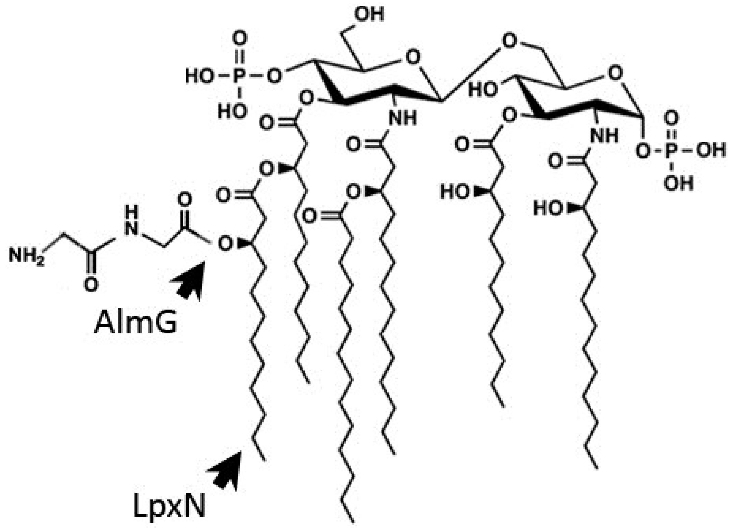
3.1. Outer Membrane Remodeling

As electrostatic interactions often play a crucial role in the initial interaction of cationic AMPs with bacterial membranes, both Gram-negative and Gram-positive bacteria have evolved strategies to neutralize the net negative charge of cell surface molecules with amine-containing substitutions. Thus, D-alanylation of teichoic acids, which are major components of the Gram-positive cell wall, confers AMP-resistance in a variety of Gram-positive bacteria including Staphylococcus aureus and Bacillus cereus [95,96]. More generally, aminoacylation of bacterial cell surface phosphatidylglycerols with L-lysine, L-alanine, or D-alanine confers resistance to cationic AMPs in both Gram-positive and Gram-negative bacteria [97].
LPS, the major constituent in the Gram-negative outer membrane, is composed of three regions: the anionic Lipid A membrane anchor, the core oligosaccharide and the O-antigen polysaccharide [98]. Hankins et al. have shown that V. cholerae O1 and O139 share identical asymmetrical hexa-acylated Lipid A structures [99] composed of a β 1'-6 linked glucosamine disaccharide with unmodified 1- and 4'-phosphate groups, which is acylated at the 2-, 3-, 2'- and 3'-positions. Myristate (C14:0) and 3-hydroxylaurate (3-OH C12:0) are ester-linked to the hydroxyl groups on the 2'- and 3'-linked fatty acyl chains (Figure 1). As in V. cholerae, the presence of a hydroxylated secondary acyl chain has been reported in the Lipid A structure of V. fischeri [100].
Figure 1. Structure of modified lipid A from V. cholerae O1 El Tor. The structure of V. cholerae lipid A was established by Hankins et al. (2011) [99]. It is composed of a β 1′-6 linked glucosamine disaccharide with unmodified 1- and 4′-phosphate groups, which is acylated at the 2-, 3-, 2′- and 3′-positions. Myristate (C14:0) and 3-hydroxylaurate (3-OH C12:0) are ester-linked to the hydroxyl groups on the 2′- and 3′-linked fatty acyl chains. The 3-hydroxylaurate secondary acyl chain transferred by the LpxN acyltransferase is required for AMP resistance. Similarly, the di-Glycine residues transferred by the AlmG to the hexa-acylated lipid A of V. cholerae O1 El Tor strains are crucial for AMP-resistance [101].
Antibiotics 03 00540 g001
Polymyxin B (PmB) has been extensively used to study the molecular basis of bacterial resistance to cationic AMPs in Gram-negative bacteria. Indeed, this peptide produced by the Gram-positive Paenibacillus polymyxa disrupts the cell envelope of Gram-negative bacteria by associating with the anionic LPS as well as with acidic glycerophospholipids [102]. To resist to AMPs, Gram-negative bacteria can neutralize their cell membrane by transferring phosphoethanolamine or aminoarabinose to phosphate groups on the lipid A domain of LPS [103].
In V. cholerae, the secondary acyltransferase VC0212 (LpxN or MsbB), which transfers a 3-hydroxylaurate group to penta-acylated Lipid A, contributes to the resistance of an El Tor strain to AMPs including PmB and LL-37 [99,104]. Thus, the higher susceptibility of the vc0212 mutant displaying incomplete Lipid A might be due to the greater permeability of its bacterial membrane. However, recent data by Hankins et al. demonstrated that the presence of a 3-hydroxyl group on the secondary acyl chain provides a site for esterification of glycine residues in a unique strategy necessary for resistance to PmB in V. cholerae [101] (Figure 1).
Three V. cholerae proteins, VC1577 (AlmG), VC1578 (AlmF), and VC1579 (AlmE) sharing sequence homology with the machinery involved in D-alanylation of teichoic acids in Gram-positive bacteria are essential for Lipid A modification with glycine and diglycine residues through aminoacyl esterification (Figure 1). Interestingly, sequence alignments comparing the classical (susceptible to PmB) and the El Tor (resistant to PmB) biotypes of V. cholerae revealed that the classical strain O395 has a nonsense mutation, resulting in a truncated AlmF carrier protein lacking the conserved serine [101]. The authors discovered that classical strains lack glycine-modified Lipid A. Upon alm mutation, the minimum inhibitory concentration (MIC) of PmB against El Tor strains dropped dramatically (~100 times) from 96–128 μg/mL to 0.5–1.0 μg/mL, showing that glycine modification of Lipid A is an essential mechanism of AMP resistance in V. cholerae. This study provides a well-defined mechanism for the different PmB-resistant phenotypes observed in V. cholerae classical and El Tor biotypes. Why classical strains appear to have lost carrier protein functionality and thus AMP resistance is a puzzling evolutionary question.
To date, it is unknown whether modifications of Vibrio LPS are induced upon exposure to sublethal concentrations of cationic AMPs, as shown in other bacterial species like Salmonella Typhimurium, which regulate their LPS structure, contributing to resistance to cationic AMP [105]. Changes in Salmonella LPS structure, regulated by the two-component system PhoPQ, include reducing average O-antigen chain-length, acylating, deacylating, and hydroxylating lipid A, derivatizing lipid A and LPS core phosphates with cationic groups (for recent review see [106]). Homologues of PhoPQ are found in Vibrio species, however, the potential role of PhoPQ in resistance to AMPs has not been described to date.

3.2. Induction of the Envelope Stress Response

As discussed above, many AMPs create damage to bacterial membranes as part of their mechanism of action. Sensing external stress is therefore crucial to combating membrane injury before the damage becomes irreversible. One of the strategies by which bacteria respond to outer membrane stress and modulate gene expression is via the alternate σE factor, encoded by the rpoE gene. Under non-stress conditions, σE is inactivated by its cognate anti-sigma factor localized to the inner membrane. When activated by envelope stress, i.e., misfolding of outer membrane proteins, σE promotes the expression of factors that help preserve and/or restore cell envelope integrity. Certain outer membrane proteins can serve as upstream signal sensors to modulate the activity of σE [107]. In V. cholerae, the major outer membrane OmpU is a key determinant of σE production [108]. Such dependence on a single factor contrasts with the regulation of σE in E. coli, in which numerous factors contribute to its activation and none is dominant.
In V. cholerae, σE plays a role in outer membrane stress response and resistance to AMPs. Thus, deficiency of σE confers to V. cholerae greater sensitivity to the antimicrobial peptide P2, a synthetic derivative of human BPI. Consistent with the ompU-dependent activation of σE, lack of OmpU in V. cholerae also conferred a greater sensitivity to AMPs [109,110]. Similar results were obtained for the oyster pathogen V. tasmaniensis LGP32 in which OmpU contributed to resistance to the oyster antimicrobials Cg-Defm and Cg-BPI [111]. However, in both V. cholerae and V. tasmaniensis, OmpU-mediated resistance was much lower than that conferred by Lipid A remodeling [101]. Moreover, in V. tasmaniensis LGP32, the major negative effect of the ompU deletion on pathogenicity was attributed to impaired capacity to invade the oyster immune cells rather than lower resistance to oyster AMPs [6].

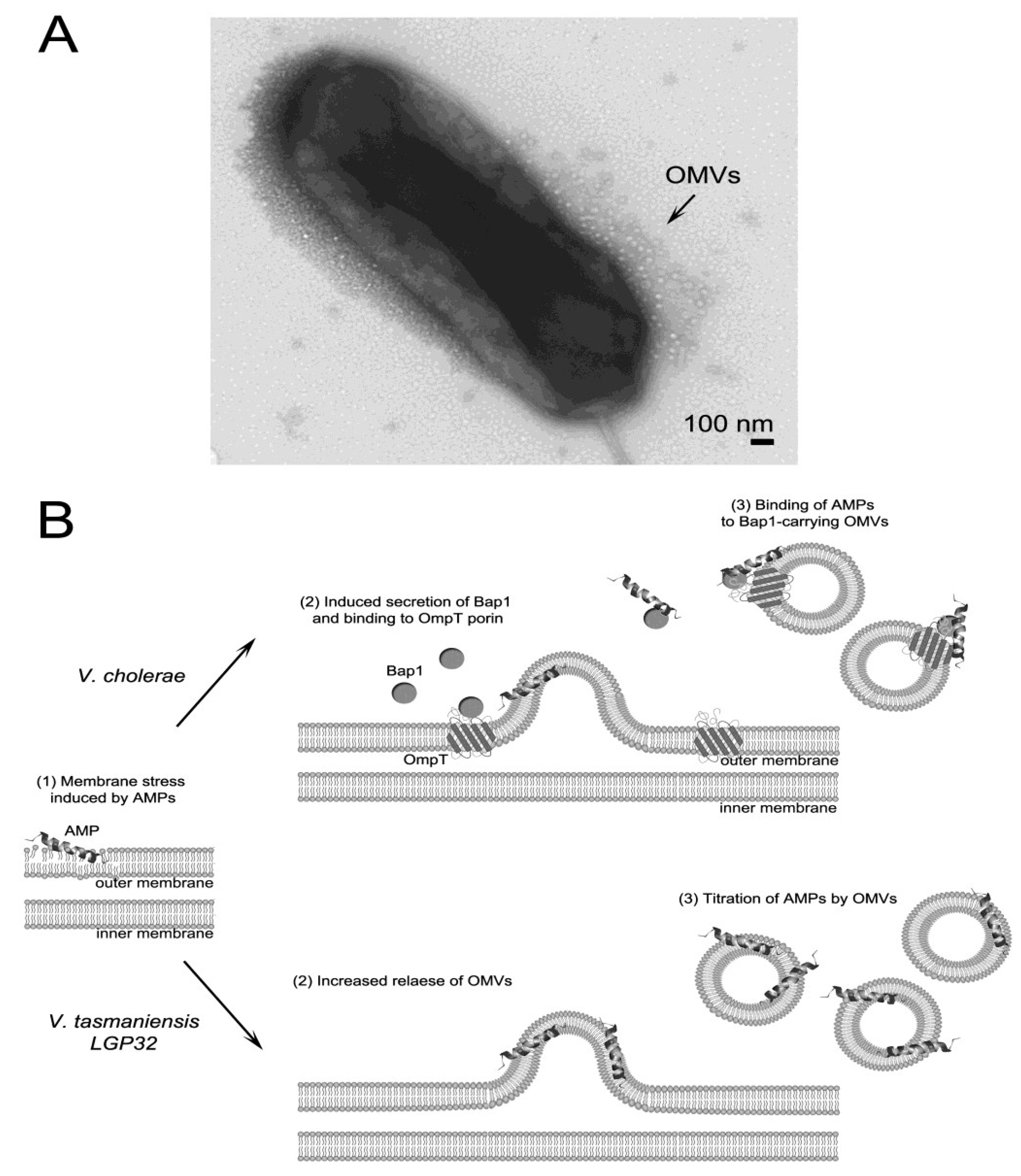
3.3. AMP Titration by Outer Membrane Vesicles

One σE-dependent mechanism whose role in AMP resistance has been less studied is outer membrane vesicle release. OMVs form the insoluble fraction of Gram-negative bacteria extracellular products; they are extruded from the bacterial cell surface and entrap some of the underlying periplasmic contents [112,113]. OMVs are key players in the interaction between Gram-negative bacteria and both the prokaryotic and eukaryotic cells from their environment [114]. Whereas it is now well established that Vibrio spp. constitutively release OMVs during cell growth [115,116,117], only recent studies in E. coli [118] and Vibrio spp. [119,120] have shown that the release of OMVs protects bacteria against membrane-active AMPs.
In V. cholerae, earlier work demonstrated that under envelope stress conditions, the small regulatory RNA VrrA is expressed in a σE-dependent manner to down-regulate OmpA, which in turn reduces envelope stress by promoting OMV release [121]. More recently, we found that physiologically relevant amounts of OMVs produced in the presence of a sub-lethal concentration of PmB provide protection against human cathelicidin LL-37, increasing the MIC of LL-37 by four-fold. This cross-protection has been attributed to the presence of the biofilm-associated extracellular matrix protein Bap1, which is associated with OMVs in larger amounts when bacteria are grown in the presence of PmB. The Bap1 protein can therefore trap LL-37, leading to increased resistance of V. cholerae towards LL-37 [119].
In V. tasmaniensis LGP32, OMVs provide significant and dose-dependent protection against AMPs [120]. Indeed, OMVs increased the MIC of PmB from 2–16-fold at OMV concentrations ranging from 6.25–50 μg/mL. This protective effect was attributed to the binding of PmB to OMVs; no proteolytic degradation of the peptide was observed. Interestingly, the addition of oyster plasma to the culture medium strongly stimulated the release of OMVs by V. tasmaniensis LGP32. This indicates that as in E. coli, in which sub-lethal concentrations of AMPs promote OMV release [118], OMV release in vibrios is likely up-regulated by membrane-active agents in oyster plasma. Consistent with this hypothesis, LGP32 lacking the major outer membrane protein OmpU, which controls envelope stress signaling in vibrios [108], showed a hypervesiculation phenotype (Figure 2A).
Altogether, these recent studies indicate that OMVs are a potent strategy used by vibrios to trap membrane-active AMPs such as PmB or LL-37, forming a protective shield that prevents interaction with the membranes of the bacterial cell (Figure 2B). Although OMVs released by vibrios can contain specific proteases like the recently identified vesicular serine protease Vsp (VS_II0815) of V. tasmaniensis LGP32, there is to date no evidence of AMP degradation by OMV-encapsulated content [119,120].
Figure 2. Model for AMP-titration by outer membrane vesicles (OMVs) in V. cholerae and V. tasmaniensis. (A) OMVs released in the extracellular medium by the hypervesiculating ΔompU mutant of V. tasmaniensis strain LGP32. Logarithmic phase cultures were negatively stained and observed by transmission electron microscopy as described in [120]; (B) The role of OMVs in the protection against AMPs has been recently described in two species of vibrios. In V. cholerae, OMVs cross-protect against the human cathelicidin LL-37 when bacteria are exposed to sublethal concentrations of PmB. Those OMVs are associated with Bap1 protein which serves as a ligand for LL-37. The association of Bap1 to OMVs is mediated by the outer membrane protein, OmpT [119]. In V. tasmaniensis, OMVs produced in the absence of AMPs are sufficient to titrate PmB and confer a potent dose-dependent protection against PmB. Although the molecular basis of PmB binding to V. tasmaniensis OMVs remain unknown, it is speculated that titration may occur by PmB insertion in the OMV membranes. The release of OMVs was shown to be strongly enhanced by the contact of V. tasmaniensis with oyster plasma [120]. In both species, OMV release is thought to create a protective membranous shield that prevents the interaction of membrane-active AMPs with the bacterial membranes.
Antibiotics 03 00540 g002

3.4. Efflux of AMPs

The involvement of efflux pumps in antimicrobial resistance, especially in antibiotic resistance, is well established in Gram-negative bacteria [122,123]. There are five different active efflux systems described in bacteria: the ATP-binding cassette superfamily (ABC), the small multidrug resistance family (SMR), the multi antimicrobial extrusion protein family (MATE), the major facilitator superfamily (MFS), and the resistance-nodulation-cell division superfamily (RND) [124]. In terms of antimicrobial resistance, the RND family efflux pumps are particularly important in Gram-negative bacteria. RND efflux systems are composed of an outer membrane protein homologous to the transmembrane β-barrel TolC protein of E. coli, a periplasmic membrane fusion protein (MFP), and an integral cytoplasmic membrane pump protein belonging to the RND superfamily of transporters (for review see [125]). These three components function to form a channel to extrude substrates from the cell envelope into the environment. The V. cholerae VexAB-TolC [126,127], the E. coli and Salmonella enterica AcrAB-TolC [128,129], and the Pseudomonas aeruginosa MexAB-OprM systems [130] function as RND efflux systems.
In V. cholerae, six RND efflux pumps have been described: VexAB, VexCD, VexEF, VexGH, VexIJK, and VexLM [131]. Among them, four are required for antimicrobial resistance in vitro. VexAB is the main efflux pump involved in the resistance to antimicrobials including bile acids, detergents, antibiotics, and PmB. The MIC of PmB dropped by four-fold (from 110–27 μg/mL) after vexB mutation in V. cholerae [127,132]. Moreover, the MIC of PmB against the vexB mutant was comparable with the MIC against the RND-null strain, indicating that only VexAB is involved in resistance to PmB [127]. Besides VexAB, VexGH also contributes to antibiotic (novobiocin and ampicillin) and detergent resistance but to a lesser extent than VexAB. Indeed, a decrease in the MIC can be observed only for a vexBH double mutant but not for the vexH single mutant, compared to the wild-type and vexB single mutant strains [133]. Finally, VexCD and VexIJK appeared to efflux bile acids and detergents, respectively [127,132]. VexEF and VexLM do not participate in antimicrobial resistance, but are required for the full virulence of V. cholerae by influencing the production of the major effectors of virulence, i.e., cholera toxin and the toxin co-regulated pilus [133]. In V. parahaemolyticus, proteomic identification of membrane proteins up-regulated in strains that artificially evolved resistant to AMPs, (including the fish AMP pleurocidin) led to the identification of TolC [134]. Unfortunately, its role in AMP resistance in V. parahaemolyticus has not been investigated further.
In addition to efflux pumps, a K+ pump encoded by the trkA gene has been described in V. vulnificus, and its role in AMP and serum resistance investigated [135]. The trkA gene product, TrkA, is a cytoplasmic protein bound to the inner side of the cytoplasmic membrane [136]. In V. vulnificus, the trkA mutant exhibited attenuated growth at intermediate potassium concentrations and was more sensitive to human serum protamine and PmB than was the wild type. Indeed, in contrast to the wild-type strain, the trkA mutant lysed in the presence of 10–20 μg/mL of protamine, and 5–15 μg/mL of PmB [135]. Moreover, TrkA was found to be important for V. vulnificus virulence in mice [135].

3.5. Suppression of AMP Expression

Pathogenic bacteria have developed multiple modalities to combat the antimicrobial response of their hosts. In addition to the structural modifications reviewed above, which increase their resistance to AMPs, they also use transcriptional repression as a strategy to evade the host immune system. Thus, the down-regulation of AMPs can be considered a general mechanism to facilitate invasion of pathogenic bacteria, including vibrios.
In humans, where the interaction of V. cholerae with intestinal epithelial cells is a critical step in the disease process, down-regulation of the cathelicidin LL-37, but not of the defensin HBD-1 has been reported in the presence of enteric pathogens including V. cholerae O139 [137]. The authors showed that cholera toxin (CT) was the predominant molecule associated with the regulation of AMPs by V. cholerae spp. in vitro and in vivo using intestinal epithelial cells and ileal loop experiments, respectively [137]. Moreover, multiple signaling pathways activated downstream of intracellular accumulation of cAMP contribute to the CT-mediated suppression of LL-37 in intestinal epithelial cells [137]. However, a more recent study on small intestine biopsies of patients with V. cholerae O1 infections did not show transcriptional repression of AMP genes in the small intestine [138], a discrepancy that might be explained by differences in transcriptional regulation in vivo and in vitro. In vivo, the expression of hBD-1, -3 and -4 did not vary with the infection, whereas hBD-2 mRNA levels were significantly higher at the acute stage of cholera than at the convalescent stage and in healthy controls. Paneth cell-derived HD-5 and HD-6, which were all expressed at high levels in controls, were not affected by the infections. While no transcriptional repression could be observed, the authors reported that hBD-2, HD-5 and LL-37 peptides are normally present in the small intestine epithelium and amounts decrease at the acute stage of watery diarrhea. Lower amounts of HD-5 could result from degranulation of the Paneth cells in response to infection. The processes regulating hBD-2 and LL-7 levels remain to be characterized.
In invertebrate hosts, similar downregulation of antimicrobial peptides and proteins has been observed during vibrioses. For instance, the coral pathogen, V. coralliilyticus, represses the expression of the damicornin, an AMP expressed by the scleractinian coral Pocillopora damicornis [28]. Indeed, damicornin transcripts increased during the first 6 days after infection with V. coralliilyticus, directly followed by a dramatic decrease from days 9–18. Conversely, no transcriptional change was observed when P. damicornis was exposed to a nonvirulent state of V. coralliilyticus [28]. Since V. coralliilyticus enters into the ectodermal coral tissue within 6 days, the authors suggested that a first phase of infection, involving bacterial recognition by host cells, triggers a nonspecific inflammatory response that activates damicornin gene transcription. In a second phase, following bacterial invasion, the pathogen suppresses damicornin transcription. This study represents the first characterization of the immunosuppression of AMP expression in an invertebrate-vibrio model of pathogenesis. More recently, using a global RNAseq approach, the same authors showed that not only damicornin, but also a mytimacin-like and a LBP-BPI gene displayed decreased expression during a successful V. coralliilyticus infection [139].
In mollusks, repression of AMP transcription has not been demonstrated per se. Indeed, upon infection of oysters with the pathogen V. tasmaniensis LGP32, major hemocyte movements occur which, by bringing AMP-producing hemocytes to infected tissues, create an apparent depletion in Cg-Defm and Cg-BPI transcripts in the circulating hemocytes. However, those transcripts accumulate at the same time in the hemocyte-infiltrated tissues [14]. A similar apparent repression of defensin expression was observed in the circulating hemocytes of a heterologous host, the mussel, infected with V. tasmaniensis LGP32 [140]. However, to date, the only AMP whose transcription is likely down-regulated by LGP32 is a proline-rich peptide from the oyster which acts by synergism with the other AMPs [14].

4. Conclusions

While vibrios have evolved the capacity to colonize immune tissues such as epithelia and phagocytes, only recent studies have started to investigate the mechanism by which they can survive the high AMP concentrations they encounter. Among their potent mechanisms of resistance to AMPs, vibrios use novel mechanisms of membrane remodeling. In particular, some highly resistant strains substitute their hexaacylated Lipid A with a diglycine residue to reduce the negative charge of their surface thereby lowering the electrostatic interaction with cationic AMPs. As a response to envelope stress, which can be induced by membrane-active agents including AMPs, vibrios release outer membrane vesicles to create a protective membranous shield that traps AMPs and prevents interaction of the peptides with their own membranes. Finally, once AMPs have breached the bacterial membrane barriers, vibrios can use RND pumps similar to those of other species to transport AMPs out of their cytoplasmic space. Although suppression of AMP transcription has been described in some host–pathogen interactions, this mechanism of immune evasion appears to be more specific to given strains/species than universal among vibrios.

Acknowledgments

We acknowledge the REVAResp project (Resistance and EVAsion from the host antimicrobial RESPonse) funded by the Languedoc-Roussillon region as well as the Vibriogen project (ANR-11-BSV7-0023) funded by the ANR for financial support.

Author Contributions

All authors have contributed to the research presented and the writing of the present review.

Conflicts of Interest

The authors declare no conflict of interest.

References

  1. Schmitt, P.; Rosa, R.D.; Duperthuy, M.; de Lorgeril, J.; Bachere, E.; Destoumieux-Garzon, D. The antimicrobial defense of the pacific oyster, Crassostrea gigas. How diversity may compensate for scarcity in the regulation of resident/pathogenic microflora. Front. Microbiol. 2012, 3, e160. [Google Scholar] [CrossRef]
  2. Jutla, A.; Whitcombe, E.; Hasan, N.; Haley, B.; Akanda, A.; Huq, A.; Alam, M.; Sack, R.B.; Colwell, R. Environmental factors influencing epidemic cholera. Am. J. Trop. Med. Hyg. 2013, 89, 597–607. [Google Scholar] [CrossRef] [PubMed]
  3. Ganz, T. Defensins: Antimicrobial peptides of innate immunity. Nat. Rev. Immunol. 2003, 3, 710–720. [Google Scholar] [CrossRef] [PubMed]
  4. Gallo, R.L.; Hooper, L.V. Epithelial antimicrobial defence of the skin and intestine. Nat. Rev. Immunol. 2012, 12, 503–516. [Google Scholar] [CrossRef] [PubMed]
  5. Ma, A.T.; McAuley, S.; Pukatzki, S.; Mekalanos, J.J. Translocation of a Vibrio cholerae type vi secretion effector requires bacterial endocytosis by host cells. Cell Host Microbe 2009, 5, 234–243. [Google Scholar] [CrossRef] [PubMed]
  6. Duperthuy, M.; Schmitt, P.; Garzon, E.; Caro, A.; Rosa, R.D.; Le Roux, F.; Lautredou-Audouy, N.; Got, P.; Romestand, B.; de Lorgeril, J.; et al. Use of ompu porins for attachment and invasion of Crassostrea gigas immune cells by the oyster pathogen Vibrio splendidus. Proc. Natl. Acad. Sci. USA 2011, 108, 2993–2998. [Google Scholar] [CrossRef] [PubMed]
  7. Brogden, K.A. Antimicrobial peptides: Pore formers or metabolic inhibitors in bacteria? Nat. Rev. Microbiol. 2005, 3, 238–250. [Google Scholar]
  8. Kragol, G.; Lovas, S.; Varadi, G.; Condie, B.A.; Hoffmann, R.; Otvos, L., Jr. The antibacterial peptide pyrrhocoricin inhibits the ATPase actions of DnaK and prevents chaperone-assisted protein folding. Biochemistry 2001, 40, 3016–3026. [Google Scholar] [CrossRef] [PubMed]
  9. Brotz, H.; Josten, M.; Wiedemann, I.; Schneider, U.; Gotz, F.; Bierbaum, G.; Sahl, H.G. Role of lipid-bound peptidoglycan precursors in the formation of pores by nisin, epidermin and other lantibiotics. Mol. Microbiol. 1998, 30, 317–327. [Google Scholar] [CrossRef] [PubMed]
  10. Park, C.B.; Kim, H.S.; Kim, S.C. Mechanism of action of the antimicrobial peptide buforin II: Buforin II kills microorganisms by penetrating the cell membrane and inhibiting cellular functions. Biochem. Biophys. Res. Commun. 1998, 244, 253–257. [Google Scholar] [CrossRef] [PubMed]
  11. Patrzykat, A.; Friedrich, C.L.; Zhang, L.; Mendoza, V.; Hancock, R.E. Sublethal concentrations of pleurocidin-derived antimicrobial peptides inhibit macromolecular synthesis in Escherichia coli. Antimicrob. Agents Chemother. 2002, 46, 605–614. [Google Scholar] [CrossRef] [PubMed]
  12. Srinivasan, S.; Beema Shafreen, R.M.; Nithyanand, P.; Manisankar, P.; Pandian, S.K. Synthesis and in vitro antimicrobial evaluation of novel fluoroquinolone derivatives. Eur. J. Med. Chem. 2010, 45, 6101–6105. [Google Scholar] [CrossRef] [PubMed]
  13. Wilmes, M.; Cammue, B.P.; Sahl, H.G.; Thevissen, K. Antibiotic activities of host defense peptides: More to it than lipid bilayer perturbation. Nat. Prod. Rep. 2011, 28, 1350–1358. [Google Scholar] [CrossRef] [PubMed]
  14. Schmitt, P.; de Lorgeril, J.; Gueguen, Y.; Destoumieux-Garzon, D.; Bachere, E. Expression, tissue localization and synergy of antimicrobial peptides and proteins in the immune response of the oyster Crassostrea gigas. Dev. Comp. Immunol. 2012, 37, 363–370. [Google Scholar] [CrossRef] [PubMed]
  15. Hilchie, A.L.; Wuerth, K.; Hancock, R.E. Immune modulation by multifaceted cationic host defense (antimicrobial) peptides. Nat. Chem. Biol. 2013, 9, 761–768. [Google Scholar] [CrossRef] [PubMed]
  16. Duquesne, S.; Destoumieux-Garzon, D.; Peduzzi, J.; Rebuffat, S. Microcins, gene-encoded antibacterial peptides from enterobacteria. Nat. Prod. Rep. 2007, 24, 708–734. [Google Scholar] [CrossRef] [PubMed]
  17. Cotter, P.D.; Ross, R.P.; Hill, C. Bacteriocins—A viable alternative to antibiotics? Nat. Rev. Microbiol. 2013, 11, 95–105. [Google Scholar]
  18. Martinez, J.L. Environmental pollution by antibiotics and by antibiotic resistance determinants. Environ. Pollut. 2009, 157, 2893–2902. [Google Scholar] [CrossRef] [PubMed]
  19. Qadri, F.; Alam, M.S.; Nishibuchi, M.; Rahman, T.; Alam, N.H.; Chisti, J.; Kondo, S.; Sugiyama, J.; Bhuiyan, N.A.; Mathan, M.M.; et al. Adaptive and inflammatory immune responses in patients infected with strains of Vibrio parahaemolyticus. J. Infect. Dis. 2003, 187, 1085–1096. [Google Scholar] [CrossRef] [PubMed]
  20. Qadri, F.; Bhuiyan, T.R.; Dutta, K.K.; Raqib, R.; Alam, M.S.; Alam, N.H.; Svennerholm, A.M.; Mathan, M.M. Acute dehydrating disease caused by Vibrio cholerae serogroups o1 and o139 induce increases in innate cells and inflammatory mediators at the mucosal surface of the gut. Gut 2004, 53, 62–69. [Google Scholar] [CrossRef] [PubMed]
  21. Vanden Broeck, D.; Horvath, C.; de Wolf, M.J. Vibrio cholerae: Cholera toxin. Int. J. Biochem. Cell. Biol. 2007, 39, 1771–1775. [Google Scholar] [CrossRef] [PubMed]
  22. Park, K.S.; Ono, T.; Rokuda, M.; Jang, M.H.; Okada, K.; Iida, T.; Honda, T. Functional characterization of two type III secretion systems of vibrio parahaemolyticus. Infect. Immun. 2004, 72, 6659–6665. [Google Scholar] [CrossRef] [PubMed]
  23. Zhou, X.; Gewurz, B.E.; Ritchie, J.M.; Takasaki, K.; Greenfeld, H.; Kieff, E.; Davis, B.M.; Waldor, M.K. A Vibrio parahaemolyticus t3ss effector mediates pathogenesis by independently enabling intestinal colonization and inhibiting tak1 activation. Cell. Rep. 2013, 3, 1690–1702. [Google Scholar] [CrossRef] [PubMed]
  24. McFall-Ngai, M.; Nyholm, S.V.; Castillo, M.G. The role of the immune system in the initiation and persistence of the euprymna scolopes—Vibrio fischeri symbiosis. Semin. Immunol. 2010, 22, 48–53. [Google Scholar] [CrossRef] [PubMed]
  25. Weber, B.; Chen, C.; Milton, D.L. Colonization of fish skin is vital for Vibrio anguillarum to cause disease. Environ. Microbiol. Rep. 2010, 2, 133–139. [Google Scholar] [CrossRef] [PubMed]
  26. Rosenberg, E.; Falkovitz, L. The Vibrio shiloi / Oculina patagonica model system of coral bleaching. Annu. Rev. Microbiol. 2004, 58, 143–159. [Google Scholar] [CrossRef] [PubMed]
  27. Daniels, N.A. Vibrio vulnificus oysters: Pearls and perils. Clin. Infect. Dis. 2011, 52, 788–792. [Google Scholar] [CrossRef] [PubMed]
  28. Vidal-Dupiol, J.; Ladriere, O.; Destoumieux-Garzon, D.; Sautiere, P.E.; Meistertzheim, A.L.; Tambutte, E.; Tambutte, S.; Duval, D.; Foure, L.; Adjeroud, M.; et al. Innate immune responses of a scleractinian coral to vibriosis. J. Biol. Chem. 2011, 286, 22688–22698. [Google Scholar] [CrossRef] [PubMed]
  29. Pitman, R.S.; Blumberg, R.S. First line of defense: The role of the intestinal epithelium as an active component of the mucosal immune system. J. Gastroenterol. 2000, 35, 805–814. [Google Scholar] [CrossRef] [PubMed]
  30. Levy, O.; Canny, G.; Serhan, C.N.; Colgan, S.P. Expression of bpi (bactericidal/permeability-increasing protein) in human mucosal epithelia. Biochem. Soc. Trans. 2003, 31, 795–800. [Google Scholar] [CrossRef] [PubMed]
  31. Bals, R.; Wang, X.; Zasloff, M.; Wilson, J.M. The peptide antibiotic ll-37/hcap-18 is expressed in epithelia of the human lung where it has broad antimicrobial activity at the airway surface. Proc. Natl. Acad. Sci. USA 1998, 95, 9541–9546. [Google Scholar] [CrossRef] [PubMed]
  32. Frohm Nilsson, M.; Sandstedt, B.; Sorensen, O.; Weber, G.; Borregaard, N.; Stahle-Backdahl, M. The human cationic antimicrobial protein (hcap18), a peptide antibiotic, is widely expressed in human squamous epithelia and colocalizes with interleukin-6. Infect. Immun. 1999, 67, 2561–2566. [Google Scholar] [PubMed]
  33. Bals, R.; Wilson, J.M. Cathelicidins—A family of multifunctional antimicrobial peptides. Cell. Mol. Life Sci. 2003, 60, 711–720. [Google Scholar] [CrossRef] [PubMed]
  34. Frohm, M.; Agerberth, B.; Ahangari, G.; Stahle-Backdahl, M.; Liden, S.; Wigzell, H.; Gudmundsson, G.H. The expression of the gene coding for the antibacterial peptide LL-37 is induced in human keratinocytes during inflammatory disorders. J. Biol. Chem. 1997, 272, 15258–15263. [Google Scholar] [CrossRef] [PubMed]
  35. Scheetz, T.; Bartlett, J.A.; Walters, J.D.; Schutte, B.C.; Casavant, T.L.; McCray, P.B., Jr. Genomics-based approaches to gene discovery in innate immunity. Immunol. Rev. 2002, 190, 137–145. [Google Scholar] [CrossRef] [PubMed]
  36. Cash, H.L.; Whitham, C.V.; Behrendt, C.L.; Hooper, L.V. Symbiotic bacteria direct expression of an intestinal bactericidal lectin. Science 2006, 313, 1126–1130. [Google Scholar] [CrossRef] [PubMed]
  37. Ouellette, A.J. Paneth cell alpha-defensins in enteric innate immunity. Cell. Mol. Life Sci. 2011, 68, 2215–2229. [Google Scholar] [CrossRef] [PubMed]
  38. Pukatzki, S.; Provenzano, D. Vibrio cholerae as a predator: Lessons from evolutionary principles. Front. Microbiol. 2013, 4, e384. [Google Scholar] [CrossRef]
  39. Salzman, N.H.; Ghosh, D.; Huttner, K.M.; Paterson, Y.; Bevins, C.L. Protection against enteric salmonellosis in transgenic mice expressing a human intestinal defensin. Nature 2003, 422, 522–526. [Google Scholar] [CrossRef] [PubMed]
  40. Chu, H.; Pazgier, M.; Jung, G.; Nuccio, S.P.; Castillo, P.A.; de Jong, M.F.; Winter, M.G.; Winter, S.E.; Wehkamp, J.; Shen, B.; et al. Human alpha-defensin 6 promotes mucosal innate immunity through self-assembled peptide nanonets. Science 2012, 337, 477–481. [Google Scholar] [CrossRef] [PubMed]
  41. Shin, O.S.; Uddin, T.; Citorik, R.; Wang, J.P.; della Pelle, P.; Kradin, R.L.; Bingle, C.D.; Bingle, L.; Camilli, A.; Bhuiyan, T.R.; et al. Lplunc1 modulates innate immune responses to Vibrio cholerae. J. Infect. Dis. 2011, 204, 1349–1357. [Google Scholar] [CrossRef] [PubMed]
  42. Masso-Silva, J.A.; Diamond, G. Antimicrobial peptides from fish. Pharmaceuticals 2014, 7, 265–310. [Google Scholar] [CrossRef] [PubMed]
  43. Rakers, S.; Niklasson, L.; Steinhagen, D.; Kruse, C.; Schauber, J.; Sundell, K.; Paus, R. Antimicrobial peptides (amps) from fish epidermis: Perspectives for investigative dermatology. J. Invest. Dermatol. 2013, 133, 1140–1149. [Google Scholar] [CrossRef] [PubMed]
  44. Smith, V.J.; Desbois, A.P.; Dyrynda, E.A. Conventional and unconventional antimicrobials from fish, marine invertebrates and micro-algae. Mar. Drugs. 2010, 8, 1213–1262. [Google Scholar] [CrossRef] [PubMed] [Green Version]
  45. Casadei, E.; Wang, T.; Zou, J.; Gonzalez Vecino, J.L.; Wadsworth, S.; Secombes, C.J. Characterization of three novel beta-defensin antimicrobial peptides in rainbow trout (Oncorhynchus mykiss). Mol. Immunol. 2009, 46, 3358–3366. [Google Scholar] [CrossRef] [PubMed]
  46. Caipang, C.M.; Lazado, C.C.; Brinchmann, M.F.; Kiron, V. Infection-induced changes in expression of antibacterial and cytokine genes in the gill epithelial cells of Atlantic cod, Gadus morhua during incubation with bacterial pathogens. Comp. Biochem. Physiol. B Biochem. Mol. Biol. 2010, 156, 319–325. [Google Scholar] [CrossRef] [PubMed]
  47. Chang, C.I.; Zhang, Y.A.; Zou, J.; Nie, P.; Secombes, C.J. Two cathelicidin genes are present in both rainbow trout (Oncorhynchus mykiss) and Atlantic salmon (Salmo salar). Antimicrob. Agents Chemother. 2006, 50, 185–195. [Google Scholar] [CrossRef] [PubMed]
  48. Kono, T.; Sakai, M. Molecular cloning of a novel bactericidal permeability-increasing protein/lipopolysaccharide-binding protein (bpi/lbp) from common carp Cyprinus carpio l. and its expression. Mol. Immunol. 2003, 40, 269–278. [Google Scholar] [CrossRef] [PubMed]
  49. Stenvik, J.; Solstad, T.; Strand, C.; Leiros, I.; Jorgensen, T.T. Cloning and analyses of a bpi/lbp cDNA of the Atlantic cod (Gadus morhua L.). Dev. Comp. Immunol. 2004, 28, 307–323. [Google Scholar] [CrossRef] [PubMed]
  50. Cho, J.H.; Park, I.Y.; Kim, H.S.; Lee, W.T.; Kim, M.S.; Kim, S.C. Cathepsin D produces antimicrobial peptide parasin I from histone H2A in the skin mucosa of fish. FASEB J. 2002, 16, 429–431. [Google Scholar] [PubMed]
  51. Krasity, B.C.; Troll, J.V.; Weiss, J.P.; McFall-Ngai, M.J. Lbp/bpi proteins and their relatives: Conservation over evolution and roles in mutualism. Biochem. Soc. Trans. 2011, 39, 1039–1044. [Google Scholar] [CrossRef] [PubMed]
  52. Gonzalez, M.; Gueguen, Y.; Destoumieux-Garzon, D.; Romestand, B.; Fievet, J.; Pugniere, M.; Roquet, F.; Escoubas, J.M.; Vandenbulcke, F.; Levy, O.; et al. Evidence of a bactericidal permeability increasing protein in an invertebrate, the Crassostrea gigas Cg-BPI. Proc. Natl. Acad. Sci. USA 2007, 104, 17759–17764. [Google Scholar] [CrossRef] [PubMed]
  53. Gueguen, Y.; Herpin, A.; Aumelas, A.; Garnier, J.; Fievet, J.; Escoubas, J.M.; Bulet, P.; Gonzalez, M.; Lelong, C.; Favrel, P.; et al. Characterization of a defensin from the oyster Crassostrea gigas. Recombinant production, folding, solution structure, antimicrobial activities, and gene expression. J. Biol. Chem. 2006, 281, 313–323. [Google Scholar] [CrossRef] [PubMed]
  54. Seo, J.K.; Lee, M.J.; Nam, B.H.; Park, N.G. Cgmolluscidin, a novel dibasic residue repeat rich antimicrobial peptide, purified from the gill of the pacific oyster, Crassostrea gigas. Fish Shellfish Immunol. 2013, 35, 480–488. [Google Scholar] [CrossRef] [PubMed]
  55. Seo, J.K.; Stephenson, J.; Noga, E.J. Multiple antibacterial histone H2B proteins are expressed in tissues of American oyster. Comp. Biochem. Physiol. B Biochem. Mol. Biol. 2011, 158, 223–229. [Google Scholar] [CrossRef] [PubMed]
  56. Poirier, A.C.; Schmitt, P.; Rosa, R.D.; Vanhove, A.S.; Kieffer-Jaquinod, S.; Rubio, T.P.; Charriere, G.M.; Destoumieux-Garzon, D. Antimicrobial histones and DNA traps in invertebrate immunity: Evidences in Crassostrea gigas. J. Biol. Chem. 2014, 289, 24821–24831. [Google Scholar] [CrossRef] [PubMed]
  57. Quayle, A.J.; Porter, E.M.; Nussbaum, A.A.; Wang, Y.M.; Brabec, C.; Yip, K.P.; Mok, S.C. Gene expression, immunolocalization, and secretion of human defensin-5 in human female reproductive tract. Am. J. Pathol. 1998, 152, 1247–1258. [Google Scholar] [PubMed]
  58. Singh, P.K.; Jia, H.P.; Wiles, K.; Hesselberth, J.; Liu, L.; Conway, B.A.; Greenberg, E.P.; Valore, E.V.; Welsh, M.J.; Ganz, T.; et al. Production of beta-defensins by human airway epithelia. Proc. Natl. Acad. Sci. USA 1998, 95, 14961–14966. [Google Scholar] [CrossRef] [PubMed]
  59. Zhao, C.; Wang, I.; Lehrer, R.I. Widespread expression of beta-defensin hBD-1 in human secretory glands and epithelial cells. FEBS Lett. 1996, 396, 319–322. [Google Scholar] [CrossRef] [PubMed]
  60. Mathews, M.; Jia, H.P.; Guthmiller, J.M.; Losh, G.; Graham, S.; Johnson, G.K.; Tack, B.F.; McCray, P.B., Jr. Production of beta-defensin antimicrobial peptides by the oral mucosa and salivary glands. Infect. Immun. 1999, 67, 2740–2745. [Google Scholar] [PubMed]
  61. O’Neil, D.A.; Porter, E.M.; Elewaut, D.; Anderson, G.M.; Eckmann, L.; Ganz, T.; Kagnoff, M.F. Expression and regulation of the human beta-defensins HBD-1 and HBD-2 in intestinal epithelium. J. Immunol. 1999, 163, 6718–6724. [Google Scholar] [PubMed]
  62. Liu, L.; Wang, L.; Jia, H.P.; Zhao, C.; Heng, H.H.; Schutte, B.C.; McCray, P.B., Jr.; Ganz, T. Structure and mapping of the human beta-defensin HBD-2 gene and its expression at sites of inflammation. Gene 1998, 222, 237–244. [Google Scholar] [CrossRef] [PubMed]
  63. Chromek, M.; Slamova, Z.; Bergman, P.; Kovacs, L.; Podracka, L.; Ehren, I.; Hokfelt, T.; Gudmundsson, G.H.; Gallo, R.L.; Agerberth, B.; et al. The antimicrobial peptide cathelicidin protects the urinary tract against invasive bacterial infection. Nat. Med. 2006, 12, 636–641. [Google Scholar] [CrossRef] [PubMed]
  64. Hase, K.; Eckmann, L.; Leopard, J.D.; Varki, N.; Kagnoff, M.F. Cell differentiation is a key determinant of cathelicidin LL-37/human cationic antimicrobial protein 18 expression by human colon epithelium. Infect. Immun. 2002, 70, 953–963. [Google Scholar] [CrossRef] [PubMed]
  65. Durr, U.H.; Sudheendra, U.S.; Ramamoorthy, A. Ll-37, the only human member of the cathelicidin family of antimicrobial peptides. Biochim. Biophys. Acta 2006, 1758, 1408–1425. [Google Scholar] [CrossRef] [PubMed]
  66. Canny, G.; Levy, O. Bactericidal/permeability-increasing protein (bpi) and bpi homologs at mucosal sites. Trends Immunol. 2008, 29, 541–547. [Google Scholar] [CrossRef] [PubMed]
  67. Falco, A.; Chico, V.; Marroqui, L.; Perez, L.; Coll, J.M.; Estepa, A. Expression and antiviral activity of a beta-defensin-like peptide identified in the rainbow trout (Oncorhynchus mykiss) EST sequences. Mol. Immunol. 2008, 45, 757–765. [Google Scholar] [CrossRef] [PubMed]
  68. Uzzell, T.; Stolzenberg, E.D.; Shinnar, A.E.; Zasloff, M. Hagfish intestinal antimicrobial peptides are ancient cathelicidins. Peptides 2003, 24, 1655–1667. [Google Scholar] [CrossRef] [PubMed]
  69. Douglas, S.E.; Gallant, J.W.; Liebscher, R.S.; Dacanay, A.; Tsoi, S.C. Identification and expression analysis of hepcidin-like antimicrobial peptides in bony fish. Dev. Comp. Immunol. 2003, 27, 589–601. [Google Scholar] [CrossRef] [PubMed]
  70. Cole, A.M.; Weis, P.; Diamond, G. Isolation and characterization of pleurocidin, an antimicrobial peptide in the skin secretions of winter flounder. J. Biol. Chem. 1997, 272, 12008–12013. [Google Scholar] [CrossRef] [PubMed]
  71. Noga, E.J.; Silphaduang, U. Piscidins: A novel family of peptide antibiotics from fish. Drug News Perspect. 2003, 16, 87–92. [Google Scholar] [CrossRef] [PubMed]
  72. Salerno, G.; Parrinello, N.; Roch, P.; Cammarata, M. cDNA sequence and tissue expression of an antimicrobial peptide, dicentracin; a new component of the moronecidin family isolated from head kidney leukocytes of sea bass, Dicentrarchus labrax. Comp. Biochem. Physiol. B Biochem. Mol. Biol. 2007, 146, 521–529. [Google Scholar] [CrossRef] [PubMed]
  73. Park, I.Y.; Park, C.B.; Kim, M.S.; Kim, S.C. Parasin I, an antimicrobial peptide derived from histone H2A in the catfish, Parasilurus asotus. FEBS Lett. 1998, 437, 258–262. [Google Scholar] [CrossRef] [PubMed]
  74. Birkemo, G.A.; Luders, T.; Andersen, O.; Nes, I.F.; Nissen-Meyer, J. Hipposin, a histone-derived antimicrobial peptide in Atlantic halibut (Hippoglossus hippoglossus L.). Biochim. Biophys. Acta 2003, 1646, 207–215. [Google Scholar] [CrossRef] [PubMed]
  75. Abd, H.; Saeed, A.; Weintraub, A.; Nair, G.B.; Sandstrom, G. Vibrio cholerae O1 strains are facultative intracellular bacteria, able to survive and multiply symbiotically inside the aquatic free-living amoeba Acanthamoeba castellanii. FEMS Microbiol. Ecol. 2007, 60, 33–39. [Google Scholar] [CrossRef] [PubMed]
  76. Abd, H.; Valeru, S.P.; Sami, S.M.; Saeed, A.; Raychaudhuri, S.; Sandstrom, G. Interaction between Vibrio mimicus and Acanthamoeba castellanii. Environ. Microbiol. Rep. 2010, 2, 166–171. [Google Scholar] [CrossRef] [PubMed]
  77. Abd, H.; Weintraub, A.; Sandstrom, G. Intracellular survival and replication of Vibrio cholerae o139 in aquatic free-living amoebae. Environ. Microbiol. 2005, 7, 1003–1008. [Google Scholar] [CrossRef] [PubMed]
  78. Ormonde, P.; Horstedt, P.; O’Toole, R.; Milton, D.L. Role of motility in adherence to and invasion of a fish cell line by Vibrio anguillarum. J. Bacteriol. 2000, 182, 2326–2328. [Google Scholar] [CrossRef] [PubMed]
  79. Wang, X.H.; Oon, H.L.; Ho, G.W.; Wong, W.S.; Lim, T.M.; Leung, K.Y. Internalization and cytotoxicity are important virulence mechanisms in vibrio-fish epithelial cell interactions. Microbiology 1998, 144, 2987–3002. [Google Scholar] [CrossRef] [PubMed]
  80. Ganz, T. Defensins: Antimicrobial peptides of vertebrates. C. R. Biol. 2004, 327, 539–549. [Google Scholar] [CrossRef] [PubMed]
  81. Duits, L.A.; Ravensbergen, B.; Rademaker, M.; Hiemstra, P.S.; Nibbering, P.H. Expression of beta-defensin 1 and 2 mrna by human monocytes, macrophages and dendritic cells. Immunology 2002, 106, 517–525. [Google Scholar] [CrossRef] [PubMed]
  82. Liu, P.T.; Modlin, R.L. Human macrophage host defense against Mycobacterium tuberculosis. Curr. Opin. Immunol. 2008, 20, 371–376. [Google Scholar] [CrossRef] [PubMed]
  83. Liu, P.T.; Stenger, S.; Li, H.; Wenzel, L.; Tan, B.H.; Krutzik, S.R.; Ochoa, M.T.; Schauber, J.; Wu, K.; Meinken, C.; et al. Toll-like receptor triggering of a vitamin D-mediated human antimicrobial response. Science 2006, 311, 1770–1773. [Google Scholar] [CrossRef] [PubMed]
  84. Sonawane, A.; Santos, J.C.; Mishra, B.B.; Jena, P.; Progida, C.; Sorensen, O.E.; Gallo, R.; Appelberg, R.; Griffiths, G. Cathelicidin is involved in the intracellular killing of mycobacteria in macrophages. Cell. Microbiol. 2011, 13, 1601–1617. [Google Scholar] [CrossRef] [PubMed]
  85. Mulero, I.; Noga, E.J.; Meseguer, J.; Garcia-Ayala, A.; Mulero, V. The antimicrobial peptides piscidins are stored in the granules of professional phagocytic granulocytes of fish and are delivered to the bacteria-containing phagosome upon phagocytosis. Dev. Comp. Immunol. 2008, 32, 1531–1538. [Google Scholar] [CrossRef] [PubMed]
  86. Cuesta, A.; Meseguer, J.; Esteban, M.A. The antimicrobial peptide hepcidin exerts an important role in the innate immunity against bacteria in the bony fish Gilthead seabream. Mol. Immunol. 2008, 45, 2333–2342. [Google Scholar] [CrossRef] [PubMed]
  87. Rosa, R.D.; Santini, A.; Fievet, J.; Bulet, P.; Destoumieux-Garzon, D.; Bachere, E. Big defensins, a diverse family of antimicrobial peptides that follows different patterns of expression in hemocytes of the oyster Crassostrea gigas. PLoS One 2011, 6, e25594. [Google Scholar] [CrossRef] [PubMed]
  88. Leippe, M.; Herbst, R. Ancient weapons for attack and defense: The pore-forming polypeptides of pathogenic enteric and free-living amoeboid protozoa. J. Eukaryot. Microbiol. 2004, 51, 516–521. [Google Scholar] [CrossRef] [PubMed]
  89. Sorensen, O.; Arnljots, K.; Cowland, J.B.; Bainton, D.F.; Borregaard, N. The human antibacterial cathelicidin, hCAP-18, is synthesized in myelocytes and metamyelocytes and localized to specific granules in neutrophils. Blood 1997, 90, 2796–2803. [Google Scholar] [PubMed]
  90. Agerberth, B.; Charo, J.; Werr, J.; Olsson, B.; Idali, F.; Lindbom, L.; Kiessling, R.; Jornvall, H.; Wigzell, H.; Gudmundsson, G.H. The human antimicrobial and chemotactic peptides LL-37 and alpha-defensins are expressed by specific lymphocyte and monocyte populations. Blood 2000, 96, 3086–3093. [Google Scholar] [PubMed]
  91. Knutson, M.D.; Oukka, M.; Koss, L.M.; Aydemir, F.; Wessling-Resnick, M. Iron release from macrophages after erythrophagocytosis is up-regulated by ferroportin 1 overexpression and down-regulated by hepcidin. Proc. Natl. Acad. Sci. USA 2005, 102, 1324–1328. [Google Scholar] [CrossRef] [PubMed]
  92. Sow, F.B.; Florence, W.C.; Satoskar, A.R.; Schlesinger, L.S.; Zwilling, B.S.; Lafuse, W.P. Expression and localization of hepcidin in macrophages: A role in host defense against tuberculosis. J. Leukoc. Biol. 2007, 82, 934–945. [Google Scholar] [CrossRef] [PubMed]
  93. Weiss, J.; Olsson, I. Cellular and subcellular localization of the bactericidal/permeability-increasing protein of neutrophils. Blood 1987, 69, 652–659. [Google Scholar] [PubMed]
  94. Calafat, J.; Janssen, H.; Tool, A.; Dentener, M.A.; Knol, E.F.; Rosenberg, H.F.; Egesten, A. The bactericidal/permeability-increasing protein (bpi) is present in specific granules of human eosinophils. Blood 1998, 91, 4770–4775. [Google Scholar] [PubMed]
  95. Peschel, A.; Otto, M.; Jack, R.W.; Kalbacher, H.; Jung, G.; Gotz, F. Inactivation of the dlt operon in Staphylococcus aureus confers sensitivity to defensins, protegrins, and other antimicrobial peptides. J. Biol. Chem. 1999, 274, 8405–8410. [Google Scholar] [CrossRef] [PubMed]
  96. Abi Khattar, Z.; Rejasse, A.; Destoumieux-Garzon, D.; Escoubas, J.M.; Sanchis, V.; Lereclus, D.; Givaudan, A.; Kallassy, M.; Nielsen-Leroux, C.; Gaudriault, S. The dlt operon of Bacillus cereus is required for resistance to cationic antimicrobial peptides and for virulence in insects. J. Bacteriol. 2009, 191, 7063–7073. [Google Scholar] [CrossRef] [PubMed]
  97. Roy, H. Tuning the properties of the bacterial membrane with aminoacylated phosphatidylglycerol. IUBMB Life 2009, 61, 940–953. [Google Scholar] [CrossRef] [PubMed]
  98. Raetz, C.R.; Whitfield, C. Lipopolysaccharide endotoxins. Annu. Rev. Biochem. 2002, 71, 635–700. [Google Scholar] [CrossRef] [PubMed]
  99. Hankins, J.V.; Madsen, J.A.; Giles, D.K.; Childers, B.M.; Klose, K.E.; Brodbelt, J.S.; Trent, M.S. Elucidation of a novel Vibrio cholerae lipid a secondary hydroxy-acyltransferase and its role in innate immune recognition. Mol. Microbiol. 2011, 81, 1313–1329. [Google Scholar] [CrossRef] [PubMed]
  100. Phillips, N.J.; Adin, D.M.; Stabb, E.V.; McFall-Ngai, M.J.; Apicella, M.A.; Gibson, B.W. The lipid a from Vibrio fischeri lipopolysaccharide: A unique structure bearing a phosphoglycerol moiety. J. Biol. Chem. 2011, 286, 21203–21219. [Google Scholar] [CrossRef] [PubMed]
  101. Hankins, J.V.; Madsen, J.A.; Giles, D.K.; Brodbelt, J.S.; Trent, M.S. Amino acid addition to Vibrio cholerae lps establishes a link between surface remodeling in Gram-positive and Gram-negative bacteria. Proc. Natl. Acad. Sci. USA 2012, 109, 8722–8727. [Google Scholar] [CrossRef] [PubMed]
  102. Guilhelmelli, F.; Vilela, N.; Albuquerque, P.; Derengowski Lda, S.; Silva-Pereira, I.; Kyaw, C.M. Antibiotic development challenges: The various mechanisms of action of antimicrobial peptides and of bacterial resistance. Front. Microbiol. 2013, 4, e353. [Google Scholar] [CrossRef]
  103. Needham, B.D.; Trent, M.S. Fortifying the barrier: The impact of lipid a remodelling on bacterial pathogenesis. Nat. Rev. Microbiol. 2013, 11, 467–481. [Google Scholar] [CrossRef] [PubMed]
  104. Matson, J.S.; Yoo, H.J.; Hakansson, K.; Dirita, V.J. Polymyxin B resistance in El Tor Vibrio cholerae requires lipid acylation catalyzed by msbb. J. Bacteriol. 2010, 192, 2044–2052. [Google Scholar] [CrossRef] [PubMed]
  105. Guo, L.; Lim, K.B.; Gunn, J.S.; Bainbridge, B.; Darveau, R.P.; Hackett, M.; Miller, S.I. Regulation of lipid a modifications by Salmonella typhimurium virulence genes phoP-phoQ. Science 1997, 276, 250–253. [Google Scholar] [CrossRef] [PubMed]
  106. Dalebroux, Z.D.; Miller, S.I. Salmonellae phopq regulation of the outer membrane to resist innate immunity. Curr. Opin. Microbiol. 2014, 17, 106–113. [Google Scholar] [CrossRef] [PubMed]
  107. Bashyam, M.D.; Hasnain, S.E. The extracytoplasmic function sigma factors: Role in bacterial pathogenesis. Infect. Genet. Evol. 2004, 4, 301–308. [Google Scholar] [CrossRef] [PubMed]
  108. Davis, B.M.; Waldor, M.K. High-throughput sequencing reveals suppressors of Vibrio cholerae rpoe mutations: One fewer porin is enough. Nucleic Acids Res. 2009, 37, 5757–5767. [Google Scholar] [CrossRef] [PubMed]
  109. Mathur, J.; Davis, B.M.; Waldor, M.K. Antimicrobial peptides activate the Vibrio cholerae sigmae regulon through an OmpU-dependent signalling pathway. Mol. Microbiol. 2007, 63, 848–858. [Google Scholar] [PubMed]
  110. Mathur, J.; Waldor, M.K. The Vibrio cholerae ToxR-regulated porin OmpU confers resistance to antimicrobial peptides. Infect. Immun. 2004, 72, 3577–3583. [Google Scholar] [CrossRef] [PubMed]
  111. Duperthuy, M.; Binesse, J.; Le Roux, F.; Romestand, B.; Caro, A.; Got, P.; Givaudan, A.; Mazel, D.; Bachere, E.; Destoumieux-Garzon, D. The major outer membrane protein ompu of Vibrio splendidus contributes to host antimicrobial peptide resistance and is required for virulence in the oyster Crassostrea gigas. Environ. Microbiol. 2010, 12, 951–963. [Google Scholar] [CrossRef] [PubMed]
  112. Beveridge, T.J. Structures of Gram-negative cell walls and their derived membrane vesicles. J. Bacteriol. 1999, 181, 4725–4733. [Google Scholar] [PubMed]
  113. Wai, S.N.; Takade, A.; Amako, K. The release of outer membrane vesicles from the strains of enterotoxigenic Escherichia coli. Microbiol. Immunol. 1995, 39, 451–456. [Google Scholar] [CrossRef] [PubMed]
  114. Kulp, A.; Kuehn, M.J. Biological functions and biogenesis of secreted bacterial outer membrane vesicles. Annu. Rev. Microbiol. 2010, 64, 163–184. [Google Scholar] [CrossRef] [PubMed]
  115. Chatterjee, S.N.; Das, J. Electron microscopic observations on the excretion of cell-wall material by Vibrio cholerae. J. Gen. Microbiol. 1967, 49, 1–11. [Google Scholar] [CrossRef] [PubMed]
  116. Hong, G.E.; Kim, D.G.; Park, E.M.; Nam, B.H.; Kim, Y.O.; Kong, I.S. Identification of Vibrio anguillarum outer membrane vesicles related to immunostimulation in the Japanese flounder, Paralichthys olivaceus. Biosci. Biotech. Biochem. 2009, 73, 437–439. [Google Scholar] [CrossRef]
  117. Kim, Y.R.; Kim, B.U.; Kim, S.Y.; Kim, C.M.; Na, H.S.; Koh, J.T.; Choy, H.E.; Rhee, J.H.; Lee, S.E. Outer membrane vesicles of Vibrio vulnificus deliver cytolysin-hemolysin VvhA into epithelial cells to induce cytotoxicity. Biochem. Biophys. Res. Commun. 2010, 399, 607–612. [Google Scholar] [CrossRef] [PubMed]
  118. Manning, A.J.; Kuehn, M.J. Functional advantages conferred by extracellular prokaryotic membrane vesicles. J. Mol. Microbiol. Biotechnol. 2013, 23, 131–141. [Google Scholar] [CrossRef] [PubMed]
  119. Duperthuy, M.; Sjostrom, A.E.; Sabharwal, D.; Damghani, F.; Uhlin, B.E.; Wai, S.N. Role of the Vibrio cholerae matrix protein bap1 in cross-resistance to antimicrobial peptides. PLoS Pathog. 2013, 9, e1003620. [Google Scholar] [CrossRef] [PubMed]
  120. Vanhove, A.S.; Duperthuy, M.; Charriere, G.M.; Le Roux, F.; Goudenege, D.; Gourbal, B.; Kieffer-Jaquinod, S.; Coute, Y.; Wai, S.N.; Destoumieux-Garzon, D. Outer membrane vesicles are vehicles for the delivery of Vibrio tasmaniensis virulence factors to oyster immune cells. Environ. Microbiol. 2014. [Google Scholar] [CrossRef]
  121. Song, T.; Mika, F.; Lindmark, B.; Liu, Z.; Schild, S.; Bishop, A.; Zhu, J.; Camilli, A.; Johansson, J.; Vogel, J.; et al. A new Vibrio cholerae sRNA modulates colonization and affects release of outer membrane vesicles. Mol. Microbiol. 2008, 70, 100–111. [Google Scholar] [CrossRef] [PubMed]
  122. Nikaido, H.; Pages, J.M. Broad-specificity efflux pumps and their role in multidrug resistance of Gram-negative bacteria. FEMS Microbiol. Rev. 2012, 36, 340–363. [Google Scholar] [CrossRef] [PubMed]
  123. Piddock, L.J. Multidrug-resistance efflux pumps—Not just for resistance. Nat. Rev. Microbiol. 2006, 4, 629–636. [Google Scholar] [CrossRef] [PubMed]
  124. Putman, M.; van Veen, H.W.; Konings, W.N. Molecular properties of bacterial multidrug transporters. Microbiol. Mol. Biol. Rev. 2000, 64, 672–693. [Google Scholar] [CrossRef] [PubMed]
  125. Hinchliffe, P.; Symmons, M.F.; Hughes, C.; Koronakis, V. Structure and operation of bacterial tripartite pumps. Annu. Rev. Microbiol. 2013, 67, 221–242. [Google Scholar] [CrossRef] [PubMed]
  126. Bina, J.E.; Mekalanos, J.J. Vibrio cholerae tolc is required for bile resistance and colonization. Infect. Immun. 2001, 69, 4681–4685. [Google Scholar] [CrossRef] [PubMed]
  127. Bina, X.R.; Provenzano, D.; Nguyen, N.; Bina, J.E. Vibrio cholerae RND family efflux systems are required for antimicrobial resistance, optimal virulence factor production, and colonization of the infant mouse small intestine. Infect. Immun. 2008, 76, 3595–3605. [Google Scholar] [CrossRef] [PubMed]
  128. Nikaido, H.; Zgurskaya, H.I. AcrAB and related multidrug efflux pumps of Escherichia coli. J. Mol. Microbiol. Biotechnol. 2001, 3, 215–218. [Google Scholar] [PubMed]
  129. Buckley, A.M.; Webber, M.A.; Cooles, S.; Randall, L.P.; La Ragione, R.M.; Woodward, M.J.; Piddock, L.J. The AcrAB-TolC efflux system of Salmonella enterica serovar Typhimurium plays a role in pathogenesis. Cell. Microbiol. 2006, 8, 847–856. [Google Scholar] [CrossRef] [PubMed]
  130. Poole, K. Multidrug efflux pumps and antimicrobial resistance in Pseudomonas aeruginosa and related organisms. J. Mol. Microbiol. Biotechnol. 2001, 3, 255–264. [Google Scholar] [PubMed]
  131. Kitaoka, M.; Miyata, S.T.; Unterweger, D.; Pukatzki, S. Antibiotic resistance mechanisms of Vibrio cholerae. J. Med. Microbiol. 2011, 60, 397–407. [Google Scholar] [CrossRef] [PubMed]
  132. Bina, J.E.; Provenzano, D.; Wang, C.; Bina, X.R.; Mekalanos, J.J. Characterization of the Vibrio cholerae vexab and vexcd efflux systems. Arch. Microbiol. 2006, 186, 171–181. [Google Scholar] [CrossRef] [PubMed]
  133. Taylor, D.L.; Bina, X.R.; Bina, J.E. Vibrio cholerae vexh encodes a multiple drug efflux pump that contributes to the production of cholera toxin and the toxin co-regulated pilus. PLoS One 2012, 7, e38208. [Google Scholar] [CrossRef] [PubMed] [Green Version]
  134. Shen, C.J.; Kuo, T.Y.; Lin, C.C.; Chow, L.P.; Chen, W.J. Proteomic identification of membrane proteins regulating antimicrobial peptide resistance in Vibrio parahaemolyticus. J. Appl. Microbiol. 2010, 108, 1398–1407. [Google Scholar] [CrossRef] [PubMed]
  135. Chen, Y.C.; Chuang, Y.C.; Chang, C.C.; Jeang, C.L.; Chang, M.C. A K+ uptake protein, TrkA, is required for serum, protamine, and polymyxin B resistance in Vibrio vulnificus. Infect. Immun. 2004, 72, 629–636. [Google Scholar] [CrossRef] [PubMed]
  136. Bossemeyer, D.; Borchard, A.; Dosch, D.C.; Helmer, G.C.; Epstein, W.; Booth, I.R.; Bakker, E.P. K+-transport protein TrkA of Escherichia coli is a peripheral membrane protein that requires other trk gene products for attachment to the cytoplasmic membrane. J. Biol. Chem. 1989, 264, 16403–16410. [Google Scholar] [PubMed]
  137. Chakraborty, K.; Ghosh, S.; Koley, H.; Mukhopadhyay, A.K.; Ramamurthy, T.; Saha, D.R.; Mukhopadhyay, D.; Roychowdhury, S.; Hamabata, T.; Takeda, Y.; et al. Bacterial exotoxins downregulate cathelicidin (hCAP-18/LL-37) and human beta-defensin 1 (HBD-1) expression in the intestinal epithelial cells. Cell. Microbiol. 2008, 10, 2520–2537. [Google Scholar] [CrossRef] [PubMed]
  138. Shirin, T.; Rahman, A.; Danielsson, A.; Uddin, T.; Bhuyian, T.R.; Sheikh, A.; Qadri, S.S.; Qadri, F.; Hammarstrom, M.L. Antimicrobial peptides in the duodenum at the acute and convalescent stages in patients with diarrhea due to Vibrio cholerae o1 or enterotoxigenic Escherichia coli infection. Microbes Infect. 2011, 13, 1111–1120. [Google Scholar] [CrossRef] [PubMed]
  139. Vidal-Dupiol, J.; Dheilly, N.M.; Rondon, R.; Grunau, C.; Cosseau, C.; Smith, K.M.; Freitag, M.; Adjeroud, M.; Mitta, G. Thermal stress triggers broad Pocillopora damicornis transcriptomic remodeling, while Vibrio coralliilyticus infection induces a more targeted immuno-suppression response. PLoS One 2014. [Google Scholar] [CrossRef]
  140. Venier, P.; Varotto, L.; Rosani, U.; Millino, C.; Celegato, B.; Bernante, F.; Lanfranchi, G.; Novoa, B.; Roch, P.; Figueras, A.; et al. Insights into the innate immunity of the mediterranean mussel Mytilus galloprovincialis. BMC Genomics 2011, 12, e69. [Google Scholar] [CrossRef]

