


Biosensors 2025, 15(12), 807; https://doi.org/10.3390/bios15120807 - 10 Dec 2025
Abstract
The West Nile Virus (WNV), transmitted by Culex mosquitoes as a major vector, has been reported worldwide. Also, West Nile neuroinvasive disease (WNND) caused by WNV lineage 1a and 2 neuroinvasive infections has been constantly reported with high fatality rates. Nevertheless, there are
[...] Read more.
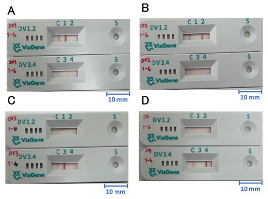
The West Nile Virus (WNV), transmitted by Culex mosquitoes as a major vector, has been reported worldwide. Also, West Nile neuroinvasive disease (WNND) caused by WNV lineage 1a and 2 neuroinvasive infections has been constantly reported with high fatality rates. Nevertheless, there are no treatments and vaccinations, so diagnosis in the early stages is important. Recently, a molecular diagnostic technique using DNA endonuclease-targeted CRISPR trans reporter (DETECTR) with the CRISPR-Cas12a system integrated with isothermal nucleic acid amplification has newly emerged. In this study, we designed a 2-Step WNV DETECTR with reverse transcription–recombinase polymerase amplification (RT-RPA) for rapid and sensitive WNV diagnosis. It successfully detected down to 1.0 × 10² RNA copies for both WNV lineage 1a and 2 with demonstrating similar sensitivity to qRT-PCR without cross-reactivity to other viruses. Additionally, we designed a 1-Step WNV DETECTR, incorporating all processing steps into a single tube, capable of detecting down to 1.0 × 10³ RNA copies for both lineages. Furthermore, we developed a more streamlined method, the 1-Step with Filter WNV DETECTR, which achieved detection limits comparable to the 2-Step method, while reducing the processing time by 5 min. This study also explored the potential of the Punch-it™ NA-Sample Kit as an efficient alternative lysis method by comparing the detection differences across various lysis methods. Through this method, we achieved rapid and simple amplification and detection processes suitable for field diagnostics with high specificity and sufficient sensitivity. Therefore, DETECTR methods presented themselves as promising alternatives to conventional diagnostic tools, potentially overcoming financial and technical constraints in diverse medical settings.
Full article
(This article belongs to the Section Biosensors and Healthcare)
►
Show Figures
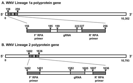
Figure 1


