Article

Morphology, Photocatalytic and Antimicrobial Properties of TiO2 Modified with Mono- and Bimetallic Copper, Platinum and Silver Nanoparticles

1
Department of Process Engineering and Chemical Technology, Faculty of Chemistry, Gdańsk University of Technology, G. Narutowicza 11/12, 80-233 Gdańsk, Poland
2
Institute for Catalysis (ICAT), Hokkaido University, N21, W10, Sapporo 001-0021, Japan
3
Department of Electrochemistry, Corrosion and Materials Engineering, Faculty of Chemistry, Gdańsk University of Technology, G. Narutowicza 11/12, 80-233 Gdańsk, Poland
4
NanoBioMedical Center, Adam Mickiewicz University, Umultowska 85, 61-614 Poznan, Poland
*
Authors to whom correspondence should be addressed.
Nanomaterials 2019, 9(8), 1129; https://doi.org/10.3390/nano9081129
Submission received: 11 July 2019 / Revised: 1 August 2019 / Accepted: 3 August 2019 / Published: 6 August 2019

Abstract

:
Noble metal nanoparticles (NMNPs) enhanced TiO2 response and extended its activity under visible light. Photocatalytic activity of TiO2 modified with noble metal nanoparticles strongly depends on the physicochemical properties of NMNPs. Among others, the differences in the size of NMNPs seems to be one of the most important factors. In this view, the effect of the metal’s nanoparticles size, type and amount on TiO2 photocatalytic and biocidal activity was investigated. TiO2 modified with mono- and bimetallic nanoparticles of Pt, Cu and Ag were prepared using chemical and thermal reduction methods. Obtained nanocomposites were characterized using transmission electron microscopy (TEM), X-ray photoelectron spectroscopy (XPS), X-ray diffraction (XRD) and diffuse-reflectance spectroscopy (DR/UV-Vis) techniques. The photocatalytic activity was examined in 2-propanol oxidation and hydrogen generation processes. The mechanism of modified TiO2 excitation was evaluated in action spectrum measurements during phenol oxidation. A possibility of using less energy-consuming light sources as a set of light-emitting diodes (LEDs) selected based on action spectrum results was examined. It was found that the differences in NMNPs size were the result of the reduction method. Moreover, coupling with a second metal strongly affected and differentiated the photocatalytic and biocidal activity of the obtained TiO2-based photocatalysts.

1. Introduction

Noble metal nanoparticles (NMNPs) have attracted much attention due to their unique properties in comparison to bulk metals. They possess exceptional optical, electrical and magnetic properties and are therefore applied in various branches of industry, such as electronics, pharmacy, catalysis, cosmetics, optoelectronics and medicine [1,2,3].
Metal nanoparticles of d block (Ag, Au, Cu, Pt and Pd) possess the ability to absorb light from the visible to the near-infrared range due to the occurrence of localized surface plasmon resonance (LSPR). The term localized surface plasmon resonance (LSPR) describes the oscillation of metal particle-free electrons. The irradiation of nanoparticles with radiation of resonant frequency results in the oscillations of free electrons. If the frequency of the exciting wavelength is the same as the particle oscillation frequency, the particle is set into oscillatory vibrations. Alternating oscillating charges of the resulting dipole emits electromagnetic waves of different frequencies. Since LSPR is an excited state, the return of a particle to the ground state proceeds via either a radiative or non-radiative way. Radiation suppression consists of photon emission, while non-radiative suppression consists of the oscillation of each electron with a charge transfer. The electron can be transferred as either interband or intraband, initiating local changes in the electric field in the surrounding environment [4,5,6,7,8,9,10,11,12,13].
The phenomenon of LSPR found an application in heterogeneous photocatalysis. The photocatalytic reaction consists in the excitation of a semiconductor with energy equal to or greater than its bandgap energy leading to the generation of charge carriers (electron–hole pair) and further formation of reactive oxygen species (ROS). Among examined semiconductors, the most widely used is titanium(IV) oxide, mainly due to its high photocatalytic activity, low cost, chemical and thermal stability [14,15]. Its photocatalytic properties have already been studied in water splitting, CO2 conversion to hydrocarbons, self-cleaning surfaces, water and air purification [16,17]. However, due to the large bandgap energy, it can be excited only by UV irradiation (λ < 388 nm), which requires the application of high-energy consuming UV lamps as an irradiation source and limits utilization of green light sources, like solar light. In order to spread TiO2 optical response over the visible light region, many methods of surface modification or doping of the semiconductor structure have been studied [18,19,20,21]. Among them, surface modification with noble and semi-noble metal (Ag, Au, Pt, Pd, Cu) nanoparticles was proposed. At the metal-semiconductor interface upon UV light irradiation, a Schottky barrier is created, which retards the charge carriers recombination by trapping electrons. Under visible light irradiation, TiO2 is activated during LSPR decay through electron transfer from metal nanoparticles (Ag, Au, Cu, Pt or Pd) to titanium(IV) oxide conduction band [5,9,22,23,24,25].
Apart from plasmonic properties, silver or copper nanoparticles deposited on TiO2 induces biocidal properties of the obtained photocatalyst [26,27,28]. Antimicrobial properties may result from the ability of the photocatalyst to generate reactive oxygen species and selected features of modifiers. Silver and copper nanoparticles are able to inhibit bacteria growth cells through binding with cysteine thiol groups leading to protein inactivation [3,29,30]. Generation of ROS leads to structural changes and the lysis of bacteria cell-walls by the peroxidation of organic components [28].
The type, size and shape of deposited metals may affect photocatalytic activity and selectivity of TiO2-based nanocomposites [4,12,21,31,32]. It was reported that, depending on the reducing agent, an oxygen saturation, the titanium(IV) oxide crystalline phase and calcination temperature, metal nanoparticles (NPs) of various size, shape, oxidation state and distribution are formed [12,19,26,31,33,34]. Dong et al. [32] investigated the effect of platinum nanoparticles size on activity and selectivity during carbon(IV) oxide photocatalytic reduction. It was found that a decrease of the size of platinum particles enhances the charge transfer efficiency leading to more effective hydrogen generation and CO2 photocatalytic reduction. However, the photocatalytic activity of TiO2 nanocomposites depends not only on metal NPs properties but also on properties of titania such as size, crystalline structure, specific surface area, surface hydroxylation and surface functionalization with non-metal and metal elements. In our previous study, we reported that photocatalytic activity strictly depended on the type and size of Pt, Pd, Cu and Au nanoparticles as well as TiO2 morphology [26,35]. Anpo et al. [36] investigated the photocatalytic activity of various commercial TiO2 JCR-TIO under UV light. The examined photocatalysts differed from the crystal structure, specific surface area, bandgap energy and a number of hydroxyl groups. They found that the most active titania was characterized by anatase structure, the large bandgap and the high concentration of surface hydroxyl groups. Murcia et al. [37] investigated the effect of surface functionalization of TiO2 with fluorine and sulfate ions combined with platinum nanoparticles deposition. They found that Pt NPs significantly enhanced the photocatalytic activity of sulfated TiO2, while it had a minor effect on the activity of fluorine titania. Thus, defining the correlation between structural properties and photocatalytic activity, as well as selectivity and biocidal properties should be of particular interest, especially when designing high-efficiency and environmentally-friendly photocatalytic processes.
In this regard, the effect of metal nanoparticles type, size, and content on photocatalytic and biocidal activity was investigated. Nanocomposites of TiO2 modified with mono-/bimetallic nanoparticles of platinum, copper and silver were obtained via chemical and thermal reduction methods. For the first time, the photocatalytic activity and properties of bimetallic TiO2-based photocatalysts in reduction and oxidation processes were studied. The spectral activity was analyzed during the phenol oxidation reaction under monochromatic irradiation in the range of 320 to 570 nm. Based on action spectrum results, a set of light-emitting diodes (LED) was selected and used as an irradiation source for the photocatalytic oxidation of organic pollutants. A biocidal activity was investigated against an Escherichia coli strain.

2. Materials and Methods

2.1. Materials

Commercial titanium (IV) oxide ST-01 (supplier: Ishihara Sangayo Ltd., Osaka, Japan) was used as TiO2 matrix. Silver nitrate, copper nitrate, potassium tetrachloroplatinate were purchased from Sigma Aldrich (Sigma Aldrich, Saint. Louis, MO, USA) and used as a starting material for the metals nanoparticles preparation. Sodium borohydride was supplied by POCh S.A. (Gliwice, Poland) and used as a reducing agent. 2-propanol and phenol, selected as model pollutants, were obtained from Fluka (Shanghai, China). Methanol was purchased from Fluka (Shanghai, China) and selected as a sacrificial agent during the hydrogen generation reaction.

2.2. Characterization Techniques

Diffuse-reflectance spectra (DR/UV-Vis) were recorded in the range of 300 to 800 nm and converted to absorption spectra. Bandgap energies were calculated from the corresponding Kubelka–Munk function F(R), which is proportional to the absorption of radiation, by plotting F(R) 0.5 Eph0.5 against Eph, where Eph is photon energy. The measurements were carried out using ThemoScientific Evolution 220 Spectrophotometer (Waltham, MA, USA) equipped with a PIN-757 (Waltham, MA, USA) integrating sphere. As reference samples, commercial TiO2 ST-01 and BaSO4 were used.
X-ray diffraction (XRD) analysis was performed using Rigaku Intelligent X-ray diffraction system SmartLab equipped with a sealed tube X-ray generator (Rigaku Corporation, Tokyo, Japan) (a copper target; operated at 40 kV and 30 mA), a D/teX high-speed position sensitive detector system and an ASC-10 automatic sample changer. Data acquisition conditions were as follows; 2θ range: 20°–90°, scan speed: 1°·min−1 and scan step 0.008. The obtained XRD patterns were analyzed by Rigaku PDXL (Version 2.0, Rigaku Corporation, Neu-Isenburg, Germany, 2007), a crystal structure analysis package including Rietveld analysis, installed in a computer controlling the diffractometer.
The surface composition was analyzed using X-ray photoelectron spectrophotometer (XPS) equipped with EDS detector. Powdered samples were attached to conductive carbon tape and a copper holder for XPS analysis and dried overnight under vacuum. All XPS spectra were recorded on Escalab 250Xi (Walthman, MA, USA), Thermofisher Scientific spectrometer using Mg K X-rays.
The morphology, size, and distribution of nanoparticles were investigated using HR-TEM HRTEM Jeol ARM 200F microscope (Jeol-USA, Peabody, MA, USA) operated at 200 kV and Cs-corrected STEM (High Angle Annular DarkField—HAADF) or high-resolution transmission electron microscope (HRTEM) Tecnai F20 X-Twin (Fei Company, Hillsboro, OR, USA).

2.3. Preparation of Photocatalysts

Two series of metal-modified titanium(IV) oxide photocatalysts were prepared. In the first series, metal ions were chemically reduced on the TiO2 surface using sodium borohydride, while in the second series metal ions were reduced thermally.
Firstly, 2 g of commercial titanium(IV) oxide (ST-01) was dispersed in 50 cm3 of water. Next, suitable volumes of 0.1 M metal salts (K2PtCl4, AgNO3 or Cu (NO3)2) solutions were added dropwise. Metal contents used for preparation ranged from 0.05 to 0.5 mol. % TiO2 (see in Table 1). Obtained dispersions have been further mixed to provide equal adsorption of metal ions on TiO2 surface. For samples reduced chemically, metal ions were reduced by the dropping of 0.1 M aqueous solution of sodium borohydride in molar excess equal to 1.5. Obtained metal-modified TiO2 powders were centrifuged, dried at 80 °C to dry mass and calcined at 400 °C for 2 h. The rate of heating during the calcination process was maintained at 2 °C·min−1.

2.4. Photocatalytic Activity

2.4.1. 2-Propanol Photocatalytic Oxidation

A total of 50 mg of the photocatalyst was dispersed in 5 cm3 of 2-propanol solution (5 vol. %). The obtained suspension was irradiated with a 300 W Xenon lamp (Hamamatsu Photonics, Hamamatsu, Japan). Measurements were conducted under UV-vis (λ > 400 nm) and vis light (λ > 450 nm). Irradiation of wavelengths shorter than 450 nm were cut-off using a Y48 filter (Hamamatsu Photonics, Hamamatsu, Japan). The photocatalytic activity was evaluated as a function of acetone generation as acetone is the first intermediate product of 2-propanol photocatalytic mineralization to carbon (IV) oxide. Acetone concentration was determined chromatographically using a Shimadzu GC-8A chromatograph equipped with PEG-20M Unipart B (Agilent Technologies, Santa Clara, CA, USA) column and FID detector (Shimadzu Corporation, Kyoto, Japan). The amount of generated CO2 during the reaction was measured using Shimadzu GC-14B with a flame-ionization detector (FID) and methanizer (Shimadzu Corporation, Kyoto, Japan).

2.4.2. Hydrogen Generation

A total of 50 mg of the photocatalyst was suspended in 5 cm3 of 50 vol. % of the aqueous methanol solution. The obtained suspension was purged with argon in order to remove dissolved oxygen. The concentration of residual oxygen was measured chromatographically with a Shimadzu GC-8A Chromatograph (column MS-5A ((Agilent Technologies, Santa Clara, CA, USA), thermal conductivity detector (TCD)) (Shimadzu Corporation, Kyoto, Japan). After the complete removal of oxygen from the reaction solution, the suspension was irradiated with a 400 W mercury lamp (Hamamatsu Photonics, Hamamatsu, Japan). Generated hydrogen was determined chromatographically using the same set-up as described for oxygen determination.

2.4.3. The Spectral Activity of Phenol Photocatalytic Oxidation

The apparent quantum yield of phenol oxidation was quantified at wavelengths 320, 380, 440, 450, 510 and 570 nm. A photocatalyst at the content of 2 G·dm−3 was added to the aqueous phenol solution (c = 20 mg·dm−3) and kept for 30 min in darkness to provide equilibrium conditions. The obtained suspension was irradiated with monochromatic light at the specific intensity in the range of 2–4 mWcm−2 emitted by a diffraction grating type illuminator (Jasco CRM-FD (Jasco Corporation, Tokyo, Japan)) equipped with a 300 W xenon lamp (Hamamatsu Photonics, Hamamatsu, Japan). The intensity of the radiation was measured using a Hioki 3664 Optical Power Meter (Hioki E.E. Corporation, Nagano, Japan). The apparent quantum yield of the phenol oxidation process was determined as a function of the 1,4-benzoquinone generation as the first photocatalytic oxidation intermediate product. The concentration of 1,4-benzoquinone was determined using a high-performance liquid chromatograph Shimadzu LC-6A ((Shimadzu Corporation, Kyoto, Japan) equipped with a WAKOSIL-II 5C18 AR column (4.6 × 250 mm) (FUJIFILM Wako Pure Chemical Corporation, Osaka, Japan) and a UV-Vis Shimadzu SPD-6A detector (Shimadzu Corporation, Kyoto, Japan)., detection wavelength at 254 nm. The mobile phase consists of water, acetonitrile and phosphoric acid (V) solution in a volume ratio 70:29.5:0.5, respectively. The mobile phase flow was maintained at 0.5 cm·dm−3.

2.4.4. Photocatalytic Phenol Oxidation in UV/Vis-LED System

A total of 1.4 g of the photocatalyst was suspended in 700 cm3 of phenol solution (C0 = 20 mg·dm−3) and mixed in darkness in order to establish equilibrium. The light source consisted of a set of light-emitting diodes (LED) covered with borosilicate glass dipped in the reaction suspension. The diodes emitted radiation in the UV-Vis range λ = 380–440 nm with the emission maximum at λmax = 415 nm. The radiation flux was maintained at 2.5 mW·cm−2 at 415 nm and controlled using Hioki 3664 m with the Hioki 9742 sensor (Hioki E.E. Corporation, Nagano, Japan). The emission spectrum of diodes used is shown in Figure S1.

2.4.5. Biocidal Properties

Biocidal activity in the aqueous phase was tested against E. coli (K12) strain. The analysis of biocidal activity was performed under visible light irradiation (λ > 450 nm) and in the dark. In this regard, 50 mg of the photocatalyst was dispersed in a test tube filled with 7 cm3 of bacterial cell suspension in physiological saline (0.9% NaCl). The optical density of the cell suspension ranged from 0.5 to 1 according to McFarland standards, which corresponds to a cell concentration of approximately 1.5 × 10⁸ CFU·cm−3 (colony-forming unit/cm3). A content of viable cells was determined using serial dilution method on Petri plates filled with PCA agar (Plate Count Agar Becton, Dickinson and Company, Sparks, MD, USA). The plates were incubated for 24 h at temperature 310 K.

3. Results

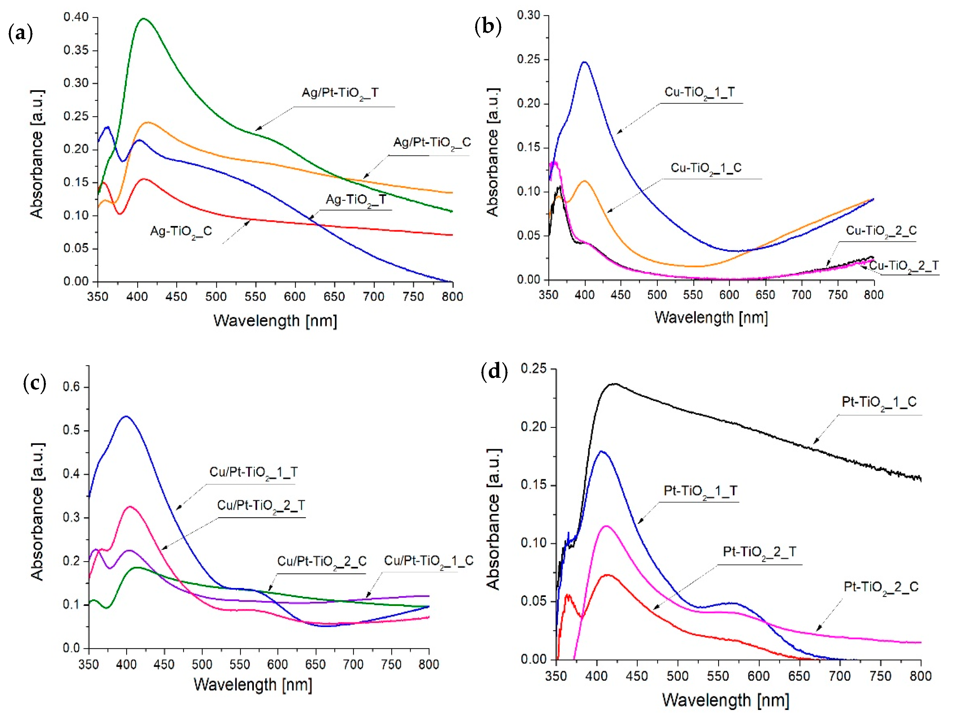
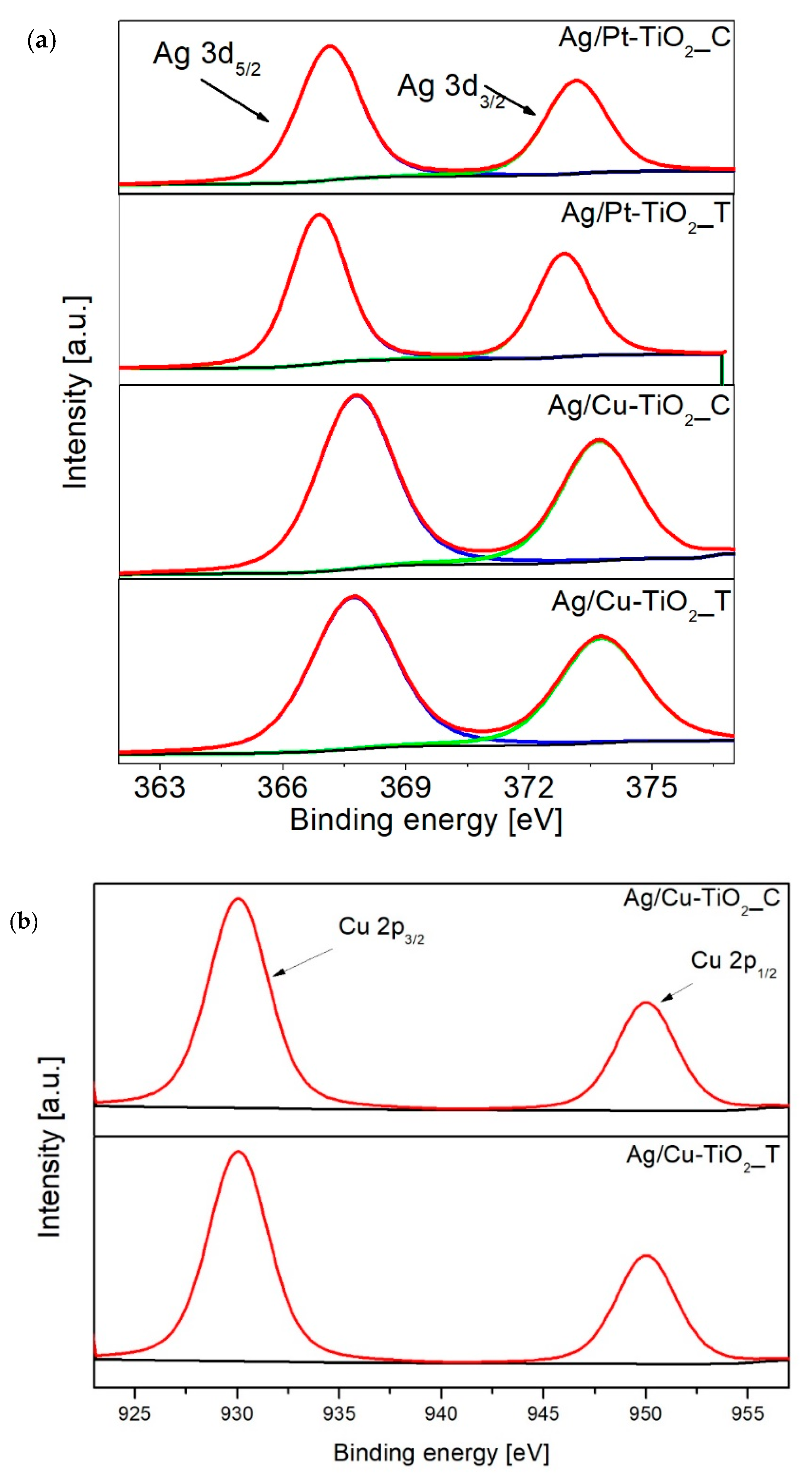
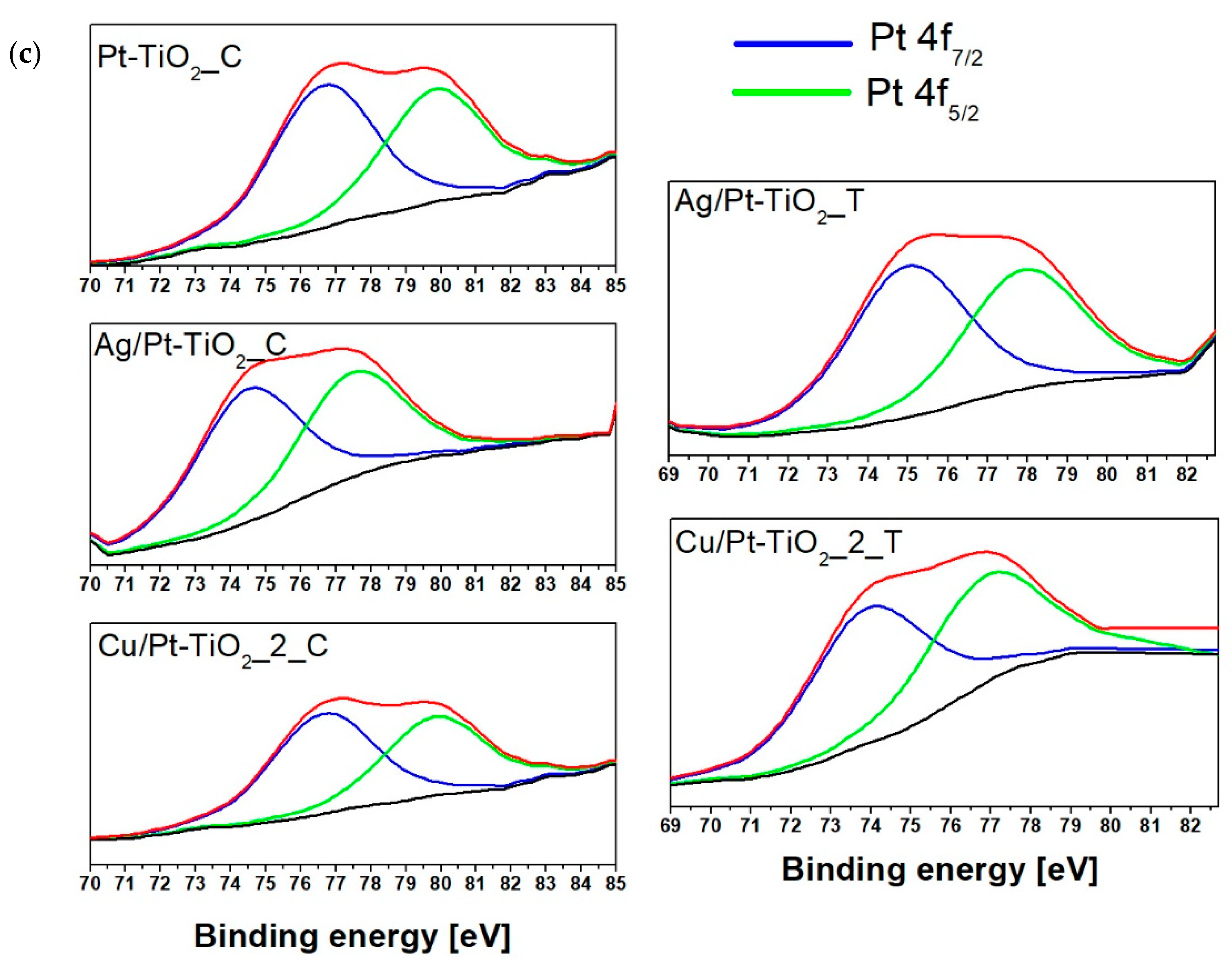
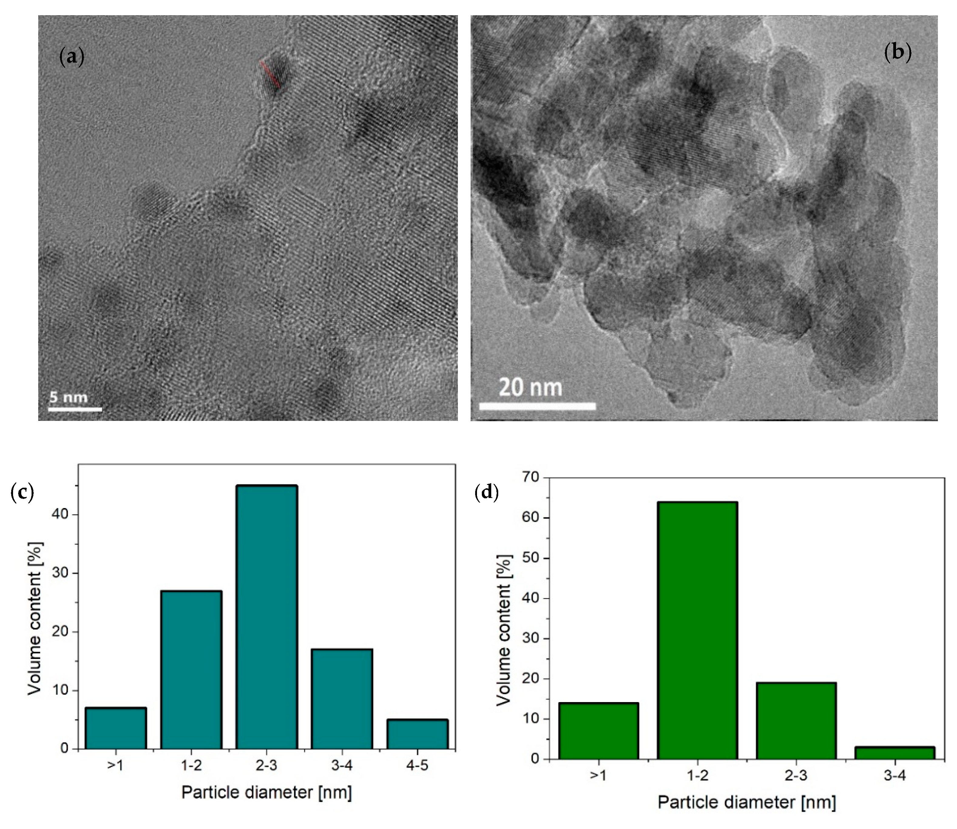
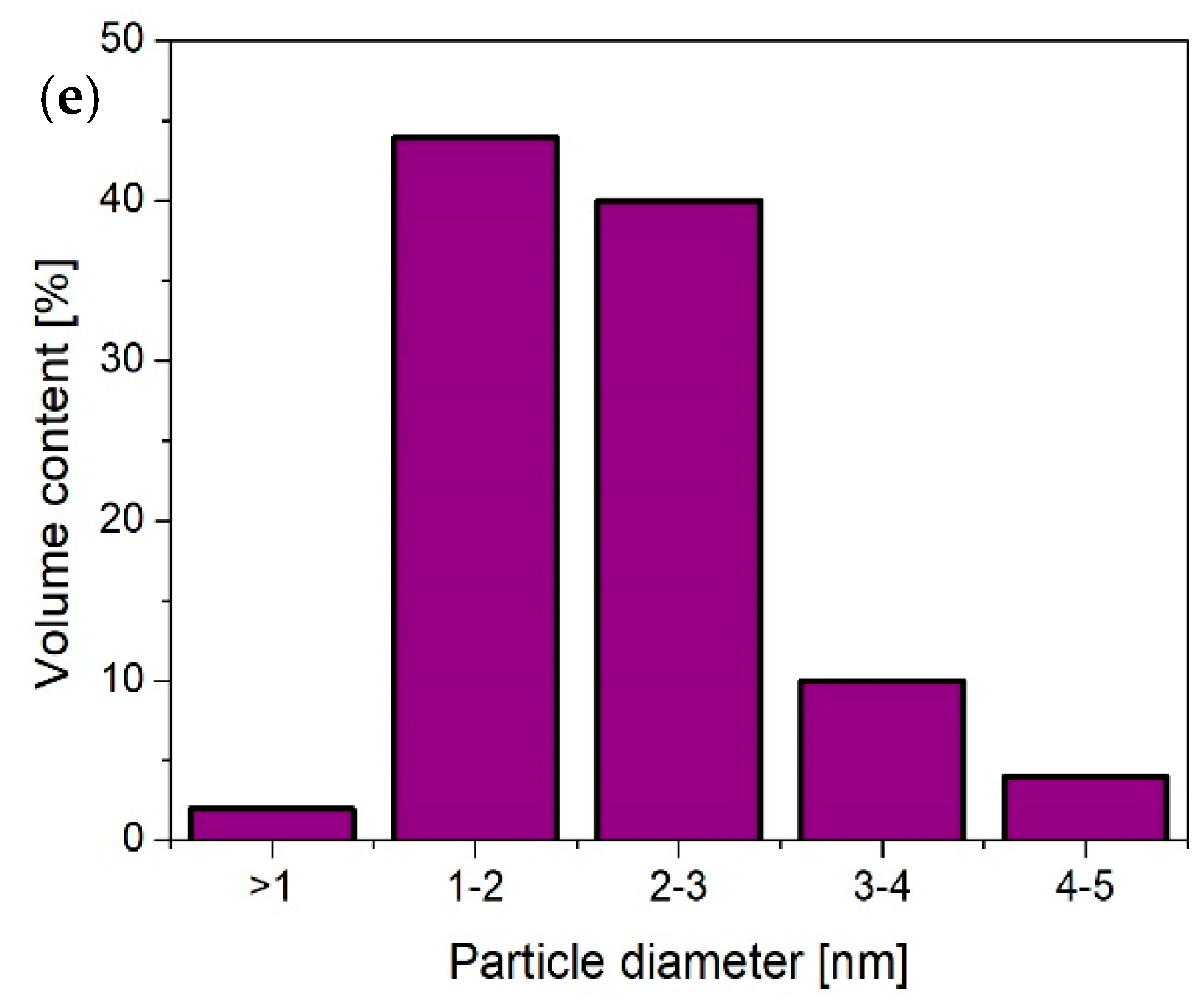
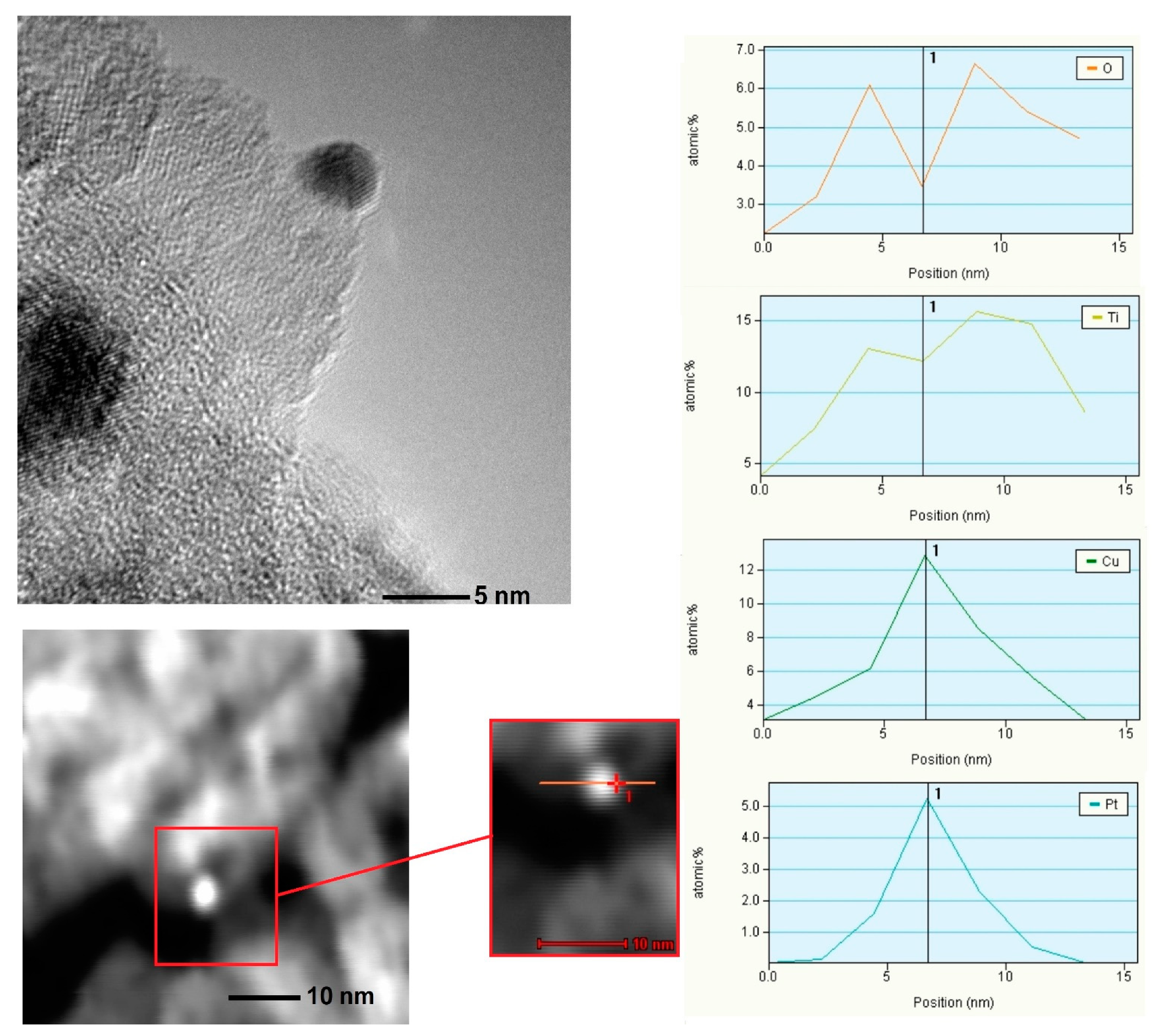
3.1. Characterization of Nanocomposites

Sample labeling, metal contents used for preparation, the average crystallite anatase size and reaction rate constants are presented in Table 1. Letters C and T refer to nanocomposites obtained by chemical and thermal reduction of metal ions, respectively.
Diffraction patterns of metal-modified TiO2 are shown in Figure S2a–c All obtained photocatalysts revealed a high level of crystallinity and consist of an anatase phase. Reflections observed at 2θ of 25.28°; 37.89°; 47.84°; 54.59°; 62.49°; 69.68°; 75.10° and 82.63° correspond to anatase crystal planes (1 0 1), (0 0 4), (2 0 0), (1 0 5), (2 0 4), (1 1 6), (2 1 5) and (0 0 7), respectively [38,39]. The average anatase crystallite size ranged from 12 nm to 14 nm. Only for Ag/Pt-TiO2_C and Ag/Pt-TiO2_T diffraction peaks attributed to metal nanoparticles (NPs) were observed (see Figure S2c). Reflections at 2θ = 32.1° and 46.1° observed for Ag/Pt-TiO2_C correspond to silver(II) oxide and platinum, respectively [26,40]. For the photocatalyst Ag/Pt-TiO2_T, the average crystallite size calculated based on the Scherrer formula was equal to 57 nm for AgO and 24 nm for Pt, while for Ag/Pt-TiO2_C the crystallite size for AgO and Pt was 27 nm and 34 nm, respectively. It was observed that the method of metal ions reduction strongly affects the size of metals crystallites. Thermal reduction of silver ions resulted in a crystallite size that was two times larger, while for platinum ions thermal reduction led to a reduction of crystallite size from 34 nm to 24 nm. The metal-TiO2 binding energies determine the rate of metal diffusion on the surface of titanium (IV) oxide and the growth of metal clusters. The higher the binding energy, the smaller the diffusion rate of deposited metal and smaller metal clusters were deposited on the TiO2 surface. Binding energy (−6.05 eV) of platinum and TiO2 in the oxidized form are significantly higher compared to the binding energy (−2.28 eV) of Pt and TiO2 in the reduced form. Therefore, the formation of smaller Pt species was observed using the thermal reduction method. For silver, the growth of crystallites results mainly from high-temperature annealing [41,42]. Therefore, chemical reduction using a strong reducing agent prevents crystallite growth and leads to the production of smaller silver particles.
Absorption spectra of thermally and chemically reduced photocatalysts are presented in Figure 1a–d. As the reference material, unmodified TiO2 was used. All metal-modified photocatalysts revealed an enhanced absorption of visible light in comparison to bare TiO2. Three main absorption bands: In the range of 400–450 nm, 550–600 nm and 700–800 nm, were observed for metal-modified TiO2 composites. An increase in absorption in the range from 400 to 450 nm is attributed to the surface plasmon resonance phenomenon of platinum and silver nanoparticles [43,44]. For photocatalysts modified with copper nanoparticles, an increase in absorption intensity in the range from 400 to 450 nm corresponds to electron transport between the TiO2 valence band and Cu(II) [45]. Absorption region from 550 to 580 nm is attributed to surface plasmon resonance of copper nanoparticles [46,47] and to the electron transport between Pt(IV) and Pt(II) species [26]. The last absorption band observed for copper-modified photocatalysts in the range from 650 to 800 nm indicates the presence of copper ions Cu(II) and Cu(I) [19,45,48]. It was observed that the metal ions reduction method strongly influences the shape of absorption properties. For chemically reduced photocatalysts, absorption bands were wide and fuzzy over the entire spectrum, whereas for thermally reduced photocatalysts narrower absorption bands were observed, as shown in Figure 1a–d. Differences in the shape of the spectra of individual metals deposited on TiO2 may result from the difference in the distribution of particle sizes [26,49]. The bandgap energy for all photocatalysts were equal to 3.2 eV, indicating surface modification of TiO2 with metal nanoparticles.
Results of surface composition analysis using X-ray photoelectron spectroscopy are presented in Table 2 and Figure 2a–c. All examined photocatalysts revealed similar element composition. The titanium content ranged from 23 at. % to 27 at. %. For each photocatalyst, the signal attributed to the Ti 2p region was deconvoluted into two-component peaks at binding energy (BE) 458.6 eV and 457.2 eV, which refer to Ti4+ and Ti3+ states, respectively [33,50,51]. The oxygen content ranged from 59.4 to 62.9 atomic percent. As a result of O 1s peak deconvolution, two-component peaks at 530 eV and 531 eV attributed to Ti–O groups and Ti–OH surface hydroxyl groups [50] were observed. Carbonaceous deposits observed on TiO2 surface ranged from 9.9 to 13.9 at. %. During the preparation of TiO2 photocatalysts, no organic compounds that could be a source of carbon were used, therefore the origin of present carbon is attributed to atmospheric contaminants. The obtained signal for carbon was divided into three component peaks at binding energy 284.8 eV, 285.2 eV and 289.0 eV, which refer to C–C, C–H and C=O bonds [52], respectively. The content of metal nanoparticles ranged from 0.1 at. % to 0.4 at. %. The surface composition analysis revealed differences in the chemical state of silver nanoparticles between chemically and thermally reduced photocatalysts. For Ag/Pt-TiO2_C and Ag/Cu-TiO2_C nanocomposites, silver appeared in the form of Ag0 and Ag2+. The Ag 3d5/2 peak at 367.3 eV and Ag 3d3/2 at 373.1 eV corresponded to silver Ag2+ in the form of oxide and Ag0 [43]. For both photocatalysts, the dominant fraction was silver Ag2+. For the nanocomposites modified with silver and platinum, as well as copper and silver obtained using thermal reduction, the presence of Ag0 and Ag+ states was also observed, in which the dominant fraction was silver(I) oxide. For Ag/Pt-TiO2_T and Ag/Cu-TiO2_T peaks were attributed to Ag 3d5/2 (BE = 367.7 eV) and Ag 3d3/2 (BE = 373.7 eV) corresponding to Ag2O and Ag0 were identified [43].
The presence of platinum species was confirmed by deconvolution of Pt 4f peak into two components Pt 4f7/2 and Pt 4f5/2. Observed signals at binding energies of 75.0, 74.8 and 74.2 eV referred to Pt0 and signals at 77.5 and 77.9 eV referred to Pt4+. Peaks attributed to Pt 4f region were characterized by a comparable surface irrespective of the reduction method. For Ag/Cu-TiO2_T and Ag/Cu-TiO2_C, the region referring to Cu 1s peaks at binding energies of 930.1 eV and 950.0 eV were observed [53]. For Cu/Pt-TiO2_2_T and Cu/Pt-TiO2_2_C, peaks attributed to copper species were not observed due to the low content of the deposited metal.
Transmission electron microscopy (TEM) images of Pt-TiO2_2_C, Pt-TiO2_2_T, Cu/Pt-TiO2_2_C, Ag/Pt-TiO2_T and Ag/Pt-TiO2_C are presented in Figure 3a–e, Figure 4 and Figures S3 and S4 in the Supplementary Materials. The analysis confirmed the surface modification of titanium(IV) oxide with platinum, silver and copper species. For Pt-TiO2_2_C, Pt-TiO2_2_T and Cu/Pt-TiO2_2_C photocatalysts, metal nanoparticles revealed the spherical shape and uniform distribution on TiO2. The size of Pt particles in the Pt-TiO2_2_C nanocomposite ranged from 1 nm to 6 nm, and the average particle size was equal to 3.4 ± 0.9 nm. The main fraction consists of nanoparticles with sizes ranging from 3 nm to 4 nm. Platinum nanoparticles obtained as a result of thermal reduction were twice as small in comparison to the Pt nanoparticles obtained by chemical reduction. For Pt-TiO2_2_T the particle sizes ranged from 1 nm to 4 nm, while the mean particle size was equal to 1.5 ± 0.6 nm. Bimetallic nanoparticles Pt/Cu deposited on the surface of titanium(IV) oxide (Pt/Cu-TiO2_C) were characterized by a size ranging from 0.9 nm to 5 nm. The average particle size was 2.2 ± 0.8 nm, while the dominant fraction were nanoparticles with sizes ranging from 1 nm to 3 nm. Based on an energy-dispersive spectroscopy (EDS) line scan, it was observed that platinum and copper form bimetallic nanoparticles with an alloy structure. Fast Fourier transform analysis confirmed the presence of platinum nanoparticles of Pt (2 0 0) and Pt (1 1 1) planes.
Bimetallic photocatalysts Ag/Pt-TiO2_T and Ag/Pt-TiO2_C (see Figures S3 and S4) consisted of small alloy particles up to 10 nm and bigger aggregates of a single metal. The largest aggregates (up to 100 nm) were observed for silver particles. For photocatalysts reduced thermally, silver aggregates were significantly larger in comparison to chemically reduced metals for Ag/Pt-TiO2_C sample. Small nanoparticles consisted of silver and platinum alloys of Ag (1 1 1), Pt (2 0 0) and Pt (1 1 1) crystalline planes.

3.2. Photocatalytic Activity

The photocatalytic activity was evaluated in 2-propanol oxidation and hydrogen generation reactions, in order to distinguish the difference in activity in oxidation and reduction reactions, respectively.

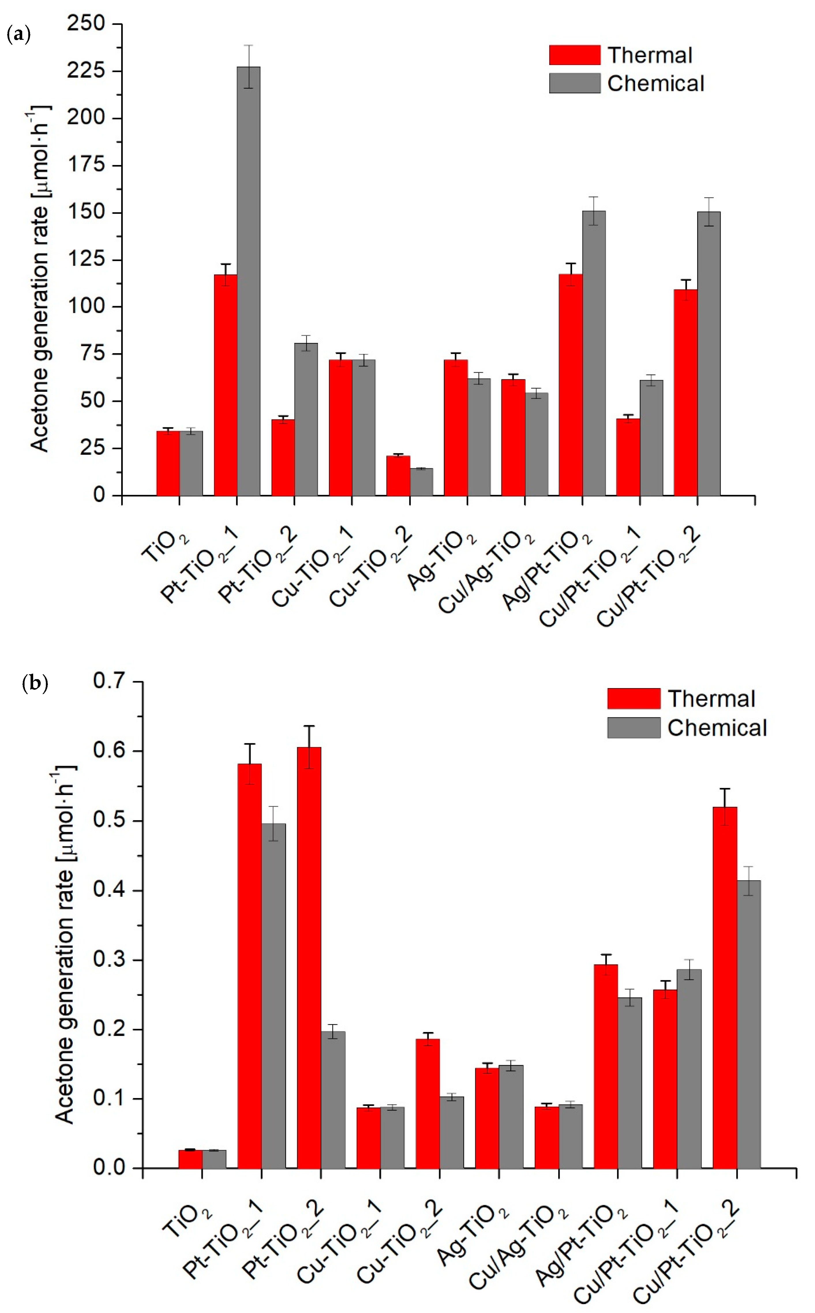
3.2.1. 2-Propanol Oxidation

Table 1 and Figure 5a,b present the results of the photocatalytic activity in 2-propanol oxidation under UV-Vis and visible light. An increase of photocatalytic activity compared to the unmodified TiO2 in the UV-Vis range was observed for all photocatalysts except for Cu-TiO2_2_T and Cu-TiO2_2_C. For photocatalysts reduced chemically, the highest activity was observed for Pt-TiO2_1_C, for which the acetone generation rate was 227.3 ± 11.4 μmol·h−1. For nanocomposites obtained via thermal reduction, the highest activity was observed for Pt-TiO2_1_T (117.1 ± 5.9 μmol·h−1). The particle size was a crucial parameter that affected photocatalytic activity. For photocatalysts modified with platinum Pt-TiO2_1_C, Pt-TiO2_2_C, Pt-TiO2_1_T and Pt-TiO2_2_T, a higher activity under UV-Vis was observed for nanocomposites with larger Pt particles obtained by chemical reduction. For monometallic photocatalysts modified with silver, a similar dependence was observed. Higher activity was noted for Ag-TiO2_T with larger Ag nanoparticles in comparison to Ag-TiO2_C. For monometallic copper modified samples, the photocatalytic activity for both chemically and thermally reduced photocatalysts was similar. However, under Vis light radiation (λ > 450 nm) higher efficiency of 2-propanol oxidation was found for thermally reduced fine Pt or Ag particles deposited on TiO2 surface, characterized by smaller metal nanoparticles compared to that obtained by chemical reduction, as shown in Figure 5b.
Among photocatalysts modified with bimetallic Pt-Cu and Ag-Pt nanoparticles, higher activity in reaction of 2-propanol oxidation under UV-Vis was also observed for photocatalysts obtained using a chemical reduction method. For Cu/Ag-TiO2_C and Cu/Ag-TiO2_T photocatalysts, a higher activity was observed for Cu/Ag-TiO2_T. In this regard, the influence of metal nanoparticles size on photocatalytic activity is the most significant for platinum and silver NPs. Under the irradiation from the visible range (λ > 450 nm) the highest activity was observed for Pt-TiO2_2_T (0.606 ± 0.030 μmol·h−1), while the lowest for TiO2 modified with Cu and Ag/Cu species (see samples Cu-TiO2_1_C (0.088 μmolh−1), Cu-TiO2_1_T (0.087 ± 0.004 μmol·h−1), Cu/Ag-TiO2_C (0.092 ± 0.005 μmol·h−1) and Cu/Ag-TiO2_T (0.089 ± 0.004 μmol·h-1), respectively.
Among bimetallic photocatalysts with a metal content of 0.5 mol % Cu and 0.1 mol % Pt, slightly higher activity was observed for Cu/Pt-TiO2_1_C than for Cu/Pt-TiO2_1_T. For Cu-TiO2_1_C, Cu-TiO2_1_T, Ag-TiO2_C, Ag-TiO2_T, Cu/Ag-TiO2_C and Cu/Ag-TiO2_T photocatalysts, no significant differences in photocatalytic activity were found. Moreover, the enhanced activity of Ag/Pt-TiO2_C compared to Ag/Pt-TiO2_T indicates the predominant role of the particle size of Pt over the size of Ag particles.
It was observed that the loading of metal nanoparticles also had an impact on photocatalytic activity. For Pt-TiO2_2_C and Pt-TiO2_1_C, an increment in Pt content from 0.05% to 0.1% mol TiO2 resulted in almost 3-times higher photooxidation of 2-propanol to acetone, whereas for Cu-TiO2_2_C and Cu-TiO2_1_C increasing the content from 0.1 to 0.5 mol % of TiO2 resulted in a five-fold increase in photocatalytic activity. For bimetallic particles modified TiO2 photocatalysts Cu/Pt-TiO2_1_C, Cu/Pt-TiO2_2_C, Cu/Pt-TiO2_1_T and Cu/Pt-TiO2_2_T, an increase in metal content for both chemically and thermally reduced photocatalysts resulted in almost twice the reduction of nanocomposite activity. Too high metal loading on the surface of titanium(IV) oxide can affect the photocatalytic activity as a result of hydroxylation degree reduction of the semiconductor surface and aggregation of metal particles that act as recombination centers for charge carriers [54]. Wang et al. [55] and Sun et al. [56] also observed a decrease in photocatalytic activity with an increase in metal content. They proposed that too high metal dispersion on the surface of TiO2 can act as an internal filter that limits the absorption of irradiation by titanium (IV) oxide.
As shown in Figure 5b and Table 1 under visible range (λ > 450 nm) a decrease in photocatalytic activity was only observed for Pt-TiO2_1_C with higher metal content than the Pt-TiO2_2_C sample. The acetone generation rate was equal to 0.496 ± 0.025 μmol·h−1 and 0.197 ± 0.010 μmol·h−1 for Pt-TiO2_1_C and Pt-TiO2_2_C, respectively.

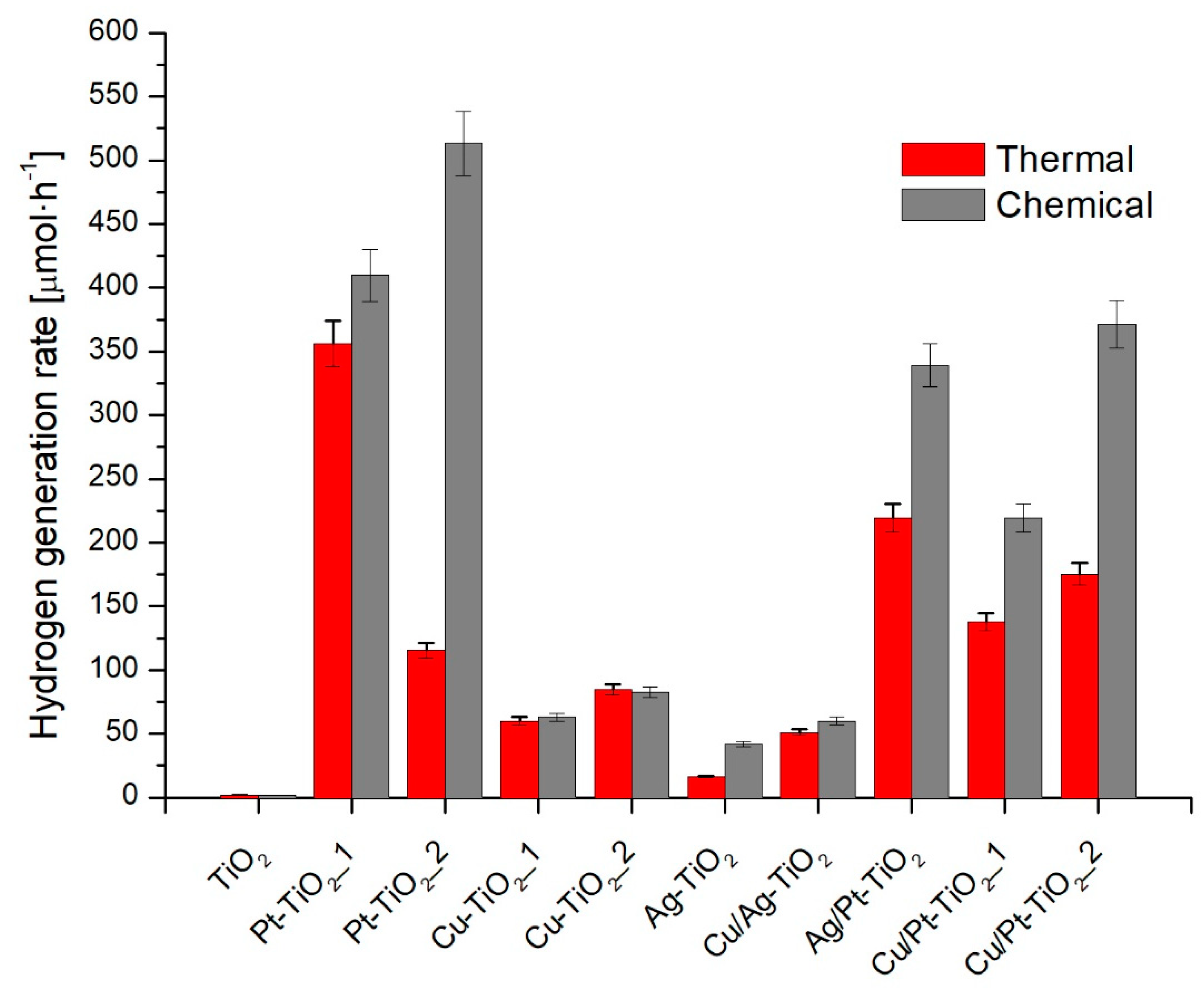
3.2.2. Hydrogen Generation

Photocatalytic activity in reduction reaction was evaluated in hydrogen generation reaction under UV-Vis irradiation in the presence of methanol as a hole scavenger. Results are presented in Figure 6 and in Table 1. As it was expected, the activity of bare titanium (IV) oxide was negligible. Hydrogen generation rate equaled to 1.9 ± 0.1 μmol·h−1. The modification of TiO2 surface with metal nanoparticles resulted in an increase in the activity for each of the metallic photocatalysts. The highest activities were observed for mono- and bi-metallic photocatalysts modified with platinum obtained by the chemical reduction method and characterized by larger Pt particles as compared to Pt particles obtained by thermal reduction. For Cu-TiO2_1_C, Cu-TiO2_2_C, Cu-TiO2_2_C and Cu-TiO2_2_T, there were no significant differences in photocatalytic activities between photocatalysts obtained by chemical and thermal reduction. Higher photocatalytic activity was observed for Ag and Ag/Cu-modified TiO2 obtained by chemical reduction, compared to thermally reduced metal ions on TiO2 surface. The larger silver particles were synthesized during thermal treatment rather than for chemical reduction using a strong reducing agent (NaBH4). For photocatalysts obtained by chemical reduction, a decrease in activity was observed with an increase in the modifier content. For monometallic photocatalysts, the highest activity was observed for TiO2 modified with platinum, higher than was found with copper and further with silver nanoparticles. For nanocomposites modified with bimetallic nanoparticles, no increase in photocatalytic activity was observed compared to the monometallic photocatalysts with the same metal content. The results obtained are consistent with literature reports. The results of hydrogen generation may be correlated with the metalwork function (Φ), which decreased according to dependence ΦPt > ΦCu > ΦAg [19,57].

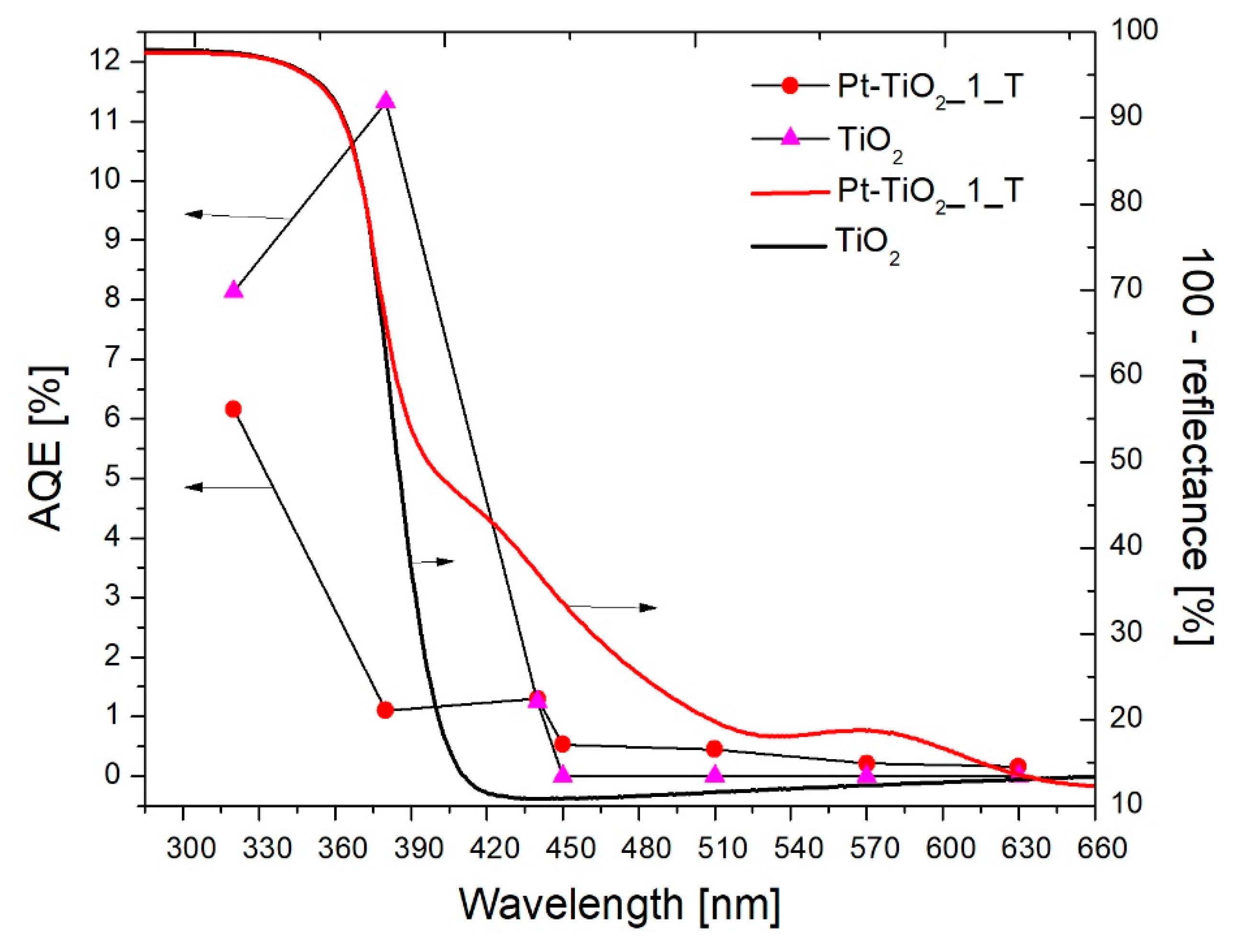
3.2.3. Action Spectra of Pt-TiO2_1_T and TiO2

The photocatalytic activity was examined in phenol oxidation reaction under monochromatic irradiation of 320, 380, 440, 450, 510, 570 and 630 nm. Results of spectral activity measurements for TiO2 and Pt-TiO2_1_T are presented in Figure 7. Apparent quantum yields of phenol decomposition were calculated in order to benzoquinone generation, as a primary reaction intermediate during phenol oxidation. In the UV region, bare titanium (IV) oxide exhibited higher activity compared to Pt-TiO2_1_T. For unmodified titanium (IV) oxide, apparent quantum yields reached 8.14% and 11.32% under 320 nm and 380 nm, respectively, while for Pt-TiO2_1_T 6.16% and 1.1%. Sun et al. [56] also observed the lower activity of platinum-modified photocatalysts in comparison to bare TiO2 during phenol oxidation under UV irradiation. They stated that lower photocatalytic activity results from decreased light absorption of TiO2 due to Pt NPs interfering. Water reduction to hydrogen consumes a part of photogenerated electrons, which can take part in oxygen reduction forming reactive oxygen species.
Under visible light irradiation (λ ≥ 440 nm) higher photocatalytic activity was observed for platinum-modified nanocomposite. Under λ = 450 nm bare TiO2 exhibited no activity, while Pt-TiO2_1_T exhibited activity in through almost whole examined region. As the wavelength of incident light increased, the quantum yield decreased and under λ = 630 nm no activity was denoted. In the visible spectrum, the highest activity was recorded in the wavelength range from 440 to 450 nm, which confirms the existence of a platinum surface plasmon resonance.
Platinum nanoparticles deposited on semiconductor surface may play a different role during Pt-TiO2 nanocomposite excitation upon UV or visible light. The schematic illustration of photocatalytic degradation mechanism is presented in Figure 8a,b. Under UV irradiation (Figure 8a) TiO2 particle is the main excitation center. Photoinduced electrons from TiO2 valence band are transferred to the conduction band and then to the metal nanoparticle. The electron transfer results from differences in a distribution of the Fermi levels (Ef) and generation of a Schottky barrier. When a metal work function is different than the electron affinity of the semiconductor conduction band, during the excitation of the TiO2 particle the excited electrons migrate towards the material of a higher value of the work function until the electrochemical equilibrium is achieved [58,59]. As a result of the Shottky barrier generation at the metal-semiconductor interface, excited electrons are trapped by a metal particle reducing the recombination of charge carriers. Under visible light, the role of an excitation center is attributed to metal nanoparticles (see Figure 8b). In the case of Pt, visible light promotes an excitation of electrons located at 5 d band and its junction to the conduction band of TiO2 [4,5,60,61]. Moreover, as it was previously reported by Lv et al. [62] under visible light Pt nanoparticles also act as a center of reactive oxygen species generation. The mechanism of charge carrier transport and trapping may be different when TiO2 is modified with other elements of compounds [63,64,65,66]. Nagakawa et al. [65,66] investigated the photocatalytic activity of CdS/WO3/CdWO4 and CdS/SiC/TiO2 composites modified with platinum nanoparticles in hydrogen generation reaction under visible light. The highest activity was observed for semiconductor systems modified with Pt NPs. The authors’ photocatalytic activity was attributed to the cascade carrier transfer of electrons between the semiconductors and capturing of electrons on platinum nanoparticles. Yoon et al. [64] investigated a mechanism of charge carrier transport in titanium(IV) oxide-graphene nanodots system. They stated that the electron transfer between TiO2 and adsorbed graphene processes through the charge transfer from HOMO (highest occupied molecular orbital) of organic adsorbate to the titanium (IV) oxide conduction band. Lee at al. [63] examined the charge transfer from adsorbed methylene blue (MB) molecule to a TiO2-graphene hybrid nanocomposite. They reported that high photocatalytic activity and efficient charge transfer between MB molecule and graphene-titanium (IV) oxide results from the narrowing of the TiO2-graphene bandgap. Narrowing of the bandgap may also lead to TiO2 excitation under visible light.

3.2.4. Photocatalytic Activity in the Vis_LED System

Based on the quantum yield analysis of phenol oxidation, it was concluded that the photocatalytic activity of TiO2 modified with platinum nanoparticles corresponds to the excitation of the nanocomposite with radiation in the range from 320 to 450 nm. In this regard, the activity of the Pt-TiO2_1_T photocatalyst was analyzed in the phenol degradation reaction under irradiation emitted by the LEDs with the emission maximum at λ = 415 nm. LED diodes may be an environmentally friendly light source with lower maintenance costs and longer operation time [67]. The phenol degradation rate constant was equal to 0.3 10−2 min−1. After 120 min of irradiation, about 30% of the phenol was oxidized. The results are shown in Figure S5. The phenol oxidation reaction proceeded linearly in the first 40 min of irradiation. After that time inhibition of the reaction was observed. Decrease of the reaction rate may result from the formation of intermediates products, including cyclohexa-2,5-diene-1,4-dione (1,4-benzoquinone). The presence of 1,4-benzoquinone in the reaction medium may significantly reduce the reaction rate due to the scavenging of superoxide anions [35,68].

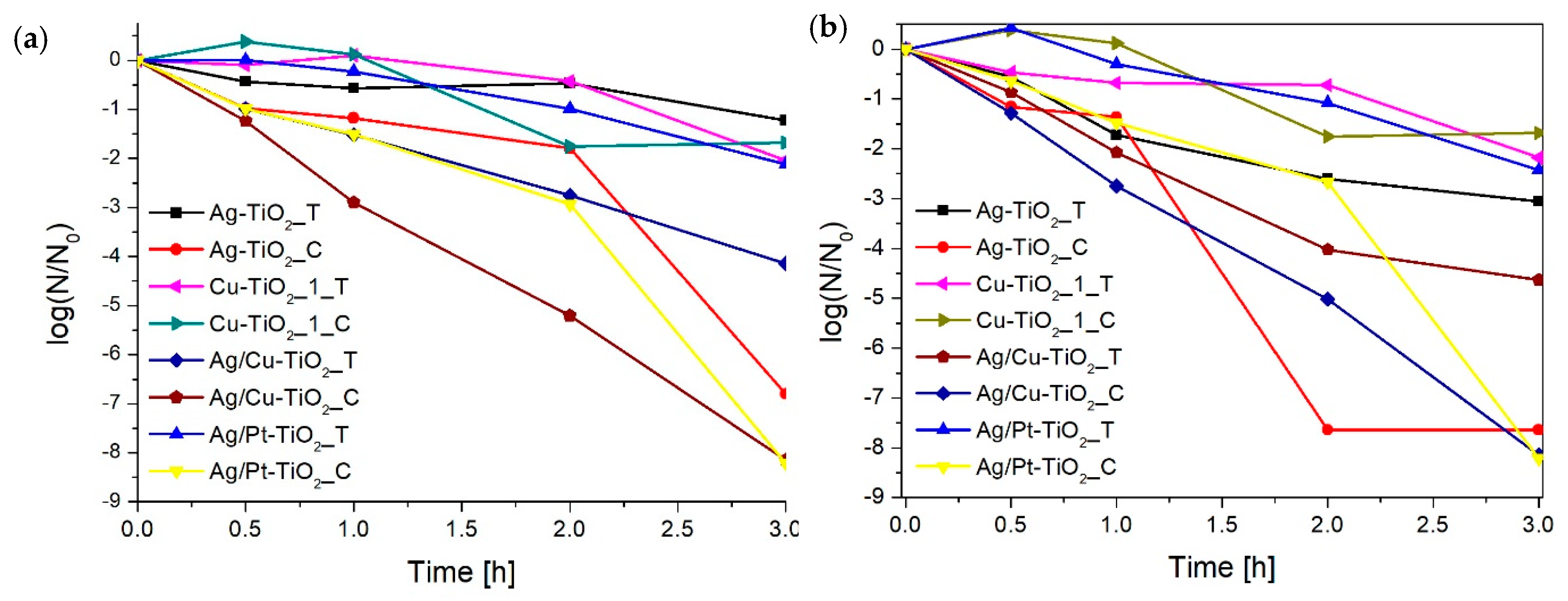
3.2.5. Antimicrobial Properties

The biocidal activity was tested on an E. coli strain in the aqueous phase. Despite the presence in the human body, E. coli was chosen as a model microorganism since infection with this bacterium can lead to serious health complications, such as food poisoning or urinary tract infection. Figure 9 and Table 3 present the results of biocidal activity. The tests were carried out under Vis radiation (λ > 450 nm) and in the dark. Table 3 lists the log(N/N0) values, where N is the number of survival bacteria, N0 is starting amount of bacteria, only for nanocomposites that have shown biocidal activity and for TiO2 as the reference material. The unmodified titanium(IV) oxide did not show antimicrobial activity. The highest activity was observed for bimetallic photocatalysts obtained by chemical reduction with small silver particles: Cu/Ag-TiO2_C and Ag/Pt-TiO2_C. For these nanocomposites, 100% of the bacterial cells were inactivated equally under irradiation and in the dark. In the case of other photocatalysts, the effect of radiation on the improvement of biocidal activity was observed. For the Cu/Ag-TiO2_T nanocomposite, the efficiency of the photocatalytic disinfection was nearly twice as high when compared to the dark. It was observed that the activity of photocatalysts results mainly from differences in the size of silver particles. The smaller the silver nanoparticles, the higher biocidal activity. The Ag-TiO2_C biocidal activity was twice as high as Ag-TiO2_T–log(N/N0) values under irradiation, which were −7.63 and −3,06, respectively. A similar dependence was observed for Cu/Ag-TiO2_C and Cu/Ag-TiO2_T, and Ag/Pt-TiO2_C and Ag/Pt-TiO2_T.

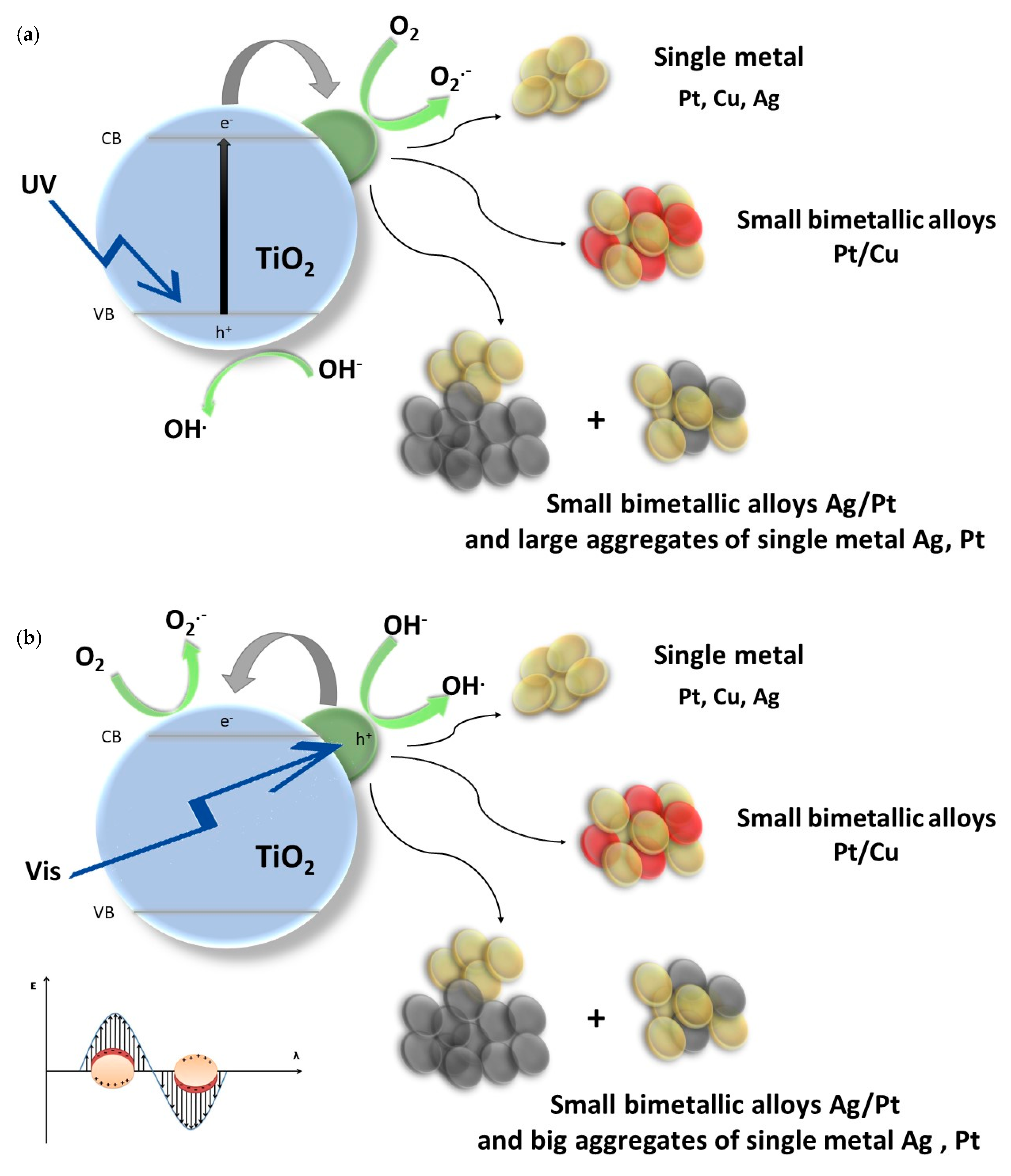
4. Discussion and Concluding Remarks

Titanium (IV) oxide photocatalysts modified with mono- and bimetallic nanoparticles of silver, platinum and copper were obtained via thermal and chemical reduction. It was observed that the size of nanoparticles deposited on TiO2 surface determined the photocatalytic activity. The differences in the metal NPs size result from the reduction method used for preparation, interactions of metal with support and between deposited metal nanoparticles [26,41,69]. Based on the results of XRD, DR/UV-Vis and TEM analysis it was found that the reduction of platinum ions via the thermal method promotes the deposition of smaller nanoparticles compared to the chemical method. For Pt-TiO2_2_C and Pt-TiO2_2_T, the average size of Pt NPs was equal to 3.4 ± 0.9 nm and 1.5 ± 0.6 nm, respectively. The influence of the ion reduction method was also observed for silver nanoparticles. In contrast to platinum, it was found that larger Ag particles were obtained using a thermal reduction method. The average size of silver oxide crystallites for Ag/Pt-TiO2_C and Ag/Pt-TiO2_T was 27 nm and 57 nm, respectively. TEM analysis also confirmed that the larger silver aggregates were formed in an annealed sample. Interactions of Pt ions with TiO2 matrix are much stronger than of silver, therefore Pt NPs are protected against aggregation. Moreover, the binding energy between Ag ions and TiO2 is lower than for Pt, which results in the growth of greater Ag clusters compared to Pt [26,41]. Silver particles tend to aggregate upon the annealing process. For copper species, no dependence was observed.
Based on TEM analysis it was also observed that the size of platinum nanoparticles is also affected by the type of coupled metal. For Ag/Pt-TiO2_C, the presence of small Ag-Pt alloy particles (5–10 nm) as well as segregation with the formation of bigger aggregates (up to 100 nm) of Pt and Ag were noticed, while for Cu/Pt-TiO2_2_C only the formation of small Cu-Pt alloy particles with an average size of was 2.2 ± 0.8 nm was observed.
Photocatalytic activity in the 2-propanol oxidation reaction varied depending on the type of metal, the range of used irradiation and the size of the particles. Under UV-Vis irradiation, the highest activity was observed for photocatalysts containing Pt nanoparticles reduced chemically, while under visible light irradiation for photocatalysts reduced thermally. The activity of monometallic TiO2 nanocomposites modified with silver and copper was similar. In the literature, it can be found that the photocatalytic activity of Pt-TiO2 photocatalysts is very sensitive to Pt NPs [26,70]. Based on our results it can be concluded that the photoactivity of tested systems deposited on TiO2 depends mainly on the platinum nanoparticles size. The small sizes of metal nanoparticles enhance the photocatalytic activity due to developing of TiO2-metal contact surface, which promotes a more efficient charge carrier transfer between the titania and metal nanoparticles.
In the hydrogen generation reaction. a higher activity was observed for photocatalysts obtained by chemical reduction. The efficiency of hydrogen generation for monometallic nanocomposites decreased in the following order, Pt > Cu > Ag. The obtained results correlate photocatalytic activity with a work function of particular metals. The efficiency of the hydrogen generation reaction may also depend on the oxidation state of the deposited metals. Xing et al. [71] investigated the effect of the platinum oxidation state on hydrogen generation efficiency. They observed a higher activity for platinum oxides than for Pt0. The authors attributed this to the lower energy of PtO2-H2 adsorption than Pt-H2. In the literature, the synergistic effect of modification of titanium(IV) oxide with bimetallic nanoparticles, e.g., Au-Pd, Au-Pt, Cu-Pt and Cu-Au, in the hydrogen generation process is reported [72,73,74,75,76]. However, modification with bimetallic nanoparticles does not always lead to synergy in the photocatalytic reduction [77,78]. In our study, TiO2 nanocomposites modified with bimetallic nanoparticles showed lower or similar activity compared to that of monometallic photocatalysts. Nadeem et al. [77] investigated the photocatalytic activity of TiO2 modified with mono- and bimetallic nanoparticles of silver and palladium in hydrogen generation under UV light. They observed that the highest hydrogen generation rates were for a sample containing only Pd NPs. They stated that the enhanced activity of Pd-TiO2 over Ag-TiO2 and Ag/Pd-TiO2 photocatalysts results from the palladium high work function and thus effective electron metal to TiO2 CB due to a high work function. They also stated that lower activity of silver-modified photocatalysts may result from the tendency of Ag to get oxidized in aqueos conditions.
The mechanism of photocatalytic activity excitation was investigated for phenol oxidation for bare TiO2 and Pt-TiO2_1_T under 320, 380, 440, 450, 510 and 570 nm. It was found that platinum-modified photocatalyst is excited by irradiation of wavelength up to 450 nm. Despite the increased absorption intensity, for Pt-TiO2_1_T, irradiation with wavelengths above 450 nm, the apparent quantum yield of reaction did not exceed 1%. Measurements of action spectra are important information in the process of selecting the radiation source. An insight into the activity of nanocomposites under specific wavelengths allows the selection of the radiation sources with a narrower emission range, e.g., LEDs.
Experiments on E. coli deactivation revealed the key effect of silver particle properties on biocidal activity. The highest activity was obtained for silver-modified nanocomposites obtained by chemical reduction, characterized by small particle size. The improvement of antimicrobial properties of metal-modified titanium(IV) oxide is possible due to the synergism of the antimicrobial properties of metals and reactive oxygen species generation on the TiO2 surface [3,79,80,81,82,83]. For gram-negative strains, silver nanoparticles are able to absorb and accumulate on the outer cell wall leading to the disintegration of the lipid bilayer and an increase in its permeability. Moreover, silver can be embedded into the DNA, leading to the slow down of the replication process and bacteria growth inhibition as a result of the bonding with oxygen, sulphur and nitrogen atoms [84,85].

Supplementary Materials

The following are available online at https://www.mdpi.com/2079-4991/9/8/1129/s1, Figure S1. Emission spectrum of a set of LED diodes, Figure S2. XRD patterns of (a) chemically reduced, (b) thermally reduced and (c) magnification in the 2θ range of 26–50° for Ag/Pt-TiO2_C and Ag/Pt-TiO2_T, Figure S3. (a) and (b) EDS mapping, (c) TEM image and (d) FFT analysis for Ag/Pt-TiO2_C, Figure S4. (a) Dark-field scanning transmission (DF-STEM) microscopy and EDS mapping of (b) silver, (c) platinum and (d) titanium for Ag/Pt-TiO2_T, Figure S5. Phenol oxidation with Pt-TiO2_1_T using LED system.

Author Contributions

Conceptualization, investigation, writing—original draft preparation, I.W., methodology, supervision of action spectra measurements, E.K.; XPS investigation, J.R., TEM analysis, G.N.; supervision, conceptualization, project administration, writing—review and editing, funding acquisition, A.Z.-J.

Funding

The research was financially supported by Gdansk University of Technology by project, The development of interdisciplinary doctoral studies at the Gdansk University of Technology in modern technologies (Project No: POKL.04.01.01-00-368/09) and National Science Centre Poland, grant number NCN 2016/23/D/ST5/01021).

Acknowledgments

Authors would like to highly acknowledge Bunsho Ohtani from the Institute for Catalysis, Hokkaido University, Japan, for the possibility of conducting research and fruitful discussions.

Conflicts of Interest

The authors declare no conflict of interest.

References

  1. Pulit, J.; Banach, M.; Kowalski, Z. Właściwości Nanocząstek Miedzi, Platyny, Srebra, Złota i Palladu. Czas. Tech. Chem. 2011, 2, 202–203. [Google Scholar]
  2. Langhammer, C.; Yuan, Z.; Zorić, I.; Kasemo, B. Plasmonic Properties of Supported Pt and Pd Nanostructures. Nano Lett. 2006, 6, 833–838. [Google Scholar] [CrossRef] [PubMed]
  3. Hajipour, M.J.; Fromm, K.M.; Akbar Ashkarran, A.; Jimenez de Aberasturi, D.; Larramendi, I.R.; de Rojo, T.; Serpooshan, V.; Parak, W.J.; Mahmoudi, M. Antibacterial Properties of Nanoparticles. Trends Biotechnol. 2012, 30, 499–511. [Google Scholar] [CrossRef] [PubMed]
  4. Zhang, X.; Chen, Y.L.; Liu, R.-S.; Tsai, D.P. Plasmonic Photocatalysis. Rep. Prog. Phys. 2013, 76, 046401. [Google Scholar] [CrossRef] [PubMed]
  5. Devi, L.G.; Kavitha, R. A Review on Plasmonic Metal-TiO2 Composite for Generation, Trapping, Storing and Dynamic Vectorial Transfer of Photogenerated Electrons across the Schottky Junction in a Photocatalytic System. Appl. Surf. Sci. 2016, 360, 601–622. [Google Scholar] [CrossRef]
  6. Clavero, C. Plasmon-Induced Hot-Electron Generation at Nanoparticle/Metal-Oxide Interfaces for Photovoltaic and Photocatalytic Devices. Nat. Photonics 2014, 8, 95–103. [Google Scholar] [CrossRef]
  7. Kelly, K.L.; Coronado, E.; Zhao, L.L.; Schatz, G.C. The Optical Properties of Metal Nanoparticles: The Influence of Size, Shape, and Dielectric Environment. J. Phys. Chem. B 2003, 107, 668–677. [Google Scholar] [CrossRef]
  8. Langhammer, C.; Larsson, E.M. Nanoplasmonic in Situ Spectroscopy for Catalysis Applications. ACS Catal. 2012, 2, 2036–2045. [Google Scholar] [CrossRef]
  9. Kumar, S.G.; Rao, K.S.R.K. Comparison of Modification Strategies towards Enhanced Charge Carrier Separation and Photocatalytic Degradation Activity of Metal Oxide Semiconductors (TiO2, WO3 and ZnO). Appl. Surf. Sci. 2016, 391, 124–128. [Google Scholar]
  10. Kowalska, E.; Mahaney, O.O.P.; Abe, R.; Ohtani, B. Visible-Light-Induced Photocatalysis through Surface Plasmon Excitation of Gold on Titania Surfaces. Phys. Chem. Chem. Phys. 2010, 12, 2344–2355. [Google Scholar] [CrossRef]
  11. Leong, K.H.; Gan, B.L.; Ibrahim, S.; Saravanan, P. Synthesis of Surface Plasmon Resonance (SPR) Triggered Ag/TiO2 Photocatalyst for Degradation of Endocrine Disturbing Compounds. Appl. Surf. Sci. 2014, 319, 128–135. [Google Scholar] [CrossRef]
  12. Wei, Z.; Rosa, L.; Wang, K.; Endo, M.; Juodkazis, S.; Ohtani, B.; Kowalska, E. Size-Controlled Gold Nanoparticles on Octahedral Anatase Particles as Efficient Plasmonic Photocatalyst. Appl. Catal. B Environ. 2017, 206, 393–405. [Google Scholar] [CrossRef] [PubMed]
  13. Sarina, S.; Waclawik, E.R.; Zhu, H. Photocatalysis on Supported Gold and Silver Nanoparticles under Ultraviolet and Visible Light Irradiation. Green Chem. 2013, 15, 1814. [Google Scholar] [CrossRef]
  14. Schneider, J.; Matsuoka, M.; Takeuchi, M.; Zhang, J.; Horiuchi, Y.; Anpo, M.; Bahnemann, D.W. Understanding TiO2 Photocatalysis: Mechanisms and Materials. Chem. Rev. 2014, 114, 9919–9986. [Google Scholar] [CrossRef]
  15. Ohtani, B. Revisiting the Fundamental Physical Chemistry in Heterogeneous Photocatalysis: Its Thermodynamics and Kinetics. Phys. Chem. Chem. Phys. 2014, 16, 1788–1797. [Google Scholar] [CrossRef] [PubMed]
  16. Fujishima, A.; Rao, T.N.; Tryk, D.A. Titanium Dioxide Photocatalysis. J. Photochem. Photobiol. C Photochem. Rev. 2000, 1, 1–21. [Google Scholar] [CrossRef]
  17. Mills, A.; Le Hunte, S. An Overview of Semiconductor Photocatalysis. J. Photochem. Photobiol. A Chem. 1997, 108, 1–35. [Google Scholar] [CrossRef]
  18. Zielińska-Jurek, A.; Hupka, J. Preparation and Characterization of Pt/Pd-Modified Titanium Dioxide Nanoparticles for Visible Light Irradiation. Catal. Today 2014, 230, 181–187. [Google Scholar] [CrossRef]
  19. Wei, Z.; Endo, M.; Wang, K.; Charbit, E.; Markowska-Szczupak, A.; Ohtani, B.; Kowalska, E. Noble Metal-Modified Octahedral Anatase Titania Particles with Enhanced Activity for Decomposition of Chemical and Microbiological Pollutants. Chem. Eng. J. 2017, 318, 121–134. [Google Scholar] [CrossRef]
  20. Dozzi, M.V.; Selli, E. Doping TiO2 with P-Block Elements: Effects on Photocatalytic Activity. J. Photochem. Photobiol. C Photochem. Rev. 2013, 14, 13–28. [Google Scholar] [CrossRef]
  21. Kowalska, E.; Rau, S.; Ohtani, B. Plasmonic Titania Photocatalysts Active under UV and Visible-Light Irradiation: Influence of Gold Amount, Size, and Shape. J. Nanotechnol. 2012, 2012, 1–11. [Google Scholar] [CrossRef] [Green Version]
  22. Kaneko, M.; Ueno, H.; Nemoto, J. Schottky Junction/Ohmic Contact Behavior of a Nanoporous TiO2 Thin Film Photoanode in Contact with Redox Electrolyte Solutions. Beilstein J. Nanotechnol. 2011, 2, 127–134. [Google Scholar] [CrossRef] [PubMed]
  23. Radecka, M.; Rekas, M.; Trenczek-Zajac, A.; Zakrzewska, K. Importance of the Band Gap Energy and Flat Band Potential for Application of Modified TiO2 Photoanodes in Water Photolysis. J. Power Sources 2008, 181, 46–55. [Google Scholar] [CrossRef]
  24. Chen, H.W.; Ku, Y.; Kuo, Y.L. Effect of Pt/TiO2 Characteristics on Temporal Behavior of o-Cresol Decomposition by Visible Light-Induced Photocatalysis. Water Res. 2007, 41, 2069–2078. [Google Scholar] [CrossRef] [PubMed]
  25. Borowska, E.; Gomes, J.; Martins, R.C.; Quinta-ferreira, R.M.; Horn, H.; Gmurek, M. Solar Photocatalytic Degradation of Sulfamethoxazole. Catalysts 2019, 9, 500. [Google Scholar] [CrossRef]
  26. Zielińska-Jurek, A.; Wei, Z.; Wysocka, I.; Szweda, P.; Kowalska, E. The Effect of Nanoparticles Size on Photocatalytic and Antimicrobial Properties of Ag-Pt/TiO2 Photocatalysts. Appl. Surf. Sci. 2015, 353, 317–325. [Google Scholar] [CrossRef]
  27. Foster, H.A.; Sheel, D.W.; Sheel, P.; Evans, P.; Varghese, S.; Rutschke, N.; Yates, H.M. Antimicrobial Activity of Titania/Silver and Titania/Copper Films Prepared by CVD. J. Photochem. Photobiol. A Chem. 2010, 216, 283–289. [Google Scholar] [CrossRef]
  28. Pulgarin, C.; Kiwi, J.; Nadtochenko, V. Mechanism of Photocatalytic Bacterial Inactivation on TiO2 Films Involving Cell-Wall Damage and Lysis. Appl. Catal. B Environ. 2012, 128, 179–183. [Google Scholar] [CrossRef]
  29. Laxma Reddy, P.V.; Kavitha, B.; Kumar Reddy, P.A.; Kim, K.H. TiO2-Based Photocatalytic Disinfection of Microbes in Aqueous Media: A Review. Environ. Res. 2017, 154, 296–303. [Google Scholar] [CrossRef] [PubMed]
  30. Gamage McEvoy, J.; Zhang, Z. Antimicrobial and Photocatalytic Disinfection Mechanisms in Silver-Modified Photocatalysts under Dark and Light Conditions. J. Photochem. Photobiol. C Photochem. Rev. 2014, 19, 62–75. [Google Scholar] [CrossRef]
  31. Gołabiewska, A.; Malankowska, A.; Jarek, M.; Lisowski, W.; Nowaczyk, G.; Jurga, S.; Zaleska-Medynska, A. The Effect of Gold Shape and Size on the Properties and Visible Light-Induced Photoactivity of Au-TiO2. Appl. Catal. B Environ. 2016, 196, 27–40. [Google Scholar] [CrossRef]
  32. Dong, C.; Lian, C.; Hu, S.; Deng, Z.; Gong, J.; Li, M.; Liu, H.; Xing, M.; Zhang, J. Size-Dependent Activity and Selectivity of Carbon Dioxide Photocatalytic Reduction over Platinum Nanoparticles. Nat. Commun. 2018, 9, 1–11. [Google Scholar] [CrossRef] [PubMed]
  33. Cybula, A.; Priebe, J.B.; Pohl, M.-M.; Sobczak, J.W.; Schneider, M.; Zielińska-Jurek, A.; Brückner, A.; Zaleska, A. The Effect of Calcination Temperature on Structure and Photocatalytic Properties of Au/Pd Nanoparticles Supported on TiO2. Appl. Catal. B Environ. 2014, 152, 202–211. [Google Scholar] [CrossRef]
  34. Parayil, S.K.; Kibombo, H.S.; Wu, C.M.; Peng, R.; Kindle, T.; Mishra, S.; Ahrenkiel, S.P.; Baltrusaitis, J.; Dimitrijevic, N.M.; Rajh, T.; et al. Synthesis-Dependent Oxidation State of Platinum on TiO2 and Their Influences on the Solar Simulated Photocatalytic Hydrogen Production from Water. J. Phys. Chem. C 2013, 117, 16850–16862. [Google Scholar] [CrossRef]
  35. Wysocka, I.; Kowalska, E.; Trzciński, K.; Łapiński, M.; Nowaczyk, G.; Zielińska-Jurek, A. UV-Vis-Induced Degradation of Phenol over Magnetic Photocatalysts Modified with Pt, Pd, Cu and Au Nanoparticles. Nanomaterials 2018, 8, 28. [Google Scholar] [CrossRef]
  36. Anpo, M.; Yamashita, H.; Ichihashi, Y.; Ehara, S. Photocatalytic Reduction of CO2 with H20 on Various Titanium Oxide Catalysts. J. Electroanal. Chem. 1995, 396, 21–26. [Google Scholar] [CrossRef]
  37. Murcia, J.J.; Hidalgo, M.C.; Navío, J.A.; Araña, J.; Doña-Rodríguez, J.M. Study of the Phenol Photocatalytic Degradation over TiO2 Modified by Sulfation, Fluorination, and Platinum Nanoparticles Photodeposition. Appl. Catal. B Environ. 2015, 179, 305–312. [Google Scholar] [CrossRef]
  38. Yadav, H.M.; Otari, S.V.; Koli, V.B.; Mali, S.S.; Hong, C.K.; Pawar, S.H.; Delekar, S.D. Preparation and Characterization of Copper-Doped Anatase TiO2 Nanoparticles with Visible Light Photocatalytic Antibacterial Activity. J. Photochem. Photobiol. A Chem. 2014, 280, 32–38. [Google Scholar] [CrossRef]
  39. Hu, Y.; Song, X.; Jiang, S.; Wei, C. Enhanced Photocatalytic Activity of Pt-Doped TiO2for NOxoxidation Both under UV and Visible Light Irradiation: A Synergistic Effect of Lattice Pt4+and Surface PtO. Chem. Eng. J. 2015, 274, 102–112. [Google Scholar] [CrossRef]
  40. Gołąbiewska, A.; Lisowski, W.; Jarek, M.; Nowaczyk, G.; Zielińska-Jurek, A.; Zaleska, A. Visible Light Photoactivity of TiO2 Loaded with Monometallic (Au or Pt) and Bimetallic (Au/Pt) Nanoparticles. Appl. Surf. Sci. 2014, 317, 1131–1142. [Google Scholar] [CrossRef]
  41. Galhenage, R.P.; Yan, H.; Tenney, S.A.; Park, N.; Henkelman, G.; Albrecht, P.; Mullins, D.R.; Chen, D.A. Understanding the Nucleation and Growth of Metals on TiO2: Co Compared to Au, Ni, and Pt. J. Phys. Chem. C 2013, 117, 7191–7201. [Google Scholar] [CrossRef]
  42. Luo, K.; St. Clair, T.P.; Lai, X.; Goodman, D.W. Silver Growth on TiO2 (110) (1 × 1) and (1 × 2). J. Phys. Chem. B 2000, 104, 3050–3057. [Google Scholar] [CrossRef]
  43. Zielińska-Jurek, A.; Kowalska, E.; Sobczak, J.W.; Lisowski, W.; Ohtani, B.; Zaleska, A. Preparation and Characterization of Monometallic (Au) and Bimetallic (Ag/Au) Modified-Titania Photocatalysts Activated by Visible Light. Appl. Catal. B Environ. 2011, 101, 504–514. [Google Scholar] [CrossRef]
  44. Kowalska, E.; Wei, Z.; Karabiyik, B.; Herissan, A.; Janczarek, M.; Endo, M.; Markowska-Szczupak, A.; Remita, H.; Ohtani, B. Silver-Modified Titania with Enhanced Photocatalytic and Antimicrobial Properties under UV and Visible Light Irradiation. Catal. Today 2015, 252, 136–142. [Google Scholar] [CrossRef]
  45. He, Z.; Fu, J.; Cheng, B.; Yu, J.; Cao, S. Cu2(OH)2CO3 Clusters: Novel Noble-Metal-Free Cocatalysts for Efficient Photocatalytic Hydrogen Production from Water Splitting. Appl. Catal. B Environ. 2017, 205, 104–111. [Google Scholar] [CrossRef]
  46. Athawale, A.A.; Katre, P.P.; Kumar, M.; Majumdar, M.B. Synthesis of CTAB-IPA Reduced Copper Nanoparticles. Mater. Chem. Phys. 2005, 91, 507–512. [Google Scholar] [CrossRef]
  47. Giuffrida, S.; Costanzo, L.L.; Ventimiglia, G.; Bongiorno, C. Photochemical Synthesis of Copper Nanoparticles Incorporated in Poly(Vinyl Pyrrolidone). J. Nanoparticle Res. 2008, 10, 1183–1192. [Google Scholar] [CrossRef]
  48. Kubacka, A.; Muñoz-Batista, M.J.; Fernández-García, M.; Obregón, S.; Colón, G. Evolution of H2 Photoproduction with Cu Content on CuOx-TiO2 Composite Catalysts Prepared by a Microemulsion Method. Appl. Catal. B Environ. 2015, 163, 214–222. [Google Scholar] [CrossRef]
  49. Wei, Z.; Janczarek, M.; Endo, M.; Colbeau-Justin, C.; Ohtani, B.; Kowalska, E. Silver-Modified Octahedral Anatase Particles as Plasmonic Photocatalyst. Catal. Today 2018, 310, 19–25. [Google Scholar] [CrossRef] [PubMed]
  50. Erdem, B.; Hunsicker, R.A.; Simmons, G.W.; Sudol, E.D.; Dimonie, V.L.; El-Aasser, M.S. XPS and FTIR Surface Characterization of TiO2 Particles Used in Polymer Encapsulation. Langmuir 2001, 17, 2664–2669. [Google Scholar] [CrossRef]
  51. Xiong, L.; Li, J.; Yang, B.; Yu, Y. Ti3+ in the Surface of Titanium Dioxide: Generation, Properties and Photocatalytic Application. J. Nanomater. 2012, 2012, 1–13. [Google Scholar] [CrossRef]
  52. Rjeb, A.; Letarte, S.; Tajounte, L.; El Idrissi, M.C.; Adnot, A.; Roy, D.; Claire, Y.; Kaloustian, J. Polypropylene Natural Aging Studied by X-ray Photoelectron Spectroscopy. J. Electron. Spectros. Relat. Phenom. 2000, 107, 221–230. [Google Scholar] [CrossRef]
  53. López, R.; Gómez, R.; Llanos, M.E. Photophysical and Photocatalytic Properties of Nanosized Copper-Doped Titania Sol-Gel Catalysts. Catal. Today 2010, 148, 103–108. [Google Scholar] [CrossRef]
  54. Ahmed, L.M.; Ivanova, I.; Hussein, F.H.; Bahnemann, D.W. Role of Platinum Deposited on TiO2 in Photocatalytic Methanol Oxidation and Dehydrogenation Reactions. Int. J. Photoenergy 2014, 2014, 1–9. [Google Scholar] [CrossRef]
  55. Wang, K.; Wei, Z.; Ohtani, B.; Kowalska, E. Interparticle Electron Transfer in Methanol Dehydrogenation on Platinum-Loaded Titania Particles Prepared from P25. Catal. Today 2018, 303, 327–333. [Google Scholar] [CrossRef]
  56. Sun, B.; Vorontsov, A.V.; Smirniotis, P.G. Role of Platinum Deposited on TiO2 in Phenol Photocatalytic Oxidation. Langmuir 2003, 19, 3151–3156. [Google Scholar] [CrossRef]
  57. Shiraishi, Y.; Sakamoto, H.; Sugano, Y.; Ichikawa, S.; Hirai, T. Pt-Cu Bimetallic Alloy Nanoparticles Supported on Anatase TiO2: Highly Active Catalysts for Aerobic Oxidation Driven by Visible Light. ACS Nano 2013, 7, 9287–9297. [Google Scholar] [CrossRef] [PubMed]
  58. Ioannides, T.; Verykios, X.E. Charge Transfer in Metal Catalysts Supported on Doped TiO2: A Theoretical Approach Based on Metal-Semiconductor Contact Theory. J. Catal. 1996, 161, 560–569. [Google Scholar] [CrossRef]
  59. Zhdanov, V.P. Nm-Sized Metal Particles on a Semiconductor Surface, Schottky Model, Etc. Surf. Sci. 2002, 512, 6–9. [Google Scholar] [CrossRef]
  60. Etacheri, V.; Di Valentin, C.; Schneider, J.; Bahnemann, D.; Pillai, S.C. Visible-Light Activation of TiO2 Photocatalysts: Advances in Theory and Experiments. J. Photochem. Photobiol. C Photochem. Rev. 2015, 25, 1–29. [Google Scholar] [CrossRef]
  61. Shiraishi, Y.; Sakamoto, H.; Fujiwara, K.; Ichikawa, S.; Hirai, T. Selective Photocatalytic Oxidation of Aniline to Nitrosobenzene by Pt Nanoparticles Supported on TiO2 under Visible Light Irradiation. ACS Catal. 2014, 4, 2418–2425. [Google Scholar] [CrossRef]
  62. Lv, J.; Gao, H.; Wang, H.; Lu, X.; Xu, G.; Wang, D.; Chen, Z.; Zhang, X.; Zheng, Z.; Wu, Y. Controlled Deposition and Enhanced Visible Light Photocatalytic Performance of Pt-Modified TiO2 Nanotube Arrays. Appl. Surf. Sci. 2015, 351, 225–231. [Google Scholar] [CrossRef]
  63. Lee, J.S.; You, K.H.; Park, C.B. Highly Photoactive, Low Bandgap TiO2 Nanoparticles Wrapped by Graphene. Adv. Mater. 2012, 24, 1084–1088. [Google Scholar] [CrossRef] [PubMed]
  64. Yoon, H.; Kim, D.; Park, M.; Kim, J.; Kim, J.; Srituravanich, W.; Shin, B.; Jung, Y.; Jeon, S. Extraordinary Enhancement of UV Absorption in TiO2 Nanoparticles Enabled by Low-Oxidized Graphene Nanodots. J. Phys. Chem. C 2018, 122, 12114–12121. [Google Scholar] [CrossRef]
  65. Nagakawa, H.; Ochiai, T.; Takekuma, Y.; Konuma, S.; Nagata, M. Effective Photocatalytic Hydrogen Evolution by Cascadal Carrier Transfer in the Reverse Direction. ACS Omega 2018, 3, 12770–12777. [Google Scholar] [CrossRef]
  66. Nagakawa, H.; Ochiai, T.; Konuma, S.; Nagata, M. Visible-Light Overall Water Splitting by CdS/WO 3 /CdWO 4 Tricomposite Photocatalyst Suppressing Photocorrosion. ACS Appl. Energy Mater. 2018, 1, 6730–6735. [Google Scholar] [CrossRef]
  67. Martín-Sómer, M.; Pablos, C.; van Grieken, R.; Marugán, J. Influence of Light Distribution on the Performance of Photocatalytic Reactors: LED vs. Mercury Lamps. Appl. Catal. B Environ. 2017, 215, 1–7. [Google Scholar] [CrossRef]
  68. Yang, M.Q.; Zhang, Y.; Zhang, N.; Tang, Z.R.; Xu, Y.J. Visible-Light-Driven Oxidation of Primary C-H Bonds over CdS with Dual Co-Catalysts Graphene and TiO2. Sci. Rep. 2013, 3, 3314. [Google Scholar] [CrossRef]
  69. Kolobov, N.S.; Svintsitskiy, D.A.; Kozlova, E.A.; Selishchev, D.S.; Kozlov, D.V. UV-LED Photocatalytic Oxidation of Carbon Monoxide over TiO2supported with Noble Metal Nanoparticles. Chem. Eng. J. 2017, 314, 600–611. [Google Scholar] [CrossRef]
  70. Vamvasakis, I.; Liu, B.; Armatas, G.S. Size Effects of Platinum Nanoparticles in the Photocatalytic Hydrogen Production Over 3D Mesoporous Networks of CdS and Pt Nanojunctions. Adv. Funct. Mater. 2016, 26, 8062–8071. [Google Scholar] [CrossRef]
  71. Xing, J.; Li, Y.H.; Jiang, H.B.; Wang, Y.; Yang, H.G. The Size and Valence State Effect of Pt on Photocatalytic H2 Evolution over Platinized TiO2photocatalyst. Int. J. Hydrogen Energy 2014, 39, 1237–1242. [Google Scholar] [CrossRef]
  72. Wu, D.; You, H.; Jin, D.; Li, X. Enhanced Inactivation of Escherichia Coli with Ag-Coated TiO2 Thin Film under UV-C Irradiation. J. Photochem. Photobiol. A Chem. 2011, 217, 177–183. [Google Scholar] [CrossRef]
  73. Gallo, A.; Marelli, M.; Psaro, R.; Gombac, V.; Montini, T.; Fornasiero, P.; Pievo, R.; Santo, V.D. Bimetallic Au–Pt/TiO2 Photocatalysts Active under UV-A and Simulated Sunlight for H2 Production from Ethanol. Green Chem. 2012, 14, 330–333. [Google Scholar] [CrossRef]
  74. Duan, K.; Liu, Z.; Li, J.; Yuan, L.; Hu, H.; Woo, S.I. Novel Pd-Au/for the Selective Catalytic Reduction of NOxby H2. Catal. Commun. 2014, 57, 19–22. [Google Scholar] [CrossRef]
  75. Oros-Ruiz, S.; Zanella, R.; Collins, S.E.; Hernández-Gordillo, A.; Gómez, R. Photocatalytic Hydrogen Production by Au-MxOy(MAg, Cu, Ni) Catalysts Supported on TiO2. Catal. Commun. 2014, 47, 1–6. [Google Scholar] [CrossRef]
  76. Zielińska-Jurek, A. Progress, Challenge, and Perspective of Bimetallic TiO2-Based Photocatalysts. J. Nanomater. 2014, 2014, 1–17. [Google Scholar] [CrossRef]
  77. Nadeem, M.A.; Al-Oufi, M.; Wahab, A.K.; Anjum, D.; Idriss, H. Hydrogen Production on Ag-Pd/TiO2 Bimetallic Catalysts: Is There a Combined Effect of Surface Plasmon Resonance with Schottky Mechanism on the Photo-Catalytic Activity? ChemistrySelect 2017, 2, 2754–2762. [Google Scholar] [CrossRef]
  78. Kowalska, E.; Janczarek, M.; Rosa, L.; Juodkazis, S.; Ohtani, B. Mono-and Bi-Metallic Plasmonic Photocatalysts for Degradation of Organic Compounds under UV and Visible Light Irradiation. Catal. Today 2014, 230, 131–137. [Google Scholar] [CrossRef]
  79. Klaine, S.J.; Alvarez, P.J.J.; Batley, G.E.; Fernandes, T.F.; Handy, R.D.; Lyon, D.Y.; Mahendra, S.; McLaughlin, M.J.; Lead, J.R. Nanomaterials in the Environment: Behavior, Fate, Bioavailability, and Effects. Environ. Toxicol. Chem. 2008, 27, 1825–1851. [Google Scholar] [CrossRef]
  80. Bhatt, I.; Tripathi, B.N. Interaction of Engineered Nanoparticles with Various Components of the Environment and Possible Strategies for Their Risk Assessment. Chemosphere 2011, 82, 308–317. [Google Scholar] [CrossRef]
  81. Zheng, K.; Setyawati, M.I.; Leong, D.T.; Xie, J. Antimicrobial Silver Nanomaterials. Coord. Chem. Rev. 2018, 357, 1–17. [Google Scholar] [CrossRef]
  82. Pietrzak, K.; Twarużek, M.; Czyżowska, A.; Kosicki, R.; Gutarowska, B. Influence of Silver Nanoparticles on Metabolism and Toxicity of Moulds. Acta Biochim. Pol. 2015, 62, 851–857. [Google Scholar] [CrossRef] [PubMed]
  83. Hoseinzadeh, E.; Makhdoumi, P.; Taha, P.; Hossini, H.; Stelling, J.; Amjad Kamal, M. A Review on Nano-Antimicrobials: Metal Nanoparticles, Methods and Mechanisms. Curr. Drug Metab. 2017, 18, 120–128. [Google Scholar] [CrossRef] [PubMed]
  84. Wang, L.; Hu, C.; Shao, L. The Antimicrobial Activity of Nanoparticles: Present Situation and Prospects for the Future. Int. J. Nanomed. 2017, 12, 1227–1249. [Google Scholar] [CrossRef] [PubMed]
  85. Rai, M.; Kon, K.; Ingle, A.; Duran, N.; Galdiero, S.; Galdiero, M. Broad-Spectrum Bioactivities of Silver Nanoparticles: The Emerging Trends and Future Prospects. Appl. Microbiol. Biotechnol. 2014, 98, 1951–1961. [Google Scholar] [CrossRef] [PubMed]
Figure 1. Absorption spectra of (a) Ag-TiO2_C, Ag-TiO2_T, Ag/Pt-TiO2_T and Ag/Pt-TiO2_C (b) Cu-TiO2_1_C, Cu-TiO2_2_C, Cu-TiO2_1_T and Cu-TiO2_2_T, (c) Cu/Pt-TiO2_1_C, Cu/Pt-TiO2_2_C, Cu/Pt-TiO2_1_T and Cu/Pt-TiO2_2_T, (d) Pt-TiO2_1_C, Pt-TiO2_2_C, Pt-TiO2_1_T and Pt-TiO2_2_T.
Nanomaterials 09 01129 g001
Figure 2. Deconvolution of X-ray photoelectron spectroscopy (XPS) spectra for (a) Ag 3d, (b) Cu 2p and (c) Pt 4f.
Nanomaterials 09 01129 g002aNanomaterials 09 01129 g002b
Figure 3. Transmission electron microscopy (TEM) images of (a) Pt-TiO2_2_C, (b) Pt-TiO2_2_T and metal particles size distribution for (c) Pt-TiO2_2_C, (d) Pt-TiO2_2_T and (e) Cu/Pt-TiO2_2_C.
Nanomaterials 09 01129 g003aNanomaterials 09 01129 g003b
Figure 4. TEM images and line analysis of Cu/Pt-TiO2_2_C.
Nanomaterials 09 01129 g004
Figure 5. Photocatalytic activity in 2-propanol oxidation under (a) UV-Vis and (b) Vis.
Nanomaterials 09 01129 g005
Figure 6. Photocatalytic activity in hydrogen generation under UV-Vis.
Nanomaterials 09 01129 g006
Figure 7. Spectral activity and absorption spectra in phenol oxidation for TiO2 and Pt-TiO2_2_T. (AQE—Apparent Quantum Efficiency).
Nanomaterials 09 01129 g007
Figure 8. Schematic illustration of metal-TiO2 nanocomposite excitation under (a) UV and (b) under Vis.
Nanomaterials 09 01129 g008
Figure 9. Antimicrobial activity (a) in the dark and (b) under visible light (λ > 450 nm).
Nanomaterials 09 01129 g009
Table 1. Characteristic of physicochemical properties and photocatalytic activity of as-prepared photocatalysts.
Sample LabelMetal Content Used for Preparation
[% mol]
Anatase Crystallite Size [nm]2-Propanol Photooxidation Rate
[µmol·h−1]
Hydrogen Generation Rate
[µmol·h−1]
UV-Vis
PtAgCuUV-VisVis
TiO20001234.3 ± 1.70.026 ± 0.0011.9 ± 0.1
Pt-TiO2_1_C0.10012227.3 ± 11.40.496 ± 0.025409.5 ± 20.5
Pt-TiO2_2_C0.05001480.8 ± 4.10.197 ± 0.010501.4 ± 25.1
Cu-TiO2_1_C000.51361.1 ± 3.10.088 ± 0.00463.1 ± 3.2
Cu-TiO2_2_C000.11514.3 ± 0.70.103 ± 0.00581.1 ± 4.1
Ag-TiO2_C00.501462.1 ± 3.10.148 ± 0.00742.2 ± 2.1
Cu/Ag-TiO2_C00.50.51254.3 ± 2.70.092 ± 0.00560.1 ± 3.1
Ag/Pt-TiO2_C0.10.5012151.1 ± 7.60.246 ± 0.012338.9 ± 16.9
Cu/Pt-TiO2_1_C0.100.51461.2 ± 3.10.286 ± 0.014219.4 ± 10.9
Cu/Pt-TiO2_2_C0.0500.113150.5 ± 7.50.414 ± 0.021371.1 ± 18.5
Pt-TiO2_1_T0.10013117.1 ± 5.90.582 ± 0.029355.9 ± 17.8
Pt-TiO2_2_T0.05001340.2 ± 2.10.606 ± 0.030113.5 ± 5.7
Cu-TiO2_1_T000.51271.9 ± 3.60.087 ± 0.00460.1 ± 3
Cu-TiO2_2_T000.11421.1 ± 1.10.186 ± 0.00982.3 ± 4.1
Ag-TiO2_T00.501272.1 ± 3.60.144 ± 0.00716.3 ± 0.8
Cu/Ag-TiO2_T00.50.51461.5 ± 3.10.089 ± 0.00451.1 ± 2.5
Ag/Pt-TiO2_T0.10.5012117.3 ± 5.90.293 ± 0.014219.1 ± 10.9
Cu/Pt-TiO2_1_T0.100.51340.7 ± 2.10.257 ± 0.013137.7 ± 6.9
Cu/Pt-TiO2_2_T0.0500.112109.1 ± 5.50.520 ± 0.026175.5 ± 8.8
Table 2. X-ray photoelectron spectroscopy (XPS) analysis of Ti, O, C, Pt, Cu and Ag.
Sample LabelElement Content [at. %]Particle Size [nm]
Ti
2p
O
1s
C
1s
Pt
4f
Cu
2p
Ag
3d
Pt-TiO2_2_C27.162.99.90.1--3.4 ± 0.9
Pt-TiO2_2_T24.763.411.9n.d.--1.5 ± 0.6
Ag/Pt-TiO2_C26.462.011.20.2-0.2-
Ag/Pt-TiO2_T26.660.812.00.2-0.4-
Ag/Cu-TiO2_C23.362.413.9-0.10.3-
Ag/Cu-TiO2_T25.260.413.9-0.20.3-
Cu/Pt-TiO2_2_C26.959.413.60.1n.d.-2.2 ± 0.8
Cu/Pt-TiO2_2_T26.961.711.40.1n.d.--
n.a.—not analyzed.
Table 3. Biocidal activity against E. coli.
Sample Labellog(N/N0)
Under Irradiation
(λ > 450 nm)
In the Dark
TiO2−0.27−0.42
Cu-TiO2_1_C−2.86−1.68
Ag-TiO2_C−7.63−6.80
Cu/Ag-TiO2_C−8.14−8.14
Ag/Pt-TiO2_C−8.21−8.21
Cu-TiO2_1_T−2.17−2.05
Ag-TiO2_T−3.06−1.22
Cu/Ag-TiO2_T−4.64−2.14
Ag/Pt-TiO2_T−2.43−2.12

