


Abstract
Ex vivo human tooth culture models preserve the native dentine–pulp complex and offer a translational platform to study pulp-capping biomaterials. This systematic review aimed to synthesize the evidence on histological pulp tissue responses to calcium silicate-based cement (CSCs) used for direct pulp capping in human tooth culture models. The review followed PRISMA 2020 guidance. Eligible studies were ex vivo whole human tooth culture models with direct pulp exposure treated with commercial or experimental CSCs and reporting histological outcomes. Risk of bias was assessed using the QUIN tool. Thirteen studies were included. Most used immature human third molars (from 15- to 19-year-old patients) and culture periods up to 28 days, with a minority extending observation to 45–90 days. Across hydraulic CSCs, Biodentine was the most frequently evaluated material, followed by ProRoot MTA and several experimental hydraulic and resin-modified formulations. Overall, hydraulic CSCs were consistently associated with biocompatible pulp responses and a pro-mineralization pattern characterized by periexposure mineralized foci/osteodentin-like tissue; where assessed, immunohistochemistry supported odontoblast-like differentiation. In contrast, the resin-modified CSC TheraCal LC and other experimental resin-modified CSCs showed more heterogeneous findings, with reports of absent, delayed, or less prominent mineralization compared with reference hydraulic CSCs. In intact human tooth culture models, hydraulic CSCs show reproducible biocompatibility and early mineralization features consistent with reparative dentinogenesis, whereas resin-modified CSCs demonstrate more variable histological performance.
1. Introduction
Vital pulp therapy (VPT) is defined as the set of endodontic therapeutic procedures applied to teeth diagnosed with pulpitis, the aim of which is to control the inflammatory process and maintain pulp vitality, wholly or partially [1]. These procedures are grounded in the defensive and reparative potential of the dentine–pulp complex in response to injury [2]. Historically, VPT was mainly indicated for immature permanent teeth, where it achieved predictable success. More recently, advances in pulpal biology and the development of bioactive dental biomaterials have expanded its indications to mature permanent teeth with pulpitis, supporting the contemporary use of VPT in a broader range of clinical scenarios [3].
When a pulp exposure occurs, either due to a coronal fracture or during the removal of carious tissue, the insult is sufficiently intense to cause odontoblast death. An inflammatory process is then triggered, leading to the release of mediators and biochemical reactions that enable the migration and proliferation of multipotent mesenchymal stem cells present in the dental pulp, which will differentiate into cells similar to odontoblasts, known as ‘odontoblast-like cells’ [4]. This cell subpopulation is of great importance, as it secretes a mineralized matrix with mineral and molecular features comparable to dentine. The resulting mineralized tissue bridge protects the pulp from further injury and contributes to tissue healing and maintenance of vitality [5].
From a biological perspective, three requirements are necessary to achieve success in VPT procedures: (1) good systemic health and reparative capacity of the patient; (2) strict control of bacterial contamination; and (3) application of a bioactive material that stimulates the migration of pulpal stem cells to form reparative dentine [2].
Classically, the material used for this type of procedure has been Ca(OH)2. It is an alkaline material with strong antibacterial capacity, capable of solubilizing bioactive molecules, such as TGF-β1, involved in reparative dentinogenesis [6]. However, this material presents important drawbacks, including solubility, limited sealing capacity, undesirable irritation of the pulpal tissue, and the presence of tunnels and structural discontinuities in the resulting dentine bridge [7].
The introduction of calcium silicate-based cements (CSCs) represented a major shift in biomaterials used for vital pulp therapy (VPT). Torabinejad et al. (1993) first reported their use in endodontic applications with favourable in vitro outcomes [8], and the first widely adopted formulation was commercialized in 1998 as ProRoot MTA. Composed mainly of C2S and C3S phases, ProRoot MTA attracted substantial clinical and scientific interest due to its bioactivity, antibacterial effects, sealing ability and cellular biocompatibility [9]. However, it also presented relevant limitations including a long setting time, challenging handling, and the incorporation of Bi2O3 as a radiopacifier, which has been associated with tooth discoloration and cytotoxic effects at the cellular level [10].
In response to these shortcomings, CSCs have undergone rapid development, yielding multiple advanced formulations intended to overcome the limitations of ProRoot MTA [9,11], alongside an expansion of clinical indications that has consolidated CSCs as materials of choice for VPT procedures [12,13]. A pivotal milestone in this evolution was the launch of Biodentine, a second-generation tricalcium silicate-based cement introduced to provide improved handling and faster setting kinetics while maintaining the bioactive profile that underpinned MTA’s clinical success. Building on this trajectory, newer calcium silicate-based formulations have continued to prioritize operability and clinical versatility. Beyond conventional powder–liquid systems, ready-to-use premixed CSCs with putty-like consistency and accelerated setting reactions have been developed to facilitate manipulation in procedures such as pulpotomies and endodontic microsurgery, where precise placement and reduced technique sensitivity are desirable [14]. Furthermore, resin-modified calcium silicate materials have been designed to enhance placement accuracy and shorten setting time through light-activated polymerization, providing additional practical advantages during clinical application [15]. While these innovations may improve operability, formulation changes can also influence physicochemical behaviour and biological interactions with pulp tissue. Consequently, the growing diversity of calcium silicate-based biomaterials highlights the need for rigorous biological characterization to enable meaningful comparisons and support evidence-based clinical decision-making.
Throughout this evolutionary process, a large number of models have been published in endodontics to characterize the biological behaviour of hydraulic CSCs, ranging from in vitro models to in vivo models. In vitro models employ cells isolated from their natural environment to conduct assays outside the organism, under controlled laboratory conditions [16]. We find two-dimensional (2D) in vitro studies and 3D models that better represent cellular environments [17]. Although far from clinical conditions, numerous researchers have studied the biological behaviour of CSCs using these types of assays [18]. Animal experimentation represents another approach, classified as either ectopic or orthotopic models according to the anatomical site of the experimental procedure [17]. Orthotopic models implant the material in its natural location; for this purpose, larger animals are used, which implies higher cost and greater handling difficulty. In endodontic procedures, dogs and, likewise, porcine species are widely used because their dentitions show growth patterns and pathophysiology similar to human teeth [19,20,21]. Nevertheless, the pulpal response to biomaterials in animals may not fully replicate human tissue behaviour, which limits direct translation of findings [22].
To bridge the gap between simplified in vitro assays and animal experimentation, Téclès et al. (2008) proposed a culture model of an intact human tooth [23]. This is an ex vivo model in which a whole organ, recently extracted and with minimal modification of its natural state, preserves vitality under controlled laboratory conditions so that the cells remain viable. This methodology has the major advantage of assessing cellular and tissue responses to CSCs in an environment and conditions similar to reality, constituting a good intermediate point between in vitro and in vivo experimentation [17]. Since its description, numerous publications have employed this model to test histologically different calcium silicate materials, both commercial and experimental. However, available studies vary considerably in culture duration, experimental conditions, and histological assessment methods, limiting comparability across reports.
Accordingly, the aim of the present systematic review is to analyze and synthesize the evidence on direct pulp capping with CSCs using the natural tooth culture model, focusing on experimental conditions as well as histological and immunohistochemical outcomes.
2. Materials and Methods
2.1. Guidelines and Protocol
This review was conducted in accordance with the PRISMA 2020 preferred reporting guidelines for systematic reviews (Supplementary Materials: PRISMA_2020_checklist-JFB2026) [24]. The review protocol was prospectively registered in the Open Science Framework (OSF) (Registration DOI 10.17605/OSF.IO/8KUX3).
2.2. Review Question
This review addressed the histological responses induced in the dental pulp by CSCs when the ex vivo human natural tooth culture model is used as the experimental system. The research question was structured according to the PICO framework, considering as P (population) ex vivo human natural tooth culture models with direct pulp exposure; I (intervention) the application of CSCs, either commercial or experimental, used as direct pulp capping agents; C (comparison) cultured teeth without material application, when available; and O (outcomes) the histological findings described in the pulp tissue, with or without complementary analyses such as immunohistochemistry (IHC), scanning electron microscopy (SEM), energy-dispersive X-ray spectroscopy (EDX), or micro-computed tomography (µCT). Therefore, the wording of the review question and focus was as follows: “In ex vivo human natural tooth culture models, does direct pulp capping with CSCs, compared with other pulp-capping materials or no material, result in different histological tissue responses?”
2.3. Search Strategy
A systematic search of electronic databases (PubMed, Embase, Scopus, and Google Scholar) was conducted from database inception to October 2025. No language restrictions were applied. Detailed search strings and any database-specific adaptations are provided in Supplementary Table S1. In addition, grey literature was screened (reference lists of included studies and related reviews; contact with field experts) to minimize publication bias.
2.4. Inclusion and Exclusion Criteria
Inclusion criteria: Ex vivo studies using the whole human natural tooth culture model with direct pulp exposure; studies testing CSCs as direct pulp capping agents (commercial or experimental); histological assessment of pulp tissue (H&E and trichrome), with or without complementary analyses (IHC, SEM, EDX, µCT); and original studies published in peer-reviewed journals.
Exclusion criteria: In vitro cell-only studies or animal models (no entire tooth model), studies focused solely on physicochemical/mechanical properties without histological evaluation, reviews/letters/abstracts or incomplete reports, duplicate publications (the most complete version was retained), and studies with no group including a calcium silicate-based material.
2.5. Study Selection
Four reviewers (A.C.-F., H.G.d.S.C., D.B.S., J.M.M.d.S.) participated in the selection process. Two reviewers independently screened titles/abstracts and full texts against the eligibility criteria (A.C.-F. and H.G.d.S.C.). A third author resolved any disagreements (J.M.M.d.S.). Rayyan app (available at https://new.rayyan.ai/reviews, accessed on 11 January 2026, Rayyan, Qatar Computing Research Institute, Qatar Foundation) was used to manage the screening workflow and to perform duplicate removal; Mendeley Desktop software (version 1.19.8, Elsevier Inc., New York, NY, USA) was used as the reference manager for organizing the final set of included studies. After full-text assessment, the final selection was established.
2.6. Data Extraction
Two independent reviewers (A.C.-F., A.D.-C.) extracted the following variables using a pre-piloted form: first author, tooth type, sample size, culture duration, culture medium, material tested (commercial/experimental), histological findings, immunohistochemical findings, and mineralization outcomes. Discrepancies were resolved through discussion and, when needed, by a third reviewer (J.M.M.d.S.); original articles were consulted to adjudicate uncertainties. When required information could not be retrieved from the publication, corresponding authors were contacted; if data remained unavailable, the study was excluded from quantitative synthesis for that outcome. The accuracy of the extracted data was verified.
2.7. Quality Assessment and Risk of Bias of Individual Studies
Risk of bias was assessed using the QUIN Tool (for in vitro dental research) [25] which is applicable to ex vivo whole-tooth culture models. The tool comprises 12 items: (1) clearly stated aims/objectives; (2) sample size calculation/justification; (3) sampling technique with inclusion/exclusion criteria; (4) details of the comparison/control group; (5) methodological detail and standardization; (6) operator details (training/calibration); (7) randomization; (8) method of measuring outcomes (procedure/justification); (9) outcome assessor details (training/calibration, inter/intra-rater reliability where applicable); (10) blinding (operators/evaluators/statistician, as applicable); (11) statistical analysis (software/methods); and (12) presentation of results. Each item was scored as 2 = adequately specified, 1 = inadequately specified, or 0 = not specified; not applicable (NA) items were excluded from the denominator. The QUIN % was calculated as (sum of points × 100)/[2 × number of applicable items] and used to classify studies as low (>70%), moderate (50–70%), or high (<50%) risk of bias. Two reviewers (A.C.-F. and D.B.S.) performed assessments independently, resolving disagreements by consensus or third-party adjudication (J.M.M.d.S.). Corresponding summary tables are provided in Supplementary Table S2 (criteria) and Supplementary Table S3 (study-level ratings).
3. Results
3.1. Study Selection
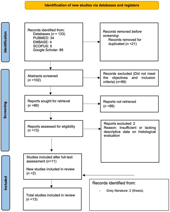
Four electronic databases—PubMed (34 records), Embase (4), Scopus (5), and Google Scholar (89)—were searched using a multistep strategy. Across all sources, 133 records were retrieved. After removing duplicates, 102 records remained for screening. Subsequently, 89 records were excluded for not meeting the prespecified inclusion criteria, leaving 13 records for full-text assessment. Of these, 2 were excluded due to insufficient or missing descriptive information in the histological assessment [26,27]. Two additional theses were identified through a hand search of the grey literature, resulting in 13 records included in the review [23,28,29,30,31,32,33,34,35,36,37,38,39]. The flowchart of the search process is displayed in Figure 1.
Figure 1.
Flowchart of the search strategy and steps of this systematic review.
3.2. Risk of Bias in the Included Studies
All included studies were assessed for risk of bias using the QUIN tool (Supplementary Tables S2 and S3). Using the prespecified thresholds, 12 of 13 studies (92.31%) were rated as having moderate risk of bias and 1 of 13 (7.69%) as low risk; none were rated as high risk. Overall reporting was adequate across most domains. All studies clearly stated their aims/objectives, described the sampling technique and methodological approach, and presented their results with sufficient clarity. Outcome measurement was adequately described in all studies except one [36]. Comparison groups were reported in most studies; however, two raised concerns regarding interpretability [34,35], and one was deemed not applicable [28].
In contrast, reporting of operator-related information was limited: six studies provided partial information [29,30,31,32,33,39], whereas seven did not report this domain [23,28,34,35,36,37,38]. Randomization and blinding were seldom described adequately: only one study reported each domain in sufficient detail [38], with the others providing insufficient or no information. Reporting outcome assessor characteristics was also incomplete: four studies provided partial information [30,31,35,38], and none reported this domain comprehensively. Statistical analysis was usually described, with one study omitting this item [34].
3.3. Data Extraction
Across the included studies, histological outcomes were reported for several commercial CSCs (Angelus MTA, Biodentine, iRoot BP Plus Putty, Neo MTA Plus, ProRoot MTA, TheraCal LC, TotalFill, White ProRoot MTA), with Biodentine being the most frequently characterized. In addition, multiple experimental calcium silicate-containing formulations were also tested (Table 1) [30,31,33,35,37]. Most studies used immature third molars extracted for orthodontic reasons from patients in the age range of 15–19; exceptions were Al-Saludi et al. [32], who used mature premolars with apical sectioning, and Khazane et al. (2022) [36], who included immature premolars in addition to third molars. The Experimental Protocol for Establishing and Maintaining Ex Vivo Whole-Tooth Cultures is presented as Supplementary Table S4.
Table 1.
Summary of included studies: design/features and histological, immunohistochemical, and mineralization findings.
Routine histology employed hematoxylin–eosin staining, whereas Kuo et al. (2021) [37] additionally used Masson’s trichrome. Immunohistochemistry was performed by Téclès et al. (2008) [23]; Laurent et al. (2012) [28]; and Pedano et al. (2021) [31] (markers included dentine sialophosphoprotein, nestin, type I collagen, and osteonectin).
Regarding follow-up, most studies set a maximum culture/observation period of 28 days [23,28,29,30,31,34,35]. Khazane et al. extended follow-up to 45 days [36] and Al-Saudi et al. to 90 days [32].
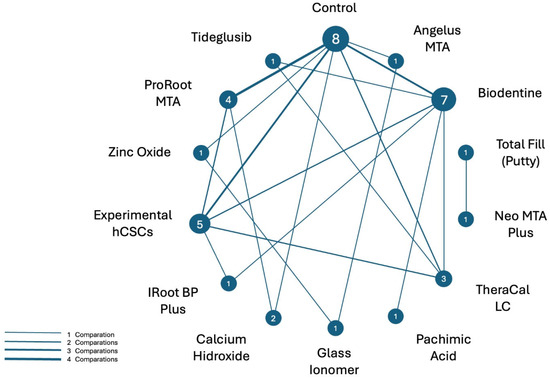
Figure 2 presents a network diagram of all direct biomaterial comparisons. Nodes represent the biomaterials included in the trials, and edges (labelled by the number of studies) indicate how frequently each comparison was evaluated. Biodentine is the most frequently studied biomaterial, followed by ProRoot MTA and various experimental hCSCs.
Figure 2.
Network diagram of all direct biomaterial comparisons assessed. Nodes represent the biomaterials, and edges (labelled by the number of studies) indicate how frequently each comparison was evaluated.
3.4. Histological and Immunohistochemical Observations
Across hydraulic calcium silicate-based cements (hCSCs), Biodentine showed the most prominent and reproducible early pro-mineralization pattern: several studies described a distinct interface zone (reported as a 50–100 μm amorphous eosinophilic/collagenous band) that delineated the exposure site and was consistently associated with multiple mineralized foci beneath it. Biodentine-associated mineralization could be detected very early (e.g., small amorphous mineralized foci from day 2, progressing to more numerous osteodentin-like foci with cavities/entrapped cells by days 14–28), and when assessed immunohistochemically, these foci frequently showed nestin positivity with DSPP/collagen I/osteonectin expression consistent with odontoblast-like differentiation within/around the mineralized structures. In longer culture conditions, Biodentine was also associated with more advanced repair with minimal inflammatory infiltration and a more organized odontoblast-like layer [30,34,35,36,37,39]. ProRoot MTA and experimental hCSCs similarly maintained a generally well-organized pulp and induced periexposure mineralized foci containing entrapped cells, with classic observations of a thin acellular necrotic interface zone and early osteodentin-like foci after MTA, and entrapped cells within foci in both MTA, TCS 50 and putty-type bioceramics (e.g., TotalFill, Neo MTA Plus) [23,32,33,37].
In contrast, the resin-modified calcium silicate-based cement (Rm-CSC) TheraCal LC showed a more variable and often delayed hard-tissue response: some models reported no pulpal reaction/no mineralized foci at earlier time points (14 days), whereas later evaluations reported small or less frequent mineralized foci [31,33,34]. For experimental Rm-CSCs, histology was strongly formulation-dependent and heterogeneous [31]. Other materials evaluated—pachymic acid, tideglusib, zinc oxide–eugenol, glass ionomer cement and the classical calcium hydroxide—generally exhibited less favourable histological performance than the reference hCSCs [34,36,38].
4. Discussion
This systematic review investigated ex vivo human tooth culture models, analyzing the histological, immunohistochemical, and biomineralization effects of CSCs used in direct pulp capping procedures. The findings, consistent with histological and immunohistochemical evidence derived from the intact human tooth culture model, confirm that hCSCs are biocompatible materials with notable bioactive potential.
Across all studies that characterized the histological response to commercially available, resin-free CSCs, researchers consistently reported early mineralization nodules and, in some cases, formation of a dentinal bridge over the follow-up period [23,28,29,30,32,33,34,35,36,37,38,39].
These observations were complemented by in vitro cell culture studies. Reis et al. (2021) demonstrated the viability of dental pulp stem cells (DPSCs) cultured in media conditioned with Biodentine extracts (1:1), showing no significant differences from controls at 24, 48 and 72 h [39]. Comparable results were reported by Sequeira et al. (2018), who examined the viability of stem cells from the apical papilla (SCAP) cultured in media conditioned with ProRoot MTA (1:1) [40]. In addition, Kuo et al. (2021) reproduced this model in pigs using an equivalent experimental protocol and obtained congruent results with the entire tooth model, showing mineralization foci in both experimental models [37].
The mineralization foci observed are associated with the deposition of an irregular, atubular mineralized matrix referred to as osteodentine, which serves as a key precursor in reparative dentinogenesis [41]. In this regard, Téclès et al. (2008), Laurent et al. (2012), and Pedano et al. (2021) complemented histological characterization with immunohistochemistry, reporting expression of type I collagen (COL-I), nestin (NES), dentine sialophosphoprotein (DSPP) and osteonectin (SPARC) [23,28,31]. The presence of proteins such as NES supports the notion that this represents an incipient form of reparative dentine, as NES is a marker of secretory human odontoblasts [42]. Likewise, DSPP expression is essential for dentine formation by regulating mineralization [43].
From a biological perspective, the localization of mineralization nodules near pulpal microvasculature may reflect the involvement of perivascular mesenchymal progenitor populations. Histological observations in the included studies frequently showed mineralized nodules located in proximity to pulpal microvascular structures. Perivascular niches are recognized sources of multipotent mesenchymal stem/stromal cells, and their presence has been demonstrated in vascular walls and microvessels, where they exhibit multilineage differentiation potential, including osteogenic commitment [44]. Moreover, mesenchymal stem cell populations have been shown to remain viable in inflamed periapical tissues, suggesting that these progenitors can actively participate in bone formation and repair processes under inflammatory conditions [45]. In parallel, several studies have reported that bioactive materials, including calcium silicate-based biomaterials, can promote mesenchymal stem cell differentiation toward mineralizing phenotypes, further supporting their role in hard tissue regeneration [46,47]. Taken together, these findings suggest that perivascular stem cell populations present within pulpal microvasculature may contribute to the pool of progenitor cells that migrate toward injury sites, differentiate into odontoblast-like cells, and participate in the formation of mineralized nodules resembling osteodentin.
Stimulation of reparative dentine formation by hCSCs correlates with their ability to induce the release of several cytokines and growth factors, notably BMPs, TGF-β1, VEGF, FGF-2 and PDGF [48]. Laurent et al. showed that Biodentine significantly increased TGF-β1 secretion by pulp cells [28]. This growth factor modulates progenitor-cell migration and differentiation, thereby contributing to reparative dentine formation. The cytokine stimulatory effect has been attributed to the release of calcium hydroxide Ca(OH)2 during material setting, which dissociates into OH− and Ca2+ ions. Ionic release has thus been described as a key driver of DPSC migration, proliferation and differentiation [49]. Consistently, extracellular Ca2+ has been shown to promote odontoblastic differentiation of DPSCs via the BMP2–Smad1/5/8 and Erk1/2 pathways [50].
In the present review, mineralization induced by ProRoot MTA was more homogeneous than that produced by the classical pulp-capping material Ca(OH)2 [23]. These data are comparable to the in vivo findings of Aeinehchi et al. [51]. Moreover, Ricucci et al. demonstrated that major causes of failure with Ca(OH)2-based pulp capping materials include tunnel defects within the dentinal bridge and progressive dissolution of the material over time [52]. In line with this, Peng et al. (2011) concluded that hCSCs promote greater DPSC proliferation and odontogenic differentiation than Ca(OH)2 [53]. A recent meta-analysis likewise showed the clinical superiority of hCSCs over traditional calcium hydroxide pastes, reporting higher success rates at all follow-up points (6, 12 and 36 months), with overall success ranging from 80% to 100% even after three years [54].
Biodentine was the most extensively characterized biomaterial within this model. Introduced in 2011 as an alternative to ProRoot MTA, it is an hCSC primarily composed of tricalcium silicate (C3S) and dicalcium silicate (C2S), with zirconium oxide (ZrO2) as radiopacifier [55]. In all studies evaluating Biodentine, mineralization nodules were observed; notably, Laurent et al. (2012) reported Biodentine particles embedded within these nodules, which further supports the material’s mineralization potential [28]. Clinical studies have corroborated these experimental findings, supporting the mineralization potential and biocompatibility of both Biodentine and ProRoot MTA [11].
Premixed hydraulic calcium silicate-based cements (hCSCs) represent a more recent alternative to conventional powder–liquid formulations, characterized by their ready-to-use presentation and simplified clinical handling. The development of fast-setting putty-type formulations has further expanded their applicability, as their cohesive consistency and improved resistance to wash-out make them suitable for a wide range of procedures, including pulpotomies and surgical endodontic interventions. Initially introduced in 2007 under different commercial names sharing similar compositions, these materials have evolved toward faster-setting premixed putty formulations with enhanced stability in moist environments [14]. Some of these materials, such as EndoSequence and TotalFill putties, achieve their composition through the incorporation of monobasic calcium phosphate into calcium silicate matrices, together with polymeric vehicles that modulate handling and setting behaviour [56].
Several studies have biologically characterized these premixed putty materials, reporting responses comparable to those of classical formulations. For instance, Lozano-Guillén et al. demonstrated similar cell viability between NeoPutty and MTA Angelus at ½ and ¼ dilutions using MTT assays, without statistically significant differences, and comparable results in cell migration tests [57]. Likewise, Cruz Hondares et al. observed no significant differences in biological behaviour between NeoPutty and EndoSequence when compared with conventional hCSCs such as ProRoot MTA and MTA Angelus [58]. These findings are consistent with those obtained using the intact tooth culture model, where Al-Saudi et al. (2019) reported complete mineralization at 90 days with TotalFill putty [32]. Clinical evidence further supports the performance of premixed hCSCs: a recent systematic review and meta-analysis demonstrated their effectiveness as pulpotomy materials in both primary and permanent teeth, with success rates comparable to MTA and reduced risk of coronal discoloration. Similar outcomes were reported by Ghaly et al. (2025), who found Well-Root PT to be effective in apexification procedures when compared with MTA [59].
From a physicochemical standpoint, Minju Song et al. showed that putty-type cements exhibit chemomechanical properties, biocompatibility, and mineralization potential comparable to ProRoot MTA, supporting the concept that their modified presentation does not compromise their bioactive performance [60].
Resin-modified CSCs are now available and offer improved clinical handling, enhanced sealing and dentine adhesion, compatibility with restorative materials, and better mechanical properties [31]. One such material is TheraCal LC. Sukajintanakarn et al. and X. Li et al. reported small, amorphous and fewer mineralization foci in histological sections [33,34]. Similarly, Pedano et al. observed a complete absence of early mineral formations [31]. Overall, these findings indicate that resin-modified CSCs demonstrate inferior histological performance, showing reduced or delayed mineralization when compared with reference materials such as Biodentine and ProRoot MTA. This may relate to the high resin content, which could polymerize incompletely and leach toxic monomers into the pulp, negatively affecting cell viability, proliferation and migration findings that are concordant with in vitro [61] and clinical studies [15]. In the work by Pedano et al., an experimental resin-containing hCSC (“Exp_HEAA”) provided favourable outcomes in terms of early mineralization and nodule formation [31]. Its organic phase incorporated a hydrophilic monomer (HEAA) that promoted hydration of calcium silicates, alongside other monomers that facilitated polymerization, thereby reducing cytotoxicity and enabling early mineralization with NES and COL-I expression. Two additional experimental hCSCs also showed promising histological outcomes: “TCS 50”, which used a pure C3S-based CSC with 50% ZrO2 added [33], and “RDSC”, a sol–gel synthesized CSC employing Bi2O3 as a radiopacifier [37].
Although several of the included studies reported numerical or semi-quantitative findings related to mineralized tissue formation, the marked heterogeneity in experimental design, outcome definitions, scoring systems, and evaluation time points precluded meaningful quantitative synthesis or direct comparisons between studies. Reported measures ranged from semi-quantitative histological scores to morphometric analyses and counts of mineralized foci, often obtained under different culture durations and methodological conditions. For example, Somudorn et al. reported higher histological scores for mineralized tissue formation in samples treated with calcium silicate-based materials (Biodentine) compared with other groups, whereas Kumar et al. used a semi-quantitative grading system for reparative responses, showing increased mineralized barrier formation in the Biodentine-treated group [35,36]. Additionally, Pedano et al. described the percentage of teeth exhibiting mineralized foci, while Duarte et al. presented semi-quantitative comparisons of hard tissue deposition associated with the evaluated materials and the degree of histological pulp involvement [31,38]. Despite these numerical observations, the lack of standardized outcome measures and differing evaluation criteria prevent reliable data aggregation or frequency-based comparisons. Nevertheless, these individual quantitative findings consistently support the trend toward early mineralized tissue deposition and odontoblast-like cell differentiation within this ex vivo model when calcium silicate-based materials are used.
The experimental model employed throughout was the intact human tooth culture. This represents an intermediate approach between in vitro cell cultures and in vivo animal models. Most in vitro cultures are two-dimensional, and cells lack their native structural arrangement and physiological interactions. Although animal experimentation remains the current benchmark in material development, interspecies differences may yield divergent tissue responses [17]. In this context, the intact human tooth culture is a valuable tool because cells remain in their physiological state under near natural conditions. It is therefore a cost-effective, rapid and reproducible methodology to investigate cellular mechanisms following pulp exposure. Histological assessment is also the most reliable method to evaluate pulp status, as it enables grading of inflammation and detection of necrosis [32].
However, the model has limitations. Although the ex vivo natural tooth culture model provides relevant information on the interaction between the dentin–pulp complex and calcium silicate-based biomaterials, as it preserves tissue cytoarchitecture and cell–extracellular matrix interactions [17], its inherent limitations must be acknowledged. First, this experimental system primarily enables the assessment of early pulp tissue responses, providing a histological snapshot of a specific culture time point, but without the possibility of dynamically monitoring the temporal evolution of the inflammatory–reparative process [33].
Moreover, because the model is isolated from systemic circulation, the lack of vascular perfusion and functional blood supply may affect tissue metabolism (e.g., hypoxia-related changes), thereby influencing the magnitude and complexity of inflammatory and reparative responses. Under these conditions, inflammatory reactions may be incomplete or limited, which should be considered when extrapolating findings to clinical scenarios. This limitation may also contribute to morphological differences in the resulting mineralized deposits, such as the absence of the typical tubular morphology described under more physiological conditions [28]. In addition, most ex vivo systems are established under “ideal” conditions of healthy pulp of immature teeth, without incorporating clinically relevant variables such as pre-existing pulp inflammation or bacterial invasion, which could substantially alter tissue responses to biomaterials; therefore, the outcomes observed in this model should be interpreted within this specific experimental context. Finally, this model is particularly suitable for investigating vital pulp therapy procedures, but it is not fully appropriate for regenerative endodontic procedures, as these require necrotic pulp conditions and interventions such as partial tissue removal, which may not meet the methodological criteria proposed for a true ex vivo model [17].
Considering these limitations, ex vivo models based on extracted teeth are especially useful for investigating dentin–pulp complex responses to biomaterials in vital pulp therapy procedures, as they enable tissue-level assessment within a preserved anatomical context. In contrast, in vitro models, including both 2D and 3D systems, are particularly valuable during early phases of biomaterials research due to their speed, low cost, and high experimental control, allowing the investigation of specific mechanisms (cytocompatibility, ion release, cell differentiation, and mineralization-related markers). Nonetheless, because they have limited capacity to replicate tissue complexity and clinically relevant pathophysiological conditions, they should be complemented by models that better preserve pulp architecture and microenvironmental cues. Finally, in vivo models represent the most physiologically relevant preclinical approach, as they incorporate vascular perfusion, immune responses, and reparative processes under complete biological conditions. However, they also involve substantial challenges, including higher cost, technical complexity, ethical considerations, and interspecies variability; therefore, they are generally reserved for advanced preclinical stages prior to translation into human studies [17].
Culture duration should also be considered: Pedano et al. showed that, even four weeks after extraction, third-molar pulps retained similar behaviours and were capable of expressing markers comparable to stem cells from freshly extracted teeth, supporting the validity of 28-day culture periods to study early mineralization phases [29]. However, studies by Kuo et al. and Al-Saudi et al. extended culture to 45 and 90 days, respectively [32,37], which may allow better characterization of CSCs’ influence on reparative dentinogenesis. Moreover, future studies could benefit from the incorporation of advanced analytical approaches beyond conventional histological and immunohistochemical assessment. Techniques such as iterative multiplex immunofluorescence and miRNA profiling may provide more comprehensive insight into the molecular and cellular responses of pulp tissue to emerging biomaterials. In addition, recent advances in transcriptomic and spatially resolved molecular analyses have been successfully applied to characterize the complex cellular landscape and spatial organization of dental tissues, particularly in developmental contexts, highlighting their potential for broader application in dental research. These unbiased and spatially contextualized approaches may offer deeper understanding of tissue heterogeneity, cell–cell interactions, and repair-associated signalling pathways [62].
In this context, the intact tooth ex vivo culture model may represent a particularly suitable platform for integrating such molecular strategies, as it preserves tissue architecture while allowing controlled experimental manipulation. Furthermore, tissue-clearing techniques combined with three-dimensional imaging have emerged as powerful tools in dental research, enabling volumetric visualization of soft and hard tissues, neurovascular networks, and mineralized structures without the limitations of conventional two-dimensional sectioning. These approaches may facilitate spatially resolved evaluation of pulp responses and hard tissue formation throughout the entire dentin–pulp complex, thereby expanding the analytical potential of ex vivo tooth culture systems [63].
Regarding the limitations of this review, most included articles presented a moderate risk of bias according to the applied assessment tool (QUIN Tool). This level of risk does not invalidate the reported findings but suggests that certain methodological aspects—such as sample size calculation, operator details, or outcome assessor details—may not have been consistently controlled across studies. Consequently, the results should be interpreted as indicative of general biological trends rather than as precise quantitative estimates of material performance.
The heterogeneity of materials and methodological variability hinders direct comparison across studies. Moreover, only non-carious molars and premolars were included, which limits generalizability and leaves open the question of whether the lack of pre-existing pulpal inflammation influenced the observed outcomes.
From a clinical perspective, the histological findings observed in this model help anticipate the early pulp tissue responses that may occur following vital pulp therapy procedures involving direct pulp exposure. The intact tooth ex vivo culture system reproduces a controlled pulpal injury and allows evaluation of the subsequent reparative processes triggered by biomaterial application, including mineralized tissue deposition and odontoblast-like cell differentiation. Within these experimental conditions, calcium silicate-based materials without resin components consistently demonstrated more organized pulp tissue architecture and more pronounced early mineralized tissue formation compared with resin-modified formulations. While clinical outcomes depend on multiple additional factors, these tissue-level observations support the use of conventional calcium silicate cements as reliable biomaterials for promoting favourable reparative responses in vital pulp therapy.
5. Conclusions
Hydraulic CSCs can stimulate reparative dentinogenesis, thereby helping to preserve the integrity of the dentine–pulp complex through localized remineralization at the injury site. Histological analysis of pulp sections across the included studies supports this conclusion. The intact human tooth model emerges as a reliable, economical and reproducible approach to examine the early stages of the pulp tissue reparative process. However, further studies using longer culture/observation periods and larger sample sizes are needed to strengthen the evidence base and to better establish this model as a screening platform for pulp-capping materials.
Supplementary Materials
The following supporting information can be downloaded at https://www.mdpi.com/article/10.3390/jfb17020078/s1, Table S1: Search strategy and eligibility criteria for the systematic search; Table S2: QUIN tool assessment of selected studies; Table S3: Overall evaluation of risk of bias of included studies using the QUIN tool; Table S4: Experimental Protocol for Establishing and Maintaining Ex Vivo Whole-Tooth Cultures; PRISMA_2020_checklist-JFB2026.
Author Contributions
Conceptualization, D.B.S. and J.M.M.d.S.; methodology, A.C.-F., H.G.d.S.C. and J.M.M.d.S.; software, A.C.-F., H.G.d.S.C. and J.M.-G.; validation, A.D.-C., J.J.S.-E. and J.P.; formal analysis, A.D.-C. and J.M.M.d.S.; investigation, A.C.-F., H.G.d.S.C., D.B.S. and J.M.M.d.S.; resources, A.C.-F. and J.P.; data curation, A.C.-F. and J.M.M.d.S.; writing—original draft preparation, A.C.-F., H.G.d.S.C. and J.M.M.d.S.; writing—review and editing, A.C.-F. and J.M.M.d.S.; visualization, A.D.-C., J.J.S.-E., J.M.-G. and J.P.; supervision, D.B.S. and J.M.M.d.S.; project administration, J.M.M.d.S.; funding acquisition, A.C.-F. and J.P. All authors have read and agreed to the published version of the manuscript.
Funding
This research received no external funding.
Institutional Review Board Statement
Not applicable.
Informed Consent Statement
Not applicable.
Data Availability Statement
The original contributions presented in the study are included in the article; further inquiries can be directed to the corresponding author.
Acknowledgments
Alberto Cabrera-Fernández is a research fellow supported by the University of Sevilla (Beca de Personal Investigador en Formación, PIF). Hebertt Gonzaga Chaves is a research fellow supported by the Coordenação de Aperfeiçoamento de Pessoal de Nível Superior—Brasil (88887.712700/2022-00 and n. 88887.994527/2024-00).
Conflicts of Interest
The authors declare no conflicts of interest.
Abbreviations
The following abbreviations are used in this manuscript:
| α-MEM | Alpha minimum essential medium |
| CSCs | Calcium silicate-based cements |
| DMEM | Dulbecco’s Modified Eagle Medium |
| DPSCs | Dental pulp stem cells |
| DSPP | Dentine sialophosphoprotein |
| FBS | Fetal bovine serum |
| hCSC | Hydraulic calcium silicate-based cements |
| Rm-CSC | Resin-modified calcium silicate-based cement |
| RDSC | Radiopaque dicalcium silicate cement |
| TCS 50 | Tricalcium silicate-based cement + 50 wt% ZrO2 |
| VPT | Vital pulp therapy |
References
- dos Santos Chaves, H.G.; Figueiredo, B.; Maia, C.A.; dos Reis-Prado, A.H.; Antunes, M.M.; de Mesquita, R.A.; Tavares, W.L.F.; Menezes, G.B.; Diniz, I.M.A.; Crovace, M.C.; et al. Tissue Response and Expression of Interleukins (IL)-1ß, IL-6, IL-10 after Pulp Capping with Bioglasses in Mice. Braz. Oral Res. 2024, 38, e096. [Google Scholar] [CrossRef] [Scilit] [PubMed]
- Duncan, H.F.; Tomson, P.L.; Simon, S.; Bjørndal, L. Endodontic Position Statements in Deep Caries Management Highlight Need for Clarification and Consensus for Patient Benefit. Int. Endod. J. 2021, 54, 2145–2149. [Google Scholar] [CrossRef] [Scilit] [PubMed]
- Santos, J.M.; Pereira, J.F.; Marques, A.; Sequeira, D.B.; Friedman, S. Vital Pulp Therapy in Permanent Mature Posterior Teeth with Symptomatic Irreversible Pulpitis: A Systematic Review of Treatment Outcomes. Medicina 2021, 57, 573. [Google Scholar] [CrossRef] [Scilit]
- Téclès, O.; Laurent, P.; Zygouritsas, S.; Burger, A.S.; Camps, J.; Dejou, J.; About, I. Activation of Human Dental Pulp Progenitor/Stem Cells in Response to Odontoblast Injury. Arch. Oral Biol. 2005, 50, 103–108. [Google Scholar] [CrossRef] [Scilit]
- Aguilar, P.; Linsuwanont, P. Vital Pulp Therapy in Vital Permanent Teeth with Cariously Exposed Pulp: A Systematic Review. J. Endod. 2011, 37, 581–587. [Google Scholar] [CrossRef] [Scilit]
- Graham, L.; Cooper, P.R.; Cassidy, N.; Nor, J.E.; Sloan, A.J.; Smith, A.J. The Effect of Calcium Hydroxide on Solubilisation of Bio-Active Dentine Matrix Components. Biomaterials 2006, 27, 2865–2873. [Google Scholar] [CrossRef] [Scilit]
- Sangwan, P.; Sangwan, A.; Duhan, J.; Rohilla, A. Tertiary Dentinogenesis with Calcium Hydroxide: A Review of Proposed Mechanisms. Int. Endod. J. 2013, 46, 3–19. [Google Scholar] [CrossRef] [Scilit]
- Lee, S.J.; Monsef, M.; Torabinejad, M. Sealing Ability of a Mineral Trioxide Aggregate for Repair of Lateral Root Perforations. J. Endod. 1993, 19, 541–544. [Google Scholar] [CrossRef] [Scilit]
- Primus, C.M.; Tay, F.R.; Niu, L. Bioactive Tri/Dicalcium Silicate Cements for Treatment of Pulpal and Periapical Tissues. Acta Biomater. 2019, 96, 35–54. [Google Scholar] [CrossRef] [Scilit]
- Pelepenko, L.E.; Marciano, M.A.; Shelton, R.M.; Camilleri, J. Leaching and Cytotoxicity of Bismuth Oxide in ProRoot MTA—A Laboratory Investigation. Int. Endod. J. 2024, 57, 1293–1314. [Google Scholar] [CrossRef] [Scilit]
- Nowicka, A.; Lipski, M.; Parafiniuk, M.; Sporniak-Tutak, K.; Lichota, D.; Kosierkiewicz, A.; Kaczmarek, W.; Buczkowska-Radlińska, J. Response of Human Dental Pulp Capped with Biodentine and Mineral Trioxide Aggregate. J. Endod. 2013, 39, 743–747. [Google Scholar] [CrossRef] [Scilit]
- Camilleri, J. Hydraulic Calcium Silicate-Based Endodontic Cements. In Endodontic Advances and Evidence-Based Clinical Guidelines; Wiley: Hoboken, NJ, USA, 2022. [Google Scholar]
- Parirokh, M.; Torabinejad, M. Mineral Trioxide Aggregate: A Comprehensive Literature Review—Part I: Chemical, Physical, and Antibacterial Properties. J. Endod. 2010, 36, 16–27. [Google Scholar] [CrossRef] [Scilit] [PubMed]
- Immich, F.; de Oliveira da Rosa, W.L.; Rossi-Fedele, G.; de Araújo, L.P.; Piva, E. Premixed Calcium Silicate-Based Materials for Pulpotomy Outcomes in Permanent and Deciduous Teeth: A Systematic Review and Meta-Analysis of Clinical Trials. Clin. Oral Investig. 2025, 29, 514. [Google Scholar] [CrossRef] [Scilit]
- Cabrera-Fernandez, A.; Dominguez-Dominguez, L.; Pérez-Pérez, A.; Santos, J.M.M.; Díaz-Cuenca, A.; Torres-Lagares, D.; Sequeira, D.B.; Segura-Egea, J.J.; Martín-González, J.; Cabrera-Fernandez, A.; et al. Clinical and Radiographic Outcomes of Vital Pulp Therapy Using Resin-Modified Versus Conventional Calcium Silicate-Based Materials: A Systematic Review and Meta-Analysis. J. Funct. Biomater. 2026, 17, 32. [Google Scholar] [CrossRef] [Scilit]
- Aslantürk, Ö.S.; Aslantürk, Ö.S. In Vitro Cytotoxicity and Cell Viability Assays: Principles, Advantages, and Disadvantages. In Genotoxicity—A Predictable Risk to Our Actual World; IntechOpen: London, UK, 2017. [Google Scholar] [CrossRef] [Scilit]
- Piglionico, S.S.; Pons, C.; Romieu, O.; Cuisinier, F.; Levallois, B.; Panayotov, I.V. In Vitro, Ex Vivo, and In Vivo Models for Dental Pulp Regeneration. J. Mater. Sci. Mater. Med. 2023, 34, 15. [Google Scholar] [CrossRef] [Scilit] [PubMed]
- Chatterjee, I.; Agrawal, P.; Sinha, Y.; Majee, N.; Bansal, S. In Vitro Response of Dental Pulp Stem Cells to Calcium Silicate-Based Cements: A Systematic Review and Meta-Analysis of Preclinical Evidence. Cureus 2025, 17, e88990. [Google Scholar] [CrossRef] [Scilit]
- Alzoubi, H.; Bshara, N.; AL-Manadili, A. Histopathological Evaluation of Pulp Response to Portland Cement Compared to MTA after Primary Canines Pulpotomy (In Vivo Study). BDJ Open 2022, 8, 29. [Google Scholar] [CrossRef] [Scilit]
- Krechina, E.K.; Volkov, A.V.; Abdurakhmanova, Z.U. Rationale for the Use of Bioactive Cements by In Vitro Simulation of Accidental Pulp Opening. Stomatologiia 2021, 100, 11–14. [Google Scholar] [CrossRef] [Scilit]
- Santos, J.M.; Marques, J.A.; Diogo, P.; Messias, A.; Sousa, V.; Sequeira, D.; Palma, P.J. Influence of Preoperative Pulp Inflammation in the Outcome of Full Pulpotomy Using a Dog Model. J. Endod. 2021, 47, 1417–1426. [Google Scholar] [CrossRef] [Scilit]
- Emara, R.; Elhennawy, K.; Schwendicke, F. Effects of Calcium Silicate Cements on Dental Pulp Cells: A Systematic Review. J. Dent. 2018, 77, 18–36. [Google Scholar] [CrossRef] [Scilit]
- Téclès, O.; Laurent, P.; Aubut, V.; About, I. Human Tooth Culture: A Study Model for Reparative Dentinogenesis and Direct Pulp Capping Materials Biocompatibility. J. Biomed. Mater. Res. B Appl. Biomater. 2008, 85B, 180–187. [Google Scholar] [CrossRef] [Scilit]
- Nagendrababu, V.; Duncan, H.F.; Tsesis, I.; Sathorn, C.; Pulikkotil, S.J.; Dharmarajan, L.; Dummer, P.M.H. PRISMA for Abstracts: Best Practice for Reporting Abstracts of Systematic Reviews in Endodontology. Int. Endod. J. 2019, 52, 1096–1107. [Google Scholar] [CrossRef] [Scilit]
- Sheth, V.H.; Shah, N.P.; Jain, R.; Bhanushali, N.; Bhatnagar, V. Development and Validation of a Risk-of-Bias Tool for Assessing in Vitro Studies Conducted in Dentistry: The QUIN. J. Prosthet. Dent. 2024, 131, 1038–1042. [Google Scholar] [CrossRef] [Scilit] [PubMed]
- Aidaros, N.; Niazy, M.; El-yassaky, M. The Effect of Incorporating Nanocalcium Phosphate Particles into Biodentine on Pulpal Tissue Response (In Vitro Study). Al-Azhar Dent. J. Girls 2018, 5, 181–186. [Google Scholar] [CrossRef] [Scilit]
- Chakka, L.R.; Vislisel, J.; Vidal, C.D.; Biz, M.T.; KSalem, A.; Cavalcanti, B.N. Application of BMP-2/FGF-2 Gene-Activated Scaffolds for Dental Pulp Capping. Clin. Oral Investig. 2020, 24, 4427–4437. [Google Scholar] [CrossRef] [Scilit] [PubMed]
- Laurent, P.; Camps, J.; About, I. BiodentineTM Induces TGF-Β1 Release from Human Pulp Cells and Early Dental Pulp Mineralization. Int. Endod. J. 2012, 45, 439–448. [Google Scholar] [CrossRef] [Scilit]
- Pedano, M.S.; Li, X.; Jeanneau, C.; Ghosh, M.; Yoshihara, K.; Van Landuyt, K.; About, I.; Van Meerbeek, B. Survival of Human Dental Pulp Cells After 4-Week Culture in Human Tooth Model. J. Dent. 2019, 86, 33–40. [Google Scholar] [CrossRef] [Scilit]
- Pedano, M.S.; Li, X.; Camargo, B.; Hauben, E.; De Vleeschauwer, S.; Yoshihara, K.; Van Landuyt, K.; Yoshida, Y.; Van Meerbeek, B. Injectable Phosphopullulan-Functionalized Calcium-Silicate Cement for Pulp-Tissue Engineering: An In-Vivo and Ex-Vivo Study. Dent. Mater. 2020, 36, 512–526. [Google Scholar] [CrossRef] [Scilit]
- Pedano, M.S.; Yoshihara, K.; Li, X.; Camargo, B.; Van Landuyt, K.; Van Meerbeek, B. Experimental Resin-Modified Calcium-Silicate Cement Containing N-(2-Hydroxyethyl) Acrylamide Monomer for Pulp Tissue Engineering. Mater. Sci. Eng. C 2021, 126, 112105. [Google Scholar] [CrossRef] [Scilit]
- Al-Saudi, K.W.; Nabih, S.M.; Farghaly, A.M.; AboHager, E.A.A. Pulpal Repair after Direct Pulp Capping with New Bioceramic Materials: A Comparative Histological Study. Saudi Dent. J. 2019, 31, 469. [Google Scholar] [CrossRef] [Scilit]
- Li, X.; Pedano, M.S.; Li, S.; Sun, Z.; Jeanneau, C.; About, I.; Hauben, E.; Chen, Z.; Van Landuyt, K.; Van Meerbeek, B. Preclinical Effectiveness of an Experimental Tricalcium Silicate Cement on Pulpal Repair. Mater. Sci. Eng. C Mater. Biol. Appl. 2020, 116, 111167. [Google Scholar] [CrossRef] [Scilit]
- Sukajintanakarn, C.; Sritanaudomchai, H.; Laptanasupkun, P.; Pongprueksa, P. Pulpal Reaction to Pulp Capping Material In Human Tooth Culture Model. In Proceedings of RSU International Research Conference (2020); Rangsit University: Pathum Thani, Thailand, 2020; p. 309. [Google Scholar]
- Somudorn, J.; SritanaudomchaI, H.; Lapthanasupkul, P.; Naruphontjirakul, P.; Pongprueksa, P. Influences of Calcium Silicate Cement Modified in Glass Ionomer Cement on Human Pulp Tissue Using Tooth Culture Model. Med. Dent. J. 2023, 43, S125–S132. [Google Scholar]
- Khazane, M.K.; Mahalaxmi, S.; Vidhya, S. Histologic Evaluation of Dentin Bridge Formation by Pachymic Acid and Biodentine in Human Tooth Culture Model. Endodontology 2022, 34, 32–37. [Google Scholar] [CrossRef] [Scilit]
- Kuo, W.T.; Li, X.; Chen, F.Y.; Wang, Y.J.; Chen, Z.; Ding, S.J.; Zhang, L. Experimental Pulp-Capping Agent Radiopaque Dicalcium Silicate Cement Facilitates Dentinogenesis. Front. Mater. 2021, 8, 738430. [Google Scholar] [CrossRef] [Scilit]
- Duarte Bittar, J. Avaliação da Resposta Pulpar a Diferentes Materiais Odontológicos Após Capeamento Direto. Estudo Ex Vivo; Universidade de Brasília: Brasília, Brazil, 2010. [Google Scholar]
- Marques dos Reis, E.M.; Marques dos Santos, J.M.; Bela Sequeira, D. Biologic Evaluation of a New Hydraulic Calcium Silicate-Based Cement for Pulpotomy in Permanent Teeth; Universidade de Coimbra: Coimbra, Portugal, 2022. [Google Scholar]
- Sequeira, D.B.; Seabra, C.M.; Palma, P.J.; Cardoso, A.L.; Peça, J.; Santos, J.M. Effects of a New Bioceramic Material on Human Apical Papilla Cells. J. Funct. Biomater. 2018, 9, 74. [Google Scholar] [CrossRef] [Scilit]
- Braut, A.; Kollar, E.J.; Mina, M. Analysis of the Odontogenic and Osteogenic Potentials of Dental Pulp in Vivo Using a Col1a1-2.3-GFP Transgene. Int. J. Dev. Biol. 2003, 47, 281–292. [Google Scholar] [CrossRef] [Scilit]
- About, I.; Laurent-Maquin, D.; Lendahl, U.; Mitsiadis, T.A. Nestin Expression in Embryonic and Adult Human Teeth Under Normal and Pathological Conditions. Am. J. Pathol. 2000, 157, 287–295. [Google Scholar] [CrossRef] [Scilit]
- Suzuki, S.; Sreenath, T.; Haruyama, N.; Honeycutt, C.; Terse, A.; Cho, A.; Kohler, T.; Müller, R.; Goldberg, M.; Kulkarni, A.B. Dentin Sialoprotein and Dentin Phosphoprotein Have Distinct Roles in Dentin Mineralization. Matrix Biol. 2009, 28, 221–229. [Google Scholar] [CrossRef] [Scilit]
- Abedin, M.; Tintut, Y.; Demer, L.L. Mesenchymal Stem Cells and the Artery Wall. Circ. Res. 2004, 95, 671–676. [Google Scholar] [CrossRef] [Scilit]
- Estrela, C.; Souza, P.O.; Barbosa, M.G.; de Carvalhosa, A.A.; Batista, A.C.; Júnior, D.D.; Yamamoto-Silva, F.P.; de Freitas Silva, B.S. Mesenchymal Stem Cell Marker Expression in Periapical Abscess. J. Endod. 2019, 45, 716–723. [Google Scholar] [CrossRef] [Scilit]
- Rathinam, E.; Rajasekharan, S.; Chitturi, R.T.; Declercq, H.; Martens, L.; De Coster, P. Gene Expression Profiling and Molecular Signaling of Various Cells in Response to Tricalcium Silicate Cements: A Systematic Review. J. Endod. 2016, 42, 1713–1725. [Google Scholar] [CrossRef] [Scilit] [PubMed]
- Bernardini, C.; Zamparini, F.; Prati, C.; Salaroli, R.; Spinelli, A.; Zannoni, A.; Forni, M.; Gandolfi, M.G. Osteoinductive and Regenerative Potential of Premixed Calcium-Silicate Bioceramic Sealers on Vascular Wall Mesenchymal Stem Cells. Int. Endod. J. 2024, 57, 1264–1278. [Google Scholar] [CrossRef] [Scilit] [PubMed]
- Morotomi, T.; Washio, A.; Kitamura, C. Current and Future Options for Dental Pulp Therapy. Jpn. Dent. Sci. Rev. 2019, 55, 5–11. [Google Scholar] [CrossRef] [Scilit] [PubMed]
- Adams, G.B.; Chabner, K.T.; Alley, I.R.; Olson, D.P.; Szczepiorkowski, Z.M.; Poznansky, M.C.; Kos, C.H.; Pollak, M.R.; Brown, E.M.; Scadden, D.T. Stem Cell Engraftment at the Endosteal Niche Is Specified by the Calcium-Sensing Receptor. Nature 2006, 439, 599–603. [Google Scholar] [CrossRef] [Scilit]
- Li, S.; Hu, J.; Zhang, G.; Qi, W.; Zhang, P.; Li, P.; Zeng, Y.; Zhao, W.; Tan, Y. Extracellular Ca2+ Promotes Odontoblastic Differentiation of Dental Pulp Stem Cells via BMP2-Mediated Smad1/5/8 and Erk1/2 Pathways. J. Cell Physiol. 2015, 230, 2164–2173. [Google Scholar] [CrossRef] [Scilit]
- Aeinehchi, M.; Eslami, B.; Ghanbariha, M.; Saffar, A.S. Mineral Trioxide Aggregate (MTA) and Calcium Hydroxide as Pulp-Capping Agents in Human Teeth: A Preliminary Report. Int. Endod. J. 2003, 36, 225–235. [Google Scholar] [CrossRef] [Scilit]
- Ricucci, D.; Loghin, S.; Lin, L.M.; Spångberg, L.S.W.; Tay, F.R. Is Hard Tissue Formation in the Dental Pulp after the Death of the Primary Odontoblasts a Regenerative or a Reparative Process? J. Dent. 2014, 42, 1156–1170. [Google Scholar] [CrossRef] [Scilit]
- Peng, W.; Liu, W.; Zhai, W.; Jiang, L.; Li, L.; Chang, J.; Zhu, Y. Effect of Tricalcium Silicate on the Proliferation and Odontogenic Differentiation of Human Dental Pulp Cells. J. Endod. 2011, 37, 1240–1246. [Google Scholar] [CrossRef] [Scilit]
- Pinto, K.P.; da Silva, G.R.; Ferreira, C.M.A.; Sassone, L.M.; da Silva, E.J.N.L. Success Rate of Direct Pulp Capping on Permanent Teeth Using Bioactive Materials: A Systematic Review and Meta-Analysis of Randomized Clinical Trials. Restor. Dent. Endod. 2024, 49, e34. [Google Scholar] [CrossRef] [Scilit]
- Kusum, B.; Rakesh, K.; Richa, K. Clinical and Radiographical Evaluation of Mineral Trioxide Aggregate, Biodentine and Propolis as Pulpotomy Medicaments in Primary Teeth. Restor. Dent. Endod. 2015, 40, 276. [Google Scholar] [CrossRef] [Scilit]
- Chavarria-Bolanos, D.; Komabayashi, T.; Vega-Baudrit, J.; Ulate, E.; Pereira-Reyes, R.; Montero-Aguilar, M. Examining the Physicochemical Composition of Three Bioceramic Putties for Endodontic Use. Eur. Endod. J. 2024, 9, 210. [Google Scholar] [CrossRef] [Scilit] [PubMed]
- Lozano-Guillén, A.; López-García, S.; Rodríguez-Lozano, F.J.; Sanz, J.L.; Lozano, A.; Llena, C.; Forner, L. Comparative Cytocompatibility of the New Calcium Silicate-Based Cement NeoPutty versus NeoMTA Plus and MTA on Human Dental Pulp Cells: An In Vitro Study. Clin. Oral Investig. 2022, 26, 7219–7228. [Google Scholar] [CrossRef] [Scilit] [PubMed]
- Cruz Hondares, T.; Hao, X.; Zhao, Y.; Lin, Y.; Napierala, D.; Jackson, J.G.; Zhang, P. Antibacterial, Biocompatible, and Mineralization-Inducing Properties of Calcium Silicate-Based Cements. Int. J. Paediatr. Dent. 2024, 34, 843–852. [Google Scholar] [CrossRef] [Scilit] [PubMed]
- Ghaly, M.S.; Abozena, N.I.; Ghouraba, R.F.; Kabbash, I.A.; EL-Desouky, S.S. Clinical and Radiographic Evaluation of Premixed Bioceramic Putty as an Apical Plug in Nonvital Immature Anterior Permanent Teeth. Sci. Rep. 2025, 15, 26487. [Google Scholar] [CrossRef] [Scilit]
- Kim, M.; Lee, S.-H.; Shin, D.-H. In Vitro Study of the Biological and Physical Properties of Dual-Cure Resin-Modified Calcium Silicate-Based Cement. Dent. J. 2023, 11, 120. [Google Scholar] [CrossRef] [Scilit]
- Jeanneau, C.; Laurent, P.; Rombouts, C.; Giraud, T.; About, I. Light-Cured Tricalcium Silicate Toxicity to the Dental Pulp. J. Endod. 2017, 43, 2074–2080. [Google Scholar] [CrossRef] [Scilit]
- Shi, Y.; Yu, Y.; Li, J.; Sun, S.; Han, L.; Wang, S.; Guo, K.; Yang, J.; Qiu, J.; Wei, W. Spatiotemporal Cell Landscape of Human Embryonic Tooth Development. Cell Prolif. 2024, 57, e13653. [Google Scholar] [CrossRef] [Scilit]
- Meng, L.; Song, X.; Wang, J.; Shi, W.; Gao, L.; Jiang, X.; Zhang, W. Tissue Clearing and Its Application in Dental Research. BMEMat 2025, 3, e12113. [Google Scholar] [CrossRef] [Scilit]

Disclaimer/Publisher’s Note: The statements, opinions and data contained in all publications are solely those of the individual author(s) and contributor(s) and not of MDPI and/or the editor(s). MDPI and/or the editor(s) disclaim responsibility for any injury to people or property resulting from any ideas, methods, instructions or products referred to in the content. |
© 2026 by the authors. Licensee MDPI, Basel, Switzerland. This article is an open access article distributed under the terms and conditions of the Creative Commons Attribution (CC BY) license.