Article

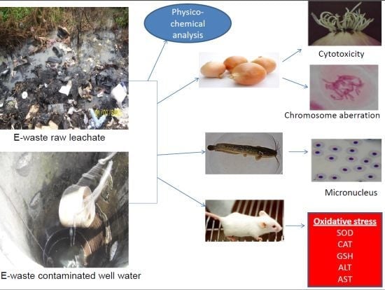
In Vivo Cytogenotoxicity and Oxidative Stress Induced by Electronic Waste Leachate and Contaminated Well Water

1
Cell Biology and Genetics Unit, Department of Zoology, University of Ibadan, Ibadan, Nigeria
2
Department of Biosciences and Biotechnology, Babcock University, Ilisan Remo, Ogun State, Nigeria
3
Ecology and Environmental Biology Unit, Department of Zoology, University of Ibadan, Ibadan, Nigeria
*
Author to whom correspondence should be addressed.
Challenges 2013, 4(2), 169-187; https://doi.org/10.3390/challe4020169
Submission received: 30 May 2013 / Revised: 14 July 2013 / Accepted: 16 July 2013 / Published: 23 July 2013
(This article belongs to the Special Issue Electronic Waste — Impact, Policy and Green Design)

Abstract

:
Environmental, plant and animal exposure to hazardous substances from electronic wastes (e-wastes) in Nigeria is increasing. In this study, the potential cytogenotoxicity of e-wastes leachate and contaminated well water samples obtained from Alaba International Electronic Market in Lagos, Nigeria, using induction of chromosome and root growth anomalies in Allium cepa, and micronucleus (MN) in peripheral erythrocytes of Clarias gariepinus, was evaluated. The possible cause of DNA damage via the assessments of liver malondialdehyde (MDA), catalase (CAT), reduced glutathione (GSH) and superoxide dismutase (SOD) as indicators of oxidative stress in mice was also investigated. There was significant (p < 0.05) inhibition of root growth and mitosis in A. cepa. Cytological aberrations such as spindle disturbance, C-mitosis and binucleated cells, and morphological alterations like tumor and twisting roots were also induced. There was concentration-dependent, significant (p < 0.05) induction of micronucleated erythrocytes and nuclear abnormalities such as blebbed nuclei and binucleated erythrocytes in C. gariepinus. A significant increase (p < 0.001) in CAT, GSH and MDA with concomitant decrease in SOD concentrations were observed in the treated mice. Pb, As, Cu, Cr, and Cd analyzed in the tested samples contributed significantly to these observations. This shows that the well water samples and leachate contained substances capable of inducing somatic mutation and oxidative stress in living cells; and this is of health importance in countries with risk of e-wastes exposure.

Graphical Abstract

1. Introduction

There has been rapid development in the Information and Communication Technology (ICT) sector in the 21st century. ICT and computer networking has penetrated nearly every aspect of modern life and is positively affecting human life, even in the most remote areas of developing countries [1]. This had been made possible by the production of varieties of Electrical and Electronic Equipment (EEE). The tremendous growth in global EEE production and consumption is attributable to frequent changes in equipment features and capabilities, product obsolescence, decreasing lifespan and prices, increasing population demand, urbanization and industrialization [2]. Despite the numerous benefits of the increasing EEE in the modern society, there is a concurrent increase in the streams of electronic waste (e-waste) generated from it after its end-of-life. At present, the annual global e-waste generation is estimated at 20–50 million metric tonnes, representing 1–3% of the world’s municipal waste [3,4]. E-waste has therefore become a global issue of public health concern, as it consists of hazardous substances [5]. This is of paramount importance especially in developing countries where infrastructure for hazardous waste management is weak and ineffective.
Nigeria has become a prime destination of e-waste dumping from developed nations [6]. Due to lack of official recycling activity and effective management policies, e-waste materials are indiscriminately dumped in homes, offices, warehouses, and informal dumpsites close to residential areas [7]. E-wastes are improperly dismantled and crudely recycled for precious metals and alloys such as steel, aluminium, copper and printed circuit boards. Open incineration of cables and electronic components is also a common practice to recover copper and other precious metals without any proper and safe working conditions [8,9]. As a result of these activities, toxic chemicals such as lead, mercury, arsenic, cadmium, selenium, chromium, barium, nickel, cobalt, silver etc., persistent organic pollutants (POPs e.g., dioxins and furans), polybrominated diphenyl ethers (PBDEs), polychlorinated bisphenyls (PCBs), polyvinyl chlorides (PVCs) and polycyclic aromatic hydrocarbons (PAHs) are released into the surrounding air, soil, plants and surface waters. Leaching of e-wastes from informal dumpsites can contaminate groundwater sources thereby exposing humans and animals to serious health hazards [7].
Previously, we reported [10] environmental contamination of soils and plants from the dumpsites of Alaba International Market, a major electronic market in Lagos, Nigeria. The soils and plants were shown to be contaminated with lead, cadmium, chromium, zinc, copper, arsenic, PAHs, PBDEs and PCBs. We have also reported the genotoxic and mutagenic effects of the e-waste leachate in mice and human peripheral blood lymphocytes [7,10]. The mechanism of DNA damage is, however, not clear. There is paucity of information on the genotoxicity of e-waste contaminated waters. Due to the proximity of the electronic market informal dumpsites to water bodies, toxic heavy metals and organic contaminants may be concentrated in surface and groundwater supplies around these e-waste dumpsites through lateral and vertical transfer of contaminants. Such contaminants may therefore bioaccumulate in aquatic organisms, become biomagnified in fishes which are at the top of the aquatic food chain and can ultimately affect humans who feed on such fishes. Hence, there is need for evaluation of the potential genotoxic effect of e-waste using aquatic organisms.
In this study, we investigated the genotoxic and cytotoxic potentials of e-waste leachate and well waters from a major electronic market in Lagos, Nigeria using piscine micronucleus and Allium cepa assays. In addition, we assessed oxidative damage in mice as a possible mechanism for DNA damage.

2. Materials and Methods
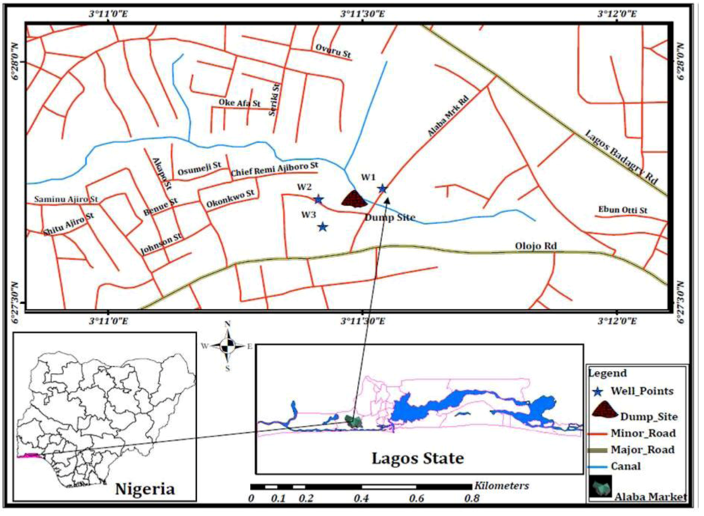
2.1. Sampling Site

The study site, Alaba International Market, Ojo, is located in the Southwestern part of Lagos State (Latitude 6°23'N and Longitude 2°42'E), Nigeria (Figure 1). The market, the largest in Africa where sales of fairly used and new electric and electronic goods are transacted, is surrounded by residential quarters. Within the market, there are many illegal dumpsites where obsolete electronics are usually dumped, dismantled for crude recycling and the remaining scraps burnt to reduce waste volume [7,8]. Well waters, used for drinking, ablution, cooking and other domestic and commercial purposes by workers and residents in the neighbourhood, are located within a 200 m circumference of the e-waste open dumpsites.
Figure 1. E-waste dumpsite and well locations (W1, W2 and W3) at Alaba International Electronics Market, Ojo, Lagos, Nigeria.
Challenges 04 00169 g001

2.2. Sample Collection

Water was collected from three different wells in the month of April, 2012 into 3 × 25 L preclean plastic containers and was labeled Alaba Well Water 1 (AWW1), Alaba Well Water 2 (AWW2) and Alaba Well Water 3 (AWW3). These wells were with apparent distance of 105.23 m, 133.36 m and 156.05 m, respectively, away from the open e-waste dumpsite as measured using Global Positioning System (GPS) coordinates (etrex LEGEND, GARMIN). Another well water sample was collected from Itire, Lagos, another community 10.91 km away from the dumpsite and without any known history of waste dumping, which served as control. Raw leachate (Alaba raw leachate, designated ARL) was also collected from different hollows on the dumpsite (holes in the ground where leachate seeps into) into clean 25 L plastic containers. These samples and control were transported to the laboratory, filtered using 15 cm filter paper (Whatman®, England) to remove debris, pH measured and stored at 4 °C throughout the period of study.

2.3. Physico-Chemical and Heavy Metal Analyses

Chemical oxygen demand (COD), alkalinity, biochemical oxygen demand (BOD), total dissolved solids (TDS), chlorides, nitrates, ammonia, and phosphates were analyzed in the leachate and well water samples in accordance with APHA [11] method. Heavy metals: Pb, Cd, Cu, Cr, Fe, Zn, Ni, Ag, and Mn were also analyzed in the samples in accordance with APHA [11] and USEPA [12] methods and the metal concentrations measured using Inductively Coupled Plasma-Atomic Emission Spectrometry (ICP-AES, Perkin Elmer Optima 3300DV, Boston, MA, USA).

2.4. Biological Materials Used for the Study

The biological materials employed are onion (Allium cepa; 2n = 16), African cat fish (Clarias gariepinus, Burchell, 1822) and albino mice (Mus musculus). Equal-sized onion bulbs were obtained commercially from Shasha market in Ibadan, Nigeria. About four times the total number of onion bulbs needed for the experiment was acquired and sun dried for 2 weeks before the commencement of the experiment. This served to replace any bulb that may dry up, rot or damaged by mould [13]. These were then used to evaluate the cytogenotoxic potentials of the well water and leachate samples using root growth inhibition and induction of chromosomal aberration as the assay end points.
Juvenile C. gariepinus (average weight of 26.27 ± 6.52 g and length 14.80 ± 1.33 cm) commercially obtained from Oyo State ministry of Agriculture and Natural Resources, Ibadan, Oyo State, Nigeria were acclimatized for a minimum of two (2) weeks in the laboratory prior to the commencement of the experiment. The fishes were maintained at 12 h photoperiod of day and night before and during the experiment and they were fed with commercial feed pellets ad libitum.
Sixty male albino mice (6–7 weeks old) obtained from Nigeria Institute of Medical Research (NIMR), Lagos, Nigeria, were used for the biochemical analysis. The mice were acclimatized for a minimum of 2 weeks in an apparently pathogen free, well-ventilated animal house of the Department of Biosciences and Biotechnology, Babcock University, Ilisan Remo, Ogun State, Nigeria. They were fed with food (Ladokun pelleted feed®) and drinking water ad libitum. All animal experiments in this study were conducted in accordance with standard guidelines [14].

2.5. Allium Cepa Assay

Twelve onion bulbs were used per concentration: 6.25, 12.5, 25, 50 and 100% (v/v; leachate/tap water) of ARL, and 100% concentration of the three well water samples. The outer dried, brown scales of the bulbs and the bottom plates (dried roots) were carefully removed leaving ring of the primordial roots intact. These were then placed in dechlorinated tap water to clean and prevent the primordial roots from drying up. Bulbs were later planted directly in the different concentrations of ARL and well water samples in 100 mL beakers at 27 ± 2 °C in a dark cupboard. Bulbs grown in well water sample from Itire served as negative control while those grown in 10 ppm lead nitrate solution served as the positive control. The test samples were changed daily to ensure continuous exposure of the onions. At 48 h, two onion bulbs with good growth were harvested; 0.5–1 cm from each root tip of each bulb was cut and fixed in ethanol:glacial acetic acid (3:1 v/v) for 24 h before the analysis of chromosome aberration. The obtained roots were hydrolyzed with 1N HCl at 60 °C for 5 min and subsequently washed in distilled water (3–4 times). Two root tips were squashed on each slide and stained with acetocarmine for 10 min. Excess stain was removed with filter paper and a cover slip carefully lowered onto each slide to exclude air bubbles. The cover slip was sealed on the slide with finger nail polish [15]. Six slides were prepared for each concentration out of which four were scored at ×1000. Cells (4000) were scored per concentration of the samples. The occurrence and frequency of aberrant cells were examined in all the stages of cell division and percentage aberrations were determined relative to the total number of dividing cells and total cell scored. The mitotic index (MI) was determined by counting the number of dividing cells per concentration including the controls, relative to the total number of cells scored.
At 72 h, the root lengths of each of the onion bulbs treated with the concentrations of ARL were harvested, measured and average root length per bulb per concentration was recorded. From the values obtained, the percentile root growth restriction in relation to the negative control and the EC50 and EC70 for the ARL was obtained. The effect of the samples on the morphology of the roots was also examined.

2.6. Micronucleus and Nuclear Abnormality Assay

Twenty fishes were randomly selected into a well aerated, rectangular and transparent 50 L plastic aquarium containing tap water at 27 ± 1.7 °C (control). Similarly, 20 fishes each were randomly selected and exposed to 50 and 100% concentrations (v/v; well water/ tap water) of AWW1 (chosen because of higher concentration of the analyzed parameters) and 12.5, 25 and 50% concentrations (v/v; leachate/tap water) of the leachate sample, for a period of 28 days in a semi-static bioassay conditions (with samples renewed twice weekly). During the time of exposure, 5 fishes were randomly selected at day 7, 14 and 28; and peripheral blood collected from their caudal vein using sterile syringes and needles, for the micronucleus (MN) assay. A thin smear of blood was made onto clean, grease free slides and air-dried overnight at room temperature before fixing in absolute methanol for 20 min and subsequently stained in May-Grunwald and 5% Giemsa respectively. Erythrocytes (2000) were scored per slide per fish at ×1000 for micronucleus (MN) and nuclear abnormalities. The nuclear abnormalities were scored along with MN as biomarkers of cytogenotoxicity in accordance with Carrasco et al. [16] and Cavas and Ergene-Gozukara [17,18].

2.7. Biochemical Assays in Mice

The mice were randomly divided into 12 groups of 5 animals per group. Group 1 received intraperitoneal (IP) injection of distilled water (0.5 mL/mouse) for five consecutive days (control A). Group 2 was given well water collected from Itire, Surulere, Lagos throughout the period of the experiment (5 weeks, equivalent of the longest exposure for groups 8–12) as their normal drinking water (control B). Groups 3, 4, 5, 6 and 7 received for five consecutive days 0.5mL IP injection of 1, 5, 10, 25 and 50% concentrations of the leachate sample, respectively; while groups 8, 9, 10, 11 and 12 were allowed to drink the well water (AWW1) without dilution (100%) for 1, 2, 3, 4 and 5 week(s), respectively. The routes of exposure (IP and drinking) were utilized purposively. In previous studies, we have shown that the tested leachate is genotoxic in both somatic and germ cells through the IP route [7], while the well water was genotoxic in mice exposed through drinking (article under review). In order to understand the mechanism of genotoxicity thus reported, we used the same route of exposure to study oxidative stress as possible mechanism of the induced genotoxicity. The IP route for leachate administration is one of the fastest routes of delivery of test sample into experimental animals. We simulated natural condition of drinking for the other groups of mice because the well water was mainly used for drinking, cooking and other domestic uses by humans residing and/or working in the electronic market on which the study site is located.
At 24 h post exposure with overnight fasting, blood was collected by cardiac puncture into lithium coated serum separator tubes under a light anesthesia and mice were sacrificed by cervical dislocation. Liver tissues were surgically removed, placed on ice bath to remove excess blood and weighed before used for biochemical analysis. The liver tissues were then homogenized in ice cold isotonic phosphate buffer; pH 7.4 and centrifuged at 10,000 g for 15 min at 4 °C using cold centrifuge. The resultant supernatant was stored at −70 °C prior to subsequent biochemical analysis [19]. The collected blood sample was allowed to coagulate, centrifuged at 3000 g for 10 min to obtain serum (supernatant) and stored at −70 °C before biochemical analysis. CAT activity was determined according to Sinha [20]. SOD was assayed using the method described by Misra and Fridovich [21]. Protein content was determined by Biuret method [22]. Reduced glutathione (GSH) was determined using the method of Habig et al. [23]. Lipid peroxidation was measured as malondialdehyde (MDA) in accordance with Shokunbi and Odetola [24] and expressed as micromoles of MDA/g tissue. Serum AST and ALT activities were determined according to Reitman and Frankel [25] using Randox kits (Randox Laboratories diagnostic Ltd, UK).

2.8. Statistical Analysis

SPSS 16.0® statistical package was used for data analysis. Frequencies of induced MN and nuclear abnormalities were expressed per 1000 erythrocytes. Analysis of the differences in mean ± SE values for all data were determined using one way ANOVA. Duncan Multiple Range Tests comparison at p < 0.05 and p < 0.001 level of significance was used to compare the treated groups and corresponding controls, when the differences among the means were significant pairwise. Spearman’s correlation coefficient (r) was used to evaluate concentration-response relationships in the experimental groups.

3. Results

3.1. Physico-Chemical and Heavy Metal Analyses

Table 1 presents the physico-chemical parameters and heavy metals analyzed in the leachate, well water and the control tap water. The pH of the samples was within the standard limits [26,27]. Alkalinity inAWW1 and AWW2, chloride and ammonia in AWW1, AWW2 and AWW3, Fe inAWW1, AWW2 and AWW3; and Mn in AWW1 and AWW2 were higher than allowable limit for drinkable water quality. The concentrations of heavy metals in ARL were higher than tap water and permissible limits in drinking water [26,27], with Pb having the highest concentration and As the least.
Table 1. Physico-chemical and heavy metals characteristics of tap and well water samples, and e-waste leachate from Alaba International market, Lagos, Nigeria.
ParameterTWIWWAWW1AWW2AWW3ARLUSEPA27NESREA26
pH7.17.47.27.16.27.86.5–8.56–9
EC640300970810650990--
COD1.57.421.679.62.6547.841090
BOD0.32.313.844.30.8324.225050
TDS56.381.641.236.249.5200.01500500
Alkalinity11.618.460.85047220150
Acidity3.61.813.6131.319--
Chloride518.4136.8457.2676.8604.83762250250
Ammonia24.617.7937.233.931.8471.30.031
PhosphatesNDND0.240.51ND0.7852
NitratesNDND0.120.23ND285.61010
SulphateNDND0.160.25ND5.69--
LeadNDND0.190.110.211.60.020.05
CadmiumNDND1.101.420.6144.480.010.2
ChromiumNDNDNDNDND18.640.10.05
CopperND0.040.12ND0.1642.151.30.5
Iron4.855.055.6515134.010.3-
Manganese0.050.030.230.20.2530.10.050.2
NickelNDNDNDNDND11.42--
Zinc0.630.961.130.250.2654.625-
SilverNDNDNDNDND17.290.1-
ArsenicNDNDNDNDND4.82--
Units of the parameters are in mg/L except for pH which has no unit and EC in μScm-1; ND = Not detected, COD = Chemical oxygen demand, BOD = Biological oxygen demand; TDS = Total dissolved solid, EC = Electrical conductivity, TW = Tap water (control); AWW = Alaba Well Water (samples 1, 2 and 3), IWW = Itire Well Water (control well water); ARL = Alaba Raw Leachate.

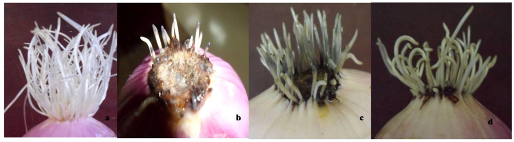
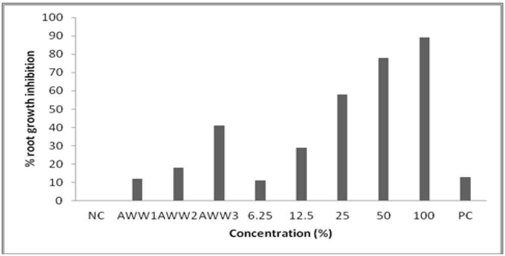
3.2. Toxicity to Root Growth in A. cepa

There was good root growth in the negative control well water. The roots of bulbs grown in the well water samples from the e-waste dumpsite showed milky-white and yellowish colors, while the roots of onions treated with ARL showed black and brownish colors, rotten basal plate (mostly at the 50 and 100% treated concentrations; Figure 2). Short, scanty, twisting and swollen (tumor) root tips were also observed in onions treated with both the well water samples and ARL. The ARL samples and positive control (10 ppm lead nitrate solution) induced concentration-dependent, significant (p<0.05) root growth inhibition in A. cepa (Figure 3). Negative correlation coefficient (r = −0.9 at P = 0.01) was observed between ARL treatment concentrations and root length growth, with EC50 and EC70 values of 34.1% and 17.7%, respectively. The well water samples induced root growth inhibitions in the order AWW3 > AWW2 > AWW1, and were significant (p<0.05) in AWW2 and AWW3 treatments (Figure 3).
Figure 2. Macroscopic effects induced in Allium cepa exposed to e-waste leachate. (a) Normal root growth in the control group, (b) short, scanty, swollen (tumour) roots with blackened root tips and rottenness at the basal plate, (c) short, scanty and blackened root length (d) short, backward bending to spiralling roots with blackened/yellowish root tips.
Challenges 04 00169 g002
Figure 3. .Effects of well water (AWW1, AWW2, AWW3) and e-waste leachate on the root growth of Allium cepa. (NC-negative control; 6.25–100%: varying concentrations of Alaba raw leachate (ARL); PC—positive control).
Challenges 04 00169 g003

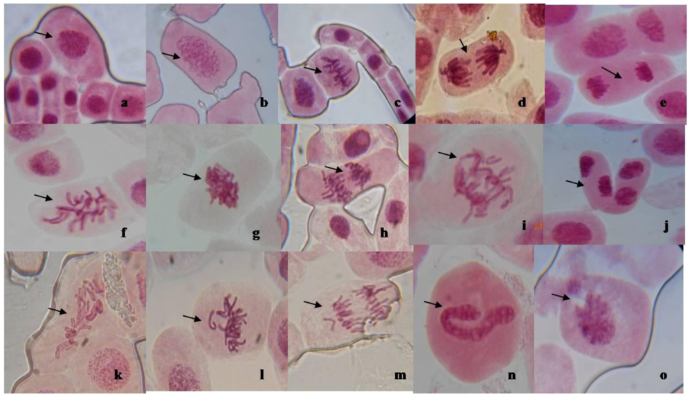
3.3. Mitotic Inhibition and Chromosomal Aberration in A. cepa

ARL induced a concentration-dependent, significant (p<0.05; r = −0.5 at P = 0.05) decrease in the mitotic index (MI), while only AWW1 and AWW3 samples induced significant decrease in MI compared to the negative control Table 2). The ARL and well water samples (in the order AWW2 > AWW1 > AWW3) also induced chromosomal aberrations in root tips of onions at all tested concentrations compared to the negative control (Table 2). The aberrations include; spindle disturbance, sticky chromosomes, polar deviations, C-mitosis, non-disjunction at anaphase, vagrant and fragment chromosomes, anaphase bridges and other nuclear abnormalities such as lobulated nuclei, nuclear buds, nucleus with nuclear point and broken/damage nuclear materials (Figure 4a–o).
Figure 4. Aberrations observed in Allium cepa root tip cells exposed to e-waste leachate and well waters. (a–e) Normal cells at Interphase (a), prophase (b), metaphase (c), anaphase (d) and telophase (e); (f) Spindle disturbance at metaphase; (g,h) stickiness at metaphase (g) and anaphase (h); (i) Bridges and non-disjunction at anaphase; (j) polar deviations at telophase; (k) C-mitosis; (l) vagrant and fragment chromosomes at metaphase; (m) vagrant chromosome at anaphase; (n,o) Nuclear abnormalities (NA) with nuclear point (n) and broken nuclear material (o) (×1000).
Challenges 04 00169 g004
Table 2. Effects of e-waste leachate and well water on mitotic activities and chromosomes of Allium cepa.
Mitotic indices and chromosomal aberration
Test sampleConc. (%)Number of dividing cellsMitotic index (%)Mitotic inhibition (%)No of cells at metaphaseNo of cells at anaphaseNo of cells at telophaseTotal aberrant cellsFrequency of aberrant cells (%) based on
Total cells scoredNo of dividing cells
ControlNC3187.9504958450a--
PC2716.7814.78566936bd0.9013.28
Well watersamplesAWW12015.0336.7912166634b0.8516.92
AWW22395.9824.8415197141b1.0317.16
AWW32225.5530.1913216829b0.7313.06
ARL(%)6.252115.2833.6518264411c0.285.21
12.52556.3819.8118256530d0.7511.77
251794.4743.719203934d0.8518.99
501684.2047.1711113033d0.8319.64
1001243.1061.018123138d0.9530.65
Values with the same superscript letter(s) are not significantly different from each other (p > 0.05) by student t-distribution. NC = Negative control, PC = Positive control.

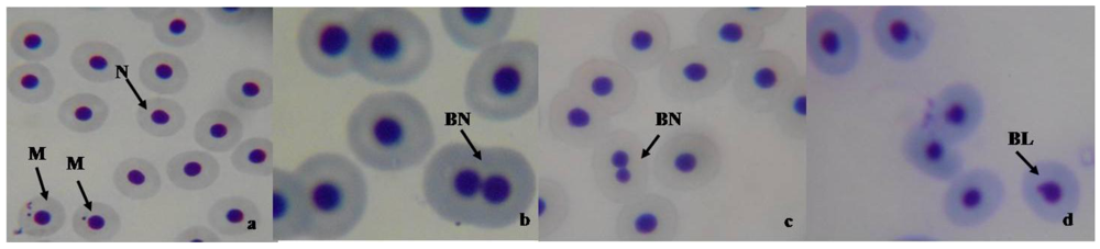
3.4. Micronucleus and Nuclear Abnormality Assay in Fish

There was reduced food intake and increase erratic movements in fish exposed to both AWW1 and ARL samples, though these were intense in fishes exposed to ARL. The frequencies of micronucleated erythrocytes and erythrocytes with nuclear abnormalities (blebbed nuclei and binucleated cells) were concentration-dependent and significant (p<0.05) but were not time dependent at tested concentrations of ARL and 100% concentration of the well water. The frequency of MN increased through the 7th day and reached a maximum on the 14th day of exposure, but decreased by the 28th day at treated concentrations of ARL (except at 12.5% and 25% where there was continuous increase with time) and well water. There was significant, concentration-dependent increase in the frequency of nuclear abnormalities (Table 3, Figure 5a–d) in fishes exposed to ARL; this was however time independent.
Table 3. Frequency of micronuclei and NA in Clarias gariepinus exposed to e-waste leachate and well water from Alaba International market, Lagos, Nigeria.
TreatmentExposure period (days)
MN (Mean ± SE)NA (Mean ± SE)
7142871428
Tap water3.73 ± 0.624.10 ± 0.911.30 ± 0.220.000.000.00
50% AWW4.93 ± 0.314.73 ± 0.413.37 ± 0.190.000.000.00
100% AWW4.60 ± 0.236.53 ± 0.523.93 ± 0.81 *0.000.000.00
12.5% ARL6.37 ± 0.41 *9.67 ± 0.66 *10.90 ± 1.03 *1.33 ± 0.580.90 ± 0.360.00
25% ARL7.87 ± 1.02 *9.40 ± 1.18 *10.10 ±0.94 *3.47 ± 1.57 *4.17 ± 1.52 *2.20 ± 0.97
50% ARL9.47 ± 0.43 *10.03 ± 1.00 *8.77 ± 0.91 *12.6 ± 1.92 *9.47 ± 1.61 *4.80 ± 1.64 *
MN = Micronucleus; NA = Nuclear abnormalities; * Significantly (p < 0.05) different from control; No. of cells scored in each treatment group per exposure period = 30,000.
Figure 5. Normal erythrocyte (N), micronucleated erythrocytes (M), binucleated cell (BN) and blebbed nuclei (BL) in Clarias gariepinus exposed to electronic waste leachate and contaminated well water (×1000).
Challenges 04 00169 g005

3.5. Biochemical Assay in Mice

Table 4 shows the effects of the well water and ARL treatment on oxidative stress biomarkers in mice. There was significant (p < 0.05) increase in the liver CAT, MDA, GSH and serum ALT and AST, with concomitant decrease in the liver SOD activities of the treated mice. The 10, 25 and 50% concentrations of ARL induced significant (p < 0.001) increase in liver MDA, CAT, GSH and serum AST and ALT activities compared to the control group. Similarly, the 10, 25 and 50% concentrations of ARL induced significant (p < 0.001) decrease in liver SOD activities compared to the control. Liver GSH, SOD and serum ALT of mice treated with the well water samples were significantly (p < 0.001) different from the control well water on the 3rd, 4th and 5th weeks of exposure. Liver CAT and serum AST were significantly (p < 0.001) different from the control on the 4th and 5th weeks of exposure while liver MDA differed from control only at the 5th week (Table 4).
Table 4. The effects of e-waste leachate and well water on liver lipid peroxidation (MDA), catalase, superoxide dismutase (SOD), reduced glutathione (GSH), alanine aminotransferase (ALT) and aspartate aminotransferase (AST) activities in mice.
E-waste MDA CAT SOD GSH ALT AST
leachate (µmol/mL) (µm/mg) (U/mL/Min) (µm/g tissue) (U/mL) (U/mL)
DW5.0 ± 0.1876.25 ± 0.964.65 ± 0.098.60 ± 0.0819.75 ± 0.9641.75 ± 0.5
1%5.4 ± 0.1476.25 ± 0.964.58 ± 0.058.65 ± 0.0921.00 ± 0.8242.50 ± 0.58
5%5.85 ± 0.0678.50 ± 0.583.9 ± 0.089.55 ± 0.1026.25 ± 0.50 *45.25 ± 0.96
10%8.0 ± 0.16 *84.25 ± 1.71 *2.33 ± 0.09 *10.85 ± 0.19 *29.75 ± 0.96 *49.25 ± 0.96 *
25%11.98 ± 0.15 *97.25 ± 1.71 *1.88 ± 0.10 *12.50 ± 0.08 *40.75 ± 1.26 *54.75 ± 0.96 *
50%18.8 ± 0.18 *134.0 ± 1.41 *1.48 ± 0.05 *14.15 ± 0.13 *47.0 ± 0.82 *61.0 ± 0.82 *
Well water
NC4.99 ± 0.1476.60 ± 0.064.66 ± 0.088.60 ± 0.0919.74 ± 0.8041.74 ± 0.9
1 week5.08 ± 0.1378.00 ± 0.824.45 ± 0.068.71 ± 0.0620.00 ± 0.8241.25 ± 0.5
2 weeks5.38 ± 0.1381.75 ± 0.964.08 ± 0.058.95 ± 0.0625.00 ± 1.4143.50 ± 0.58
3 weeks5.78 ± 0.0983.02 ± 0.963.55 ± 0.13 *10.05 ± 0.06 *28.75 ± 0.50 *48.25 ± 0.50
4 weeks7.68 ± 0.1789.0 ± 0.82 *3.58 ± 0.15 *11.48 ± 0.05 *32.00 ± 1.41 *51.25 ± 1.50 *
5 weeks8.35 ± 0.10 *96.0 ± 0.82 *2.20 ± 0.08 *12.38 ± 0.09 *36.75 ± 0.50 *56.25 ± 0.50 *
* significantly (p < 0.001) from corresponding control, DW = Distilled water, NC = Negative control (Itire well water).

4. Discussion

Human exposure to chemical substances in the environment may be from air, water and soil. E-waste contamination/pollution of the terrestrial and aquatic environments may increase the level of human exposure to heavy metals and organic contaminants. In developing countries including Nigeria, there are confirmations that harmful chemicals and microorganisms from unsanitary dumpsites are introduced into adjacent surface and ground waters used as drinking water by communities [28,29]. Epidemiological data from consumption of unsafe water showed increased risk of nephrotoxicity, cancer and central nervous system defects [30,31]. Exposure to chemicals through drinking contaminated water is capable of inducing DNA damage and enhancing genetic changes in somatic cells that can result in decreased cell survival or transformation and eventual reproductive abnormalities and cancer formation in organisms [32,33]. The results of our study showed that the tested well water samples were contaminated by harmful substances. It also showed the cytogenotoxic potentials of well water samples from the vicinity of open e-waste dumpsite and e-waste leachate in A. cepa and peripheral erythrocytes of C. gariepinus. There was also induction of oxidative damage by constituents of the tested samples in mice.
The induction of chromosome aberration and decreasing mitotic index in A. cepa, as well as micronucleus and nuclear abnormalities in peripheral erythrocytes of C. gariepinus treated with the well water and e-waste leachate suggest that these samples contained clastogenic and/or aneugenic substances capable of increasing DNA damage and genome instability in the tested organisms. Gomez-Arroyo et al. [34] similarly reported that well water contaminated by arsenic from Zimapan, Hidalgo town in Mexico induced sister chromatid exchange in treated Vicia faba root tips. Kong and Ma [35] reported that shallow well water collected from the vicinity of five pesticide farms induced chromosome aberration and micronucleated cells in Allium root anaphase aberration, Tradescantia-micronucleus and Tradescantia stamen hair mutation.
Cytological aberrations and MI observations in plant test systems are excellent monitoring tests for detecting environmental chemicals that pose risk to the cytoplasm and genetic materials mostly during mitosis and meiosis. Studies using A. cepa assay have shown good correlation with in vivo cytogenetic studies in mammalian systems [15,36]. It can be inferred that the inhibitory effects of the tested samples on root growth and cell proliferation in A. cepa was by inhibition of DNA synthesis at S-phase [37], complete destruction of metabolic activities that prevented the cell from entering mitosis [38] or disturbances of cell cycle or chromatin materials [39]. Stickiness of chromosomes may be due to increase chromosome contraction and condensation or DNA depolymerization [40,41] and nucleoproteins dissolution [42]. Similarly, anaphase bridges are probably formed during breakage and fusion of chromosomes and chromatids [43], suggesting that the constituents of e-waste leachate and well water have clastogenic effect on the genetic materials of the exposed A. cepa [36]. The presence of C-mitosis may indicate the inhibitory effects on spindle formation (tubargenic effect) due to chemicals in the tested samples [44].The observed lobulated nuclei and polynuclei cells may indicate cell death process in the root system of A. cepa [45].
We are not aware of studies on e-waste contaminated well water and leachate induced genetic damage in C. gariepinus. The available information is on contaminated rivers. For instance, studies have shown that Berdan river, Turkey receiving discharges from industrial and municipal wastes induced increased MN in the peripheral erythrocytes, gill cells and caudal fin epithelial of Nile tilapia (Oreochromis niloticus) in a 2, 4 and 6 days exposure study [46]. In an in situ biomonitoring of polluted marine environment of the southern Mediterranean coast of Turkey, an increase in the frequency of MN induction in the peripheral erythrocytes and gill cells of grey mullet (Mugilcephalus) indicated that clastogenic and aneugenic substances from industrial effluents were discharged into the sea [17]. Formation of nuclear abnormalities along with MN in erythrocytes of fish is considered as possible indicators of genotoxicity when investigating the effects of pollutants in aquatic species [47]. In in vivo studies under controlled laboratory conditions, MN and NAs were measured to assess the genotoxic potentials of municipal landfill leachates in C. gariepinus [48], textile mill effluent in O. niloticus [17], crude oil in Scophthalmus maximus and Gadus morua [49] and heavy metal in Puntius altus [50]. Significant increase in binucleated erythrocytes of e-waste leachate treated C. gariepinus may indicate cytokinesis blocking of a normal dividing cell during M phase of the cell cycle (cytotoxicity) by constituents of the leachate, mostly the toxic metals [48,50]. Blebbed and notch nuclei are associated with aneuploidy probably originated from tubuline failure, hence extruding from the nucleus as damage [51]. The presence of NAs can lead to genetic imbalance and carcinogenesis [52], thus they complement the scoring of MN in cytogenotoxicity studies [53].
In recent time, research focus on possible mechanisms of complex mixture induced DNA damage is increasing. Li et al. [54] and Bakare et al. [55] reported that the possible mechanism of municipal landfill and sludge leachates induced genotoxicity and toxicity in mice was by oxidative damage. Similarly, oxidative stress was implicated in incinerated bottom ash and municipal landfill leachates induced toxicity and genotoxicity in plant test systems [56,57,58]. These are in concert with our findings that e-waste leachate and contaminated well water induced elevated levels of lipid peroxidation and alterations in oxidative stress enzymes in liver of treated mice. MDA, an end product of lipoperoxydation, is considered a biomarker of oxidative stress and cellular damage [59,60]. GSH, an antioxidant, plays a crucial role in protecting the cells from oxidative damage [61], and change in the concentrations of GSH was observed during increases in oxidative stress [62]. Superoxide radicals or their transformation product, hydrogen peroxide (H2O2), are capable of causing the oxidation of cysteine which will lead to decreased SOD activity [63]. Activities of SOD were markedly decreased by the tested samples which resulted in an increase in CAT activity, since the degradation of H2O2, a potent oxidant at high cellular concentration, is affected by CAT due to its induction against increased oxidative stress. It is plausible that the observed cytogenotoxic effects in A. cepa and C. gariepinus is via generation of reactive oxygen species.
Serum ALT and AST are the most used markers of hepatocellular necrosis and are considered sensitive indicators of hepatic injury [64,65] and cell membrane damage and leakage [66]. Concomitant increase in the activities of ALT and AST in the serum of treated mice indicates acute hepatocellular injury. This is supported by previous finding wherein rats exposed to municipal landfill leachates showed concomitant increase in the serum activities of ALT and AST; severe necrosis, congestion and periportal cellular infiltrations of the liver tissues [67]. These observations further suggest that free radicals generated by the toxic constituents in the e-waste contaminated well water and leachate induced the cytogenotoxic effects in A. cepa and C. gariepinus.
The heavy metals and organic compounds present in the well water samples and e-waste leachate [10] are known to generate ROS that caused DNA, protein and lipid damage in eukaryotic cells [57]. These chemicals mostly heavy metals can bind to phosphate and base residues of DNA, to alter its primary and secondary structures [68]. They can also interfere with protein structure and function to cause DNA damage [69]. Therefore, free radical generation and oxidative damage by these chemicals may be responsible for the observed cytogenotoxic damage herein. The concentrations of the heavy metals and other compounds in the well water and e-waste leachate indicate environmental contamination due to indiscriminate disposal and open burning of e-waste. The high concentration of the chemicals can cause severe degradation in the groundwater quality and palatability to human consumption. This has been implicated with human gastrointestinal irritation and laxative effects [28,31], abnormal sperm quality [70], chromosome aberration and DNA damage [71], and reduced fecundity and adverse birth effects [72].

5. Conclusions

In conclusion, e-waste leachate and contaminated well water induced cytogenotoxicity in C. gariepinus and A. cepa, and oxidative stress in mice. Heavy metals and organic compounds present in the tested samples provoked the observed DNA damage through ROS formation. Inappropriate e-waste management in Nigeria and other developing countries may impact on human populations and other living organisms, and contaminate the environment. It is important that appropriate regulatory authorities implement sustainable methods of managing e- wastes so as to protect human and environmental health.

Acknowledgements

We thank A. A. Sowunmi of Hydrobiology and Fisheries unit of the Department of Zoology, University of Ibadan for his assistance on the piscine MN and nuclear abnormality assay.

Conflict of Interest

The authors declare no conflict of interest.

References

  1. Osibanjo, O.; Nnorom, I.C. The challenges of electronic waste (e-waste) management in developing countries. Waste Manag. Res. 2007, 25, 489–501. [Google Scholar] [CrossRef]
  2. Nnorom, I.C.; Osibanjo, O. Electronic waste (e-waste): Material flows and management practices in Nigeria. Waste Manag. 2008, 28, 1472–1479. [Google Scholar] [CrossRef]
  3. Bridgen, K.; Labunska, I.; Santillo, D.; Johnston, P. (Eds.) Greenpeace. Chemical Contamination at E-waste Recycling and Disposal Sites in Accra and Korforidua, Ghana. August 2008. Available online: http://www.greenpeace.org/ghanacontamination (accessed on 5 February 2010).
  4. Robinson, B.H. E-waste: An assessment of global production and environmental impacts. Sci. Total Environ. 2009, 408, 183–191. [Google Scholar] [CrossRef]
  5. Chen, A.; Dietrich, K.N.; Huo, X.; Ho, S. Development of Neurotoxicants in E-waste: An emerging health concern. Environ. Health Perspect. 2011, 119, 431–438. [Google Scholar]
  6. Puckett, J. (Ed.) Basel Action Network. The digital dump: Exporting re-use and abuse to Africa. Basel Action Network, 24 October 2005. Available online: http://www.ban.org (accessed on 18 February 2010).
  7. Alabi, O.A.; Bakare, A.A. Genotoxicity and mutagenicity of electronic waste leachates using animal bioassays. Toxicol. Environ. Chem. 2011, 93, 1073–1088. [Google Scholar] [CrossRef]
  8. Manhart, A.; Osibanjo, O.; Aderinto, A.; Prakash, S. Informal e-waste management in Lagos, Nigeria—Socio-economic impacts and feasibility of international recycling co-operations. Final Report of Component 3 of the UNEP SBC E-waste Africa Project. Öko-Institut e.V.: Lagos & Freiburg, Germany, 2011. [Google Scholar]
  9. Osibanjo, O.; Nnorom, I.O.; Bakare, A.A.; Alabi, O.A. Environmental and public health consequences of adopting crude recovery techniques in e-waste management in developing countries: An emerging global crisis. In Advances in Environmental Research Vol. 17; Daniels, J.A., Ed.; Nova Science Publishers Inc.: Hauppauge, NY, USA, 2012; ISBN: 978-1-61209-965-1. [Google Scholar]
  10. Alabi, O.A.; Bakare, A.A.; Xu, X.; Li, B.; Zhang, Y.; Huo, X. Comparative evaluation of environmental contamination and DNA damage induced by electronic waste in Nigeria and China. Sci. Total Environ. 2012, 423, 62–72. [Google Scholar] [CrossRef]
  11. American Public Health Association (APHA), Standard Methods for the Examination of Water and Wastewater, 20th ed.; APHA: Washington, DC, USA, 1988.
  12. United State Environmental Protection Agency (USEPA). Method 8270C revision 3, semi volatile organic compounds by gas chromatography/mass spectrometry (GC/MS). 1996. Available online: www.epa.gov/epaoswer/hazwaste/test/main.htm (accessed on 15 January 2010).
  13. Fiskesjo, G. The Allium test as a standard in environmental monitoring. Hereditas 1985, 102, 99–112. [Google Scholar] [CrossRef]
  14. CIOMS. International guiding principles for biomedical research involving animals. 1985. Available online: http://www.cioms.ch/publications/guidelines/1985_texts_of_guidelines.htm (accessed on 07 April 2010).
  15. Grant, W.F. Chromosome aberration assay in Allium. A report of the U.S. environmental protection agency gene-tox program. Mutat. Res. 1982, 99, 273–291. [Google Scholar] [CrossRef]
  16. Carrasco, K.R.; Tilbury, K.L.; Mayers, M.S. Assessment of the piscine micronuclei test as an in situ biological indicator of chemical contaminants effects. Can. J. Fish Aquat. Sci. 1990, 47, 2123–2136. [Google Scholar] [CrossRef]
  17. Cavas, T.; Ergene-Gozukara, S. Evaluation of the genotoxic potential of lambda–cyhalothrin using nuclear and nucleolar biomarkers on fish cells. Mutat. Res. 2003, 534, 93–99. [Google Scholar] [CrossRef]
  18. Cavas, T.; Ergene-Gozukara, S. Micronucleus test in fish cells: A bioassay for in situ monitoring of genotoxic pollution in the marine environment. Environ. Mol. Mutagen. 2005, 46, 64–70. [Google Scholar] [CrossRef]
  19. Agnihotri, N.; Kaur, H.; Kaur, N.; Sarotra, P. Role of oxidative stress in lansoprasole mediated gastric and hepatic protection in Wistar rats. Ind. J. Gastroenterol. 2007, 26, 118–121. [Google Scholar]
  20. Sinha, K.A. Colorimetric assay of catalase. Anal. Biochem. 1972, 47, 389–394. [Google Scholar] [CrossRef]
  21. Misra, H.P.; Fridovich, I. The role of superoxide anion in the autooxidation of epinephrine and a simple assay for superoxide dismutase. J. Biol. Chem. 1972, 247, 3170–3175. [Google Scholar]
  22. Gornall, A.G.; Barawill, J.C.; David, M.M. Determination of serum protein by means of biuret reaction. J. Biol. Chem. 1949, 177, 751–761. [Google Scholar]
  23. Habig, W.H.; Pabst, M.J.; Jakoby, W.B. Glutathione-s-transferases: The first enzymatic step in mercapturic acid formation. J. Biol. Chem. 1974, 249, 7130–7139. [Google Scholar]
  24. Shokunbi, O.S.; Odetola, A.A. Gastroprotective and antioxidant activities of Phyllanthus amarus extracts on absolute ethanol induced ulcer in albino rats. J. Med. Plants Res. 2008, 2, 261–267. [Google Scholar]
  25. Reitman, S.; Frankel, S. A colorimetric method for determination of serum glucose oxaloacetate and glutamic pyruvate transaminases. Am. J. Clin. Pathol. 1957, 28, 53–56. [Google Scholar]
  26. National Environmental Standards and Regulation Enforcement Agency (NESREA), (Federal Republic of Nigeria Official Gazette), National Environmental (Sanitation and Waste Control). Federal Government of Nigeria Printer: Abuja, Nigeria, 2009; FGP 112/102009/L000 (OL54). No.60 (96); pp. 1057–1102.
  27. United State Environmental Protection Agency (USEPA). Drinking water contaminants. Washington, DC, USA, 2009. Available online: http://water.epa.gov/drink/contaminants/index.cfm#List (accessed on 5 February 2010).
  28. Sia Su, G. Impact on drinking water sources in close proximity to the Payatas dumpsite, Philippianes. J. Public Health 2007, 15, 51–55. [Google Scholar] [CrossRef]
  29. Okonko, I.O.; Adejoye, O.D.; Ogunnusi, T.A.; Fajobi, E.A.; Shittu, O.B. Microbiological and physicochemical analysis of different water samples used for domestic purposes in Abeokuta and Ojota, Lagos State, Nigeria. Afr. J. Biotech. 2008, 7, 617–621. [Google Scholar]
  30. Gupta, S.K.; Gupta, R.C.; Gupta, A.B. Recurrent diarrhea in children living in areas with high levels of nitrate in drinking water. Arch. Environ. Health 2001, 56, 369–373. [Google Scholar] [CrossRef]
  31. Sia Su, G. Water-borne illness from contaminated drinking water sources in close proximity to a dumpsite in Payatas, The Philippianes. J. Rural Trop. Public Health 2005, 4, 43–48. [Google Scholar]
  32. Shugart, L.R.; McCarthy, J.F.; Halbrook, R.S. Biological markers of environmental and ecological contamination: An overview. Risk Anal. 1992, 12, 353–360. [Google Scholar] [CrossRef]
  33. Russo, C.; Rocco, L.; Morescalchi, M.A.; Stingo, V. Assessment of environmental stress by the micronucleus test and comet assay on the genome of teleost populations from two natural environments. Ecotoxicol. Environ. Saf. 2004, 57, 168–174. [Google Scholar] [CrossRef]
  34. Gómez-Arroyo, S.; Armienta, M.A.; Cortés-Eslava, J.; Villalobos-Pietrini, R. Sister chromatid exchanges in Vicia faba induced by arsenic-contaminated drinking water from Zimapan, Hidalgo, Mexico. Mutat. Res. 1997, 394, 1–7. [Google Scholar] [CrossRef]
  35. Kong, M.S.; Ma, T.H. Genotoxicity of contaminated soil and shallow well water detected by plant bioassays. Mutat. Res. 1999, 426, 221–228. [Google Scholar] [CrossRef]
  36. Leme, D.M.; Marin-Morales, M.A. Allium cepa in environmental monitoring: A review on its application. Mutat. Res. 2009, 682, 71–81. [Google Scholar] [CrossRef]
  37. Sudhakar, R.; Gowda, N.; Venu, G. Mitotic abnormalities induced by silk Dyeing Industry Effluents in the cells of Allium cepa. Cytologia 2001, 66, 235–239. [Google Scholar] [CrossRef]
  38. Metin, M.; Burun, B. Effects of the high doses of Urginea maritima (L.) baker extract on chromosomes. Caryologia 2010, 63, 367–375. [Google Scholar]
  39. Glinska, S.; Bartezak, M.; Oleksiaka, S.; Wolska, A.; Gabara, B.; Posmyk, M.; Janas, K. Effects of anthocyanin-rich extract from red cabbage leaves on meristematic cells of Allium cepa L. roots treated with heavy metals. Ecotoxicol. Environ. Saf. 2007, 68, 343–350. [Google Scholar] [CrossRef]
  40. Ahmed, M.; Grant, W.F. Cytological effects of the pesticides phosdrin and bladex in Tradescantia and Vicia faba. Can. J. Genet. Cytol. 1972, 14, 157–165. [Google Scholar]
  41. Klasterska, I.; Natarjan, A.T.; Ramel, C. An interpretation of the origin of subchromatid aberrations and chromosome stickiness as a category of chromatid aberration. Hereditas 1976, 83, 153–162. [Google Scholar] [CrossRef]
  42. Kaufman, B.P. Cytochemical studies of changes induced in cellular materials by ionizing ra­diations. Ann. N. Y. Acad. Sci. 1958, 59, 553–559. [Google Scholar] [CrossRef]
  43. Halıem, A.S. Cytological effects of the herbicide sencorer on mitosis of Allium cepa. Egypt. J. Bot. 1990, 33, 93–104. [Google Scholar]
  44. Shahın, S.A.; El-Amoodı, K.H.H. Induction of numerical chromosomal aberrations during DNA synthesis using the fungicides nimrod and rubigan-4 in root tips of Vicia faba L. Mutat. Res. 1991, 261, 169–176. [Google Scholar] [CrossRef]
  45. Leme, D.M.; Angelis, D.F.; Marin-Morales, M.A. Action mechanisms of petroleum hydrocarbons present in waters impacted by an oil spill on the genetic material of Allium cepa root cells. Aquat. Toxicol. 2008, 88, 214–219. [Google Scholar] [CrossRef]
  46. Ergene, S.; Cava, T.; Celik, A.; Koleli, N.; Aymak, C. Evaluation of river water genotoxicity using piscine micronucleus test. Environ. Mol. Mutag. 2007, 48, 421–429. [Google Scholar] [CrossRef]
  47. DeLemos, C.T.; Rodel, P.M.; Terra, N.R.; Erdtmann, B. Evaluation of basal micronucleus frequency and hexavalent chromium effects in fish erythrocytes. Environ. Toxicol. Chem. 2007, 20, 1320–1324. [Google Scholar]
  48. Alimba, C.G.; Saliu, J.K.; Adesanya, A.; Bakare, A.A. Evaluation of Genotoxicity of municipal landfill leachate by micronucleus test using Clarias gariepinus. Res. Environ. Life Sci. 2011, 4, 1–6. [Google Scholar]
  49. Barsiene, J.; Dedonyte, V.; Rybakovas, A.; Andreikenaite, L.; Andersen, O.K. Investigation of micronuclei and other nuclear abnormalities in peripheral blood and kidney of marine fish treated with crude oil. Aquat. Toxicol. 2006, 78, S99–S104. [Google Scholar] [CrossRef]
  50. Ozkan, F.; Gündüz, S.G.; Berköz, M.; Hunt, A.O. Induction of micronuclei and other nuclear abnormalities in peripheral erythrocytes of Nile tilapia, Oreochromis niloticus, following exposure to sublethal cadmium doses. Turkish J. Zool. 2011, 35, 585–592. [Google Scholar]
  51. Ventura, B.C.; Angelis, D.F.; Marin-Morales, M.A. Mutagenic and genotoxic effects of the atrazine herbicide in Oreochromis niloticus (Perciformes, Cichlidae) detected by the micronuclei test and the comet assay. Pestic. Biochem. Phys. 2008, 90, 42–51. [Google Scholar] [CrossRef]
  52. Rodilla, V. Origin and evolution of binucleated cells and binucleated cell with micronuclei in cisplatin—Treated CHO cultures. Mutat. Res. 1993, 300, 281–291. [Google Scholar] [CrossRef]
  53. Cavas, T. In vivo genotoxicity of mercury chloride and lead acetate: Micronucleus test on acridine orange stained fish cells. Food Chem. Toxicol. 2008, 46, 352–358. [Google Scholar] [CrossRef]
  54. Li, H.; Han, M.; Hou, L.; Li, G.; Sang, N. Landfill leachate ingestions induced protein oxidation and DNA–protein crosslinks in mouse viscera. J. Hazard. Mat. 2010, 174, 54–58. [Google Scholar] [CrossRef]
  55. Bakare, A.A.; Patel, S.; Pandey, A.K.; Bajpayee, M.; Dhawan, A. DNA and oxidative damage induced in somatic organs and tissues of mouse by municipal sludge leachate. Toxicol. Ind. Health 2012, 28, 614–623. [Google Scholar] [CrossRef]
  56. Ferrari, B.; Radetski, C.M.; Veber, A.M.; Ferard, J.F. Ecotoxicological assessment of solid wastes: A combined liquid—And solid—Phase testing approach using a battery of bioassays and biomarkers. Environ. Toxicol. Chem. 1999, 18, 1195–1202. [Google Scholar]
  57. Radetski, C.M.; Ferrari, B.; Cotelle, S.; Masfaraud, J.F.; Ferard, J.F. Evaluation of the genotoxic, mutagenic and oxidant stress potentials of municipal solid waste incinerator bottom ash leachates. Sci. Total Environ. 2004, 333, 209–216. [Google Scholar] [CrossRef]
  58. Sang, N.; Han, M.; Li, G.; Mingzhu-Huang, M. Landfill leachate affects metabolic responses of Zea mays L. seedlings. Waste Manag. 2010, 30, 856–862. [Google Scholar] [CrossRef]
  59. Kim, H.S.; Kwack, S.J.; Lee, B.M. Lipid peroxidation, antioxidant enzymes, and benzo[a]pyrene- quinones in the blood of rats treated with benzo[a]pyrene. Chem. Biol. Interact. 2000, 127, 139–150. [Google Scholar] [CrossRef]
  60. Dotan, Y.; Lichtenberg, D.; Pinchuk, I. Lipid peroxidation cannot be used as a universal criterion of oxidative stress. Progress Lipid Res. 2004, 43, 200–227. [Google Scholar] [CrossRef]
  61. Guoyao, W.; Yun-Zhong, F.; Sheng, Y.; Joanne, R.; Nancy, D. Glutathione metabolism and its implications for health. J. Nutr. 2004, 134, 489–492. [Google Scholar]
  62. Bray, T.M.; Taylor, C.G. Tissue glutathione, nutrition and oxidative stress. Can. J. Physiol. Pharmacol. 1993, 71, 746–751. [Google Scholar] [CrossRef]
  63. Dimitrova, M.S.T.; Tsinova, V.; Velcheva, V. Combined effect of zinc and lead on the hepatic superoxide dismutase-catalase system in carp (Cyprinus carpio). Comp. Biochem. Physiol. 1994, 108, 43–46. [Google Scholar]
  64. Molander, D.W.; Wroblewsk, F.; La Due, J.S. Transaminase compared with cholinesterase and alkaline phosphatase an index of hepatocellular integrity. Clin. Res. Proc. 1955, 3, 20–24. [Google Scholar]
  65. Friedman, L.S.; Martin, P.; Munoz, S.J. Liver function tests and the objective evaluation of the patient with liver disease. In Hepatology: A Textbook of Liver Disease, 3rd ed.; Zakin, D., Boyer, T.D., Eds.; WB Saunders: Philadelphia, 1996; pp. 791–833. [Google Scholar]
  66. Kaplan, M.M. Laboratory tests. In Diseases of the Liver, 7th ed.; Schiff, L., Schiff, E.R., Eds.; JB Lippinocott: Philadephia, PA, USA, 1993; pp. 108–144. [Google Scholar]
  67. Alimba, C.G.; Bakare, A.A.; Aina, O.O. Liver and kidney dysfunction in wistar rats exposed to municipal landfill leachate. Res. Environ. 2012, 2, 150–163. [Google Scholar]
  68. Bridgewater, L.; Manning, F.; Woo, E.; Patierno, S. DNA polymerase arrest by adducted trivalent chromium. Mol. Carcinog. 1994, 9, 122–133. [Google Scholar] [CrossRef]
  69. Resit, M.; Jenner, P.; Halliwell, B. Sulphite enhances peroxynitrite dependent a1- antiproteinase inactivation: A mechanism of lung injury by sulphur dioxide. FEBS Lett. 1998, 43, 231–234. [Google Scholar]
  70. Akutsu, K.; Takatori, S.; Nozawa, S.; Yoshiike, M.; Nakazawa, H.; Hayakawa, K.; Makino, T.; Iwamoto, T. Polybrominateddiphenyl ethers in human serum and sperm quality. Bull. Environ. Contam. Toxicol. 2008, 80, 345–350. [Google Scholar] [CrossRef]
  71. Liu, Q.; Cao, J.; Li, K.Q.; Miao, X.H.; Li, G.; Fan, F.Y. Chromosomal aberrations and DNA damage in human populations exposed to the processing of electronics waste. Environ. Sci. Pollut. Res. Int. 2009, 16, 329–338. [Google Scholar] [CrossRef]
  72. Xu, X.; Yang, H.; Chen, A.; Zhou, Y.; Wu, K.; Liu, J.; Guo, Y.; Huo, X. Birth outcomes related to informal E-waste recycling in Guiyu, China. Reprod. Toxicol. 2012, 33, 94–98. [Google Scholar] [CrossRef]

