Article

In Vivo Hypolipidemic Effects and Antioxidant Capacity of Pinus morrisonicola Hay Extracts by Supercritical Carbon Dioxide Extraction

1
Department of Food Science, National Quemoy University, Jinning Township, Kinmen County 892, Taiwan
2
Department of Health Food, Chung Chou University of Science and Technology, Changhua 510, Taiwan
3
Department of Cosmeceutics, China Medical University, Taichung 406, Taiwan
4
Department of Medicinal Botanicals and Foods on Health Applications, Da-Yeh University, Changhua 515, Taiwan
*
Author to whom correspondence should be addressed.
Agriculture 2023, 13(3), 535; https://doi.org/10.3390/agriculture13030535
Submission received: 23 November 2022 / Revised: 19 February 2023 / Accepted: 20 February 2023 / Published: 23 February 2023

Abstract

:
Pinus morrisonicola hay (PM) is a pine tree unique to Taiwan, whose needles are used as traditional medicine and as functional drink. PME3-1 was made using supercritical extraction to evaluate the prevention of hyperlipidemia. This study explored the hypolipidemic effect of PME3-1 on hamsters on a high fat and cholesterol (HFC) diet. Three groups of hamsters were fed with PME3-1 (0.2, 1.0, and 5.0 mg/kg bw). After feeding for eight weeks, PME3-1 reduced the serum cholesterol, triglyceride levels, the low-density lipoprotein cholesterol/high-density lipoprotein cholesterol (LDL/HDL) ratio, and the swelling of the liver and kidney significantly (p < 0.05). In addition, feeding the hamsters with 5.0 mg/kg bw of PME3-1 could significantly reduce their total lipid (TL) content, total cholesterol (TC) content, total triglyceride (TG) content, and the HMG-CoA reductase activity in the liver (p < 0.05). Meanwhile, the antioxidant enzymes in the liver, glutathione peroxidase (GPx) and glutathione S-transferase (GST), can also improve, promoting the excretion of triglycerides (TG) and total cholesterol (TC) in the feces (p < 0.05). Therefore, these results confirm that PME3-1 hypolipidemic and antioxidant regulating functions in vivo.

1. Introduction

An excessive intake of cholesterol in the daily diet increases the concentration of cholesterol in the blood. Excess cholesterol accumulates in the arterial intima, which can lead to arteriosclerosis and cause myocardial infarction and cerebrovascular disease. High levels of total cholesterol (TC), triglycerides (TG), low-density lipoprotein cholesterol (LDL-C), low levels of high-density lipoprotein cholesterol (HDL-C), and reduced antioxidant capacity in vivo, all increase the risk of cardiovascular disease [1,2]. In order to maintain normal physiological functions, cells can consume stored cholesterol. When the stored cholesterol within the cells is exhausted, liver cells are prompted to increase the number and activity of LDL receptors (LDLRs). It is known that SIM can regulate the sterol regulatory element-binding protein (SREBP) pathway, to increase the activity of LDLR on the liver cell membrane, thus resulting in more LDL-C being taken up and utilized by the hepatocytes. In turn, this further reduces the total cholesterol and LDL-C in the blood and affects the metabolism of other lipids [3,4,5]. In addition, SIM can inhibit the synthesis of apolipoprotein B-100 in the hepatocytes. It also inhibits the synthesis and secretion of triglyceride-rich lipoproteins [6]. Previous studies have found that SIM did not significantly affect cholesterol absorption but did increase sterol excretion [7].
The Taiwan pine (Pinus morrisonicola hay) is an endangered species of pine tree that is unique to Taiwan. Its leaves are needle-like, with five needles in a bunch, and are popular in traditional medicine. Pine needle tea is also used as a functional drink in Asia, especially in Taiwan, China, and Korea. Previous in vitro studies have confirmed that it has anti-oxidative, anti-inflammatory, and anti-cancer properties, lowering blood lipids and cholesterol, preventing arteriosclerosis, and diminishing inflammatory responses [8,9,10]. Its functional components are rich in chlorophyll, polyphenolic compounds (epicatechin, anthocyanin, p-Coumaric acid, and β-sitosterol), abietic acid (abietinic acid), and flavonoids (chrysin, tectochrysin, and pinobanksin) [11,12,13,14]. Hyperlipidemia causes arteriosclerosis, which can lead to myocardial infarction, cerebrovascular disease, and peripheral vascular disease, and has a high mortality rate. Hyperlipidemia is the main risk factor for cardiovascular disease. Therefore, the development of nutraceuticals with blood lipid-lowering properties is an important contemporary issue.
Supercritical fluid extraction (SCFE) is an environmentally-friendly, convenient, rapid, and low-polluting separation technology [15], that has been widely used in the food, chemical, pharmaceutical, and petrochemical industries. SCFE with carbon dioxide has the advantages of a high extraction rate, high stability, high safety, and no solvent residue. It is colorless, odorless, and non-toxic, and is often used in the food industry [16]. SCFE improves the functional components extraction of natural ingredients and Chinese herbal medicines by traditional extraction, such as using a solvent or steam [17].
In previous in vitro studies, the highest antioxidant activity extract (PME3) was found using supercritical fluid extraction with carbon dioxide, using ethanol as an adjuvant solvent under 25 MPa for 15 min at 40 °C. Purified from PME3 by column and thin-layer chromatography, PME3-1-inhibited LDL oxidation more effectively than PME3 in a cell-free system to decrease conjugated diene levels and the foam cell formation induced by ox-LDL. Through GC-MS, 1-docosene, cedrane-8,13-diol, neophytadiene, and methyl abietate were identified in PME3-1. These have been shown to have hypolipidemic and antioxidant activities [18,19,20,21]. As the hamster’s plasma LDL-C ratio and lipoprotein distribution of hamsters are similar to those of human beings, this study mainly examines the hypolipidemic effect of PME3-1 on hamsters with a high fat and cholesterol (HFC) diet in order to provide parameters for PM applications and the development of hypolipidemic health foods.

2. Materials and Methods

2.1. Materials and Chemicals

Cholesterol, casein, α-cellulose, minerals, and vitamins were purchased from MP Biomedicals Inc. (Santa Ana, CA, USA). Choline bitartrate and methionine were purchased from Sigma-Aldrich Co., Ltd. (St. Louis, MO, USA). Corn oil was purchased from Jia-Ger Foods Co. (Taipei, Taiwan). Sucrose was purchased from Taiwan Sugar Corp. (Tainan, Taiwan). Corn starch was purchased from Gem Font Corp. (Taipei, Taiwan). Assay kits for triglycerides, cholesterol, LDL-cholesterol, and HDL-cholesterol were purchased from Randox Laboratories Ltd. (Berlin, UK). Analytical grade chemicals such as chloroform, methanol, potassium hydroxide, n-hexane, methyl pentadecanoate, boron trifluoride-methanol (purity 20%), sodium sulfate anhydrous, silicic acid, isopropyl alcohol, sodium periodate, acetylacetone, ethanol, acetone, acetic acid, digitonin, sulfuric acid, perchloric acid, molybdate, ascorbic acid, sodium bicarbonate methylene chloride, and ethyl acetate were purchased from Echo Chemical Co., Ltd. (Taichung, Taiwan). P. morrisonicola Hay pine needles were provided by the KARA Legend Biotechnology Co., Ltd. (Taoyuan, Taiwan).

2.2. Sample Preparation

Following Cheng et al. [21], 20 g of freeze-dried pine needle powder was added to a 30 mL extraction tank. Then, 3 mL of ethanol was added as an adjuvant solvent, the system temperature was set to 40 °C, and the supercritical carbon dioxide pressure was set to 25 MPa using a high-pressure pump. After the extraction tank reached the set pressure, the high-pressure pump was stopped and the sample equilibrate was held in the extraction tank for 15 min for static extraction. The high-pressure pump was then turned on, and the flow rate of supercritical carbon dioxide was maintained at 6 mL/min for 15 min for dynamic extraction. The PME3-1 was separated by thin-layer chromatography and column chromatography. The yield was approximately 3.9%.

2.3. In Vivo Treatment and Analysis

2.3.1. Treatments

Following and modifying Lin et al. [22], 48 male hamsters (six weeks old) were acclimated by BioLASCO Co. (Taipei, Taiwan) for 2 days at 24 ± 1 °C and a 50 ± 10% relative humidity with a 12 h light–dark cycle per day. Before the start of the experiment, all hamsters were randomly divided into six groups (n = 8). The 0.2% cholesterol and 15.0% fat (12% corn oil with 3% lard) diet hamsters were fed on the high–fat and cholesterol (HFC) diet for the first 4 weeks, followed by Simvastatin or different levels of PME3-1 to week twelve. The different feed formulations, which were provided from 5 to 12 weeks, are shown in Table 1. The weight of the hamsters were recorded and 15 g of fresh feed and enough fresh water were changed daily. All experimental procedures involving animals were conducted in accordance with National Institutes of Health (NIH) guidelines. This experiment was approved by the Institutional Animal Care and Use Committee (IACUC, no. 011) of the Chung Chou University of Science and Technology.

2.3.2. Analysis

All hamsters were fasted for 16 h, anesthetized with carbon dioxide, and had blood was drawn from their celiac arteries at week 4 and 12. Their blood was immediately centrifuged (3200 rpm, 20 min) to collect serum. It was then stored at −20 °C for the analysis of triglycerides (TG), total cholesterol (TC), low-density lipoprotein cholesterol (LDL-C), and high-density lipoprotein cholesterol (HDL-C) content. After the hamsters were euthanized, the livers, kidneys, and spleens were collected and frozen for analysis. Organs used for the evaluation of pathological sections were immersed in 10% formalin for 24 h.
The liver lipid analysis procedure followed and was modified from Folch et al. [23]. An amount of 0.5 g of the liver was extracted five times with 5 mL of chloroform–methanol (2:1, v/v) solution to extract the lipids. Afterward, it was stored at −20 °C. The triglyceride content analysis followed and was modified from the method from Fletcher [24]. The nitrogen-blown liver extract was mixed with chloroform (1:10, v/v) and 1 g of silicic acid. It was activated at 120 °C for 3 h and shaken for 15 min. After centrifugation (1500 rpm, 20 min), 2 mL of supernatant was taken, dried with nitrogen, and then mixed with isopropanol: water (9:1, v/v) and 0.6 mL reagent S (KOH: isopropanol: distilled water = 5:60:40) and redissolved. After saponification at 60–70 °C for 15 min, 1 mL of 0.003 M NaIO4 solution and 0.5 mL of acetylacetone were added and maintained in a 50 °C water bath for 30 min. The absorbance was then measured at 405 nm. The cholesterol content determination followed and was modified from the method from Sperry & Webb [25]. An amount of 2 mL of the dried liver extract with nitrogen was mixed with 0.5 mL of ethanol–acetone (1:1, v/v). Next, 0.02 mL of 50% KOH solution was added for saponification in a shaking water bath at 40 °C for 30 min. After cooling down, 0.02 mL of 0.1% phenol, 0.12 mL of 10% acetic acid, and 0.2 mL of 0.5% digitonin were added sequentially. After being maintained at 25 °C overnight and centrifuged (1500 rpm, 5 min), and the precipitate was placed at 110 °C for 30 min to remove the water. Afterward, 0.5 mL of acetic acid was added. After cooling 1 mL of color-forming agent (anhydrous acetic acid: concentrated acid = 20:1) was added and the mixture was left to stand at 25 °C for 3 min to develop color. The absorbance was measured at 620 nm. An analysis of dried feces followed the method described above.
The determination of the GST enzymatic activity followed and was modified from the method from Dierickx [26]. Taking 100 μL of the homogeneous solution, 880 μL of 100 mM GSH was added to 100 mM potassium phosphate buffer (pH 6.5) and 2 μL of 50 mM 1-chloro-2,4-dinitrobenzene (CDNB). The change in absorbance was measured within 5 min at 340 nm. The formula for calculating the enzyme activity followed Formula (1). The liver tissue homogenate was measured using Bio-Rad’s protein reaction reagent.
E340 = 9.6 mM−1cm−1
The determination of GPx enzymatic activity followed and was modified from the method from Mohandas et al. [27]. Taking 100 μL of homogenate solution, 800 μL of mixed solution (1 mM EDTA, 1 mM NaN3, 0.2 mM NADPH, 1 U/mL GSH reductase, and 1 mM GSH) was added to 100 mM of potassium phosphate buffer (pH = 7.0) and maintained for 5 min. Them, 100 μL of 2.5 mM H2O2 was added and the absorbance was measured within 5 min at 340 nm. The formula for the enzyme activity calculation is as followes.
E340 = 6220 M−1cm−1
The determination of malondialdehyde (MDA) followed and was modified from Richard et al. [28] and Ko et al. [29]. In sequence, 0.2% BHT and 0.4% TBA were added (1:0.3:2, v/v/v), mixed well, and heated at 90 °C for 45 min. The reaction was then stopped immediately with an ice bath, followed by mixture with n-butanol (1:1, v/v) for extraction and centrifugation (3000 rpm, 3 min). The absorbance value of the supernatant was measured at 535 nm.

2.4. Statistical Analysis

All analytical treatments were conducted with eight replications. All data are presented as the mean ± standard deviation. An analysis of variance (ANOVA) was performed using SPSS (17.0) statistical software, and Duncan’s multiple range test was used to determine whether there was a significant difference (p < 0.05) or not in each group.

3. Results and Discussion

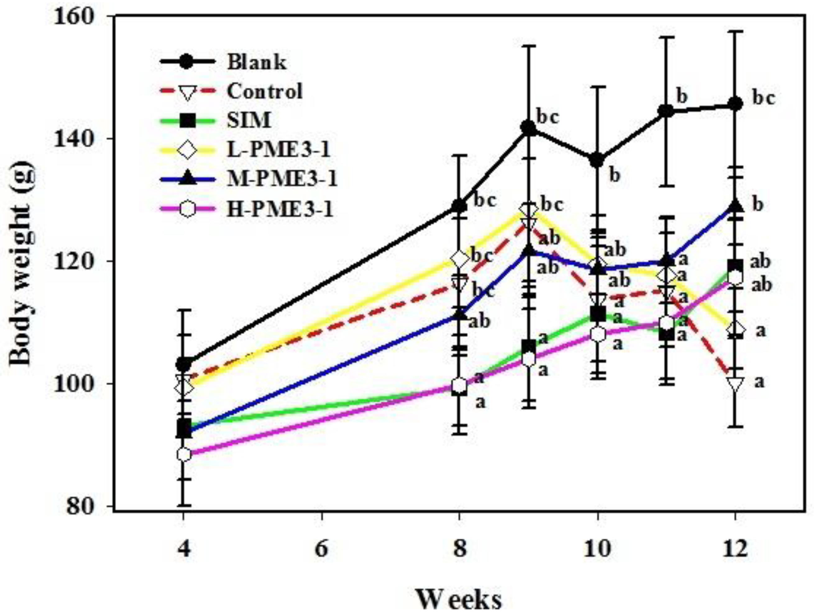
3.1. Weight Change of Hamster Bodies and Organ

In an earlier study, it was shown that the metabolism of cholesterol and bile acid in hamsters is closer to human beings than that of rats, making hamsters ideal in vivo test animals for studying lipid metabolism [30,31]. However, when hamsters were fed using an HFC diet, their appetite was reduced from a normal diet of around 10 g to a diet of 7–8 g. This was the main reason for their weight loss. In Figure A1, it can be seen that the weight of the hamsters increased significantly from weeks 4 to 8, and that the weight values of the blank group were significantly higher than those of the other groups. For those on the HFC diet, the body weight increased in the SIM, M-PME3-1, and H-PME3-1 groups (p > 0.05), indicating that the addition of Simvastatin or medium and high doses of PME3-1 can increase the weight level of hamsters. However, both the control and lower-dose PME3-1 groups showed a significant trend of weight loss (p < 0.05), indicating that the addition of a lower dose of PME3-1 or being in the control group did not maintain the body weight of the hamsters.
In past study indicated that fat infiltration may lead to symptoms of discomfort in hamsters, such as a loss of appetite, fatigue, and weight loss [32]. The experimental results show that an HFC diet causes damage to hamsters, and the phenomenon of appetite loss occurs. It is speculated that under the continuous induction of an HFC diet, hamsters will develop non-alcoholic fatty liver disease, leading to loss of appetite and weight loss [33,34]. Gan and Watts [35] pointed out that non-alcoholic fatty liver disease is easily caused by an HFC diet [36]. Patients with fatty liver disease are prone to a loss of appetite. Non-alcoholic fatty liver disease is a type of change in liver fat that can be caused by a variety of factors, with hyperlipidemia being one of the most common [37]. Hamden et al. [38] noted that the livers and kidneys of rats with hyperlipidemia had obvious fat accumulation. Due to fat accumulation in the livers and kidneys caused by the HFC diet of hamsters in this study, the organ weight (control group) was heavier than that of the blank group. As can be seen from Table 2, the liver weight increased from 3.9 to 6.3 g and the kidney weight increased from 1.1 to 2.1 g, suggesting that PME3-1 has the potential to reduce non-alcoholic fatty liver disease caused by an HFC diet.
Table 2 shows that, in the control and blank groups, the HFC diets significantly increased the liver and kidney weights by 0.62 and 0.91 times, respectively. In addition to the low-dose PME3-1 kidney weight, the groups supplemented with Simvastatin and PME3-1 significantly reduced the liver and kidney weight. Continuously feeding medium and high doses of PME3-1 for 12 weeks can significantly reduce liver and kidney hypertrophy caused by high lipids and cholesterol. The liver weight was reduced by approximately 36.51% and 46.03% and the kidney weight was reduced by approximately 38.10% and 38.33%, respectively. The liver and kidney weights of the hamsters given middle and high doses of PME3-1 showed no significant difference between the blank and the SIM groups (p > 0.05). At low doses of PME3-1, the liver weight was reduced by approximately 17.46% (p < 0.05), while the kidney weight was reduced by approximately 14.29% (p > 0.05). In addition, the weight of the spleen under the inducement of the HFC diet was significantly reduced (p < 0.05) compared to the blank group, by a figure of approximately 8.00%. The spleen weight in the groups given low, medium, and high doses of PME3-1 increased by approximately 0.50, 1.25, and 1.50 times, respectively. Only the high dose of PME3-1 gave a significant increase (p < 0.05), better than the SIM group. Meanwhile, the H-PME3-1 group demonstrated no significant difference from the blank group.

3.2. Serum Analysis

From Table 3, it can be seen that, compared with the blank group, the serum cholesterol, triglycerides, high-density lipoprotein, and low-density lipoprotein of the HFC diet-fed hamsters increased by 1.58, 0.35, 0.87, and 3.34 times, respectively. After 12 weeks of continuous feeding with low, medium, and high doses of PME3-1, the high cholesterol, triglycerides, and high-density lipids induced by the HFC diet were significantly reduced (p < 0.05). The cholesterol content decreased by 33.49%, 42.11%, and 64.47%, respectively, and the effects of the high dose of PME3-1 especially were better than in the SIM group (in which the cholesterol and triglyceride contents were decreased by 6.78 and 21.92%, respectively). The high-density lipoprotein content decreased by 20.86%, 27.74%, and 35.13%, respectively. And there was no significant difference between the medium and high doses of PME3-1 and the SIM group (p < 0.05). The low-density lipoprotein content decreased by 58.64%, 68.11%, and 71.60%, respectively, again with no significant difference from the SIM group (p < 0.05). The level of cholesterol in the blood is most commonly used to evaluate the risk of arteriosclerosis. A higher LDL/HDL value indicates greater difficulty in removing cholesterol from the serum. In contrast, a higher HDL value makes it easier to remove the cholesterol in the serum, and the risk of arteriosclerosis will also be decreased. Table 3 shows that the control group had an increasing trend over the blank group, but was no different from the other groups, indicating that PME3-1 has the potential to improve LDL/HDL.
The atherogenic index is the atherosclerosis index. A higher value indicates a higher risk of atherosclerosis. In Table 3, the control group shows a significantly higher value than the blank group. After 12 weeks of feeding, the atherogenic index for the SIM and the low, medium, and high doses of PME3-1 groups showed a downward trend, with the high-dose PME3-1 group in particular being better than the SIM group (0.31 < 0.82). According to a previous study [21], PME3-1 demonstrated an antioxidant activity in in vitro tests. It also demonstrated the hypolipidemic potential of inhibiting Cu2+-induced LDL oxidation and promoting RAW264.7 to form foam cells [10]. This research was designed to discover whether PME3-1 has hypolipidemic ability in vivo and to offer a preliminary exploration of its possible mechanism. The levels of total cholesterol, triglycerides, and low-density lipoprotein in serum were significantly increased using an HFC diet for 12 weeks (p < 0.05). Liu et al. [39] confirmed that adding cholesterol to the diet can increase the concentration of cholesterol in the liver and promote the activity of Acyl-CoA cholesterol acyltransferase (ACAT). ACAT promotes the esterification of cholesterol in the liver into a cholesterol ester, thus increasing the accumulation of cholesterol in the liver. Therefore, the cholesterol in the control group was significantly higher than in the blank group. The addition of medium and high doses of PME3-1 can effectively reduce the concentrations of TG and TC in the liver (Table 4). In addition to its activity in inhibiting HMG-CoA reductase in the liver, it is speculated that PME3-1 may have a role in inhibiting ACAT in the liver, thus reducing cholesterol synthesis and accumulation. Once the cholesterol content has been reduced, it promotes the liver cells to increase LDLR activity. This promotes the uptake of LDL-C into the liver, thereby reducing LDL-C in the serum [3]. Furthermore, PME3-1 may inhibit the level of liver lipids since it is speculated that it can inhibit the activity of fatty acid synthase (FAS), thus reducing the accumulation of lipids in the liver. However, this mechanism still needs to be explored. Cholesteryl ester transfer protein (CETP) can transfer neutral lipids (cholesteryl ester and triglycerides) and phospholipids to lipoproteins, and lipoprotein particles to a cholesteryl ester and triglycerides. The net transfer depends on the different lipoprotein contents and lipid compositions [40]. In summary, CEPT can mediate the transfer of a cholesteryl ester and TG between HDL and LDL to increase the ratio of LDL-C to HDL-C, thus increasing the risk of arteriosclerosis. This research discovered that PME3-1 could significantly reduce the ratio of LDL to HDL and arteriosclerosis in hamster serum; it is therefore speculated that PME3-1 may have the ability to inhibit CEPT activity.

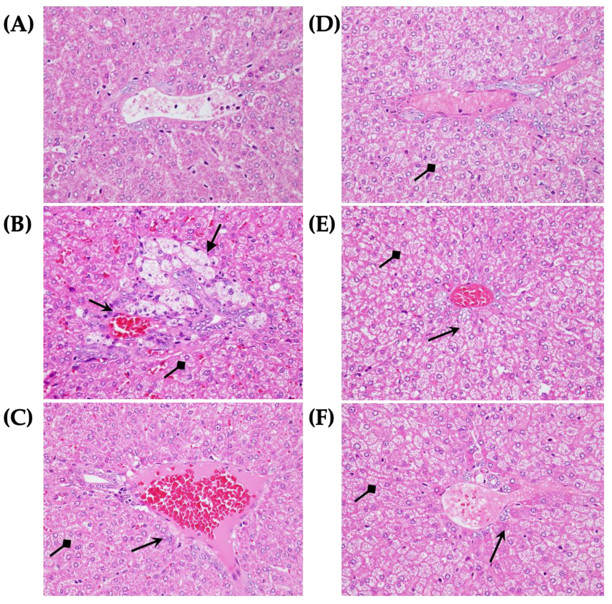
3.3. Morphological and Pathological Slices

Figure 1 shows the liver injuries in the HFC-fed hamsters. The liver pathological sections indicated that, the blank group had many fat particles accumulated in the hepatocytes and moderate-to-severe liver damage, fat infiltration, fatty degeneration, and necrosis when compared to the control group. However, there was no evident difference in the liver pathological sections of the SIM and the low, medium, and high dose of PME3-1 groups. In the histopathological statistics, the livers of the control group showed significant fatty change and infiltration, and the kidneys demonstrated chronic progressive nephrosis (CPN). After PME3-1 was continuously fed to the hamsters for 12 weeks, only the medium- and high-dose groups showed a slight improvement in the macro-fatty change in the livers and CPN lesions in the kidneys, while the rest showed no obvious improvement trend (Table 4).
Oxidative stress causes lipids in cell membranes to mutate. This leads to abnormal lipid metabolisms such as total cholesterol, triglycerides, low-density lipoprotein, and MDA in the blood. These will increase with a higher oxidative stress in the body [41,42]. In addition, patients with an abnormal lipid metabolism have a higher risk of arteriosclerosis [43]. Therefore, the production of many serum enzymes such as cholesterol, TG, LDL, HDL, and TBARs is regarded as a biochemical indicator of lipid metabolism. This research confirmed that feeding hamsters with PME3-1 could significantly protect the hamsters from HFC-diet-induced hyperlipidemia by reducing the cholesterol and TG content in the serum, reducing the LDL/HDL ratio, and inhibiting lipid peroxidation.

3.4. Lipid and Cholesterol Content of Liver Tissue and Feces

A previous study confirmed that Pinus morrisonicola hay of Taiwan can regulate blood pressure in rats, reduce the phenomenon of arterial wall hypertrophy, and reduce the content of plasma cholesterol and low-density lipoprotein cholesterol in hamsters on HFC diets [8]. Lee [44] found that pine needle powder (Pinus densiflora Sieb. et Zucc.) can reduce total cholesterol and thiobarbituric acid reactive substances (TBARS) in rats fed a high-cholesterol diet. Park et al. [45] also found that pine needle oil can improve ethanol-induced liver cell damage and hyperlipidemia in rats. Jeon and Kim [46] proved that pine needle water extracts can reduce hyperlipidemia induced by a high-fat diet.
Table 5 shows the effect of PME3-1 on the production of total fat, total cholesterol, and triglycerides in the livers and feces of hamsters treated with a high-fat diet. When compared with the blank group, the total fat, total cholesterol, and triglycerides in the livers of hamsters increased by 4.2, 1.2, and 1.2 times, respectively. After 12 weeks of continuously feeding hamsters with SIM and low, medium, and high doses of PME3-1, the total fat, total cholesterol, and triglycerides in the liver tissue had significantly reduced (p < 0.05). When the hamsters were fed with an HFC diet, the total cholesterol in their feces decreased by 1.8 times and the triglycerides increased by 2.2 times. The amount of feces produced by the hamsters also decreased. Compared with the control group, the total cholesterol and triglycerides increased significantly (p < 0.05).
The fecal triglycerides of the control group were significantly higher than that of the blank group (p < 0.05). However, the cholesterol level was lower (p < 0.05). This phenomenon may be because excess cholesterol is stored in the liver due to some body mechanism, thus reducing the concentration of cholesterol in the feces (Table 5). There are two main methods of cholesterol excretion. One is through the action of cholesterol 7α-hydroxylase in the liver, the limiting enzyme responsible for the conversion of cholesterol and bile acid, which converts cholesterol into bile acid and excretes it as feces. The second is excretion in the form of neutral steroids [47]. In the past, studies have pointed out that natural substances can combine with cholesterol in the diet to inhibit the absorption of cholesterol, alter enterohepatic circulation, and accelerate the metabolism of cholesterol in the body. Reducing blood cholesterol promotes the excretion of bile and cholesterol in the intestine and increases the excretion of neutral/acid sterols and bile acid [48,49]. Since PME3-1 can increase the content of cholesterol in the feces of HFC diet-fed hamsters, it is inferred that PME3-1 may promote the excretion of cholesterol in the feces, rather than binding to cholesterol directly.
A high-fat diet will increase the synthesis of fatty acids in the liver, and excess free fatty acids will be transported to the liver, reducing the β-oxidation of fatty acids and causing fat accumulation in the liver. In turn, this induces liver cell dysfunction and damages the liver parenchyma [50]. Many studies on blood-lipid-lowering mechanisms by natural products demonstrate that most of them can reduce lipids and cholesterol by inhibiting lipid and cholesterol absorption and synthesis and promoting decomposition and anti-oxidation. In this study, PME3-1 not only inhibited the HFC-diet-induced cholesterol, triglyceride, and LDL-C levels in the serum effectively, but also significantly reduced the total lipid, TC, and TG levels in the liver (p < 0.05). However, its high-dose inhibitory ability showed no significant difference from the SIM group (p > 0.05). The TG and TC contents excreted in the feces were also significantly higher than those of the control group (p < 0.05) but demonstrated no significant difference from the SIM group (p > 0.05). It is speculated, therefore, that PME3-1 and SIM have a similar mechanism for inhibiting high-fat-diet-induced hyperlipidemia.

3.5. Antioxidant Capacity and Cholesterol Synthesis Inhibition

From Table 6, it can be seen that in hamsters with a high cholesterol level induced by a high-fat diet, the presence of the lipid peroxidation product MDA in the liver tissue increased significantly: approximately 2.09 times (p < 0.05). Compared to the control group, when low, medium, or high doses of PME3-1 were fed to the hamsters for 12 weeks continuously, the MDA formation induced by the high-fat diet was reduced (p < 0.05). Additionally, there was no significant difference between the SIM, M-PME3-1, and H-PME3-1 groups (p < 0.05). The effects of a high-fat diet on GST and GPx activities in the liver tissues of hamsters with liver injury can be seen in Table 6. In comparison to the blank group, the activities of GST and GPx decreased by approximately 14.29% and 8.01%, respectively. When hamsters were fed with low, medium, and high doses of PME3-1 for 12 weeks, the GPx activity in the hamster liver tissue increased by approximately 0.04, 0.04, and 0.17 times, respectively. It is clear that the H-PME3-1 group was better than the SIM group (156.3 > 141.3). GST levels showed no significant difference between the three groups. The low, medium, and high dose PME3-1 groups all had slightly increased GST levels of approximately 0.15, 0.23, and 0.21 times, respectively, which was, again, better than in the SIM group.
HMG-CoA reductase is the most critical enzyme (the rate-limiting enzyme) in the process of cholesterol synthesis. This process reduces HMG-CoA to mevalonate and synthesizes the cholesterol [51]. HMG-CoA reductase is thus the most important indicator of cholesterol synthesis. As can be seen from Figure 2, compared with the blank group, the HMG-CoA reductase content in the liver was slightly increased by approximately 0.30 times. The low-dose, medium-dose, and high-dose groups all inhibited the formation of HMG-CoA reductase, with figures of around 0.3%, 17.5%, and 24.1%, respectively, and there was no significant difference between the groups with the middle and high doses of PME3-1 and the SIM group (an inhibition of approximately 28.6%).

4. Conclusions

This study confirmed that PME3-1 has a reduced ability on TC, TG, the LDL/HDL ratio, and the atherosclerotic index in hamster serum under a high fat and cholesterol diet. In addition, the content of lipid peroxidation product MDA and the activity of HMG-CoA reductase in the liver decreased as well. The excretion of TC and TG in feces was also increased. It is clear that PME3-1 is an effective inhibitor of cardiovascular-related diseases. It can be speculated that the hypolipidemic mechanism of PME3-1 is related to the inhibition of cholesterol, lipid synthesis, and lipid peroxidation in the liver and the promotion of fecal cholesterol excretion in hamsters. However, the subcutaneous mechanism of PME3-1 requires further investigation. In summary, PME3-1 has the potential to reduce serum hyperlipidemia and lipid accumulation in the liver. This is helpful for developing nutraceuticals for preventing cardiovascular-related diseases in the future.

Author Contributions

Conceptualization, M.-C.C. and T.-Y.S.; methodology, C.-L.C.; formal analysis, C.-L.C. and M.-C.C.; resources, H.-C.C.; writing—original draft preparation, Y.-J.L. and T.-Y.S.; writing—review and editing, Y.-J.L. and T.-Y.S.; visualization, H.-C.C.; data curation, C.-L.C. All authors have read and agreed to the published version of the manuscript.

Funding

This research received no external funding.

Institutional Review Board Statement

Not applicable.

Data Availability Statement

The datasets generated for this study are available upon request from the corresponding author.

Conflicts of Interest

The authors declare no conflict of interest.

Appendix A

Figure A1. Effects of PME3-1 on the body weight change in hamsters fed with high fat and cholesterol diet. The HFC-diet hamsters were orally supplemented with low (L-PME3-1), middle (M-PME3-1) and high (H-PME3-1) doses of PME3-1 (0.2, 1.0, 5.0 mg/kg b.w.) or Simvastatin (SIM) (5.0 mg/kg b.w.). Body weights were measured once a week. Each value is expressed as mean ± standard deviation (n = 8). Value (a–c) with different letters within different groups at the same week indicates significant difference (p < 0.05).
Agriculture 13 00535 g0a1

References

  1. Grundy, S.M. Cholesterol and coronary heart disease: A new era. JAMA 1986, 256, 2849–2858. [Google Scholar] [CrossRef] [PubMed]
  2. Diaz, M.N.; Frei, B.; Vita, J.A.; Keaney, J.F., Jr. Antioxidants and atherosclerotic heart disease. N. Engl. J. Med. 1997, 337, 408–416. [Google Scholar] [CrossRef] [PubMed]
  3. Jia, Y.J.; Xu, R.X.; Sun, J.; Tang, Y.; Li, J.J. Enhanced circulating PCSK9 concentration by berberine through SREBP-2 pathway in high fat diet-fed rats. J. Transl. Med. 2014, 12, 1–8. [Google Scholar] [CrossRef] [PubMed] [Green Version]
  4. Brown, M.S.; Goldstein, J.L. Receptor-mediated endocytosis: Insights from the lipoprotein receptor system. Proc. Natl. Acad. Sci. USA 1979, 76, 3330–3337. [Google Scholar] [CrossRef] [PubMed] [Green Version]
  5. Horton, J.D.; Goldstein, J.L.; Brown, M.S. SREBPs: Activators of the complete program of cholesterol and fatty acid synthesis in the liver. J. Clin. Investig. 2002, 109, 1125–1131. [Google Scholar] [CrossRef]
  6. Maron, D.J.; Fazio, S.; Linton, M.F. Current perspectives on statins. Circulation 2000, 101, 207–213. [Google Scholar] [CrossRef] [Green Version]
  7. Sudhop, T.; Reber, M.; Tribble, D.; Sapre, A.; Taggart, W.; Gibbons, P.; Musliner, T.; von Bergmann, K.; Lütjohann, D. Changes in cholesterol absorption and cholesterol synthesis caused by ezetimibe and/or simvastatin in men. J. Lipid Res. 2009, 50, 2117–2123. [Google Scholar] [CrossRef] [Green Version]
  8. Hsu, T.Y.; Sheu, S.C.; Liaw, E.T.; Wang, T.C.; Lin, C.C. Anti-oxidant activity and effect of Pinus morrisonicola Hay. on the survival of leukemia cell line U937. Phytomedicine 2005, 12, 663–669. [Google Scholar] [CrossRef]
  9. Yen, G.C.; Duh, P.D.; Huang, D.W.; Hsu, C.L.; Fu, T.Y.C. Protective effect of pine (Pinus morrisonicola Hay.) needle on LDL oxidation and its anti-inflammatory action by modulation of iNOS and COX-2 expression in LPS-stimulated RAW 264.7 macrophages. Food Chem. Toxicol. 2008, 46, 175–185. [Google Scholar] [CrossRef]
  10. Chiu, H.F.; Wang, H.M.; Shen, Y.C.; Venkatakrishnan, K.; Wang, C.K. Anti-inflammatory properties of fermented pine (Pinus morrisonicola Hay.) needle on lipopolysaccharide-induced inflammation in RAW 264.7 macrophage cells. J. Food Biochem. 2019, 43, e12994. [Google Scholar] [CrossRef]
  11. Nyman, B.F. Protein-proanthocyanidin interactions during extraction of Scots pine needles. Phytochemistry 1985, 24, 2939–2944. [Google Scholar] [CrossRef]
  12. Fang, J.M.; Chang, C.F.; Cheng, Y.S. Flavonoids from Pinus morrisonicola. Phytochemistry 1987, 26, 2559–2561. [Google Scholar] [CrossRef]
  13. Fang, J.M.; Su, W.C.; Cheng, Y.S. Flavonoids and stilbenes from armand pine. Phytochemistry 1988, 27, 1395–1397. [Google Scholar] [CrossRef]
  14. Kong, Z.; Liu, Z.; Ding, B. Study on the antimutagenic effect of pine needle extract. Mutat. Res. Lett. 1995, 347, 101–104. [Google Scholar] [CrossRef] [PubMed]
  15. Chen, M.H.; Huang, T.C. Volatile and nonvolatile constituents and antioxidant capacity of oleoresins in three Taiwan citrus varieties as determined by supercritical fluid extraction. Molecules 2016, 21, 1735. [Google Scholar] [CrossRef] [PubMed] [Green Version]
  16. Coelho, J.A.P.; Pereira, A.P.; Mendes, R.L.; Palavra, A.M.F. Supercritical carbon dioxide extraction of Foeniculum vulgare volatile oil. Flavour Fragr. J. 2003, 18, 316–319. [Google Scholar] [CrossRef]
  17. Guan, W.; Li, S.; Yan, R.; Tang, S.; Quan, C. Comparison of essential oils of clove buds extracted with supercritical carbon dioxide and other three traditional extraction methods. Food Chem. 2007, 101, 1558–1564. [Google Scholar] [CrossRef]
  18. Carretero, M.E.; López-Pérez, J.L.; Abad, M.J.; Bermejo, P.; Tillet, S.; Israel, A.; Noguera-P, B. Preliminary study of the anti-inflammatory activity of hexane extract and fractions from Bursera simaruba (Linneo) Sarg.(Burseraceae) leaves. J. Ethnopharmacol. 2008, 116, 11–15. [Google Scholar] [CrossRef] [PubMed]
  19. Cheng, M.C.; Lin, L.Y.; Yu, T.H.; Peng, R.Y. Hypolipidemic and antioxidant activity of mountain celery (Cryptotaenia japonica Hassk) seed essential oils. J. Agric. Food Chem. 2008, 56, 3997–4003. [Google Scholar] [CrossRef] [PubMed]
  20. Jung, M.J.; Jung, H.A.; Kang, S.S.; Hwang, G.S.; Choi, J.S. A new abietic acid-type diterpene glucoside from the needles of Pinus densiflora. Arch. Pharmacal Res. 2009, 32, 1699–1704. [Google Scholar] [CrossRef]
  21. Cheng, M.C.; Chang, W.H.; Chen, C.W.; Li, W.W.; Tseng, C.Y.; Song, T.Y. Antioxidant properties of essential oil extracted from Pinus morrisonicola hay needles by supercritical fluid and identification of possible active compounds by GC/MS. Molecules 2015, 20, 19051–19065. [Google Scholar] [CrossRef] [Green Version]
  22. Lin, L.Y.; Chuang, C.H.; Chen, H.C.; Yang, K.M. Lime (Citrus aurantifolia (Christm.) Swingle) essential oils: Volatile compounds, antioxidant capacity, and hypolipidemic effect. Foods 2019, 8, 398. [Google Scholar] [CrossRef] [Green Version]
  23. Folch, J.; Lees, M.; Sloane Stanley, G.H. A simple method for the isolation and purification of total lipids from animal tissues. J. Biol. Chem. 1957, 226, 497–509. [Google Scholar] [CrossRef] [PubMed]
  24. Fletcher, M.J. A colorimetric method for estimating serum triglycerides. Clin. Chim. Acta 1968, 22, 393–397. [Google Scholar] [CrossRef] [PubMed]
  25. Sperry, W.M.; Webb, M. A revision of the Schoenheimer-Sperry method for cholesterol determination. J. Biol. Chem. 1950, 187, 97–106. [Google Scholar] [CrossRef] [PubMed]
  26. Dierickx, P.J. The influence of picolines on glutathione transferase activity and subunit composition in human liver derived Hep G2 cells. Biochem. Pharmacol. 1994, 48, 1976–1978. [Google Scholar] [CrossRef]
  27. Mohandas, J.; Marshall, J.J.; Duggin, G.G.; Horvath, J.S.; Tiller, D.J. Differential distribution of glutathione and glutathione-related enzymes in rabbit kidney: Possible implications in analgesic nephropathy. Biochem. Pharmacol. 1984, 33, 1801–1807. [Google Scholar] [CrossRef]
  28. Richard, M.J.; Guiraud, P.; Meo, J.; Favier, A. High-performance liquid chromatographic separation of malondialdehyde—Thiobarbituric acid adduct in biological materials (plasma and human cells) using a commercially available reagent. J. Chromatogr. B Biomed. Sci. Appl. 1992, 577, 9–18. [Google Scholar] [CrossRef]
  29. Ko, W.S.; Guo, C.H.; Yeh, M.S.; Lin, L.Y.; Hsu, G.S.W.; Chen, P.C.; Luo, M.C.; Lin, C.Y. Blood micronutrient, oxidative stress, and viral load in patients with chronic hepatitis C. World J. Gastroenterol. WJG 2005, 11, 4697. [Google Scholar] [CrossRef]
  30. Spady, D.K.; Turley, S.D.; Dietschy, J.M. Dissociation of hepatic cholesterol synthesis from hepatic low-density lipoprotein uptake and biliary cholesterol saturation in female and male hamsters of different ages. Biochim. Biophys. Acta (BBA)-Lipids Lipid Metab. 1983, 753, 381–392. [Google Scholar] [CrossRef]
  31. Lee, C.L.; Tsai, T.Y.; Wang, J.J.; Pan, T.M. In vivo hypolipidemic effects and safety of low dosage Monascus powder in a hamster model of hyperlipidemia. Appl. Microbiol. Biotechnol. 2006, 70, 533–540. [Google Scholar] [CrossRef]
  32. Newton, J.L. Systemic symptoms in non-alcoholic fatty liver disease. Dig. Dis. 2010, 28, 214–219. [Google Scholar] [CrossRef]
  33. Matos, S.L.; Paula, H.D.; Pedrosa, M.L.; Santos, R.C.D.; Oliveira, E.L.D.; Chianca Júnior, D.A.; Silva, M.E. Dietary models for inducing hypercholesterolemia in rats. Braz. Arch. Biol. Technol. 2005, 48, 203–209. [Google Scholar] [CrossRef] [Green Version]
  34. Hartvigsen, K.; Binder, C.J.; Hansen, L.F.; Rafia, A.; Juliano, J.; Hörkkö, S.; Steinberg, D.; Palinski, W.; Witztum, J.L.; Li, A.C. A diet-induced hypercholesterolemic murine model to study atherogenesis without obesity and metabolic syndrome. Arterioscler. Thromb. Vasc. Biol. 2007, 27, 878–885. [Google Scholar] [CrossRef]
  35. Gan, S.K.; Watts, G.F. Is adipose tissue lipolysis always an adaptive response to starvation?: Implications for non-alcoholic fatty liver disease. Clin. Sci. 2008, 114, 543–545. [Google Scholar] [CrossRef] [Green Version]
  36. Zeng, W.C.; Zhang, Z.; Gao, H.; Jia, L.R.; He, Q. Chemical composition, antioxidant, and antimicrobial activities of essential oil from pine needle (Cedrus deodara). J. Food Sci. 2012, 77, C824–C829. [Google Scholar] [CrossRef]
  37. Arslan, U.; Türkoglu, S.; Balcioglu, S.; Tavil, Y.; Karakan, T.; Çengel, A. Association between nonalcoholic fatty liver disease and coronary artery disease. Coron. Artery Dis. 2007, 18, 433–436. [Google Scholar] [CrossRef]
  38. Hamden, K.; Mnafgui, K.; Amri, Z.; Aloulou, A.; Elfeki, A. Inhibition of key digestive enzymes related to diabetes and hyperlipidemia and protection of liver-kidney functions by trigonelline in diabetic rats. Sci. Pharm. 2013, 81, 233–246. [Google Scholar] [CrossRef] [Green Version]
  39. Liu, C.H.; Huang, M.T.; Huang, P.C. Sources of triacylglycerol accumulation in livers of rats fed a cholesterol-supplemented diet. Lipids 1995, 30, 527–531. [Google Scholar] [CrossRef]
  40. Guerin, M.; Dolphin, P.J.; Chapman, M.J. A new in vitro method for the simultaneous evaluation of cholesteryl ester exchange and mass transfer between HDL and apoB-containing lipoprotein subspecies. Identification of preferential cholesteryl ester acceptors in human plasma. Arterioscler. Thromb. J. Vasc. Biol. 1994, 14, 199–206. [Google Scholar] [CrossRef] [Green Version]
  41. Kugiyama, K.; Doi, H.; Motoyama, T.; Soejima, H.; Misumi, K.; Kawano, H.; Nakagawa, O.; Yoshimura, M.; Ogawa, H.; Matsumura, T.; et al. Association of remnant lipoprotein levels with impairment of endothelium-dependent vasomotor function in human coronary arteries. Circulation 1998, 97, 2519–2526. [Google Scholar] [CrossRef] [Green Version]
  42. Chan, S.H.; Tai, M.H.; Li, C.Y.; Chan, J.Y. Reduction in molecular synthesis or enzyme activity of superoxide dismutases and catalase contributes to oxidative stress and neurogenic hypertension in spontaneously hypertensive rats. Free Radic. Biol. Med. 2006, 40, 2028–2039. [Google Scholar] [CrossRef]
  43. Castelli, W.P.; Garrison, R.J.; Wilson, P.W.; Abbott, R.D.; Kalousdian, S.; Kannel, W.B. Incidence of coronary heart disease and lipoprotein cholesterol levels: The Framingham Study. JAMA 1986, 256, 2835–2838. [Google Scholar] [CrossRef]
  44. Lee, E. Effects of powdered pine needle (Pinus densiflora seib et Zucc.) on serum and liver lipid composition and antioxidative capacity in rats fed high oxidized fat. J. Korean Soc. Food Sci. Nutr. 2003, 32, 926–930. [Google Scholar] [CrossRef]
  45. Park, K.J.; Kim, K.S.; Ahn, K.H.; Rhee, J.S. Pine needle oil and Korean medicinal herb complex protect hyperlipidemia and liver cell damage induced by alcohol. Korean J. Environ. Biol. 2003, 21, 410–414. [Google Scholar]
  46. Jeon, J.R.; Kim, J.Y. Effects of pine needle extract on differentiation of 3T3-L1 preadipocytes and obesity in high-fat diet fed rats. Biol. Pharm. Bull. 2006, 29, 2111–2115. [Google Scholar] [CrossRef] [Green Version]
  47. Murakami, S.; Kondo, Y.; Toda, Y.; Kitajima, H.; Kameo, K.; Sakono, M.; Fukuda, N. Effect of taurine on cholesterol metabolism in hamsters: Up-regulation of low density lipoprotein (LDL) receptor by taurine. Life Sci. 2002, 70, 2355–2366. [Google Scholar] [CrossRef]
  48. Yang, T.T.; Koo, M.W. Chinese green tea lowers cholesterol level through an increase in fecal lipid excretion. Life Sci. 1999, 66, 411–423. [Google Scholar] [CrossRef]
  49. Muramatsu, K.; Fukuyo, M.; Hara, Y. Effect of green tea catechins on plasma cholesterol level in cholesterol-fed rats. J. Nutr. Sci. Vitaminol. 1986, 32, 613–622. [Google Scholar] [CrossRef] [Green Version]
  50. Sheth, S.G.; Gordon, F.D.; Chopra, S. Nonalcoholic steatohepatitis. Ann. Intern. Med. 1997, 126, 137–145. [Google Scholar] [CrossRef]
  51. Massy, Z.A.; Keane, W.F.; Kasiske, B.L. Inhibition of the mevalonate pathway: Benefits beyond cholesterol reduction? Lancet 1996, 347, 102–103. [Google Scholar] [CrossRef]
Figure 1. Pathological section images of the livers in high fat and cholesterol diet hamsters: (A) blank, (B) control, (C) SIM, (D) L-PME3-1, (E) M-PME3-1, and (F) H-PME3-1.
Agriculture 13 00535 g001
Figure 2. Effects of PME3-1 on HMG-CoA reductase capacity of the liver tissue in high fat and cholesterol diet in hamsters. Each value is expressed as mean ± standard deviation (n = 8). Value (a–c) with different letters within the same row indicate significant difference (p < 0.05).
Agriculture 13 00535 g002
Table 1. The animal feed formula for testing the effect of hypolipidemic and antioxidant of PME3-1 in male hamsters.
GroupBlankControlSIML-PME3-1M-PME3-1H-PME3-1
Feed composition (%)
Casein202020202020
Sucrose3524.824.824.824.824.8
Corn starch303030303030
Corn oil41212121212
Lard133333
Cholesterol00.20.20.20.20.2
Ain 76 mineral3.53.53.53.53.53.5
Ain 76 vitamin111111
A-cellulose555555
D,L-methionine0.30.30.30.30.30.3
Choline bitartrate0.20.20.20.20.20.2
Total100100100100100100
Supplementary addition (mg/kg b.w.)
SIM--5.00.00.00.0
PME3-1--0.00.21.05.0
PME3-1 is the first separated fraction by column and thin layer chromatography of PME3, which is the third separated fraction extracted by supercritical CO2 from Pinus morrisonicola. L-PME3-1, M-PME3-1, and H-PME3-1 are represented as low, medium, and high doses of PME3-1, respectively. SIM is the abbreviation of Simvastatin.
Table 2. Effects of PME3-1 on the relative organ weights of hamsters fed with high fat and cholesterol diet.
TreatmentLiverKidneySpleen
Blank3.9 ± 0.8 c1.1 ± 0.3 b0.12 ± 0.08 a
Control6.3 ± 0.8 a2.1 ± 0.5 a0.04 ± 0.07 b
SIM3.5 ± 0.7 c1.2 ± 0.2 b0.04 ± 0.08 b
L-PME3-15.2 ± 1.0 b1.8 ± 0.6 a0.06 ± 0.09 b
M-PME3-14.0 ± 0.6 bc1.3 ± 0.3 b0.05 ± 0.07 b
H-PME3-13.4 ± 0.3 c1.4 ± 0.2 b0.10 ± 0.04 a
Each value is expressed as mean ± standard deviation (n = 8). Values (a–c) with different letters within the same column indicates significant difference (p < 0.05). The unit of relative tissue weight is g/100 g b.w. L-PME3-1, M-PME3-1, and H-PME3-1 are represented as low, medium, and high doses of PME3-1, respectively. SIM is the abbreviation of Simvastatin.
Table 3. Effects of PME3-1 on serum biochemical index of hamsters fed with high fat and cholesterol diet.
TreatmentTC 1TG 2HDL-C 3LDL-C 4LDL/HDL RatioAtherogenic Index 5
Blank93.2 ± 18.3 c205.3 ± 22.7 bc53.7 ± 17.1 c11.2 ± 3.0 c0.200.75
Control240.1 ± 17.8 a278.0 ± 90.1 a100.2 ± 15.9 a48.6 ± 9.1 a0.481.40
SIM91.5 ± 23.6 c232.2 ± 46.1 ab50.5 ± 22.8 c13.6 ± 2.0 c0.260.82
L-PME3-1159.7 ± 12.9 b181.7 ± 98.2 bc79.3 ± 10.2 b20.1 ± 4.1 bc0.251.01
M-PME3-1139.0 ± 13.5 b184.0 ± 54.6 c72.4 ± 15.5 bc15.5 ± 3.0 c0.210.93
H-PME3-185.3 ± 13.9 c181.3 ± 20.4 c65.0 ± 9.8 bc13.8 ± 3.0 c0.200.31
Each value is expressed as mean ± standard deviation (n = 8). Value (a–c) with different letters within the same column indicates significant difference (p < 0.05). L-PME3-1, M-PME3-1, and H-PME3-1 are represented as low, medium, and high doses of PME3-1, respectively. SIM is the abbreviation of Simvastatin. 1 TC means total cholesterol. (Unit: mg/g). 2 TG means triglyceride. (Unit: mg/g). 3 HDL-C means low-density lipoprotein cholesterol. (Unit: mg/g). 4 LDL-C means high-density lipoprotein cholesterol. (Unit: mg/g). 5 Atherogenic index = (Total-C-HDL-C)/HDL-C.
Table 4. The effects of PME3-1 on histopathological changes of livers in high fat and cholesterol diet in hamsters.
OrganHistopathologyBlankControlSIML-PME3-1M-PME3-1H-PME3-1
LiverInfiltration fat diffuse1.8 ± 0.4 b4.4 ± 0.7 a3.9 ± 0.3 a4.1 ± 0.6 a4.0 ± 0.7 a4.2 ± 0.4 a
Micro fatty change1.8 ± 0.4 ab4.4 ± 0.7 a3.9 ± 0.3 a4.1 ± 0.6 a4.0 ± 0.7 a4.2 ± 0.4 a
Macro fatty changeN.D.0.4 ± 0.7 a0.4 ± 0.7 a0.4 ± 0.5 a0.1 ± 0.3 ab0.1 ± 0.3 ab
Infiltration mononuclear cells0.3 ± 0.7 b0.8 ± 0.7 a1.1 ± 0.8 a1.0 ± 0.5 a0.9 ± 0.6 a0.7 ± 0.5 ab
Necrosis0.6 ± 0.7 a0.5 ± 0.7 a1.0 ± 0.9 a0.3 ± 0.7 a0.2 ± 0.6 a0.4 ± 0.5 a
KidneyCPN 11.1 ± 0.0 c4.4 ± 1.3 a3.1 ± 2.0 bc4.4 ± 1.0 a3.8 ± 1.6 ab3.5 ± 1.5 ab
Each value is expressed as mean ± standard deviation (n = 8). Values (a–c) with different letters within the same row indicate significant difference (p < 0.05). L-PME3-1, M-PME3-1, and H-PME3-1 are represented as low, medium, and high doses of PME3-1, respectively. SIM is the abbreviation of Simvastatin. N.D. means not detected. 1 CPN: chronic progressive nephrosis.
Table 5. Effects of PME3-1 on lipids and cholesterol content of the liver tissue and fecal in high fat and cholesterol diet in hamsters.
OrganParameterBlankControlSIML-PME3-1M-PME3-1H-PME3-1
LiverTL 130.3 ± 2.9 f126.0 ± 4.3 a60.3 ± 6.9 e113 ± 2.0 b92.3 ± 3.2 c70.3 ± 4.6 de
TC 215.5 ± 0.9 c18.3 ± 0.4 a15.7 ± 0.9 c18.9 ± 1.2 a17.4 ± 0.6 bc16.0 ± 0.6 c
TG 320.6 ± 0.6 b24.2 ± 0.7 a20.9 ± 1.0 b24.2 ± 1.0 a21.0 ± 0.6 b20.6 ± 0.9 b
Dried fecalTC 225.1 ± 3.0 a13.7 ± 0.4 e17.4 ± 1.6 bc14.5 ± 2.2 de14.7 ± 3.4 de16.1 ± 1.3 cd
TG 310.8 ± 1.3 d23.6 ± 2.0 c27.7 ± 1.0 ab27.0 ± 2.0 ab24.3 ± 4.9 bc29.0 ± 2.5 ab
Each value is expressed as mean ± standard deviation (n = 8). Values (a–f) with different letters within the same row indicate significant difference (p < 0.05). L-PME3-1, M-PME3-1, and H-PME3-1 are represented as low, medium, and high doses of PME3-1, respectively. SIM is the abbreviation of Simvastatin. 1 TL means total lipid. (Unit: mg/g). 2 TC means total cholesterol. (Unit: mg/g). 3 TG means triglyceride. (Unit: mg/g).
Table 6. Effects of PME3-1 on antioxidant activities of the liver tissue in high fat and cholesterol diet in hamsters.
ParameterBlankControlSIML-PME3-1M-PME3-1H-PME3-1
MDA 1107.3 ± 48.1 c331.4 ± 93.7 ab206.2 ± 57.1 bc378.4 ± 79.0 a208.1 ± 56.2 abc269.7 ± 33.1 abc
GPx 2144.8 ± 27.8 bc133.2 ± 29.6 c141.3 ± 29.0 bc138.1 ± 16.5 bc138.5 ± 16.3 bc156.3 ± 20.9 ab
GST 349.0 ± 6.47 a42.0 ± 7.03 a43.4 ± 7.1 a48.1 ± 6.3 a51.7 ± 8.0 a50.8 ± 10.7 a
Each value is expressed as mean ± standard deviation (n = 8). Value (a–c) with different letters within the same row indicates significant difference (p < 0.05). L-PME3-1, M-PME3-1, and H-PME3-1 are represented as low, medium, and high doses of PME3-1, respectively. SIM is the abbreviation of Simvastatin. 1 MDA means malondialdehyde. (Unit: µmol. MDA/g protein). 2 GPx means glutathione peroxidase. (Unit: µmol. NADPH/min/g protein). 3 GST means glutathione S-transferase. (Unit: mol. CDNB-GSH/min/g protein).
Disclaimer/Publisher’s Note: The statements, opinions and data contained in all publications are solely those of the individual author(s) and contributor(s) and not of MDPI and/or the editor(s). MDPI and/or the editor(s) disclaim responsibility for any injury to people or property resulting from any ideas, methods, instructions or products referred to in the content.

