Article

OTUD7A Regulates Inflammation- and Immune-Related Gene Expression in Goose Fatty Liver

1
College of Animal Science and Technology, Yangzhou University, Yangzhou 225009, China
2
Joint International Research Laboratory of Agriculture and Agri-Product Safety of the Ministry of Education of China, Yangzhou University, Yangzhou 225009, China
*
Author to whom correspondence should be addressed.
Agriculture 2022, 12(1), 105; https://doi.org/10.3390/agriculture12010105
Submission received: 28 November 2021 / Revised: 9 January 2022 / Accepted: 11 January 2022 / Published: 13 January 2022
(This article belongs to the Special Issue Application of Genetics and Genomics in Livestock Production)

Abstract

:
OTU deubiquitinase 7A (OTUD7A) can suppress inflammation signaling pathways, but it is unclear whether the gene can inhibit inflammation in goose fatty liver. In order to investigate the functions of OTUD7A and identify the genes and pathways subjected to the regulation of OTUD7A in the formation of goose fatty liver, we conducted transcriptomic analysis of cells, which revealed several genes related to inflammation and immunity that were significantly differentially expressed after OTUD7A overexpression. Moreover, the expression of interferon-induced protein with tetratricopeptide repeats 5 (IFIT5), tumor necrosis factor ligand superfamily member 8 (TNFSF8), sterile alpha motif domain-containing protein 9 (SAMD9), radical S-adenosyl methionine domain-containing protein 2 (RSAD2), interferon-induced GTP-binding protein Mx1 (MX1), and interferon-induced guanylate binding protein 1-like (GBP1) was inhibited by OTUD7A overexpression but induced by OTUD7A knockdown with small interfering RNA in goose hepatocytes. Furthermore, the mRNA expression of IFIT5, TNFSF8, SAMD9, RSAD2, MX1, and GBP1 was downregulated, whereas OTUD7A expression was upregulated in goose fatty liver after 12 days of overfeeding. In contrast, the expression patterns of these genes showed nearly the opposite trend after 24 days of overfeeding. Taken together, these findings indicate that OTUD7A regulates the expression of inflammation- and immune-related genes in the development of goose fatty liver.

1. Introduction

Some fish and birds are able to pre-deposit large amounts of fat in the liver for use during migration, and then the liver can return to its normal state without any obvious pathological symptoms. Geese, as the offspring of migratory birds, also have this characteristic. In agricultural production, this ability of geese is often used for fatty liver production. As a well-known liver-producing species, the fatty liver (typically composed of approximately 60% fat) of Landes geese can reach an 8–10-fold higher weight than the normal liver in a short period through overfeeding [1]. The changes that occur in goose fatty liver are physiological, with no overt injury or pathological symptoms [2,3]. Recent work indicates that the pro-inflammatory factor is suppressed in goose fatty liver vs. normal liver [4]. However, nonalcoholic fatty liver disease (NAFLD) in humans and mammals is frequently accompanied by inflammation [5,6]. Human NAFLD is prone to developing from simple steatosis to nonalcoholic steatohepatitis (NASH), cirrhosis, and even liver cancer, posing a serious threat to human health [7]. In addition, the incidence of fatty liver in livestock and poultry has also increased due to improved feed nutrition levels, reduced animal activity, and increased environmental stress in modern intensive livestock production, thereby bringing economic losses to livestock production [8]. The differences between geese and other animals suggest that the goose liver utilizes a protection mechanism, although the underlying mechanisms remain unclear. Studies of this mechanism may provide information for developing approaches for preventing or treating NAFLD-associated complications in humans and economically important animals.
A previous study demonstrated that inflammation is important in the progression from simple steatosis to NASH [9]. The hepatic inflammatory response plays an important role in insulin resistance, oxidative stress, and endoplasmic reticulum stress. Inflammation can interact with insulin resistance, and inflammatory factors can interfere with insulin signaling pathways and deepen the degree of insulin resistance, leading to a further decrease in insulin sensitivity in liver cells. On the other hand, inflammation causes the activation of Kupffer cells and stellate cells in the liver, and induces the development of liver fibrosis and cirrhosis [10]. Therefore, inflammation has been widely studied as a possible target for NASH therapy. The nuclear factor κB (NF-κB) signaling pathway is activated in human and animal NAFLD models, and the pathway is a key pro-inflammatory signaling pathway leading to the progression of NAFLD to NASH [5,6]. Inhibition of the NF-κB pathway has been reported to reduce the expression of inflammatory factors in the liver and alleviate the progression of NAFLD [11].
The results of our preliminary transcriptomic analysis showed that a number of deubiquitinating enzymes, including ovarian tumor deubiquitinase 7A (OTUD7A), were significantly altered at the transcriptional level in the livers of overfed geese compared to controls, suggesting that deubiquitinating modifications are involved in the formation of fatty liver in geese. OTUD7A, also known as cellular zinc-finger anti-NF-κB 2, is a member of the ovarian tumor deubiquitinase family [12,13]. OTUD7A promotes deubiquitination of its target proteins, including tumor necrosis factor receptor-associated factor 6 (TRAF6) [13], thus suppressing the NF-κB signaling pathway. Thus, we speculated that OTUD7A could inhibit inflammation. However, the functions of OTUD7A in the development of NAFLD are unclear. Therefore, we investigated the function of OTUD7A, and identified genes and pathways involved in regulating OTUD7A, using goose fatty liver as a model.

2. Materials and Methods

2.1. Animal Experiment

Thirty-two healthy 63-day-old male Landes geese were selected and randomly assigned to a control group and an overfeeding group (16 geese per group). Geese in the control group were provided water and feed ad libitum, whereas the geese in the overfeeding treatment were overfed using previously described procedures and diets [14]. In brief, geese in the overfeeding group were subjected to 1 week of pre-overfeeding, followed by 24 days of overfeeding. During the period of pre-overfeeding, the feed intake was gradually increased from 100 g to 300 g per day. For formal overfeeding, the daily feed intake was 500 g for three meals per day in the first 5 days, followed by 1200 g for 5 meals per day in the remaining time. The feed used in this study was cooked maize (maize boiled for 5 min) supplemented with 1% plant oil and 1% salt. All geese were raised in cages. At 81 and 93 days of age, six geese per treatment were randomly selected and fasted overnight with free access to water; in the next morning (at 82 and 94 days of age), the geese were weighed and killed with an electrolethaler. After the geese were exsanguinated, the liver samples were collected and stored at −80 °C. All animal protocols were approved by the Institutional Animal Ethics Committee of Yangzhou University, with permission number 202103309.

2.2. Preparation of Goose Primary Hepatocytes

Hepatocytes were isolated from Landes goose embryos after 23 days of incubation [2]. Specifically, the goose embryo was removed from the egg and placed in a pre-sterilized tray. The abdominal quills were gently removed and the embryo was sterilized with 75% alcohol. The liver was quickly harvested, immersed in PBS, and rinsed 2–3 times. The chopped liver was transferred to a Petri dish and digested with 0.1% type IV collagenase (Worthington Biochemical Corporation, Lakewood, NJ, USA) at 37 °C for 25 min. Subsequently, an equal volume of pre-warmed complete medium that consisted of Dulbecco′s modified Eagle’s medium (DMEM Gibco, Grand Island, NY, USA), 0.02 mL/L epidermal growth factor (PeproTech, London, UK), 100 IU/mL penicillin (Sigma-Aldrich, St. Louis, MO, USA), 10% fetal bovine serum (Gibco, USA), and 100 mg/mL streptomycin (Sigma-Aldrich, St. Louis, MO, USA) was added to terminate the digestion. The hepatocyte suspension was obtained by filtering through a 220-mesh sterile nylon mesh to remove large tissue clumps and cell clusters. After treating the cells with erythrocyte lysate (Solarbio Co., Ltd., Beijing, China), complete medium was added to the hepatocytes to form a new hepatocyte suspension. The cells were inoculated into 12-well plates at a density of 1 × 10⁶/well and transferred to a 37 °C incubator with 5% CO2. The medium was renewed after the first 6 h of incubation, and then every 24 h during subsequent incubation.

2.3. Overexpression of Goose OTUD7A

The pcDNA3.1(+) vector containing the goose OTUD7A coding sequence (CDS) and the empty vector were designed, isolated, and purified from Shanghai GenePharma Co., Ltd. (Shanghai, China). The CDS of OTUD7A was found to be 2808 base pairs. The OTUD7A sequence fragment was obtained by PCR; the fragment was digested separately using enzymatic digestion from the pcDNA3.1 vector, and the product was purified and ligated. The ligated products were transformed into bacterial receptor cells, and the clones grown were first identified by enzymatic cleavage to demonstrate that the target gene had been connected to the target vector. The positive clones were then sequenced and analyzed for comparison, and those that were correct were considered to be successful. The recombinant vector was extracted via ultrapure extraction to obtain the pcDNA3.1-OTUD7A vector. Goose primary hepatocytes that were transformed with empty vectors were taken as controls (control treatment), and the OTUD7A CDS vector was used for overexpression. The vectors were transfected using Lipofectamine 2000 (Biosharp, Hefei, China) according to the instructions of the manufacturer. Six replicates were used for each treatment. After 6 h of transfection, the culture medium was changed from Opti-MEM (Thermo Fisher Scientific, Waltham, MA, USA) to complete medium. Cells were harvested after 24 h of culture.

2.4. Transcriptome Analysis

The samples of the control treatment and OTUD7A overexpression were subjected to RNA sequencing analysis. Briefly, mRNA was purified from total RNA using poly-T oligo-attached magnetic beads (Sigma-Aldrich, St. Louis, MO, USA), after which fragmentation was performed. RNA degradation and contamination were monitored on 1% agarose gels. The RNA purity was checked using a spectrophotometer (IMPLEN, Westlake Village, CA, USA), and the integrity was assessed using the RNA Nano 6000 Assay Kit of the Bioanalyzer 2100 system (Agilent Technologies, Santa Clara, CA, USA). A total of 1 µg of RNA per sample was used as input material for cDNA library preparation. The libraries were generated using the NEBNext® UltraTM RNA Library Prep Kit for Illumina® (NEB, Ipswich, MA, USA), following the manufacturer’s recommendations. After library construction, initial quantification was carried out using a Qubit 2.0 Fluorometer (Thermo Fisher Scientific, Waltham, MA, USA). The insert size of the library was then measured using an 2100 Bioanalyzer (Agilent Technologies, Santa Clara, CA, USA). When the insert size met expectations, real-time quantitative PCR (RT-qPCR) was performed to accurately quantify the effective library concentration (effective library concentration should be higher than 2 ng/μL) in order to ensure library quality. Eight cell lanes were used for each cDNA library.
The clustering of the index-coded samples was performed on a cBot Cluster Generation System using the TruSeq PE Cluster Kit v3-cBot-HS (Illumina), according to the manufacturer’s instructions. After cluster generation, the library preparations were sequenced on an Illumina NovaSeq platform (San Diego, CA, USA) using four fluorescently labelled dNTP, DNA polymerase, and splice primers, and 150 bp paired-end reads were generated. Raw data (raw reads) of FASTQ format were firstly processed through in-house Perl scripts. In this step, clean data (clean reads) were obtained by removing reads containing adapters, reads containing poly-N, and low-quality reads from raw data. The average clean base (clean reads × 150 bp) was 8.19 G. At the same time, the Q20, Q30, and GC content of the clean data were calculated. All of the downstream analyses were based on the clean data, with high quality; the reference genome was Anser cygnoides domesticus (assembly AnsCyg_PRJNA183603_v1.0, https://www.ncbi.nlm.nih.gov/genome/?term=Anser+cygnoides+domesticus, accessed on 28 November 2021). The reads per kilobase million (FPKM) of each gene were calculated based on the length of the gene and the reads count mapped to the gene. Differential expression analysis of two groups was performed using the DESeq2 R package (Bioconductor, version 1.16.1, http://www.bioconductor.org/about/, accessed on 28 November 2021). The resulting p-values were adjusted using the Benjamini–Hochberg approach for controlling the false discovery rate. The differentially expressed genes (DEGs) were defined as genes with a fold change of treatment over control >2 or <0.5, and p-value < 0.05. In addition, the clusterProfiler R package (Bioconductor, version 3.4.4, http://www.bioconductor.org/about/, accessed on 28 November 2021) was used to test the statistical enrichment of differential expression genes in Kyoto Encyclopedia of Genes and Genomes (KEGG) pathways.

2.5. RNA Interference Assay

The small interfering RNA (siRNA) was designed to target the goose OTUD7A CDS, and was generated by Shanghai GenePharma Co., Ltd. (Shanghai, China). The siRNAs were separately transfected into the primary hepatocytes (prepared as described in Section 3.2), and cultured in serum-free and antibiotic-free Opti-MEM (Thermo Fisher Scientific, Waltham, MA, USA) using Lipofectamine 2000 (Biosharp). Briefly, 5 μL of Lipofectamine 2000 was added to 95 μL of Opti-MEM and incubated at room temperature for 5 min to prepare solution A, whereas 5 μL of siRNA or negative control was added to 95 μL of Opti-MEM to prepare solution B. Subsequently, 200 μL of the liquid was added to each well after solutions A and B were mixed and incubated at room temperature for 23 min. The Opti-MEM was replaced with complete culture medium after transfection, and the transfection time was 6 h. The siRNA dose was 100 nM. Scrambled siRNA was taken as a negative control. Six replicates were used for the negative control and RNA interference groups. The best siRNA was selected based on its ability to suppress OTUD7A expression (Supplementary Figure S2). After evaluation, the sense strand sequence of the chosen siRNA was 5′-GCGUGUACAGUGAAGAUUUTT-3′, while the antisense strand sequence was 5′-AAAUCUUCACUGUACACGCTT-3′.

2.6. Gene Expression Analysis

Total RNA from liver samples and primary hepatocytes that were transformed with an empty vector or OTUD7A CDS vector, or with scrambled siRNA or siRNA targeting OTUD7A, was obtained using TRIzol reagent (TaKaRa Biotechnology, Shiga, Japan). The quality and quantity of mRNA were assessed using a NanoDrop 1000 spectrophotometer (Thermo Scientific, Wilmington, DE, USA). A total of 600 ng of RNA per sample was reverse-transcribed into cDNA using PrimeScript RT Master Mix kits (TaKaRa Biotechnology, Shiga, Japan). Real-time PCR was performed using an ABI 7500 real-time quantitative PCR (RT-qPCR) system (Applied Biosystems, Foster City, CA, USA) using SYBR® Premix Ex TaqTM kits (Takara Biotechnology Co., Ltd., Dalian, China). The reactions were as follows: 95 °C for 30 s, followed by 40 cycles of 95 °C for 5 s and 60 °C for 30 s, then 95 °C for 15 s, 60 °C for 60 s, and 95 °C for 15 s. Two technical replicates were made for each sample. The primers used are listed in Table 1. Relative quantification methods were used, and glyceraldehyde-3-phosphate dehydrogenase (GAPDH) was chosen as the reference gene. The fold change level of mRNA was analyzed using the 2−ΔΔCT method [15].

2.7. Statistical Analysis

The data were confirmed for normal distribution using the Shapiro–Wilk test. Significance was determined using Student’s t-test for pairwise comparisons, and considered significant at p < 0.05.

3. Results

3.1. Genes and Pathways Affected by OTUD7A

RNA sequencing analysis of the transcriptomes of goose hepatocytes transfected with the OTUD7A overexpression vector vs. empty vector (as control) revealed 34 DEGs (19 upregulated and 15 downregulated) (Supplementary Figure S1). The upregulated and downregulated genes are shown in Table 2 and Table 3, respectively. The enriched KEGG pathways were “cytokine–cytokine receptor interaction”, “tropane, piperidine, and pyridine alkaloid biosynthesis”, “isoquinoline alkaloid biosynthesis”, “Janus kinase-signal transducer and activator of transcription (JAK-STAT) signaling pathway”, and “PI3K-Akt signaling pathway” (Figure 1). RT-qPCR analysis of nine DEGs was performed to confirm the results of transcriptome analysis. Consistently, the expression of OTUD7A, membrane primary amine oxidase (MPAO), and interleukin-23 receptor (IL23R) was markedly induced by OTUD7A overexpression, whereas that of interferon-induced protein with tetratricopeptide repeats 5 (IFIT5), TNF ligand superfamily member 8 (TNFSF8), sterile alpha motif domain-containing protein 9 (SAMD9), radical S-adenosyl methionine domain-containing protein 2 (RSAD2), interferon-induced GTP-binding protein Mx1 (MX1), and interferon-induced guanylate binding protein 1-like (GBP1) was significantly inhibited (p < 0.05, Figure 2).
These findings were validated in a knockdown assay in goose primary hepatocytes with siRNA against goose OTUD7A. Compared with the controls, the mRNA expression of MPAO, IFIT5, TNFSF8, SAMD9, RSAD2, MX1, and GBP1 was significantly increased by OTUD7A siRNA, whereas the IL23R expression was decreased (p < 0.05, Figure 3).

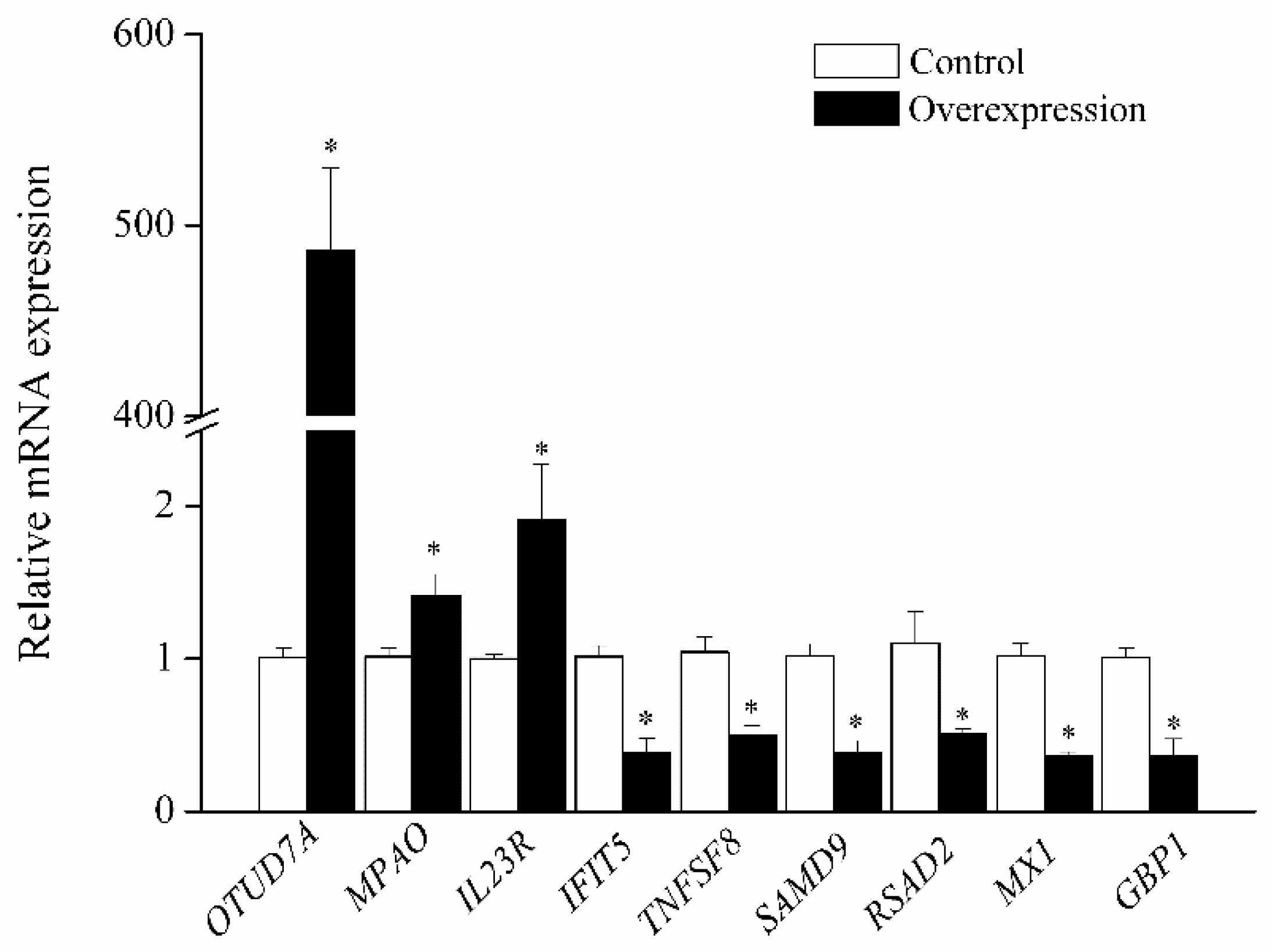
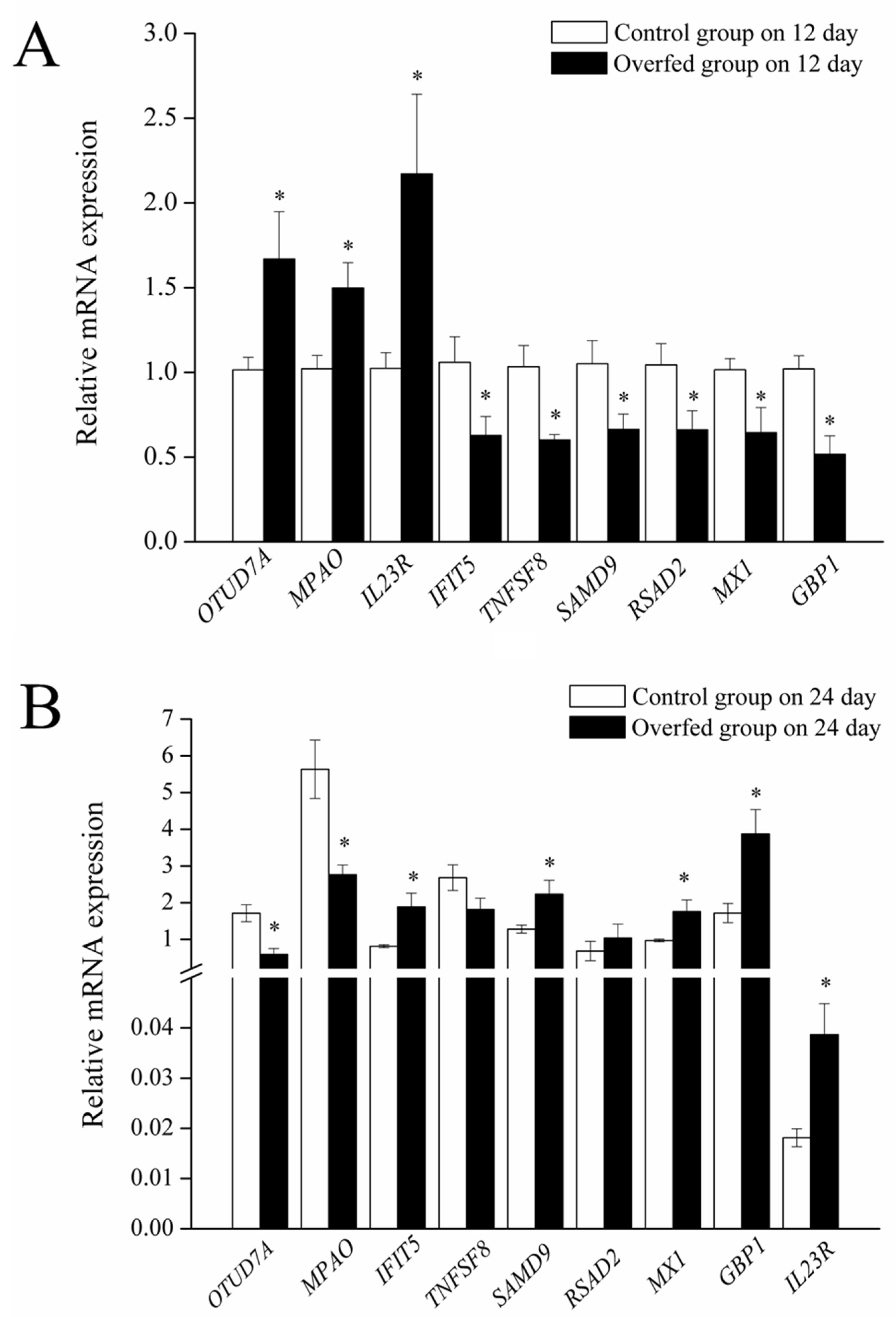
3.2. Expression of OTUD7A and Its Downstream Genes

The expression of OTUD7A, MPAO, and IL23R was upregulated, whereas that of IFIT5, TNFSF8, SAMD9, RSAD2, MX1, and GBP1 was downregulated at day 12 of overfeeding (p < 0.05, Figure 4A). However, the OTUD7A and MPAO expression was downregulated in goose fatty liver, whereas that of IL23R, IFIT5, SAMD9, MX1, and GBP1 was upregulated on day 24 of overfeeding (p < 0.05, Figure 4B).

4. Discussion

The pathogenesis of NAFLD is still unclear. Previous studies have indicated that inflammatory responses play important roles in NAFLD, and inflammation is considered to be a marker of progression from simple steatosis to NASH [5,16,17]. Inflammation not only aggravates insulin resistance, but also causes hepatic stellate cell activation, and induces liver fibrosis and cirrhosis [10,18]. The NF-κB pathway—a major inflammatory signaling pathway—is activated in patients with NAFLD and in animal models [5,6]. Activation of the NF-κB pathway can promote transcription of downstream inflammatory response genes, resulting in an increase in inflammatory factor production and release. Inflammatory factors can successively reactivate NF-κB, creating positive feedback regulation that leads to further amplification of the initial inflammatory signal [19]. In addition, NF-κB activation can activate pro-apoptotic proteins on mitochondria, such as BAX, leading to apoptosis of hepatocytes and further exacerbating the inflammatory response [20]. Reducing inflammatory factor expression in the liver can alleviate NAFLD development [11]; therefore, inflammation has received considerable attention as a possible target for NAFLD therapy.
As an excellent waterfowl product, goose fatty liver can be used as both a high-grade food and a unique model for NAFLD research. Our preliminary research has identified some characteristics that are different from mammalian NAFLD, such as elevated fatty acid desaturase, increased expression of lipocalin receptor genes, increased expression of mitochondria-related genes, and downregulated expression of complement genes and pro-inflammatory factors in goose fatty liver [2,3,4,21]. These results suggest the existence of certain mechanisms in the formation of goose fatty liver that can resist the occurrence and development of inflammation. Some studies suggest that deubiquitinating enzymes are involved in protecting against inflammation in NAFLD. Ubiquitin-specific protease 18—a member of the deubiquitinating enzyme family—is downregulated in the livers of obese mice [22]. Similarly, USP4 protects against the inflammatory response, as USP4 depletion was shown to exacerbate inflammation in mice with high-fat-diet-induced NAFLD, and USP4 may suppress activation of the downstream NF-κB pathway [23]. Mevissen et al. [12] found that OTUD7A is a potential tumor suppressor, and can regulate multiple signaling pathways. Another study indicated that OTUD7A inhibits the NF-κB pathway through deubiquitination of TRAF6 protein in HepG2 cells [10]. Therefore, OTUD7A may be involved in the development of fatty liver.
In this study, transcriptome sequencing analysis showed that a total of 19 genes were upregulated and 15 genes were downregulated in the OTUD7A overexpression group compared to controls. The DEGs were mainly enriched in cytokine–cytokine receptor interaction; tropane, piperidine, and pyridine alkaloid biosynthesis; isoquinoline alkaloid biosynthesis, the JAK-STAT signaling pathway; and the PI3K-Akt signaling pathway. Further analysis showed that numerous DEGs affected by OTUD7A were related to inflammation, as their mRNA expression was subject to OTUD7A overexpression or suppression by siRNA against OTUD7A, which is similar to the OTUD7A subfamily that can regulate some inflammation-related genes [24,25]. Our findings also support the hypothesis that OTUD7A can regulate the NF-κB pathway via deubiquitination of TRAF6. Pro-inflammatory factors can activate the inflammatory cascade response and increase the expression of genes such as IL-6 and inducible nitric oxide synthase, causing inflammatory responses and tissue damage in the body. During the formation of mammalian NAFLD, the levels of pro-inflammatory factors are usually significantly elevated [18,26]. These results suggest that OTUD7A may be associated with the inhibition of inflammatory responses in goose fatty liver—at least in the mid-overfeeding period.
Interestingly, cytokine–cytokine receptor interactions, which are important for the maintenance and regulatory functions of multicellular organisms [27], were among the enriched KEGG pathways in the OTUD7A assay. Tumor necrosis factor superfamily (TNFSF) is a cytokine secreted by immune cells. The interaction between TNFSF and tumor necrosis factor receptor is involved in regulating cell growth, immune response, apoptosis, and inflammatory response [28]. The TNFSF8 protein can activate the NF-κB pathway by binding to its receptor, TNFRSF8, thereby mediating the secretion of IL-2, IL-6, TNF-α, and other cytokines [29]. The expression of TNFSF8 was downregulated by OTUD7A overexpression in primary goose hepatocytes, as well as in goose fatty liver after 12 days of overfeeding, whereas the expression of TNFSF8 was increased after OTUD7A suppression in primary goose hepatocytes, suggesting that TNFSF8 is regulated by OTUD7A in goose hepatocytes through the cytokine receptor interaction pathway.
The JAK-STAT pathway, as a downstream pathway of cytokine receptors, was also enriched according to the transcriptomic analysis of goose primary hepatocytes overexpressing OTUD7A. Several cytokines, including interferons, can modulate intracellular signaling by activating the JAK-STAT pathway [30]. Upon the binding of cytokines to their cognate receptors, STATs can modulate the expression of their target genes and participate in inflammation [31,32]. Enrichment of DEGs in the JAK-STAT signaling pathway resulting from OTUD7A overexpression suggests that OTUD7A regulates inflammation through the JAK-STAT signaling pathway in goose hepatocytes. The IFIT family, as interferon-induced genes, participate in the immune response [33]. Previous studies indicated that IFIT5 promotes NF-κB activation and synergizes NF-κB-mediated gene expression, whereas knockdown of IFIT5 inhibits NF-κB pathway activation and downstream gene expression [34,35]. Our data indicate that IFIT5 expression was significantly decreased by OTUD7A overexpression, but was induced by OTUD7A knockdown in primary goose hepatocytes. Downregulation of IFIT5 was accompanied by upregulation of OTUD7A on day 12 of overfeeding, whereas upregulation of IFIT5 was accompanied by downregulation of OTUD7A on day 24 of overfeeding, which is consistent with the results of the cell research. Thus, OTUD7A may regulate IFIT5 expression in the goose fatty liver. In addition, previous studies have suggested that SAMD9 is a downstream target of inflammatory cytokines [36,37], and may function as an anti-inflammatory factor [38]; it is also associated with the immune response [39]. Our data indicate that SAMD9, like IFIT5, is regulated by OTUD7A.
Moreover, many studies have indicated that innate immune response is connected with inflammation in NAFLD/NASH, thereby promoting the development of fibrosis, cirrhosis, and carcinogenesis. The results from our study showed that OTUD7A regulates the expression of some immune-related genes—for example, IL23R—in goose fatty liver. IL23R mediates the stimulation of T cells, natural killer cells, and possibly certain macrophage/myeloid cells—likely through the JAK-STAT pathway [40]. In addition, the mRNA expression of RSAD2, MX1, and GBP1 was downregulated by OTUD7A overexpression, but upregulated by OTUD7A knockdown. These results suggest that RSAD2, MX1, and GBP1 are downstream of OTUD7A. RSAD2, also known as viperin, is an important component of innate immunity [41]. MX1 is also a vital antiviral protein during the innate immune response [42,43]. Some studies have shown that GBP1 can be induced by IFN-α, IFN-β, IFN-γ, and inflammatory cytokines [44,45]. GBP1 expression is increased in inflammatory skin diseases, and the gene is a cellular activation marker characterizing the inflammatory cytokine-activated phenotype of cells [46]. As immune-related genes, IL23R, RSAD2, MX1, and GBP1 were regulated by OTUD7A, suggesting that OTUD7A can regulate immune response in liver physiology and pathology.
Additionally, KEGG results suggested that the DEGs were related to tropane, piperidine, pyridine, and isoquinoline alkaloid biosynthesis following OTUD7A overexpression; for example, MPAO, which participates in the metabolism of alkaloid biosynthesis, was upregulated by OTUD7A overexpression in goose primary hepatocytes. Consistently, upregulation of MPAO was accompanied by upregulation of OTUD7A on day 12 of overfeeding, whereas the downregulation of MPAO was accompanied by downregulation of OTUD7A on day 24 of overfeeding. Therefore, OTUD7A may participate in alkaloid biosynthesis in the goose fatty liver. Bour et al. [47] found that MPAO expression was increased in the adipose tissue, suggesting that it is involved in adipogenesis. Fat metabolism is very active in the formation of goose fatty liver [3]; thus, MPAO may be involved in lipid metabolism in the goose fatty liver. Further research is required in order to confirm this hypothesis.

5. Conclusions

Transcriptome sequencing analysis showed that OTUD7A is involved in the development of goose fatty liver, mainly through cytokine–cytokine receptor interaction; tropane, piperidine, and pyridine alkaloid biosynthesis; isoquinoline alkaloid bio-synthesis; the JAK-STAT signaling pathway, and the PI3K-Akt signaling pathway. In addition, OTUD7A may regulate the expression of inflammation- and immune-related genes such as TNFSF8, IFIT5, IL23R, RSAD2, MX1, and GBP1 in the goose fatty liver.

Supplementary Materials

The following are available online at https://www.mdpi.com/article/10.3390/agriculture12010105/s1: Figure S1: Genes affected by OTU deubiquitinase 7A (OTUD7A) overexpression; Figure S2: Screening of small interfering RNAs (siRNAs) for OTU deubiquitinase 7A (OTUD7A).

Author Contributions

Conceptualization, T.G. and D.G.; methodology, K.W., D.J. and M.K.K.; software, M.Z.; validation, M.Z. and L.L.; formal analysis, M.Z. and X.F.; investigation, D.G.; resources, M.Z.; data curation, Q.S.; writing—original draft preparation, M.Z.; writing—review and editing, T.G., D.J. and M.K.K.; visualization, D.G.; supervision, D.G.; project administration, D.G.; funding acquisition, M.Z. and D.G. All authors have read and agreed to the published version of the manuscript.

Funding

This research was funded by the National Nature Science Foundation of China, grant numbers 31802052, 31972546, and 32072785; the China Postdoctoral Science Foundation (2017M621840); and the Priority Academic Program Development of Jiangsu Higher Education Institutions (PAPD).

Institutional Review Board Statement

This study was approved by the Institutional Ethics Committee of Yangzhou University (protocol code 202103309, 9 March 2021).

Data Availability Statement

The data presented in this study are available upon request from the corresponding author.

Conflicts of Interest

The authors declare no conflict of interest.

References

  1. Hermier, D.; Rousselot-Pailley, D.; Peresson, R.; Sellier, N. Influence of orotic acid and estrogen on hepatic lipid storage and secretion in the goose susceptible to liver steatosis. Biochim. Biophys. Acta 1994, 1211, 97–106. [Google Scholar] [CrossRef]
  2. Geng, T.Y.; Xia, L.L.; Li, F.Y.; Xia, J.; Zhang, Y.H.; Wang, Q.Q.; Yang, B.; Montgomery, S.; Cui, H.M.; Gong, D.Q. The role of endoplasmic reticulum stress and insulin resistance in the occurrence of goose fatty liver. Biochem. Biophys. Res. Commun. 2015, 465, 83–87. [Google Scholar] [CrossRef]
  3. Geng, T.Y.; Yang, B.; Li, F.Y.; Xia, L.L.; Wang, Q.Q.; Zhao, X.; Gong, D.Q. Identification of protective components that prevent the exacerbation of goose fatty liver: Characterization, expression and regulation of adiponectin receptors. Comp. Biochem. Physiol. B Biochem. Mol. Biol. 2016, 194–195, 32–38. [Google Scholar] [CrossRef]
  4. Liu, L.; Zhao, X.; Wang, Q.; Sun, X.X.; Xia, L.L.; Wang, Q.Q.; Yang, B.; Zhang, Y.H.; Montgomery, S.; Meng, H.; et al. Prosteatotic and protective components in a unique model of fatty liver: Gut microbiota and suppressed complement system. Sci. Rep. 2016, 6, 31763. [Google Scholar] [CrossRef] [Green Version]
  5. Baker, R.G.; Hayden, M.S.; Ghosh, S. NF-κB, inflammation, and metabolic disease. Cell Metab. 2011, 13, 11–22. [Google Scholar] [CrossRef] [PubMed] [Green Version]
  6. Zhou, Y.; Ding, Y.L.; Zhang, J.L.; Zhang, P.; Wang, J.Q.; Li, Z.H. Alpinetin improved high fat diet-induced non-alcoholic fatty liver disease (NAFLD) through improving oxidative stress, inflammatory response and lipid metabolism. Biomed. Pharmacother. 2018, 97, 1397–1408. [Google Scholar] [CrossRef]
  7. Hossain, N.; Kanwar, P.; Mohanty, S.R. A comprehensive updated review of pharmaceutical and nonpharmaceutical treatment for NAFLD. Gastroenterol. Res. Pract. 2016, 144, 7109270. [Google Scholar]
  8. Rahman, A.M.; Mahdavi, A.H.; Rahmani, H.R.; Jahanian, E. Clove bud (Syzygium aromaticum) improved blood and hepatic antioxidant indices in laying hens receiving low n-6 to n-3 ratios. J. Anim. Physiol. Anim. Nutr. 2017, 101, 881–892. [Google Scholar] [CrossRef] [PubMed]
  9. Marra, F. Nuclear factor-κB inhibition and non-alcoholic steatohepatitis: Inflammation as a target for therapy. Gut 2008, 57, 570–572. [Google Scholar] [CrossRef] [PubMed]
  10. Tilg, H.; Moschen, A.R. Evolution of inflammation in nonalcoholic fatty liver disease: The multiple parallel hits hypothesis. Hepatology 2010, 52, 1836–1846. [Google Scholar] [CrossRef] [PubMed]
  11. Tian, Y.L.; Ma, J.T.; Wang, W.D.; Zhang, L.J.; Xu, J.; Wang, K.; Li, D.F. Resveratrol supplement inhibited the NF-κB inflammation pathway through activating AMPKα-SIRT1 pathway in mice with fatty liver. Mol. Cell. Biochem. 2016, 422, 75–84. [Google Scholar] [CrossRef]
  12. Mevissen, T.E.; Hospenthal, M.K.; Geurink, P.P.; Elliott, P.R.; Akutsu, M.; Arnaudo, N.; Ekkebus, R.; Kulathu, Y.; Wauer, T.; Oualid, F.E.I.; et al. OTU deubiquitinases reveal mechanisms of linkage specificity and enable ubiquitin chain restriction analysis. Cell 2013, 154, 169–184. [Google Scholar] [CrossRef] [Green Version]
  13. Xu, Z.; Pei, L.; Wang, L.; Zhang, F.; Hu, X.; Gui, Y. Snail1-dependent transcriptional repression of Cezanne2 in hepatocellular carcinoma. Oncogene 2014, 33, 2836–2845. [Google Scholar] [CrossRef]
  14. Zhao, M.M.; Liu, T.J.; Wang, Q.; Zhang, R.; Liu, L.; Gong, D.Q.; Geng, T.Y. Fatty acids modulate the expression of pyruvate kinase and arachidonate-lipoxygenase through PPARγ/CYP2C45 pathway: A link to goose fatty liver. Poult. Sci. 2019, 98, 4346–4358. [Google Scholar] [CrossRef] [PubMed]
  15. Schmittgen, T.D.; Livak, K.J. Analyzing real-time PCR data by the comparative CT method. Nat. Protoc. 2008, 3, 1101–1108. [Google Scholar] [CrossRef] [PubMed]
  16. Parola, M.; Vajro, P. Nocturnal hypoxia in obese-related obstructive sleep apnea as a putative trigger of oxidative stress in pediatric NAFLD progression. J. Hepatol. 2016, 65, 470–472. [Google Scholar] [CrossRef] [Green Version]
  17. Wruck, W.; Graffmann, N.; Kawala, M.A.; Adjaye, J. Concise review: Current status and future directions on research related to nonalcoholic fatty liver disease. Stem Cells 2017, 35, 89–96. [Google Scholar] [CrossRef] [Green Version]
  18. Pirozzi, C.; Lama, A.; Simeoli, R.; Paciello, O.; Pagano, T.B.; Mollica, M.P.; Di Guida, F.; Russo, R.; Magliocca, S.; Canani, R.B.; et al. Hydroxytyrosol prevents metabolic impairment reducing hepatic inflammation and restoring duodenal integrity in a rat model of NAFLD. J. Nutr. Biochem. 2016, 30, 108–115. [Google Scholar] [CrossRef] [PubMed]
  19. Kondylis, V.; Kumari, S.; Vlantis, K.; Pasparakis, M. The interplay of IKK, NF-κB and RIPK1 signaling in the regulation of cell death, tissue homeostasis and inflammation. Immunol. Rev. 2017, 277, 113–127. [Google Scholar] [CrossRef]
  20. John, J.L.; Qian, T.; He, L.H.; Kim, J.S.; Elmore, S.P.; Cascio, W.E.; Brenner, D.A. Role of mitochondrial inner membrane permeabilization in necrotic cell death, apoptosis, and autophagy. Antioxid. Redox. Signal. 2002, 4, 769–781. [Google Scholar]
  21. Xing, Y.; Xu, C.; Lin, X.; Zhao, M.M.; Gong, D.Q.; Liu, L.; Geng, T.Y. Complement C3 participates in the development of goose fatty liver potentially by regulating the expression of FASN and ETNK1. Anim. Sci. J. 2021, 92, e13527. [Google Scholar] [CrossRef] [PubMed]
  22. An, S.; Zhao, L.P.; Shen, L.J.; Wang, S.; Zhang, K.; Qi, Y.; Zheng, J.; Zhang, X.J.; Zhu, X.Y.; Bao, R.; et al. USP18 protects against hepatic steatosis and insulin resistance through its deubiquitinating activity. Hepatology 2017, 666, 1866–1884. [Google Scholar] [CrossRef]
  23. Zhao, Y.C.; Wang, F.; Gao, L.C.; Xu, L.W.; Tong, R.Y.; Lin, N.; Su, Y.Y.; Yan, Y.; Gao, Y.; He, J.; et al. Ubiquitin-specific protease 4 is an endogenous negative regulator of metabolic dysfunctions in nonalcoholic fatty liver disease in mice. Hepatology 2018, 68, 897–917. [Google Scholar] [CrossRef] [PubMed] [Green Version]
  24. Lin, D.D.; Shen, Y.; Qiao, S.; Liu, W.W.; Zheng, L.S.; Wang, Y.N.; Cui, N.P.; Wang, Y.F.; Zhao, S.L.; Shi, J.H. Upregulation of OTUD7B (cezanne) promotes tumor progression via AKT/VEGF pathway in lung squamous carcinoma and adenocarcinoma. Front. Oncol. 2019, 9, 862. [Google Scholar] [CrossRef] [Green Version]
  25. Deng, J.J.; Hou, G.X.; Fang, Z.X.; Liu, J.L.; Lv, X.D. Distinct expression and prognostic value of OTU domain-containing proteins in non-small-cell lung cancer. Oncol. Lett. 2019, 5, 5417–5427. [Google Scholar] [CrossRef] [PubMed] [Green Version]
  26. Ceccarelli, S.; Panera, N.; Mina, M.; Gnani, D.; Stefanis, C.D.; Crudele, A.; Rychlicki, C.; Petrini, S.; Bruscalupi, G.; Agostinelli, L.; et al. LPS-induced TNF-α factor mediates pro-inflammatory and pro-fibrogenic pattern in non-alcoholic fatty liver disease. Oncotarget 2015, 6, 41434–41452. [Google Scholar] [CrossRef] [PubMed] [Green Version]
  27. Leonard, W.J.; Lin, J.X. Cytokine receptor signaling pathways. J. Allergy Clin. Immunol. 2000, 105, 877–888. [Google Scholar] [CrossRef]
  28. Croft, M.; Duan, W.; Choi, H.; Eun, S.Y.; Madireddi, S.; Mehta, A. TNF superfamily in inflammatory disease: Translating basic insights. Trends Immunol. 2012, 33, 144–152. [Google Scholar] [CrossRef] [PubMed] [Green Version]
  29. Horie, R.; Aizawa, S.; Nagai, M.; Ito, K.; Higashihara, M.; Ishida, T.; Inoue, J.; Watanabe, T. A novel domain in the CD30 cytoplasmic tail mediates NFkappaB activation. Int. Immunol. 1998, 10, 203–210. [Google Scholar] [CrossRef] [Green Version]
  30. McGillicuddy, F.C.; Chiquoine, E.H.; Hinkle, C.C.; Kim, R.J.; Shah, R.; Roche, H.M.; Smyth, E.M.; Reilly, M.P. Interferon γ attenuates insulin signaling, lipid storage, and differentiation in human adipocytes via activation of the JAK/STAT pathway. J. Biol. Chem. 2009, 284, 31936–31944. [Google Scholar] [CrossRef] [Green Version]
  31. Imada, K.; Leonard, W.J. The Jak-STAT pathway. Mol. Immunol. 2000, 37, 1–11. [Google Scholar] [CrossRef]
  32. Coskun, M.; Salem, M.; Pedersen, J.; Nielsen, O.H. Involvement of JAK/STAT signaling in the pathogenesis of inflammatory bowel disease. Pharmacol. Res. 2013, 76, 1–8. [Google Scholar] [CrossRef] [PubMed]
  33. Rohaim, M.A.; Santhakumar, D.; Naggar, R.F.E.; Iqbal, M.; Hussein, H.A.; Munir, M. Chickens expressing IFIT5 ameliorate clinical outcome and pathology of highly pathogenic avian influenza and velogenic newcastle disease viruses. Front. Immunol. 2018, 9, 2025. [Google Scholar] [CrossRef] [PubMed]
  34. Zhang, B.H.; Liu, X.Y.; Chen, W.; Chen, L. IFIT5 potentiates anti-viral response through enhancing innate immune signaling pathways. Acta Biochim. Biophys. Sin. 2013, 45, 867–874. [Google Scholar] [CrossRef] [Green Version]
  35. Zheng, C.S.; Zheng, Z.H.; Zhang, Z.H.; Meng, J.; Liu, Y.; Ke, X.L.; Hu, Q.X.; Wang, H.Z. IFIT5 positively regulates NF-κB signaling through synergizing the recruitment of IκB kinase (IKK) to TGF-β-activated kinase 1 (TAK1). Cell. Signal. 2015, 27, 2343–2354. [Google Scholar] [CrossRef]
  36. Chefetz, I.; Ben, A.D.; Browning, S.; Skorecki, K.; Adir, N.; Thomas, M.G.; Kogleck, L.; Topaz, O.; Indelman, M.; Uitto, J.; et al. Normophosphatemic familial tumoral calcinosis is caused by deleterious mutations in SAMD9, encoding a TNF-α responsive protein. J. Investig. Dermatol. 2008, 128, 1423–1429. [Google Scholar] [CrossRef] [Green Version]
  37. Hershkovitz, D.; Gross, Y.; Nahum, S.; Yehezkel, S.; Sarig, O.; Uitto, J.; Sprecher, E. Functional characterization of SAMD9, a protein deficient in normophosphatemic familial tumoral calcinosis. J. Investig. Dermatol. 2011, 131, 662–669. [Google Scholar] [CrossRef] [Green Version]
  38. He, P.; Wu, L.-F.; Bing, P.-F.; Xia, W.; Wang, L.; Xie, F.-F.; Lu, X.; Lei, S.-F.; Deng, F.-Y. SAMD9 is a (epi-) genetically regulated anti-inflammatory factor activated in RA patients. Mol. Cell Biochem. 2019, 456, 135–144. [Google Scholar] [CrossRef]
  39. Nounamo, B.; Li, Y.B.; Byrne, P.O.; Kearney, A.M.; Khan, A.; Liu, J. An interaction domain in human SAMD9 is essential for myxoma virus host-range determinant M062 antagonism of host anti-viral function. Virology 2017, 503, 94–102. [Google Scholar] [CrossRef]
  40. Parham, C.; Chirica, M.; Timans, J.; Vaisberg, E.; Travis, M.; Cheung, J.; Pflanz, S.; Zhang, R.; Singh, K.P.; Vega, F.; et al. A receptor for the heterodimeric cytokine IL-23 is composed of IL-12Rβ1 and a novel cytokine receptor subunit, IL-23R. J. Immunol. 2002, 168, 5699–5708. [Google Scholar] [CrossRef] [Green Version]
  41. Rivieccio, M.A.; Suh, H.S.; Zhao, Y.M.; Zhao, M.L.; Chin, K.C.; Lee, S.C.; Brosnan, C.F. TLR3 ligation activates an antiviral response in human fetal astrocytes: A role for viperin/cig5. J. Immunol. 2006, 177, 4735–4741. [Google Scholar] [CrossRef] [PubMed] [Green Version]
  42. Wisskirchen, C.; Ludersdorfer, T.H.; Müller, D.A.; Moritz, E.; Pavlovic, J. Interferon-induced antiviral protein MxA interacts with the cellular RNA helicases UAP56 and URH49. J. Biol. Chem. 2011, 286, 34743–34751. [Google Scholar] [CrossRef] [PubMed] [Green Version]
  43. Xiao, J.; Yan, J.; Chen, H.; Li, J.; Tian, Y.; Tang, L.S.; Feng, H. Mx1 of black carp functions importantly in the antiviral innate immune response. Fish Shellfish Immunol. 2016, 58, 584–592. [Google Scholar] [CrossRef]
  44. Tripal, P.; Bauer, M.; Naschberger, E.; Mörtinger, T.; Hohenadl, C.; Cornali, E.; Thurau, M.; Stürzl, M. Unique features of different members of the human guanylate-binding protein family. J. Interferon Cytokine Res. 2007, 27, 44–52. [Google Scholar] [CrossRef]
  45. Britzen-Laurent, N.; Bauer, M.; Berton, V.; Fischer, N.; Syguda, A.; Reipschläger, S.; Naschberger, E.; Herrmann, C.; Stürzl, M. Intracellular trafficking of guanylate-binding proteins is regulated by heterodimerization in a hierarchical manner. PLoS ONE 2010, 12, e14246. [Google Scholar] [CrossRef] [PubMed] [Green Version]
  46. Lubeseder-Martellato, C.; Guenzi, E.; Jörg, A.; Töpolt, K.; Naschberger, E.; Kremmer, E.; Zietz, C.; Tschachler, E.; Hutzler, P.; Schwemmle, M.; et al. Guanylate-binding protein-1 expression is selectively induced by inflammatory cytokines and is an activation marker of endothelial cells during inflammatory diseases. Am. J. Pathol. 2002, 161, 1749–1759. [Google Scholar] [CrossRef] [Green Version]
  47. Bour, S.; Daviaud, D.; Gres, S.; Lefort, C.; Prévot, D.; Zorzano, A.; Wabitsch, M.; Saulnier-Blache, J.-S.; Valet, P.; Carpéné, C. Adipogenesis-related increase of semicarbazide-sensitive amine oxidase and monoamine oxidase in human adipocytes. Biochimie 2007, 89, 916–925. [Google Scholar] [CrossRef] [PubMed]
Figure 1. Kyoto Encyclopedia of Genes and Genomes (KEGG) analysis of the control and ovarian tumor deubiquitinase 7A (OTUD7A) overexpression groups in goose hepatocytes. Primary hepatocytes were isolated from goose embryos after 23 days of incubation. The cells transfected with the empty pcDNA3.1(+) vector were used as controls, while those transfected with pcDNA3.1(+) containing the OTUD7A coding sequence were used for overexpression. Differentially expressed genes (DEGs) were identified using DESeq2 R software. The DEGs were selected based on genes showing a fold change in OTUD7A overexpression compared to the control group of >2 or <0.5, and p-value < 0.05. The clusterProfiler R package was used to statistically analyze the enrichment of the DEGs in the KEGG pathways.
Agriculture 12 00105 g001
Figure 2. Validation of differentially expressed genes of the control and ovarian tumor deubiquitinase 7A (OTUD7A) overexpression groups in goose hepatocytes; * p < 0.05. Data are shown as the mean ± SEM of six replicates for each treatment. MPAO: membrane primary amine oxidase; MX1: interferon-induced GTP-binding protein Mx1; IL23R: interleukin-23 receptor; IFIT5: interferon-induced protein with tetratricopeptide repeats 5; TNFSF8: tumor necrosis factor ligand superfamily member 8; RSAD2: radical S-adenosyl methionine domain-containing protein 2; GBP1: interferon-induced guanylate binding protein 1-like; SAMD9: sterile alpha motif domain-containing protein 9.
Agriculture 12 00105 g002
Figure 3. Effects of transfection with siRNA targeting ovarian tumor deubiquitinase 7A (OTUD7A) on the expression of downstream genes in goose primary hepatocytes. Scrambled siRNA was used as a control; * p < 0.05. Six replicates were evaluated for each treatment. Data are shown as the mean ± SEM.
Agriculture 12 00105 g003
Figure 4. Effects of overfeeding on expression of downstream genes. Control group denotes geese that were fed normally; overfed group denotes geese subjected to 12 (A) and 24 days (B) of overfeeding; * p < 0.05. Samples in the control group on day 12 of overfeeding were used as calibrators for all groups. Six replicates were evaluated for each treatment. Data are shown as the mean ± SEM.
Agriculture 12 00105 g004
Table 1. Primer sequences for real-time quantitative PCR analysis.
Gene 1GenBank NumberPrimer Sequence (5′ to 3′)Product Size
OTUD7AXM_013177443.1F: CAGACTTTGTTCGGTCCACA236
R: AGCAATGTCATCCTGCCTCT
MPAOXM_013199765.1F: TCCGACATGCAGTCCTTGTA201
R: AACCTTGTTGCCGTAGTTGG
IFIT5XM_013194152.1F: CCTTGAAGAAGTCCCTGCTG194
R: ATTTTCCAGGGCTTCCTTGT
IL23RXM_013193090.1F: CTGATGGGCTCCAACATCTC161
R: CTGTAGGGCAGCCTGAAGTC
TNFSF8XM_013182840.1F: AGGCTTGGAGCCACTAACAA201
R: GGTCGGGACATTCTGTAGGA
SAMD9XM_013187038.1F: CTCTCGCACAAATGGCACTA157
R: AGCTTTTGCAGCTCCAATGT
RSAD2XM_013172803.1F: GAGAGCGGTGGTTCAAGAAG238
R: TTGAGTGCCATGATCTGCTC
MX1XM_013181384.1F: TGGAGCAAGTAAACGCCTCT216
R: TTCAGGCACTGGTAGGCTTT
GBP1XM_013170751.1F: GAGCAGGAGAGAGAGGCTGA190
R: TGTTCTTCTCGGAGCCACTT
GAPDHXM_013199522.1F: GCCATCAATGATCCCTTCAT155
R: CTGGGGTCACGCTCCTG
1OTUD7A: ovarian tumor deubiquitinase 7A; MPAO: membrane primary amine oxidase; IFIT5: interferon-induced protein with tetratricopeptide repeats 5; IL23R: interleukin-23 receptor; TNFSF8: tumor necrosis factor ligand superfamily member 8; SAMD9: sterile alpha motif domain-containing protein 9; RSAD2: radical S-adenosyl methionine domain-containing protein 2; GBP1: interferon-induced guanylate binding protein 1-like; MX1: interferon-induced GTP-binding protein Mx1.
Table 2. The upregulated genes in hepatocytes transfected with pcDNA3.1(+) containing goose OTU deubiquitinase 7A CDS and an empty vector.
NameDescriptionp-ValueFold Change
SNCGGamma-synuclein isoform0.0192.11
GPRIN2G protein-regulated inducer of neurite outgrowth 2 isoform0.0392.29
NMUNeuromedin-U isoform0.0047.86
OTUD7AOTU domain-containing protein 7A<0.001134.69
LOC106034831CMRF35-like molecule 3 isoform0.0184.68
ARHGAP28Rho GTPase-activating protein 28 isoform0.0402.10
SH3BP1SH3 domain-binding protein 10.0402.82
MESTMesoderm-specific transcript homolog protein, partial0.0452.75
PROCREndothelial protein C receptor0.0042.61
CLMPCXADR-like membrane protein0.0122.40
KLHDC8AKelch domain-containing protein 8A0.0473.32
MYL10Myosin regulatory light chain 2B, cardiac muscle isoform0.0192.07
IL23RInterleukin-23 receptor0.0172.14
LOC106045877Alpha-1,3-mannosyl-glycoprotein 4-beta-N-acetylglucosaminyltransferase C-like0.0422.06
ACEAngiotensin-converting enzyme0.0022.05
TMEM114Transmembrane protein 1140.0043.64
PPP1R27Protein phosphatase 1 regulatory subunit 270.052.99
MPAOMembrane primary amine oxidase0.0452.03
LOC106048707Uncharacterized protein LOC106048707 isoform0.0454.52
Table 3. The downregulated genes in hepatocytes transfected with pcDNA3.1(+) containing goose OTU deubiquitinase 7A CDS and an empty vector.
NameDescriptionp-ValueFold Change
IFIT5Interferon-induced protein with tetratricopeptide repeats 5<0.0010.42
OTOGLOtogelin-like protein0.0450.45
LOC106030637Interleukin-7-like0.0220.17
RSAD2Radical S-adenosyl methionine domain-containing protein 2<0.0010.48
DDX60Probable ATP-dependent RNA helicase DDX60 isoform<0.0010.42
MX1Interferon-induced GTP-binding protein Mx1<0.0010.47
NCAM2Neural cell adhesion molecule 2 isoform0.0070.27
TNFSF8Tumor necrosis factor ligand superfamily member 80.0410.35
PIH1D3Protein PIH1D30.0350.39
LOC106038693Low-affinity vacuolar monovalent cation antiporter0.0430.08
SAMD9Sterile alpha motif domain-containing protein 9<0.0010.45
LOC106040189Arylacetamide deacetylase-like 40.0100.42
LOC106041938Placenta-specific gene 8 protein-like isoform<0.0010.44
B3GNT7UDP-GlcNAc:betaGal beta-1,3-N-acetylglucosaminyltransferase 7<0.0010.36
GBP1Interferon-induced guanylate-binding protein 1<0.0010.45
Publisher’s Note: MDPI stays neutral with regard to jurisdictional claims in published maps and institutional affiliations.

