Assessment of Inner Retinal Layers and Choroidal Thickness in Type 1 Diabetes Mellitus: A Cross-Sectional Study

 , ,
, ,  , ,
, , 
Abstract
1. Introduction
2. Experimental Section
2.1. Patients
2.2. Clinical Assessment
2.3. Ophthalmic Examination
2.4. Statistical Analysis
3. Results
3.1. Ophthalmologic Examination
3.2. Ophthalmological Associations
4. Discussion
4.1. RNFL and GCL
4.2. Choroid
4.3. Study Strengths and Limitations
5. Conclusions
Supplementary Materials
Author Contributions
Funding
Acknowledgments
Conflicts of Interest
References
- Wong, T.Y.; Cheung, C.M.; Larsen, M.; Sharma, S.; Simó, R. Diabetic retinopathy. Nat. Rev. Dis. Primers 2016, 17, 16012. [Google Scholar] [CrossRef] [PubMed]
- Lee, R.; Wong, T.Y.; Sabanayagam, C. Epidemiology of diabetic retinopathy, diabetic macular edema and related vision loss. Eye Vis. 2015, 2, 17. [Google Scholar] [CrossRef] [PubMed]
- Simó, R.; Stitt, A.W.; Gardner, T.W. Neurodegeneration in diabetic retinopathy: Does it really matter? Diabetologia 2018, 61, 1902–1912. [Google Scholar] [CrossRef] [PubMed]
- El-Fayoumi, D.; Badr Eldine, N.M.; Esmael, A.F.; Ghalwash, D.; Soliman, H.M. Retinal nerve fiber layer and ganglion cell complex thicknesses are reduced in children with type 1 diabetes with no evidence of vascular retinopathy. Investig. Ophthalmol. Vis. Sci. 2016, 57, 5355–5360. [Google Scholar] [CrossRef] [PubMed]
- Gundogan, F.C.; Akay, F.; Uzun, S.; Yolcu, U.; Çaǧlltay, E.; Toyran, S. Early Neurodegeneration of the Inner Retinal Layers in Type 1 Diabetes Mellitus. Ophthalmologica 2016, 235, 125–132. [Google Scholar] [CrossRef] [PubMed]
- Van Dijk, H.W.; Verbraak, F.D.; Kok, P.H.B.; Garvin, M.K.; Sonka, M.; Lee, K.; Devries, J.H.; Michels, R.P.J.; van Velthoven, M.E.J.; Schlingemann, R.O.; et al. Decreased retinal ganglion cell layer thickness in patients with type 1 diabetes. Investig. Ophthalmol. Vis. Sci. 2010, 51, 3660–3665. [Google Scholar] [CrossRef] [PubMed]
- Karti, O.; Nalbantoglu, O.; Abali, S.; Ayhan, Z.; Tunc, S.; Kusbeci, T.; Ozkan, B. Retinal Ganglion Cell Loss in Children With Type 1 Diabetes Mellitus without Diabetic Retinopathy. Ophthalmic Surg. Lasers Imaging Retin. 2017, 48, 473–477. [Google Scholar] [CrossRef]
- Chen, Y.; Li, J.; Yan, Y.; Shen, X. Diabetic macular morphology changes may occur in the early stage of diabetes. BMC Ophthalmol. 2016, 16, 12. [Google Scholar] [CrossRef]
- Gołȩbiewska, J.; Olechowski, A.; Wysocka-Mincewicz, M.; Baszyńska-Wilk, M.; Groszek, A.; Czeszyk-Piotrowicz, A.; Szalecki, M.; Hautz, W. Choroidal Thickness and Ganglion Cell Complex in Pubescent Children with Type 1 Diabetes without Diabetic Retinopathy Analyzed by Spectral Domain Optical Coherence Tomography. J. Diabetes Res. 2018, 2018, 5458015. [Google Scholar] [CrossRef]
- Gupta, P.; Thakku, S.G.; Sabanayagam, C.; Tan, G.; Cheung, C.M.G.; Lamoureux, E.L.; Wong, T.Y.; Cheng, C.Y. Characterisation of choroidal morphological and vascular features in diabetes and diabetic retinopathy. Br. J. Ophthalmol. 2017, 101, 1038–1044. [Google Scholar] [CrossRef]
- Melancia, D.; Vicente, A.; Cunha, J.P.; Abegão Pinto, L.; Ferreira, J. Diabetic choroidopathy: A review of the current literature. Graefe’s Arch. Clin. Exp. Ophthalmol. 2016, 254, 1453–1461. [Google Scholar] [CrossRef] [PubMed]
- Ferrara, D.; Waheed, N.K.; Duker, J.S. Progress in Retinal and Eye Research Investigating the choriocapillaris and choroidal vasculature with new optical coherence tomography technologies. Prog. Retin. Eye Res. 2015. [Google Scholar] [CrossRef]
- Çağıltay, E.; Toyran, S.; Akay, F.; Uzun, S.; Gundogan, F.C. Choroidal and macular thickness changes in type 1 diabetes mellitus patients without diabetic retinopathy AU—Yolcu, Umit. Postgrad. Med. 2016, 128, 755–760. [Google Scholar]
- Esmaeelpour, M.; Brunner, S.; Ansari-Shahrezaei, S.; Nemetz, S.; Povâzay, B.; Kajic, V.; Drexler, W.; Binder, S. Choroidal thinning in diabetes type 1 detected by 3-dimensional 1060 nm optical coherence tomography. Investig. Ophthalmol. Vis. Sci. 2012, 53, 6803–6809. [Google Scholar] [CrossRef] [PubMed]
- Vujosevic, S.; Midena, E. Retinal Layers Changes in Human Preclinical and Early Clinical Diabetic Retinopathy Support Early Retinal Neuronal and Müller Cells Alterations. J. Diabetes Res. 2013, 2013, 905058. [Google Scholar] [CrossRef] [PubMed]
- Sayin, N.; Kara, N.; Pirhan, D.; Vural, A.; Ersan, H.B.A.; Onal, H.; Cinar, S. Evaluation of subfoveal choroidal thickness in children with type 1 diabetes mellitus: An EDI-OCT study. Semin. Ophthalmol. 2014, 29, 27–31. [Google Scholar] [CrossRef] [PubMed]
- Pekel, E.; Altıncık, S.A.; Pekel, G. Evaluation of optic disc, retinal nerve fiber and macular ganglion cell layers in pediatric diabetes. Int. Ophthalmol. 2018, 38, 1955–1961. [Google Scholar] [CrossRef] [PubMed]
- Elhabashy, S.A.; Elbarbary, N.S.; Nageb, K.M.; Mohammed, M.M. Can optical coherence tomography predict early retinal microvascular pathology in type 1 diabetic adolescents without minimal diabetic retinopathy? A single-centre study. J. Pediatr. Endocrinol. Metab. 2015, 28, 139–146. [Google Scholar] [CrossRef] [PubMed]
- Jonsson, K.B.; Frydkjaer-Olsen, U.; Grauslund, J. Vascular Changes and Neurodegeneration in the Early Stages of Diabetic Retinopathy: Which Comes First? Ophthalmic Res. 2016, 56, 1–9. [Google Scholar] [CrossRef] [PubMed]
- Carbonell, M.; Castelblanco, E.; Valldeperas, X.; Betriu, À.; Traveset, A.; Granado-Casas, M.; Hernández, M.; Vázquez, F.; Martín, M.; Rubinat, E.; et al. Diabetic retinopathy is associated with the presence and burden of subclinical carotid atherosclerosis in type 1 diabetes. Cardiovasc. Diabetol. 2018, 17, 66. [Google Scholar] [CrossRef] [PubMed]
- Alonso, N.; Traveset, A.; Rubinat, E.; Ortega, E.; Alcubierre, N.; Sanahuja, J.; Hernández, M.; Betriu, A.; Jurjo, C.; Fernández, E.; et al. Type 2 diabetes-associated carotid plaque burden is increased in patients with retinopathy compared to those without retinopathy. Cardiovasc. Diabetol. 2015, 14, 33. [Google Scholar] [CrossRef] [PubMed]
- Boonarpha, N.; Zheng, Y.; Stangos, A.N.; Lu, H.; Raj, A.; Czanner, G.; Harding, S.P.; Nair-Sahni, J. Standardization of choroidal thickness measurements using enhanced depth imaging optical coherence tomography. Int. J. Ophthalmol. 2015, 8, 484–491. [Google Scholar] [PubMed]
- Wilkinson, C.P.; Ferris, F.L.; Klein, R.E.; Lee, P.P.; Agardh, C.D.; Davis, M.; Dills, D.; Kampik, A.; Pararajasegaram, R.; Verdaguer, J.T.; et al. Proposed international clinical diabetic retinopathy and diabetic macular edema disease severity scales. Ophthalmology 2003, 110, 1677–1682. [Google Scholar] [CrossRef]
- Scarinci, F.; Picconi, F.; Virgili, G.; Giorno, P.; Di Renzo, A.; Varano, M.; Frontoni, S.; Parravano, M. Single retinal layer evaluation in patients with type 1 diabetes with no or early signs of diabetic retinopathy: The first hint of neurovascular crosstalk damage between neurons and capillaries? Ophthalmologica 2017, 237, 223–231. [Google Scholar] [CrossRef]
- Simó, R.; Hernández, C. Novel approaches for treating diabetic retinopathy based on recent pathogenic evidence. Prog. Retin. Eye Res. 2015, 48, 160–180. [Google Scholar] [CrossRef]
- Santos, A.R.; Ribeiro, L.; Bandello, F.; Lattanzio, R.; Egan, C.; Frydkjaer-Olsen, U.; García-Arumí, J.; Gibson, J.; Grauslund, J.; Harding, S.P.; et al. Functional and structural findings of neurodegeneration in early stages of diabetic retinopathy: Cross-sectional analyses of baseline data of the EUROCONDOR project. Diabetes 2017, 66, 2503–2510. [Google Scholar] [CrossRef]
- Holm, K.; Adrian, M.L. In diabetic eyes, multifocal ERG reflects differences in function between the nasal part and the temporal part of the macula. Graefe’s Arch. Clin. Exp. Ophthalmol. 2012, 250, 1143–1148. [Google Scholar] [CrossRef]
- Simão, S.; Costa, M.Â.; Sun, J.K.; Cunha-Vaz, J.; Simó, R. Development of a Normative Database for Multifocal Electroretinography in the Context of a Multicenter Clinical Trial. Ophthalmic Res. 2017, 57, 107–117. [Google Scholar] [CrossRef]
- Simó, R.; Hernández, C.; Porta, M.; Bandello, F.; Grauslund, J.; Harding, S.P.; Aldington, S.J.; Egan, C.; Frydkjaer-Olsen, U.; García-Arumí, J.; et al. Effects of Topically Administered Neuroprotective Drugs in Early Stages of Diabetic Retinopathy: Results of the EUROCONDOR Clinical Trial. Diabetes 2019, 68, 457–463. [Google Scholar] [CrossRef] [PubMed]
- Nickla, D.L.; Wallman, J. The multifunctional choroid. Prog. Retin. Eye Res. 2010, 29, 144–168. [Google Scholar] [CrossRef] [PubMed]
- Lutty, G.A. Diabetic choroidopathy. Vis. Res. 2017, 139, 161–167. [Google Scholar] [CrossRef] [PubMed]
- Anderson, O.A.; Finkelstein, A.; Shima, D.T. A2E Induces IL-1ß Production in Retinal Pigment Epithelial Cells via the NLRP3 Inflammasome. PLoS ONE 2013, 8, e67263. [Google Scholar] [CrossRef] [PubMed]
- Kim, M.; Ha, M.J.; Choi, S.Y.; Park, Y.H. Choroidal vascularity index in type-2 diabetes analyzed by swept-source optical coherence tomography. Sci. Rep. 2018, 8, 70. [Google Scholar] [CrossRef] [PubMed]


| Variables 1 | Control | Type 1 Diabetes | p Value |
|---|---|---|---|
| N | 69 | 242 | - |
| Age, years | 45.1 (11.2) | 44.5 (10.7) | 0.668 |
| Sex, male | 30 (43.5%) | 114 (47.1%) | 0.692 |
| Ethnicity, Caucasian | 69 (100%) | 239 (98.8%) | 1.000 |
| Current or former smoker | 35 (50.7%) | 128 (53.2%) | 0.387 |
| Antiplatelet agents | 0 (0.00%) | 67 (27.7%) | <0.001 |
| Dyslipidaemia | 5 (7.25%) | 95 (39.3%) | <0.001 |
| Hypertension | 7 (10.1%) | 58 (24.0%) | 0.020 |
| Systolic blood pressure, mmHg | 118 (13.2) | 126 (17.1) | <0.001 |
| Diastolic blood pressure mmHg | 71.8 (9.38) | 74.4 (10.0) | 0.049 |
| Body mass index, kg/m2 | 24.6 (3.62) | 25.6 (4.05) | 0.059 |
| Waist circumference, cm | 87.2 (11.0) | 88.9 (12.6) | 0.290 |
| HbA1c, % | 5.28 (0.31) | 7.60 (1.03) | <0.001 |
| Total cholesterol, mg/dL | 203 (30.8) | 180 (28.9) | <0.001 |
| HDL cholesterol, mg/dL | 59.9 (12.4) | 64.3 (15.4) | 0.017 |
| LDL cholesterol, mg/dL | 123 (29.3) | 102 (24.2) | <0.001 |
| Triglycerides, mg/dL | 103 (69.4) | 74.4 (34.7) | 0.002 |
| Creatinine, mg/dL | 0.79 (0.13) | 0.77 (0.16) | 0.151 |
| Urine albumin/creatinine ratio, mg/g | 7.65 (10.4) | 7.02 (13.6) | 0.684 |
| Diabetes duration, years | - | 20.6 (10.4) | - |
| Variables 1 | Control | No DR | Mild DR | Advanced DR | p Overall | p CT vs. No DR | p CT vs. Mild DR | p CT vs. Adv. DR | p No DR vs. Mild DR | p No DR vs. Adv. DR | p Mild DR vs. Adv. DR |
|---|---|---|---|---|---|---|---|---|---|---|---|
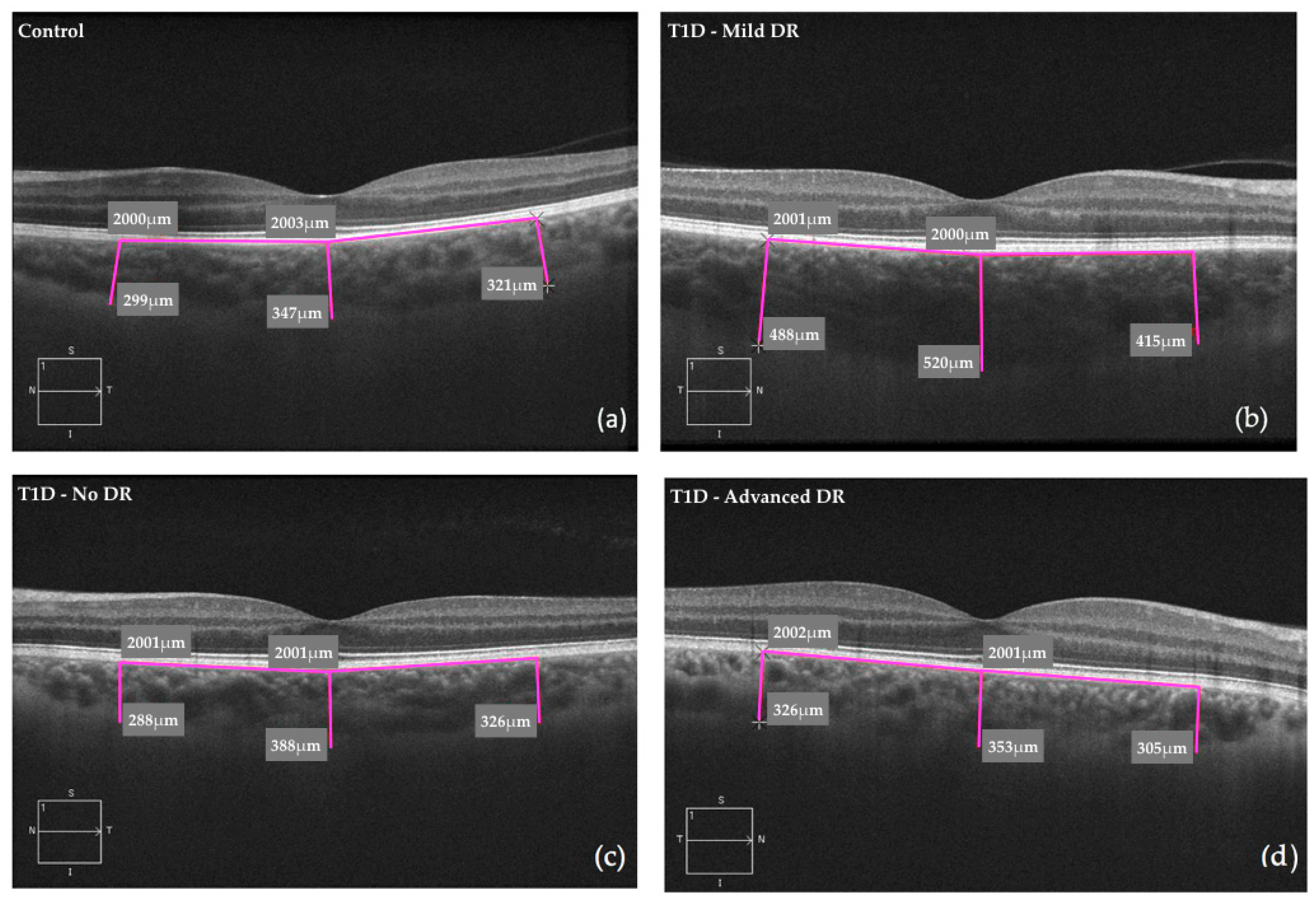
| CT (subfoveal) | 322 (92.6) | 367 (93.8) | 386 (97.7) | 341 (83.7) | <0.001 | 0.006 | <0.001 | 0.812 | 0.549 | 0.526 | 0.148 |
| CT (nasal) | 228 (86.9) | 278 (87.4) | 300 (89.5) | 249 (72.4) | <0.001 | 0.001 | <0.001 | 0.706 | 0.314 | 0.372 | 0.045 |
| CT (temporal) | 291 (76.2) | 321 (80.5) | 337 (81.8) | 289 (76.3) | 0.002 | 0.056 | 0.005 | 0.998 | 0.542 | 0.206 | 0.038 |
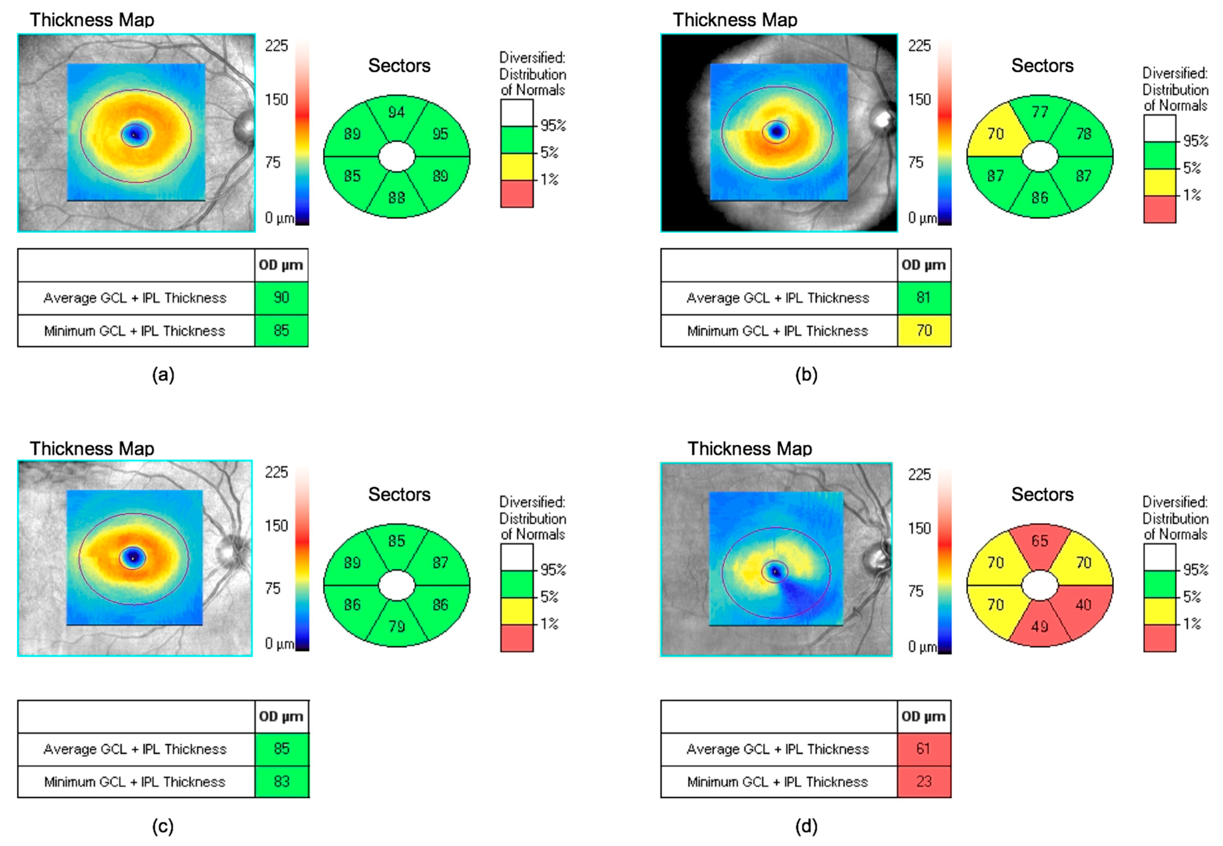
| GCL (mean) | 82.4 (6.6) | 84.3 (7.2) | 81.4 (10.8) | 78.5 (18.8) | 0.018 | 0.561 | 0.925 | 0.275 | 0.175 | 0.024 | 0.533 |
| GCL (minimum) | 79.6 (10.4) | 80.7 (9.9) | 77.8 (12) | 68 (25.0) | <0.001 | 0.932 | 0.821 | <0.001 | 0.373 | <0.001 | 0.003 |
| RNFL (mean) | 91.6 (9.8) | 95.1 (10.7) | 92.5 (19.3) | 94.6 (16.7) | 0.287 | 0.305 | 0.980 | 0.763 | 0.545 | 0.998 | 0.898 |
| RNFL (temporal) | 66.9 (12.5) | 65.4 (10.1) | 62.9 (13.3) | 70.2 (16) | 0.056 | 0.822 | 0.197 | 0.681 | 0.499 | 0.303 | 0.063 |
| RNFL (superior) | 110 (17.5) | 116 (18.0) | 119 (16.8) | 120 (21) | 0.026 | 0.104 | 0.035 | 0.141 | 0.837 | 0.852 | 0.995 |
| RNFL (nasal) | 110 (18.2) | 75.9 (14.8) | 75.5 (18.3) | 76.4 (16.0) | <0.001 | <0.001 | <0.001 | <0.001 | 0.999 | 0.999 | 0.997 |
| RNFL (inferior) | 111 (17.7) | 125 (19.1) | 124 (23.9) | 118 (31.0) | <0.001 | <0.001 | 0.003 | 0.484 | 0.954 | 0.471 | 0.725 |
| Variables | Mild DR | Advanced DR | ||
|---|---|---|---|---|
| Estimated β (SE) | p Value | Estimated β (SE) | p Value | |
| CT (subfoveal) | 28.17 (13.31) | 0.126 | −7.52 (20.50) | 0.892 |
| CT (nasal) | 28.84 (12.64) | 0.126 | −13.75 (19.46) | 0.687 |
| CT (temporal) | 23.83 (11.40) | 0.126 | −17.46 (17.56) | 0.535 |
| GCL (mean) | −2.39 (1.52) | 0.330 | −4.96 (2.36) | 0.126 |
| GCL (minimum) | −2.06 (1.91) | 0.514 | −11.34 (2.98) | 0.004 |
| RNFL, mean | −2.43 (2.12) | 0.514 | 0.54 (3.34) | 0.917 |
| RNFL, temporal | −2.48 (1.78) | 0.412 | 6.99 (3.01) | 0.126 |
| RNFL, superior | 2.23 (2.69) | 0.629 | 1.17 (4.56) | 0.917 |
| RNFL, nasal | −0.22 (2.43) | 0.929 | 0.71 (4.11) | 0.917 |
| RNFL, inferior | −1.34 (3.28) | 0.892 | −6.10 (5.56) | 0.514 |
© 2019 by the authors. Licensee MDPI, Basel, Switzerland. This article is an open access article distributed under the terms and conditions of the Creative Commons Attribution (CC BY) license (http://creativecommons.org/licenses/by/4.0/).
Share and Cite
Carbonell, M.; Alonso, N.; Castelblanco, E.; Real, J.; Ramírez-Morros, A.; Simó, R.; Hernández, C.; Jurjo, C.; Traveset, A.; Valldeperas, X.; et al. Assessment of Inner Retinal Layers and Choroidal Thickness in Type 1 Diabetes Mellitus: A Cross-Sectional Study. J. Clin. Med. 2019, 8, 1412. https://doi.org/10.3390/jcm8091412
Carbonell M, Alonso N, Castelblanco E, Real J, Ramírez-Morros A, Simó R, Hernández C, Jurjo C, Traveset A, Valldeperas X, et al. Assessment of Inner Retinal Layers and Choroidal Thickness in Type 1 Diabetes Mellitus: A Cross-Sectional Study. Journal of Clinical Medicine. 2019; 8(9):1412. https://doi.org/10.3390/jcm8091412
Chicago/Turabian StyleCarbonell, Marc, Núria Alonso, Esmeralda Castelblanco, Jordi Real, Anna Ramírez-Morros, Rafael Simó, Cristina Hernández, Carme Jurjo, Alícia Traveset, Xavier Valldeperas, and et al. 2019. "Assessment of Inner Retinal Layers and Choroidal Thickness in Type 1 Diabetes Mellitus: A Cross-Sectional Study" Journal of Clinical Medicine 8, no. 9: 1412. https://doi.org/10.3390/jcm8091412
APA StyleCarbonell, M., Alonso, N., Castelblanco, E., Real, J., Ramírez-Morros, A., Simó, R., Hernández, C., Jurjo, C., Traveset, A., Valldeperas, X., & Mauricio, D. (2019). Assessment of Inner Retinal Layers and Choroidal Thickness in Type 1 Diabetes Mellitus: A Cross-Sectional Study. Journal of Clinical Medicine, 8(9), 1412. https://doi.org/10.3390/jcm8091412