The Eye as a Window to Systemic Infectious Diseases: Old Enemies, New Imaging

 ,
,
Abstract
1. Introduction
2. Experimental Section
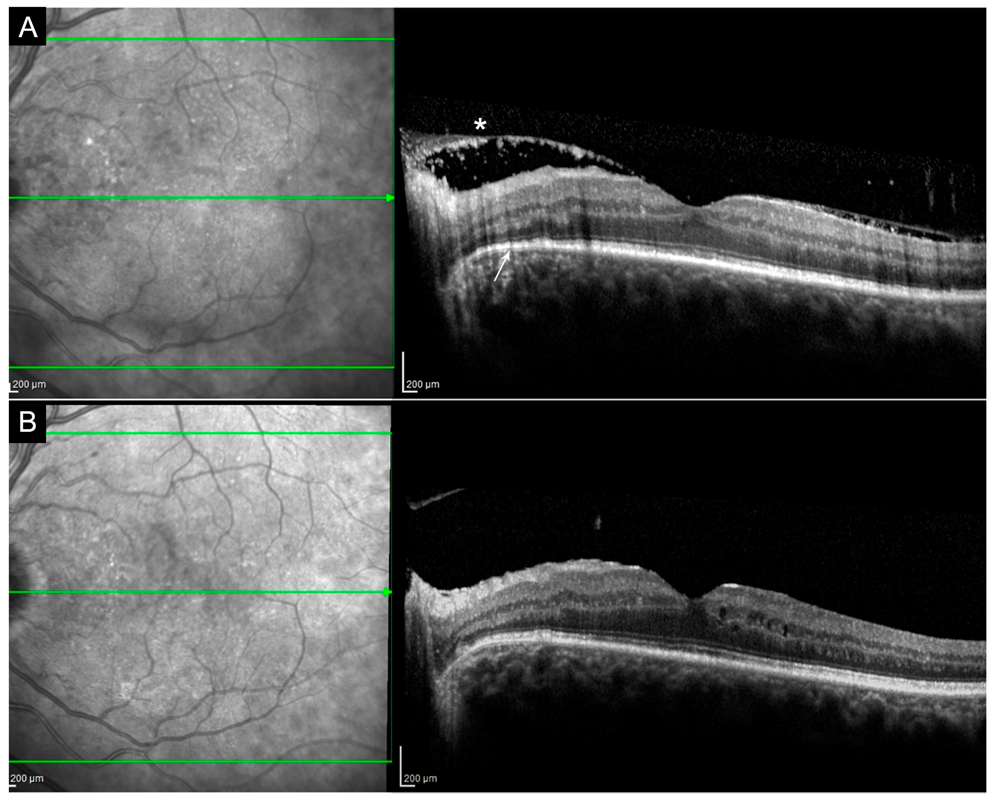
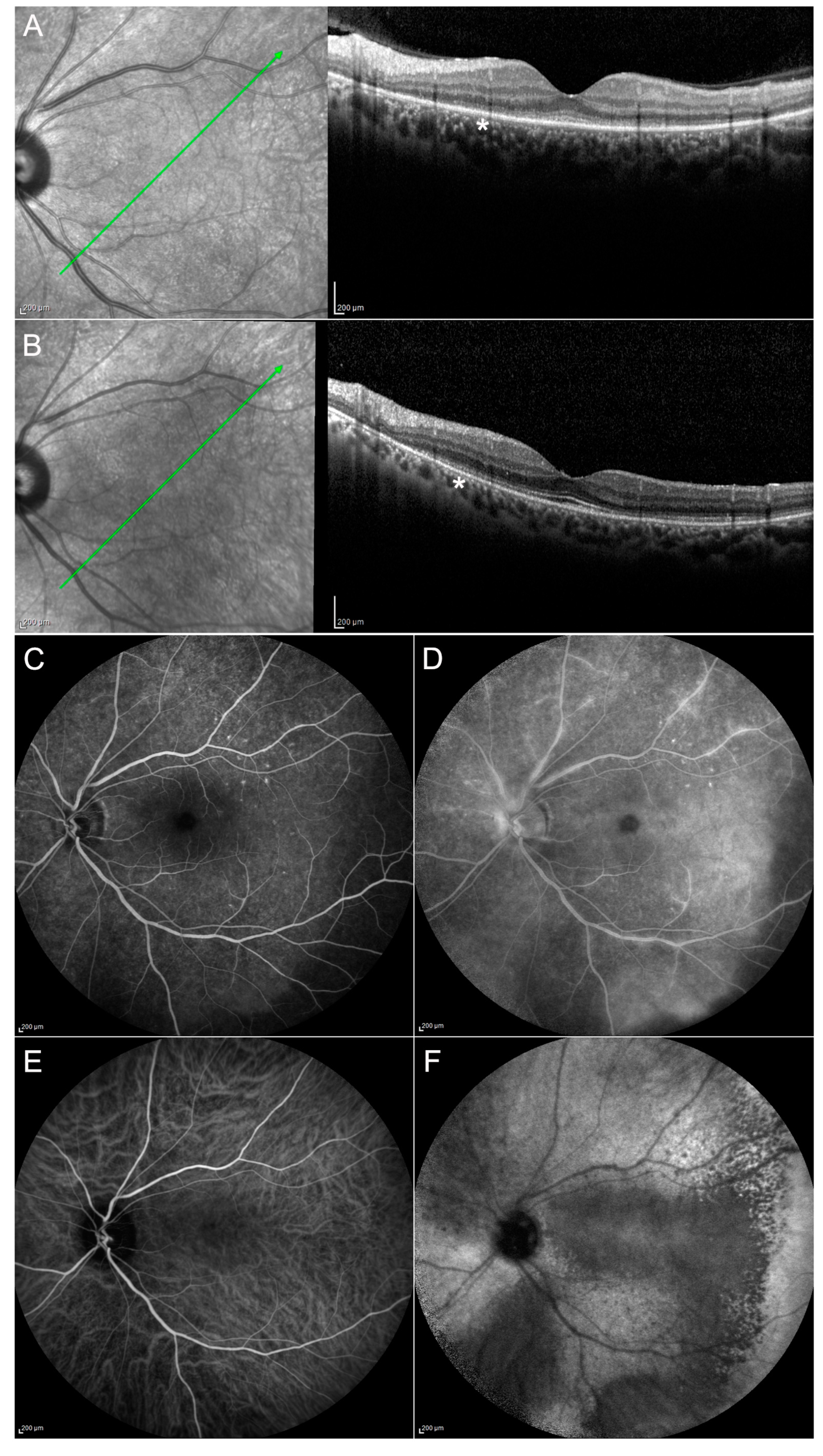
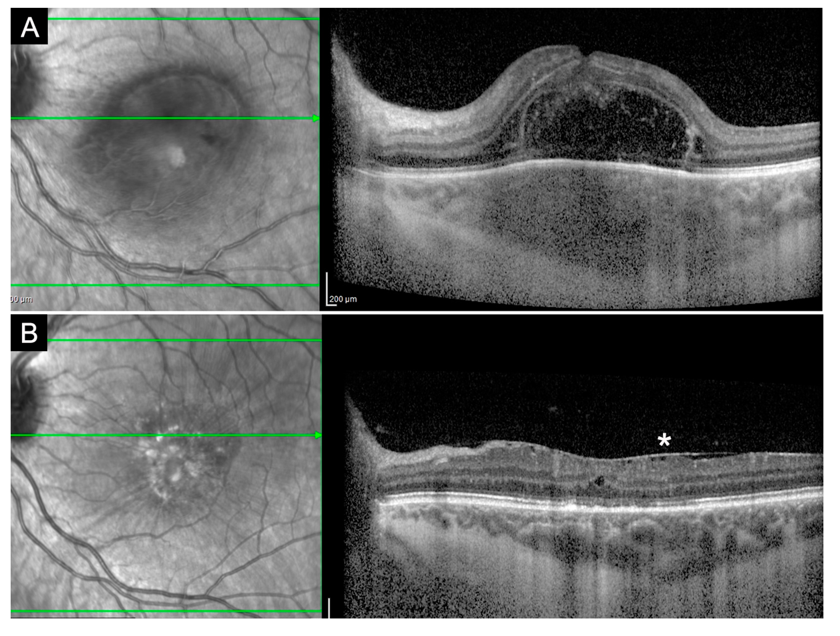
2.1. Syphilis
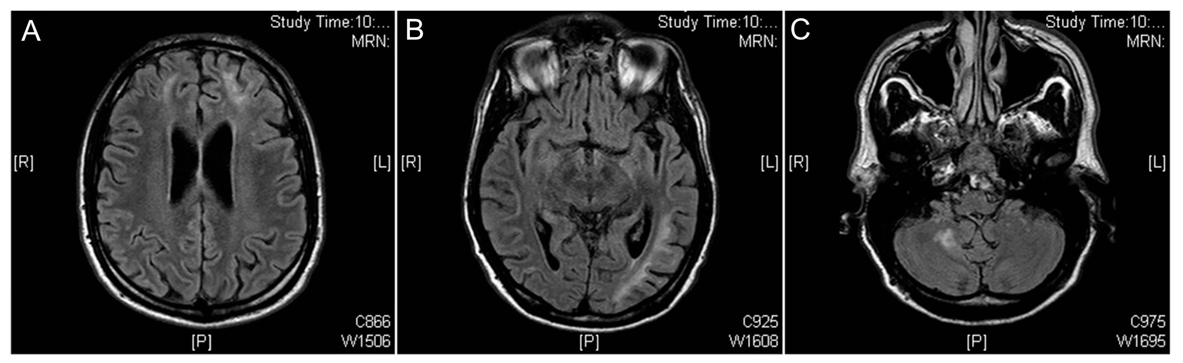
2.2. Tuberculosis
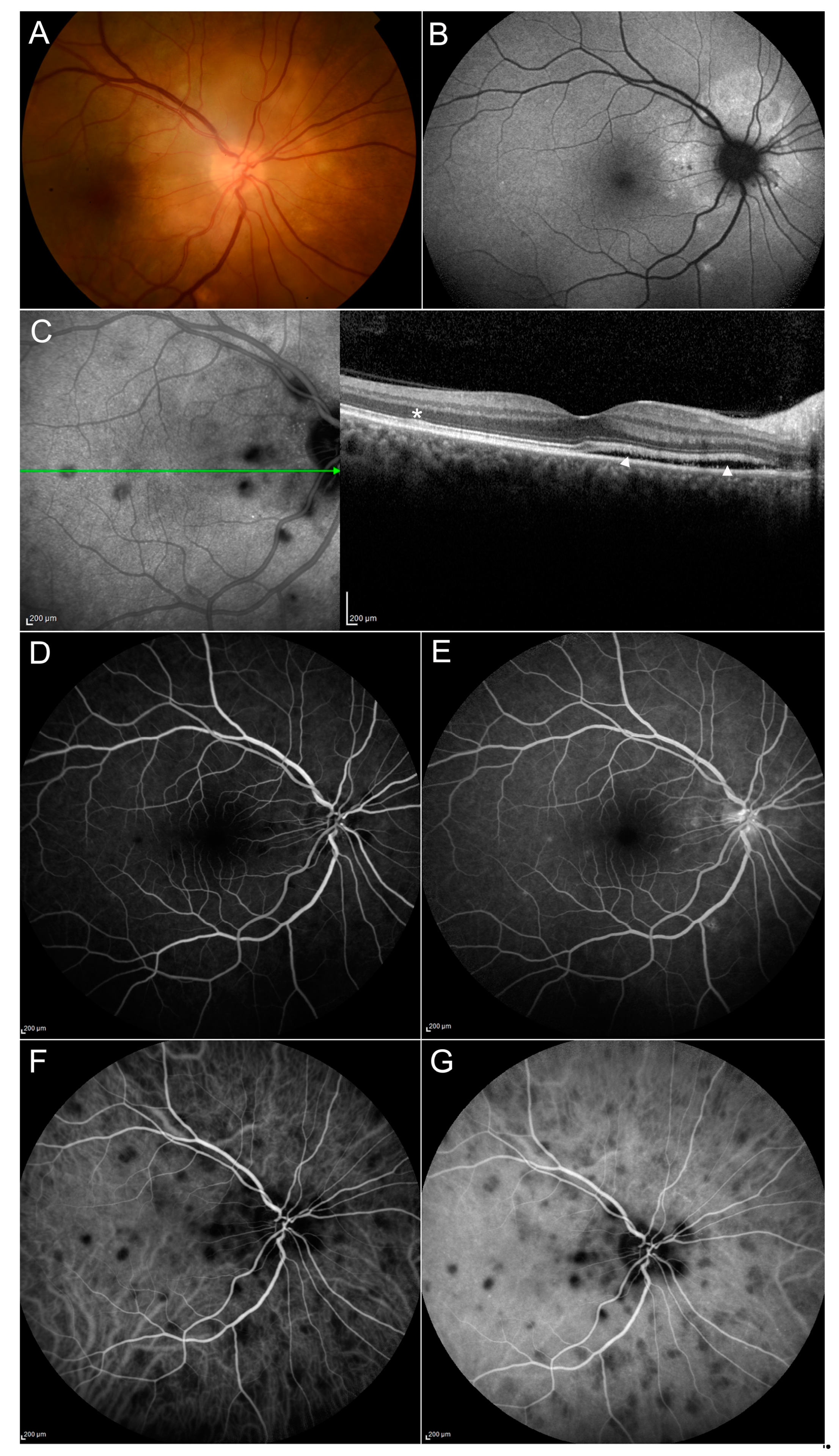
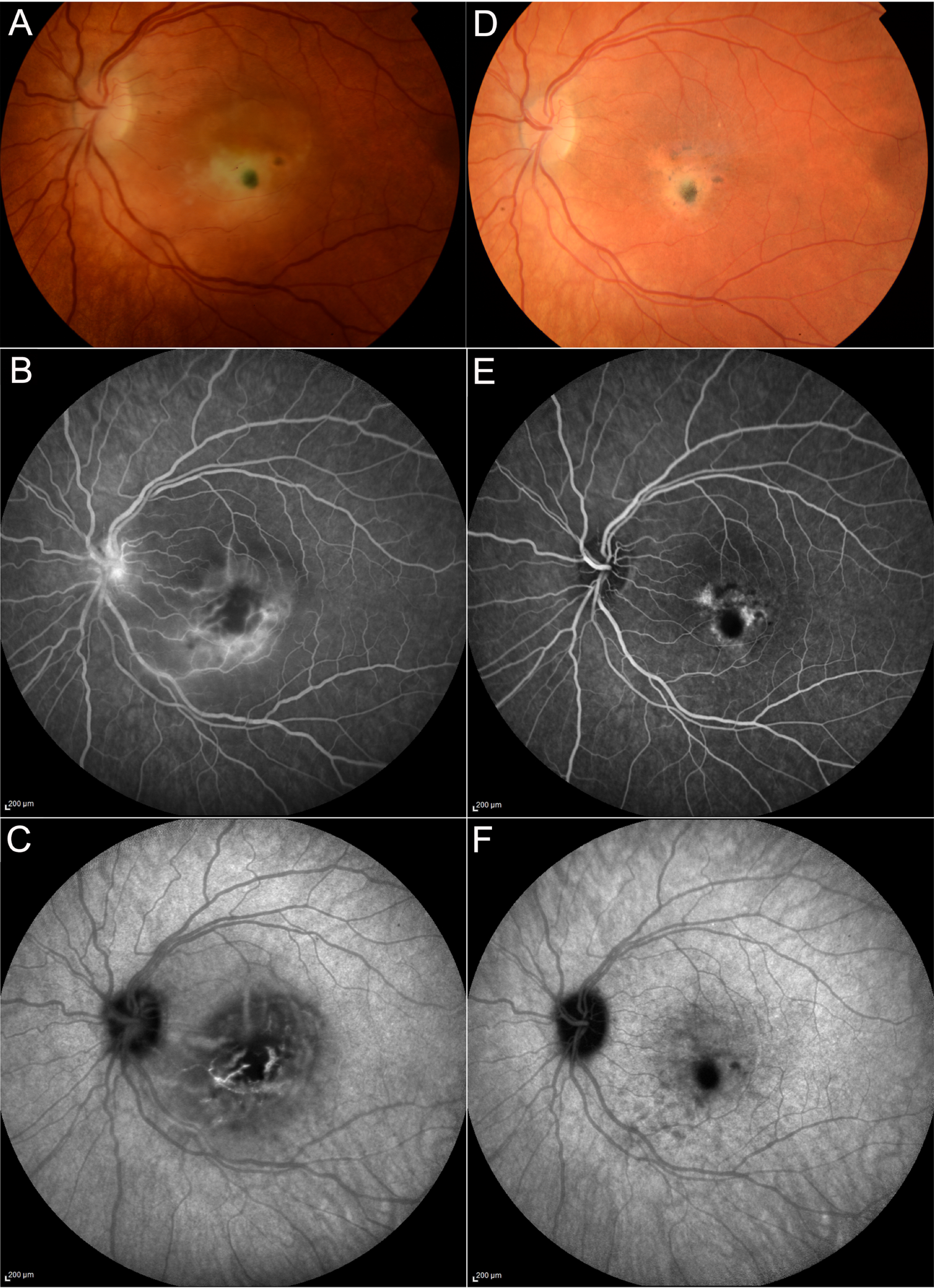
2.3. Toxoplasmosis
3. Results and Discussion
Author Contributions
Conflicts of Interest
References
- Saloojee, H.; Velaphi, S.; Goga, Y.; Afadapa, N.; Steen, R.; Lincetto, O. The prevention and management of congenital syphilis: An overview and recommendations. Bull. World Health. Organ. 2004, 82, 424–430. [Google Scholar]
- Hook, E.W.; Peeling, R.W. Syphilis Control—A Continuing Challenge. N. Engl. J. Med. 2004, 351, 122–124. [Google Scholar] [CrossRef]
- Centers for Disease Control and Prevention. Sexually Transmitted Disease Surveillance. 2017. Available online: https://www.cdc.gov/std/stats17/default.htm (accessed on 5 August 2019).
- Sparling, P.F. Diagnosis of Neurosyphilis: New Tools. Sex. Transm. Dis. 2010, 37, 1. [Google Scholar] [CrossRef]
- Dutta Majumder, P.; Chen, E.J.; Shah, J.; Ching Wen Ho, D.; Biswas, J.; See Yin, L.; Gupta, V.; Pavesio, C.; Agrawal, R. Ocular Syphilis: An Update. Ocul. Immunol. Inflamm. 2019, 7, 117–125. [Google Scholar] [CrossRef]
- Tsuboi, M.; Nishijima, T.; Yashiro, S.; Teruya, K.; Kikuchi, Y.; Katai, N.; Oka, S.; Gatanaga, H. Prognosis of ocular syphilis in patients infected with HIV in the antiretroviral therapy era. Sex. Transm. Infect. 2016, 92, 605–610. [Google Scholar] [CrossRef]
- Haug, S.J.; Cunningham, E.T. Syphilitic Uveitis. In Emerging Infectious Uveitis; Springer Science and Business Media LLC: Cham, Switzerland, 2017; pp. 35–42. [Google Scholar]
- World Health Organization. WHO Global Tuberculosis Report 2016; WHO Press: Geneve, Switzerland, 2016; ISBN 978 92 4 156539 4. [Google Scholar]
- Houben, R.M.G.J.; Dodd, P.J. The Global Burden of Latent Tuberculosis Infection: A Re-estimation Using Mathematical Modelling. PLoS Med. 2016, 13, e1002152. [Google Scholar] [CrossRef]
- Churchyard, G.; Kim, P.; Shah, N.S.; Rustomjee, R.; Gandhi, N.; Mathema, B.; Dowdy, D.; Kasmar, A.; Cardenas, V. What We Know About Tuberculosis Transmission: An Overview. J. Infect. Dis. 2017, 216, S629–S635. [Google Scholar] [CrossRef]
- Albert, D.M.; Raven, M.L. Ocular Tuberculosis. Microbiol. Spectr. 2016, 4, 1–36. [Google Scholar]
- Gupta, V.; Gupta, A.; Rao, N.A. Intraocular Tuberculosis—An Update. Surv. Ophthalmol. 2007, 52, 561–587. [Google Scholar] [CrossRef]
- Bansal, R.; Basu, S.; Gupta, A.; Rao, N.; Invernizzi, A.; Kramer, M. Imaging in tuberculosis-associated uveitis. Indian J. Ophthalmol. 2017, 65, 264–270. [Google Scholar]
- Saadatnia, G.; Golkar, M. A review on human toxoplasmosis. Scand. J. Infect. Dis. 2012, 44, 805–814. [Google Scholar] [CrossRef]
- Maenz, M.; Schlüter, D.; Liesenfeld, O.; Schares, G.; Gross, U.; Pleyer, U. Ocular toxoplasmosis past, present and new aspects of an old disease. Prog. Retin. Eye Res. 2014, 39, 77–106. [Google Scholar] [CrossRef]
- Besirli, C.G.; Ozgonul, C. Recent Developments in the Diagnosis and Treatment of Ocular Toxoplasmosis. Ophthalmic Res. 2016, 57, 1–12. [Google Scholar]
- Butler, N.J.; Furtado, J.M.; Winthrop, K.L.; Smith, J.R. Ocular Toxoplasmosis II: Clinical Features, Pathology and Management. Clin. Experiment. Ophthalmol. 2013, 41, 95–108. [Google Scholar] [CrossRef]
- Kiss, S.; Damico, F.M.; Young, L.H. Ocular Manifestations and Treatment of Syphilis. Semin. Ophthalmol. 2005, 20, 161–167. [Google Scholar] [CrossRef]
- Villanueva, A.V.; Sahouri, M.J.; Ormerod, L.D.; Puklin, J.E.; Reyes, M.P. Posterior Uveitis in Patients with Positive Serology for Syphilis. Clin. Infect. Dis. 2000, 30, 479–485. [Google Scholar] [CrossRef]
- Mauro, J.; Samson, C.M.; Foster, C.S. Syphilis. In Diagnosis and Treatment of Uveitis, 2nd ed.; Foster, C.S., Vitale, A.T., Eds.; Jaypee Brothers Medical Publishers: New Delhi, India, 2013; pp. 337–345. [Google Scholar]
- Lee, S.Y.; Cheng, V.; Rodger, D.; Rao, N. Clinical and laboratory characteristics of ocular syphilis: A new face in the era of HIV co-infection. J. Ophthalmic Inflamm. Infect. 2015, 5, 56. [Google Scholar] [CrossRef]
- Johns, D.R.; Tierney, M.; Felsenstein, D. Alteration in the Natural History of Neurosyphilis by Concurrent Infection with the Human Immunodeficiency Virus. N. Engl. J. Med. 1987, 316, 1569–1572. [Google Scholar] [CrossRef]
- Davis, J.L. Ocular Syphilis. Curr. Opin. Ophthalmol. 2014, 25, 513–518. [Google Scholar] [CrossRef]
- Yokoi, M.; Kase, M. Retinal Vasculitis Due to Secondary Syphilis. Jpn. J. Ophthalmol. 2004, 48, 65–67. [Google Scholar] [CrossRef]
- Savir, H.; Kurz, O. Fluorescein angiography in syphilitic retinal vasculitis. Ann. Ophthalmol. 1976, 8, 713–716. [Google Scholar]
- Jumper, M.J.; Machemer, R.; Gallemore, R.P.; Jaffe, G.J. EXUDATIVE RETINAL DETACHMENT AND RETINITIS ASSOCIATED WITH ACQUIRED SYPHILITIC UVEITIS. Retina 2000, 20, 190–194. [Google Scholar] [CrossRef]
- Pratas, A.C.; Goldschmidt, P.; Lebeaux, D.; Aguilar, C.; Ermak, N.; Benesty, J.; Charlier, C.; Benveniste, E.; Merabet, L.; Sedira, N.; et al. Increase in Ocular Syphilis Cases at Ophthalmologic Reference Center, France, 2012–2015. Emerg. Infect. Dis. 2018, 24, 193–200. [Google Scholar] [CrossRef]
- Zhang, T.; Zhu, Y.; Xu, G. Clinical Features and Treatments of Syphilitic Uveitis: A Systematic Review and Meta-Analysis. J. Ophthalmol. 2017, 2017, 1–15. [Google Scholar] [CrossRef]
- Gass, J.D.M.; Braunstein, R.A.; Chenoweth, R.G. Acute Syphilitic Posterior Placoid Chorioretinitis. Ophthalmol. 1990, 97, 1288–1297. [Google Scholar] [CrossRef]
- Cavallero, E.; Pirani, V.; Cesari, C.; Carrozzi, G.; Giovannini, A.; Mariotti, C. Multimodal Imaging Analysis of Acute Syphilitic Posterior Placoid Chorioretinitis: A Case Report. Ophthalmic Surg. Lasers Imaging Retina 2019, 50, e179–e184. [Google Scholar] [CrossRef]
- Yang, P.; Zhang, N.; Li, F.; Chen, Y.; Kijlstra, A. OCULAR MANIFESTATIONS OF SYPHILITIC UVEITIS IN CHINESE PATIENTS. Retina 2012, 32, 1906–1914. [Google Scholar] [CrossRef]
- Bellmann, C.; Holz, F.G.; Breitbart, A.; Völcker, H.E. Bilateral acute syphilitic posterior placoid chorioretinopathy–Angiographic and autofluorescence characteristics. Ophthalmologe 1999, 96, 522–528. [Google Scholar] [CrossRef]
- Matsumoto, Y.; Spaide, R.F. AUTOFLUORESCENCE IMAGING OF ACUTE SYPHILITIC POSTERIOR PLACOID CHORIORETINITIS. Retin. Cases Brief Rep. 2007, 1, 123–127. [Google Scholar] [CrossRef]
- Eandi, C.M.; Neri, P.; Adelman, R.A.; Yannuzzi, L.A.; Cunningham, E.T. International Syphilis Study Group. Acute Syphilitic Posterior Placoid Chorioretinitis: Report of a Case Series and Comprehensive Review of the Literature. Retina 2012, 32, 1915–1941. [Google Scholar] [CrossRef]
- Workowski, K.A.; Bolan, G.A. Sexually Transmitted Diseases Treatment Guidelines, 2015. MMWR. Recomm. Rep. 2015, 64, 1–137. [Google Scholar]
- Jay, C.A. Treatment of neurosyphilis. Curr. Treat. Options Neurol. 2006, 8, 185–192. [Google Scholar] [CrossRef]
- Zifko, U.; Lindner, K.; Wimberger, D.; Volc, B.; Grisold, W. Jarisch-Herxheimer reaction in a patient with neurosyphilis. J. Neurol. Neurosurg. Psychiatry 1994, 57, 865–867. [Google Scholar] [CrossRef]
- Júnior, A.D.; Rodrigues, L.D.S.; Costa, L.C. Jarisch-Herxheimer reaction in a patient with syphilis and human immunodeficiency virus infection. Rev. Soc. Bras. Med. Trop. 2018, 51, 877–878. [Google Scholar] [CrossRef]
- World Health Organization. Consolidated ARV Guidelines 2013; WHO Press: Geneve, Switzerland, 2013. [Google Scholar]
- Basu, S.; Monira, S.; Modi, R.R.; Choudhury, N.; Mohan, N.; Padhi, T.R.; Balne, P.K.; Sharma, S.; Panigrahi, S.R. Degree, duration, and causes of visual impairment in eyes affected with ocular tuberculosis. J. Ophthalmic Inflamm. Infect. 2014, 4, 3. [Google Scholar] [CrossRef]
- Donahue, H.C. Ophthalmologic Experience in a Tuberculosis Sanatorium. Am. J. Ophthalmol. 1967, 64, 742–748. [Google Scholar] [CrossRef]
- Abu El-Asrar, A.M.; Abouammoh, M.; Al-Mezaine, H.S. Tuberculous Uveitis. Middle East Afr. J. Ophthalmol. 2009, 16, 188–201. [Google Scholar] [CrossRef]
- Agrawal, R.; Gunasekeran, D.V.; Grant, R.; Agarwal, A.; Kon, O.M.; Nguyen, Q.D.; Pavesio, C.; Gupta, V.; Collaborative Ocular Tuberculosis Study (COTS)-1 Study Group. Clinical Features and Outcomes of Patients with Tubercular Uveitis Treated with Antitubercular Therapy in the Collaborative Ocular Tuberculosis Study (COTS)-1. JAMA Ophthalmol. 2017, 135, 1318–1327. [Google Scholar] [CrossRef]
- Helm, C.J.; Holland, G.N. Ocular Tuberculosis. J. Neuro-Ophthalmology. 1994, 14, 127. [Google Scholar] [CrossRef]
- Zhang, M.; Zhang, J.; Liu, Y. CLINICAL PRESENTATIONS AND THERAPEUTIC EFFECT OF PRESUMED CHOROIDAL TUBERCULOSIS. Retina 2012, 32, 805–813. [Google Scholar] [CrossRef]
- Lekha, T.; Karthikeyan, R. Multimodal imaging of choroidal tubercles. Indian J. Ophthalmol. 2018, 66, 995–996. [Google Scholar] [CrossRef]
- Mehta, S.; Chauhan, V.; Hastak, S.; Jiandani, P.; Dalal, P. Choroidal Tubercles in Neurotuberculosis: Prevalence and Significance. Ocul. Immunol. Inflamm. 2006, 14, 341–345. [Google Scholar] [CrossRef]
- Salman, A.; Parmar, P.; Rajamohan, M.; Vanila, C.G.; Thomas, P.A.; Jesudasan, C.A.N. Optical Coherence Tomography in Choroidal Tuberculosis. Am. J. Ophthalmol. 2006, 142, 170–172. [Google Scholar] [CrossRef]
- Bansal, R.; Kulkarni, P.; Gupta, A.; Gupta, V.; Dogra, M.R. High-resolution spectral domain optical coherence tomography and fundus autofluorescence correlation in tubercular serpiginouslike choroiditis. J. Ophthalmic Inflamm. Infect. 2011, 1, 157–163. [Google Scholar] [CrossRef]
- Bansal, R.; Gupta, A.; Gupta, V.; Dogra, M.R.; Sharma, A.; Bambery, P. Tubercular Serpiginous-Like Choroiditis Presenting as Multifocal Serpiginoid Choroiditis. Ophthalmology 2012, 119, 2334–2342. [Google Scholar] [CrossRef]
- Gupta, A.; Bansal, R.; Gupta, V.; Sharma, A.; Bambery, P. Ocular Signs Predictive of Tubercular Uveitis. Am. J. Ophthalmol. 2010, 149, 562–570. [Google Scholar] [CrossRef]
- Kumar, V.; Chandra, P.; Kumar, A. Ultra-wide field angiography in the management of Eales disease. Indian J. Ophthalmol. 2016, 64, 504–507. [Google Scholar] [CrossRef]
- Yang, S.; Salek, S.; Rosenbaum, J.T. Ocular Sarcoidosis: New diagnostic modalities and treatment. Curr. Opin. Pulm. Med. 2017, 23, 458–467. [Google Scholar] [CrossRef]
- Oliver, G.F.; Stathis, R.M.; Furtado, J.M.; Arantes, T.E.; McCluskey, P.J.; Matthews, J.M.; Smith, J.R.; International Ocular Syphilis Study Group. Current Ophthalmology Practice Patterns for Syphilitic Uveitis. Br. J. Ophthalmol. 2019. Available online: https://doi.org/10.1136/bjophthalmol-2018-313207 (accessed on 5 August 2019). [CrossRef]
- Jabs, D.A. Epidemiology of Uveitis. Ophthalmic Epidemiol. 2008, 15, 283–284. [Google Scholar] [CrossRef]
- Balasundaram, M.B.; Andavar, R.; Palaniswamy, M.; Venkatapathy, N. Outbreak of Acquired Ocular Toxoplasmosis Involving 248 Patients. Arch. Ophthalmol. 2010, 128, 28. [Google Scholar] [CrossRef]
- Bosch-Driessen, L.E.H.; Berendschot, T.T.J.M.; Ongkosuwito, J.V.; Rothova, A. Ocular toxoplasmosis: clinical features and prognosis of 154 patients. Ophthalmol. 2002, 109, 869–878. [Google Scholar] [CrossRef]
- Kim, S.J.; Scott, I.U.; Brown, G.C.; Brown, M.M.; Ho, A.C.; Ip, M.S.; Recchia, F.M. Interventions for Toxoplasma Retinochoroiditis: A Report by the American Academy of Ophthalmology. Ophthalmology 2013, 120, 371–378. [Google Scholar] [CrossRef]
- Friedmann, C.T.; Knox, D.L. Variations in Recurrent Active Toxoplasmic Retinochoroiditis. Arch. Ophthalmol. 1969, 81, 481–493. [Google Scholar] [CrossRef]
- Park, Y.-H.; Nam, H.-W. Clinical Features and Treatment of Ocular Toxoplasmosis. Korean J. Parasitol. 2013, 51, 393–399. [Google Scholar] [CrossRef]
- Wakefield, D.; Cunningham, E.T.; Pavesio, C.; Garweg, J.G.; Zierhut, M. Controversies in Ocular Toxoplasmosis. Ocul. Immunol. Inflamm. 2011, 19, 2–9. [Google Scholar] [CrossRef]
- Westfall, A.C.; Lauer, A.K.; Suhler, E.B.; Rosenbaum, J.T. Toxoplasmosis retinochoroiditis and elevated intraocular pressure: a retrospective study. J. Glaucoma 2005, 14, 3–10. [Google Scholar] [CrossRef]
- Pichi, F.; Veronese, C.; Lembo, A.; Invernizzi, A.; Mantovani, A.; Herbort, C.P.; Cunningham, E.T.; Morara, M.; Ricci, F.; Neri, P.; et al. New Appraisals of Kyrieleis Plaques: A Multimodal Imaging Study. Br. J. Ophthalmol. 2017, 101, 316–321. [Google Scholar] [CrossRef]
- Doft, B.H.; Gass, J.D.M. Punctate Outer Retinal Toxoplasmosis. Arch. Ophthalmol. 1985, 103, 1332–1336. [Google Scholar] [CrossRef]
- Bonfioli, A.A.; Orefice, F. Toxoplasmosis. Semin. Ophthalmol. 2005, 20, 129–141. [Google Scholar] [CrossRef]
- Matthews, J.D.; Weiter, J.J. Outer Retinal Toxoplasmosis. Ophthalmol. 1988, 95, 941–946. [Google Scholar] [CrossRef]
- Goldenberg, D.; Goldstein, M.; Loewenstein, A.; Habot-Wilner, Z. Vitreal, retinal, and choroidal findings in active and scarred toxoplasmosis lesions: A prospective study by spectral-domain optical coherence tomography. Graefe’s Arch. Clin. Exp. Ophthalmol. 2013, 251, 2037–2045. [Google Scholar] [CrossRef]
- Lavinsky, D.; Romano, A.; Muccioli, C.; Jr, R.B. Imaging in Ocular Toxoplasmosis. Int. Ophthalmol. Clin. 2012, 52, 131–143. [Google Scholar] [CrossRef]
- Pereira-Chioccola, V.L.; Vidal, J.E.; Su, C. Toxoplasma gondiiinfection and cerebral toxoplasmosis in HIV-infected patients. Future Microbiol. 2009, 4, 1363–1379. [Google Scholar] [CrossRef]
- Zhang, X.; Du, Q.; Ma, F.; Lu, Y.; Wang, M.; Li, X. Characteristics of syphilitic uveitis in northern China. BMC Ophthalmol. 2017, 17, 95. [Google Scholar] [CrossRef]
- Cimino, L.; Aldigeri, R.; Marchi, S.; Mastrofilippo, V.; Viscogliosi, F.; Coassin, M.; Soldani, A.; Savoldi, L.; De Fanti, A.; Belloni, L.; et al. Changes in patterns of uveitis at a tertiary referral center in Northern Italy: analysis of 990 consecutive cases. Int. Ophthalmol. 2018, 38, 133–142. [Google Scholar] [CrossRef]


| Clinical Features | % |
|---|---|
| Retinitis | 14 |
| Optic neuritis | 14 |
| Anterior uveitis (plus iris gumma) | 3.5 |
| Neuroretinitis | 3.5 |
| Intermediate uveitis | 3.5 |
| Retinal vasculitis | 3.5 |
| Posterior placoid chorioretinitis | 58 |
| Ocular Complications | % |
|---|---|
| Cataract | 12.9 |
| Ocular Hypertension | 4.7 |
| Posterior Synechiae | 4.7 |
| Chorioretinal Scarring | 3.8 |
| Epiretinal Membrane | 3.8 |
| Macular Edema | 3.1 |
| Optic Disc Atrophy | 3.1 |
| Retinal Detachment | 2.5 |
| Proliferative Vitreoretinopathy | 1.6 |
| Phthisis Bulbi | 1.3 |
| Others | 2.2 |
| Total | 43.7 |
| Clinical Features | % | |
|---|---|---|
| Uveitis | Posterior uveitis | 36.3 |
| Intermediate uveitis | 15.9 | |
| Anterior uveitis | 12.5 | |
| Panuveitis | 35.3 | |
| Vitreous haze | 45.4 | |
| Snowballs | 16.2 | |
| Snowbanking | 6.1 | |
| Disc hyperemia/edema | 20.5 | |
| Macular edema | 17.6 | |
| Choroidal involvement | 64.4 | |
| Retinal vasculitis (with occlusive features) | 41.4 | |
| Retinal vasculitis (without occlusive features) | 31.5 | |
| Ocular Complications | % |
|---|---|
| Chorioretinitis | 50 |
| Isolated Retinal Tear | 6 |
| Retinal Detachment | 6 |
| Retinal Vascular Occlusion | 5 |
| Pre-Retinal Membrane | 7 |
| Macular Edema | 12 |
| Choroidal Neovascularization | <1 |
| Vitreous Hemorrhage | 2 |
| Optic Atrophy | 4 |
| Cataract | 5–13 |
| Raised Intraocular Pressure | 30–38 |
| Retinochoroiditis recurrence (at 5 years) | 79 |
© 2019 by the authors. Licensee MDPI, Basel, Switzerland. This article is an open access article distributed under the terms and conditions of the Creative Commons Attribution (CC BY) license (http://creativecommons.org/licenses/by/4.0/).
Share and Cite
Pirani, V.; Pelliccioni, P.; De Turris, S.; Rosati, A.; Franceschi, A.; Cesari, C.; Nicolai, M.; Mariotti, C. The Eye as a Window to Systemic Infectious Diseases: Old Enemies, New Imaging. J. Clin. Med. 2019, 8, 1392. https://doi.org/10.3390/jcm8091392
Pirani V, Pelliccioni P, De Turris S, Rosati A, Franceschi A, Cesari C, Nicolai M, Mariotti C. The Eye as a Window to Systemic Infectious Diseases: Old Enemies, New Imaging. Journal of Clinical Medicine. 2019; 8(9):1392. https://doi.org/10.3390/jcm8091392
Chicago/Turabian StylePirani, Vittorio, Paolo Pelliccioni, Serena De Turris, Alessandro Rosati, Alessandro Franceschi, Claudia Cesari, Michele Nicolai, and Cesare Mariotti. 2019. "The Eye as a Window to Systemic Infectious Diseases: Old Enemies, New Imaging" Journal of Clinical Medicine 8, no. 9: 1392. https://doi.org/10.3390/jcm8091392
APA StylePirani, V., Pelliccioni, P., De Turris, S., Rosati, A., Franceschi, A., Cesari, C., Nicolai, M., & Mariotti, C. (2019). The Eye as a Window to Systemic Infectious Diseases: Old Enemies, New Imaging. Journal of Clinical Medicine, 8(9), 1392. https://doi.org/10.3390/jcm8091392