Article

Comparison of the Characteristic Mass Fragmentations of Phenethylamines and Tryptamines by Electron Ionization Gas Chromatography Mass Spectrometry, Electrospray and Matrix-Assisted Laser Desorption Ionization Mass Spectrometry

Department of Chemistry, National Taiwan Normal University, 88 Sec. 4 Tingchow Road, Taipei 11677, Taiwan
*
Author to whom correspondence should be addressed.
Appl. Sci. 2018, 8(7), 1022; https://doi.org/10.3390/app8071022
Submission received: 18 April 2018 / Revised: 7 June 2018 / Accepted: 19 June 2018 / Published: 22 June 2018
(This article belongs to the Special Issue Photoionization Mass Spectrometry)

Abstract

:
Characteristic mass fragmentation of 20 phenethylamine/tryptamine standards were investigated and compared by means of matrix assisted laser desorption/time-of-flight mass spectrometry (MALDI/TOFM), gas chromatography–electron ionization–mass spectrometry (GC-EI/MS) and liquid chromatography–electrospray ionization/mass spectrometry (LC-ESI/MS) methods. As a result, three characteristic peaks ([M]+ and fragments from the Cβ-Cα bond breakage) were found to be unique and contained information useful in identifying 2C series compounds based on the GC-EI/MS method. We found that the protonated molecular ion ([M+H]+) and two types of fragments produced from the α-cleavage and β-cleavage processes were useful mass spectral information in the rapid screening and confirmation of phenethylamine and tryptamine derivatives when ESI/MS and MALDI/TOFMS methods were applied. This assay was successfully used to determine samples that contain illicit drugs.

1. Introduction

Thus far, gas chromatography/mass spectrometry (GC/MS) is the officially prescribed and most popular technique for the analysis of illicit drugs such as phenethylamines, tryptamines and their analogs [1,2,3,4,5,6,7,8,9]. 2C series compounds, invented by Alexander Shulgin, are general names for the family of psychedelic phenethylamines containing methoxy groups on the 2 and 5 positions of a benzene ring. The increased availability of 2C-series of phenethylamine derivatives on the illicit market has become a serious social problem. However, little mass spectrum information is available concerning their detection. Because of this, GC/MS analysis has dominated the field of drug screening for many years, and a huge database is typically available in commercial libraries. However, the sample preparation for GC/MS analysis is still complicated and time consuming. Typically, various samples (such as clandestine tablets, hair, urine, etc.) must be pretreated to extract the analytes by either liquid–liquid or solid-phase extraction methods. Each analyte has unique properties for extraction, and the conditions required for the subsequent individual derivatization, prior to their injection into the GC/MS system, are also different. Thus, thousands of samples, especially those that contain multiple components, are frequently involved in routine testing. Due to the large number of samples, a rapid and soft-ionization method, which is also reliable and complementary to GC/MS for use in forensic analysis, would be highly desirable. Two types of soft ionization methods—electrospray ionization/mass spectrometry (ESI/MS) and matrix-assisted laser desorption ionization/time-of-flight mass spectrometry (MALDI/TOFMS)—have become more popular and have widespread use in many fields. In this paper, we report on the characteristic mass fragmentations of 11 phenethylamine and 9 tryptamine standards, including 2C series compounds, which were obtained by GC-EI/MS, LC-ESI/MS and MALDI/TOFMS.

2. Materials and Methods

2.1. Apparatus

A gas chromatograph (GC 6890 Hewlett-Packard, Avondale, PA, USA) equipped with a mass spectrometer (Hewlett-Packard 5973 mass selective detector) was used. Herein, a capillary column (30 m × 0.25 μm I.D.) with an HP-5MS (cross-linked 5% PH ME siloxane) bonded stationary phase film, 0.25 μm in thickness (Agilent Technologies, Santa Clara, CA, USA) was used. For the MALDI/TOFMS experiments, a linear type of time-of-flight mass spectrometer (TOFMS), the modified Wiley–McLaren design (R. M. Jordan Co., Grass Valley, CA, USA), the laser source (Nd:YAG laser; 355 nm) and the data acquisition system used were similar to those used in previously described methods [10,11]. In this case, the inlet temperature was maintained at 250 °C and the column oven was held at 70 °C for 1 min; programmed from 70 to 200 °C at 15 °C/min; held for 2 min; programmed from 200 to 260 °C at 20 °C/min; and, finally, held for 8.84 min (carrier gas: helium, flow-rate 1 mL/min). The mass spectrometry conditions were as follows: ionization energy, 70 eV; ion source temperature, 230 °C. For the ESI-MS experiments, a LC/MS system (Finnigan LCQ Classic LC/MS/MS) consisting of an electrospray ionization probe operated in the positive ion mode and an Xcalibur data system was used. Infusion into the mass spectrometer was performed by a built-in syringe pump at a flow rate of 0.01 mL/min. The tube lens offset was set at −5 V; sheath gas and auxiliary gas flow rates were 60 and 10 (arb), respectively. Scan mode used was full scan; the capillary temperature and spray voltage were set to 150 °C and 4.5 kV, respectively [12].

2.2. Reagents

The procedures for the synthesis of these drugs have been described previously by Ann and Alexander Shulgin in publications titled PiHKAL (Phenethylamines I Have Known and Loved) and TiHKAL (Tryptamines I Have Known and Loved) [13,14]. Shulgin also invented the term 2C, being an abbreviation for the 2 carbon atoms between the benzene ring and the amino group, as described below. The following drugs were generously donated by the Military Police Command, Forensic Science Center, Taipei city, Taiwan, including methamphetamine (MA), 2,4-dimethoxy-5-bromoamphetamine (2,4-DOB), 3,4-methylenedioxyamphetamine (3,4-MDA), 3,4-benzodioxol-5-yl-2-butylamine (3,4-BDB), 3,4-methylenedioxymethamphetamine (3,4-MDMA), N-methyl-1-(1,3-benzodioxol-5-yl)-2-butylamine (MBDB), 4-chloro-2,5-dimethoxy-β-phenethylamine (2C-C), 4-ethylthio-2,5-dimethoxy-β-phenethylamine (2C-T-2), 4-propylthio-2,5-dimethoxy-β-phenethylamine (2C-T-7), 4-bromo-2,5-dimethoxy-β-phenethylamine (2C-B), 4-iodo-2,5-dimethoxy-β-phenethylamine (2C-I), α-methyltryptamine (AMT), N,N-dimethyltryptamine (DMT), 5-methoxy-α-methyltryptamine (5-MeO-AMT), N,N-diethyltryptamine (DET), N,N-dipropyltryptamine (DPT), N,N-dibutyltryptamine (DBT), N,N-diisopropyltryptamine (DIPT), 5-methoxy-N,N-dimethyltryptamine (5-MeO-DMT) and 5-methoxy-N,N-diisopropyltryptamine (5-MeO-DIPT). Following synthesis, the final products were identified by NMR and IR and verified by GC-MS. LSD (lysergic acid diethylamide) was acquired from Radian International (Austin, TX, USA). α-Cyano-4-hydroxycinnamic acid (CHCA) and 2,5-dihydroxybenzoic acid (2,5-DHB) were purchased from Sigma-Aldrich (St. Louis, MO, USA). All other chemicals were of analytical grade and were obtained from commercial sources.

2.3. Sample Preparation

Matrices: In the case of 2,5-DHB, 10.0 mg were dissolved in a 1.0 mL aliquot of water/acetonitrile (v/v, 50/50), which also contained 0.1% trifluoroacetic acid. In contrast to this, 10 mg of CHCA were dissolved in a 1.0 mL aliquot of a water/acetonitrile (v/v, 50/50) solution that contained 0.1% trifluoroacetic acid.
Standards: The preparation of analytes for GC/MS and LC/MS was the same as described below, but without any matrix. Solutions for each of the analogs were prepared by dissolving the appropriate amount in methanol to obtain a final concentration. A drop of the aqueous (0.001 mL) matrix compound solution was mixed with the analyte solution (0.01 mg mL−1 methanol) and dried, resulting in a solid deposit of analyte-doped matrix crystals, which was used in the MALDI/TOFMS for analysis.
Tablet: The tablets were ground into a fine powder, and approximately 1.0 mg was dissolved in 1.0 mL methanol. After 2 min of sonication and a 1 min centrifugation at 5000 rpm at room temperature, the upper layer was collected and used directly in the analysis by MALDI/TOFMS.
Hair: The blank hair samples (black and white hair) were collected from two adult male volunteers, respectively. After a rinsing, the hair samples were immersed in a methanol stock solution containing a 3,4-MDMA standard (0.2 mg mL−1) for making 3,4-MDMA spiked samples. Two days later, the hair samples were moved to the substrate for drying. Then, 0.001 mL of CHCA-solution was loaded on the hair substrate, and the samples were directly used for the subsequent MALDI/TOFMS experiments.
Urine: The blank urine sample was collected from an adult male volunteer. A 0.9 mL filtered aliquot of urine was spiked with the 0.1 mL LSD standard solution (0.01 mg mL−1 methanol) to make an LSD-spiked urine solution. Then, 0.001 mL aliquot of the LSD-spiked urine solution was mixed with 0.001 mL of 2,5-DHB-solution and loaded on the MALDI substrate for drying.

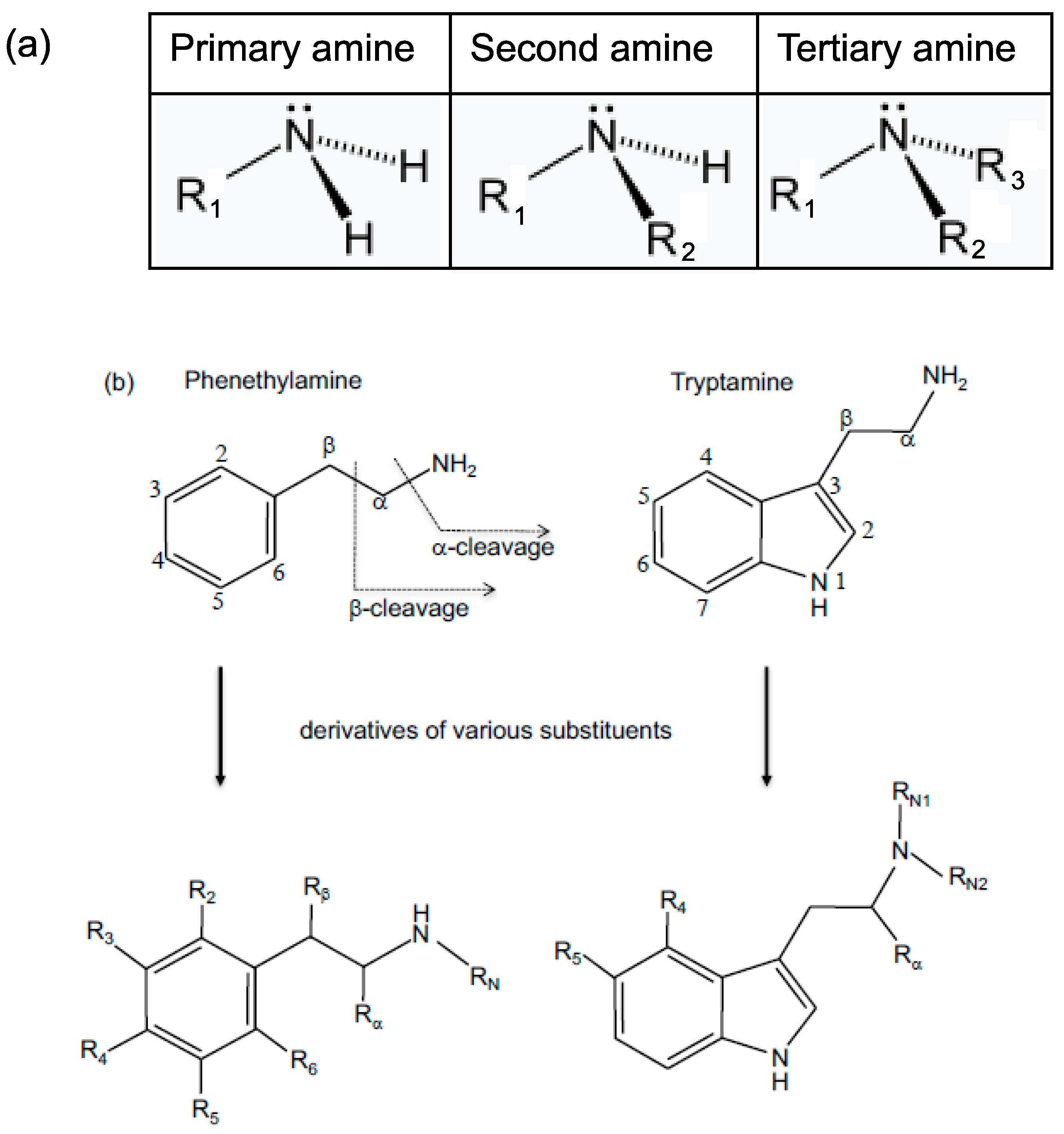
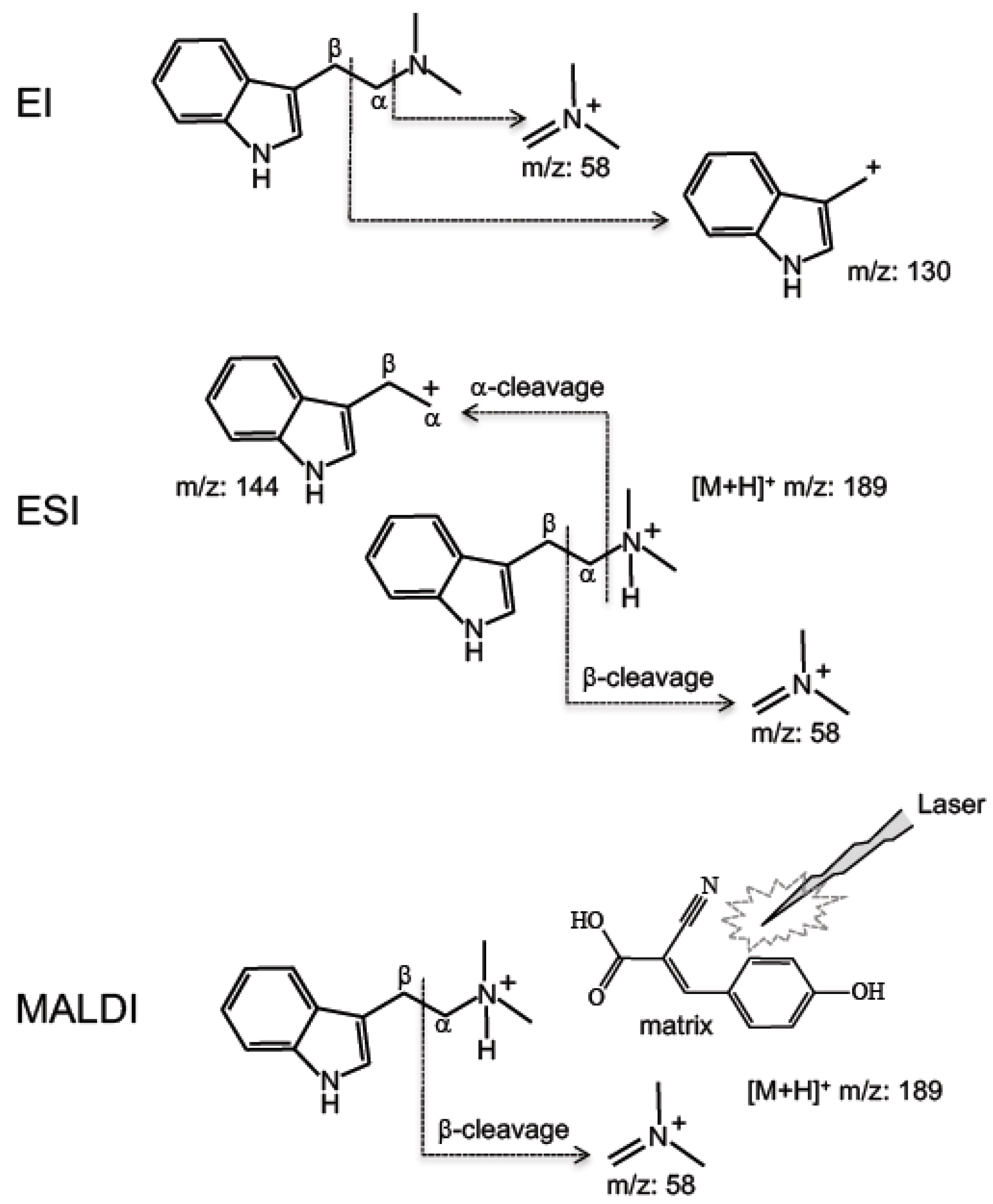
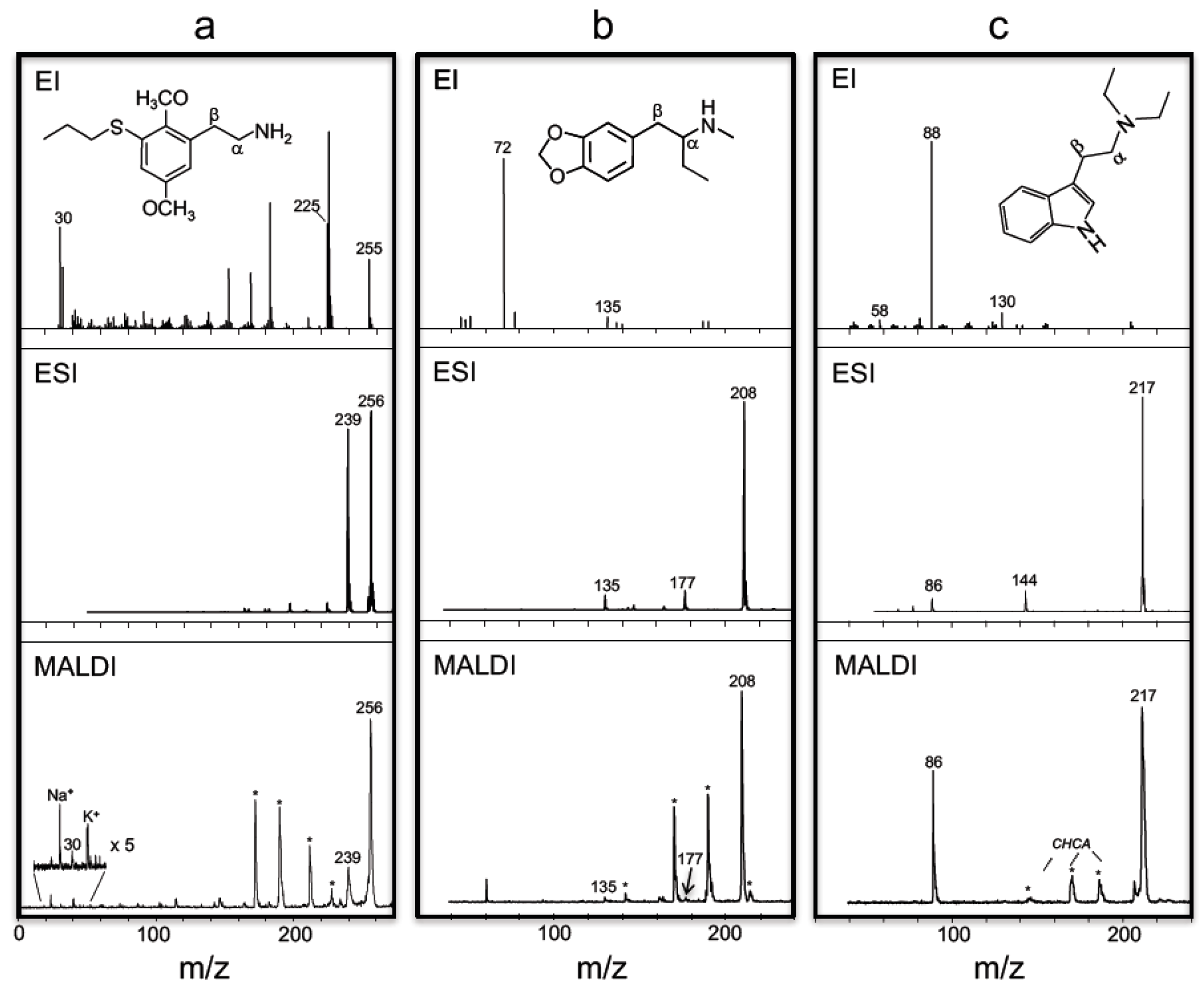
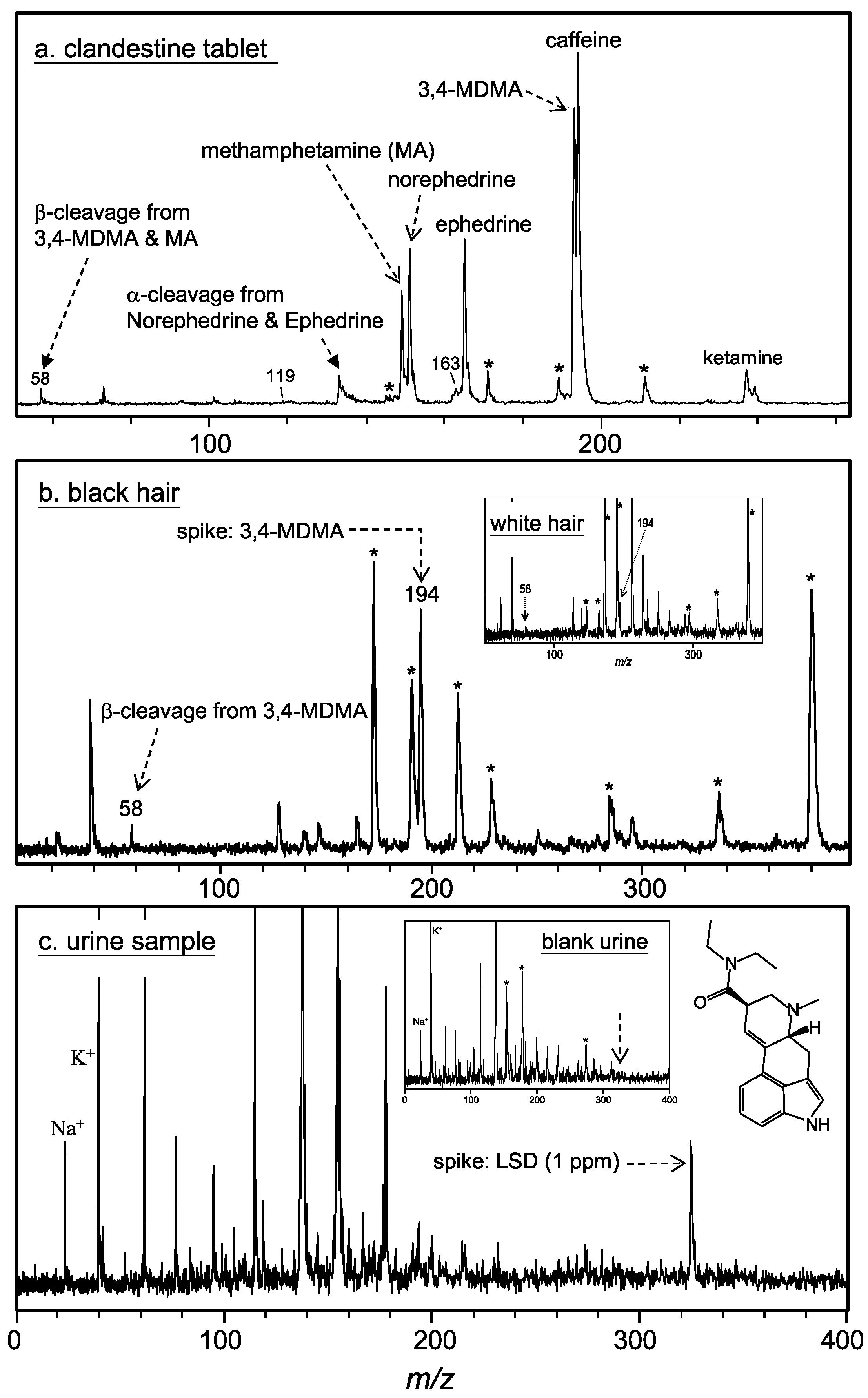
3. Results and Discussion

The abbreviations of the 11 phenethylamine and 9 tryptamine standards (tabulated by structure) used in this study are shown in Table 1. In fact, 55 tryptamine and 179 phenethylamine compounds were synthesized and described by Ann and Alexander Shulgin in TiHKAL and PiHKAL, respectively [13,14]. These compounds have been abused since the mid-1980s. Hence, more detailed information about the characteristic mass fragments, produced under different types of ionization methods, is highly desirable for the screening and confirmation of phenethylamine and tryptamine derivatives. Figure 1a shows the basic structures of primary (1°), secondary (2°), and tertiary (3°) amines, respectively; the chemical structures of phenethylamine, tryptamine and their derivatives of various substituents are shown in Figure 1b. Figure 2 shows a general schematic outline for the α-cleavage and β-cleavage based on ESI and MALDI modes, respectively, when N,N-dimethyltryptamine (DMT; Mw: 188) was selected as the model compound. This scheme shows that, if a tryptamine (or phenethylamine) acquires a proton, it will form a [M+H]+ ion. The α-cleavage would progress at the Cα-N bond, and, as a result, a cation (indole-containing group) and small neutral amine (NHRN1RN2) are formed. In this case, [M+H]+ ion (m/z: 189) and the cation (m/z: 58) can be observed. However, the proton also could be donated to the Cβ atom, leading to a Cα-Cβ bond breakage (i.e., β-cleavage). As a result, an iminium ion (CH2=N+RN1RN2) and a neutral fragment would form.
Table 2 shows the differences and similarities in the mass spectra of these phenethylamine/tryptamine compounds when different ionization methods are applied, including EI-MS, ESI-MS and MALDI/TOFMS. It can be found that, in the case of ESI/MS, the ions of m/z = 58, 144 and 189 come from β-cleavage, α-cleavage and [M+H]+, respectively. In the case of MALDI/TOFMS, only α-cleavage and [M+H]+ can be found. In contrast, different fragments are found when EI mode was used. All of these characteristic peaks are very important to identify tryptamine and phenethylamine. Among these data, our group selected 2C-T-7 (a primary amine), 3,4-MBDB (a secondary amine), and DET (a tertiary amine) as the model compounds for comparison, as described below. Figure 3 shows the EI/MS, ESI/MS and MALDI/TOFMS spectra of the three model compounds. As shown in Figure 3a (the upper mass spectrum; based on the EI mode), a parent ion of 2C-T-7, at m/z = 255, can be clearly observed. The other 2C series compounds also show similar consequences, as shown in Table 2, i.e., the parent ions obviously can be obtained. This is not a common phenomenon because, when the EI mode is used, in general, the energy of the electron makes it too difficult to obtain the parent ion. The reason for this could be that the functional groups (-OCH3 at R3/R5 and halogens at R4) attach at the benzene ring, resulting in stronger Cβ-Cα and Cα-N bonds. In fact, the Cα-N bond does not break for any compound investigated in this study when the EI mode was used. Two fragment ions (m/z = 30, 225) clearly form because of the Cβ-Cα bond breakage; the Cα-N bond seems very difficult to break. The three characteristic peaks ([M]+ and fragments from the Cβ-Cα bond breakage) are very unique and provide useful information for identifying 2C series compounds in actual forensic cases using the SIM (select ion monitoring) mode [12]. In the cases of the ESI and MALDI methods (the middle and bottom mass spectra of Frame 3a, respectively), a stable protonated parent ion ([M+H]+, m/z = 256) can be clearly observed. The α-cleavage process would produce a major fragment at m/z = 239, indicating the loss of a neutral fragment, 256 − 239 = 17 (NH3). In the β-cleavage process, a peak at m/z = 30 (H2C=N+H2) should be observed, indicating the loss of a larger neutral fragment, 256 − 30 = 226. However, the peak of m/z = 30 cannot be seen on the ESI-spectrum because the detection range of the instrument (Finnigan LCQ Classic) is limited to m/z above 50. Our group used the MALDI method to examine this and found that the fragment of m/z = 30 does indeed exist, although its intensity is very weak. Figure 3b shows the EI/MS, ESI/MS and MALDI/TOFMS spectra of 3,4-MBMB. As shown in the upper mass spectrum (EI), the parent ion (m/z = 208) is not formed, whereas two fragment ions (m/z = 58, 177) are produced from the Cβ-Cα bond breakage. No peak can be observed from the Cα-N bond breakage again. The peak at m/z = 58 is the major fragment of 3,4-MBMB, suggesting that this is the most stable fragment ion that forms after electron impact. It is obvious in this case, the EI mode is not suited for rapid screening if it is dependent only on the major peak at m/z = 58. 3,4-MDMA shows similar pattern with 3,4-MBMB. In traditional method, 3,4-MDMA can be performed via reaction with acetic anhydride, which provides specific fragments (m/z = 58, 100 and 162), permitted its characterization [15]. However, while such a specific derivative-reagent is suitable for one compound, it may not be suitable for others. In contrast, stable protonated parent ions ([M+H]+, m/z = 208) can be clearly observed by means of ESI and MALDI modes (the middle and bottom mass spectra of Frame B, respectively). In the case of ESI/MS, α-cleavage is the major process that would produce a fragment at m/z = 177, indicating the loss of a neutral fragment, 208 − 177 = 31 [NH2(CH3)], but the peak from β-cleavage cannot be found. It should be noted that, in the case of MALDI/TOFMS, fragment ions from both α-cleavage and β-cleavage could be observed, even though the fragment from α-cleavage is insignificant. Figure 3c shows the EI/MS, ESI/MS and MALDI/TOFMS spectra of DET. A parent ion (m/z = 217) is formed by the EI mode even though its intensity is insignificant, and two fragment ions (m/z = 86, 144) are produced from the Cβ-Cα bond breakage. In the case of ESI/MS, stable protonated parent ions ([M+H]+, m/z = 217) and two fragments (m/z = 86, 144), which are formed through α-cleavage and β-cleavage processes, can be clearly observed. The α-cleavage is the major process that would produce a fragment at m/z = 144, indicating the loss of a neutral fragment, 217 − 144 = 73 ([NH(CH2CH3)2]). Meanwhile, the β-cleavage process also occurs, in which a peak at m/z = 58 [H2C=N+(CH3)2] was observed, indicating the loss of a larger neutral fragment, 217 – 86 = 131. The ion intensity order of DMT is as follows: [M+H]+ >> fragment formed by the α-cleavage process > fragment formed by the β-cleavage process. In the case of MALDI/TOFMS, the ion intensity of [M+H]+ is greater than the fragment from the β-cleavage process, but the fragment (m/z = 144) from α-cleavage is missing. Thus, our group concludes that the three characteristic peaks, ([M+H]+ and the peaks from the α-cleavage and β-cleavage processes) are very unique and provide useful information in identifying phenethylamines and tryptamines in actual forensic cases based on the ESI/MS or MALDI/TOFMS methods. Although a direct sample injection is possible based on some LC-ESI/MS methods, which would be an ideal method for rapid screening, thus far, such studies also can be performed by means of MALDI/TOFMS since the sample substrate used in MALDI/TOFMS can be much easily inserted and changed. Our group selected MALDI/TOFMS as the rapid screening tool for the following studies, including actual clandestine tablet, hair and urine samples.
Figure 4a shows a typical mass spectrum obtained by the MALDI/TOFMS method from an actual clandestine tablet, which was seized from the illicit market. CHCA and Nd:YAG laser (355 nm) were used as the matrix and excitation source, respectively. As can be seen from the mass spectrum in Figure 4a, the ion peaks at m/z = 194 and m/z = 58/163 correspond to the protonated parent ion and the fragments from β-cleavage and α-cleavage, respectively, indicating that the tablet could contain 3,4-MDMA. The ion peaks at m/z = 150 and m/z = 58/119 correspond to the protonated parent ion and the fragments from the β-/α-cleavage, respectively, indicating that the tablet also contains methamphetamine. Furthermore, the ion peaks belong to norephedrine (m/z = 152) and ephedrine (m/z = 166), raw materials for the synthesis of methamphetamine. The fragment (m/z = 136) from α-cleavage also was found. Peaks corresponding to caffeine and ketamine also were found, showing that this was a tablet with multiple components. Herein, it should be noted that 3,4-BDB (3,4-benzodioxol-5-yl-2-butylamine) can provide similar pattern with 3,4-MDMA. Hence, in this case, the clandestine tablet should be examined in detail if ion peaks at m/z = 194 and m/z = 58 were found. In other words, a rapid screening method is important and useful for saving time. Figure 4b shows a typical MALDI-TOF mass spectrum for a 3,4-MDMA spiked hair (black color). The inset shows a 3,4-MDMA spiked white hair. The results indicate that the MALDI/TOFMS method is also useful for rapid screening of hair samples without any pretreatment; however, a black hair absorbs laser emission better than a white hair. Finally, a 0.01 mg mL−1 DMT standard was spiked to a blank urine sample. The findings show that ion peaks at m/z = 189 and m/z = 58 can be found (data not shown). The limit of detection of DMT in urine is about 0.001 mg mL−1. Instead, Figure 4c shows the mass spectrum of a LSD (a tertiary amine with a tryptamine-like structure) spiked urine sample, since LSD is very difficult to detect by GC/MS without derivatization process. As seen in the mass spectrum, several peaks appear, but the peak of the spiked LSD urine sample (0.001 mg mL−1) can be clearly observed. The matrix used was 2,5-DHB, since it works better than CHCA. The inset shows the same urine sample before spiking. In Taiwan, if the concentration of amphetamines or related metabolites exceeds 0.0005 g mL−1, a suspect is judged guilty. The method proposed here provides sufficient sensitivity to permit identification at such a low level of LSD.

4. Conclusions

Our group demonstrated that a MALDI/TOFMS method can be used successfully for rapid screening of tablet, hair and urine samples. This is because the protonated parent ion and the fragments from β-cleavage and α-cleavage are unique and useful for the identification of phenethylamines and tryptamines. The results from this study show that MALDI/TOFMS is accurate, sensitive and quick, which makes it a viable method for use in rapid drug screening. The MALDI/TOFMS method is sufficiently reliable to serve as a complementary alternative to the officially prescribed method, GC/MS, for use in this field.

Author Contributions

C.-H.L. conceived and designed the experiments; B.-H.C., H.-M.C., and W.-X.C. performed the experiments and analyzed the data; J.-T.L. contributed reagents/materials/analysis tools; and C.-H.L. wrote the paper.

Funding

This work was supported by grants from the National Science Council of Taiwan under Contracts of No. 95-2745-M-003-001.

Conflicts of Interest

The authors declare no conflict of interest.

References

  1. Lin, L.-C.; Liu, J.-T.; Lin, C.-H. Identification of 2,5-dimethoxy-4-ethylthiophenethylamine and its metabolites in the urine of rats by gas chromatography–mass spectrometry. J. Chromatogr. B 2003, 798, 241–247. [Google Scholar] [CrossRef]
  2. Valentine, J.L.; Middleton, R. GC-MS identification of sympathomimetic amine drugs in urine: Rapid methodology applicable for emergency clinical toxicology. J. Anal. Toxicol. 2000, 24, 211–222. [Google Scholar] [CrossRef] [PubMed]
  3. Jurado, C.; Gimenez, M.P.; Soriano, T.; Menendez, M.; Repetto, M. Rapid analysis of amphetamine, methamphetamine, MDA, and MDMA in urine using solid-phase microextraction, direct on-fiber derivatization, and analysis by GC-MS. J. Anal. Toxicol. 2000, 24, 11–16. [Google Scholar] [CrossRef] [PubMed]
  4. Pujadas, M.; Pichini, S.; Poudevida, S.; Menoyo, E.; Zuccaro, P.; Farre, M.; Torre, R. Development and validation of a gas chromatography-mass spectrometry assay for hair analysis of amphetamine, methamphetamine and methylenedioxy derivatives. J. Chromatogr. B 2003, 798, 249–255. [Google Scholar] [CrossRef]
  5. Peters, F.T.; Schaefer, S.; Staack, R.F.; Kraemer, T.; Maurer, H.H. Screening for and validated quantification of amphetamines and of amphetamine- and piperazine-derived designer drugs in human blood plasma by gas chromatography/mass spectrometry. J. Mass Spectrom. 2003, 38, 659–676. [Google Scholar] [CrossRef] [PubMed]
  6. Cody, J.T.; Valtier, S. Differentiation of the 2,3-methylenedioxy regioisomer of 3,4-MDMA (ecstasy) by gas chromatography-mass spectrometry. J. Anal. Toxicol. 2002, 26, 537–539. [Google Scholar] [CrossRef] [PubMed]
  7. Stout, P.R.; Horn, C.K.; Klette, K.L. Rapid simultaneous determination of amphetamine, methamphetamine, 3,4-methylenedioxyamphetamine, 3,4-methylenedioxymethamphetamine, and 3,4-methylenedioxyethylamphetamine in urine by solid-phase extraction and GC-MS: A method optimized for high-volume laboratories. J. Anal. Toxicol. 2002, 26, 253–261. [Google Scholar] [PubMed]
  8. Weinmann, W.; Renz, M.; Vogt, S.; Pollak, S. Automated solid-phase extraction and two-step derivatisation for simultaneous analysis of basic illicit drugs in serum by GC/MS. Int. J. Legal Med. 2000, 113, 229–235. [Google Scholar] [CrossRef] [PubMed]
  9. Marquet, P.; Lacassie, E.; Battu, C.; Faubert, H.; Lachâtre, G. Simultaneous determination of amphetamine and its analogs in human whole blood by gas chromatography-mass spectrometry. J. Chromatogr. B Biomed. Sci. Appl. 1997, 700, 77–82. [Google Scholar] [CrossRef]
  10. Su, A.-K.; Liu, J.-T.; Lin, C.-H. Rapid drug-screening of clandestine tablets by MALDI-TOF mass spectrometry. Talanta 2005, 67, 718–724. [Google Scholar] [CrossRef] [PubMed]
  11. Su, A.-K.; Liu, J.-T.; Lin, C.-H. Rapid drug-screening and quantitation of 3,4-methylenedioxymethamphetamine in urine by MALDI-TOF mass spectrometr. Anal. Chim. Acta. 2005, 546, 193–198. [Google Scholar] [CrossRef]
  12. Chen, B.-H.; Liu, J.-T.; Chen, W.-X.; Chen, H.-M.; Lin, C.-H. A general approach to the screening and confirmation of tryptamines and phenethylamines by mass spectral fragmentation. Talanta 2008, 74, 512–517. [Google Scholar] [CrossRef] [PubMed]
  13. Shulgin, A.; Shulgin, A. PHINKAL (Phenethylamines I Have Known and Loved): The Continuation; Transform Press: Berkeley, CA, USA, 1991. [Google Scholar]
  14. Shulgin, A.; Shulgin, A. THINKAL (Tryptamines I Have Known and Loved): The Continuation; Transform Press: Berkeley, CA, USA, 1997. [Google Scholar]
  15. Fang, C.; Chung, Y.-L.; Liu, J.-T.; Lin, C.-H. Rapid analysis of 3,4-methylenedioxymethamphetamine: A comparison of nonaqueous capillary electrophoresis/fluorescence detection with GC/MS. Forensic Sci. Int. 2000, 125, 142–148. [Google Scholar] [CrossRef]
Figure 1. (a) basic structure of primary (1°), secondary (2°), and tertiary (3°) amines; and (b) chemical structures of phenethylamine, tryptamine and their derivatives of various substituents.
Applsci 08 01022 g001
Figure 2. A general schematic outline for α-cleavage and β-cleavage when the EI/MS (electron ionization–mass spectrometry), ESI/MS (electrospray ionization/mass spectrometry) and MALDI/TOFMS (matrix assisted laser desorption/time-of-flight mass spectrometry) methods were used, respectively. N,N-dimethyltryptamine (DMT; Mw: 188) was selected as the model compound.
Applsci 08 01022 g002
Figure 3. EI/MS; ESI/MS; and MALDI/TOFMS spectra of (a) 2C-T-7, (b) 3,4-MBMB and (c) DET (each sample, 0.1 mg mL−1 methanol), respectively. The major fragments of CHCA are indicated as “*”.
Applsci 08 01022 g003
Figure 4. (a) MALDI-TOF mass spectrum obtained from an actual clandestine tablet, which was seized from the illicit market in 2001, using CHCA and Nd:YAG laser (355 nm) as the matrix and excitation source, respectively. (b) MALDI-TOF mass spectrum for a 3,4-MDMA spiked hair (black color); inset, a 3,4-MDMA spiked white hair. (c) MALDI-TOF mass spectrum obtained from of LSD spiked urine sample, using DHB as the matrix. The major fragments of matrix (CHCA) are indicated as “*”.
Applsci 08 01022 g004
Table 1. Abbreviations used for the phenethylamine and tryptamine standards (tabulated structure) in this study.
A. Phenethylamines
Abbrev.RR2R3RaR4R5R6RN1Full Name
MACH3HH HHHCH3methamphetamine
2,4-DOBCH3OCH3H OCH3BrHH2,4-dimethoxy-5-bromoamphetamine
3,4-MDAHH -OCH2O-H HH3,4-methylenedioxyamphetamine
3,4-BDBCH2CH3H -OCH2O-H HH3,4-benzodioxol-5-yl-2-butylamine
3,4-MDMACH3H -OCH2O-H HCH33,4-methylenedioxymethamphetamine
MBDBCH2CH3H -OCH2O-H HCH3N-methyl-1-(1,3-benzodioxol-5-yl)-2-butylamine
2C-T-7HHOCH3 SCH2CH2CH3HOCH3H4-propylthio-2,5-dimethoxy-β-phenethylamine
2C-T-2HHOCH3 SCH2CH3HOCH3H4-ethylthio-2,5-dimethoxy-β-phenethylamine
2C-CHHOCH3 ClHOCH3H4-chloro-2,5-dimethoxy-β-phenethylamine
2C-BHHOCH3 BrHOCH3H4-bromo-2,5-dimethoxy-β-phenethylamine
2C-IHHOCH3 IHOCH3H4-iodo-2,5-dimethoxy-β-phenethylamine
B. Tryptamines
Abbrev.R5RRN1RN2Full Name
AMTHCH3HHα-methyltryptamine
DMTHHCH3CH3N,N-dimethyltryptamine
5-MeO-AMTOCH3CH3HH5-methoxy-α-methyltryptamine
DETHHCH2CH3CH2CH3N,N-diethyltryptamine
DPTHHCH2CH2CH3CH2CH2CH3N,N-dipropyltryptamine
DBT HHCH2CH2CH2CH3CH2CH2CH2CH3N,N-dibutyltryptamine
5-MeO-DMTOCH3HCH3CH35-methoxy-N,N-dimethyltryptamine
DIPTHHCH(CH3)2CH(CH3)2N,N-diisopropyltryptamine
5-MeO-DIPTOCH3HCH(CH3)2CH(CH3)2 5-methoxy-N,N-diisopropyltryptamine
a: A ring formed with both R3 and R4.
Table 2. Mass fragmentations (m/z values) of phenethylamines and tryptamines based on EI/MS, ESI/MS and MALDI/TOFMS, respectively. The three types of amines show different characteristic mass fragments. The symbols of 1°, 2° and 3° are abbreviations of primary, secondary and tertiary amines, respectively.
Abbrev.EI/MSESI/MSMALDI/TOFMS
Cβ··CαCα··NM+β-Cleavageα-Cleavage[M+H]+β-Cleavageα-Cleavage[M+H]+
2C-T-730, 225--255--23925630239256
2C-T-230, 211--241--22524230225242
2C-C30, 185--215--19921630199216
2C-B30, 229--259--24326030243260
2C-I30, 277--307--29130830291308
AMT44, 130--174--15817544158175
5-MeO-AMT44, 160------18820544188205
2,4-DOB44, 230------25727444257274
3,4-MDA44, 135------16318044163180
3,4-BDB58, 135------17719458177194
3,4-MDMA58, 135------16319458163194
3,4-MBDB72, 135------17720872177208
MA58, 91------11915058119150
DMT58, 130--1885814418958--189
DET86, 130----8614421786--217
DPT114, 130----114144245114--245
DBT 130, 142----142144273142--273
5-MeO-DMT58, 160--2185817421958--219
DIPT114, 130----114144245114--245
5-MeO-DIPT114, 160----114174275114--275

