Antimicrobial Effect of Titanium Hydroxyapatite in Denture Base Resin


1
Department of Removable Prosthodontics and Occlusion, Osaka Dental University, Hirakata 573-1144, Japan
2
Department of Microbiology, School of Dentistry, Aichi Gakuin University, Nagoya 464-8650, Japan
*
Author to whom correspondence should be addressed.
Appl. Sci. 2018, 8(6), 963; https://doi.org/10.3390/app8060963
Submission received: 15 May 2018
/
Revised: 30 May 2018
/
Accepted: 9 June 2018
/
Published: 12 June 2018
(This article belongs to the Section Applied Biosciences and Bioengineering)
Abstract
:In the current study, we investigated the antimicrobial effect of titanium hydroxyapatite (TiHA), a photo-oxidizing organic material, in denture base resin on single-species biofilms formed by laboratory bacteria and on multispecies biofilms formed by bacteria from the human saliva. Although TiHA reportedly restricts the growth of planktonic bacteria upon ultraviolet A (UVA) irradiation, the antimicrobial effect of TiHA on bacterial biofilms remains to be elucidated. Resin specimens were prepared by adding TiHA to polymethyl methacrylate-based, denture base resin. The specimens were incubated with biofilm-forming Streptococcus sanguinis, Actinomyces naeslundii, Staphylococcus aureus, Escherichia coli, or bacteria from the human saliva obtained from volunteers. After UVA irradiation, the colony-forming units (CFUs) from the biofilms formed on the specimens were determined. CFU numbers for S. sanguinis, A. naeslundii, and S. aureus that formed biofilms on TiHA-containing specimens were significantly lower than those formed on specimens without TiHA. TiHA did not reduce the CFUs of biofilm-forming E. coli. In all cases, CFU numbers in the biofilms formed on TiHA-containing specimens by the salivary bacteria were significantly reduced. In addition, neither a 56 h UVA irradiation nor a 28 d soaking in water diminished the antibacterial effect of TiHA. TiHA in denture base resin exerts an antimicrobial effect on single-species bacterial biofilms and biofilms formed by a wide variety of bacteria from human saliva.
1. Introduction
With a recent increase of the aging population, the number of people missing teeth is also increasing. Oral function compromised by tooth loss is generally restored by prosthodontic treatments, including the use of dentures [1]. Dentures, which have hard and non-shedding surfaces like teeth, accumulate dental plaque and calculus in a similar way to teeth [2]. The adhesion process is influenced by several factors, such as the structure and composition of the surface of these materials and the chemical/physical properties of microbial cell surfaces [3,4]. Poor denture cleansing not only leads to denture stomatitis [5], but also results in more serious infectious diseases, such as aspiration pneumonia [6,7].
Although cleaning of dentures is effective at ensuring oral hygiene and general health, denture cleanliness and oral hygiene of denture wearers is generally poor, thereby facilitating the formation and accumulation of an oral biofilm [8,9]. Clinical studies have reported that disrupting the biofilm may be more important than the use of antifungal or antimicrobial agents in the prevention and treatment of denture stomatitis [10,11]. Mechanical cleansing is an effective measure for routine biofilm control on dentures [12]. Chemical cleansing also aids denture wearers, especially geriatric patients and those with limited motor capacity [5], because the complicated shapes and rough surfaces of dentures make them difficult to clean [13,14]. Several materials, including the photocatalyst, have recently been used to manage denture stomatitis by killing the microorganisms attached to dentures. The application of titanium dioxide (TiO2), a representative photocatalytic material [1], on prosthodontic materials [15,16], including dentures [1,17,18] and implants [19,20,21], has been investigated. This material exerts an antimicrobial effect by photo-oxidizing in response to ultraviolet A (UVA) irradiation [22,23,24]. However, the effect might not be necessarily pronounced, since organisms, such as bacteria and proteins, do not adsorb onto TiO2 [25,26]. Titanium hydroxyapatite (TiHA; Taihei Chemical Industrial Co. Ltd., Osaka, Japan), in which Ca2+ is partially replaced by Ti4+ [25], is a recently developed photocatalytic material. In contrast to TiO2, TiHA exhibits a high affinity for organic material, thereby effectively killing bacteria [25]. Upon UVA irradiation, methylene blue, an organic compound, is effectively removed from a TiHA-containing denture [27]. Several planktonic bacteria from TiHA-containing tissue-conditioning materials are also killed upon UVA irradiation [28]. However, denture plaque mostly consists of bacterial biofilms composed of multilayers of a wide variety of oral bacteria, not of planktonic bacteria. The antimicrobial effect of TiHA on bacterial biofilms has not yet been reported.
In the current study, we evaluated the antimicrobial effect of TiHA against bacterial biofilms formed on denture base resin, and the effect against planktonic bacteria. For a clinical perspective, the biofilms used in the evaluation were composed not only by a single bacterial species but also by various species of bacteria from the human saliva. We also examined the effect of irradiation and extensive soaking on the antimicrobial effect of TiHA.
2. Materials and Methods
2.1. Preparation of Test Specimens
Experimental denture base resin specimens were prepared by adding TiHA (3%, 6%, or 9% [w/w]) to the polymer powder of polymethyl methacrylate (PMMA)-based, self-curing denture base resin (DUPE RESIN, dupe pink, GC Co., Tokyo, Japan). The resin without TiHA was used as a control. The polymer powder and resin monomer were mixed in a powder-liquid ratio of 5 g/3.5 mL at 25 °C, in accordance with the manufacturer’s instructions, poured into putty-type silicon rubber molds, and pressed by glass plates. After polymerization for 30 min, all specimens (φ 10 × 1 mm) were polished with four kinds of waterproof abrasive papers in the sequence #600, #800, #1000, and #1500 (Riken Corundum Co., Ltd., Konosu, Japan). This was followed by 24 h soaking in distilled water to remove the residual monomer. For experiments with bacteria, the specimens were first disinfected using 0.01% (v/v) sodium hypochlorite, and then rinsed with sterile distilled water three times.
2.2. Scanning Electron Microscopy (SEM) and Energy Dispersive X-ray Spectroscopy (EDX)
After coating with a 20 nm carbon layer, surface morphologies of the specimens containing no (control) or different TiHA concentrations (3%, 6%, or 9%) were observed using scanning electron microscopy (SEM; JXA-8530FA, JOEL, Tokyo, Japan) at 100× magnification. Energy dispersive X-ray spectroscopy (EDX; JOEL) was employed to determine elemental content at randomly selected areas (5 × 5 μm, or 500 × 500 μm) of the SEM images. The JOEL EPMA (JOEL), which is a software designated for the apparatus, was used for analysis.
2.3. Bacterial Strains and Culture Conditions
Streptococcus sanguinis JICC136 [29] and Actinomyces naeslundii 12104 [30] were grown anaerobically (under 80% N2, 10% H2, and 10% CO2) at 37 °C in the Brain Heart Infusion broth (BHI; Becton, Dickinson and Company, Franklin Lakes, NJ, USA). Staphylococcus aureus FDA 209P [31] and Escherichia coli DH5α (GE Healthcare Japan, Tokyo, Japan) were grown aerobically at 37 °C in the LB broth (Becton, Dickinson). The working bacterial cultures were prepared by inoculating fresh medium with an overnight culture at a ratio of 20:1, and incubating at 37 °C for 12 h. The optical density at 595 nm (OD595) of the bacterial cultures was adjusted to approximately 1.0, and the adjusted cultures were used for experiments described below. To determine the number of colony-forming units (CFUs), the microorganism suspensions were diluted and then plated on the BHI or LB plates. Bacteria from the human saliva were plated on Brucella HK agar (Kyokuto Pharmaceutical Industrial Co., Ltd., Tokyo, Japan) containing 5% rabbit blood, and then anaerobically incubated.
2.4. Evaluation of the Antimicrobial Effect of TiHA against Planktonic Bacteria
To evaluate the antimicrobial effect of TiHA against planktonic bacteria, an aliquot (100 μL) of microorganism suspension, with OD595 approximately 1.0, was transferred to the surface of each specimen that had been placed on a wet filter paper in a petri dish to maintain moisture. After 2 or 4 h irradiation with UVA (352 nm) from a distance of 20 cm, the microorganism suspension was diluted and then serially plated to determine the CFU value. Bacteria from specimens placed in a shade for 2 or 4 h (i.e., without irradiation) were used as negative controls.
2.5. Evaluation of the Antimicrobial Effect of TiHA against Biofilms
To evaluate the antimicrobial effect of TiHA against biofilms formed by single bacterial species (S. sanguinis, A. naeslundii, S. aureus, or E. coli), the specimens were placed in petri dishes and soaked in 20 mL of fresh medium containing 1 mL of each bacterial culture, and then incubated at 37 °C for 12 h. After incubation, the specimens were gently and carefully washed with sterile distilled water, three times, to remove non-adherent cells. Each specimen was placed on a wet filter paper and then irradiated with UVA for 2 h. The bacteria that adhered to the back of the specimens, which received no irradiation, were removed with a cotton swab saturated with 70% ethanol. After irradiation, the specimens were soaked in 5 mL of PBS, and then exposed for 15 min to ultrasound using an ultrasonic cleaner B-220 (Branson Ultrasonic, Danbury, CT, USA) set at approximately 50 kHz to remove the attached bacteria. After serial dilution with PBS, the microorganism suspensions were plated to determine CFU values. The bacteria from non-irradiated specimens used as negative controls were placed in a shade for 2 h without irradiation, as described above.
The effect of TiHA on bacteria from human saliva was also evaluated. Saliva samples were provided by eight volunteers (four males and four females, 31 ± 20 years old [mean ± SD]). The volunteers were asked to refrain from oral activities (such as eating, drinking, chewing, sucking, brushing, and mouth rinsing) for at least 3 h before the collection of the saliva. The collected saliva was stored on ice and used for experiments within 30 min. The specimens (n = 3) were incubated anaerobically at 37 °C for 12 h in petri dishes containing the mixture of saliva and fresh BHI at a ratio of 1:3, or only saliva. Other procedures, including UVA irradiation, the removal of bacteria from the specimens, and determining the CFU values, were performed as described above.
2.6. Inductively Coupled Plasma Atomic Emission Spectroscopy Analysis (ICPA)
Each specimen containing 0% or 9% TiHA was rinsed three times with ultrapure water (Synergy UV, Merck Millipore Co., Darmstadt, Germany) and then soaked in 20 mL of ultrapure water for 0 (control), 7, 14, 21, or 28 d. Ca2+ and Ti4+, the constituent elements of TiHA [25], were quantified using ICPA (Optima 7300DV, Perkin Elmer Japan Co., Ltd., Yokohama, Japan) to evaluate the release of TiHA from the specimens into water. The antimicrobial effect of the soaked specimens against biofilm-forming bacteria was evaluated as described above.
2.7. Statistical Analysis
Differences between groups were analyzed by using one-way analysis of variance, followed by the Student-Newman-Keuls multiple-comparisons test. Differences were considered significant when p < 0.05.
2.8. Ethics Statement
The study design and the procedure for obtaining the informed consent were approved by the Institutional Ethics Committee (110953) of the School of Dentistry, Osaka Dental University, Hirakata, Japan. All experiments were performed in accordance with the approved guidelines.
3. Results
3.1. SEM and EDX Evaluation of TiHA-Containing Resin Specimens
When present on the specimen surface, TiHA exhibits antimicrobial effect by adsorbing various organic materials and decomposing them under irradiation [19]. Therefore, the surface properties of TiHA-containing specimens were examined using SEM and EDX (Figure 1). SEM analysis revealed that the specimen surface mainly contained two different electron ray-contrast areas (Figure 1a). The bright areas contained more elements with higher atomic weight and appeared to contain more TiHA than the dark areas. To evaluate TiHA content of the specimens (Figure 1b), five bright and five dark areas (5 × 5 μm each) were randomly selected from the SEM images and analyzed using EDX (Figure 1b). The weight percentage of C and O, which are present in both denture base resin and TiHA, to the total weight of the elements detected in the analyzed areas was almost 100% in the dark areas. By contrast, Ca, P, and Ti, which are the constituent elements of TiHA, were detected only in the bright areas. When wider regions of the SEM images, containing both dark and bright areas (500 × 500 μm), were analyzed using EDX, the weight ratio of P and Ca to the total element weight increased in a concentration-dependent manner with the TiHA content (p < 0.05) (Figure 1c).
3.2. The Antimicrobial Effect of TiHA against Planktonic Bacteria
The antimicrobial effect of TiHA against planktonic bacteria, including S. sanguinis, A. naeslundii, S. aureus, and E. coli, was evaluated (Figure 2). In the absence of UVA irradiation, the CFU values of planktonic bacterial cultures placed on specimens containing different concentrations of TiHA were not significantly different from those on the control (data not shown).
Upon 2 h of UVA irradiation, the numbers of CFUs of planktonic S. sanguinis on specimens containing 6% and 9% TiHA were significantly lower than those on the control (p < 0.05). Under the same conditions, the CFU values of A. naeslundii, S. aureus, and E. coli on specimens containing 3%, 6%, and 9% TiHA were not significantly different from those of the control. When S. sanguinis, A. naeslundii, and S. aureus placed on the specimens were irradiated for 4 h, the CFU values were significantly reduced with an increasing TiHA content of the specimens (p < 0.05). By contrast, the CFU values of planktonic E. coli on TiHA-containing specimens were not significantly different from those of the control even after a 4 h irradiation. Thus, TiHA exerted an antimicrobial effect against planktonic S. sanguinis, A. naeslundii, and S. aureus upon a 4 h UVA irradiation but not against planktonic E. coli.
3.3. The Antimicrobial Effect of TiHA against Single-Species Bacterial Biofilms
The antimicrobial effect of TiHA against bacteria forming biofilms was next evaluated (Figure 3). The specimens were soaked in a fresh medium supplemented with 5% (v/v) of each bacterial culture to allow for biofilm formation. The bacteria that formed biofilms on specimens containing 0% (control), 3%, 6%, or 9% TiHA were quantified after irradiation. No viable bacteria were retrieved from S. aureus biofilms on specimens that had been irradiated for 4 h. Therefore, the antimicrobial effect of TiHA was evaluated only after 2 h of irradiation.
Without UVA irradiation, the CFU values of S. sanguinis, A. naeslundii, S. aureus, and E. coli biofilms on the specimens were not significantly different from those of each control (data not shown). When S. sanguinis, A. naeslundii, and S. aureus biofilms on the TiHA-containing specimens were irradiated, the CFUs were significantly reduced as the TiHA content of the specimens increased (p < 0.05). By contrast, the CFU values for E. coli biofilms on the TiHA-containing specimens were not significantly different from those of the control, with or without UVA irradiation.
3.4. The Antimicrobial Effect of TiHA against Biofilms Formed by Multiple Species of Bacteria from the Human Saliva
The antimicrobial effect of TiHA against biofilms formed by bacteria from the saliva of human volunteers was also investigated (Figure 4). When the specimens were incubated in BHI containing 25% saliva, the CFU values for bacteria in biofilms formed on the specimens containing 9% TiHA were significantly lower than those formed on the control, for the samples from all volunteers (p < 0.05) (Figure 4a). When the specimens were incubated in 100% saliva, the numbers of CFUs of bacteria from the TiHA-containing specimens were significantly reduced for samples from six out of eight volunteers, compared with the control (p < 0.05) (Figure 4b). These findings demonstrated that TiHA was largely effective against not only single-bacterium species biofilms (Figure 3) but also multispecies of biofilms formed by bacteria from the human saliva (Figure 4).
3.5. TiHA Resistance to Long-Time Irradiation and Soaking
Knowing the durability of TiHA is important from a clinical application standpoint. Hence, we evaluated the impact of long-term irradiation on the antimicrobial effect of TiHA (Figure 5a). Specimens containing 9% TiHA were prepared by pre-irradiation for 0 (control), 14, 28, 42, or 56 h, which corresponded to 0, 7, 14, 21, or 28 d use, respectively, assuming that TiHA-containing dentures were UVA-irradiated 2 h a day. The CFU values for S. sanguinis and A. naeslundii biofilms that formed on specimens that underwent 14 and 28 h pre-irradiation were not significantly different from those without pre-irradiation (Figure 5a). Unexpectedly, the CFU values for A. naeslundii biofilms from specimens pre-irradiated for 42 h, and for S. sanguinis and A. naeslundii biofilms from specimens pre-irradiated for 56 h, were significantly lower than those of the control (p < 0.05). These observations suggested that the antimicrobial effect of TiHA was not readily reduced by UVA irradiation.
Next, we examined the effect of soaking of the TiHA-containing specimens in ultrapure water on the antimicrobial effect of TiHA (Figure 5b). After soaking for 0 (control), 7, 14, 21, or 28 d, specimens that contained 9% TiHA were used in biofilm experiments. The CFU values for S. sanguinis and A. naeslundii biofilms formed on the pre-soaked specimens were not significantly different from those formed on specimens without pre-soaking (Figure 5b). Further, ultrapure water used for soaking was analyzed using ICPA (Figure 6). The concentrations of Ca2+ released from the specimens containing 9% TiHA increased significantly in a time-dependent manner until 21 d after soaking (p < 0.05). However, no significant differences in the Ca2+ concentrations were observed between samples soaked for 21 d and 28 d. The amount of Ca2+ released from specimens without TiHA was below the detection limit. The amount of Ti4+ released from all the tested specimens was undetectable (data not shown). Preliminary experiments revealed that the Ca2+ and Ti4+ detection limits of the apparatus used in the current study were 0.087 μg/cm3 and 0.013 μg/cm3, respectively.
4. Discussion
Bacterial biofilm formation in the oral cavity starts after the adherence of early colonizers, including S. sanguinis and A. naeslundii, to a tooth surface covered with acquired pellicles [32]. S. aureus and E. coli are generally used in investigations of the antimicrobial properties of photocatalytic materials [33,34]. Therefore, S. sanguinis, A. naeslundii, S. aureus, and E. coli were selected for the evaluation of the antimicrobial effects of TiHA-containing denture base resin in the current study.
The addition of other compounds to the PMMA polymer inhibits polymerization of denture base resin, leading to a low flexural strength of the denture base resin [1,35,36]. The interstitial areas between PMMA particles in the TiHA-containing specimens increased with an increasing TiHA concentration (Figure 1a), suggesting an inhibition of denture base resin polymerization by TiHA. Cheng et al. [27] reported that the flexural strength of TiHA-containing specimens is lower than that of specimens lacking TiHA. It is hence necessary to monitor the reduction of strength of denture base resin associated with the addition of TiHA. By contrast, the concentration of the photocatalyst had a significant effect on surface roughness of the specimens [27].
The TiHA sensitivity of the planktonic bacteria tested was ranked as follows: S. aureus > S. sanguinis ≥ A. naeslundii > E. coli (Figure 2). This mostly agreed with a bacterial ranking in a previous study: S. aureus > S. mutans ≥ E. coli [28]. These data, together with ones presented in the current study, indicate that the sensitivity of planktonic bacteria to TiHA depends on the bacterial species. Interestingly, Uchimaru et al. [28] reported that the antimicrobial effect of TiHA in tissue-conditioning materials against planktonic S. aureus was ca. 1000-fold greater than that against E. coli. Such a high discrepancy in the TiHA sensitivity of S. aureus and E. coli was not apparent in the current study (Figure 2). This might be because of the different experimental base materials containing TiHA used (tissue-conditioning material and denture base resin).
From a clinical viewpoint, the antimicrobial effect of TiHA against biofilm-forming bacteria is more important than that against planktonic bacteria, since the denture plaque is a bacterial biofilm. In the current study, we demonstrated that TiHA incorporated in the tested specimens killed S. sanguinis, A. naeslundii, and S. aureus biofilms in a concentration-dependent manner (Figure 3). By contrast, TiHA was ineffective against planktonic E. coli (Figure 2), as well as E. coli biofilms (Figure 3). The photokilling mechanism of Gram-positive bacteria is different from that of Gram-negative bacteria [37,38]. In Gram-positive bacteria, hydroxyl radicals generated by photocatalytic materials upon irradiation directly destroy the cytoplasmic membrane and abolish cellular activity. On the other hand, in Gram-negative cells, the outer membrane prevents the hydroxyl radicals from severely damaging the cytoplasmic membrane. Hence, in the Gram-negative cells, the hydroxyl radicals penetrate the cytoplasmic membrane only after partially destroying the outer membrane [37,38]. Consequently, Gram-negative cells, including E. coli, are relatively resistant to the photokilling. Under the same UVA-irradiation conditions, the antimicrobial effect of TiHA against bacterial biofilms was more pronounced than that against planktonic bacteria (Figure 2 and Figure 3), probably because the liquid media surrounding the planktonic bacteria weakened the UVA.
The denture plaque does not consist of select single bacterial species but contains a wide variety of multiple bacterial species. Therefore, evaluation of the antimicrobial effect of TiHA against multiple bacterial species from human saliva containing various bacteria from different niches of the oral cavity [39,40,41] was required. TiHA was highly effective against all the tested biofilms formed by bacteria present in the different saliva samples, and killed more than 90% of biofilm-forming bacteria in seven of eight samples (Figure 4). Even when the saliva was the only nutrition source for the biofilm-forming salivary bacteria, TiHA was effective in six out of eight cases. The observed antimicrobial effect of TiHA against a wide variety of oral bacteria indicates the great clinical application potential of this material. It would be interesting to observe the change of bacteria on the TiHA-containing dentures and on the oral mucosa of the wearers.
If TiHA was used in clinical applications, dentures containing TiHA would have to be repeatedly irradiated and soaked in water to maintain hygiene. Hence, it was necessary to investigate the duration of the antimicrobial effect of TiHA. A 56 h UVA irradiation, corresponding to 28 d of use (2 h per day), did not result in a reduction of the antimicrobial effect of TiHA (Figure 5a). Unexpectedly, the observed antimicrobial effect was more pronounced with longer pre-irradiation times (Figure 5a). This might be associated with the decomposition of organic substances attached to TiHA, including proteins, by pre-irradiation [25]. Further studies are needed to clarify the effect of the irradiation on the decomposition of organic substances by TiHA.
It was reported that 180 d soaking does not reduce the ability of TiHA to decompose methylene blue [27]. Similarly, based on the experiments presented herein, the soaking of TiHA-containing specimens in water for 28 d did not result in a reduction of the antimicrobial effect of TiHA (Figure 5b). The concentration of Ca2+ released from TiHA-containing specimens increased significantly in a time-dependent manner until 21 d after soaking, whereas the concentration of Ti4+ remained below the detection limits (Figure 6). Considering that the detection limits of the apparatus used in the current study were 0.087 μg/cm3 and 0.013 μg/cm3 for Ca2+ and Ti4+, respectively, it is reasonable to assume that the amount of released Ti4+ was extremely limited. A hybrid orbital is apparently formed between Ti 3d and O 2p in a TiHA crystal [42], suggesting that Ti is covalently bound to PO4. Thus, Ti release would be difficult and, consequently, the antimicrobial effect of TiHA would not be reduced for at least 28 d (Figure 5b).
5. Conclusions
In conclusion, TiHA in denture base resin exerts an antimicrobial effect on biofilms composed of not only a single species of select bacteria but also those composed of multiple bacteria from the human saliva.
Author Contributions
Conceptualization: W.S., S.K., and J.O.; methodology: W.S. and Y.Y.; validation: S.K.; formal analysis: Y.Y.; data curation: S.W.; writing-original draft preparation: S.W. and Y.Y.; writing-review & editing: S.K., Y.H., and J.O.; supervision: J.O.
Funding
This research received no external funding.
Acknowledgments
The authors thank Masato Wakamura (Fujitsu, Tokyo, Japan) for providing specific information about TiHA.
Conflicts of Interest
The authors declare no conflicts of interest.
References
- Shibata, T.; Hamada, N.; Kimoto, K.; Sawada, T.; Sawada, T.; Kumada, H.; Umemoto, T.; Toyoda, M. Antifungal effect of acrylic resin containing apatite-coated TiO2 photocatalyst. Dent. Mater. J. 2007, 26, 437–444. [Google Scholar] [CrossRef] [PubMed]
- Neill, D.J. A study of materials and methods employed in cleaning dentures. Br. Dent. J. 1968, 124, 107–115. [Google Scholar]
- Busscher, H.J.; Cowan, M.M.; van der Mei, H.C. On the relative importance of specific and non-specific approaches to oral microbial adhesion. FEMS Microbiol. Rev. 1992, 8, 199–209. [Google Scholar] [CrossRef] [PubMed]
- Bellon-Fontaine, M.N.; Mozes, N.; van der Mei, H.C.; Sjollema, J.; Cerf, O.; Rouxhet, P.G.; Busscher, H.J. A comparison of thermodynamic approaches to predict the adhesion of dairy microorganisms to solid substrata. Cell Biophys. 1990, 17, 93–106. [Google Scholar] [CrossRef] [PubMed]
- Kulak-Ozkan, Y.; Kazazoglu, E.; Arikan, A. Oral hygiene habits, denture cleanliness, presence of yeasts and stomatitis in elderly people. J. Oral Rehabil. 2009, 29, 300–304. [Google Scholar] [CrossRef]
- El-Solh, A.A. Association between pneumonia and oral care in nursing home residents. Lung 2011, 189, 173–180. [Google Scholar] [CrossRef] [PubMed]
- Scannapieco, F.A.; Papandonatos, G.D.; Dunford, R.G. Associations between oral conditions and respiratory disease in a national sample survey population. Ann. Periodontol. 1998, 3, 251–256. [Google Scholar] [CrossRef] [PubMed]
- Frenkel, H.F.; Harvey, I.; Newcombe, R.G. Oral health care among nursing home residents in Avon. Gerodontology 2000, 17, 33–38. [Google Scholar] [CrossRef] [PubMed]
- De Visschere, L.M.; Grooten, L.; Theuniers, G.; Vanobbergen, J.N. Oral hygiene of elderly people in long-term care institutions a cross-sectional study. Gerodontology 2006, 23, 195–204. [Google Scholar] [CrossRef] [PubMed]
- Catalán, A.; Pacheco, J.G.; Martínez, A.; Mondaca, M.A. In vitro and in vivo activity of Melaleuca alternifolia mixed with tissue conditioner on Candida albicans. Oral Surg. Oral Med. Oral Pathol. Oral Radiol. Endod. 2008, 105, 327–332. [Google Scholar] [CrossRef] [PubMed]
- Geerts, G.A.; Stuhlinger, M.E.; Basson, N.J. Effect of an antifungal denture liner on the saliva yeast count in patients with denture stomatitis: A pilot study. J. Oral Rehabil. 2008, 35, 664–669. [Google Scholar] [CrossRef] [PubMed]
- Kulak-Ozkan, Y.; Kazazoglu, E.; Arikan, A. Use of microwave energy to disinfect a long-term soft lining material contaminated with Candida albicans or Staphylococcus aureus. J. Prosthet. Dent. 1998, 79, 454–458. [Google Scholar]
- Radford, D.R.; Challacombe, S.J.; Walter, J.D. Denture plaque and adherence of Candida albicans to denture-base materials in vivo and in vitro. Crit. Rev. Oral Biol. Med. 1999, 10, 99–116. [Google Scholar] [CrossRef] [PubMed]
- Ramage, G.; Tomsett, K.; Wickes, B.L.; López-Ribot, J.L.; Redding, S.W. Denture stomatitis: A role for Candida biofilms. Oral Surg. Oral Med. Oral Pathol. Oral Radiol. Endod. 2004, 98, 53–59. [Google Scholar] [CrossRef] [PubMed]
- Su, W.; Wei, S.S.; Hu, S.Q.; Tang, J.X. Preparation of TiO2/Ag colloids with ultraviolet resistance and antibacterial property using short chain polyethylene glycol. J. Hazard Mater. 2009, 172, 716–720. [Google Scholar] [CrossRef] [PubMed]
- Yao, Y.; Ohko, Y.; Sekiguchi, Y.; Fujishima, A.; Kubota, Y. Self-sterilization using silicone catheters coated with Ag and TiO2 nanocomposite thin film. J. Biomed. Mater. Res. B Appl. Biomater. 2008, 85, 453–460. [Google Scholar] [CrossRef] [PubMed]
- Sawada, T.; Sawada, T.; Kumasaka, T.; Hamada, N.; Shibata, T.; Nonami, T.; Kimoto, K. Self-cleaning effects of acrylic resin containing fluoridated apatite-coated titanium dioxide. Gerodontology 2014, 31, 68–75. [Google Scholar] [CrossRef] [PubMed]
- Kado, D.; Sakurai, K.; Sugiyama, T.; Ueda, T. Evaluation of Cleanability of a Titanium Dioxide (TiO2)-coated Acrylic Resin Denture Base. Prosthodont. Res. Pract. 2005, 4, 69–76. [Google Scholar] [CrossRef]
- Zhang, R.; Wan, Y.; Ai, X.; Liu, Z.; Zhang, D. Corrosion resistance and biological activity of TiO2 implant coatings produced in oxygen-rich environments. Proc. Inst. Mech. Eng. Part H 2017, 231, 20–27. [Google Scholar] [CrossRef] [PubMed]
- Hoshi, N.; Negishi, H.; Okada, S.; Nonami, T.; Kimoto, K. Response of human fibroblasts to implant surface coated with titanium dioxide photocatalytic films. J. Prosthodont. Res. 2010, 54, 185–191. [Google Scholar] [CrossRef] [PubMed]
- Lilja, M.; Forsgren, J.; Welch, K.; Astrand, M.; Engqvist, H.; Strømme, M. Photocatalytic and antimicrobial properties of surgical implant coatings of titanium dioxide deposited though cathodic arc evaporation. Biotechnol. Lett. 2012, 34, 2299–2305. [Google Scholar] [CrossRef] [PubMed]
- Fujishima, A.; Honda, K. Electrochemical photolysis of water at a semiconductor electrode. Nature 1972, 238, 37–38. [Google Scholar] [CrossRef] [PubMed]
- Fujishima, A.; Honda, K. Electrochemical evidence for the mechanism of the primary stage of photosynthesis. Bull. Chem. Soc. Jpn. 1971, 44, 1148–1150. [Google Scholar] [CrossRef]
- Hisanaga, T.; Harada, K.; Tanaka, K. Photocatalytic degradation of organochlorine compounds in suspended TiO2. J. Photochem. Photobiol. A Chem. 1990, 54, 113–118. [Google Scholar] [CrossRef]
- Wakamura, M.; Hashimoto, K.; Watanabe, T. Photocatalysis by Calcium Hydroxyapatite Modified with Ti (IV): Albumin Decomposition and Bactericidal Effect. Langmuir 2003, 19, 3428–3431. [Google Scholar] [CrossRef]
- Zeng, H.; Chittur, K.K.; Lacefield, W.R. Dissolution reprecipitation of calcium phosphate thin films produced by ion beam sputter deposition technique. Biomaterials 1999, 20, 443–451. [Google Scholar] [CrossRef]
- Cheng, Y.; Sakai, T.; Moroi, R.; Nakagawa, M.; Sakai, H.; Ogata, T.; Terada, Y. Self-cleaning ability of a photocatalyst-containing denture base material. Dent. Mater. J. 2008, 27, 179–186. [Google Scholar] [CrossRef] [PubMed]
- Uchimaru, M.; Sakai, T.; Moroi, R.; Shiota, S.; Shibata, Y.; Deguchi, M.; Sakai, H.; Yamashita, Y.; Terada, Y. Antimicrobial and antifungal effects of tissue conditioners containing a photocatalyst. Dent. Mater. J. 2011, 30, 691–699. [Google Scholar] [CrossRef] [PubMed] [Green Version]
- Yoshida, Y.; Konno, H.; Nagano, K.; Abiko, Y.; Nakamura, Y.; Tanaka, Y.; Yoshimura, F. The influence of a glucosyltransferase, encoded by gtfP, on biofilm formation by Streptococcus sanguinis in a dual-species model. APMIS 2014, 122, 951–960. [Google Scholar] [CrossRef] [PubMed]
- Cisar, J.O.; Sandberg, A.L.; Abeygunawardana, C.; Reddy, G.P.; Bush, C.A. Lectin recognition of host-like saccharide motifs in streptococcal cell wall polysaccharides. Glycobiology 1995, 5, 655–662. [Google Scholar] [CrossRef] [PubMed]
- Sugai, M.; Komatsuzawa, H.; Ooku-Inomata, K.; Miyake, Y.; Ishida, E.; Suginaka, H. Isolation and characterization of Staphylococcus aureus mutants which form altered cell clusters. Microbiol. Immunol. 1994, 38, 995–999. [Google Scholar] [PubMed]
- Kolenbrander, P.E.; Andersen, R.N.; Blehert, D.S.; Egland, P.G.; Foster, J.S.; Palmer, R.J., Jr. Communication among oral bacteria. Microbiol. Mol. Biol. Rev. 2002, 66, 486–505. [Google Scholar] [CrossRef] [PubMed]
- Li, P.; Tong, Z.; Huo, L.; Yang, F.; Su, W. Antibacterial and biological properties of biofunctionalized nanocomposites on titanium for implant application. J. Biomater. Appl. 2016, 31, 205–214. [Google Scholar] [CrossRef] [PubMed]
- Haenle, M.; Fritsche, A.; Zietz, C.; Bader, R.; Heidenau, F.; Mittelmeier, W.; Gollwitzer, H. An extended spectrum bactericidal titanium dioxide (TiO2) coating for metallic implants: In vitro effectiveness against MRSA and mechanical properties. J. Mater. Sci. Mater. Med. 2011, 22, 381–387. [Google Scholar] [CrossRef] [PubMed]
- Kanie, T.; Arikawa, H.; Fujii, K.; Inoue, K. Physical and mechanical properties of PMMA resins containing γ-methacryloxypropyltrimethoxysilane. J. Oral Rehabil. 2004, 31, 166–171. [Google Scholar] [CrossRef] [PubMed]
- Pesci-Bardon, C.; Fosse, T.; Madinier, I.; Serre, D. In vitro new dialysis protocol to assay the antiseptic properties of a quaternary ammonium compound polymerized with denture acrylic resin. Lett. Appl. Microbiol. 2004, 39, 226–231. [Google Scholar] [CrossRef] [PubMed] [Green Version]
- Sunada, K.; Watanabe, T.; Hashimoto, K. Studies on photokilling of bacteria on TiO2 thin film. J. Photochem. Photobiol. A Chem. 2003, 156, 227–233. [Google Scholar] [CrossRef]
- Sunada, K.; Watanabe, T.; Hashimoto, K. Bactericidal activity of copper-deposited TiO2 thin film under weak UV light illumination. Environ. Sci. Technol. 2003, 37, 4785–4789. [Google Scholar] [CrossRef] [PubMed]
- Denepitiya, L.; Kleinberg, I. A comparison of the microbial compositions of pooled human dental plaque and salivary sediment. Arch. Oral Biol. 1982, 27, 739–745. [Google Scholar] [CrossRef]
- Mager, D.L.; Haffajee, A.D.; Socransky, S.S. Effects of periodontitis and smoking on the microbiota of oral mucous membranes and saliva in systemically healthy subjects. J. Clin. Periodontol. 2003, 30, 1031–1037. [Google Scholar] [CrossRef] [PubMed]
- Kononen, E.; Paju, S.; Pussinen, PJ.; Hyvonen, M.; Di Tella, P.; Suominen-Taipale, L.; Knuuttila, M. Population-based study of salivary carriage of periodontal pathogens in adults. J. Clin. Microbiol. 2007, 45, 2446–2451. [Google Scholar] [CrossRef] [PubMed]
- Tsukada, M.; Wakamura, M.; Yoshida, N.; Watanabe, T. Band gap and photocatalytic properties of Ti-substituted hydroxyapatite: Comparison with anatase-TiO2. J. Mol. Catal. A Chem. 2011, 338, 18–23. [Google Scholar] [CrossRef]
Figure 1.
Characterization of the TiHA-containing specimen surface. (a) SEM images of the surface morphologies of specimens containing the following concentrations of TiHA: 0% (control), 3%, 6%, or 9%. The fields were scanned at 100× magnification. Bar, 100 μm. Arrows indicate dark and bright areas; (b) Element weight percentages in the dark and bright areas on the specimens. The weights of C, O, Ca, P, and Ti relative to the sum of weights of all detected elements were determined using EDX and are shown as percentages for the dark and bright areas (5 × 5 μm). Each area was randomly selected from the SEM image of a specimen containing 9% TiHA (in A). Data are presented as the mean ± standard deviation for five areas; (c) Element weight percentages in specimens containing different concentrations of TiHA. The wide regions containing both dark and bright areas (500 × 500 µm) were analyzed using EDX, and the weights of Ca and P relative to the total weights of all detected elements were determined and are shown as percentages. Data are presented as the mean ± standard deviation for five areas, which were randomly selected from the SEM images of specimens containing 0% (control), 3%, 6%, or 9% TiHA. The asterisk indicates a significant difference between the samples (p < 0.05).

Figure 2.
The antimicrobial effect of TiHA against planktonic bacteria. The specimens containing 0% (control), 3%, 6%, or 9% TiHA were used. An aliquot (100 μL) of S. sanguinis, A. naeslundii, S. aureus, or E. coli cultures was placed on the specimen surface, and the specimens were irradiated for 2 or 4 h. The bacteria were then plated to determine the CFU values. Data are presented as the mean ± standard deviation for three specimens. The asterisk indicates a significant difference between the CFU values of the control and TiHA-containing specimens (p < 0.05).
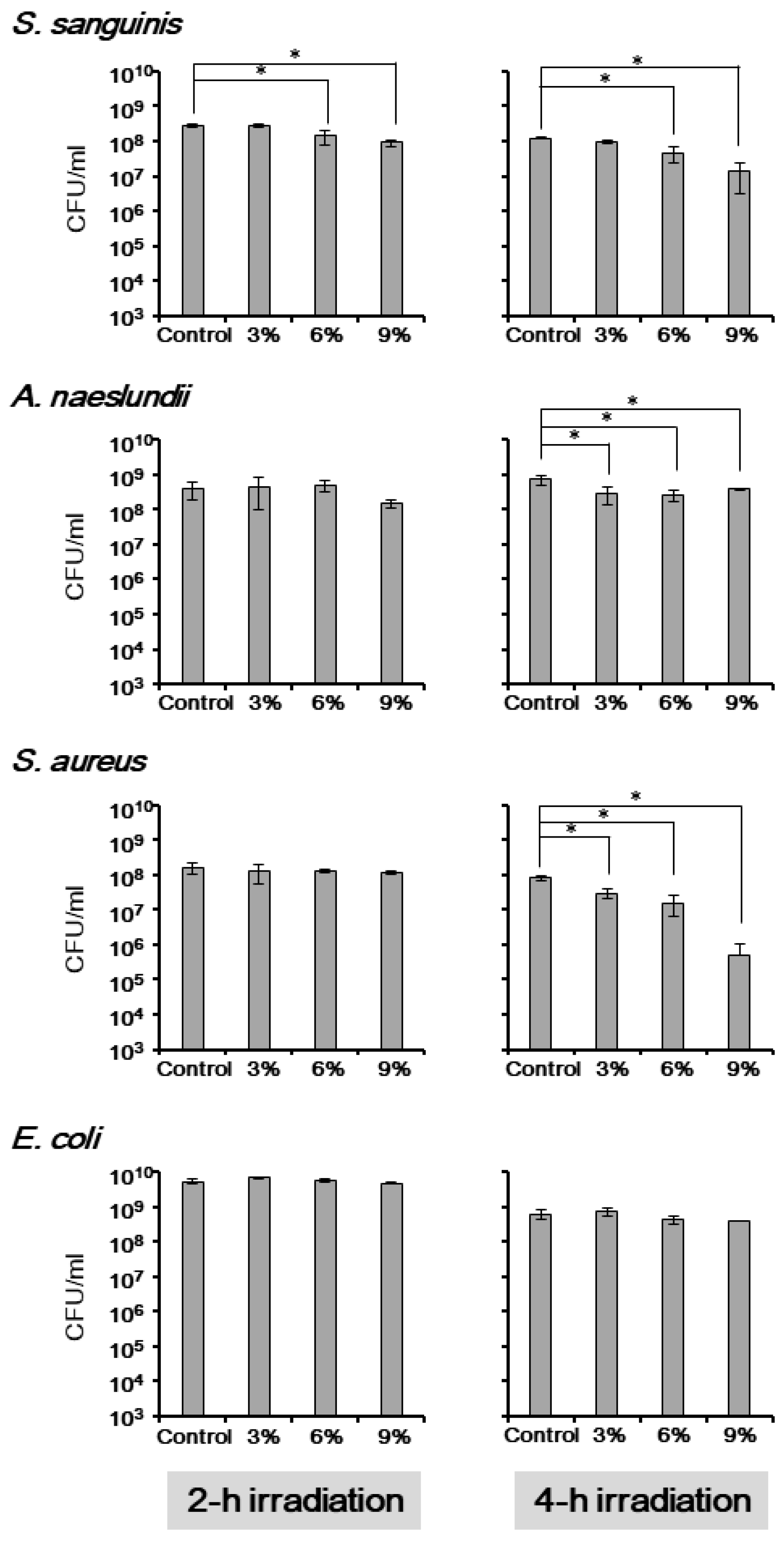
Figure 3.
The antimicrobial effect of TiHA against single-species bacterial biofilms. The specimens containing 0% (control), 3%, 6%, or 9% TiHA were used. The specimens, on which biofilms of S. sanguinis, A. naeslundii, S. aureus, or E. coli were formed, were irradiated for 2 h. The bacteria were then plated to determine the CFU values, which were divided by the specimen surface area. Data are presented as the mean ± standard deviation for three specimens. The asterisk indicates a significant difference in the CFU values of the control and the TiHA-containing specimens (p < 0.05).
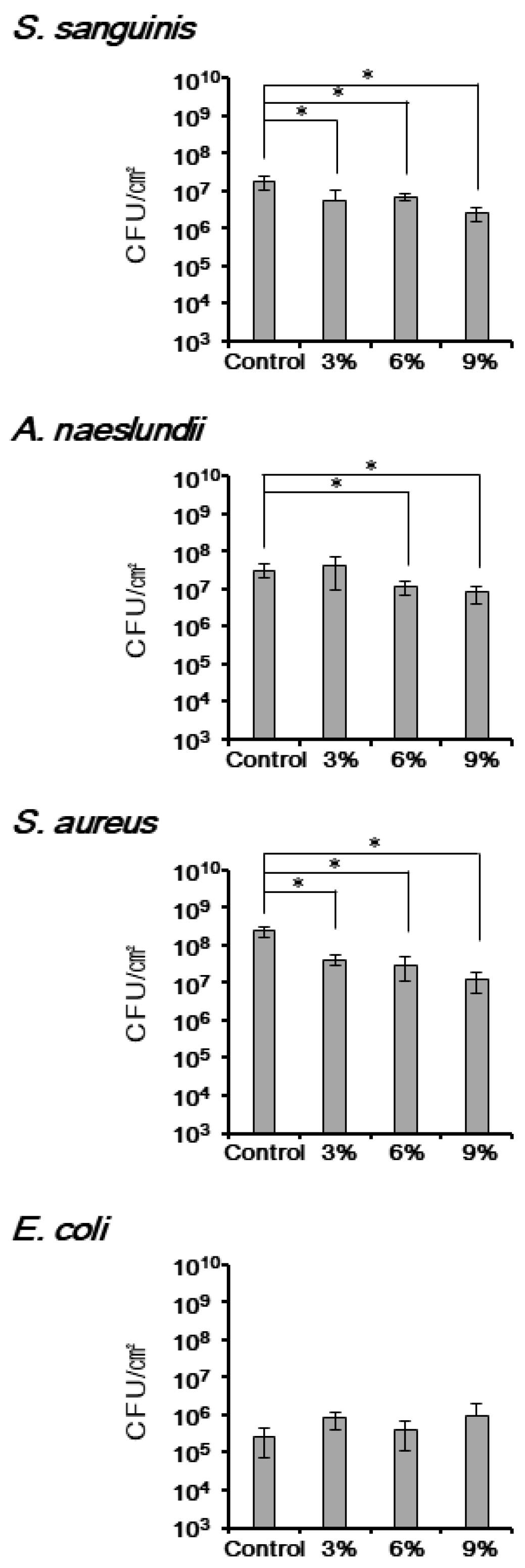
Figure 4.
The antimicrobial effect of TiHA against biofilms formed by bacteria from the human saliva. The specimens containing 0% (control) and 9% TiHA were soaked in BHI containing 25% saliva (a) or 100% saliva (b) for 12 h to allow the biofilms to form on the specimens. After 2 h of irradiation, the bacteria were plated to determine the CFU values, which were divided by the specimen surface area. The values from specimens containing 9% TiHA were compared with those from the control for each volunteer. Data are presented as the mean ± standard deviation for three specimens.
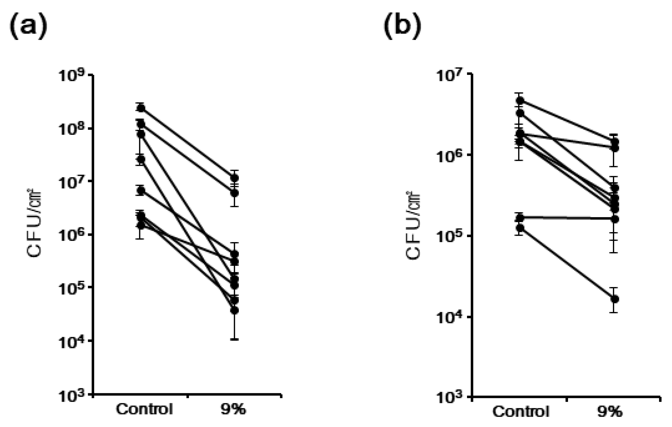
Figure 5.
The resistance of TiHA to long-time irradiation and soaking. Before the experiments, specimens containing 9% TiHA were pre-irradiated for 0 (control), 14, 28, 42, or 56 h (a), or soaked in sterile distilled water for 0 (control), 7, 14, 21, or 28 d (b) and then examined. The specimens on which S. sanguinis or A. naeslundii formed biofilms were irradiated for 2 h. The bacteria were plated to determine the CFU values, which were divided by the specimen surface area. Data are presented as the mean ± standard deviation for three specimens. The asterisk indicates a significant difference in the CFU values of the control and the TiHA-containing specimens (p < 0.05).
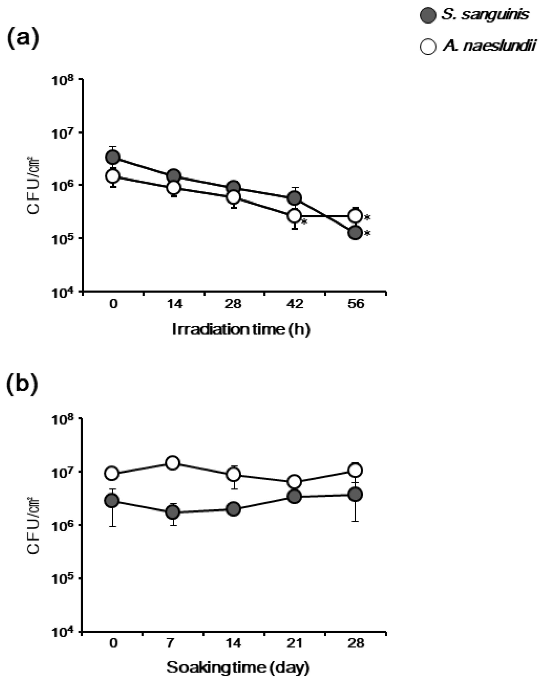
Figure 6.
Ca2+ released from the TiHA-containing specimens. The concentration of Ca2+ in the ultrapure water, in which the specimens were soaked for 0, 7, 14, 21, or 28 d, was determined using ICPA. The concentration value (μg/cm2) was obtained by dividing the total amount of Ca2+ in the ultrapure water by the specimen surface area. Data are presented as the mean ± standard deviation for three specimens. The asterisk indicates a significant difference between the compared groups (p < 0.05).

© 2018 by the authors. Licensee MDPI, Basel, Switzerland. This article is an open access article distributed under the terms and conditions of the Creative Commons Attribution (CC BY) license (http://creativecommons.org/licenses/by/4.0/).
Share and Cite
MDPI and ACS Style
Sato, W.; Yoshida, Y.; Komasa, S.; Hasegawa, Y.; Okazaki, J. Antimicrobial Effect of Titanium Hydroxyapatite in Denture Base Resin. Appl. Sci. 2018, 8, 963. https://doi.org/10.3390/app8060963
AMA Style
Sato W, Yoshida Y, Komasa S, Hasegawa Y, Okazaki J. Antimicrobial Effect of Titanium Hydroxyapatite in Denture Base Resin. Applied Sciences. 2018; 8(6):963. https://doi.org/10.3390/app8060963
Chicago/Turabian StyleSato, Wataru, Yasuo Yoshida, Satoshi Komasa, Yoshiaki Hasegawa, and Joji Okazaki. 2018. "Antimicrobial Effect of Titanium Hydroxyapatite in Denture Base Resin" Applied Sciences 8, no. 6: 963. https://doi.org/10.3390/app8060963
Note that from the first issue of 2016, this journal uses article numbers instead of page numbers. See further details here.