
Fluorescent Composite Cotton Fabric Modified with Crosslinked Chitosan for Theranostic Applications

1
Department of Textile, Leather and Fuels, University of Chemical Technology and Metallurgy, 1756 Sofia, Bulgaria
2
Faculty of Medicine, Sofia University “St. Kliment Ohridski”, 1407 Sofia, Bulgaria
*
Authors to whom correspondence should be addressed.
Appl. Sci. 2023, 13(23), 12660; https://doi.org/10.3390/app132312660
Submission received: 20 October 2023
/
Revised: 17 November 2023
/
Accepted: 23 November 2023
/
Published: 25 November 2023
(This article belongs to the Special Issue Hydrogels and Microgels: Fundamentals, Fabrication and Applications)
Abstract
:Developing multifunctional textile material for wound dressing is challenging due to the variety of wounds and their differing healing stages. Therefore, theranostics replaces the traditional approach to provide patient comfort and accelerated healing. In this study, we developed and compared three different materials. For this purpose, for the first time, chitosan was modified with 4-nitro-1,8-naphthalic anhydride in N,N-dimethylformamide (DMF) suspension, and subsequent nucleophilic substitution of the nitro group with N,N-dimethylamino group, whereby chitosan with a yellow color and fluorescence was obtained. Cotton fabric was impregnated successively with a citric acid solution and solution from chitosan and chitosan modified with 1,8-naphthalimide fluorophore (CN material). The same experimental protocol was applied for the second material, but indomethacin was added to the chitosan solution (CNI material). The third material was prepared similarly to the second but was immersed in an alginate solution as a last step (CNIA material). The obtained materials have been characterized by optical and scanning electron microscopy and thermal analysis (TG-DTA-DTG). Indomethacin release from composite materials and hydrogel swelling and erosion in phosphate buffer pH 7.4 at 37 °C was examined using gravimetric analysis, UV-vis absorption, and fluorescence spectroscopy. The antimicrobial activity of the cotton samples has been evaluated against B. cereus and P. aeruginosa as model bacterial strains. The analysis showed that CN material inhibited about 98.8% of the growth of P. aeruginosa and about 95.5% of the growth of B. cereus. Other composite materials combine antimicrobial properties with a sustained release of biologically active substances that can observed visually.
1. Introduction
Wound healing is a dynamic process that depends on the patient’s health status. Traditional wound dressings use a passive approach and aim to create a suitable environment for the healing process [1,2]. The current understanding of restoration without leaving a scar is for a breathable dressing that creates a clean, moist, and warm environment. These conditions are suitable for rapid revascularization, providing oxygen, nutrients, and growth factors, and for re-epithelialization, preventing the development of chronic wounds. In recent years, modern trends in wound care have focused on a more personalized approach based on the theranostics idea, or the simultaneous diagnosis of the wound condition and treatment as needed [3]. The new advanced materials development performing multiple functions is a promising area for improving patient care. Providing antimicrobial activity to prevent infection and monitoring wound health without removing the dressing creates greater patient treatment comfort [4,5]. Various methods of coating and surface modification of textile materials are being investigated in order to create and enhance their antibacterial activity. For this purpose, synthetic low and high molecular weight compounds, biopolymers and natural compounds, nanoparticles, and peptides are used [6,7,8,9]. Textile material modified with a layer of fluorescent stimuli-responsive hydrogel is a forceful tool for theranostic applications in skin care. This composite material has multifunctional properties aiding effective diagnosis, imaging, and successful therapy in different situations, such as skin diseases, injuries, and cosmetic implementation.
Among the various approaches, the thin film application on the fabric surface allows localized and precisely controlled release of active therapeutics [10]. Strategies for obtaining ultrathin layers include the Langmuir–Blodgett method, monolayer deposition techniques, and layer-by-layer (LbL) construction [11]. The LbL technique offers enormous freedom in the choice of materials and flexibility in the structural design. It includes multiple components, each with its role within the unified function of releasing a biologically active substance (BAS) [12]. This method does not impose restrictions on the size or shape of the substrate and does not require high temperature or pressure. The deposition on the substrate surface occurs by alternative adsorption of interacting materials through electrostatic forces, hydrogen bonds, covalent bonds, or bio-specific interactions. Different materials can be used for LbL, such as polyelectrolytes, micelles, graphene oxide, nanoparticles, and proteins [13]. Depending on the materials used to produce the multilayer film and the external conditions, the resulting layer can degrade, allowing for the controlled release of BAS.
In recent years, natural polyelectrolytes, such as chitosan, alginate, pectin, collagen, etc., have found significant application in this field because they are non-toxic, biodegradable, and biocompatible. Chitosan is a polycationic polymer with a broad spectrum of biological activity against bacteria and fungi and hemostatic properties [14,15]. Alginate is a polyanionic polymer used for wound dressing as it is a highly absorbent material and very suitable for wounds with high exudate. These natural polymers are stimuli-responsive to pH and temperature, and are applied in drug delivery and textile, especially for textile-based transdermal therapy [16]. Positively charged chitosan can interact with alginate to enhance layer stability and cytocompatibility [17]. Such supramolecular systems have been found to have applications in various fields, as they combine their ability to release drugs in a controlled manner with sensing properties for processes occurring inside and outside the studied system [18].
Recently, we have prepared new composite materials with potential applications as antimicrobial wound dressings releasing BAS. For this purpose, cotton fabric is modified with chitosan, cross-linked with different amounts of citric acid and containing the anti-inflammatory BAS, indomethacin. Their investigation showed that the BAS release combines diffusion processes with the swelling and erosion of the chitosan gel. The obtained materials have moderate antimicrobial activity against the Gram-positive bacteria Bacillus cereus and the Gram-negative bacteria Pseudomonas aeruginosa [19].
The fluorescent polymer can be used to visualize changes in hydrogel structure [20,21]. Covalent binding of the dye to polymer functional groups prevents the leaching of the fluorescent dye from the polymer and provides modification resistance to the solvents. The most widely used method for the chemical modification of chitosan is N-substitution in which the primary amino group of chitosan is the functional group that reacts. [22]. In this way, new fluorescent derivatives of chitosan were obtained by its primary amino group’s reaction with 4-bromonaphthalic anhydride and the bromine atom replacement with amino, hydroxyl, and acyl groups [23,24,25,26,27,28]. The electron-donating amino substituents introduction at the fourth position of the 1,8-naphthalimide structure changes the polarization of the chromophore system, resulting in intense yellow-green fluorescence emission and bright yellow color [29]. Such modified chitosan with 1,8-naphthalimide was used for rapid detection of 2,4-dinitrophenylhydrazine [26], Hg2+ [27], or Cu2+ [28] in the environment.
The optical properties of fluorescent 1,8-naphthalimides depend on the environment, and in polar solvents their fluorescence emission is quenched. Thus, the swelling of a hydrogel based on 1,8-naphthalimide modified chitosan and its erosion can be detected by a sensitive fluorescence analysis.
The study aims to obtain new antimicrobial textile materials with simultaneous drug-released properties and fluorescence imaging of this process.
2. Materials and Methods
Commercial 100% cotton fabrics with a surface weight of 135 ± 5 g/m2 (Vratciza-Vratza, JSC, Vratza, Bulgaria); Chitosan with Mw = 600,000–800,000 g mol−1, indomethacin and sodium alginate (Acros Organics, Geel, Belgium); acetic glacial acid (Merck, Darmstadt, Germany); citric acid (Sigma Aldrich, Darmstadt, Germany); sodium dihydrogen phosphate dihydrate (Merck, Darmstadt, Germany); disodium hydrogen phosphate dodecanhydrate (Merck, Darmstadt, Germany).
2.1. Modification of Chitosan with 4-Dimethylamino-1,8-naphtalimide-Ch-NI
Here, 2.00 g of chitosan were suspended in 50 mL of DMF and 0.20 g 4-nitro-1,8-naphthalic anhydride dissolved in 10 mL DMF was added slowly at a temperature of 70 °C. The mixture was stirred for 3 h and the reaction process was monitored by thin layer chromatography with liquid system-n-heptane acetone 1:1. After establishing the end of the reaction, the reaction mixture was cooled to 30 °C and 0.6 mL N,N-dimethylamine was added and stirring continued for 24 h. The final product was isolated by filtering and washing 3 times with 50 mL ethanol and drying at room temperature. Yield 2.16 g.
2.2. Preparation of Composite Materials
The cotton fabric was soaked in a citric acid solution (10.0%; w/v) at a bath modulus 1:2 and dried at room temperature. Three samples named CN, CNI, and CNIA were obtained from this fabric by the following procedure:
2.3. CN Composite Material
Chitosan (4.0%; w/v) was placed in water, and glacial acetic acid (1.0%; v/v) was added gradually. The resulting mixture was stirred until it obtained a transparent viscous solution. The solution was left at room temperature for 24 h to remove air bubbles formed during agitation. To a part of this solution in the amount of 5:1 volume of solution to fabric) was added modified chitosan with 1,8-naphthalimide Ch-NI (0.5%; w/w) and stirred with a magnetic stirrer until dissolved. The treated with citric acid fabric was soaked with the resulting solution. The sample has been dried at room temperature for 24 h.
2.4. CNI Composite Material
In this material, indomethacin (26 mg g−1 chitosan) dissolved in ethanol was added to the solution containing chitosan and chitosan modified with 4-dimethylamino-1,8-naphthalimide (prepared in the same way as for CN material). Next, the cotton fabric was treated the same as the sample CN.
2.5. CNIA Composite Material
The CNIA composite material was treated initially in the same manner as the CNI material and further coated with alginate by immersion for 15 min in 20.0 mL, 0.5% w/v aqueous alginate solution at pH 5.5 [30], after which the sample was allowed to dry at room temperature for 24 h.
2.6. Characterization of the Composite Materials
The fabric weight gain, after modification, was calculated with the following Equation (1):
where W and W0 are the weights of the starting fabric and modified fabric after its immersion in distilled water to remove the unreacted substances and its drying to constant weight.
2.7. Determination of the Degree of Gel Swelling
The dried modified textiles were weighed and immersed in 20.0 mL phosphate buffer pH = 7.4 at 37 °C to constant weight. At specified intervals, the samples were removed, excess water was absorbed with filter paper from the surface and then weighed. Equation (2) has been used to determine the degree of swelling:
where Wd and W1 are the weights of the dry and swollen material.
Swelling degree (%) = [(W1 − Wd)/Wd] × 100
2.8. In Vitro Gel Erosion
The in vitro degradation of the hydrogels was carried out in a phosphate-buffered solution with pH = 7.4 at 37 °C for 24 h. Textile samples were immersed in 20 mL of buffer for a specified time, then removed and dried to constant weight. The weight loss of the samples is determined by Equation (3).
where W0 is the initial sample weight, and Wt is the sample weight at time t.
weight loss % = [(W0 − Wt)/W0] × 100
2.9. Scanning Electron Microscope (SEM)
The studied samples were coated with gold using a DC magnetron sputtering Au K500X (Quorum Technologies, Lewes, UK). The surface morphology of the composite materials was analyzed using SEM-EDX equipment: SEM/FIB LYRA I XMU SEM (Tescan Group, Brno-Kohoutovice, Czech Republic). The instrument provided a resolution of 3.5 nm at 30 kV, with an accelerating voltage range of 200 V to 30 kV.
2.10. IR, 1H-NMR, and Fluorescence Analysis
1H spectrum was recorded at ambient temperature in D2O/CD3COOD on a Bruker Avance II+ 600 spectrometer operating at 600.13 MHz. The fluorescence spectra were recorded on a “Cary Eclipse” spectrofluorometer, respectively, using 1 cm optical path length quartz cuvettes (Hellma, Müllheim in Markgräflerland, Germany). Infrared spectra were conducted using an Infrared Fourier Transform spectrometer (IRAffinity-1, Shimadzu, Kyoto, Japan) equipped with a diffuse-reflectance attachment (MIRacle TM Attenuated Total Reflectance Attachment).
2.11. Thermal Properties Measurement
Simultaneous thermogravimetric analysis (TGA) and differential thermal analysis (DTA) were carried out on an STA PT1600 TG-DTA/DSC analyzer (LINSEIS Messgeräte GmbH, Selb, Germany) with a heating rate of 10 °C/min, covering the temperature range from room temperature to 600 °C in an air atmosphere.
2.12. In Vitro Release of Indomethacin from Composite Materials
The in vitro release of indomethacin from the composite materials was monitored in phosphate-buffered saline (pH = 7.4, 0.01 M) at 37 ± 0.5 °C by measuring the absorption intensity of the solution at λ = 320 nm using an ONDA spectrophotometer UV-31 SCAN, 190–1100 nm. Each sample was immersed in 20.0 mL of buffer. At specified time intervals, 5.0 mL of the solution was taken and replaced with 5.0 mL of a new amount of phosphate buffer at 37 °C. A standard curve was used to determine the concentration of the analyzed solutions.
2.13. Antibacterial Assay
The antimicrobial activity of the investigated composite materials CN, CNI, and CNIA has been tested against Gram-positive Bacillus cereus ATCC 11778 and Gram-negative Pseudomonas aeruginosa 1310 used as model strains (Collection of the Institute of Microbiology, Bulgarian Academy of Sciences). Microbial cultures were maintained at 4 °C on meat-peptone agar (MPA) slants and transferred monthly. The tubes containing sterile meat-peptone broth and cotton-sized samples (10 mm × 10 mm) were inoculated with each bacterial suspension. Tubes with untreated cotton samples and no samples were also prepared as controls. After incubation at 28 °C for 24 h and shaking at 240 rpm, the samples were removed, and bacterial growth was determined by measuring the turbidity of the medium at 600 nm (OD600). The antimicrobial activity of the treated cotton samples was assessed by the decrease in cell density after incubation. All antimicrobial tests were performed in triplicate (standard deviations less than 5%).
3. Results and Discussion
3.1. Synthesis and Characterization of Ch-NI
The modification of chitosan with 1,8-naphthalimide was carried out in a two-step synthetic route. Initially, 4-nitro-1,8-naphthalic anhydride interacts with the primary amino groups of chitosan at 70 °C through its anhydride group, forming a strong covalent bond. The nitro group has electron-accepting properties, and, for this reason, in the structure of 4-nitro-1,8-naphthalimide, a donor-acceptor interaction cannot take place, which would cause the polarization of the molecule and the appearance of color and fluorescent emission [31]. For this reason, the nitro group was nucleophilically substituted with an N,N-dimethylamino group by adding dimethylamine to the reaction mixture at a low temperature for 24 h (Scheme 1). In this, the nucleophilic substitution of the nitro group is favored by both the electron-accepting properties of the carbonyl group of 1,8-naphthalimide and DMF as a solvent, and the reaction proceeds at low temperature [32]. The chemical structure of the modified chitosan was confirmed by 1H-NMR and IR spectroscopy.
1H-NMR spectroscopy was used to prove the structure of the chitosan modified with 4-dimethylamino-1,8-naphthalimide (Figure 1). The characteristic signals of the chitosan, such as H7 at 2.06 ppm (the protons from CH3 of the NHCOCH3 group), H2 at 3.20 ppm, H3–6 in the range 3.48–4.03 ppm (the overlapping signals at the non-anomeric carbon atoms from in the glucopyranose ring), and H1 at 4.59 ppm (from the anomeric proton), were recorded in the proton NMR spectrum of the modified chitosan Ch-NI. Since the chitosan used has a 90% deacetylation degree of chitin, the signal at 2.77 ppm refers to the H2″ proton, which is offset from the signal at H2. The signals from the structure of the 4-dimethylamino-1,8-naphthalimide fragment are observed in the spectrum also. The characteristic signals for the aromatic protons from the naphthalene nucleus of 1,8-naphthalimide are located in the range of 7.27 ÷ 8.66 ppm. The protons from -N(CH3)2 are appeared at 3.02 ppm. The signal at 2.86 ppm refers to the H2′ proton and differs from that at H2 due to a polarization change after covalent coupling of the 1,8-naphthalimide fragment to the glucosamide structure.
Figure 2 presents the IR spectra of the modified chitosan with 4-dimethylamino-1,8-naphthalimide Ch-NI in the region from 4000 cm−1 to 400 cm−1. For comparison, the original unmodified commercial product chitosan was used. Due to the small amount of 1,8-naphthalimide fragments attached to the chitosan molecule, its principal characteristic peaks overlap with those of chitosan. An increase in the intensity of the bands of the modified chitosan compared to the initial one is observed, probably due to the presence of 1,8-naphthalimides and a change in the polarization of the monosaccharide nuclei to which they are attached. The absorption band at 3220–3336 cm−1 refers to N-H and O-H stretching due to possible intramolecular hydrogen bonds in the chitosan structure. Absorption bands at around 2919 cm−1 and 2878 cm−1 are due to C-H symmetric and asymmetric stretching of the monosaccharides ring. The carbonylimide groups (C=O) of the 1,8-naphthalimide structure have two characteristic vibrational bands caused by asymmetric and symmetric vibration. The band of the asymmetric vibration is recorded at 1699 cm−1, while the band of the symmetric vibration at 1652 cm−1 is almost overlapped by the absorption band of NHCOCH3 (C=O stretching of amide I) at about 1646 cm−1. A band at 1576 cm−1 corresponds to the N-H bending of the primary amino groups of the chitosan structure [33]. The symmetric deformation vibrations of the -CH2- groups have an absorption band at 1422. The band at 1323 cm−1 is due to the C-N-C from the imid (O=C-NR-C=O) from the 1,8-naphthalimides. The absorption band at 1153 cm−1 can be attributed to the asymmetric stretching of the C-O-C bond. The C-O stretching bands were recorded at 1066 and 1028 cm−1. The major chitosan bands outlined in this study are similar to samples reported by other investigations [33,34].
In the solid state, the structurally modified chitosan is of yellow-brown color, while the initial chitosan is white (Figure 3). The presence of 1,8-naphthalimide fragments in the main chain of chitosan did not affect its solubility. Good solubility has been observed in acidic water medium with pH values less than pH = 5. In such solutions, chitosan has a yellow color with an absorption maximum of λA = 411 nm and emits yellow-green fluorescence with a maximum of λF = 502 nm. In water solution, at neutral and alkaline pH media, due to deprotonation of the amino groups, the solubility of modified chitosan Ch-NI in water decreases, and the solution color becomes lighter with decreased fluorescence intensity (Figure 4). These results also confirm that the 1,8-naphthalimide fragments are covalently attached to the chitosan matrix, giving it new fascinating properties such as color and fluorescence emission.
Chitosan is a positively charged biopolymer at a pH less than 6.5 due to the protonation of its amino groups in aqueous solution. Citric acid is a weak polycarboxylic acid that finds application for crosslinking chitosan. The deprotonated carboxylic groups of citric acid can form ionic bonds with the ammonium groups of chitosan, as shown in Figure 5A. Indomethacin has a pKa of 4.5 and dissociates at a pH above pKa. In the pH range of 4.5 ÷ 6.5, ionic bonds occur between negatively charged indomethacin and positively charged chitosan and between chitosan and alginate, which has a pKa of 3.5 ÷ 4.6. The interaction of chitosan with alginate is shown in Figure 5B.
3.2. Thermogravimetric Analysis
Figure 6A shows that the thermal degradation of both chitosans is approximately the same. Two stages can be distinguished in the TG curves. The first stage is related to water separation. This effect is endothermal, as DTA curves show. Between the first and second stages, the stability region of the mass lost is observed from 150 °C to 250 °C. The most significant mass loss is observed in the next stage. After that, the degradation continues evenly. At 600 °C, the polymers have not decomposed completely, with about 20% dry residue remaining. The difference between chitosan and Ch-NI is minimal. The modified Ch-NI is more hydrophobic, so its mass loss is less initially. Figure 6B shows that cotton fabric and composite materials have the same TG profile as powdered chitosans. The cotton degraded completely at 600 °C, while the chitosan-coated fabrics remained more stable with the same dry residue as in chitosans. The addition of indomethacin and alginate does not significantly affect the thermal resistance.
3.3. Optical Microscope Examination
The micrographs obtained by optical microscope observation of the starting cotton fabric (CO) and the CN, CNI, and CNIA composite materials are shown in Figure 7. Irregularly scattered yellow-orange patches of fluorescent chitosan are observed on the surface of the composite materials, which are attributed to its poor solubility in acidic media due to the reduced number of free amino groups remaining after its modification.
3.4. Scanning Microscope Examination
A scanning electron microscope was used for better surface morphology characterization of the composite materials and the resulting films. Figure 8 shows the micrographs of the initial cotton fabric (CO) and the CN, CNI, and CNIA composites at different magnifications. It is apparent that in CN and CNI material, chitosan covered the cotton fibers and the space between them with a thin film, which is well visible in CNI material. A layer with an irregular grain structure tightly covers the fibers and fills the gaps between them after sample processing with alginate.
3.5. Determination of the Swelling Degree of the Material Coatings
The hydrogel swelling indicates the release mechanism of the biologically active substance [35]. At pH = 7.4, at which the study was carried out, the amino groups of chitosan are deprotonated and do not significantly affect the swelling of the hydrogel. In contrast, most free carboxyl groups are deprotonated, and electrostatic repulsion forces arise between them [36,37]. Figure 9 shows the swelling of the materials studied as a function of time. The sizeable swelling of material CN can be explained by the deprotonation of the free groups of citric acid and electrostatic repulsion between them at pH = 7.4 and the penetration of more water molecules into the gel structure. The addition of alginate to CNIA leads to an increase in the amount of carboxyl groups. The observed swelling enhances slightly with a longer residence time of the sample, which is necessary for the alginate macromolecule rearrangement. The CNI material showed the lowest swelling due to the hydrophobic indomethacin molecules incorporation into the coating structure and a reduction in the access of water molecules to the carboxyl groups.
3.6. Erosion of the Chitosan Gel
Essential for the use of the obtained materials as a delivery system for poorly water-soluble BAS is the erosion of the hydrogel produced on their surface over time. Supramolecular interactions are inherently dynamic and allow slow erosion of the gel [38]. Gel erosion and indomethacin release experiments were performed in phosphate buffer pH = 7.4 at 37 °C. After standing the materials for 24 h under these conditions, the gel was found to have eroded: 0.71% for CN material, 2.82% for CNI material, and 1.36% for CNIA material.
There should be no separation of chitosan molecules from the material surface since all its amino groups are deprotonated at pH = 7.4, and it is insoluble in water. Thus, only the citric acid used for the cotton fabric impregnation was set free. The indomethacin addition to the chitosan solution engages some of its amino groups. As a result, a more citric acid amount remains free and released from CNI material. After applying an alginate layer to the chitosan in material CNIA, many electrostatic bonds formed between the two polymers. These bonds would hardly break simultaneously to allow the alginate molecules to detach from the material surface.
3.7. Fluorescence Analysis
The presence of fluorescent chitosan modified with 4-dimethylamino-1,8-naphthalimide allows the change in the structure of the materials upon contact with the buffer solution. Figure 10 compares the change in fluorescence intensity of CNI and CNIA composites when immersed in phosphate buffer for 120 min. It can be seen that the fluorescence emission decreases more strongly for the CNIA material due to the pronounced gel swelling, as it was shown in Figure 9. As a result, the 4-dimethylamino-1,8-naphthalimide molecules were surrounded by more water molecules, and this change in the polarity of the environment affects the fluorescence emission of the materials.
3.8. Release of Indomethacin from CNI and CNIA Composites
The cumulative release of indomethacin from CNI and CNIA samples is presented in Figure 11. The study was conducted in vitro in phosphate buffer at pH = 7.4 and 37 °C for 30 h. For the first 360 min, an initial rapid release of indomethacin was observed due to the desorption of its less bound molecules in the resulting layers of the materials. In the CNI sample, 58.24 μg/cm2 of indomethacin was released, while in the CNIA sample, the amount released was slightly higher at 69.75 μg/cm2. There was then a delay in the delivery of the biologically active substance due to the more difficult separation of its molecules from inside the chitosan hydrogel. In the samples studied, the release of indomethacin continued uniformly after 24 h. In 30 h, the released quantity was 98.91 μg/cm2 for CNIA material and 92.71 μg/cm2 for the CNI sample. The faster release of indomethacin from the CNIA sample was due to the considerable swelling of the coating relative to CNI rather than its sizeable erosion.
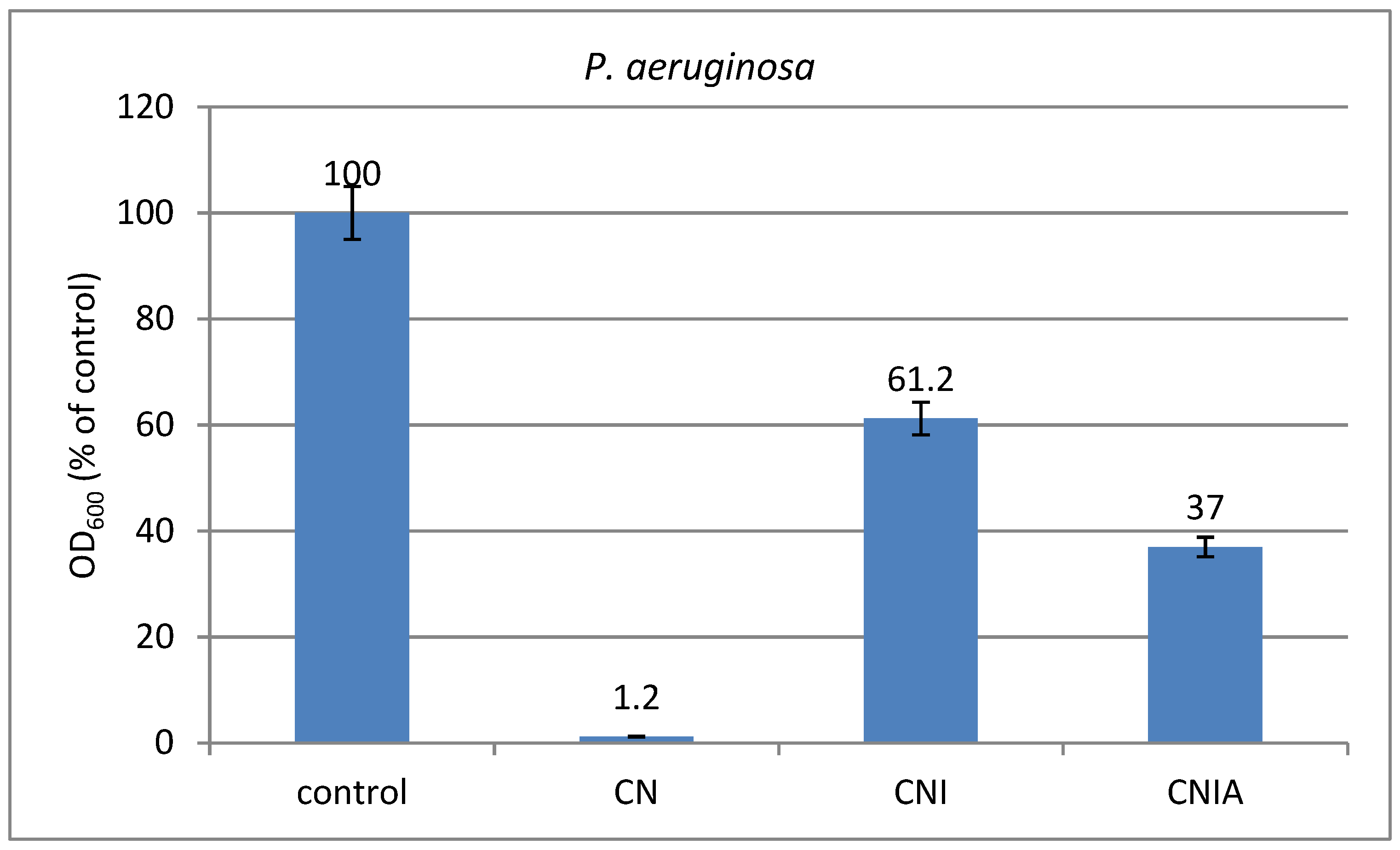
3.9. Antimicrobial Activity of Composite Materials against Bacterial Strains of P. aeruginosa and B. cereus
Chitosan is a well-known natural antimicrobial agent. The mechanism of its action after immobilization on the fabric surface is associated with altering cell permeability, chelation of essential metals, and preventing nutrients from being taken up from cells extracellularly [39]. The presence of 1,8-naphthalimide derivative on the fabric surface is responsible for the hydrophobic interactions with bacteria.
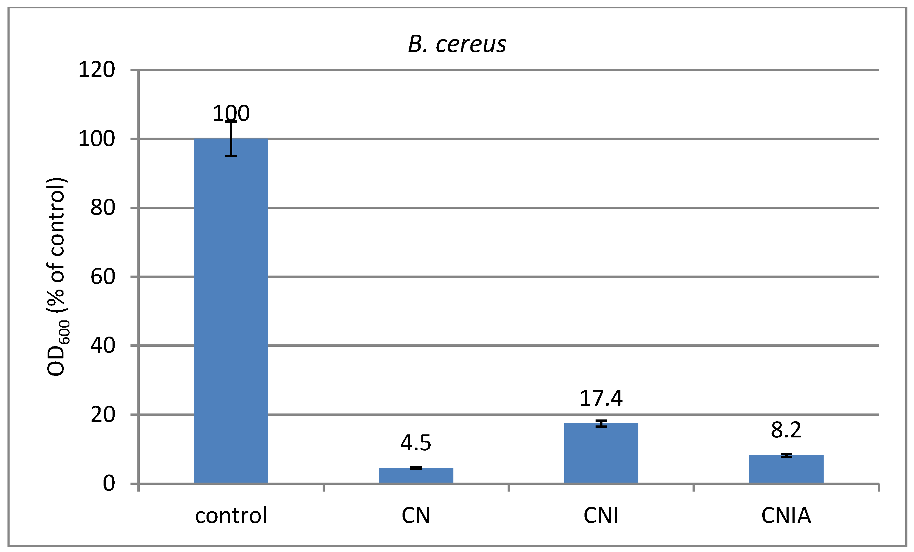
The composite material CN, CNI, and CNIA have been screened for antimicrobial activity in liquid medium against Gram-positive Bacillus cereus and Gram-negative Pseudomonas aeruginosa used as model bacterial strains. The results obtained are shown in Figure 12. It is seen as a reduction in cell growth of both strains of all CN, CNI, and CNIA samples tested compared to the control. In the case of CN material, the inhibition is about 98.8% of the growth of P. aeruginosa and about 95.5% of the growth of B. cereus, which is probably due to the synergistic effect of the hydrophobicity due to chitosan modified with 1,8-naphthalimide, the presence of the protonated cationic amino groups of chitosan and its chelating properties for divalent cations, mainly Mg2+ and Ca2+, that linked lipopolysaccharides in the outer membrane of Gram-negative bacteria [40]. In the CNI material, some of the polar groups of chitosan are involved in the electrostatic bond formation with indomethacin molecules, resulting in a decrease in its antibacterial activity, more pronounced against Gram-negative bacteria. The inhibition of P. auroginosa was the same as the result obtained in our previous study, where the chitosan coating did not contain 1,8-naphthalimide-modified chitosan [19]. Therefore, the addition of indomethacin inhibits the antimicrobial activity of the coating. The incorporation of alginate in the coating of CNIA material results in good antimicrobial activity against Gram-positive B. Seraeus and weaker against Gram-negative bacteria P. aeruginosa. Alginate has as good chelating properties as chitosan. For this reason, the alginate addition in the coating restores the antimicrobial properties of the material CNIA, close to the material CN and better than the material CNI.
4. Conclusions
Chitosan was modified with 4-dimethylamino-1,8-naphthalimide fluorescent units and characterized by NMR and IR spectroscopy. In a solid state and a water–acid solution, the modified chitosan has an intense yellow color and emits a yellow-green fluorescence. This chitosan has been used to prepare cotton fabric composites in the presence of indomethacin and alginate. The indomethacin release was followed in phosphate buffer pH = 7.4 and found to proceed uniformly over 24 h from both CNI and CNIA materials. Coating with an additional alginate layer accelerates the release of indomethacin due to faster swelling of the layer due to the possibility of breaking some bonds from its interior. However, this does not affect the gel erosion as alginate remains on the material surface. The resulting CN, CNI, and CNIA composites possess very good antimicrobial activity against Gram-positive bacteria B. cereus and Gram-negative bacteria P. aeruginosa. The sample containing only modified chitosan CN was found to almost completely inhibit the growth of both bacterial strains. The resulting three composite materials may find application as wound dressings as they possess antimicrobial properties. Two of the materials release an anti-inflammatory biologically active substance in a controlled manner.
Author Contributions
Conceptualization, D.S. and I.G.; methodology, D.S. and I.G.; formal analysis, D.S., D.A.; investigation, D.S., D.A. and I.G.; writing—original draft preparations, D.S. and I.G.; writing—review and editing, D.S. and I.G.; visualization, D.S. and D.A.; project administration, D.S. and I.G. All authors have read and agreed to the published version of the manuscript.
Funding
This study is financed by the European Union-NextGenerationEU, through the National Recovery and Resilience Plan of the Republic of Bulgaria, project № BG-RRP-2.004-0008-C01. This study is funded by the European Union-NextGenerationEU, through the National Recovery and Resilience Plan of the Republic of Bulgaria, project № BG-RRP-2.004-0002-C01, “BiOrgaMCT”.
Institutional Review Board Statement
Not applicable.
Informed Consent Statement
Not applicable.
Data Availability Statement
Data are contained within the article.
Conflicts of Interest
The authors declare no conflict of interest.
References
- Tehrany, P.M.; Rahmanian, P.; Rezaee, A.; Ranjbarpazuki, G.; Fard, F.S.; Asadollah Salmanpour, Y.M.; Zandieh, A.; Ranjbarpazuki, A.; Asghari, S.; Javani, N.; et al. Multifunctional and theranostic hydrogels for wound healing acceleration: An emphasis on diabetic-related chronic wounds. Environ. Res. 2023, 238, 117087. [Google Scholar] [CrossRef] [PubMed]
- Saberianpour, S.; Melotto, G.; Forss, R.; Redhead, L.; Elsom, J.; Terrazzini, N.; Sandeman, S.; Sarker, D.; Bucca, G.; Hesketh, A.; et al. Development of theranostic wound dressings: Harnessing the knowledge of biospecific interactions at the biomaterial interface to promote healing and identify biomarkers. Expert Rev. Med. Devices 2023, 20, 163–165. [Google Scholar] [CrossRef] [PubMed]
- Yang, C.; Yang, C.; Chen, Y.; Liu, J.; Liu, Z.; Chen, H.-J. The trends in wound management: Sensing, therapeutic treatment, and theranostics. J. Sci. Adv. Mater. Devices 2023, 8, 100619. [Google Scholar] [CrossRef]
- Avossa, J.; Pota, G.; Vitiello, G.; Macagnano, A.; Zanfardino, A.; Di Napoli, M.; Pezzella, A.; D’Errico, G.; Varcamonti, M.; Luciani, G. Multifunctional mats by antimicrobial nanoparticles decoration for bioinspired smart wound dressing solutions. Mater. Sci. Eng. C 2021, 123, 111954. [Google Scholar] [CrossRef] [PubMed]
- Derakhshandeh, H.; Kashaf, S.S.; Aghabaglou, F.; Ghanavati, I.O.; Tamayol, A. Smart Bandages: The Future of Wound Care. Trends Biotechnol. 2018, 36, 1259–1274. [Google Scholar] [CrossRef] [PubMed]
- Andrade-Guel, M.; Ávila-Orta, C.A.; Cabello-Alvarado, C.; Cadenas-Pliego, G.; Esparza-González, S.C.; Pérez-Alvarez, M.; Quiñones-Jurado, Z.V. Non-Woven Fabrics Based on Nanocomposite Nylon 6/ZnO Obtained by Ultrasound-Assisted Extrusion for Improved Antimicrobial and Adsorption Methylene Blue Dye Properties. Polymers 2021, 13, 1888. [Google Scholar] [CrossRef] [PubMed]
- Spasova, M.; Stoilova, O.; Manolova, N.; Rashkov, I.; Naydenov, M. Electrospun Eco-Friendly Materials Based on Poly(3-hydroxybutyrate) (PHB) and TiO2 with Antifungal Activity Prospective for Esca Treatment. Polymers 2020, 12, 1384. [Google Scholar] [CrossRef]
- Georgieva, S.; Todorov, P.; Staneva, D.; Grozdanov, P.; Nikolova, I.; Grabchev, I. Metal–Peptide Complexes with Antimicrobial Potential for Cotton Fiber Protection. J. Funct. Biomater. 2023, 14, 106. [Google Scholar] [CrossRef]
- Staneva, D.; Said, A.I.; Vasileva-Tonkova, E.; Grabchev, I. Enhanced Photodynamic Efficacy Using 1,8-Naphthalimides: Potential Application in Antibacterial Photodynamic Therapy. Molecules 2022, 27, 5743. [Google Scholar] [CrossRef]
- Park, S.; Han, U.; Choi, D.; Hong, J. Layer-by-layer assembled polymeric thin films as prospective drug delivery carriers: Design and applications. Biomater. Res. 2018, 22, 29. [Google Scholar] [CrossRef]
- Li, W.; Lei, X.; Feng, H.; Li, B.; Kong, J.; Xing, M. Layer-by-Layer Cell Encapsulation for Drug Delivery: The History, Technique Basis, and Applications. Pharmaceutics 2022, 14, 297. [Google Scholar] [CrossRef]
- Aubert-Viard, F.; Mogrovejo-Valdivia, A.; Tabary, N.; Maton, M.; Chai, F.; Neut, C.; Martel, B.; Blanchemain, N. Evaluation of antibacterial textile covered by layer-by-layer coating and loaded with chlorhexidine for wound dressing application. Mater. Sci. Eng. C 2019, 100, 554–563. [Google Scholar] [CrossRef] [PubMed]
- Lengert, E.V.; Koltsov, S.I.; Li, J.; Ermakov, A.V.; Parakhonskiy, B.V.; Skorb, E.V.; Skirtach, A.G. Nanoparticles in Polyelectrolyte Multilayer Layer-by-Layer (LbL) Films and Capsules—Key Enabling Components of Hybrid Coatings. Coatings 2020, 10, 1131. [Google Scholar] [CrossRef]
- Hameed, A.Z.; Raj, S.A.; Kandasamy, J.; Baghdadi, M.A.; Shahzad, M.A. Chitosan: A Sustainable Material for Multifarious Applications. Polymers 2022, 14, 2335. [Google Scholar] [CrossRef]
- Inta, O.; Yoksan, R.; Limtrakul, J. Hydrophobically modified chitosan: A bio-based material for antimicrobial active film. Mater. Sci. Eng. C 2014, 42, 569–577. [Google Scholar] [CrossRef] [PubMed]
- Chatterjee, S.; Hui, P.C. Review of Stimuli-Responsive Polymers in Drug Delivery and Textile Application. Molecules 2019, 24, 2547. [Google Scholar] [CrossRef] [PubMed]
- El-Feky, G.S.; El-Banna, S.T.; El-Bahy, G.S.; Abdelrazek, E.M.; Kamal, M. Alginate coated chitosan nanogel for the controlled topical delivery of Silver sulfadiazine. Carbohydr. Polym. 2017, 177, 194–202. [Google Scholar] [CrossRef]
- Gontero, D.; Lessard-Viger, M.; Brouard, D.; Guillermo Bracamonte, A.; Boudreau, D.; Veglia, A.V. Smart multifunctional nanoparticles design as sensors and drug delivery systems based on supramolecular chemistry. Microchem. J. 2017, 130, 316–328. [Google Scholar] [CrossRef]
- Atanasova, D.; Staneva, D.; Grabchev, I. Modified with chitosan cotton fabric for control release of indomethacin. IOP Conf. Ser. Mater. Sci. Eng. 2021, 1188, 012004. [Google Scholar] [CrossRef]
- Brasselet, C.; Pierre, G.; Dubessay, P.; Dols-Lafargue, M.; Coulon, J.; Maupeu, J.; Vallet-Courbin, A.; de Baynast, H.; Doco, T.; Michaud, P.; et al. Modification of Chitosan for the Generation of Functional Derivatives. Appl. Sci. 2019, 9, 1321. [Google Scholar] [CrossRef]
- Staneva, D.; Grabchev, I.; Bosch, P. Fluorescent Hydrogel–Textile Composite Material Synthesized by Photopolymerization. Int. J. Polym. Mater. Polym. Biomater. 2015, 64, 838–847. [Google Scholar] [CrossRef]
- Madera-Santana, T.J.; Herrera-Méndez, C.H.; Rodríguez-Núñez, J.R. An overview of the chemical modifications of chitosan and their advantages. Green Mater. 2018, 6, 131–142. [Google Scholar] [CrossRef]
- Munro, N.H.; Hanton, L.R.; Robinson, B.H.; Simpson, J. Synthesis and characterisation of fluorescent chitosan derivatives containing substituted naphthalimides. React. Funct. Polym. 2008, 68, 671–678. [Google Scholar] [CrossRef]
- Sarkar, K.; Kundu, P.P. PAMAM conjugated chitosan through naphthalimide moiety for enhanced gene transfection efficiency. Carbohydr. Polym. 2013, 98, 495–504. [Google Scholar] [CrossRef] [PubMed]
- Kumar, S.; Koh, J. Synthesis, physiochemical and optical properties of chitosan based dye containing naphthalimide group. Carbohydr. Polym. 2013, 94, 221–228. [Google Scholar] [CrossRef]
- Meng, Z.; Yin, J.; Zhao, F.; Li, M.; Zhang, Y.; Liang, Y.; Wang, Z.; Yang, Y. An efficient chitosan-based naphthalimide-modified fluorescent sensor for rapid detection of 2,4-dinitrophenylhydrazine and its applications in environmental analysis. Eur. Polym. J. 2021, 158, 110705. [Google Scholar] [CrossRef]
- Meng, Z.; Li, X.; Liang, Y.; Gu, Y.; Xu, X.; Wang, Z.; Yang, Y.; Wang, S. An efficient chitosan-naphthalimide fluorescent probe for simultaneous detection and adsorption of Hg2+ and its application in seafood, water and soil environments. Int. J. Biol. Macromol. 2023, 247, 125807. [Google Scholar] [CrossRef] [PubMed]
- Meng, Z.; Wang, Z.; Liang, Y.; Zhou, G.; Li, X.; Xu, X.; Yang, Y.; Wang, S. A naphthalimide functionalized chitosan-based fluorescent probe for specific detection and efficient adsorption of Cu2+. Int. J. Biol. Macromol. 2023, 239, 124261. [Google Scholar] [CrossRef]
- Dodangeh, M.; Grabchev, I.; Staneva, D.; Gharanjig, K. 1,8-Naphthalimide Derivatives as Dyes for Textile and Polymeric Materials: A Review. Fibers Polym. 2021, 22, 2368–2379. [Google Scholar] [CrossRef]
- Shu, X.Z.; Zhu, K.J.; Song, W. Novel pH-sensitive citrate cross-linked chitosan film for drug controlled release. Int. J. Pharm. 2001, 212, 19–28. [Google Scholar] [CrossRef]
- Alexiou, M.S.; Tychopoulos, V.; Ghorbanian, S.; Tyman, J.H.P.; Brown, R.G.; Brittain, P. The UV-Visible Absorption and Fluorescence of some Substituted 1,8-Naphthalimides and Naphthalic Anhydrides. J. Chem. Soc. Perkin Ttrans. 1990, 2, 837–842. [Google Scholar] [CrossRef]
- Grabchev, I.; Moneva, I.; Bojinov, V.; Guittonneau, S. Synthesis and Properties of Fluorescent 1,8-Naphthalimide Derivatives as dyes for Liquid Crystals. J. Mat. Chem. 2000, 10, 1291–1296. [Google Scholar] [CrossRef]
- Fernandes Queiroz, M.; Melo, K.R.T.; Sabry, D.A.; Sassaki, G.L.; Rocha, H.A.O. Does the Use of Chitosan Contribute to Oxalate Kidney Stone Formation? Mar. Drugs 2015, 13, 141–158. [Google Scholar] [CrossRef] [PubMed]
- Song, C.; Yu, H.; Zhang, M.; Yang, Y.; Zhang, G. Physicochemical properties and antioxidant activity of chitosan from the blowfly Chrysomya megacephala larvae. Int. J. Biol. Macromol. 2013, 60, 347–354. [Google Scholar] [CrossRef]
- Ofridam, F.; Tarhini, M.; Lebaz, N.; Gagnière, É.; Mangin, D.; Elaissari, A. pH-sensitive polymers: Classification and some fine potential applications. Polym. Adv. Technol. 2021, 32, 1455–1484. [Google Scholar] [CrossRef]
- Gomes, A.P.; Mano, J.F.; Queiroz, J.A.; Gouveia, I.C. Layer-by-layer deposition of antibacterial polyelectrolytes on cotton fibres. J. Polym. Environ. 2012, 20, 1084–1094. [Google Scholar] [CrossRef]
- Sun, Z.; Zhang, X.; Wang, X.; Liang, S.; Li, N.; An, H. Progress in research on natural cellulosic fibre modifications by polyelectrolytes. Carbohydr. Polym. 2022, 278, 118966. [Google Scholar] [CrossRef]
- Visan, A.I.; Popescu-Pelin, G.; Socol, G. Degradation Behavior of Polymers Used as Coating Materials for Drug Delivery—A Basic Review. Polymers 2021, 13, 1272. [Google Scholar] [CrossRef]
- Ke, C.-L.; Deng, F.-S.; Chuang, C.-Y.; Lin, C.-H. Antimicrobial Actions and Applications of Chitosan. Polymers 2021, 13, 904. [Google Scholar] [CrossRef]
- Paracini, N.; Schneck, E.; Imberty, A.; Micciulla, S. Lipopolysaccharides at Solid and Liquid Interfaces: Models for Biophysical Studies of the Gram-negative Bacterial Outer Membrane. Adv. Colloid Interface Sci. 2022, 301, 102603. [Google Scholar] [CrossRef]
Scheme 1.
Synthesis of modified with 4-dimethylamino-1,8-naphthalimide chitosan—Ch-NI.

Figure 1.
1H-NMR spectrum of modified chitosan Ch-NI solubilized in D2O/CD3COOD.

Figure 2.
Infrared spectra of chitosan and modified chitosan Ch-NI.
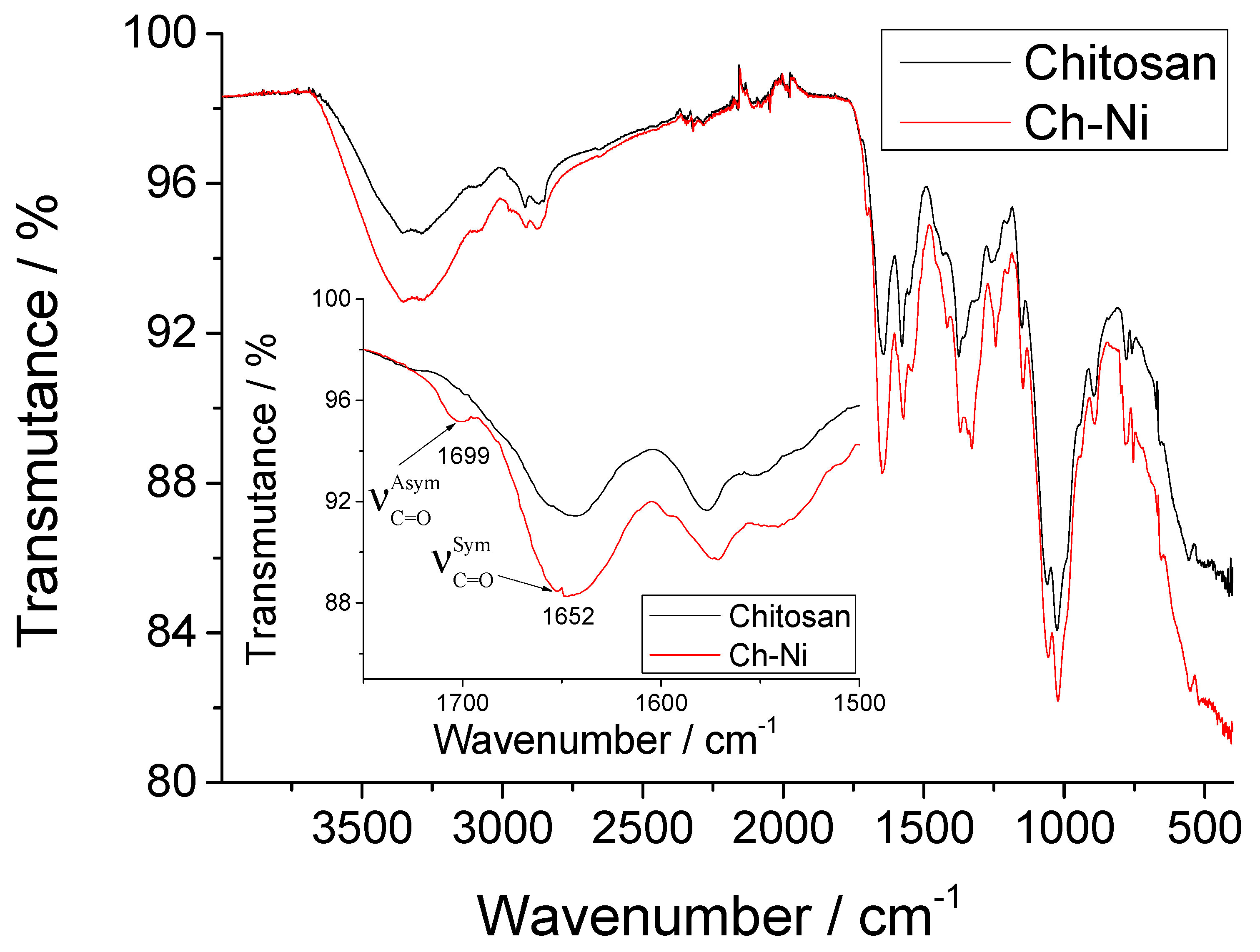
Figure 3.
Photograph of the unmodified chitosan (A) and the modified with 4-dimethylamino-1,8-naphthalimide in the solid state (B).
Figure 3.
Photograph of the unmodified chitosan (A) and the modified with 4-dimethylamino-1,8-naphthalimide in the solid state (B).

Figure 4.
Photographs of modified chitosan Ch-NI solution in water at different pH: at visible light (A) and after irradiation with monochromatic light at λext = 365 nm (B).
Figure 4.
Photographs of modified chitosan Ch-NI solution in water at different pH: at visible light (A) and after irradiation with monochromatic light at λext = 365 nm (B).
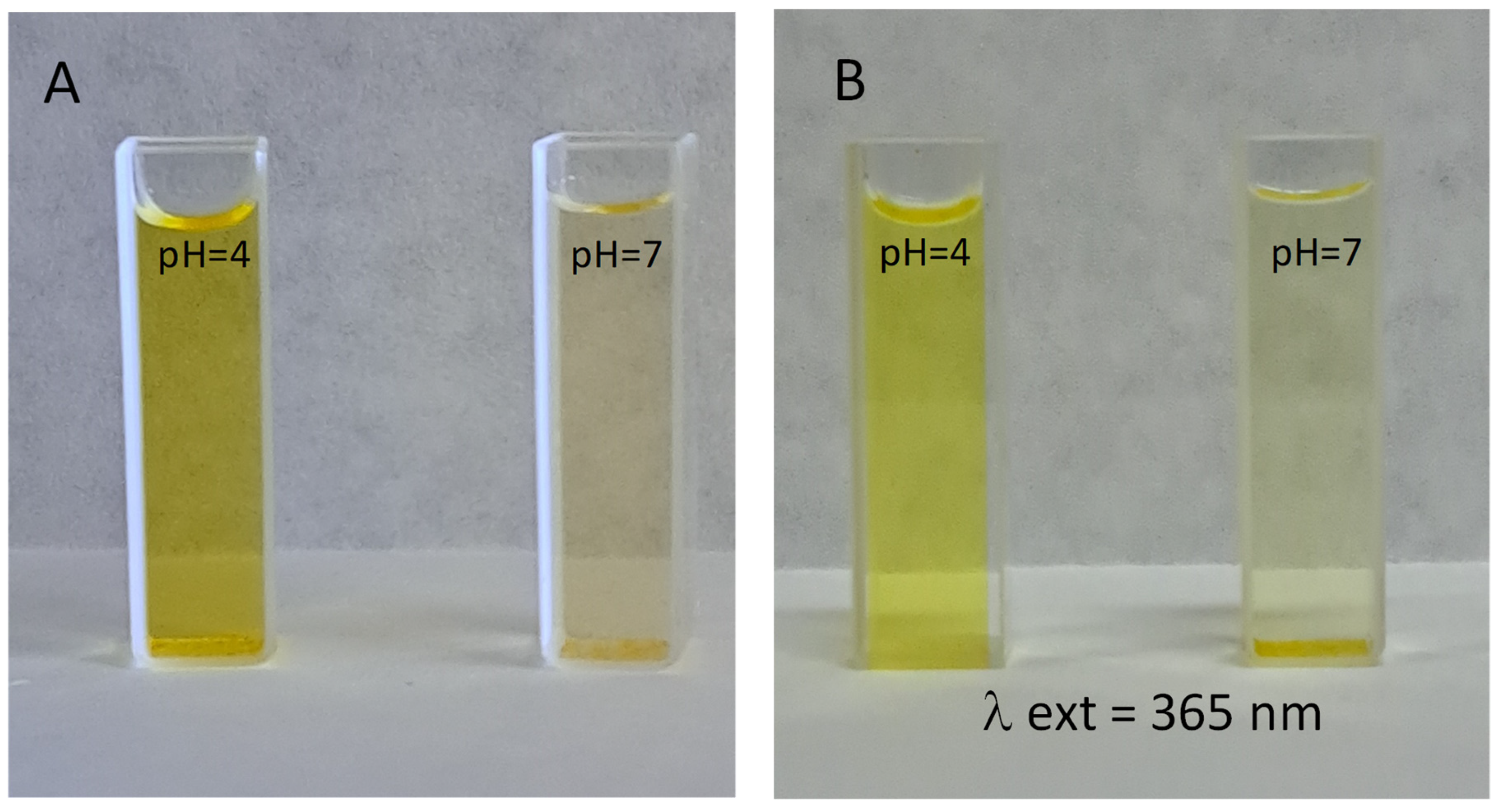
Figure 5.
Interaction of chitosan with citric acid and with indomethacin (A) and interaction of chitosan with alginate (B).
Figure 5.
Interaction of chitosan with citric acid and with indomethacin (A) and interaction of chitosan with alginate (B).
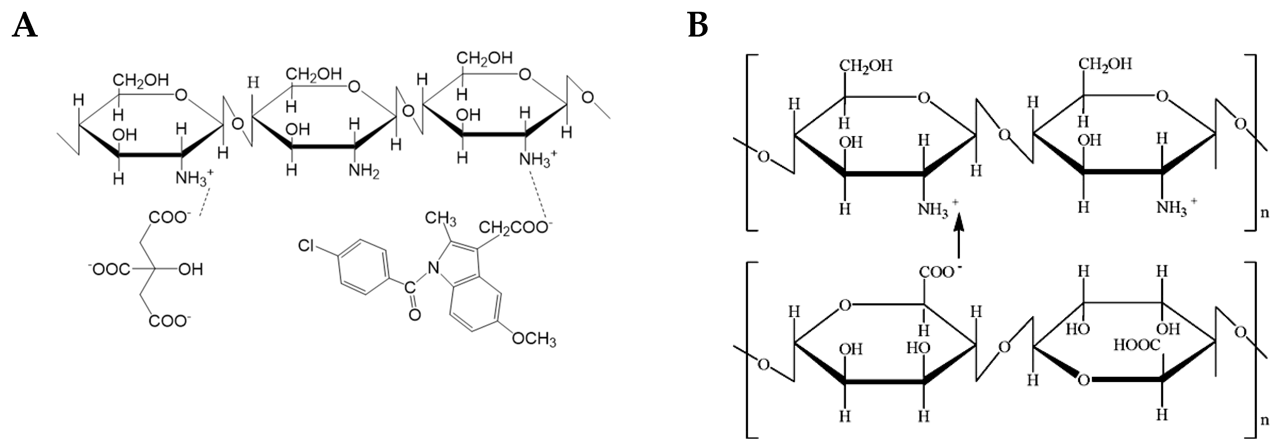
Figure 6.
TG and DTA of chitosan and Ch-NI (A) and TG of the cotton fabric and composite materials CN, CNI, and CNIA (B).
Figure 6.
TG and DTA of chitosan and Ch-NI (A) and TG of the cotton fabric and composite materials CN, CNI, and CNIA (B).

Figure 7.
Optical microscopy images of cotton fabric (CO) and CN, CNI, and CNIA composites (at ×40 and ×100 magnification).
Figure 7.
Optical microscopy images of cotton fabric (CO) and CN, CNI, and CNIA composites (at ×40 and ×100 magnification).
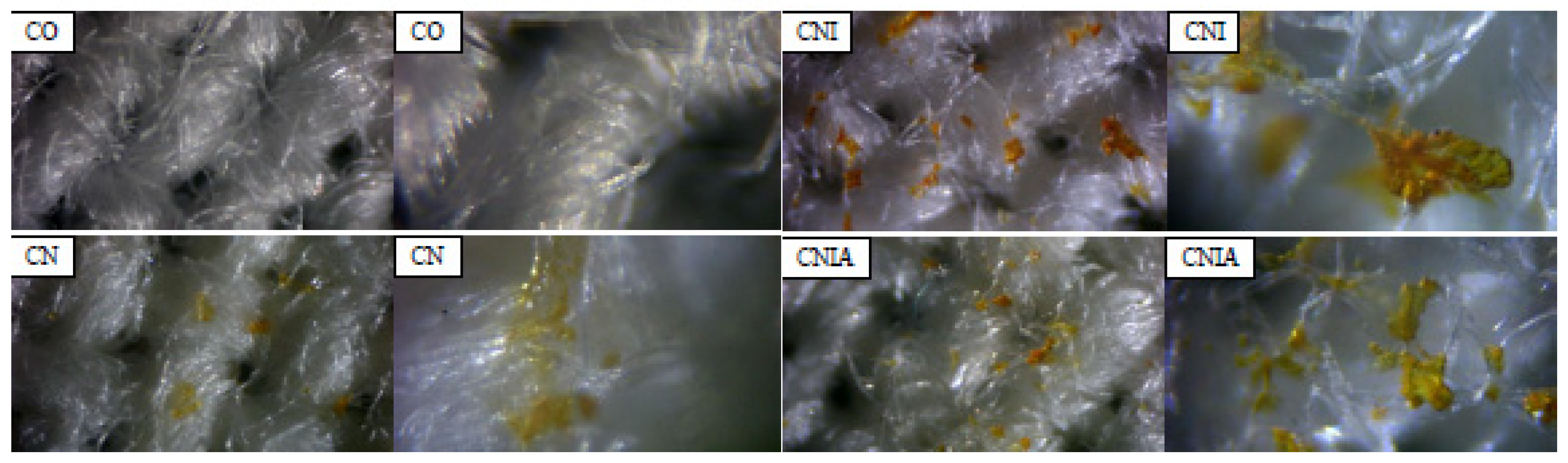
Figure 8.
SEM micrographs at different magnifications of cotton fabric (CO) and CN, CNI, and CNIA composites with the magnification of the images ×100; ×1000; ×10,000.
Figure 8.
SEM micrographs at different magnifications of cotton fabric (CO) and CN, CNI, and CNIA composites with the magnification of the images ×100; ×1000; ×10,000.
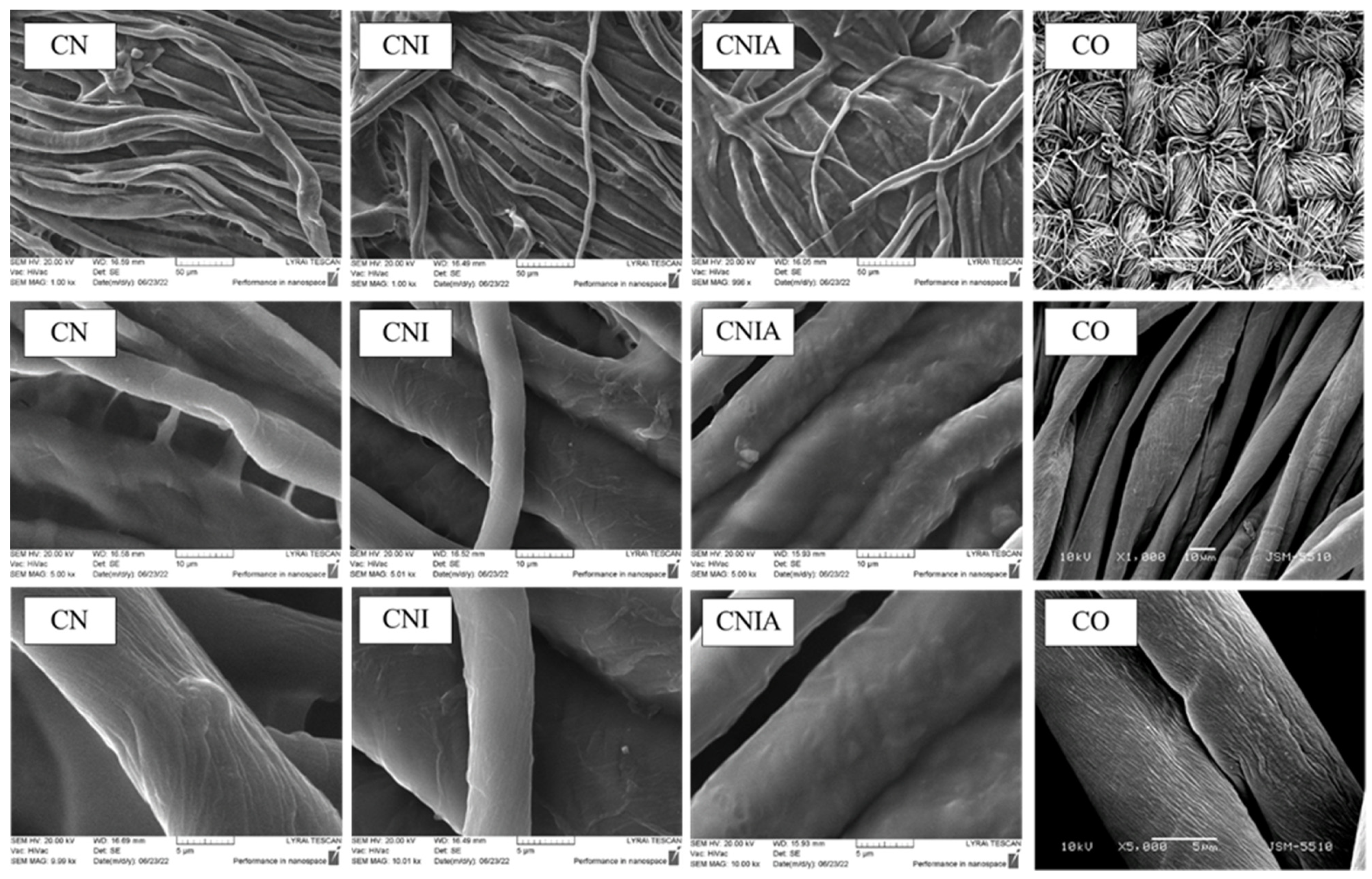
Figure 9.
Gel swelling of CN, CNI, and CNIA composites in phosphate buffer pH = 7.4, at 37 °C.
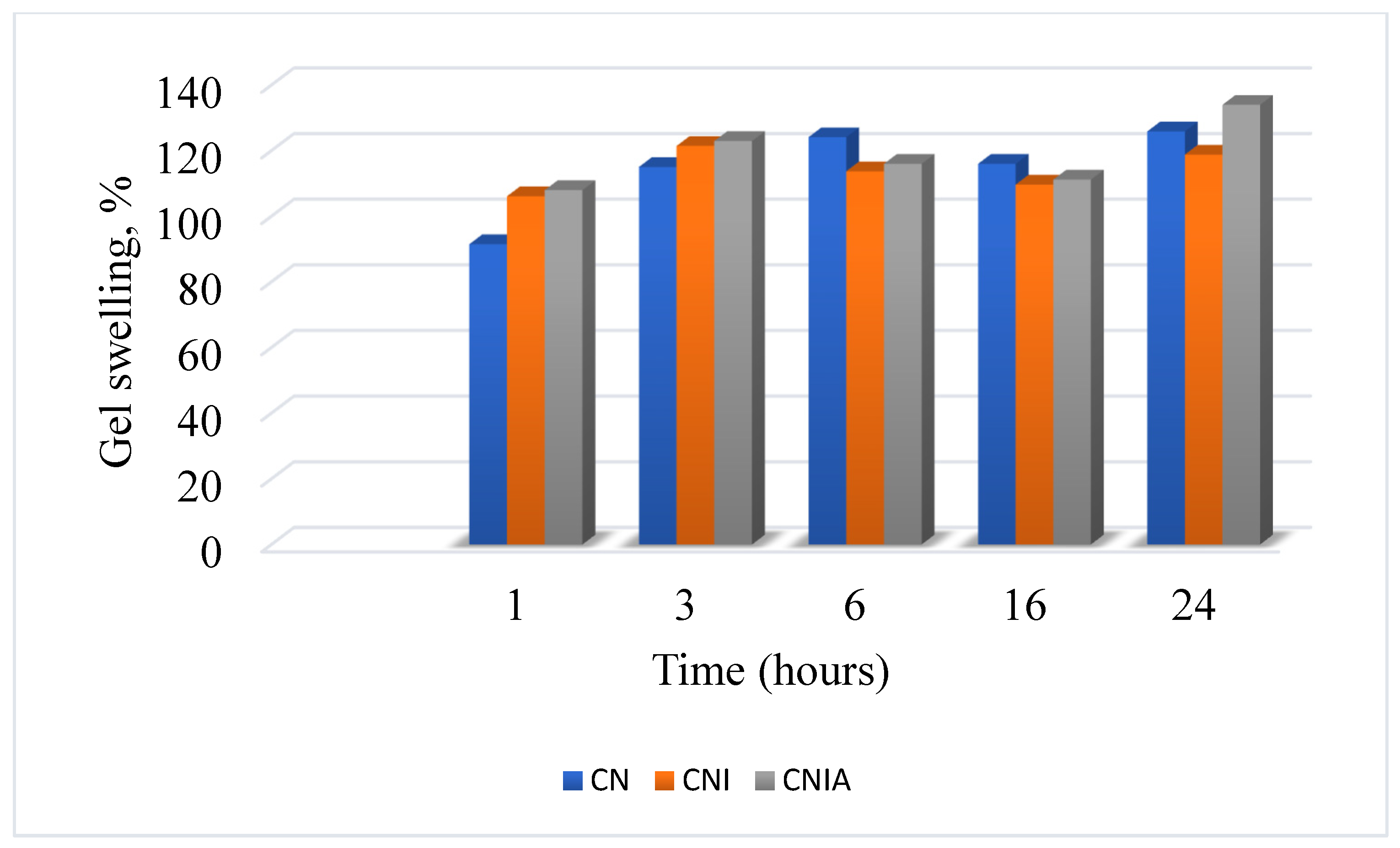
Figure 10.
Change with time in fluorescence intensity at λF = 485 nm of CNI and CNIA when immersed in phosphate buffer with pH = 7.4.
Figure 10.
Change with time in fluorescence intensity at λF = 485 nm of CNI and CNIA when immersed in phosphate buffer with pH = 7.4.
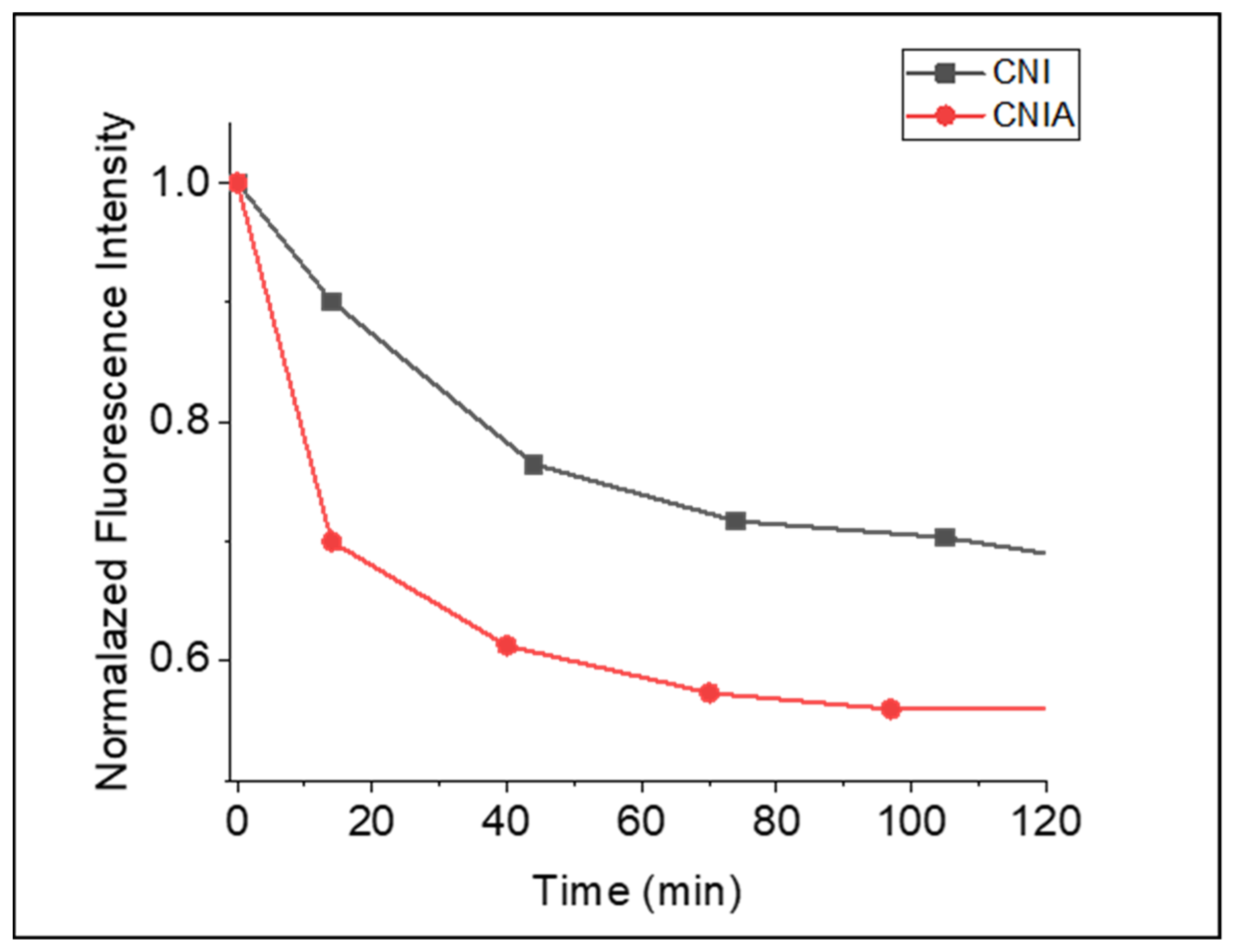
Figure 11.
In vitro release of indomethacin from CNI and CNIA composites in phosphate buffer pH = 7.4, at 37 °C, (at λA = 320 nm).
Figure 11.
In vitro release of indomethacin from CNI and CNIA composites in phosphate buffer pH = 7.4, at 37 °C, (at λA = 320 nm).

Figure 12.
Antimicrobial activity of cotton fabric (control) and CN, CNI, and CNIA materials on the growth of P. aeruginosa and B. cereus.
Figure 12.
Antimicrobial activity of cotton fabric (control) and CN, CNI, and CNIA materials on the growth of P. aeruginosa and B. cereus.
Disclaimer/Publisher’s Note: The statements, opinions and data contained in all publications are solely those of the individual author(s) and contributor(s) and not of MDPI and/or the editor(s). MDPI and/or the editor(s) disclaim responsibility for any injury to people or property resulting from any ideas, methods, instructions or products referred to in the content. |
© 2023 by the authors. Licensee MDPI, Basel, Switzerland. This article is an open access article distributed under the terms and conditions of the Creative Commons Attribution (CC BY) license (https://creativecommons.org/licenses/by/4.0/).
Share and Cite
MDPI and ACS Style
Staneva, D.; Atanasova, D.; Grabchev, I. Fluorescent Composite Cotton Fabric Modified with Crosslinked Chitosan for Theranostic Applications. Appl. Sci. 2023, 13, 12660. https://doi.org/10.3390/app132312660
AMA Style
Staneva D, Atanasova D, Grabchev I. Fluorescent Composite Cotton Fabric Modified with Crosslinked Chitosan for Theranostic Applications. Applied Sciences. 2023; 13(23):12660. https://doi.org/10.3390/app132312660
Chicago/Turabian StyleStaneva, Desislava, Daniela Atanasova, and Ivo Grabchev. 2023. "Fluorescent Composite Cotton Fabric Modified with Crosslinked Chitosan for Theranostic Applications" Applied Sciences 13, no. 23: 12660. https://doi.org/10.3390/app132312660
Note that from the first issue of 2016, this journal uses article numbers instead of page numbers. See further details here.