Article

Alterations in Surface Roughness and Chemical Characteristics of Sandblasted and Acid-Etched Titanium Implants after Irradiation with Different Diode Lasers

1
Department of Periodontology, School of Dentistry, Kyungpook National University, Daegu 41940, Korea
2
Advanced Dental Device Development Institute, Kyungpook National University, Daegu 41940, Korea
3
Department of Dental Science, Graduate School, Kyungpook National University, Daegu 41940, Korea
4
Department of Orthodontics, School of Dentistry, Kyungpook National University, Daegu 41940, Korea
5
Department of Prosthodontics, School of Dentistry, Kyungpook National University, Daegu 41940, Korea
*
Authors to whom correspondence should be addressed.
Appl. Sci. 2020, 10(12), 4167; https://doi.org/10.3390/app10124167
Submission received: 26 May 2020 / Revised: 15 June 2020 / Accepted: 16 June 2020 / Published: 17 June 2020
(This article belongs to the Special Issue Applied Sciences in Dentistry)

Abstract

:
The purpose of this study was to evaluate the effects of diode laser irradiation with different wavelengths on the surface roughness (Ra) and chemical composition of sandblasted and acid-etched (SLA) titanium implants. Three types of diode lasers with different wavelengths were irradiated on the titanium implants at output powers of 1.0, 2.0, and 3.0 W. The mean Ra values for all spots were measured using a scanning probe microscope. Analysis of variance tests were performed to verify the differences in the Ra between groups according to the type of lasers or power out (α = 0.05). For analyzing chemical composition, atomic and weight percent ratios of titanium, oxygen, and carbon were measured using energy-dispersive spectrometry (EDS). The mean Ra of titanium disc was higher in the 3.0-W output than in 1.0-W or 2.0-W output, but there was no statistically significant difference (p > 0.05). In EDS analysis, it was difficult to find a clear difference in the titanium, oxygen, and carbon element ratios between the laser-irradiated and nonirradiated groups. The irradiation of diode laser with 1.0, 2.0, and 3.0 W output for 15 s decontaminated the SLA titanium surface without damage. However, additional clinical trials will be needed to verify the results of the present study.

1. Introduction

Dental implants are viable treatment options for the replacement of missing teeth [1]. However, bacterial adhesion on implant surfaces can provoke inflammation, resulting in progressive alveolar bone loss [2]. Therefore, decontaminating implant surfaces is essential to treat peri-implantitis [3].
Various therapeutic techniques, such as tetracycline administration and the use of plastic curettes and ultrasonic devices have been used for peri-implantitis treatment [4,5,6]. However, these conventional methods are inadequate in eliminating bacteria on roughened implant surfaces [7]. Narrow and deep bony defects make it difficult to remove dental biofilms from the implant surface [4]. Furthermore, cleaning dental implants with curettes might cause crack lines on the implant surface which could increase plaque deposition [8].
Recently, with their bactericidal effects, applications of laser systems have been suggested to be adjunctive methods for decontaminating infected implant surfaces to the mechanical debridement [9]. The main goal of a laser can be the elimination of bacteria on rough implant surfaces. Moritz et al. [10] reported that laser application on the periodontal pocket can significantly reduce total bacterial counts as well as specific bacteria, such as actinobacillus actinomycetemcomitans, prevotella intermedia, and porphyromonas gingivalis. It is also known that laser irradiation has other advantages, such as tissue ablation without contact, less bleeding, and limited pain and scar formation [9].
Diverse laser systems with various wavelengths, such as neodymium (Nd:YAG), erbium (Er:YAG), and CO2 lasers, have been used for the decontamination of the implant surface [11]. While typical diode lasers have a wide range of wavelengths, dental diode lasers with wavelengths ranging from 805 to 980 nm have been used in the clinical practice of dentistry for at least 15 years [7] because of their small size and reduced cost, compared to other types of laser systems [11].
However, some studies have demonstrated damage and alterations on titanium surfaces because of the laser irradiation [12,13]. Therefore, the irradiation treatment should be carefully selected to avoid thermal or physical damage to the implant surface [14]. To the best of our knowledge, there have been few reports regarding the change in titanium surfaces after various diode laser applications and appropriate energy for decontaminating titanium surfaces without changing these surfaces. The aim of this study was to compare the surface roughness and component changes of sandblasted and acid-etched (SLA) titanium implants before and after various diode laser irradiations so that the proper irradiation power and exposure time of diode laser for decontaminating a titanium disc surface can be addressed without damage. The null hypothesis was that there would be no difference in surface roughness according to the different dental diode lasers and output power.

2. Materials and Methods

2.1. Dental Laser

Since the previous study verified the stability of implant treatment using a diode laser with 940 nm [14], three dental diode lasers with wavelengths from 940 to 980 nm were used in the present study: K2 mobile (Hulaser, Seoul, Korea) with a wavelength of 980 nm, EpicTM 10 (Biolase, CA, USA) with a wavelength 940 nm, and Saeshin diode laser with a wavelength 980 nm (Saeshin, Daegu, Korea). The laser fiber tip has a circular shape with a diameter of 400 µm, and an identical tip was used for three dental diode lasers. The laser fiber tip was unmoved, and in contact vertically with the titanium disk surface while irradiation was performed for a period of 15 s. The three dental diode lasers were used only in the continuous wave mode with a flexible fiber with a focused spot size of 0.4 mm diameter. All the preselected laser parameters for the three lasers can be seen in Table 1. All the preselected laser parameters were determined using a calibrated power meter system (NovaII; Ophir Photonics, North Logan, UT, USA).

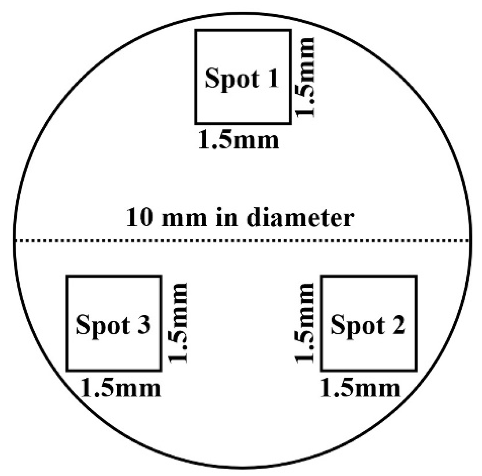
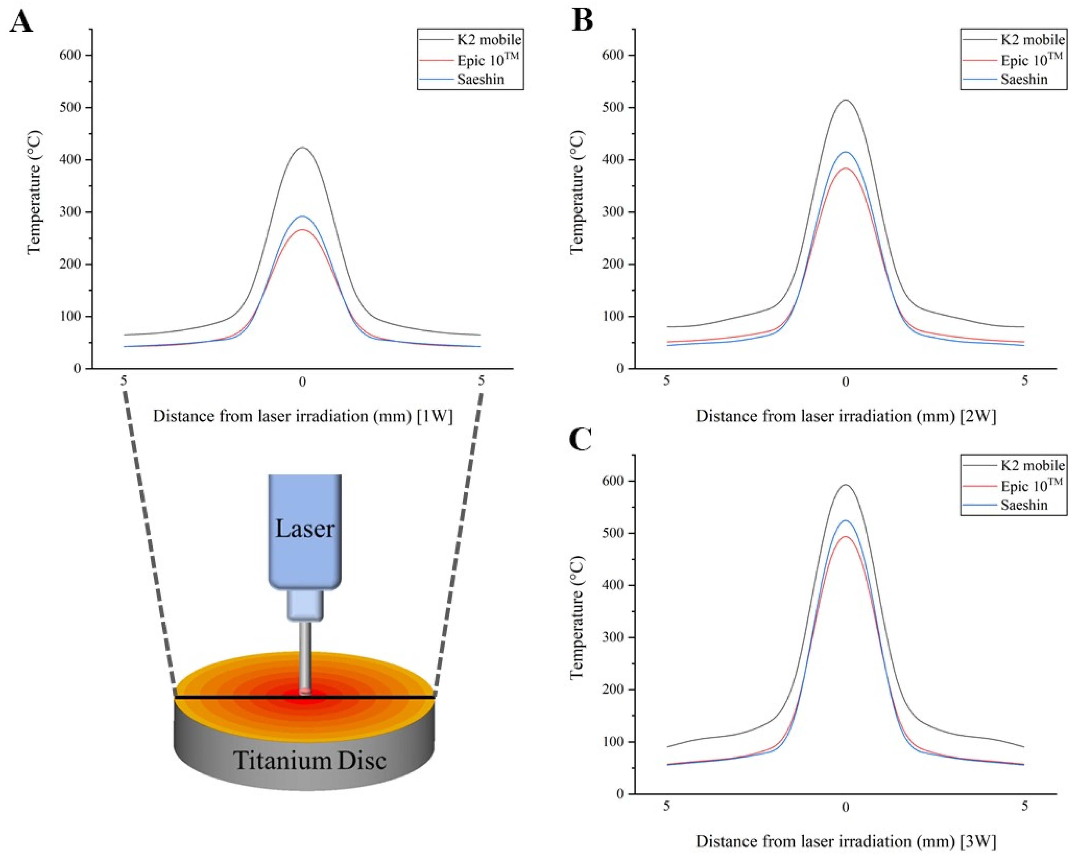
2.2. Preparation of Titanium Implants and Laser Irradiations

SLA (sandblasted and acid-etched) titanium implants (10 mm in diameter and 2 mm thick; Dentis, Daegu, Korea) were used. The reason the titanium disc was SLA treated prior to the experiment is to reproduce the surface of a commercial dental implant. The SLA of the titanium disc was performed by the implant manufacturer (Dentis, Daegu, Korea). A total of 10 titanium implants were prepared. Nine out of 10 discs were designated as experimental groups and divided into three groups based on the laser used: group 1 was irradiated using the K2 mobile laser, group 2 was irradiated using the Epic 10TM laser, and group 3 was irradiated using the Saeshin diode laser. The remaining one titanium disc was designated as a control without laser irradiation.
Three titanium implants in each experimental group were irradiated with lasers at different power settings. The first disc of each experimental group was irradiated at 1.0 W power setting for a period of 15 s with continuous wave mode. The second disc of each experimental group was irradiated at 2.0 W, and the third disc was irradiated at 3.0 W for the same period in the continuous wave mode.
Titanium disks were sterilized before irradiation and stored individually in sterile microtubes after irradiation. Titanium implants were irradiated at three spots, and the titanium implants were irradiated with just a single π (0.4/2)2 area of the beam within a 1.5 × 1.5 mm square (Figure 1). Within the range of each spot per specimen was irradiated under the same conditions at the same power setting with the same type of laser. Before using three types of dental diode lasers, the temperature of implant surface was confirmed during irradiation using a calibrated thermometer system (midi LOGGER GL240; GRAPHTEC, Yokohama, Japan). The laser fiber tip has a circular shape, and the same fiber tip was used for three dental diode lasers. The laser fiber tip was unmoved, and in contact vertically with the titanium disk surface while irradiation was performed for a period of 15 s. After cooling completely, the laser was irradiated at the next spot. All diode lasers were irradiated by the same investigator (H.-K.K.), and when irradiating the spots, the laser was irradiated carefully within spot area to prevent irradiation of other spots. The energy-dispersive spectrometry (EDS) was analyzed immediately after laser irradiation, considering the contamination of the sample.

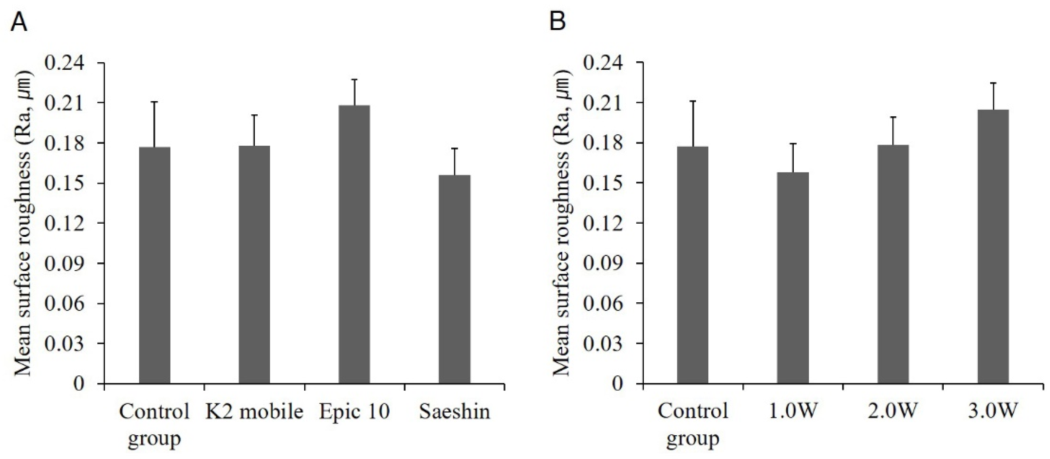
2.3. Surface Roughness

For measuring the surface roughness of titanium discs, scanning probe microscope (Veeco Instrument, New York, NY, USA) was used. The topographic analysis was performed at each experimental and control spot, and surface roughness (Ra) values, defined as the arithmetic mean deviation of the roughness profile, were recorded. The surface roughness of titanium implants was measured five times for each area irradiated, and the average for each spot was recorded.

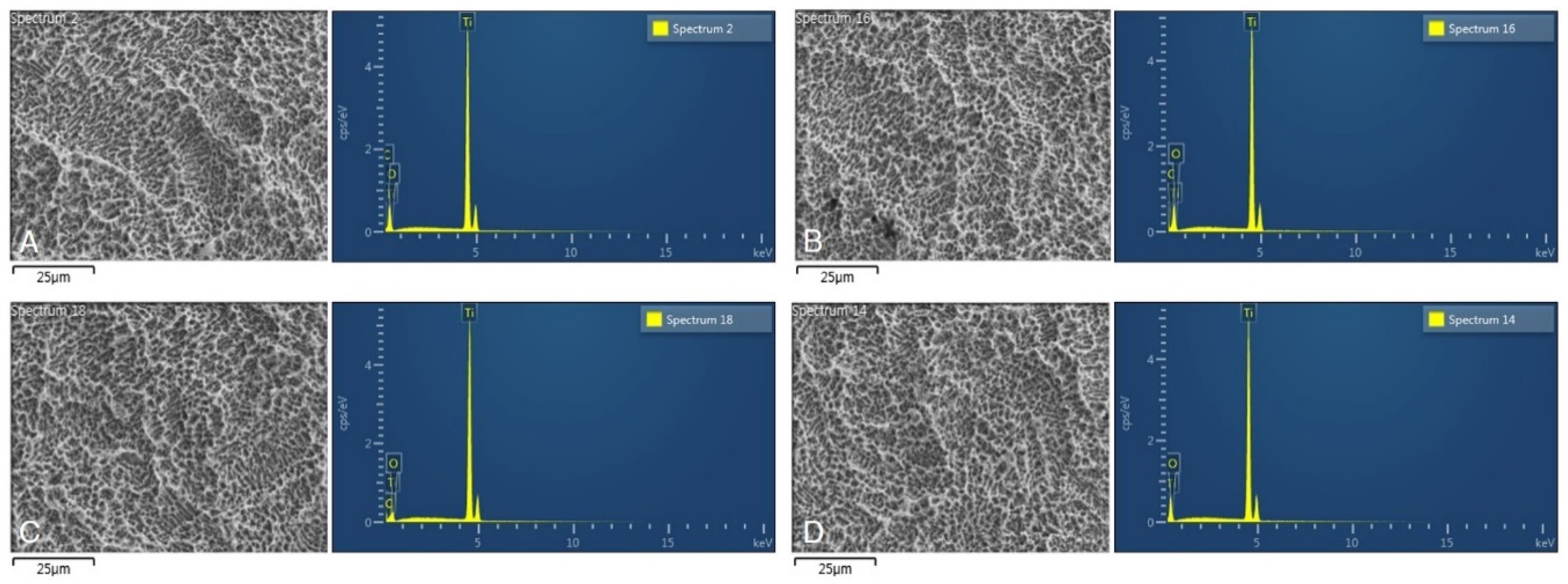
2.4. Energy-Dispersive Spectrometry

For analyzing chemical characterization of each spot, EDS was performed using a field emission scanning electron microscope (S-4800, Hitachi, Ltd., Tokyo, Japan). Spectra were obtained under the following conditions: 5.1 × 10⁶ Pa vacuum, 20 kV accelerating voltage, 73 nA beam current, ×500 original magnification with a 0.26 × 0.26 mm sampling window, 100 s acquisition time, and from 30% to 40% dead time. EDS represented the atomic and weight percent ratios of titanium, oxygen, and carbon for each spot.

2.5. Statistical Analysis

To determine the sample size, pilot experiments were conducted, and the size of the appropriate sample was calculated to be three per group, using the power analysis software (G * Power v3.1.9.2; Heinrich-Heine-Universität) (effect size (f) = 3.34; actual power = 99.99%; power = 99%; α = 0.05). Mean values and standard deviations of Ra values for three spots in one specimen were calculated. One-way ANOVA (analysis of variance) tests were performed to verify the differences in the surface roughness between groups according to the type of lasers, output power, and surface temperature. Tukey honestly significant difference tests were performed as a post hoc test. Results were considered to be significant for p values < 0.05.

3. Results

Figure 2 shows the results of comparison of temperature after laser irradiation using three types of dental diode lasers.
The surface temperature per output power was significantly different depending on the type of laser (p < 0.001) (Table 2). On the other hand, as a result of post hoc test, there were no significant differences in the surface temperatures of Epic 10TM and Saeshin (p > 0.05) (Table 2).

3.1. Surface Roughness

Table 3 shows the mean surface roughness (Ra) value for each SLA titanium disc. Each specimen consisted of three irradiation spots; therefore, the average of Ra values was represented. Ten specimens were under different conditions for the laser types and output power.
Figure 3 shows the difference in mean surface roughness (Ra) values of SLA titanium implants according to various laser types and output powers. Figure 2A represents mean Ra values of four groups according to the laser types irradiated. The control group was the specimen without laser irradiation, and its Ra value was measured to be 0.177 ± 0.034 µm. Mean Ra values of the experimental groups after laser irradiation were measured to be 0.178 ± 0.023 µm for the K2 mobile group, 0.208 ± 0.020 µm for the Epic 10TM group, and 0.156 ± 0.020 µm for the Saeshin diode laser group, respectively. Mean Ra values were higher in the K2 and Epic 10TM groups and lower in the Saeshin diode laser group than that of the control group. However, the difference in mean Ra values was not statistically significant between any two groups. Figure 2B represents mean Ra values of four groups based on the output power. Mean Ra values were measured to be 0.158 ± 0.021 µm for 1.0 W group, 0.178 ± 0.021 µm for 2.0 W group, and 0.205 ± 0.020 µm for 3.0 W group, respectively. In the 1.0 W group, mean Ra values were lower than those of the control group. In the 2.0 and 3.0 W laser groups, mean Ra values were higher than those of the control group. As the output values increased, the average Ra values also tended to increase. However, the difference in mean Ra values was not statistically significant between any two groups (p > 0.05).

3.2. Atomic and Weight Percentage

Table 4 shows the atomic and weight percentages of carbon, oxygen, and titanium atoms on the SLA titanium implants under various types of laser and output powers. The atomic percentage represents the percentage as a function of the number of atoms, whereas the weight percentage represents the percentage as a function of weight. The atomic and weight percentages of carbon, oxygen, and titanium atoms in the control group were similar to those in the experimental groups. Statistical comparisons between groups could not be performed because of the small number of samples.

3.3. Scanning Electron Microscope Images and EDS

Figure 4 represents the scanning electron microscope (SEM) images and EDS analyses of SLA titanium surfaces irradiated with various types of lasers and nonirradiated specimens. Figure 4A shows a nonirradiated titanium surface with the highest peak of titanium in the EDS analysis. Carbon and oxygen showed lower peaks compared to titanium. The peaks of titanium, oxygen, and carbon on the nonirradiated titanium surface represented similar patterns on titanium specimens irradiated with diverse laser types. Additionally, SEM images of titanium implants, which were irradiated with various types of diode lasers were similar to those without laser irradiation (Figure 4).

4. Discussion

We examined the changes in the surfaces of SLA titanium implants irradiated with diverse types of diode lasers. Previous studies [7,8] have focused on only the alteration in SEM images when diode lasers are irradiated on titanium implants or implants. Romanos et al. [7] reported that the diode laser (980 nm) did not damage the titanium surface. Fox et al. [8] reported that the titanium surface was significantly altered when scaling using a metal instrument. In this work, however, we intended to observe the changes in surface roughness and chemical characterization beyond alterations in SEM images when the diode laser was irradiated at titanium discs.
Diode lasers emit light between 805 and 980 nm [15]. In this study, three kinds of diode lasers were used. The wavelengths of the K2 mobile, Epic 10TM, and Saeshin diode lasers were 980, 940, and 980 nm, respectively. In the case of Epic 10TM having a wavelength of 940 nm, which is relatively smaller than 980 nm, the surface roughness values were slightly higher than those of the other two laser groups. This is because diode lasers with shorter wavelengths tend to have higher energy per photon, which might damage the surface, such as by forming a crack or crevice, consequently increasing the surface roughness [9]. In the case of the 3.0-W irradiation, the surface roughness values were slightly higher than those irradiated at 1.0 and 2.0 W. This is likely due to the higher intensity of laser irradiation. Laser irradiation might lower the surface roughness by melting the titanium surface [16]. We observed lower surface roughness values at lower powers, such as 1.0 W, compared to the control specimen. However, there were no statistical differences in surface roughness between laser-irradiated and nonirradiated discs, so more studies must be conducted for validation. The different laser types with different wavelengths used in this work did not significantly affect the Ra of titanium disks. Further research is needed using lasers with more diverse wavelengths.
When diode lasers were irradiated at titanium implants, Tosun et al. [12] reported a decrease of 28% at 0.25 W, 61% at 0.5 W, 95% at 0.75 W, and 100% at 1.0 W in Staphylococcus aureus. In the present study, the energy density at 1.0-W power was 14, 9, and 10 kJ/cm2 by K2 mobile, Epic 10TM, and Saeshin, respectively (Table 1), and the energy density at 1.0-W power in the Tosun et al. study was 12 kJ/cm2 [12]. Tosun et al. confirmed a 95% bactericidal effect at 9 kJ/cm2 [12]. Therefore, 1.0-W output power was selected as a minimum intensity, and it was assumed that diode lasers had a bactericidal effect under all conditions. Further research is needed for the bactericidal effect through laser irradiation in peri-implantitis treatment [17].
An increase in surface temperature was inevitable when a diode laser was irradiated on a titanium disc. A temperature rise of more than 10 °C has been reported to adversely affect bone tissues due to alkaline phosphatase denaturation [11,18]. Eriksson and Albreksson et al. [19] reported that, with an air flow of 2.5 L/min, irradiating a diode laser at 3.0 W in the continuous mode for 10 s avoided exceeding the threshold temperature. Here, diode lasers were irradiated at Ti disks for 15 s at a maximum output power of 3.0 W to see if titanium surface changes occurred at temperatures slightly above the threshold temperature. The present study was conducted without water cooling and showed a high surface temperature of above 200 °C at all output powers at the laser irradiation site. In addition, further study is needed to confirm a setting value suitable for more clinical use by lowering the temperature rise of the implant surface by applying less time and power than setting value applied in the present study. Additionally, in clinical practice, open flap treatment is necessary to ensure sufficient cooling of the implant surface. Therefore, additional research is required under water cooling conditions, and the temperature rise must be taken into account when laser irradiation is applied to dental implants in clinical practice.
Titanium is a highly reactive element and exists in the form of TiO2 [20]. Titanium and oxygen signals in the EDS analysis showed that the titanium disc surface consisted of a titanium oxide layer [21]. The thickness of the titanium oxide is known to be 2–6 nm, and EDS analysis provides compositional information averaged over a depth of 1 μm [22]. The carbon signal in the EDS analysis could be due to surface contamination [21]. The SLA surface has undergone an acid etching process, which washes away any remaining elements on the titanium surface [23]. Thus, it was difficult to find a clear difference in the titanium, oxygen, and carbon element ratios between the laser-irradiated and nonirradiated groups. Therefore, it is suggested that the diode laser does not seem to change the chemical composition of the titanium disc surface.
On the other hand, the effects of laser irradiation on the implant surface under salivary conditions have not been evaluated in this work. The chemical composition of the titanium surface can be influenced by microbial pH occurring in vivo. Therefore, further studies are needed under intraoral conditions. In decontaminating the implant surface, an increase in surface roughness can make the osseointegration difficult after treatment [5]. Therefore, it is necessary to irradiate a laser that can remove the contamination and increase the proper surface temperature without increasing the surface roughness of the implant. Further research is needed to confirm these conditions. Finally, the possibility that the beam at the fiber tip has a non-uniform intensity distribution has not been eliminated from the measurements in the present study. Further study is needed the measurement of the beam profile using a charged coupled device camera system.

5. Conclusions

In this study, alterations in the surface roughness and chemical composition of SLA titanium implants were investigated after irradiation with diode lasers of various wavelengths. The temperature of the implant surface during laser irradiation was significantly different according to the type of dental diode laser. The mean surface roughness of the titanium disc irradiated at 940 nm was slightly higher than that at 980 nm. Moreover, the mean surface roughness of the titanium disc was higher at 3.0 W compared with those at 1.0 and 2.0 W. However, the type of laser and power did not significantly change the RA, so the irradiation of a diode laser with 1.0, 2.0, and 3.0 W for 15 s can decontaminate the SLA titanium surface without damaging titanium. Nevertheless, these findings were conducted in vitro, and clinical trials will be needed to verify the results of this study.

Author Contributions

Conceptualization, H.-K.K.; Methodology, S.-Y.P. and K.S.; Validation, K.-B.L.; Formal Analysis, Y.-G.K.; Investigation, W.-J.Y.; Data Curation, J.-M.L.; Writing—Original Draft, H.-K.K. and S.-Y.P.; Visualization, H.-K.K.; Supervision, J.-M.L.; Project Administration, K.-B.L.; Writing—Review and Editing, K.S. All authors have read and agreed to the published version of the manuscript.

Funding

This work was supported by Korea Institute for Advancement of Technology (KIAT) through the National Innovation Cluster R&D program (P0006691).

Conflicts of Interest

The authors declare no conflicts of interest. The funders had no role in the design of the study; in the collection, analyses, or interpretation of the data; in the writing of the manuscript, or in the decision to publish the results.

References

  1. Shin, S.I.; Min, H.K.; Park, B.H.; Kwon, Y.H.; Park, J.B.; Herr, Y.; Heo, S.J.; Chung, J.H. The effect of Er:YAG laser irradiation on the scanning electron microscopic structure and surface roughness of various implant surfaces: An in vitro study. Lasers Med. Sci. 2011, 26, 767–776. [Google Scholar] [CrossRef]
  2. Gholami, G.A.; Karamlou, M.; Fekrazad, R.; Ghanavati, F.; Hakimiha, N.; Romanos, G. Comparison of the Effects of Er, Cr: YSGG Laser and Super-Saturated Citric Acid on the Debridement of Contaminated Implant Surfaces. J. Lasers Med. Sci. 2018, 9, 254–260. [Google Scholar] [CrossRef] [Green Version]
  3. Bernardi, S.; Bianchi, S.; Tomei, A.R.; Continenza, M.A.; Macchiarelli, G. Microbiological and SEM-EDS evaluation of titanium surfaces exposed to periodontal gel: In vitro study. Materials 2019, 12, 1448. [Google Scholar] [CrossRef] [Green Version]
  4. Deppe, H.; Horch, H.H.; Neff, A. Conventional versus CO2 laser-assisted treatment of peri-implant defects with the concomitant use of pure-phase beta-tricalcium phosphate: A 5-year clinical report. Int. J. Oral Maxillofac. Implants 2007, 22, 79–86. [Google Scholar]
  5. Rühling, A.; Kocher, T.; Kreusch, J.; Plagmann, H.C. Treatment of subgingival implant surfaces with Teflon-coated sonic and ultrasonic scaler tips and various implant curettes. An in vitro study. Clin. Oral Implants Res. 1994, 5, 19–29. [Google Scholar] [CrossRef]
  6. Shetty, B.; Dinesh, A.; Seshan, H. Comparitive effects of tetracyclines and citric acid on dentin root surface of periodontally involved human teeth: A scanning electron microscope study. J. Indian Soc. Periodontol. 2008, 12, 8–15. [Google Scholar] [CrossRef]
  7. Romanos, G.E.; Everts, H.; Nentwig, G.H. Effects of diode and Nd:YAG laser irradiation on titanium discs: A scanning electron microscope examination. J. Periodontol. 2000, 71, 810–815. [Google Scholar] [CrossRef]
  8. Fox, S.C.; Moriarty, J.D.; Kusy, R.P. The effects of scaling a titanium implant surface with metal and plastic instruments: An in vitro study. J. Periodontol. 1990, 61, 485–490. [Google Scholar] [CrossRef]
  9. Stubinger, S.; Etter, C.; Miskiewicz, M.; Homann, F.; Saldamli, B.; Wieland, M.; Sader, R. Surface alterations of polished and sandblasted and acid-etched titanium implants after Er:YAG, carbon dioxide, and diode laser irradiation. Int. J. Oral Maxillofac. Implants 2010, 25, 104–111. [Google Scholar]
  10. Moritz, A.; Schoop, U.; Goharkhay, K.; Schauer, P.; Doertbudak, O.; Wernisch, J.; Sperr, W. Treatment of periodontal pockets with a diode laser. Lasers Surg. Med. 1998, 22, 302–311. [Google Scholar] [CrossRef]
  11. Rios, F.G.; Viana, E.R.; Ribeiro, G.M.; González, J.C.; Abelenda, A.; Peruzzo, D.C. Temperature evaluation of dental implant surface irradiated with high-power diode laser. Lasers Med. Sci. 2016, 31, 1309–1316. [Google Scholar] [CrossRef]
  12. Tosun, E.; Tasar, F.; Strauss, R.; Kıvanc, D.G.; Ungor, C. Comparative evaluation of antimicrobial effects of Er:YAG, diode, and CO₂ lasers on titanium discs: An experimental study. J. Oral Maxillofac. Surg. 2012, 70, 1064–1069. [Google Scholar] [CrossRef]
  13. Block, C.M.; Mayo, J.A.; Evans, G.H. Effects of the Nd:YAG dental laser on plasma-sprayed and hydroxyapatite-coated titanium dental implants: Surface alteration and attempted sterilization. Int. J. Oral Maxillofac. Implants 1992, 7, 441–449. [Google Scholar]
  14. Torkzaban, P.; Kasraei, S.; Torabi, S.; Farhadian, M. Low-level laser therapy with 940 nm diode laser on stability of dental implants: A randomized controlled clinical trial. Lasers Med. Sci. 2018, 33, 287–293. [Google Scholar] [CrossRef] [PubMed]
  15. Romanos, G.E.; Gutknecht, N.; Dieter, S.; Schwarz, F.; Crespi, R.; Sculean, A. Laser wavelengths and oral implantology. Lasers Med. Sci. 2009, 24, 961–970. [Google Scholar] [CrossRef]
  16. Ercan, E.; Arin, T.; Kara, L.; Çandirli, C.; Uysal, C. Effects of Er, Cr:YSGG laser irradiation on the surface characteristics of titanium discs: An in vitro study. Lasers Med. Sci. 2014, 29, 875–880. [Google Scholar] [CrossRef]
  17. Faggion, C.M. Laser Therapy as an Adjunct Treatment for Peri-Implant Mucositis and Peri-Implantitis Provides No Extra Benefit for Most Clinical Outcomes. J. Evid. Based Dent. Pract. 2019, 19, 203–206. [Google Scholar] [CrossRef]
  18. Geminiani, A.; Caton, J.G.; Romanos, G.E. Temperature change during non-contact diode laser irradiation of implant surfaces. Lasers Med. Sci. 2012, 27, 339–342. [Google Scholar] [CrossRef]
  19. Eriksson, A.R.; Albrektsson, T. Temperature threshold levels for heat-induced bone tissue injury: A vital-microscopic study in the rabbit. J. Prosthet. Dent. 1983, 50, 101–107. [Google Scholar] [CrossRef]
  20. Massaro, C.; Rotolo, P.; De Riccardis, F.; Milella, E.; Napoli, A.; Wieland, M.; Textor, M.; Spencer, N.D.; Brunette, D.M. Comparative investigation of the surface properties of commercial titanium dental implants. Part I: Chemical composition. J. Mater. Sci. Mater. Med. 2002, 13, 535–548. [Google Scholar] [CrossRef]
  21. Lausmaa, J.; Kasemo, B. Surface spectroscopic characterization of titanium implant materials. Appl. Surf. Sci. 1990, 44, 133–146. [Google Scholar] [CrossRef]
  22. Shibli, J.A.; Marcantonio, E.; d’Avila, S.; Guastaldi, A.C.; Marcantonio, E. Analysis of failed commercially pure titanium dental implants: A scanning electron microscopy and energy-dispersive spectrometer x-ray study. J. Periodontol. 2005, 76, 1092–1099. [Google Scholar] [CrossRef] [Green Version]
  23. Kim, H.; Choi, S.H.; Ryu, J.J.; Koh, S.Y.; Park, J.H.; Lee, I.S. The biocompatibility of SLA-treated titanium implants. Biomed. Mater. 2008, 3, 025011. [Google Scholar] [CrossRef]
Figure 1. Schematic illustration showing three laser irradiation spots on one specimen.
Applsci 10 04167 g001
Figure 2. Comparison of temperature after laser irradiation using three types of dental diode lasers. (A) 1W, (B) 2W, (C) 3W.
Applsci 10 04167 g002
Figure 3. Bar diagrams showing the difference in mean surface roughness (Ra) values according to laser types and output powers: (A) a comparison of mean Ra values between various laser types, (B) a comparison of mean Ra values between various output powers.
Applsci 10 04167 g003
Figure 4. Scanning electron microscope (SEM) images and energy-dispersive spectrometry (EDS) analyses of SLA titanium surfaces irradiated with various types of lasers and nonirradiated specimens: (A) control specimen, (B) specimen irradiated with K2 mobile laser, (C) Epic 10TM laser, and (D) Saeshin laser.
Applsci 10 04167 g004
Table 1. Parameters of three dental diode lasers.
Laser TypesOutput PowerActual PowerPower Density (W/cm2)Energy Density (kJ/cm2)
K2 mobile
(λ = 980 nm)
1.0 W1.1812 W969.214
2.0 W2.2378 W1905.228
3.0 W3.4866 W271540
Epic 10 TM
(λ = 940 nm)
1.0 W0.7628 W607.49
2.0 W1.4548 W1157.417
3.0 W2.2612 W178826
Saeshin
(λ = 980 nm)
1.0 W0.8604 W668.610
2.0 W1.6334 W1334.620
3.0 W2.4698 W1957.829
Table 2. Comparison of temperature after laser irradiation using three types of dental diode lasers.
Output PowerLaser TypesDistance from Laser Irradiation
0 mm1 mm2 mm3 mm4 mm5 mm
Surface Temperature (Mean ± SD, °C)
1.0 WK2 mobile436.6 ± 18 a118.3 ± 24.3 a88.3 ± 6.2 a75.1 ± 5.7 a69.2 ± 5.2 a67.8 ± 4.3 a
Epic 10 TM301.7 ± 21.6 b61.8 ± 2.5 b55.2 ± 4.7 b48.5 ± 3.5 b45.1 ± 2.1 b42.4 ± 2.4 b
Saeshin274.6 ± 11.3 b74.4 ± 15.3 b55.5 ± 3.9 b47.2 ± 3.5 b43.5 ± 3.3 b42.7 ± 2.7 b
p<0.001 §<0.001 §<0.001 §<0.001 §<0.001 §<0.001 §
2.0 WK2 mobile630.8 ± 24.8 a135.6 ± 18.3 a109.3 ± 4.9 a95.2 ± 4.7 a86 ± 2.6 a82.2 ± 2.8 a
Epic 10 TM429.7 ± 16.8 b77.5 ± 12.3 b61.8 ± 6.4 b50.7 ± 3.5 b49 ± 3.2 b44.7 ± 2.7 b
Saeshin396.7 ± 15.5 b85.3 ± 11.5 b68.8 ± 3.1 b59.9 ± 2.9 b54.1 ± 1.6 b51.7 ± 1.7 b
p<0.001 §<0.001 §<0.001 §<0.001 §<0.001 §<0.001 §
3.0 WK2 mobile811.4 ± 23.2 a171.5 ± 24.8 a129.6 ± 12.2 a107.6 ± 3.3 a101.3 ± 4.2 a91.5 ± 3.5 a
Epic 10 TM543.3 ± 15.9 b91.6 ± 4.6 b78.4 ± 7.8 b66.5 ± 3.4 b61.6 ± 0.8 b55.9 ± 1.9 b
Saeshin510.3 ± 14.5 b107.9 ± 15.6 b81.5 ± 7.6 b67.7 ± 2.1 b63.7 ± 2.6 b57.6 ± 2.2 b
p<0.001 §<0.001 §<0.001 §<0.001 §<0.001 §<0.001 §
§ Significant by one-way ANOVA; p < 0.05. Different letters (a,b) indicate the significant differences among the laser groups by Tukey HSD test; p < 0.05.
Table 3. Mean surface roughness (Ra) values of sandblasted and acid-etched (SLA) titanium implants under various laser types and output powers.
No. of DiscGroupLaser TypesOutput PowerRa Value (n = 3, µm)
No. 1Control groupNo irradiation0.177 ± 0.018
No. 2Experimental Group 1K2 mobile
(λ = 980 nm)
1.0 W0.147 ± 0.007
No. 32.0 W0.204 ± 0.029
No. 43.0 W0.183 ± 0.073
No. 5Experimental Group 2Epic 10TM
(λ = 940 nm)
1.0 W0.182 ± 0.070
No. 62.0 W0.174 ± 0.043
No. 73.0 W0.267 ± 0.095
No. 8Experimental Group 3Saeshin
(λ = 980 nm)
1.0 W0.144 ± 0.067
No. 92.0 W0.158 ± 0.056
No. 103.0 W0.166 ± 0.040
Table 4. Atomic and weight percentages of carbon, oxygen, and titanium atoms on the SLA titanium implants under various laser types and output powers.
GroupLaser TypesOutput PowersWeight PercentageAtomic Percentage
COTiCOTi
Experimental Group 1K2 mobile1.0 W04.5095.50012.3687.64
2.0 W03.5096.5009.090.20
3.0 W2.915.0192.089.7712.6477.59
Experimental Group 2Epic 10TM1.0 W2.104.2493.657.3111.0881.62
2.0 W2.435.0592.528.2712.8978.85
3.0 W2.174.9192.927.4312.6479.92
Experimental Group 3Saeshin1.0 W1.895.7992.326.4514.7978.76
2.0 W04.1195.89011.3888.62
3.0 W04.5295.48012.4087.60
Control GroupNo irradiation2.034.3393.657.0511.2981.66

