1. Introduction
This report describes the rediscovery of a historic North American fossil wood locality, its geologic setting and a detailed re-examination of newly-collected specimens using a variety of petrographic and geochemical methods.
In 1895, land surveyors working 11 km northwest of Bliss, Idaho, discovered intact branches of a fossil tree protruding a meter above the ground surface [
1]. When excavated down to the 8-m level, the trunk’s diameter was nearly 2 m. The fossil tree was believed to be standing where it grew, and no other fossil wood was found in the vicinity. Approximately two tons (1800 kg) of the opalized wood were shipped to Philadelphia mineral dealer A.E. Foote by 1897 [
1]. All Foote labels corresponding to the Clover Creek material bear the number 212 [
2] as a reference to opal’s order in the sixth edition of
Dana’s System of Mineralogy. The Twentieth Report of the United States Geological Survey of 1898 [
3] included Foote’s description of the Clover Creek fossil find and highlights the amazing preservation of the cellular tissue and apparent woody structure and color. The report indicated that the largest intact branch sections measured 12 inches (30 cm) in diameter.
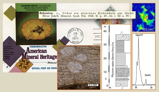
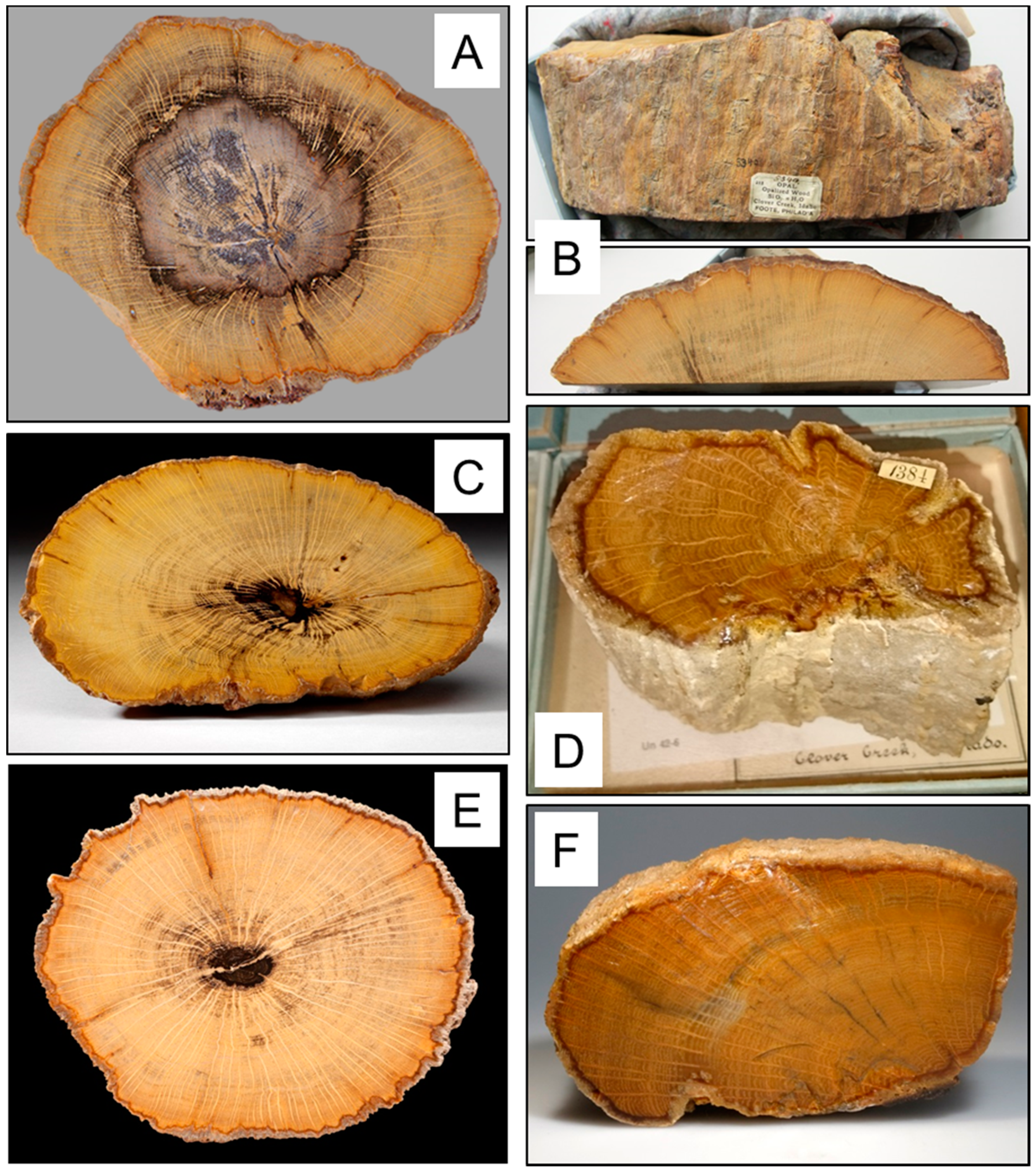
Clover Creek specimens found their way into museums in the United States, Canada and Europe, commonly displayed in systematic mineral collections as an example of opalization (
Figure 1). Clover Creek samples are likewise described in the mineralogical literature as examples of wood preserved by opal [
4,
5,
6]. Even philatelists are confronted with a picture of a Smithsonian Clover Creek sample on two first-day covers of the 1974 stamp issue of the U.S. postal office celebrating U.S. mineral heritage; another serious reason to clearly establish the provenance of Clover Creek opalized wood.
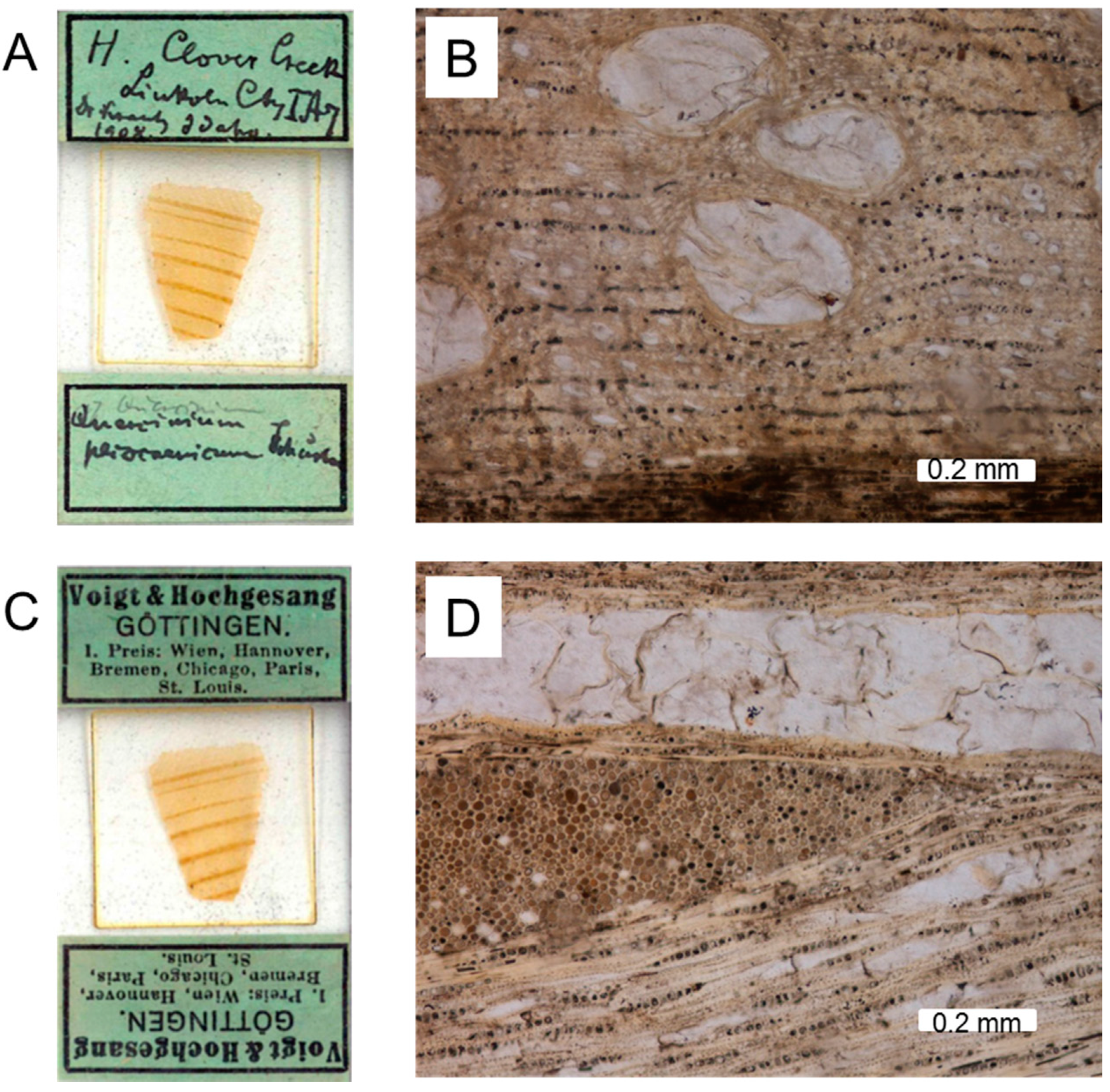
Prior to our investigations, scientific study of the Clover Creek fossil wood was limited to a single report in 1908 [
7]. The author Julius Schuster received Clover Creek samples from Johann Traugott Sterzel, a German teacher, paleobotanist and first director of the Natural History Collections of Chemnitz, Germany, who obtained them from the German mineral dealer Dr. Friedrich. Krantz. Sterzel assigned the specimens to
Quercinium and asked Schuster for a detailed examination of thin sections prepared by the company Voigt and Hochgesang, Goettingen (
Figure 2), today part of the Sterzel collection at Museum für Naturkunde Chemnitz, Germany. Schuster established a new species name,
Q. pliocaenicum [
7]. Subsequently, the German mineral dealer Krantz offered Clover Creek specimens for sale in 1908 [
8], adding the new scientific name in 1909 [
9].
Quercinium was established as a morpho-genus by Unger [
10] to describe petrified wood having a cellular anatomy characteristic of the modern oak family (
Fagaceae). In later years, fossilized oak-like wood has been described as
Quercoxylon [
11,
12,
13,
14], but
Quercinium continues to be used [
14,
15]. We have not attempted to emend the taxonomy of
Q. pliocaenicum, but instead focus on geologic and mineralogical aspects.
Clover Creek opalized wood has been referenced in several compendia [
16,
17,
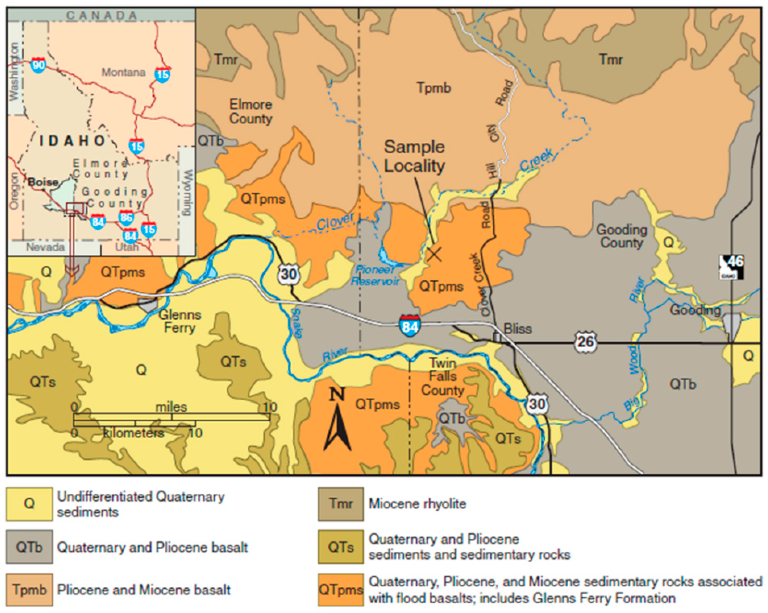
18], but an exact location or story behind the discovery is not given. Modern paleobotany books do not mention the Clover Creek find, even though many specimens are displayed in major museums. Originally, the locality was reported as being in Lincoln County, Idaho, but present county boundaries now place the site in Gooding County (
Figure 3, [
19]). The 2014 rediscovery of the site is described by Viney
et al. [
20]. The GPS coordinates are 42°59′46.70′′N, 115°00′28.66′′W, elevation 1009 m (3300 feet).
The purpose of our study is to provide a comprehensive re-evaluation of this locality, using contemporary knowledge of the geologic setting, and to analyze newly-discovered specimens by contemporary analytical methods not available in 1908. This research is based on examination of the original 1908 thin sections and new thin sections prepared from specimens we collected in 2014. In addition, we extend the taxonomic work of Schuster [
7] by providing analyses of the composition and microstructure of Clover Creek
Q. pliocaenicum.
2. Geology of the Area
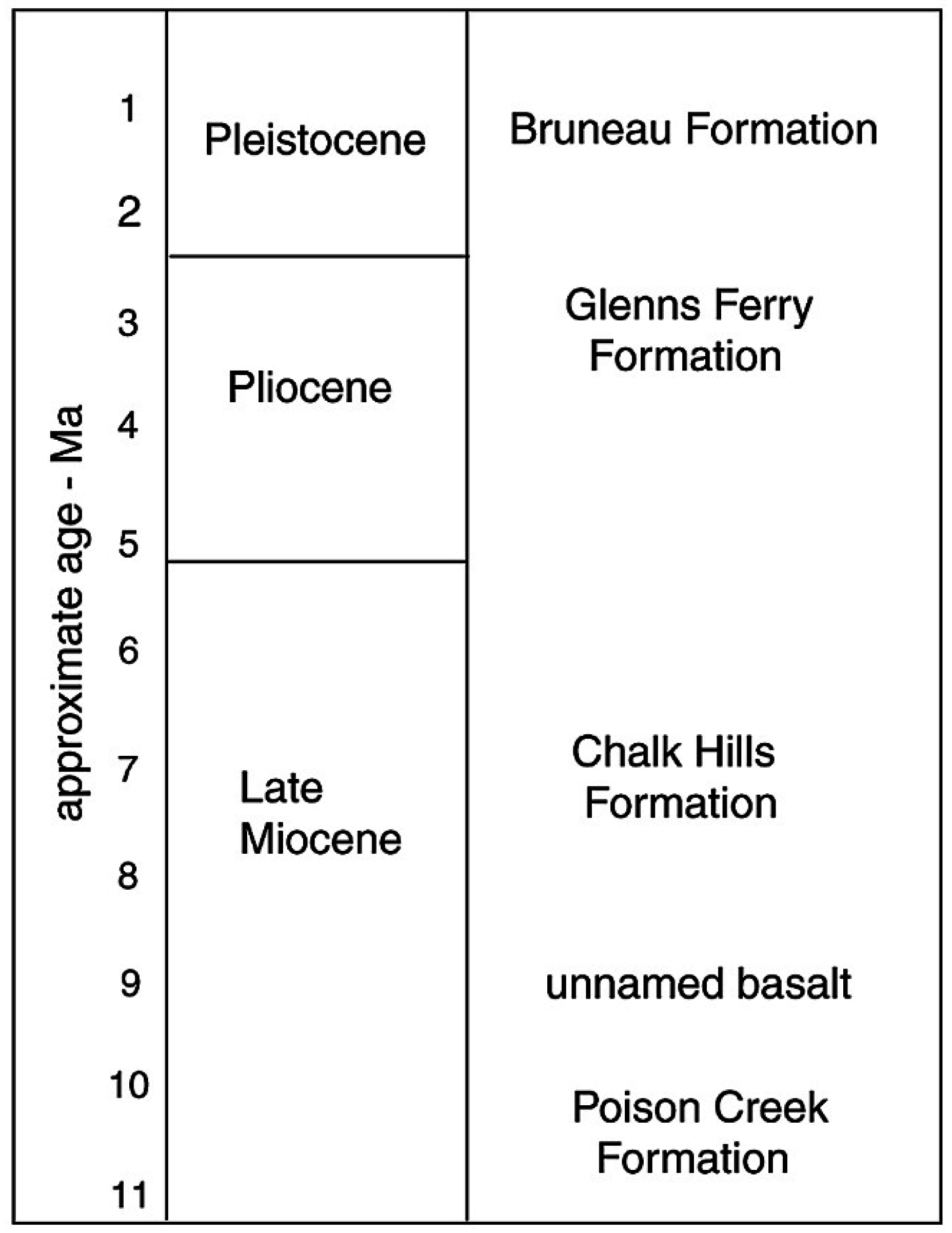
The opalized remnants of oak wood are found in flat-lying sandstone and siltstone of the Glenns Ferry Formation, at an elevation 1009 m, on the east side of Clover Creek. The light-gray and light-brown siltstone and sandstone of the fossil site are in the upper member of the Glenns Ferry Formation, stratigraphically above the Clover Creek basalt flow and below the Shoestring Road basalt. The strata are of Pliocene age and lie above the rhyolitic Fossil Gulch ash, with dates of about 3.3 Ma [
21,
22] (
Figure 4).
The Pliocene Glenns Ferry Formation of the Idaho Group contains sediment deposited in Lake Idaho, an inland lake that occupied the Snake River Plain from about 6.5 to 1.5 million years ago. The initial spillover of Lake Idaho was about four million years ago, but the age is not tightly constrained [
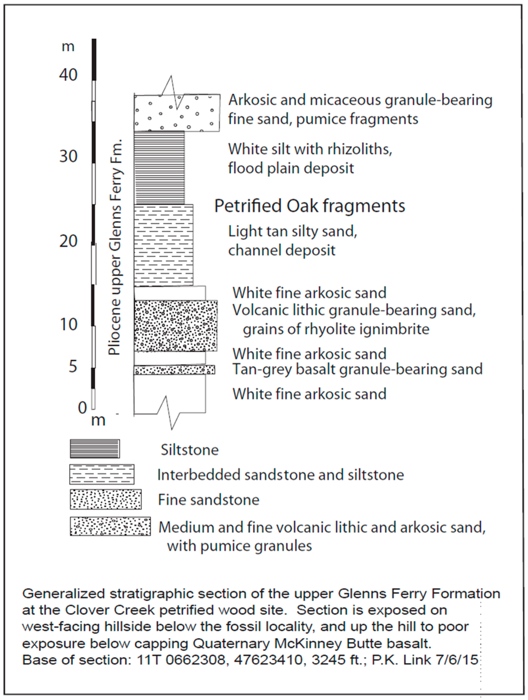
23]. At the Clover Creek fossil site, about 60 m of sediment are exposed below the Shoestring Road Basalt, which forms the upland land surface (
Figure 5, stratigraphic column). Exposure is intermittent. The sediment contains poorly- to well-sorted, subangular, medium to granule-sized volcanic lithic sandstone, interbedded with white to gray siltstone that bears small rhizoliths. The volcanic lithic grains are mainly rhyolite and pumice, with subordinate basalt. The sandstone also contains feldspar, quartz and biotite, attesting to the derivation from granitic rocks of the Cretaceous Idaho batholith to the north.
Though exposure is poor, we estimate that about half of the strata are sand and half are siltstone. The sandstone represents fluvial channels, and the siltstone represents the adjacent flood plain. The most abundant fossil wood fragments are in a siltstone interval, suggesting the tree grew in the flood plain. Fragments of well-cemented coarse sandstone are also found with the wood fragments, suggesting that the sand acted as a permeable zone for silica-bearing fluids.
Detrital-zircons from central Idaho suggest that the Glenns Ferry Formation represents the paleo-Wood River, rather than the paleo-Snake River [
24]. The Glenns Ferry Formation contains extraordinary vertebrate fossils at Hagerman Fossil Beds National Monument, 20 km to the southeast [
25].
3. Materials and Methods
Specimens were cut to reveal transverse, tangential and radial planes of subsequently polished thin-sections. Optical Microscopy (OM) was performed using a Nikon SMZ1500 stereomicroscope (Tokio, Japan), a Nikon Eclipse ME600 petrographic microscope equipped with a Nikon DS-5M-L1 digital camera and a Zeiss Photomicroscope II equipped with a Spot Infinity digital camera. Specimens, thin sections and digital photomicrographs are archived at the Geology Department, Western Washington University, USA, at Museum für Naturkunde Chemnitz and Professur Werkstoff- und Oberflächentechnik, Technische Universität Chemnitz, Germany.
Bulk powdered samples were analyzed by means of an X-ray diffractometer (Bruker AXS D8, Berlin, Germany) using Ni-filtered Cu Kα radiation.
Scanning Electron Microscopy (SEM) was done using a VEGA (Tescan, Brno, Czech Republic) for secondary electron (SE) images, a NEON40EsB (Zeiss, Jena, Germany) combined with an GEMINI (EDAX, Mahwah, Mahwah, NJ, USA) energy-dispersive X-ray detector for backscatter electron (BSE) images and elemental analysis and an Nova NanoSEM (FEI, Hillsboro, Hillsboro, OR, USA) equipped with a cathodoluminescence (CL) detector (GATAN monoCL, Pleasanton, CA, USA). CL spectroscopy and CL imaging (CL-OM) were done on carbon-coated, polished thin sections in the SEM operated at 20 kV and a “hot cathode” CL microscope HC1-LM (lumic, Dortmund, Germany) operated at a 14-kV accelerating voltage and a current of 0.2 mA (current density of about 10 µA/mm2). Luminescence images were captured “on-line” during CL operations using a Peltier cooled digital video camera (OLYMPUS DP72, Tokio, Japan). CL spectra in the wavelength range 380 nm to 1000 nm were recorded with an Acton Research SP-2356 digital triple-grating spectrograph with a Princeton Spec-10 CCD detector that was attached to the CL microscope by a silica-glass fiber guide. CL spectra were measured under standardized conditions (wavelength calibration by an Hg halogen lamp, spot width 30 µm, measuring time 2 s). Raman spectroscopic measurements were performed with a 785-nm laser for excitation in a confocal Raman microscope (Renishaw inVia Reflex, Wotton-under-Edge, Gloucestershire, UK).
4. Results
4.1. Microscopy
Analytical methods for studying silicification have greatly advanced since the work of Schuster [
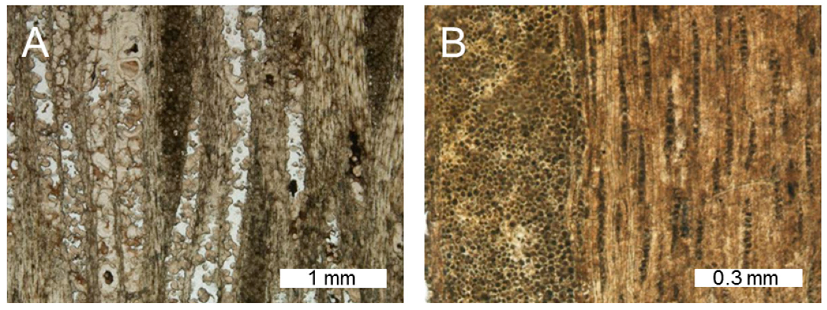
7]. However, his keen powers of observation are evident in the description of the anatomical characteristics with which he established the new species. Petrographic thin sections made from a sample collected in 2014 compare closely to photomicrographs of Schuster’s original thin sections (
Figure 6 and
Figure 7), confirming that the recently-collected material matches the original 1895 discovery.
Schuster [
7] focused his work on taxonomy, based on the well-preserved cellular detail. However, he also provided a description of petrographic features, observing that polished wood cross-sections were light yellow, with brownish discoloration along the outer margins. Schuster believed that brownish zones are probably caused by the presence of hydrated ferric oxide. In radial sections, the opal was reported to have a blue color.
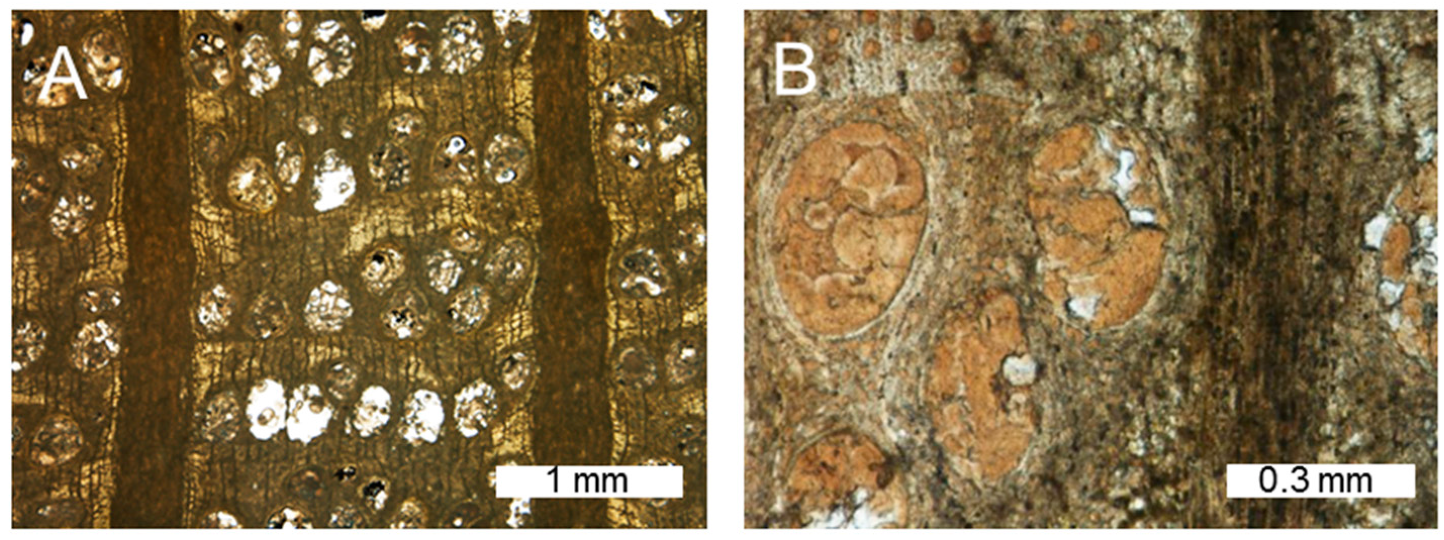
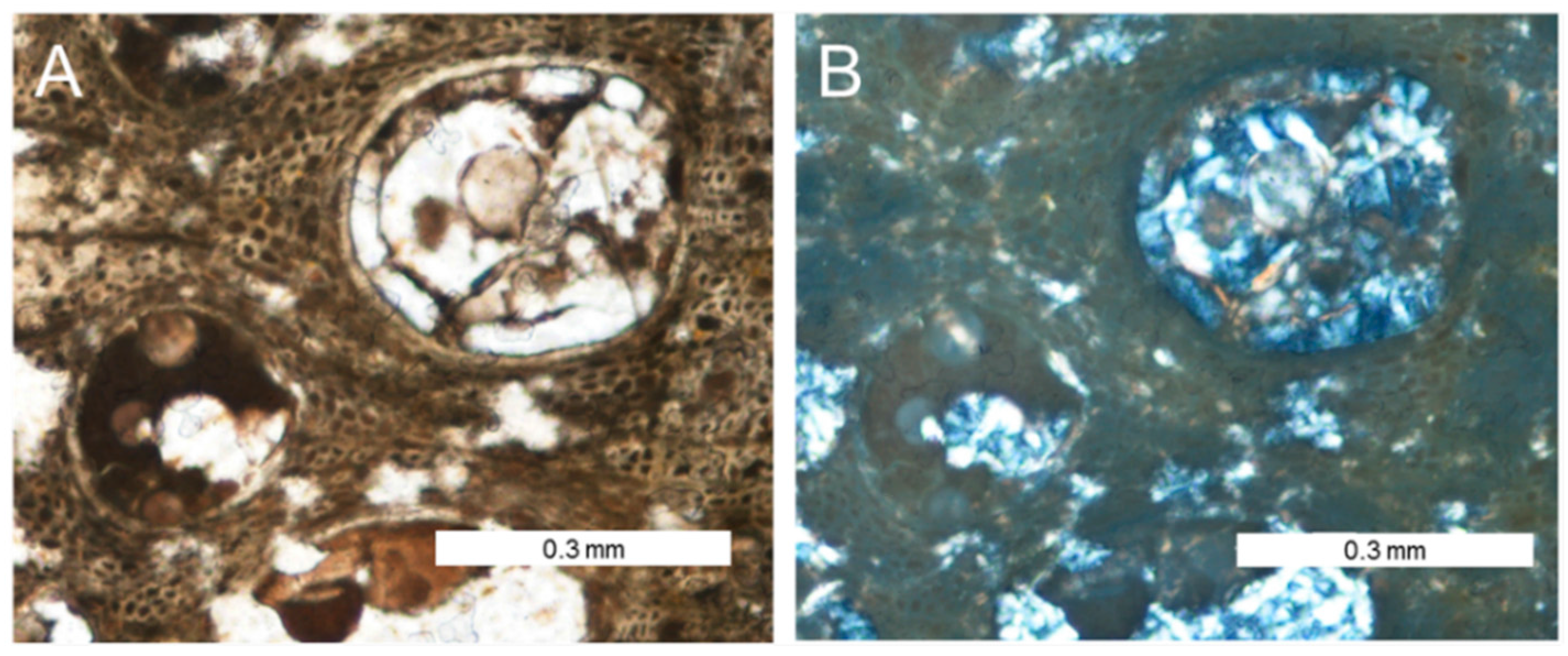
Schuster’s comments on mineralogy are brief, but he made an important observation: the silicification primarily consists of common opal, isotropic under polarized light, but most of the vessels are filled with chalcedony. Schuster [
7] believed that the “more or less spherulitic chalcedony” originated from devitrification of the original opal. As discussed later, a more likely explanation is that the chalcedony filling the vessels represents a second stage of silica deposition, not a transformation of opal to chalcedony. The coexistence of opal and chalcedony is evident in thin sections prepared from the 2014 material (
Figure 8).
Most of the cellular tissue is mineralized with opal-CT. Opalization of tracheids commonly includes mineralization of spaces between adjacent cells, accounting for the vitreous luster of hand specimens. The transparency of the silicified tissue makes transmitted light microscopy an effective method for revealing the detailed anatomical preservation. Cellular tissue has been mineralized with opal-CT, but vessels remained open during this phase of silicification. Later, chalcedony was precipitated by silica-bearing fluids permeating these vessels. Frequently, vessels were not completely filled with silica. Chalcedony surfaces that face these openings typically have a botryoidal texture. Polarized light microscopy allows for a clear distinction between opal-CT, chalcedony and open spaces. Opal-CT appears dark in cross-polarized light illumination; open spaces appear bright; and chalcedony exhibits birefringence (
Figure 8B).
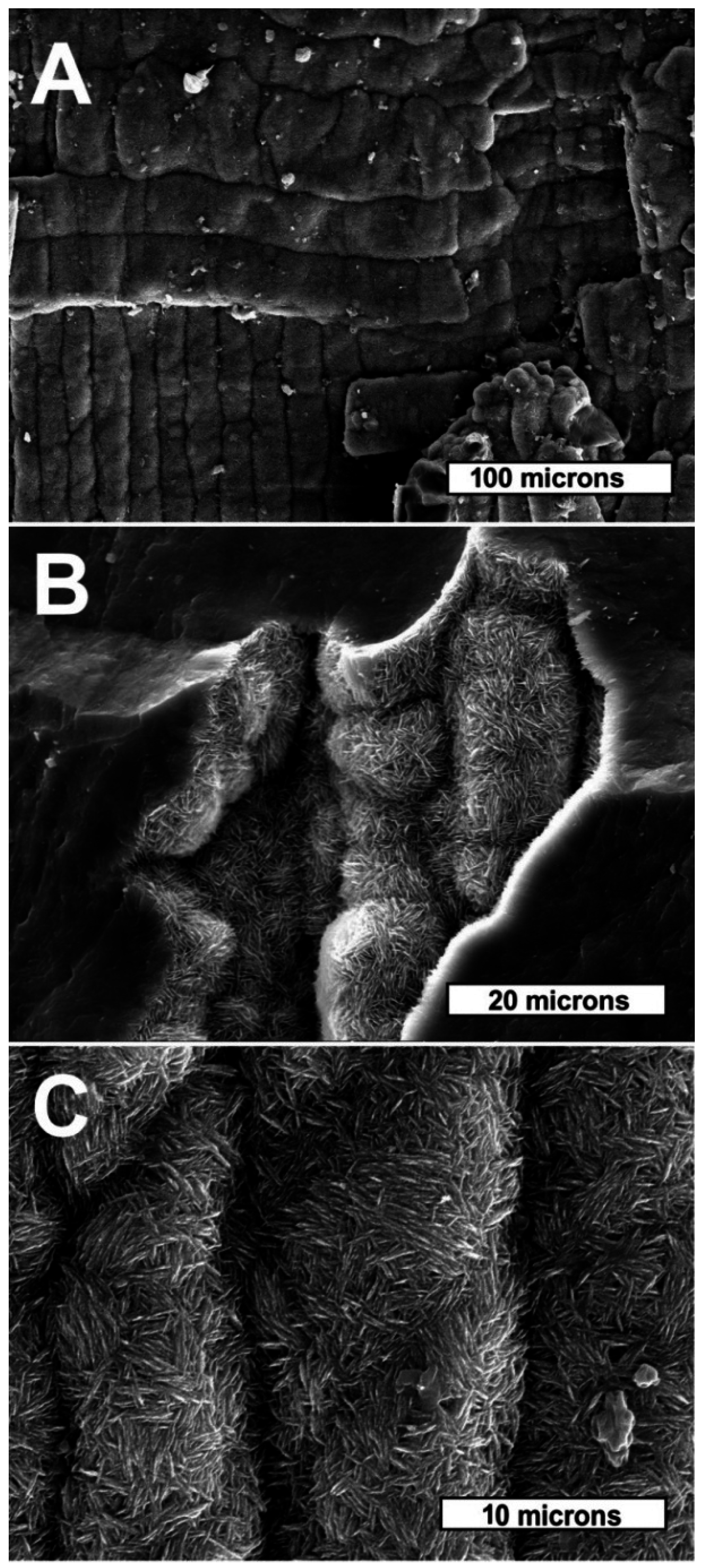
Opalized regions typically appear rather featureless in SEM views because the mineralization of intercellular spaces obscures cellular topography. In regions where intracellular spaces are not completely mineralized, the morphology of individual adjacent cells is visible. In areas of incomplete silicification, the morphologies of individual tracheids and mineral textures are evident (
Figure 9). Tracheids are mineralized with tabular microcrystals that are a common form of opal-CT (
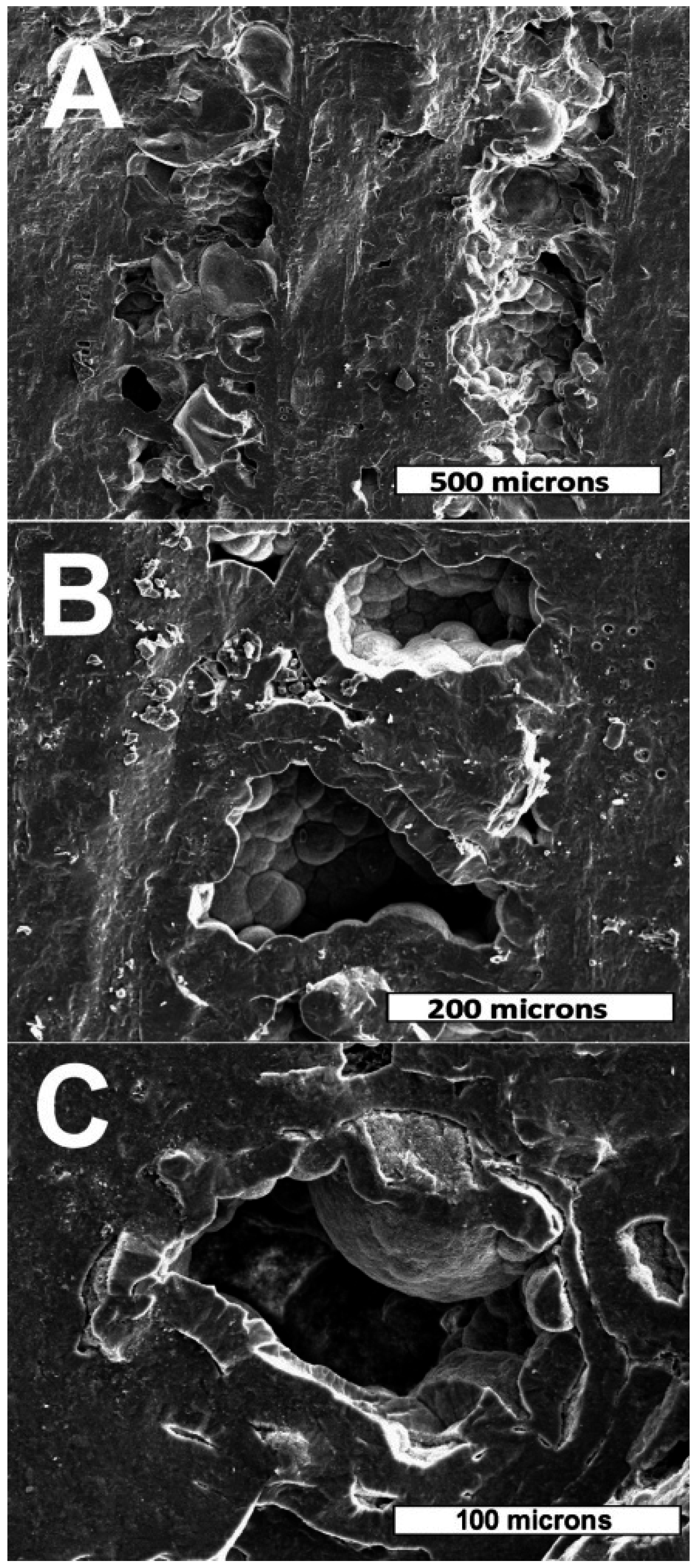
Figure 9B,C). Mineralization of vessels is very different from that of the surrounding wood. Vessels remain partially unmineralized. Silica in the vessels is commonly present in the form of chalcedony (
Figure 10). SE SEM images clearly show the cellular tissue mineralized with opal-CT to form a vitreous groundmass, in contrast to adjacent vessels that have been partially filled with chalcedony. In both transverse and radial views (
Figure 10B,C), the semi-hemispherical shape of individual chalcedony masses facing the hollow interior of vessels can be observed.
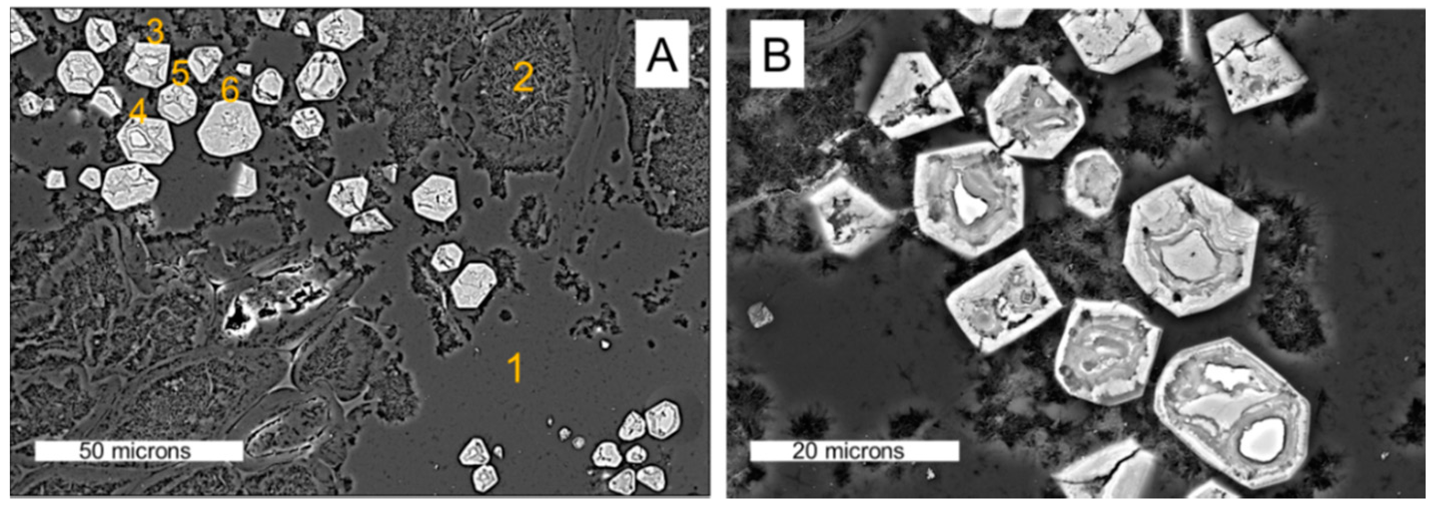
The coloration of the fossil wood may be at least in part the result of trace amounts of iron. In some regions, silicified cells contain iron-rich microcrystals (
Figure 11,
Table 1). Atomic percentages calculated from EDS spectra indicate the presence of several iron minerals. Crystal shapes most commonly appear to be variations of the trigonal system, suggesting hematite as a possible parent mineral. Other symmetries are present, however, including orthorhombic forms that could have originated as marcasite. The presence of diverse iron minerals is perhaps the result of alteration related to multiple episodes of mineralization. The internal architecture of these crystals suggests complex paragenesis.
4.2. Diffraction and Spectroscopy
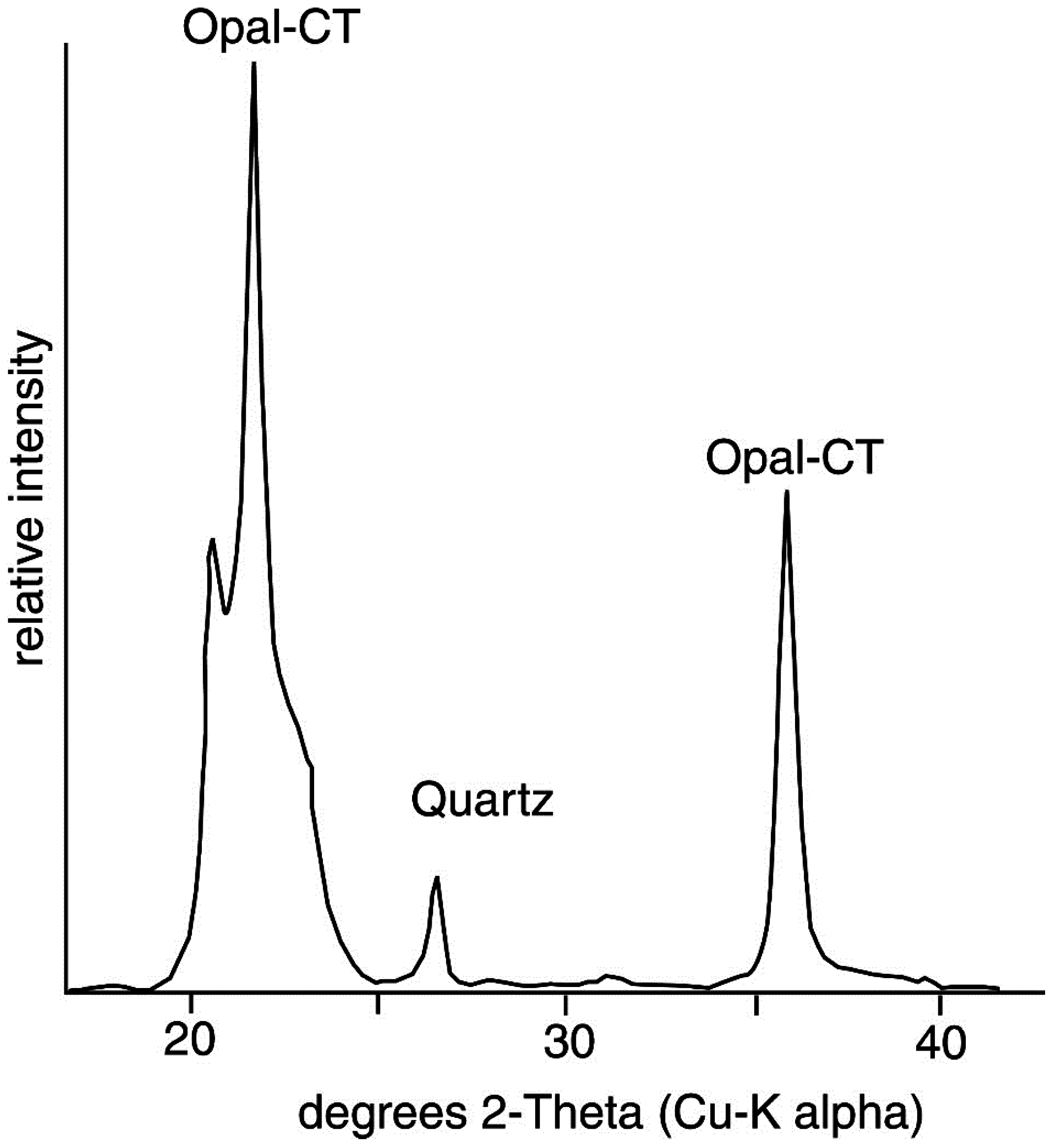
X-ray diffraction, Raman spectroscopy and cathodoluminescence microscopy were used to better understand the mineralogy. The X-ray diffraction pattern from a bulk powdered sample (
Figure 12) shows the presence of two forms of silica, consistent with the evidence from optical and scanning electron microscopy. Opal-CT peaks are very evident. Modeling this mineral phase as interstratified layers of cristobalite and tridymite [
26] results in a cristobalite/tridymite ratio around 0.54 and crystallite sizes around 25 nm. Quartz peaks apparently indicate chalcedony present as vessel filling; no other forms of quartz were observed by microscopy. The size of those crystals is between 60 and 80 nm, which is consistent with the expectation of fibrous chalcedony.
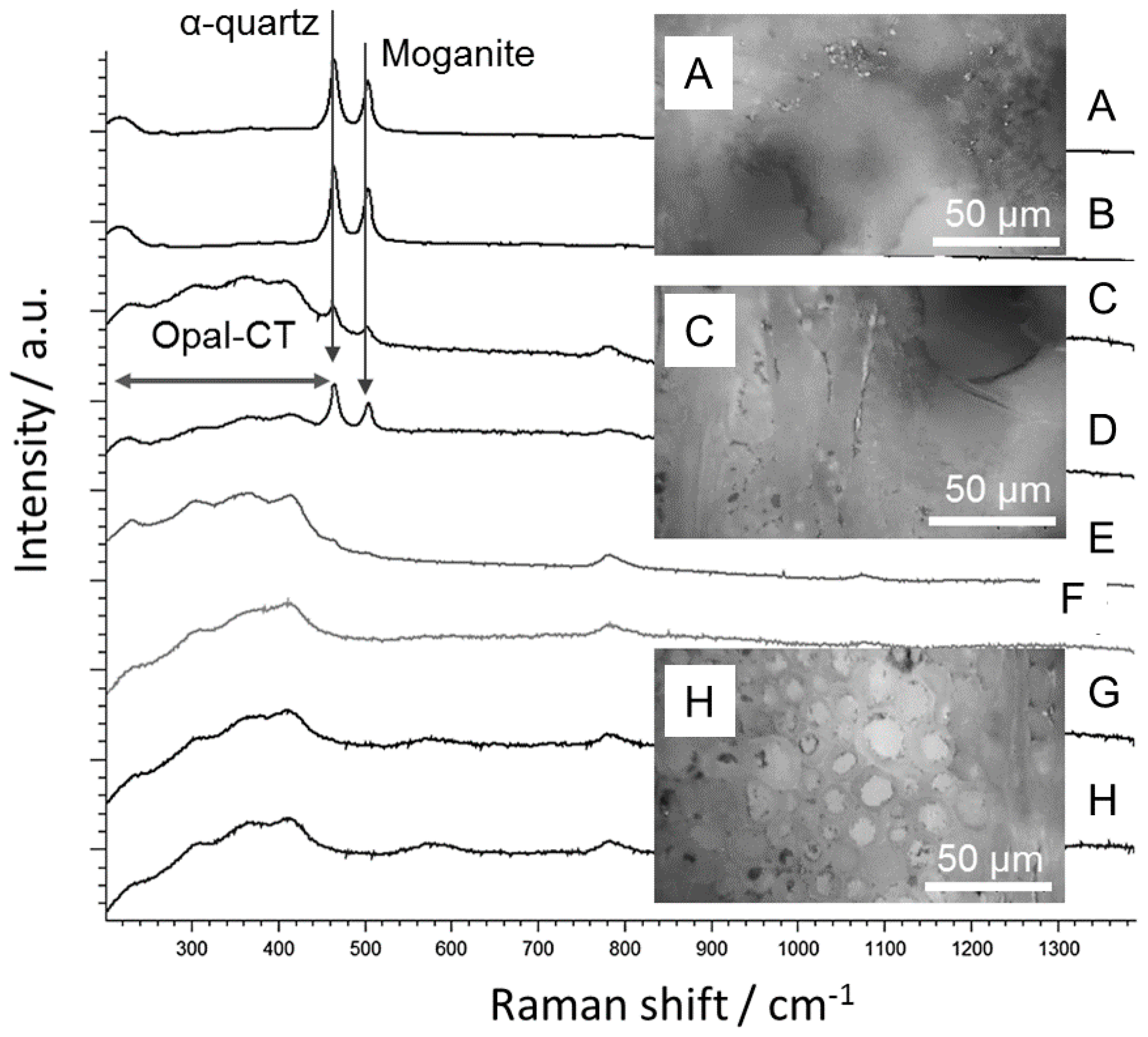
Raman spectroscopy provides molecular information by inelastic scattering of photons and complements the crystallographic information achieved by elastic scattering of X-rays. Structure-building units, which are composed of a few silicon-oxygen tetrahedra, can be identified by means of vibrational, rotational and other low-frequency modes in the silica system. Different parts of the Clover Creek sample selected by OM indicate different silica polymorphs (
Figure 13). Spectra gained from tracheids and medullary rays of the Clover Creek sample (
Figure 13, Spectra c to h) exhibit peaks at 220 cm
−1, 310 cm
−1, 365 cm
−1 and 415 cm
−1 with varying intensities. These peaks are attributed to Si–O–Si bending vibrations, suggesting that the structure is close to an opal-CT type consisting of a matrix of intergrown tridymite- and cristobalite-type nano-domains according to [
27]. Additionally, a higher portion of tridymite-like structural units can be inferred, which is in accordance with our XRD measurements.
Raman shifts at 464 cm
−1 and 502 cm
−1 are evident at early wood vessels (
Figure 13, Spectra A,B). The peak at 464 cm
−1 is attributed to α-quartz and the peak at 502 cm
−1 to moganite. First described in 1984, but not formally recognized as a valid mineral species until 2007, moganite is a monoclinic SiO
2 polymorph that commonly occurs as a constituent in chalcedony and other varieties of microcrystalline quartz [
28]. The moganite content of chalcedony has been reported to range from 26% to 82%, [
29], but because moganite identification is usually based on Raman spectroscopy, the possible presence of this mineral in silicified wood has historically been overlooked. Götze
et al. [
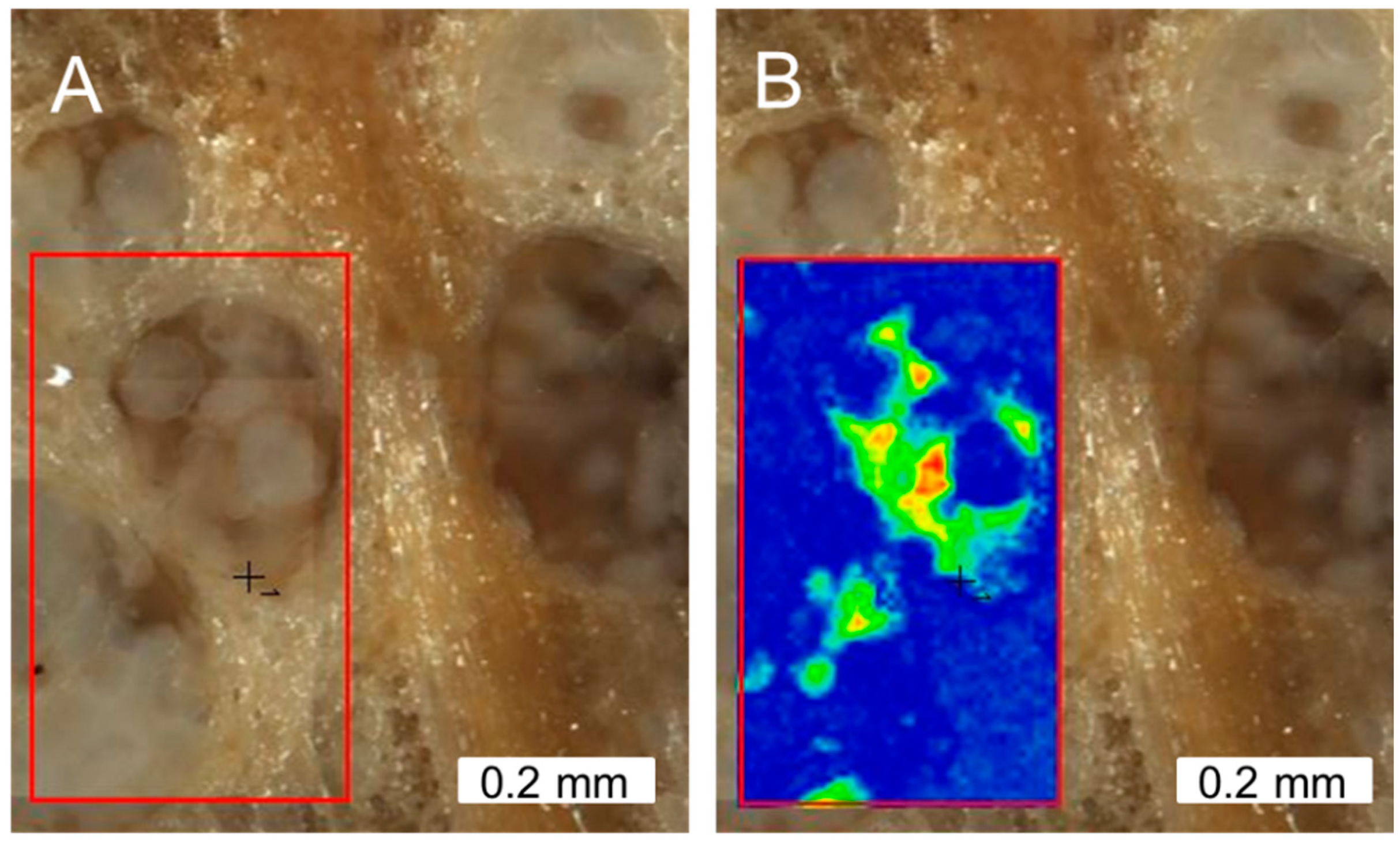
30] provided a calibration curve for determining the moganite content in nano-crystalline α-quartz (chalcedony). Accordingly, the measured peaks were fitted by Lorentzian distributions for calculating the peak integral ratio. The result (0.5 to 0.7) indicates moganite content between 70 and 78%. Moganite occurs in chalcedony as nano-range lamellae down to single Brazil law twin planes detectable by Raman spectroscopy, but too small for the long-range order-dependent XRD. The spatially-resolved distribution of moganite in a chalcedony-preserved early wood vessel is shown by an intensity map of the moganite-related Raman peak (
Figure 14).
CL spectroscopy in combination with microscopy provides information about the distribution of impurities and structural defects. This information can indicate mineralization sequences or hydrothermal mineralization and alteration of silica polymorphs. Four CL peaks were detected, with intensities that varied with respect to sample position and the CL analytical method. CL peaks at ~380 nm, 450 nm and 650 nm were detected by electron beam excitation with the SEM. Peaks at ~550 nm and 650 nm were detected using the optical microscope hot cathode CL system [
30,
31]. In the latter system, UV radiation cannot be detected because of the absorption effects of the glass optics.
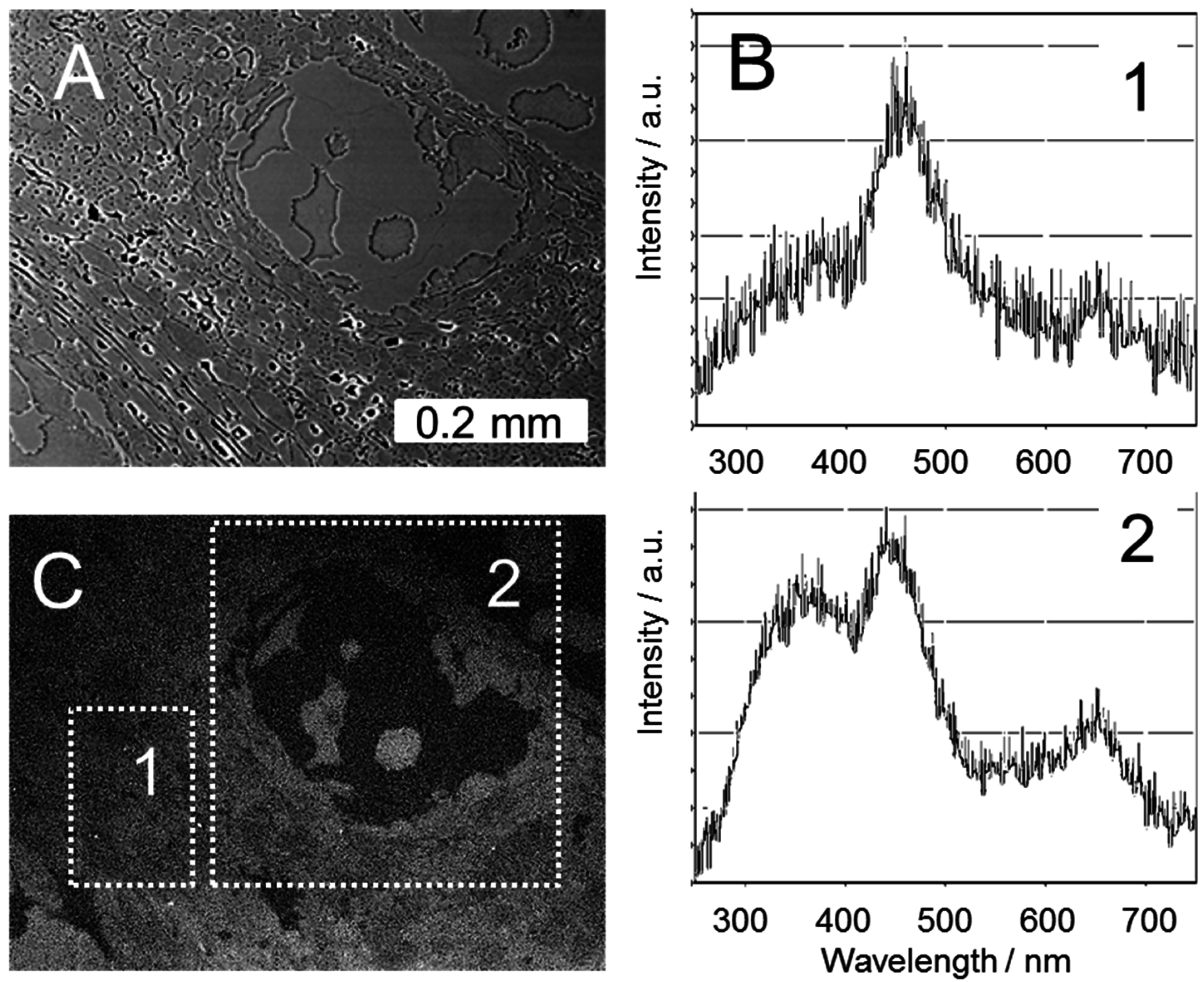
A silicified early wood vessel in the transverse section is shown by SEM BSE imaging, panchromatic SEM-CL imaging and corresponding CL spectra (
Figure 15). The peaks at 450 nm, attributed to an oxygen-deficiency center [
32], and 650 nm, attributed to a non-bridging oxygen hole center [
30], are common in quartz and opal from igneous, volcanic and metamorphic processes. The chalcedony, which filled the early wood vessel, is the source of an additional peak at ~380 nm, which is typical for cryptocrystalline quartz with a hydrothermal origin [
33].
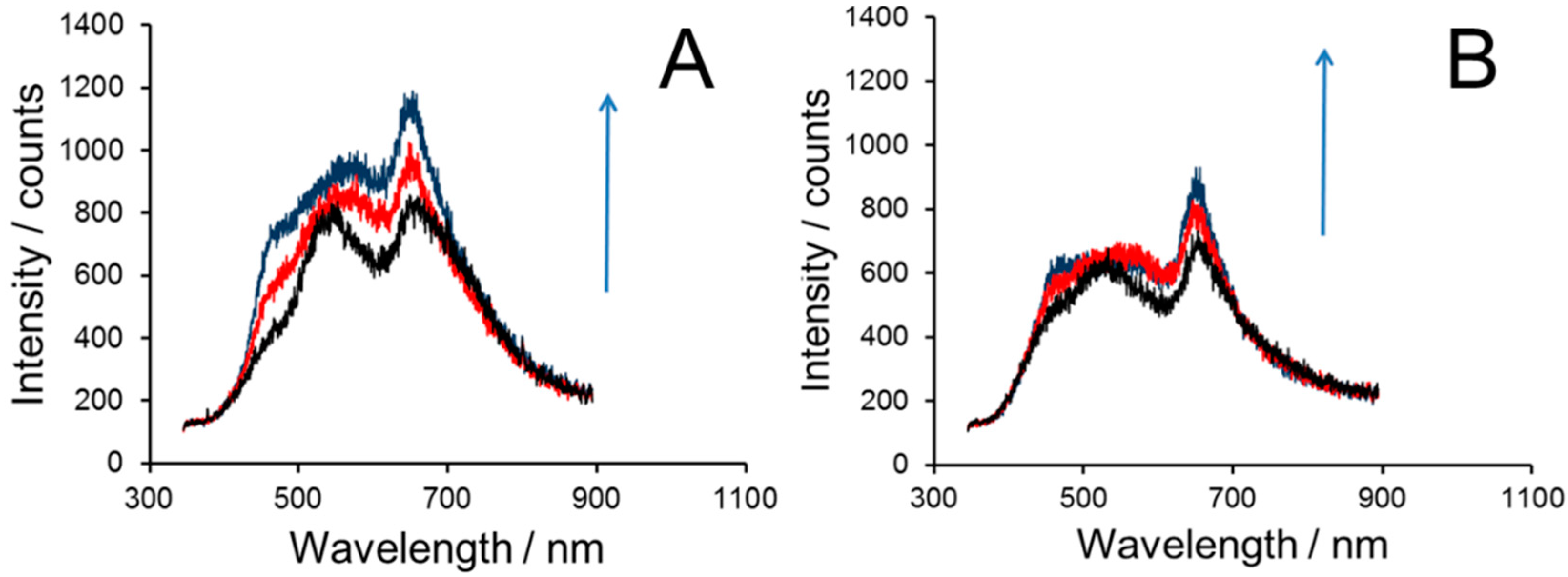
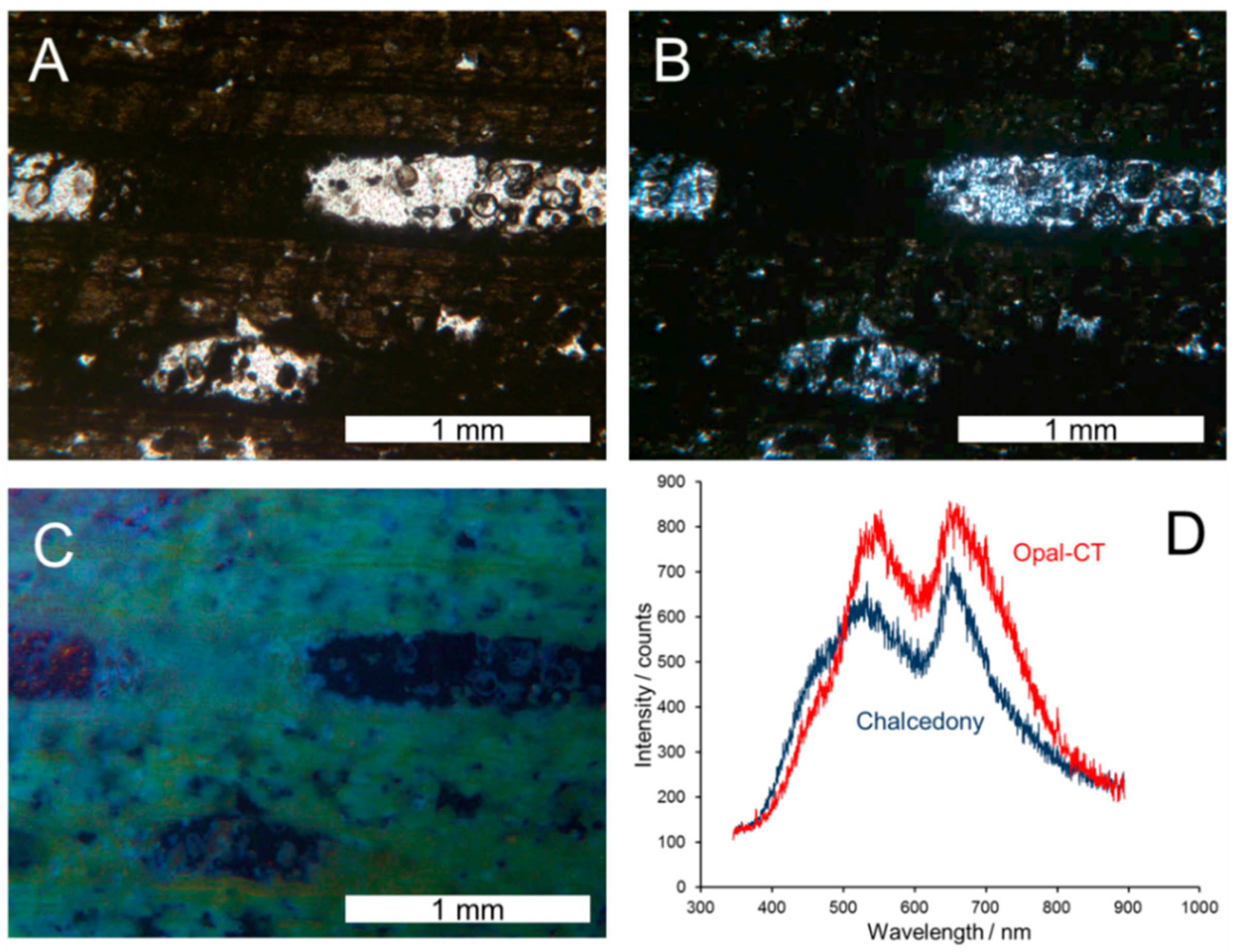
The observation of CL peaks was accomplished using an optical CL microscope with a defocused electron beam. OM-CL images (
Figure 16) show early wood vessels in the tangential longitudinal section by polarized light microscopy and OM-CL. The polarized light micrograph confirms that the silicified wood consists of a relative dense opal and larger vessels partially lined with chalcedony. The OM-CL images reveal bluish luminescence in the opal matrix and blue-violet luminescence in the vessels. OM-CL spectra are similar for Opal-CT and Chalcedony. The OM-CL spectrum shows only a slight shoulder of the UV band because of the system’s detection limits. Cathodoluminescence changed in intensity with increasing radiation dose, this change being more evident in opal as compared to chalcedony (
Figure 17). The bluish CL of opal turns to slight yellowish due to the activation of more defects. The CL spectrum of chalcedony is characterized by an increase of the 650 nm band and a decrease of the UV band around 380 nm (not visible in the OM-CL spectra). This is typical behavior for quartz crystallized from low temperature aqueous solutions (e.g., hydrothermal fluids) [
33] and indicates a multi-stage process of silicification for the Clover Creek samples.
5. Discussion
5.1. Taxonomy
The proposed taxonomy for fossil oak wood has not received universal agreement. The first description of fossil oak was by Gӧppert [
34] who introduced
Klödenia quercoides as a name for wood from northern Germany and Silesia. This taxonomy became confused because
Klödenia was later accepted as a name for an ostracod genus [
35]. Unger [
10] proposed
Quercinium as the genus to be used for fossil oak wood exhibiting the cellular anatomy characteristic of the modern oak family (
Fagaceae). This taxonomy received scant attention until Felix [
36,
37] published descriptions of known species and added four new ones.
As noted, Schuster [
7] named the Clover Creek wood
Q. pliocaenicum, and Boeshore and Jump [
38] described
Q. album from the Miocene Payette Formation of southwestern Idaho. Boeshore and Jump [
38] include a table in their paper that listed all
Quercinium species recognized in the literature at the time (1938); interestingly, Schuster’s
Q. pliocaenicum was not included. Other investigators have used other taxonomic strategies. Penhallow [
39] described Holocene oak wood from Illinois as
Quercus, a practice that has enjoyed continued use, e.g., [
40]. Hofman [
11] suggested
Quercoxylon as a form genus name for fossil oak wood, but did not name a type species. Kräusel [
12] designated
Q. retzianum as a type species for this new genus.
It is not our intent to resolve these taxonomic complexities. We have retained the original
Quercinium pliocaenicum nomenclature because of historical precedence and because this fossil genus has been applied to most other North American occurrences of fossil oak wood (e.g., [
15,
41,
42]). For a more detailed discussion of these taxonomic issues, see Iamandei
et al. [
14].
5.2. Paleoclimate and Paleoenvironment
At the time of the 1895 discovery and the subsequent published study [
7], knowledge of the regional geology was rather scanty. Prior to the advent of radiometric dating, fossils offered a way to estimate ages of sedimentary strata and, by inference, the ages of associated igneous rocks. In recent decades, the geology of the Snake River Plain has received considerable attention, and our knowledge of the ages of rock units has been greatly refined thanks to precise radiometric ages determined for the many volcanic interbeds [
21,
43,
44,
45]. Snake River Plain sediments range from late Miocene to early Pleistocene.
Quercus leaves are preserved in the late Miocene to early Pliocene Poison Creek and Chalk Hills Formations; Cherry Creek fossil oak wood is probably from the late Pliocene Glenns Ferry Formation.
While Schuster did not offer any thoughts on the paleoclimate associated with
Q. pliocaenicum, he did use the presence of narrow growth rings to infer that the tree was deciduous and growing in poor conditions. Today, the Snake River Plain sediments provide one of the best-documented records of Neogene paleoclimate in the western USA. Paleoclimatic inferences have been derived from fossil mammals [
46], fish [
47,
48] and pollen [
49,
50], as well as isotopic investigations [
51]. The paleoflora document the presence of diverse deciduous trees in the central Idaho region during the late Miocene. The Pliocene Glenns Ferry flora records a significant climate change, with the appearance of
Artemisia (sagebrush),
Sarcobatus (saltbush) and abundant Poaceae (grasses). Pollen from aquatic plants indicates the presence of ponds and lakes. Tree pollen includes
Pinus (pine) as the most common conifer, but diverse hardwood pollen includes
Quercus. Mustoe and Leopold [
50] believed that this floral evolution is the result of the late Miocene or early Pliocene rise of the Cascade Range, resulting in a rain shadow. Glenns Ferry sediments appear to have been deposited in a region of gentle topography, with grasslands, areas of temperate forest and scattered wetlands. The overlying early Pliocene Bruneau Formation shows a marked increase in the abundance of
Artemisia and
Sarcobatus and a decline in Poaceae and aquatics, indicating increasing aridity. Mustoe and Leopold [
50] used Jaccard similarity coefficients to estimate paleoclimatic conditions for Neogene paleoflora of Washington, Oregon and Idaho. These data suggest that the floral composition of the Glenns Ferry Formation flora resembled modern vegetation from Lewiston, Idaho, ~600 km to the northwest. The Glenns Ferry paleoflora is estimated to have had a mean annual temperature (MAT) of ~12 °C and annual precipitation of 300 to 325 mm. This compares to the modern climate at nearby Hagerman, Idaho, with MAT of 10 °C and annual precipitation of ~250 mm. Pollen studies for Glenns Ferry Formation and Idaho Group strata between 12 and 4 Ma suggest a savannah-like setting, with an annual precipitation of up to ~500 mm [
22,
49,
52].
The ancient oak tree whose remains were preserved at Clover Creek presumably flourished at a time when the climate was somewhat warmer and wetter than at present. One of the most significant climatic differences is seasonality; estimated July precipitation was ~17 mm during Glenns Ferry time, compared to ~5 mm in the modern era. This increased seasonality was an important factor in the appearance of sagebrush steppe flora and explains the decline in habitat for oaks and other broadleaf hardwoods in the central Idaho region.
5.3. Silicification Pathway
One of the most significant aspects of the Clover Creek fossil wood is the evidence it provides for the silicification processes. Schuster [
7] observed that the wood cells were mineralized with common opal, but that vessels contain chalcedony. Schuster believed that the “more or less spherulitic chalcedony” originated from devitrification of the original opal. During that era, processes of wood silicification were poorly understood, and although Schuster did comment on transformation processes, he did not propose possible mineralization processes.
Pathways for the formation of petrified wood have been discussed as early as 1665 when Robert Hooke used his newly-invented microscope to inspect the cellular structure of petrified wood for the first time [
53]. In 1976, Leo and Barghoorn [
54] established a key concept for wood silicification that includes two basic assumptions. First, wood acts as a template for silica, even as it slowly degrades and is replaced. Second, silica sol is the silicifying agent that forms hydrogen bonds with hydroxyl groups exposed on cell walls. This concept has been incorporated into every silicification model that has since been proposed. Dietrich
et al. provide a review of studies on artificial and natural silicification [
55]. More recently, Mustoe [
56] studied the evidence of multiple silicification pathways of Neogene fossil woods. Mustoe’s model includes early mineralization of cell walls followed by a later silica deposition in lumina, vessels and rot pockets from groundwater that permeated open spaces. He also described the process of open-space filling as analogous to the genesis of geodes and veins, where multiple episodes of hydrothermal precipitation produce opal, chalcedony and quartz as deposits within a single cavity.
Clover Creek fossil wood is a good example of polyphasic and polymineralic petrifaction. Although most of the cellular tissue has been mineralized with opal, the extent of mineralization is not homogeneous. In many regions of the specimens, cell walls, lumen and intracellular spaces are completely mineralized to produce a solid mass of opal. X-ray diffraction and Raman spectroscopic data reveal this material to consist of opal-CT. SEM images show the platy crystalline structure of this opal, but there is no visible evidence of opal lepispheres, the common architecture of opal-A. Instead, opal-CT appears to have formed as a primary mineral. During this initial phase of silica deposition, vessels remained open.
At a later stage of mineralization, many of the vessels became partially filled with chalcedony. The geode-like architecture suggests that the chalcedony was precipitated from hydrothermal fluids during a later episode, unrelated to the initial silicification phases that mineralized both tracheids and ray parenchyma with opal-CT. The observation that vessels are only partially filled with chalcedony is evidence that a silica-bearing hydrothermal fluid was able to permeate the wood even after the tracheids enclosing the vessels were thoroughly opalized. This characteristic suggests that the conductive components of the xylem (tracheids, vessels and fibers) were an important conduit for the entry of silica sol during early stages of petrifaction. This interpretation is consistent with the experimental evidence [
54] that suggests that the earliest phase of silica deposition involves precipitation on cell wall surfaces because of the affinity between cellulose and hemicellulose with silicic acid (silica sol). Because some intracellular spaces and some vessels remain open, Clover Creek fossil wood remains potentially susceptible to additional future episodes of mineralization. The initial mineralization of cell walls followed by a later episode of silica deposition in vessels from hydrothermal fluids that permeated these open spaces is consistent with similar observations [
56]. The coloration of the fossil is in part the result of hydrated iron oxide. Some silicified cells additionally contain trigonal and orthorhombic pseudomorphs. The internal architecture of these crystals suggests complex paragenesis and may provide additional evidence in support of multiple episodes of mineralization.
6. Conclusions
Clover Creek opalized wood is displayed as a mineral variant instead of a fossil in major museums in the U.S., Canada and Europe, even though the mineral dealer Foote [
2] described the specimen as “the most perfect and beautiful petrifaction in existence”. Interestingly, only a single scientific work in German describing the anatomy and providing the scientific name of
Quercinium pliocaenicum for this historic find existed up until this publication. Schuster’s paper [
7] has recently been translated to English (see the
Supplementary Material). Historical research uncovering the provenance of museum specimens, rediscovery of Schuster’s original thin section slides, along with rediscovery of the original site and the collection of new specimens allowed for a thorough scientific investigation of the geologic setting and wood mineralization. Clover Creek museum specimens can now be understood in the context of modern analyses of the geologic setting and mineralization.
The mineralization of Clover Creek opalized wood adds to a growing body of evidence suggesting a multistage pathway for wood silicification. X-ray diffraction indicates that the wood is mineralized with opal-CT and chalcedony. OM and SEM reveal that most of the cellular tissue is mineralized with tabular opal-CT microcrystals with no evidence of opal lepisphere precursors, whereas inner vessel walls are lined with botryoidal chalcedony. Raman spectroscopy provides evidence for the intergrowth of chalcedony and moganite. The observation that opal-CT has replaced most of the tissue, with vessels partially filled by chalcedony, each silica polymorph originating as a primary mineral, was substantiated by CL. This is interpreted as evidence for a multiple stage mineralization process. EDS spectra reveal the presence of authigenic iron-rich microcrystals having both trigonal and orthorhombic shapes. The low abundance of these crystals suggest they are not a major cause of the wood’s lustrous brown coloration, which is more likely caused by submicroscopic particles. However, the internal architecture of these crystals suggests complex paragenesis, perhaps representing further evidence for multiple episodes of mineralization.
The current study verifies that the fossil tree was recovered from the upper Glenns Ferry Formation along Clover Creek in Idaho. The geologic setting in which the tree was deposited and fossilized reveals that it grew in a flood plain along a fluvial channel. The tree grew at a time when the climate was warmer and wetter; however, the influence of the Cascade rain shadow slowly produced a dryer climate that over time would be less conducive to growing oaks, such as Q. pliocaenicum.
Supplementary Materials
The following are available online at
https://www.mdpi.com/2076-3263/6/2/21/s1: pdf file 1: Translation Schuster Q pliocaenicum, pdf file 2: Supplement XRD”, pdf file 3: Clover Creek Museum specimens.
Acknowledgments
The authors would like to thank the following people for answering requests and providing photographs: Katherine Dunnell, Royal Ontario Museum, Canada; Peta Hayes and Mike Rumsey Natural History Museum London, U.K.; Bruce Geller, Beth Simmons, and Sam Allen, Colorado School of Mines Geology Museum, USA.; Klaus Thalheim, Senckenberg Naturhistorische Sammlungen Dresden/Museum für Mineralogie und Geologie, Germany; Achim Reisdorf, Geologisch-Paläontologisches Institut der Universität Basel and Thomas Burri, Naturhistorisches Museum der Burgergemeinde Bern, Switzerland; Zina Fihl, Statens Naturhistoriske Museum Kobenhavn, Denmark; Ursula Müller-Krantz, Dr. F. Krantz Rheinisches Mineralien-Kontor GmbH and Co. KG, Germany. We thank William W. Besse, Associate Cartographer for Rocks and Minerals magazine, Philadelphia, USA, for use of his Clover Creek map.
Thomas Mehner, Professur Werkstoff- und Oberflächentechnik, TU Chemnitz, performed the XRD analyses. Daniel Wett, Professur Verbundwerkstoffe, TU Chemnitz, and Massimiliano Rocchia, Thermo Fisher Scientific, Milano, Italy, provided Raman data. Steffen Schulze, Professur Analytik an Festkörperoberflächen, TU Chemnitz, achieved the SEM-CL data. We are much obliged to Alex Brandl, Colorado State University Colorado, USA, for his assistance in translating the paper of Schuster into English (see the
Supplementary Material).
Author Contributions
Mike Viney revived the history of the samples. Mike Viney and Paul Link rediscovered the fossil site and investigated the stratigraphy. George Mustoe performed Optical Microscopy (OM) and Scanning Electron Microscopy (SEM) studies. Ronny Rößler provided OM on ancient and newly-prepared thin sections. Jens Götze performed CL measurements. Dagmar Dietrich performed energy dispersive spectroscopy (EDS) analysis. Thomas Lampke contributed analytical tools and supervised Dagmar Dietrich, who was responsible for collecting, analyzing and interpreting the analytic results. Mike Viney, George Mustoe, Paul Link and Dagmar Dietrich wrote the paper.
Conflicts of Interest
The authors declare no conflict of interest.
Abbreviations
The following abbreviations are used in this manuscript:
| XRD | X-Ray Diffraction |
| SEM | Scanning Electron Microscopy |
| EDS | Energy Dispersive Spectroscopy |
| T | Transverse |
| TL | Tangential Longitudinal |
| RL | Radial Longitudinal |
| OM | Optical Microscopy |
| SE | Secondary Electron |
| BSE | backscattered electron |
| CL | cathodoluminescence |
References
- Penrose, C.W. A Singular Natural Curiosity on Clover Creek Idaho; The Salt Lake Herald: Salt Lake, UT, USA, 1897; p. 3. [Google Scholar]
- Foote, W.M. A complete catalogue of minerals. Available online: http://catalog.hathitrust.org/Record/008423524 (accessed on 3 March 2016).
- Walcott, C.D. Twentieth Annual Report of the United States Geological Survey. 1898–1899, Part VI—Mineral Resources of the United States 1898; USGS: Reston, VA, USA, 1899.
- Bauer, M.H. Edelsteinkunde; C.H. Tauchnitz: Leipzig, Germany, 1909. [Google Scholar]
- Brauns, R.A. Das Mineralreich; Fritz Lehmann Verlag: Stuttgart, Germany, 1903. [Google Scholar]
- Whitlock, H.P. The Story of Minerals; The American Museum of Natural History Handbook Series No. 12; American Museum of Natural History: New York, NY, USA, 1932. [Google Scholar]
- Schuster, J. Über ein pliocänes Eichenholz aus Idaho. Neue Jahrb. Mineral. Geol. Pal. 1908, 2, 49–54. [Google Scholar]
- Müller-Krantz, U.; (Dr. F. Krantz Rheinisches Mineralien-Kontor GmbH & Co. KG, Bonn, Germany). Mineralogisches Semester-Verzeichnis Nr.8, Dezember 1908. personal communication, 2014. [Google Scholar]
- Müller-Krantz, U.; (Dr. F. Krantz Rheinisches Mineralien-Kontor GmbH & Co. KG, Bonn, Germany). Paläontologisches Semester-Verzeichnis Nr.35, Dezember 1909. personal communication, 2014. [Google Scholar]
- Unger, F. Synopsis lignorum fossilium plantarum acramphibryarum. In Genera Plantarum Secundum Ordines Naturales Disposita (Suppl. II), Appendix, Apud Fridericum Beck; Endlicher, S., Ed.; Universitatis Bibliopolam: Wien, Austria, 1842; pp. 100–102. [Google Scholar]
- Hofmann, E. Verkieselte Hölzer aus dem Sarmat des Tokaj-Eperjeser-Gebirges. Föld. közlöny 1939, 12, 260–270. [Google Scholar]
- Kräusel, R. Ergebnisse der Forschungsreisen Prof. E. Stromers in den Wüsten Ägyptens. IV. Die fossilen Floren Ägyptens; Antiquariat Thomas Haker GmbH and Co. KG: Berlin, Deutschland, 1939; Volume 47, p. 140. [Google Scholar]
- Gros, J.P. La dénomination des bois fossiles identifiés à des chênes. Bull. Mens. Soc. Linn. Lyon 1988, 57, 250–260. [Google Scholar]
- Iamandei, S.; Iamandei, E.; Bozukov, V.; Tsenov, B. Oligocene woods from Rhodopes, Bulgaria. Acta Palaentol. Rom. 2012, 9, 15–25. [Google Scholar]
- Wheeler, E.F.; Scott, R.A.; Barghoorn, E.S. Fossil dicotyledonous woods from Yellowstone National Park II. J. Arnold Arbor. 1978, 59, 1–25. [Google Scholar]
- Nickles, J.M. Bibliography of North American Geology for 1908 with Subject Index; United States Geological Survey: Reston, VA, USA, 1909; Volume 409.
- Edwards, W.N. Dicotyledones (Ligna). In Fossilium Catalogus II; Jongmans, W., Ed.; Plantae. Pars 17; W. Junk: Berlin, Greman, 1931; pp. 63–69. [Google Scholar]
- LaMotte, R.S. Catalogue of the Cenozoic plants of North America through 1950; Geological Society of America: New York, NY, USA, 1952; Volume 51. [Google Scholar]
- Lewis, R.S.; Link, P.K.; Stanford, L.R.; Long, S.L. Geologic Map of Idaho 2012: Scale 1:750,000; Idaho Geologic Survey: Moscow, ID, USA, 2012.
- Viney, M.; Mills, J.; Link, P. Opalized wood from Clover Creek, Gooding County, Idaho: Museum Mineral Specimens lead to the Rediscovery of an 1897 Fossil Dig. Rocks Miner. 2016, 91, 258–268. [Google Scholar]
- Malde, H.E.; Powers, H.A. Geologic map of the Glenns Ferry-Hagerman area, west-central Snake River Plain, Idaho; scale 1:48,000; Map I-696; U.S. Geological Survey Miscellaneous Investigations; U.S. Geological Survey: Reston, VA, USA, 1972.
- McDonald, H.G.; Link, P.K.; Lee, D.E. An overview of the geology and paleontology of the Pliocene Glenns Ferry Formation, Hagerman Fossil Beds National Monument. Northwest Geol. 1996, 26, 16–45. [Google Scholar]
- Wood, S.H.; Clemens, D.M. Geologic and Tectonic History of the Western Snake River Plain, Idaho and Oregon. In Tectonic and Magmatic Evolution of the Snake River Plain Volcanic Province; Bonnichsen, B., White, C.M., McCurry, M., Eds.; Idaho Geologic Survey Bulletin: Moscow, ID, USA, 2002; Volume 30, pp. 69–103. [Google Scholar]
- Link, P.K.; McDonald, H.G.; Fanning, C.M.; Godfrey, A.E. Detrital zircon evidence for Pleistocene drainage reversal at Hagerman Fossil Beds National Monument, central Snake River Plain, Idaho. In Tectonic and Magmatic Evolution of the Snake River Plain Volcanic Province; Bonnichsen, B., White, C.M., McCurry, M., Eds.; Idaho Geological Survey Bulletin: Moscow, ID, USA, 2002; Volume 30, pp. 105–119. [Google Scholar]
- Baxter, K.; Shallat, T. Secrets of the Magic Valley and Hagerman's Remarkable Horse; Faculty Authored Books, Book 367; Black Canyon Communications: Boise, ID, USA, 2002. [Google Scholar]
- Guthrie, G.D., Jr.; Dish, D.L.; Reynolds, R.C., Jr. Modeling the X-ray diffraction pattern of opal-CT. Am. Miner. 1995, 80, 869–872. [Google Scholar] [CrossRef]
- Ilieva, A.; Mihailova, B.; Tsintsov, Z.; Petrov, O. Structural state of microcrystalline opals: A Raman spectroscopic study. Am. Miner. 2007, 92, 1325–1333. [Google Scholar] [CrossRef]
- Heaney, P.J.; Post, J.E. The widespread distribution of a novel silica polymorph in microcrystalline quartz varieties. Science 1992, 255, 441–443. [Google Scholar] [CrossRef] [PubMed]
- Bustillo, M.A.; Pérez-Jiménez, J.L.; Alonzo-Zarza, A.M.; Furio, M. Moganite in the chalcedony variations of continental cherts (Miocene, Madrid basin, Spain). Spectrosc. Lett. 2012, 45, 109–113. [Google Scholar] [CrossRef]
- Götze, J.; Nasdala, L.; Kleeberg, R.; Wenzel, M. Occurrence and distribution of—Moganite'' in agate/chalcedony: A combined micro-Raman, Rietveld, and cathodoluminescence study. Contrib. Miner. Petrol. 1998, 133, 96–105. [Google Scholar] [CrossRef]
- Neuser, R.D.; Bruhn, F.; Götze, J.; Habermann, D.; Richter, D.K. Kathodolumineszenz: Methodik und Anwendung. Z. Geol. Paläontol. Teil I 1995, 1, 287–306. [Google Scholar]
- Götze, J.; Kempe, U. A comparison of Optical Microscope (OM) and Scanning Electron Microscope (SEM) based cathodoluminescence (CL) imaging and spectroscopy applied to geosciences. Miner. Mag. 2008, 72, 909–924. [Google Scholar] [CrossRef]
- Götze, J.; Plötze, M.; Habermann, D. Origin, spectral characteristics and practical applications of the cathodoluminescence (CL) of quartz—A review. Miner. Petrol. 2001, 71, 225–250. [Google Scholar]
- Göppert, J.H.R. Über die fossile Flora der Quadersandsteinformation in Schlesien, als ersten Beitrag zur Flora der Tertiärgebilde. Verh. Kais. Leopoldinisch Carol. Akad. Natur. 1842, 19, 99–134. [Google Scholar]
- Jones, T.R. Notes on Palaeozoic bivalved Entomostraca II. Some British and foreign species of Beyrichia. Ann. Mag. Nat. Hist. 1855, 2, 163–176. [Google Scholar] [CrossRef]
- Felix, J. Untersuchungen über fossile Hölzer. Z. dt. Geol. Ges. 1883, 35, 59–91. [Google Scholar]
- Felix, J. Die Holzopale Ungarns in palaeophytologischer Hinsicht. Mitth. Jahrb. Ungar. Geol. Anst. 1884, 7, 1–43. [Google Scholar]
- Boeshore, I.; Jump, J.A. A new fossil oak wood from Idaho. Am. J. Bot. 1938, 25, 307–311. [Google Scholar] [CrossRef]
- Penhallow, D.P. Two Species From the Post-Glacial of Ilinois. Available online: http://www.biodiversitylibrary.org/bibliography/59738#/summary (accessed on 3 March 2016).
- Cheng, Y.; Yi, T.; Li, C. Identification of the Early Pliocene fossil woods from Ninghai in Zhejiang Province and its palaeoenvironmental implications. J. Palaeogeogr. 2013, 15, 105–112. [Google Scholar]
- Knowlton, F.H. Fossil flora of the Yellowstone National Park. In Geology of the Yellowstone National Park; Geological Survey Monograph: Washington, DC, USA, 1899; Volume 32, pp. 651–882. [Google Scholar]
- Platen, P. Untersuchungen Fossiler Hölzer aus dem Westen der Vereinigten Staaten von Nordamerika; Leipzig: Quelle, Meyer, 1908. [Google Scholar]
- Malde, H.E. Stratigraphy of the Glenns Ferry from Hammet to Hagerman, Idaho; Geological Survey Bulletin 1331-D; United States Government Printing Office: Washington, DC, USA, 1972; p. 19. [Google Scholar]
- Kimmel, P.G. Stratigraphy, age and tectonic setting of the Miocene-Pliocene lacustrine sediments of the western Snake River Plain, Oregon and Idaho. In Cenozoic Geology of Idaho; Bonnichsen, B., Breckenridge, R.M., Eds.; Idaho Bureau of Mines and Geology Bulletin; Idaho Geological Survey: Moscow, ID, USA, 1982; Volume 26, pp. 559–578. [Google Scholar]
- Jenks, M.D.; Bonnichsen, B.; Godchaux, M.M. Geologic Map of the Grand-View Bruneau Area, Owyhee County, Idaho: Digitally Revised Version, Idaho Geological Survey Technical Report 98–1; unpublished work. 1998.
- Ruez, D.R., Jr. Middle Pliocene paleoclimate in the Glenns Ferry Formation of Hagerman Fossil Beds National Monument, Idaho: A baseline for evaluating faunal change. J. Idaho Acad. Sci. 2006, 42, 1–16. [Google Scholar]
- Smith, G.R. Fishes of the Pliocene Glenns Ferry Formation southwest Idaho. Univ. Mich. Museum Paleontol. Pap. 1975, 14, 1–68. [Google Scholar]
- Smith, G.R.; Swirydczuk, K.S.; Kimmel, P.G.; Wilkinson, B.H. Fish biostratigraphy of late Miocene to Pleistocene sediments of the western Snake River Plain, Idaho. In Cenozoic Geology of Idaho; Bonnichsen, B., Breckenridge, R.M., Eds.; Idaho Bureau of Mines and Geology Bulletin; Idaho Geological Survey: Moscow, ID, USA, 1982; Volume 26, pp. 519–541. [Google Scholar]
- Leopold, E.B.; Wright, V.C. Pollen Profiles from the Pliocene-Pleistocene Transition in the Snake River Plane, Idaho. In Late Cenozoic history of the Pacific Northwest: Pacific Division; Smiley, C.J., Ed.; American Association for the Advancement of Sciences: Washington, DC, USA, 1983; pp. 323–348. [Google Scholar]
- Mustoe, G.E.; Leopold, E.B. Paleobotanical evidence for the post-Miocene uplift of the Cascade Range. Can. J. Earth Sci. 2014, 51, 809–824. [Google Scholar] [CrossRef]
- Smith, G.R.; Patterson, W.P. Mio-Pliocene seasonality of the Snake River plain: Comparison of faunal and oxygen isotopic evidence. Palaeogeogr. Palaeoclimatol. Palaeoecol. 1974, 107, 291–302. [Google Scholar] [CrossRef]
- Davis, O.K.; Ellis, B. Early occurrence of sagebrush steppe, Miocene (12 Ma) on the Snake River Plain. Rev Palaeobot. Palynol. 2010, 160, 172–180. [Google Scholar] [CrossRef]
- Hooke, R. Observ. 17. Of wood, and other bodies, petrified. In Micrographia or Some Physiological Descriptions of Minute Bodies Made by Magnifying Glasses with Observations and Inquiries Thereupon; Royal Society London: London, UK, 1665; pp. 107–110. [Google Scholar]
- Leo, R.P.; Barghoorn, E.S. Silicification of wood. Bot. Mus. Leafl. Harv. Univ. 1976, 25, 1–47. [Google Scholar]
- Dietrich, D.; Viney, M.; Lampke, T. Petrifactions and wood-templated ceramics: Comparisons between natural and artificial silicification. IAWA J. 2015, 36, 167–185. [Google Scholar] [CrossRef]
- Mustoe, G.E. Late Tertiary petrified wood from Nevada, USA: Evidence of multiple silicification pathways. Geosciences 2015, 5, 286–309. [Google Scholar]
© 2016 by the authors; licensee MDPI, Basel, Switzerland. This article is an open access article distributed under the terms and conditions of the Creative Commons by Attribution (CC-BY) license (http://creativecommons.org/licenses/by/4.0/).


 ,
, 


