Article

The Role of the TLR4-MyD88 Signaling Pathway in the Immune Response of the Selected Scallop Strain “Hongmo No. 1” to Heat Stress

1
Guangdong Marine Invertebrates Science and Technology Innovation Center, Fisheries College, Guangdong Ocean University, Zhanjiang 524088, China
2
Guangdong Provincial Engineering Laboratory for Mariculture Organism Breeding, Fisheries College, Guangdong Ocean University, Zhanjiang 524088, China
3
Key Laboratory of Marine Ecology and Aquaculture Environment of Zhanjiang, Fisheries College, Guangdong Ocean University, Zhanjiang 524088, China
4
Department of Food Science and Nutrition, The Hong Kong Polytechnic University, Hong Kong 999077, China
*
Authors to whom correspondence should be addressed.
Animals 2024, 14(3), 497; https://doi.org/10.3390/ani14030497
Submission received: 26 December 2023 / Revised: 23 January 2024 / Accepted: 30 January 2024 / Published: 2 February 2024
(This article belongs to the Special Issue Second Edition of Aquatic Animal Disease and Immunity)

Abstract

:

Simple Summary

The innate immunity of marine bivalves is challenged upon exposure to heat stress, but there is limited knowledge of its molecular mechanism. In this study, genes in the TLR4-MyD88 signaling pathway were investigated in a new scallop strain: “Hongmo No. 1” (Bohai Red ♂ × A. irradians concentricus ♀). This is the first study reporting that acute heat stress obviously inhibited the TLR4-MyD88 signaling pathway of bivalves, even with LPS stimulation. The subsequent RNAi targeting TLR4 gene in the new scallop strain “Hongmo No. 1” indicated the key role of TLR4-MyD88 signal transduction in the immune response. Acute heat stress seemed to affect the response of downstream mediator molecules to the inhibition of HMTLR4. These results provide more information about bivalve immunity under heat stress, which is expected to improve heat tolerance to cope with more frequent and extreme heat wave events.

Abstract

The innate immunity of marine bivalves is challenged upon exposure to heat stress, especially with increases in the frequency and intensity of heat waves. TLR4 serves a classical pattern recognition receptor in recognizing pathogenic microorganisms and activating immune responses. In this study, three genes, HMTLR4, HMMyD88 and HMTRAF6, were characterized as homologs of genes in the TLR4-MyD88 signaling pathway in the selected scallop strain “Hongmo No. 1”. According to RT-PCR, acute heat stress (32 °C) inhibited genes in the TLR4-MyD88 signaling pathway, and LPS stimulation-induced activation of TLR4-MyD88 signal transduction was also negatively affected at 32 °C. ELISA showed LPS-induced tumor necrosis factor alpha (TNF-α) or lysozyme (LZM) activity, but this was independent of temperature. RNA interference (RNAi) confirmed that HMTLR4 silencing suppressed the expression of its downstream gene, whether at 24 °C or at 32 °C. The level of TNF-α and the activity of LZM also decreased after injection with dsRNA, indicating a negative effect on the innate immunity of scallops. Additionally, acute heat stress affected the suppression of downstream gene expression when compared with that at 24 °C, which led us to the hypothesis that heat stress directly influences the downstream targets of HMTLR4. These results enrich the knowledge of scallop immunity under heat stress and can be beneficial for the genetic improvement of new scallop strains with higher thermotolerance.

1. Introduction

The global ocean continues to experience the consequences of increases in the frequency and intensity of heat waves [1], and acute heat stress poses a significant threat to biodiversity, including economically important marine organisms, especially species with a limited ability to move [2]. Marine bivalves are poikilotherms whose body temperature changes as the ambient water temperature changes, which means they are vulnerable to harm from acute heat stress. Various physiological and biochemical changes have been observed in bivalves under heat stress, such as negatively correlated growth, increased energy metabolism and weakened or strengthened immunity [3,4,5].
The bay scallop Argopecten irradians concentricus was originally introduced from the Atlantic coast of America to China and cultured in Guangdong Province in the 1990s. Wang et al. [6] crossed A. irradians concentricus and Argopecten purpuratus, continued the selection of offspring by population breeding, and produced a new strain called Bohai Red (a scallop variety in China, variety registration no. GS-01-003-2015) with faster growth but relatively low heat stress tolerance (semi-lethal temperature 29 °C for 96 h) [7]. For further genetic improvement with environmental adaption to high temperatures in subtropical areas, a newly selected strain of scallop, “Hongmo No. 1” (Bohai Red ♂ × A. irradians concentricus ♀), was produced, and this strain exhibited higher heat stress tolerance (semi-lethal temperature 32.4 °C for 96 h) [8]. Despite the genetic improvement, more frequent and extreme heat wave events still cause scallop cultures in South China acute stress. Further understanding of the response of scallops to heat stress is required for the future genetic improvement of the “Hongmo No. 1” strain. The immune response has received a great deal of research attention for its close relationship with heat stress-induced mortality.
The innate immunity of bivalves is their primary line of defense, including at humoral and cellular levels. Immunological recognition is regarded as the initial step of effective defense, during which pathogen-associated molecular patterns (PAMPs) are detected by pattern recognition receptors (PRRs) [9,10]. Toll-like receptors (TLRs) are the most widespread class of membrane-bound innate immune receptors, responsible for PAMPs recognition, such as lipopolysaccharides (LPS) [11]. Besides the role of PRRs, TLRs also play a critical role in the production of immune effectors through the activation of intracellular signaling cascades [12]. According to an analysis of TLR repertoires in 85 metazoans, marine bivalves present the largest and most diversified repertoire of TLRs in the animal kingdom [13]. It was hypothesized that the expansion of the TLR gene family in mussels follows a functional diversity motivated by the biological features of these organisms and their habitats [13]. This highlighted the role of TLRs in the innate immune system of bivalves.
Toll-like receptor 4 (TLR4) is a major member of the TLR family and plays a fundamental role in pathogen recognition and the activation of innate immunity [12]. The stimulation of TLR4 by LPS has been intensively investigated, and the relatively conserved LPS/TLR4 signal transduction pathway has been characterized in various vertebrates [11,14]. TLR4 signaling has generally been divided into myeloid differentiation primary response gene 88 (MyD88)-dependent and MyD88-independent pathways, responsible for proinflammatory cytokine expression and the induction of Type I interferons and interferon-inducible genes, respectively [15,16,17]. Nowadays, increasing evidence suggests that bivalves have a TLR4-MyD88 pathway, although the TLRs of these animals are not orthologous to those of vertebrates [18]. This pathway in bivalves is also supposed to be critical against bacterial infection and/or environmental stress, though it is not well studied in invertebrates. For example, Xing et al. [19] systematically characterized 18 TLR genes in Yesso scallops (Patinopecten yessoensis) and confirmed their inducible expression patterns under acidifying exposure, including one TLR4 gene. Recent transcriptome analysis indicated the involvement of the TLR4-MyD88 pathway in the immune response of Crassostrea hongkongensis against Vibrio parahemolyticus [20]. Subsequent RNA interference (RNAi) and Vibrio challenge assay provided a novel insight into TLR4 in the innate immune response of C. hongkongensis [21]. According to a comparative proteomics and transcriptomics analysis, the TLR4 gene also played a vital role in innate immunity induction when pearl oyster Pinctada fucata martensii was implanted with a spherical nucleus, which is a common process in artificial pearl production [22]. As for TLR4 signaling in bivalves under heat stress, this still needs to be investigated.
Previous transcriptome analysis of F3 generation of “Hongmo No. 1” scallops revealed that the expression of numerous genes involved in the immune system was affected under heat stress, including genes in the TLR4 signaling pathway (unpublished data). The aim of this study was to further investigate the TLR4 signaling pathway under heat stress. The full-length transcripts of TLR4, MyD88 and the TNF receptor-associated factor 6 gene (TRAF6) in “Hongmo No. 1” (HMTLR4, HMMyD88 and HMTRAF6) were discovered using RACE, and the expression patterns of four genes in the TLR4-MyD88 signaling pathway were also detected when scallops were exposed to heat stress and/or LPS stimulation. Furthermore, HMTLR4 was silenced by RNA interference in vivo, followed by transfer to an environment with a temperature of 30 °C. Then, the expression of downstream genes and immunological indicators was analyzed to investigate the role of HMTLR4 in the immune response to heat stress. This work will enrich the knowledge of scallop immunity challenged upon exposure to heat stress, with the aim of improving the health of aquaculture and resistance to disease and abiotic stress.

2. Materials and Methods

2.1. Animal Information

All scallops used in this study were from the sixth generation of scallop strain “Hongmo No. 1”. In total, four hundred three-month-old scallops (shell length 40~50 mm) were collected from a local scallop farm in Zhanjiang, Guangdong province, China, in September 2022. Before treatment, they were acclimated over a week in 28–30‰ filtered seawater (FSW) at 23–24 °C. During acclimation, the scallops were not fed.

2.2. Heat Stress Treatment

After acclimation, some scallops were maintained in 24 °C FSW as a control and others were transferred to 26 °C, 28 °C, 30 °C and 32 °C FSW, respectively. Each treatment consisted of three replicates, with 15 scallops per replicate. Two specimens were taken randomly from each replication tank at 6, 12, 24, 48 and 72 h post-transfer. Part of the shell edges (about 10 mm long and 5 mm wide) of each scallop was chipped away and hemolymph was taken from the adductor muscle of the shell through the notch with a syringe. Then, it was centrifuged at 800× g for 10 min at 4 °C. After removing the supernatant liquid, 1 mL Trizol (Thermo Fisher Scientific, Waltham, MA, USA) was added, and the hemolymph sample was stored at −80 °C. For the control group, the gill, adductor muscle and mantle of each scallop were then dissected immediately. For the treatment group, the gill tissue of each scallop was sampled. Then, tissue samples were frozen immediately using liquid nitrogen and stored at −80 °C.

2.3. Injection of Lipopolysaccharide (LPS) at 24 °C and 32 °C

For this experiment, acclimated scallops were divided randomly into four groups, with 40 scallops per group. One hundred microliters of LPS (10 μg/mL) from Escherichia coli 0111:B4 (Sigma–Aldrich, St. Louis, MO, USA) were injected into the adductor muscle of each scallop in two of the groups. Those in the other two groups were injected with 100 μL PBS (0.01 M, pH 7.4). After injection, two groups of scallops injected with LPS were transferred to 24 °C and 32 °C FSW, respectively. Those injected with PBS were treated in the same way. Six scallops were randomly sampled from each group at 3, 6, 12, 24 and 48 h post-transfer. All tissue samples were obtained and stored as described above.

2.4. RNA Extraction

The total RNA of the scallop hemolymph and tissues was extracted using RNAiso Plus (Takara, Kusatsu, Japan) according to the manufacturer’s protocol. The integrity of total RNA was analyzed using RNase-free agarose gel electrophoresis (1.5% agarose gel). The concentration and purity of the total RNA were assessed using SimpliNano (Biochrom, Berlin, Germany).

2.5. Molecular Cloning and Sequencing

The total RNA of scallop gill tissues was used as the template for RACE library construction and first-strand cDNA was produced by the SMARTer® RACE 5′/3′ Kit (Clontech, San Jose, CA, USA). Then, 5′- and 3′-RACE PCRs were performed with the primer sets in Table 1 and the PCR products were purified. The target fragment was introduced into the pEAZY®-Blunt Zero vector (TransGen Biotech, Beijing, China) and connected at 25 °C for 20 min. The ligating system was as follows: 1 µL pEAZY®-Blunt Zero vector and 4 µL PCR product. The ligation products were transformed into E. coli DH5α competent cells by the heat shock method and an appropriate amount of bacterial liquid was pipetted and evenly coated on an LB ampicillin resistance solid medium. Ten different independent positive clones were picked for each RACE insert and then sequenced by Sangon Biotech Co., Ltd. (Shanghai, China).

2.6. Bioinformatic Analysis

The complete cDNAs detected by RACE were analyzed by the NCBI ORF Finder (https://www.ncbi.nlm.nih.gov/orffinder/) (accessed on 24 March 2023) for the gene open reading frame (ORF) and deduced amino acid sequences. Then, the isoelectric point and molecular weight were predicted by Expasy pI/Mw (https://www.expasy.org/resoures/compute-pi-mw) (accessed on 21 March 2023) and the conserved domains were analyzed according to the protein information resource SMART database (https://smart.embl.de/) (accessed on 24 March 2023). Full-length protein sequences of orthologs were downloaded from NCBI and multiple sequence alignments were performed using ClustalX with default parameters. After manual adjustments, maximum likelihood trees were constructed using MEGA 7.0.26 with the 1000 bootstrap repetitions [23,24,25].

2.7. Real-Time Quantitative PCR (RT-PCR)

The total RNA was used for first-strand cDNA synthesis. First-strand cDNA was prepared with six biological replicates and synthesized from 1 μg total RNA by the Prime Script TM RT reagent Kit with the gDNA Eraser (Takara, Kusatsu, Japan). The RT-PCR was performed with the primers in Table 1 on the LightCycler 96 real-time PCR instrument (Roche, Welwyn, UK) using the SYBR Green real-time PCR Kit (Takara, Kusatsu, Japan). β-actin gene expression was used to normalize gene expression. Cycling parameters, amplification efficiency analysis, standard curve construction and control settings of the microplate were the same as those in our previous study on Crassostrea gigas [26]. Relative gene expression levels were calculated by the 2−(ΔΔCt) method [27] and then data were analyzed by one-way analysis of variance (one-way ANOVA) with a significant threshold of p < 0.05.

2.8. RNA Interference (RNAi) of HMTLR4 Gene Expression In Vivo

A partial cDNA fragment of HMTLR4 was amplified using the primer pair TLR4-sense and TLR4-antisense, listed in Table 1. The purified PCR product was ligated into the pEAZY®-Blunt Zero vector (TransGen Biotech, Beijing, China) and, after being transferred, the positive clones were selected for Sanger sequencing. The plasmids containing either the forward insert or the reverse insert were chosen and linearized separately by the Pst I enzyme (New England Biolabs, Hitchin, UK). The purified products were used as the template for in vitro transcription and the double-strand RNA (dsRNA) was synthesized using the T7 RNAi Transcription Kit (Vazyme, Nanjing, China) following the manufacturer’s instructions.
After acclimating at 24 °C for one week, scallops were randomly divided into four groups. One hundred microliters of dsRNA (1 μg/μL) was injected into the adductor muscle of each scallop in the two groups. Those in the other two groups were injected with 100 μL PBS (0.01 M, pH 7.4) as a control. After injection with dsRNA, two groups of scallops were transferred to FSW at temperatures of 24 °C and 32 °C, respectively. Scallops injected with PBS were treated in the same manner. Six scallops were randomly sampled from each group at 24, 48, 72 and 96 h post-injection. All tissue samples were obtained and stored as described above.

2.9. Enzyme-Linked Immunosorbent Assay (ELISA)

Sandwich-type ELISAs were performed for the detection of tumor necrosis factor alpha (TNF-α) and lysozyme (LZM) in scallop gills using the commercially available kits for tilapia (Oreochromis niloticus) (Shanghai Enzyme-linked Biotechnology Co., Ltd., Shanghai, China). Antibodies against TNF-α and LZM of tilapia were used to pre-coat the micro-wells in the two ELISA kits, respectively. Reconstituted TNF-α and LZM of tilapia were used as the standard samples. Briefly, tissues were homogenized in 0.01 M PBS and then centrifuged at 1000× g for 10 min. Fifty microliters of standard substance and sample supernatant were added to micro-wells that were coated with polyclonal rabbit anti-TNF-α or anti-LZM. Each well was spiked with 100 μL of HRP-labeled antibody and then incubated at 37 °C for 1 h. After washing, substrate solution was added and incubated at 37 °C for 15 min. When the reaction was terminated, the average optical density of each well was detected by the Synergy H1 Multi-Mode Microplate Reader (BioTek, Winooski, VT, USA) at a 450 nm wavelength and the content was determined by near regression equations based on gradient-diluted standard chemicals. The data were analyzed by one-way ANOVA with a significant threshold of p < 0.05.

3. Results

3.1. Gene Cloning and Sequence Analysis of HMTLR4, HMMyD88 and HMTRAF6

The transcript of HMTLR4 was obtained by the RACE method, and it was 3016 bp in length, containing the open-reading frame (ORF) of 2643 bp, 5′-untranslated regions (UTR) of 311 bp and 3′-UTR of 63 bp (Figure 1A). It was predicted to encode a protein of 880 amino acids (aa) with a molecular weight (MW) of 101.306 KDa and isoelectric point (pI) of 5.76. The molecular structure prediction for the deduced amino acid sequence revealed one signal peptide (1-24 aa), one LRR-NT conserved domain (56-92 aa), seven LRR conserved domains (109-128 aa, 134-153 aa, 243-265 aa, 326-348 aa, 350-365 aa, 559-577 aa, 582-601 aa), one LRR-TYP conserved domain (535-558 aa), one membrane-spanning domain (681-703 a) and one TIR conserved domain (738-880 aa) (Figure 1A). When compared with orthologs in several other bivalves, TIR was the most conserved domain, and the predicted TLR4 protein in hybrid scallop “Hongmo No. 1” showed the closest homology to that predicted in Mizuhopecten yessoensis (67.3%) (Figure S1). The full sequence of the HMTLR4 transcript was submitted to GenBank under accession number OQ750685.
The transcript of HMMyD88 (GenBank accession number OQ750686) was 2393 bp, which contained 5′-UTR of 914 bp, ORF of 1062 bp and 3′ UTR of 417 bp (Figure 1B). HMMyD88 was predicted to encode a protein of 353 aa with a MW of 42.408 kDa and pI of 6.80, including a death domain (30-118 aa) and a TIR domain (164-299 aa), as shown in Figure 1B. The two predicted domains of MyD88 were highly conserved in bivalves, and the predicted MyD88 protein in “Hongmo No. 1” showed the closest homology to that in Argopecten irradians (99.7%) (Figure S2). The transcript of HMTRAF6 (GenBank accession number OQ750687) was 3254 bp, which contained 5′-UTR of 312 bp, ORF of 2046 bp and 3′ UTR of 896 bp (Figure 1C). It was predicted to encode a protein of 681 aa with a MW of 77.269 kDa and pI of 6.10. The molecular structure prediction for the deduced amino-acid sequence revealed one RING domain (123-163 aa), two zinc finger domains (208-262 aa and 262-320 aa), a coiled-coil region (480-525 aa) and a MATH domain (532-659 aa) (Figure 1C). The predicted TRAF6 protein in “Hongmo No. 1” showed the closest homology to that in Pecten maximus (89.3%) (Figure S3).

3.2. Phylogenetic Analysis of HMTLR4, HMMyD88 and HMTRAF6

Phylogenetic analysis based on the deduced amino acid sequence was performed to investigate the relationship between the above three genes and their gene family members. As shown in Figure 1D, the TLR4 proteins of Mollusca formed a major group distinct from those of vertebrates, including mammals, aves and teleosts. Within the Mollusca clade, the predicted protein encoded by HMTLR4 firstly clustered with TLR4 from scallops (M. yessoensis and P. maximus) and then clustered with TLR4 from oysters, mussels and abalones (Figure 1D). Similar phylogenetic relationships were also found in HMMyD88 and HMTRAF6 (Figure 1E,F).

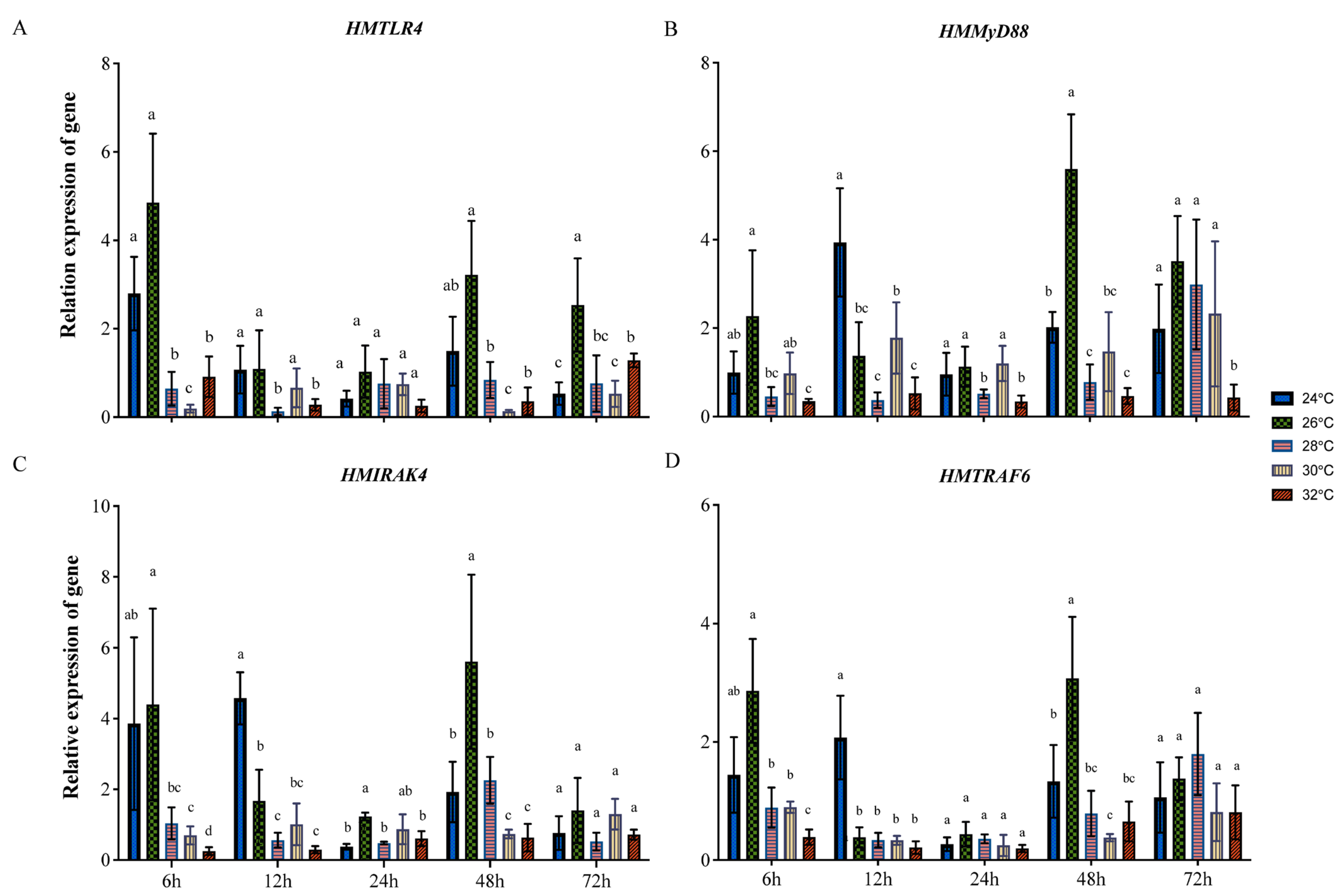
3.3. Expression of HMTLR4, HMMyD88, HMIRAK4 and HMTRAF6 at Different Temperatures

Besides the three genes above, the expression patterns of another gene in the TLR4-MyD88 pathway, interleukin receptor associated kinase 4 (HMIRAK4), were also investigated. For a clear understanding about the tissue specificity of gene expression, the expression of four genes in the TLR4-MyD88 pathway were detected in the gill, adductor muscle, mantle and hemolymph of scallops sampled from the control group (24 °C) at 0 h. The relative expression of HMTLR4 in the mantle was the highest among the four kinds of tissues, and the expression of HMTLR4 in the gill was also significantly higher than that in the adductor muscle and hemolymph (Figure S4A). As for HMMyD88, HMIRAK4 and HMTRAF6, they showed similar expression patterns, and their transcripts in gill tissue were the most abundant (Figure S4B–D).
The expression patterns of the above four genes under heat stress were primarily investigated in scallop gill tissues for their key roles in responses to environmental changes. At 6 h after transfer, HMTLR4 expression was higher in the gills of scallops transferred to 26 °C FSW but decreased significantly in those transferred to 28 °C, 30 °C and 32 °C FSW (Figure 2A). At 12 h, HMTLR4 expression exhibited no significant difference in samples from 24 °C, 26 °C and 30 °C groups and a relative low level in those from 28 °C and 32 °C groups (Figure 2A). There was no significant difference in HMTLR4 expression between all groups at 24 h. The expression patterns of HMTLR4 at 48 h were similar to those at 6 h, except for an insignificant decrease in those exposed to 28 °C FSW (Figure 2A). At 72 h, HMTLR4 expression in samples exposed to 26 °C FSW remained at a relatively high level, but, interestingly, HMTLR4 expression in 32 °C groups increased slightly when compared to those in 24 °C groups (Figure 2A). As for the expression of HMMyD88 at 6 h, it was similar to the expression pattern of HMTLR4, except for an insignificant decrease in samples exposed to 28 °C and 30 °C (Figure 2B). The expression of HMMyD88 in gills sampled from the control group was significantly higher than in others at 12 h, and there was no significant difference in HMMyD88 expression between all groups at 24 h (Figure 2B). At 48 h and 72 h, HMMyD88 expression in samples exposed to 26 °C FSW was relatively high, and there was no significant difference between HMMyD88 expression in gill samples from 24 °C and 28 °C groups (Figure 2B). It should be noted that HMMyD88 expression in scallops exposed to 32 °C remained at the lowest level within 72 h. HMIRAK4 and HMTRAF6 showed similar expression patterns in scallops under heat stress. As shown in Figure 2C,D, their expression in scallops exposed to 26 °C FSW was significantly higher than that of others at 6 h after transfer, but, at 12 h, gene expression in samples from the control group was at the highest level. At 48 h, the expression patterns of HMIRAK4 and HMTRAF6 were similar to that of HMTLR4 (Figure 2C,D). There was no significant difference in gene expression between all groups at 72 h (Figure 2C,D).

3.4. The Expression of Genes in the TLR4-MyD88 Pathway after LPS Injection at 24 °C and 32 °C

The response of four TLR4 pathway genes to LPS stimulation was investigated at different temperatures. At 24 °C, the expression of HMTLR4 increased significantly 3 h after injection with LPS and reached the highest level at 6 h (Figure 3A). Then, the expression of HMTLR4 in scallops injected with LPS decreased to the same level as those injected with PBS at 24 h (Figure 3A). When transferred to 32 °C FSW after injection, the expression of HMTLR4 showed no significant difference between samples injected with LPS and those injected with PBS (Figure 3B). HMMyD88 presented a different expression pattern than that of HMTLR4. At 24 °C, a significant increase in HMMyD88 could also be found 3 h after injection with LPS, and the relatively higher level under LPS stimulation persisted until 24 h (Figure 3C). When transferred to 32 °C FSW after injection, the expression of HMMyD88 in samples injected with LPS was relatively higher than those injected with PBS within 6 h, but a smaller difference was shown when compared with those at 24 °C (Figure 3D). HMIRAK4 and HMTRAF6 showed similar expression patterns in response to LPS stimulation. Their expression increased significantly 6 h after injection with LPS, whether at 24 °C or at 32 °C, and the time was shorter in maintaining a higher level of gene expression at 32 °C than that at 24 °C (Figure 3E–H).

3.5. Levels of TNF-α and LZM under Different Stress Conditions

Levels of TNF-α and LZM were preliminarily investigated in the gill tissue of scallops under heat stress, and partial samples exposed to 32 °C FSW were selected for comparison with those in 24 °C FSW. As shown in Figure S5A, the concentrations of TNF-α were not influenced by the increase in the environmental temperature at 6 h and 48 h, although the three TLR4 pathway-related genes in this study showed significant changes in abundance. Whereas, the activity of LZM increased significantly at 48 h after being transferred to 32 °C FSW (Figure S5). Levels of TNF-α and LZM were also detected in the gill tissue of scallops injected with LPS, and they increased significantly at 6 h and 48 h when compared with those injected with PBS, whether at 24 °C or at 32 °C (Table 2). In addition, no obvious impact of heat stress could be observed because both TNF-α and LZM showed similar levels between samples injected with LPS and PBS at different temperatures (Table 2).

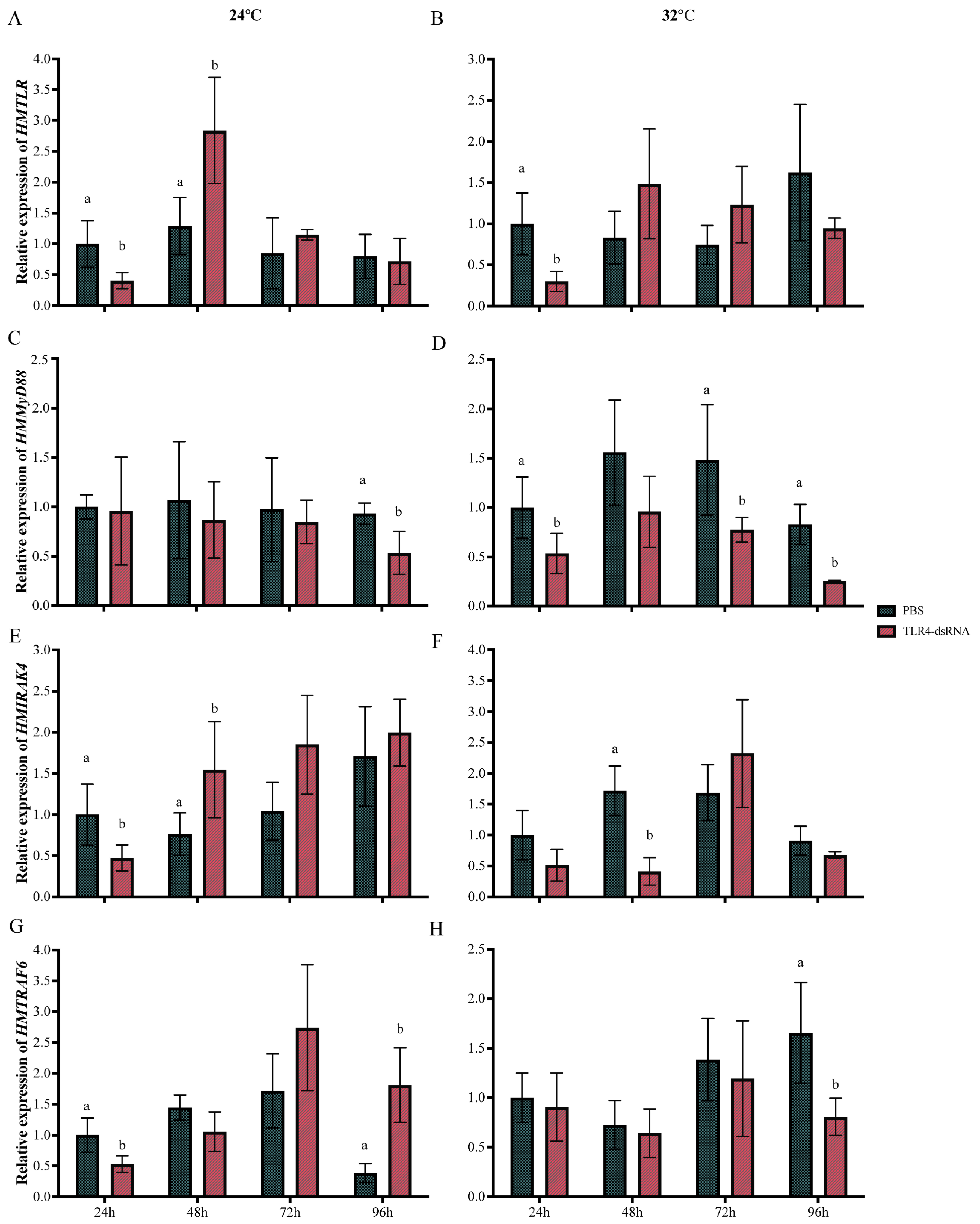
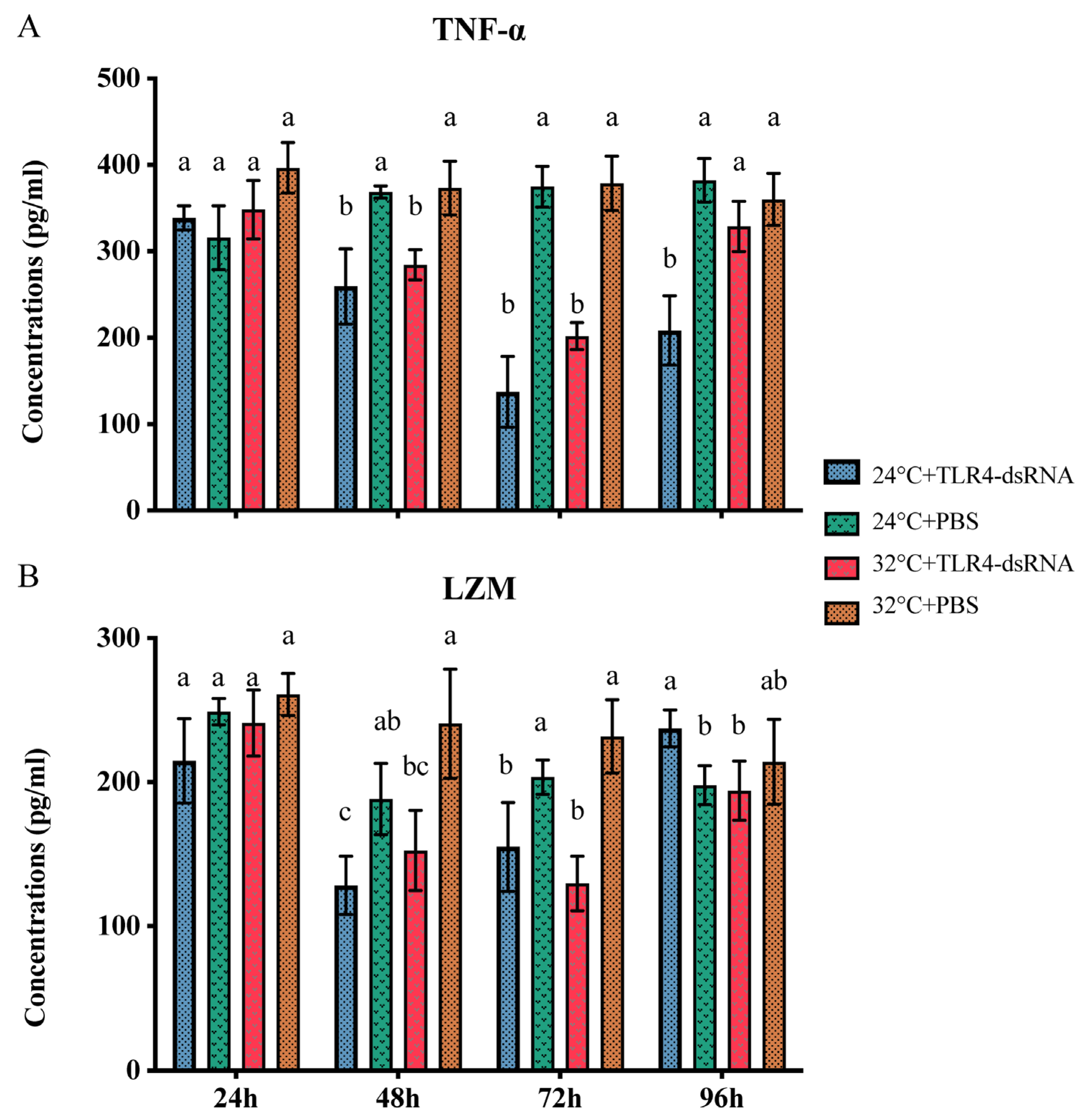
3.6. The Effect of HMTLR4 Suppression on the Expression of Downstream Genes and the Levels of TNF-α and LZM

The expression of HMTLR4 was primarily investigated in the gills of scallops injected with PBS and dsRNA at 24 °C. As shown in Figure 4A, the expression of HMTLR4 in gills decreased significantly at 24 h but increased at 48 h after injection with dsRNA. Then, it was reduced to the same level as the expression of HMTLR4 in samples injected with PBS (Figure 4A). Similar suppression of HMTLR4 could be observed in scallops injected with dsRNA that underwent immediate transfer to 32 °C FSW, except for an insignificant difference at 48 h and a relatively higher level in the control group at 96 h (Figure 4B).
Then, we tested the effect of HMTLR4 suppression on the expression of its downstream genes, HMMyD88, HMIRAK4 and HMTRAF6. At 24 °C, HMMyD88 did not show significant differences in transcript abundance until 96 h after injection with dsRNA targeting HMTLR4 (Figure 4C). By contrast, when scallops were injected with dsRNA targeting HMTLR4 at 32 °C, the expression of HMMyD88 was significantly lower than that in the control group, except at 48 h after injection (Figure 4D). When HMTLR4 was silenced at 24 °C, HMIRAK4 showed similar expression trends within 96 h (Figure 4E). But, at 32 °C, a significant decrease appeared later at 48 h after injection (Figure 4F). As for the expression of HMTRAF6, it decreased immediately at 24 h but showed a higher level than that in the control group at 72 h after injection with dsRNA at 24 °C (Figure 4G). At 32 °C, there was no significant difference in the expression of HMTRAF6 between the PBS and dsRNA treatment groups within 72 h, and a lower expression level of HMTRAF6 in scallops injected with dsRNA appeared at 96 h (Figure 4H).
Scallops injected with dsRNA and PBS were selected, and the levels of TNF-α and LZM in gill tissues were determined by ELISA assay. At 48 h, TNF-α presented significantly lower concentrations in scallops injected with dsRNA when compared with those injected with PBS at 24 °C (Figure 5A). At 32 °C, a similar decrease in TNF-α was found in scallops injected with dsRNA, but the level was restored at 96 h (Figure 5A). LZM showed similar concentration variation trends when scallops were injected with dsRNA targeting HMTLR4, whether at 24 °C or at 32 °C (Figure 5B). Interestingly, the LZM of samples injected with dsRNA showed relatively higher levels than those of samples injected with PBS at 96 h after being transferred to 32 °C FSW (Figure 5B).

4. Discussion

Acute heat stress poses significant challenges to the immune system of marine bivalves, which are particularly vulnerable to extremely high temperatures. Recent advancements in molecular biotechnologies, such as transcriptome sequencing, have highlighted the importance of signal transduction pathways in understanding this process. In this study, we identified transcripts of three crucial genes involved in the TLR4-MyD88 signaling pathway in a novel scallop strain “Hongmo No. 1”. Our aim was to investigate the role of this specific signal transduction pathway in the immune response of scallops to acute heat stress.
Three genes, HMTLR4, HMMyD88 and HMTRAF6, in the TLR4-MyD88 signaling pathway were identified in “Hongmo No. 1” and predicted to encode proteins with typical function domains. Seven LRR domains and one TIR domain, responsible for ligand recognition and the recruitment of signaling adapters, respectively [12,28], were presented in the deduced amino acids of HMTLR4. There was obvious variation in the number of LRR domains and TLR4 proteins among species, but, generally, more LRRs were present in the TLR4 proteins of invertebrates [29]. This supported the idea that TLR genes in both vertebrates and invertebrates are the result of independent gene family expansion and loss events. Such evolution of TLRs may be attributed to a “response to a changing array of binding requirements” [18,30,31,32]. MyD88 was the critical downstream adapter on the MyD88-dependent signaling pathway that interacted with TLR4 through the TIR domain [15,33]. In addition to the TIR domain, MyD88 also contains a death domain by which it recruits and activates a death domain-containing kinase, IRAK4 [14]. We found high conservation in the putative protein of HMMyD88 with typical TIR and death domains but failed to obtain the transcript of its downstream IRAK4 coding gene. Fortunately, a homolog of the adaptor protein TRAF6 coding gene, critical for IRAK4, was identified in “Hongmo No. 1” scallops, and showed high similarity in deduced amino acids with homologs in other species. In our opinion, the identification of HMTLR4, HMMyD88 and HMTRAF6 supported the view that bivalves have a TLR- and MyD88-based immune signaling pathway [19,20,21,22,34].
Then, the expression patterns of four genes in the TLR4-MyD88 signaling pathway, including HMIRAK4, were investigated in “Hongmo No. 1” scallops at different temperatures for a global view of their transcriptional response to heat stress. Generally, these genes were downregulated when scallops were exposed to extremely high temperatures, especially those over 30 °C. This is different from the heat stress-mediated activation of the TLR signaling pathway in most homeothermic organisms, such as Bama miniature pigs [35], which is supposed to trigger the release of cytokines and coordinate a proinflammatory response [36,37,38]. But, similar suppression of the TLR4-MyD88 pathway can also be found in both homeothermic and poikilothermal animals under acute heat stress. For example, the TLR4 coding gene was significantly downregulated in tissues of Indian major carp catla Catla catla at temperatures over 30 °C [39]. These results suggest an immediate and direct effect of heat on TLR pathway activation; however, conclusive results and the significance of these findings in the pathogenesis of heat-stressed animals have not been clearly defined [38]. In addition, there was no significant difference in the level of TNF-α between “Hongmo No. 1” scallops at 24 °C and 32 °C FSW within 48 h, although genes in the TLR4-MyD88 pathway were significantly downregulated at 32 °C. Thus, this requires more evidence to confirm whether the TLR4-MyD88 signaling pathway affects innate immunity in scallops.
The increase in immune parameters at 24 °C was preceded by an increase in gene expression, which may indicate a relationship. This was consistent with the role of TLR4 as an important sensor for LPS to induce the release of critical proinflammatory cytokines that are necessary to activate potent immune responses [11,40]. Under acute heat stress, the LPS-induced TLR4-MyD88 signaling pathway in scallops was obviously suppressed, which was revealed by the smaller increase in gene expression. This supported the negative effect of heat on TLR pathway activation, as shown in the initial heat stress treatment experiment, where the decline was visible at 28 °C. A previous in vitro study on innate memory CD8+ T cells of mice indicated that heat shock protein (HSP) 70 could down-regulate TLR4 [41], and this provided a clue to the activation mechanism of the TLR4-MyD88 signaling pathway in scallops under heat stress. The TLR4 genes in invertebrates are quite different from those in mammals, and it remains to be further investigated whether the activation of TLR4-MyD88 under heat stress is relatively conserved between vertebrates and invertebrates. Moreover, TNF-α and LZM were not significantly affected by heat stress and showed an obvious increase when injected with LPS, both at 24 °C and at 32 °C. This may indicate that adequate signal molecules were transduced for the activation of innate immunity in scallops, although the TLR4-MyD88 signaling pathway was negatively affected.
Subsequent RNAi targeting HMTLR4 provided more information on the TLR4-MyD88 signaling pathway in the immune response of “Hongmo No. 1” scallops. HMTLR4 silencing, to a greater or lesser extent, suppressed the expression of HMMyD88, HMIRAK4 and HMTRAF6, whether at 24 °C or at 32 °C. Previous studies on several bivalves, such as Chlamys farreri and C. hongkongensis, also indicated that the inhibition of the TLR gene induced a decrease in its downstream mediator molecules [21,42], demonstrating the key role of TLR in the activation of TLR- and MyD88-based signaling pathways in bivalves [43]. TNF-α and LZM showed lower levels at different time points after injection with dsRNA targeting HMTLR4, which indicated that the block of TLR4-MyD88 signal transduction negatively affected the innate immunity of scallops. According to the gene expression at 32 °C, acute heat stress seemed to weaken or strengthen the response of downstream mediator molecules to the inhibition of HMTLR4. This led us to a hypothesis that heat stress could directly influence the downstream targets of HMTLR4, but more convincing evidence needs to be provided. Investigating the potential involvement of heat shock proteins, such as HSP 70, in downstream signaling events could be a promising area for further study on the TLR4-MyD88 pathway of bivalves. Such research could deepen our understanding of the complex molecular mechanisms underlying the immune response to acute heat stress in marine bivalves and enhance our ability to develop effective strategies for mitigating the impacts of extreme temperature events on these aquaculture organisms.

5. Conclusions

In this study, HMTLR4, HMMyD88 and HMTRAF6 were characterized from a new scallop strain, “Hongmo No. 1” (Bohai Red ♂ × A. irradians concentricus ♀); then, the expression of four genes in the TLR4-MyD88 signaling pathway, including HMIRAK4, was investigated in scallops under heat stress and/or LPS stimulation. Acute heat stress obviously inhibited the TLR4-MyD88 signaling pathway, even with LPS stimulation, but innate immunity in scallops was activated successfully according to the levels of proinflammatory cytokines and immune enzymes. RNAi targeting HMTLR4 at different temperatures indicated the key role of TLR4-MyD88 signal transduction in the immune response of “Hongmo No. 1” scallops. Acute heat stress seemed to affect the response of downstream mediator molecules to the inhibition of HMTLR4. These results provide more information about bivalve immunity under heat stress and can be beneficial for the genetic improvement of heat tolerance to cope with more frequent and extreme heat wave events.

Supplementary Materials

The following supporting information can be downloaded at https://www.mdpi.com/article/10.3390/ani14030497/s1: Figure S1: The predicted TLR4 amino acid sequences of several bivalves; Figure S2: The predicted MyD88 amino acid sequences of several bivalves; Figure S3: The predicted TRAF6 amino acid sequences of several bivalves; Figure S4: Relative expression of HMMyD88, HMIRAK4 and HMTRAF6 in different tissues of scallops in the control group; Figure S5: Levels of TNF-α and LZM in gill tissue of scallops at 24 °C and 32 °C.

Author Contributions

Conceptualization, C.Y. and Z.L.; data curation, C.Y., K.Z. and X.S.; funding acquisition, Z.L.; investigation, K.Z. and X.S.; methodology, W.L., H.G., L.Z. and J.K.-H.F.; supervision, Z.L.; writing—original draft, C.Y.; writing—review and editing, W.L., H.G., L.Z. and J.K.-H.F. All authors have read and agreed to the published version of the manuscript.

Funding

This study was supported by grants from the International Science and Technology Cooperation Project of Guangdong Province in 2020 (2020A0505100057), GuangDong Basic and Applied Basic Research Foundation (2022A1515110002), the Project of 2019 Annual Guangdong Province Key Field R&D Program (2019B020238002), South China Sea Economic Animal Germplasm Innovation and Utilization Innovation Team (2021KCXTD026), the Guangdong Province Shellfish Industry Technology System Innovation Team Project in 2019 (2021KJ146), the 2020 Provincial Rural Revitalization Strategy Project Funds (Development of Fishery Industry) (Guangdong Cainong (2020) 4), the Department of Education of Guangdong Province (2021KCXTD026), and the program for scientific research start-up funds of Guangdong Ocean University (060302022105).

Institutional Review Board Statement

Ethical review and approval were waived for this study due to the absence of ethical approval files for bivalves. Animal Ethics Committee in Guangdong Ocean University can only provide ethical approval files for research on vertebrates, not invertebrates. Although ethical and regulatory oversight of research animals is focused on vertebrates and rarely includes low invertebrates, we guarantee that we comply with GDOU guidelines for animal experimentation.

Informed Consent Statement

Consent was obtained from the scallop farmer.

Data Availability Statement

The data presented in this study are available upon request from the corresponding author. The data are not publicly available due to the requirement of Guangdong Ocean University.

Conflicts of Interest

The authors declare no conflict of interest.

References

  1. Perkins-Kirkpatrick, S.E.; Stone, D.A.; Mitchell, D.M.; Rosier, S.; King, A.D.; Lo, Y.T.E.; Pastor-Paz, J.; Frame, D.; Wehner, M. On the attribution of the impacts of extreme weather events to anthropogenic climate change. Environ. Res. Lett. 2022, 17, 024009. [Google Scholar] [CrossRef]
  2. Islam, M.J.; Kunzmann, A.; Slater, M.J. Responses of aquaculture fish to climate change-induced extreme temperatures: A review. J. World Aquac. Soc. 2022, 53, 314–366. [Google Scholar] [CrossRef]
  3. Dégremont, L.; Bédier, E.; Soletchnik, P.; Ropert, M.; Huvet, A.; Moal, J.; Samain, J.-F.; Boudry, P. Relative importance of family, site, and field placement timing on survival, growth, and yield of hatchery-produced Pacific oyster spat (Crassostrea gigas). Aquaculture 2005, 249, 213–229. [Google Scholar] [CrossRef]
  4. Liu, S.; Jiang, X.; Hu, X.; Gong, J.; Hwang, H.; Mai, K. Effects of temperature on non-specific immune parameters in two scallop species: Argopecten irradians (Lamarck 1819) and Chlamys farreri (Jones & Preston 1904). Aquac. Res. 2004, 35, 678–682. [Google Scholar] [CrossRef]
  5. Ferreira-Rodríguez, N.; Fernández, I.; Cancela, M.L.; Pardo, I. Multibiomarker response shows how native and non-native freshwater bivalves differentially cope with heat-wave events. Aquat. Conserv. 2018, 28, 934–943. [Google Scholar] [CrossRef]
  6. Wang, C.; Liu, B.; Li, J.; Liu, S.; Li, J.; Hu, L.; Fan, X.; Du, H.; Fang, H. Introduction of the Peruvian scallop and its hybridization with the bay scallop in China. Aquaculture 2011, 310, 380–387. [Google Scholar] [CrossRef]
  7. Wang, C.; Liu, B.; Liu, X.; Ma, B.; Zhao, Y.; Zhao, X.; Liu, F.; Liu, G. Selection of a new scallop strain, the Bohai Red, from the hybrid between the bay scallop and the Peruvian scallop. Aquaculture 2017, 479, 250–255. [Google Scholar] [CrossRef]
  8. Zhang, Y.; Liu, Z.; Wang, C.; Yao, G.; Zhang, K.; Zhan, J.; Lu, W.; Zhong, M.; Liufu, S.; Fang, J. Growth performance and model fitting of the selected strain of scallop “Hongmo No. 1” cultivated during different seasons. Aquaculture 2023, 578, 740104. [Google Scholar] [CrossRef]
  9. Janeway, C.A., Jr.; Medzhitov, R. Innate immune recognition. Annu. Rev. Immunol. 2002, 20, 197–216. [Google Scholar] [CrossRef]
  10. Medzhitov, R.; Janeway, C.A. Innate immunity: The virtues of a nonclonal system of recognition. Cell 1997, 91, 295–298. [Google Scholar] [CrossRef]
  11. Hoshino, K.; Takeuchi, O.; Kawai, T.; Sanjo, H.; Ogawa, T.; Takeda, Y.; Takeda, K.; Akira, S. Cutting edge: Toll-like receptor 4 (TLR4)-deficient mice are hyporesponsive to lipopolysaccharide: Evidence for TLR4 as the Lps gene product. J. Immunol. 1999, 162, 3749–3752. [Google Scholar] [CrossRef] [PubMed]
  12. Akira, S.; Takeda, K. Toll-like receptor signaling. Nat. Rev. Immunol. 2004, 4, 499–511. [Google Scholar] [CrossRef] [PubMed]
  13. Saco, A.; Novoa, B.; Greco, S.; Gerdol, M.; Figueras, A. Bivalves present the largest and most diversified repertoire of Toll-like receptors in the animal kingdom, suggesting broad-spectrum pathogen recognition in marine waters. Mol. Biol. Evol. 2023, 40, msad133. [Google Scholar] [CrossRef] [PubMed]
  14. Chow, J.C.; Young, D.W.; Golenbock, D.T.; Christ, W.J.; Gusovsky, F. Toll-like receptor-4 mediates lipopolysaccharide- induced signal transduction. J. Biol. Chem. 1999, 274, 10689–10692. [Google Scholar] [CrossRef] [PubMed]
  15. Medzhitov, R.; Preston-Hurlburt, P.; Kopp, E.; Stadlen, A.; Chen, C.; Ghosh, S.; Janeway, C.A., Jr. MyD88 is an adaptor protein in the hToll/IL-1 receptor family signaling pathways. Mol. Cell 1998, 2, 253–258. [Google Scholar] [CrossRef] [PubMed]
  16. Muzio, M.; Natoli, G.; Saccani, S.; Levrero, M.; Mantovani, A. The human Toll signaling pathway: Divergence of nuclear factor κB and JNK/SAPK activation upstream of tumor necrosis factor receptor–associated factor 6 (TRAF6). J. Exp. Med. 1998, 187, 2097–2101. [Google Scholar] [CrossRef]
  17. Horng, T.; Barton, G.; Medzhitov, R. TIRAP: An adapter molecule in the Toll signaling pathway. Nat. Immunol. 2001, 2, 835–841. [Google Scholar] [CrossRef] [PubMed]
  18. Gerdol, M.; Venier, P.; Edomi, P. Diversity and evolution of TIR-domain-containing proteins in bivalves and Metazoa: New insights from comparative genomics. Dev. Comp. Immunol. 2017, 70, 145–164. [Google Scholar] [CrossRef]
  19. Xing, Q.; Liao, H.; Xun, X.; Wang, J.; Zhang, Z.; Yang, Z.; Huang, X.; Bao, Z. Genome-wide identification, characterization and expression analyses of TLRs in Yesso scallop (Patinopecten yessoensis) provide insight into the disparity of responses to acidifying exposure in bivalves. Fish Shellfish Immun. 2017, 68, 280–288. [Google Scholar] [CrossRef]
  20. Chen, J.; Lin, J.; Yu, F.; Zhong, Z.; Liang, Q.; Pang, H.; Wu, S. Transcriptome analysis reveals the function of TLR4-MyD88 pathway in immune response of Crassostrea hongkongensis against Vibrio Parahemolyticus. Aquacult. Rep. 2022, 25, 101253. [Google Scholar] [CrossRef]
  21. Yu, F.; Chen, J.; Lin, J.; Zhong, Z.; Lu, Y.; Zeng, X.; Lei, X. TLR4 involved in immune response against Vibrio Parahaemolyticus by MyD88-dependent pathway in Crassostrea hongkongensis. Fish Shellfish Immun. 2023, 134, 108591. [Google Scholar] [CrossRef]
  22. Lu, J.; Zhang, M.; Liang, H.; Shen, C.; Zhang, B.; Liang, B. Comparative proteomics and transcriptomics illustrate the allograft-induced stress response in the pearl oyster (Pinctada fucata martensii). Fish Shellfish Immun. 2022, 121, 74–85. [Google Scholar] [CrossRef] [PubMed]
  23. Felsenstein, J. Confidence limits on phylogenies: An approach using the bootstrap. Evolution 1985, 39, 783–791. [Google Scholar] [CrossRef] [PubMed]
  24. Jones, D.T.; Taylor, W.R.; Thornton, J.M. The rapid generation of mutation data matrices from protein sequences. Bioinformatics 1992, 8, 275–282. [Google Scholar] [CrossRef]
  25. Kumar, S.; Stecher, G.; Tamura, K. MEGA7: Molecular Evolutionary Genetics Analysis Version 7.0 for Bigger Datasets. Mol. Biol. Evol. 2016, 33, 1870–1874. [Google Scholar] [CrossRef] [PubMed]
  26. Yue, C.; Li, Q.; Yu, H. Integrated analysis of miRNA and mRNA expression profiles identifies potential regulatory interactions during sexual development of Pacific oyster Crassostrea gigas. Aquaculture 2022, 546, 737294. [Google Scholar] [CrossRef]
  27. Schmittgen, T.D.; Livak, K.J. Analyzing real-time PCR data by the comparative CT method. Nat. Protoc. 2008, 3, 1101–1108. [Google Scholar] [CrossRef] [PubMed]
  28. Matsushima, N.; Tanaka, T.; Enkhbayar, P.; Mikami, T.; Taga, M.; Yamada, K.; Kuroki, Y. Comparative sequence analysis of leucine-rich repeats (LRRs) within vertebrate toll-like receptors. BMC Genom. 2007, 8, 124. [Google Scholar] [CrossRef]
  29. Matsushima, N.; Miyashita, H.; Enkhbayar, P.; Kretsinger, R.H. Comparative geometrical analysis of leucine-rich repeat structures in the nod-like and toll-like receptors in vertebrate innate immunity. Biomolecules 2015, 5, 1955–1978. [Google Scholar] [CrossRef]
  30. Hughes, A.L.; Piontkivska, H. Functional diversification of the toll-like receptor gene family. Immunogenetics 2008, 60, 249–256. [Google Scholar] [CrossRef]
  31. Zhang, Q.; Zmasek, C.M.; Godzik, A. Domain architecture evolution of pattern recognition receptors. Immunogenetics 2010, 62, 263–272. [Google Scholar] [CrossRef]
  32. Buckley, K.M.; Rast, J.P. Dynamic evolution of toll-like receptor multigene families in echinoderms. Front. Immunol. 2012, 3, 136. [Google Scholar] [CrossRef]
  33. Wesche, H.; Henzel, W.J.; Shillinglaw, W.; Li, S.; Cao, Z. MyD88: An adapter that recruits IRAK to the IL-1 receptor complex. Immunity 1997, 7, 837–847. [Google Scholar] [CrossRef]
  34. Zhang, Y.; He, X.; Yu, F.; Xiang, Z.; Li, J.; Thorpe, K.L.; Yu, Z. Characteristic and functional analysis of toll-like receptors (TLRs) in the lophotrocozoan, Crassostrea gigas, reveals ancient origin of TLR-mediated innate immunity. PLoS ONE 2013, 8, e76464. [Google Scholar] [CrossRef]
  35. Ju, X.-H.; Xu, H.-J.; Yong, Y.-H.; An, L.-L.; Jiao, P.-R.; Liao, M. Heat stress upregulation of toll-like receptors 2/4 and acute inflammatory cytokines in peripheral blood mononuclear cell (PBMC) of Bama miniature pigs: An in vivo and in vitro study. Animal 2014, 8, 1462–1468. [Google Scholar] [CrossRef] [PubMed]
  36. Basu, S.; Binder, R.J.; Suto, R.; Anderson, K.M.; Srivastava, P.K. Necrotic but not apoptotic cell death releases heat shock proteins, which deliver a partial maturation signal to dendritic cells and activate the NF-κB pathway. Int. Immunol. 2000, 12, 1539–1546. [Google Scholar] [CrossRef] [PubMed]
  37. Martine, P.; Rébé, C. Heat Shock Proteins and Inflammasomes. Int. J. Mol. Sci. 2019, 20, 4508. [Google Scholar] [CrossRef] [PubMed]
  38. Cantet, J.M.; Yu, Z.; Ríus, A.G. Heat Stress-Mediated Activation of Immune–Inflammatory Pathways. Antibiotics 2021, 10, 1285. [Google Scholar] [CrossRef] [PubMed]
  39. Basu, M.; Paichha, M.; Swain, B.; Lenka, S.S.; Singh, S.; Chakrabarti, R.; Samanta, M. Modulation of TLR2, TLR4, TLR5, NOD1 and NOD2 receptor gene expressions and their downstream signaling molecules following thermal stress in the Indian major carp catla (Catla catla). Biotech 2015, 5, 1021–1030. [Google Scholar] [CrossRef] [PubMed]
  40. Ciesielska, A.; Matyjek, M.; Kwiatkowska, K. TLR4 and CD14 trafficking and its influence on LPS-induced pro-inflammatory signaling. Cell. Mol. Life Sci. 2021, 78, 1233–1261. [Google Scholar] [CrossRef] [PubMed]
  41. Ghosh, A.K.; Sinha, D.; Mukherjee, S.; Biswas, R.; Biswas, T. LPS stimulates and Hsp70 down-regulates TLR4 to orchestrate differential cytokine response of culture-differentiated innate memory CD8+ T cells. Cytokine 2015, 73, 44–52. [Google Scholar] [CrossRef] [PubMed]
  42. Wang, M.; Yang, J.; Zhou, Z. A primitive Toll-like receptor signaling pathway in mollusk Zhikong scallop Chlamys farreri. Dev. Comp. Immunol. 2011, 35, 511–520. [Google Scholar] [CrossRef] [PubMed]
  43. Parisi, M.; Baranzini, N.; Dara, M.; La Corte, C.; Vizioli, J.; Cammarata, M. AIF-1 and RNASET2 are involved in the inflammatory response in the Mediterranean mussel Mytilus galloprovincialis following Vibrio infection. Fish Shellfish Immunol. 2022, 127, 109–118. [Google Scholar] [CrossRef] [PubMed]
Figure 1. HMTLR4, HMMyD88 and HMTRAF6 genes in scallop “Hongmo No. 1” and phylogenetic trees of their putative proteins. (AC) Schematic presentation of HMTLR4, HMMyD88 and HMTRAF6, respectively. Sky-blue and white boxes show transcript UTRs and their ORF. Pink box show putative proteins. In predicted amino acid sequences, conserved domains were labeled with texts and different colors. (DF) Phylogenetic trees based on the predicted amino acid sequences of HMTLR4, HMMyD88 and HMTRAF6, respectively. Species names are abbreviated as BT for Bos taurus, SS for Sus scrofa, HS for Homo sapiens, OC for Oryctolagus cuniculus, MM for Mus musculus, RN for Rattus norvegicus, SC for Serinus canaria, VC for Vidua chalybeata, PF for Patagioenas fasciata monilis, GG for Gallus gallus, PC for Phasianus colchicus, FA for Ficedula albicollis, OT for Onychostruthus taczanowskii, AC for Acanthisitta chloris, AP for Anas platyrhynchos, EG for Egretta garzetta, CC for Cyanistes caeruleus, TG for Taeniopygia guttata, IP for Ictalurus punctatus, TF for Tachysurus fulvidraco, DR for Danio rerio, CC for Cyprinus carpio, MA for Megalobrama amblycephala, SR for Sinocyclocheilus rhinocerous, LC for Larimichthys crocea, ON for Oreochromis niloticus, CA for Carassius auratus, OT for Oncorhynchus tshawytscha, PF for Pinctada fucata, AI for Argopecten irradians, AB for Anadara broughtonii, HD for Haliotis diversicolor, CS for Cyclina sinensis, HR for Haliotis rufescens, OE for Ostrea edulis, MY for Mizuhopecten yessoensis, PM for Pecten maximus, CG for Crassostrea gigas, MC for Mytilus californianus, MG for Mytilus galloprovincialis, AF for Azumapecten farreri and PI for Pinctada imbricata.
Animals 14 00497 g001
Figure 2. Expression profiles of HMTLR4 (A), HMMyD88 (B), HMIRAK4 (C) and HMTRAF6 (D) in gills of scallops at different temperatures. Bars represent standard deviation. For statistical analysis, a separate one-factor analysis of variance (ANOVA) was performed at each time point and levels were accepted as significant if p value < 0.05. The different letters indicate significant differences in gene expression between groups at the same time point.
Animals 14 00497 g002
Figure 3. Expression profiles of HMTLR4, HMMyD88, HMIRAK4 and HMTRAF6 in gills of scallops under LPS stimulation at 24 °C and 32 °C. (A,B) Expression profiles of HMTLR4 at 24 °C and 32 °C. (C,D) Expression profiles of HMMyD88 at 24 °C and 32 °C. (E,F) Expression profiles of HMIRAK4 at 24 °C and 32 °C. (G,H) Expression profiles of HMTRAF6 at 24 °C and 32 °C. Bars represent standard deviation. For statistical analysis, a separate one-factor analysis of variance (ANOVA) was performed at each time point and levels were accepted as significant if p value < 0.05. The different letters indicate significant differences in gene expression between groups at the same time point.
Animals 14 00497 g003
Figure 4. Expression profiles of HMTLR4 (A,B),HMMyD88 (C,D), HMIRAK4 (E,F) and HMTRAF6 (G,H) in gills of scallops injected with dsRNA at 24 °C and 32 °C. Bars represent standard deviations. Statistical analyses were carried out by one-way ANOVA and levels were accepted as significant if p value < 0.05. The different letters indicate significant differences in gene expression between groups at the same time points.
Animals 14 00497 g004
Figure 5. Levels of TNF-α (A) and LZM (B) in gills of scallops injected with dsRNA targeting HMTLR4 at 24 °C and 32 °C. Bars represent standard deviations. Statistical analyses were carried out by one-way ANOVA and levels were accepted as significant if p value < 0.05. The different letters indicate significant differences between groups at the same time points.
Animals 14 00497 g005
Table 1. Primers used in this study.
PrimerSequence (5′–3′)Application
HMTLR4-5-1TGGAGACAGAGCCGTAACCCACG5′RACE
HMTLR4-5-2GGTGGAGACAGAGCCGTAACCCA5′RACE
HMMyD88-5-1AATTGTGGACACTATAGTTATGCTTGAC5′RACE
HMMyD88-5-2CATCACCGCCTTTAACCACAAT5′RACE
HMTRAF6-5-1GCCATTGCCTGTCTTTCAACCGT5′RACE
HMTLR4-3-1TAGATTTGTCGGGTGAAGTCGGTAGC3′RACE
HMTLR4-3-2TTCACGCTGGGAAGGTATCTGCT3′RACE
HMMyD88-3-1TTACAGGGCAAGTTGGGGTGT3′RACE
HMMyD88-3-2AAATTATGATGATTTCGCAGCTATG3′RACE
HMTRAF6-3-1GAAGGCTGTGATACGCAGGTGGT3′RACE
HMTLR4-FCCTTACCTGTATTTCCCCCCRT-PCR
HMTLR4-RGGTTTCTGACTGGACCCTTCRT-PCR
HMMyD88-FTTTTAGGAACAGGCTTTART-PCR
HMMyD88-RTGTGGACACGTTGATAATRT-PCR
HMIRAK4-FTGTCGTACTGCTGCCTGATRT-PCR
HMIRAK4-RTGGAAGCGTAGACTGGAGART-PCR
HMTRAF6-FTCAGGTGAAGCGATTGGGRT-PCR
HMTRAF6-RTCCTGGTGGAAAGGTAATRT-PCR
β-actin-FCCGTGACTTGACCGATTACCRT-PCR
β-actin-RCCTTGATGTCCCTGACGATTRT-PCR
HMTLR4-senseGGTCAGTGTTTCGATTGGCGDsRNA synthesis
HMTLR4-antiCCCAAGTTGTTGTTGCTGATGADsRNA synthesis
Table 2. Levels of TNF-α and LZM in scallops injected with LPS and PBS.
Time (h) Concentrations (pg/mL)
24 °C + LPS24 °C + PBS32 °C + LPS32 °C + PBS
TNF-α6720.17 ± 95.03 *368.70 ± 7.11791.65 ± 71.13 373.44 ± 30.98
48746.63 ± 57.24 *382.12 ± 25.34749.39 ± 58.73 360.01 ± 30.10
LZM6367.00 ± 54.31 *188.30 ± 24.78424.84 ± 27.20 240.54 ± 37.83
48387.73 ± 38.14 *458.63 ± 36.42458.63 ± 36.42 214.01 ± 9.44
Values represent the mean value with standard deviation in six different individuals. Significant differences from the value in the control group at the same time points are represented by * and at 24 °C and 32 °C, respectively (p < 0.05).
Disclaimer/Publisher’s Note: The statements, opinions and data contained in all publications are solely those of the individual author(s) and contributor(s) and not of MDPI and/or the editor(s). MDPI and/or the editor(s) disclaim responsibility for any injury to people or property resulting from any ideas, methods, instructions or products referred to in the content.

