


Simple Summary
The present study describes for the first time the differences in the serum and saliva proteomes between healthy bitches and bitches with mammary tumors using a high-throughput proteomic approach. More than 1000 proteins were identified and 35 in serum and 49 in saliva were significantly modulated. Additionally, their related pathways were discussed in order to improve understanding of the pathophysiology of the disease and one protein, serum albumin, was further validated. The results of the present study could be a source of potential biomarkers for canine mammary tumors in saliva and serum and increase the knowledge on the pathophysiology of the disease.
Abstract
The aim of this study was to evaluate changes in serum and saliva proteomes in canine mammary tumors (CMT) using a high-throughput quantitative proteomic analysis in order to potentially discover possible biomarkers of this disease. Proteomes of paired serum and saliva samples from healthy controls (HC group, n = 5) and bitches with CMT (CMT group, n = 5) were analysed using a Tandem Mass Tags-based approach. Twenty-five dogs were used to validate serum albumin as a candidate biomarker in an independent sample set. The proteomic analysis quantified 379 and 730 proteins in serum and saliva, respectively. Of those, 35 proteins in serum and 49 in saliva were differentially represented. The verification of albumin in serum was in concordance with the proteomic data, showing lower levels in CMT when compared to the HC group. Some of the modulated proteins found in the present study such as haptoglobin or S100A4 have been related to CMT or human breast cancer previously, while others such as kallikrein-1 and immunoglobulin gamma-heavy chains A and D are described here for the first time. Our results indicate that saliva and serum proteomes can reflect physiopathological changes that occur in CMT in dogs and can be a potential source of biomarkers of the disease.
1. Introduction
Mammary tumors are the most common cancer in intact female dogs, accounting for almost 50% of all tumors [1]. Approximately half of all canine mammary tumors (CMT) are diagnosed as malignant by histological examination, leading to death due to distant metastases in many cases [2]. Several conditions such as hormonal imbalance, viruses, obesity, diet, genetic components, and oxidative stress contribute to CMT [1,3], although the pathogenesis has not been fully elucidated to date. It is known that many biological characteristics of mammary tumors are shared between humans and dogs such as histopathologic features, biological behaviour, metastatic pattern, antigenic phenotype and expression patterns of proliferating cell nuclear antigen, cytokeratins, and calponin [4]. Therefore, dogs have been proposed as an ideal model for the investigation of human breast cancer [5]. Furthermore, since humans and dogs share the same environment and are exposed to the same cancerogenic factors, spontaneously occurring malignancies in dogs have been considered as a valuable experimental model for investigation of human neoplasias [6].
Saliva is a non-invasive biological fluid that can reflect both systemic and local health-related conditions and possesses several benefits when used for sampling instead of invasive methods such as blood sampling. The advantages of using saliva include that it is a safer method for personnel and patients, it is easier to collect, and collecting saliva reduces sampling stress as it is pain-free [7]. In humans, proteomic analyses of saliva have proved to be a useful technique for discovering disease-related biomarkers in different pathologies including cancer [8]. In dogs, gel-free proteomics approaches such as Tandem Mass Tag (TMT) have been successfully used in serum and/or saliva in different infectious diseases such as pyometra [9], parvoviral enteritis [10] and leishmaniosis [11], as well as for metabolic diseases such as obesity [12].
Changes in serum and saliva proteomes in women with breast cancer have been described. In serum, higher levels of histone deacetylase proteins were detected in women with recurrent breast cancer in comparison to non-recurrent cases [13]. In saliva, Delmonico et al. [14] identified differences in alpha-2-macroglobulin, ceruplasmin, haptoglobin, hemopexin and vitamin D-binding, among others, in women with impalpable breast cancer in comparison to healthy women; and Giri et al. [15] identified 166 proteins with different expression in saliva of women with breast cancer in comparison to healthy controls [15]. In dogs, two studies were reported in which 2-DE gel proteomic analyses of canine mammary tumor tissues were performed. In one of the studies [16], of 21 proteins differentially modulated in healthy and diseased dogs, 19 were previously reported for human breast cancer. The second study [17] compared normal canine mammary gland tissue proteomes with tissues at different stages of malignant progression, identifying 48 proteins with significant changes. However, to the best of the authors’ knowledge, there are no studies addressing changes in serum or saliva proteomes in dogs with mammary tumors.
The current study aims to explore for the first time changes in paired serum and saliva proteomes in bitches with mammary tumors compared to healthy ones by using TMT label-based gel-free proteomic approach. This study is expected to increase the knowledge on the pathogenesis of CMT and potentially discovering possible biomarkers for the detection of this disease.
2. Materials and Methods
2.1. Animals
A total of 10 client-owned bitches presenting at private veterinary clinics in Murcia, Spain, during January 2019 were involved in this study. A group of five bitches (3 mixed breeds, 1 Pinscher and 1 German shepherd; all spayed; aged 9.7 ± 3.6 years old) classified as healthy based on a complete physical examination, haematology and biochemistry were included as the control group (HC). Five bitches (4 mixed breeds and 1 Pinscher; all spayed; aged 12.0 ± 1.93 years old) diagnosed with mammary tumors (3 simple tubulopapillary carcinomas and 2 simple tubular carcinoma) were included as the mammary tumor (CMT) group. All dogs were submitted to general physical examination and general haematology and biochemistry were performed. Tumor staging was based on the ‘Tumor, Node, Metastasis classification system’ (TNM) [18], and all cases were diagnosed as grades 1 and 2 (tumor diameter ≤3 cm, and absence of regional lymph node or distant metastases). Dogs were excluded if they were spayed less than one year ago, if there was possibility of concurrent diseases based on physical examination and laboratory analyses, or if gingivitis was detected. None of the serum and saliva samples showed haemolysis. All dogs with TNM were treated by surgical removal of the neoplasm and were alive and without relapse 6 months after the surgery.
All the procedures were approved by the ethics committees of the University of Murcia and the Ministry of Agriculture, Livestock, Fishing and Aquaculture of the Region of Murcia (A13170503).
2.2. Serum and Saliva Sampling
Surplus serum remainings after routine clinical biochemical analyses were stored at −80 °C until their use for the present study. Venipuncture of the jugular or cephalic vein was performed for blood collection. Whole blood was stored in tubes containing a coagulation activator and a gel separator and kept at room temperature (25 °C) until visible clot reaction. Samples were centrifuged (3500× g, 10 min) and the supernatants were stored at −80 °C until analysis.
Saliva samples were obtained as previously reported [19]. In brief, a small sponge was placed into the mouth and, when it was thoroughly moistened, it was then removed and placed into collection devices (Salivette saliva collection tube/V-Bottom, Sarstedt, Aktiengesellschaft & Co, Nümbrecht, Germany). After the samples were centrifuged (3000× g, 10 min, 4 °C), the supernatants were stored at −80 °C until analysis [20]. At least 0.2 mL of saliva was collected from each patient.
2.3. Proteomics Study of Saliva and Serum Samples and LC–MS/MS Analysis
A total of 35 µg of protein per sample was subjected to reduction, alkylation, digestion and labelling using 6-plex Tandem Mass Tag reagents, according to manufacturer instructions (Thermo Scientific) as described previously [10,11]. In brief, after protein determination using Bradford assay [21], 35 µg of sample and internal standards (consisting of a pool of 35 µg of each sample) were reduced for 1 h with 200 mM DTT (Sigma-Aldrich), alkylated for 30 min with 375 mM iodoacetamide (Sigma-Aldrich) and precipitated with ice-cold acetone (VWR, Llinars del Vallés, Barcelona, Spain). The next day, the samples were centrifuged, the acetone was decanted and the pellets were resuspended with 50 μL of 100 mM TEAB buffer and subsequently digested with trypsin (Promega, Madison, WI, USA) overnight at 37 °C (2.5 μg of trypsin per 100 μg of protein). The TMT labelling reagents were resuspended in LC–MS-grade anhydrous acetonitrile (Thermo Scientific, Waltham, MA, USA) and mixed with each sample for 1 h at room temperature. The reaction was quenched by adding 5% hydroxylamine (Thermo Scientific) for 15 min and the samples were combined at equal amounts. Then, 6 μg of each mixed sample set was placed in a well of a microplate, vacuum-dried and stored at −80 °C before further LC–MS/MS analysis. LC−MS/MS analysis was performed using the Dionex Ultimate 3000 RSLC nano-flow system (Dionex, Camberley, UK) and an Orbitrap Q Exactive Plus mass spectrometer (Thermo Fisher Scientific), as described elsewhere [22]. The SEQUEST algorithm, Proteome Discoverer (version 2.0., Thermo Fisher Scientific, Waltham, MA, USA), was used for peptide identification and relative quantification. A NCBI database search for Canis Lupus FASTA files was performed considering two missed trypsin cleavage sites, a precursor tolerance of 10 ppm and a fragment mass tolerance of 0.02 Da. The Percolator algorithm within the Proteome Discoverer workflow was used to determine the false discovery rate (FDR) for peptide identification, which was set at 1%.
2.4. Validation of Serum Biomarkers
For validation of serum biomarkers detected by proteomic analysis, serum samples from 25 bitches that were presented to the Department and Clinic of Animal Reproduction, University of Life Sciences, Lublin, Poland, were employed—healthy controls (n = 10; aged 7.5–11) and dogs with CMT stage 1–2 tumors (n = 15; aged 8–13).
Albumin in serum was selected for validation since it is commonly performed in routine biochemistry analysis in our laboratory and, therefore, that data were already available. Serum albumin was determined using a commercially available kit (Albumin OSR 6102; Olympus Life and Material Science Europe GmbH, Irish branch, Ennis, Ireland), according to manufacturer instructions.
2.5. Statistical Analysis
All statistics were performed using R v3.2.2 [23]. First, proteins with fewer than two unique peptides and proteins with >90% missing data were removed from the analysis. Sample outliers were detected for each of the proteins using Dixon’s test from R package outliers v0.14 [24]. If a sample outlier was significant (p < 0.05), it was removed from further analysis. As the majority of the analysed proteins did not follow normal distribution, as tested by the Shapiro–Wilk test, the Wilcoxon–Mann–Whitney test was performed in order to test for differences in protein abundance between groups. The fold change between the two groups was calculated as the mean protein abundance for the CMT group divided by the mean protein abundance for the HC group.
For the validation of serum biomarkers, the D’Agostino and Pearson omnibus normality test was employed to determine the distribution of data and, since data were not normally distributed, the non-parametric statistical Mann–Whitney U (two-way) test was used to compare between groups.
2.6. Bioinformatics Analysis
The proteins’ GI accession numbers were converted into official gene symbols either using the DAVID conversion tool (https://david.ncifcrf.gov/conversion.jsp), UniProtKB ID mapping (https://www.uniprot.org/uploadlists/) or the SEQUEST search engine implemented into Proteome Discoverer [25]. Genes encoding the differentially abundant proteins in the CMT and HC groups were used to determine the GO terms overrepresented in CMT using the Protein Analysis Through Evolutionary Relationships (PANTHER) classification tool (http://www.pantherdb.org/).
Heatmaps were designed using R package pheatmap v1.0.12 [26].
3. Results
3.1. Proteomic Analysis in Serum
After the removal of proteins with fewer than 2 unique peptides, NMT <5% FDR, missing data and outliers, 379 serum proteins remained for statistical analysis (Table A1). The Wilcoxon–Mann–Whitney test revealed statistically significant different abundances in the HC and CMT groups for 35 proteins, corresponding to 16 unique genes after the removal of duplicates and isoforms, as summarized in Table 1 and Figure A1. The 35 proteins differentially expressed in serum in CMT and HC were used for subsequent bioinformatics analysis in terms of functional clusters, according to the PANTHER classification system (Table 2). The identified differentially modulated proteins in CMT and HC had three molecular functions: binding (40%), catalytic activity (30%) or molecular function regulators (30%). Three different biological processes were involved: 66% of proteins were involved in cellular process, 17% of proteins in biological adhesion and another 17% in localization. Regarding biological pathways, half of the proteins were involved in blood coagulation, while 25% were involved in the plasminogen activating cascade and in angiotensin II-stimulated signalling through G protein and beta-arrestin. Regarding protein class, 50% of proteins were enzyme modulators, followed by transfer/carrier proteins (17%), signalling molecules (17%), and receptors (16%).
Table 1.
Proteins downregulated and upregulated in serum. In serum, 35 proteins were initially identified with statistically significant different abundances in CMT and HC. After the removing of duplicates and isoforms, there were 9 downregulated and 7 upregulated proteins.
Table 2.
Molecular function, biological process, pathways, and protein class as the number of genes and gene percentage of the differentially expressed proteins in the HC and CMT groups in serum and saliva based on the Protein Analysis Through Evolutionary Relationships (PANTHER) classification system.
Of the sixteen modulated proteins, seven were upregulated and nine were downregulated in CMT. Proteins with a fold increase greater than 2 were fibrinogen A-alpha chain (FGA) and IgA heavy chain constant region [Canis lupus familiaris]. The most downregulated proteins in CMT were interleukin-13 receptor subunit alpha-2 precursor [Canis lupus familiaris], immunoglobulin gamma-heavy chain A [Canis lupus familiaris], immunoglobulin gamma-heavy chain D [Canis lupus familiaris], and interleukin-13 receptor subunit alpha-2 precursor [Canis lupus familiaris].
3.2. Proteomic Analysis in Saliva
After the removal of proteins with fewer than two unique peptides, NMT <5% FDR, missing data and outliers, 730 proteins remained for statistical analysis (Table A2). The Wilcoxon–Mann–Whitney test identified 49 proteins (corresponding to 28 unique genes) with different abundances between the HC and CMT groups (p < 0.05), as summarized after the removal of duplicates and isoforms in Table 3 and Figure A2. Proteins differentially expressed in saliva between CMT and HC were used for subsequent bioinformatics analysis in terms of functional clusters, according to the PANTHER classification system, as shown in Table 2. The identified differentially modulated proteins in CMT and HC had five molecular functions: binding (37%), catalytic activity (28%), structural molecule activity (21%), molecular function regulators (7%), and molecular transducer activity (7%). Furthermore, proteins were involved in nine different biological processes—with 35%, 22%, and 13% of proteins involved with cellular process, metabolic process, and localization, respectively. Most proteins were involved in nucleic acid binding (50%), although there were also cytoskeletal proteins, isomerases and transfer/carrier proteins. Regarding biological pathways, the differentially modulated proteins were involved in de novo purine biosynthesis, de novo pyrimidine deoxyribonucleotide biosynthesis, de novo pyrimidine ribonucleotide biosynthesis, glycolysis (P00024), and salvage pyrimidine ribonucleotides (20% of proteins for each).
Table 3.
Proteins downregulated and upregulated in saliva. In saliva, 49 proteins were initially identified with statistically significant different abundances in CMT and HC. After the removing of duplicates and isoforms, there were 27 upregulated and 1 downregulated proteins.
Of the 28 proteins, 27 were upregulated and 1 was downregulated. Protein S100-A6 (S100A6), major allergen Can f1 (CANF1), protein S100-A4 (S100A4), malate dehydrogenase (MDH2), and protein S100-A2 (S100A2) were the most upregulated proteins in MT, with >2 fold change. Kallikrein-1 (KLK1) was the only protein that was downregulated in the CMT group.
Finally, when proteomic results were compared between saliva and serum, the abundance of no protein changed statistically significantly in both serum and saliva.
3.3. Validation of Albumin
When the CMT and HC groups were compared, lower circulating concentrations of albumin were found in bitches with CMT (median (25–75th percentile), 2.9 (2.6–3.2) g/dL) in comparison to healthy controls (3.1 (3.1–3.4) g/dL) (p = 0.0173).
4. Discussion
In this report, serum and salivary proteomes showed changes in bitches with CMT when compared to healthy individuals. Some of the proteins that were modulated in the present study such as S100A4 or haptoglobin were also described in previous studies on canine mammary tumors [27] or human breast cancer [14,15,28,29], while other proteins including immunoglobulin gamma-heavy chains A and D or kallikrein-1 were described here for the first time as CMT-related proteins.
In serum, 35 of 379 proteins were differentially modulated in CMT. Fibrinogen A-alpha chain (FGA) was the most upregulated protein in serum in CMT. This protein is a degradation product of the alpha chain of fibrinogen [30] and its protective role against tumor growth and metastasis has been proposed previously [31]. Increases in fibrinogen degradation have been described in neoplasms and seem related to an increase in fibrin deposition in the tumor [32]. FGA has been proposed as a biomarker for HER2-positive breast cancer [33], although it can also appear increased in oral cancer [34] and in infectious diseases such as dirofilariosis or leishmaniosis [35].
The two most downregulated proteins in serum of bitches with CMT were interleukin-13 receptor subunit alpha-2 precursor and immunoglobulin gamma-heavy chains A and D. Interleukins are key immunoregulatory and anti-inflammatory cytokines in tumor cells, produced by different immune cells such as B, T, mast cells, dendritic or natural killer cells [36]. In dogs, the upregulation of interleukin-13 receptor subunit alpha-2 genes was observed in macrophages grown as a co-culture with canine mammary cancer cells [37]. In human breast cancer tissues, overexpression of interleukin-13 receptor subunit alpha-2 precursor has been suggested as an independent predictor of poorer outcome, playing important roles in cancer cell survival and progression, although it was dependent on the breast cancer subtype [38]. In the present study, dogs had cancers of low TNM grade and did not have relapses for at least 6 months, which could explain at least partially the observed low levels of this protein. Immunoglobulin gamma is the most abundant class of antibodies, predominant in the immune secondary response and participating in different important roles such as the activation of the complement cascade, or mediation of antibody-dependent cell cytotoxicity [39]. There is a lack of information on the possible relation between IgG and CMT and, therefore, further studies would be diserable in order to discern the possible reasons for the IgG downregulation observed in our study.
Additionally in serum, modulations of known acute-phase proteins (APP) in dogs with increases in haptoglobin and decreases in albumin were observed in the proteomic analysis of this study. While haptoglobin is considered a moderate APP in dogs that increases by approximately 2- to 5-fold, albumin is a negative APP that decreases during inflammation [40]. In agreement with our results, increases in serum haptoglobin concentrations were observed in dogs with mammary tumors when compared to healthy controls [41,42], as well as in dogs with hemolymphatic, mesenchymal, and epithelial tumors [40,43,44]. The downregulation of albumin in serum in CMT bitches was further verified previously by a commercially available kit using an automated analyser, showing a decrease in albumin in dogs with CMT in comparison to controls.
In saliva, three proteins of the S100A family, namely S100A2, S100A4, and S100A6, were among the most upregulated proteins in bitches with mammary tumors when compared to healthy controls. S100A proteins are a family of calcium-binding proteins with potential roles in different cancers including breast [45]. In breast cancer, the positive correlation between S100A4 expression and cancer progression has been described in several studies [28,46,47]. A controversial role in carcinogenesis has been described for S100A2, which has been proposed both as a tumor suppressor [48] and a tumor promotor [49]. Higher tissue expression of S100A2 was significantly correlated with worse overall survival in ovarian cancer patients [15], and S100A2 protein expression in saliva was observed to be higher in oral squamous cell carcinoma patients in comparison to other oral potentially malignant disorders. S100A6 expression has been associated with worse prognosis or the progression of several cancers including oncogynecology [50], colorectal [51], gastric [52], and renal [53]. In contrast, a recent study suggests a relation between S100A6 expression and better prognosis in breast cancer [54]. However, to the best of our knowledge, this is the first report in which the S100A family is described in saliva of bitches with mammary tumors. Based on our results and previous studies in women, evaluation of the S100A family as possible biomarkers of prognosis and evolution of mammary tumors in dogs could be of interest in order to evaluate whether dogs with poorer prognosis can have higher values of these proteins.
Calmodulin-like proteins, namely CALM2 and CALM3, were also upregulated in saliva of bitches with mammary tumors. These proteins are described here as modulated in saliva in CMT for the first time. Previous studies in humans reported overexpression of calmodulins in serum of patients with breast cancer [55] and the use of calmodulin antagonists as a therapeutic strategy [56].
Kallikrein-1-like (KLK1) protein was the only downregulated protein in saliva of bitches with CMT. Both kallikrein genes and proteins might promote or inhibit cancer cell growth, angiogenesis, invasion and metastasis by different mechanisms [57]. Therefore, the role of KLK in cancer is still controversial, with several studies associating KLK with better or worse income. For example, the in vitro inhibition of KLK1 suppresses the invasiveness of breast cancer cells, and therefore it could be a defence mechanism [58]. On the other hand, KLK-mediated degradation of extracellular matrix proteins facilitates tumor cell invasion and metastasis [59], and higher serum levels of other kallikreins such as KLK5 [60], KLK10 [61], and KLK14 [62] have been found in women with breast cancer when compared to healthy ones. Hormonal regulation of KLK1 expression was suggested in human prostate and breast tissues [63] and its salivary secretion was proposed as modulated by age [64]. Nevertheless, to the best of the author’s knowledge, this is the first report describing a downregulation of KLK1 in saliva in CMT, and the possible causes for its modulation should be further studied.
When serum and salivary proteomes were compared, approximately twice as many proteins were identified in saliva compared to serum (730 in saliva vs. 379 in serum). This is in agreement with previous studies reporting a higher number of proteins in saliva [65], and indicates that saliva can provide opportunities for the identification of new non-invasive biomarkers. This fact also suggests that a high number of proteins that appear in saliva are not transported from blood but may come from salivary glands, nasal and bronchial secretions, gingival crevicular fluid among others, and, thus, their origin should be investigated in future studies. In our study, none of the proteins were significantly differentially modulated in both serum and saliva at the same time, which reinforces the possible different origin of many proteins in serum and saliva.
The results of the present study suffer from some limitations. First, the included animals were client-owned bitches of different breeds, which may have influenced our results and increased variability since, for example, higher expression of p53 (which is generally associated with poorer overall survival) has been reported in large-breed dogs [66]. In addition, although these biomarkers were not studied in our reports, cancer-related biomarkers including estrogen receptor alpha (ERα), progesterone receptor (PR) and human epidermal growth factor receptor 2 (HER2) differ between the type and subtype of CMT and could have a potential influence in the results [67,68]. Finally, the sample size was relatively small, although it was greater than the minimum of three biological replicates recommended for proteomic studies [69]. Therefore, this study should be considered as a pilot study, and further research with larger populations are needed in order to confirm our findings and to evaluate the potential influence of variables such as age, breed, tumor type based on immunophenotype, and hormonal influence in addition to other possible confounding factors such as other types of neoplasms, different diseases or the presence of oral alterations such as gingivitis.
5. Conclusions
Dogs with CMT have changes in serum and saliva proteomes as compared to healthy dogs. Bioinformatic analyses in serum and saliva showed that most modulated proteins have binding or catalytic activity molecular functions. Proteins such as kallikrein-1 and immunoglobulin gamma-heavy chains A and D were related to CMT in this study for the first time, and could be considered as potential novel CMT biomarkers. In addition, some of the modulated proteins found in the present study such as haptoglobin or S100A4 have been related to CMT or human breast cancer previously, which confirms the validity of our study design for CMT evaluation and the use of CMT as a model for breast cancer research. Overall, data presented in this report reflect the changes that occur in the proteome of serum and saliva in dogs with CMT and could be a potential source for the study of new biomarkers for this disease.
Author Contributions
Conceptualization, V.M., J.J.C., S.M.-S. and A.T.; formal analysis, L.F.-M. and A.H.; resources, R.D.; software, A.G.; supervision, V.M. and J.J.C.; validation, M.D.C.-A.; writing—original draft, L.F.-M. All authors have read and agreed to the published version of the manuscript.
Funding
L.F.-M. and M.D.C.-A. were granted with pre-doctoral contract ‘FPU’ of University of Murcia, Spain. A.T. has a post-doctoral fellowship “Ramón y Cajal” supported by the “Ministerio de Economía y Competitividad”, Spain. This work was supported by a grant from the Program for Research Groups of Excellence of the Seneca Foundation, Murcia, Spain (grant 19894/GERM/15), European Commission ERA chair FP7 grant (VetMedZg #621394), and by the Seneca Foundation-Agency of Science and Technology of the Region of Murcia through the Subprogram of Support to the Scientific Leadership and the Transition to the Independent Investigation (20649/JLI/18).
Acknowledgments
The authors would like to thank the pet owners and their colleagues that kindly agreed to collaborate in this study.
Conflicts of Interest
The authors declare no conflict of interest.
Appendix A
Figure A1.
Heatmap showing the relative abundance (color) of serum proteins in healthy (HC) dogs and dogs with canine mammary tumors (CMT).
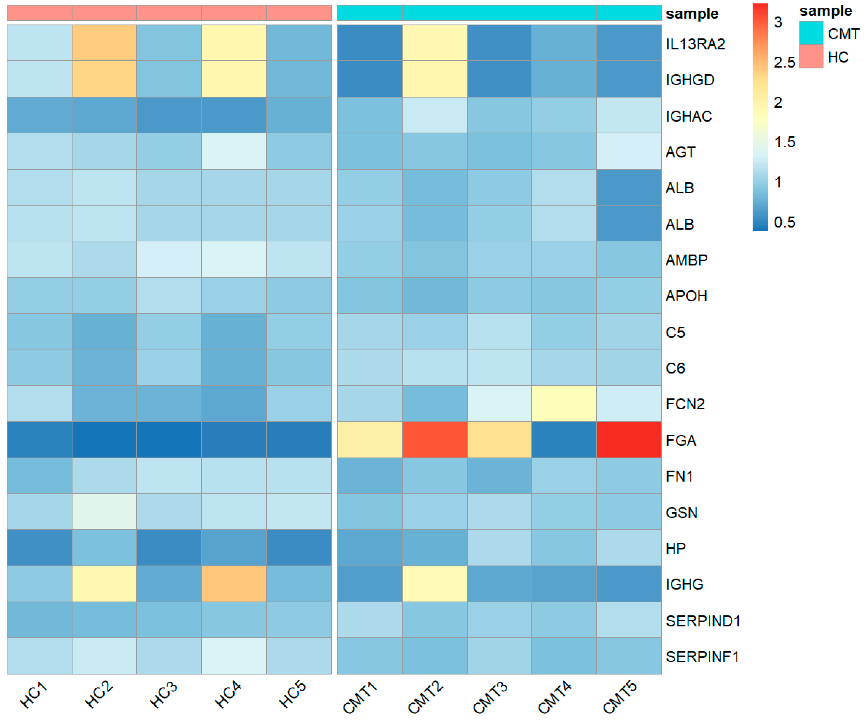
Figure A2.
Heatmap showing the relative abundance (color) of salivary proteins in healthy (HC) dogs and dogs with canine mammary tumors (CMT).
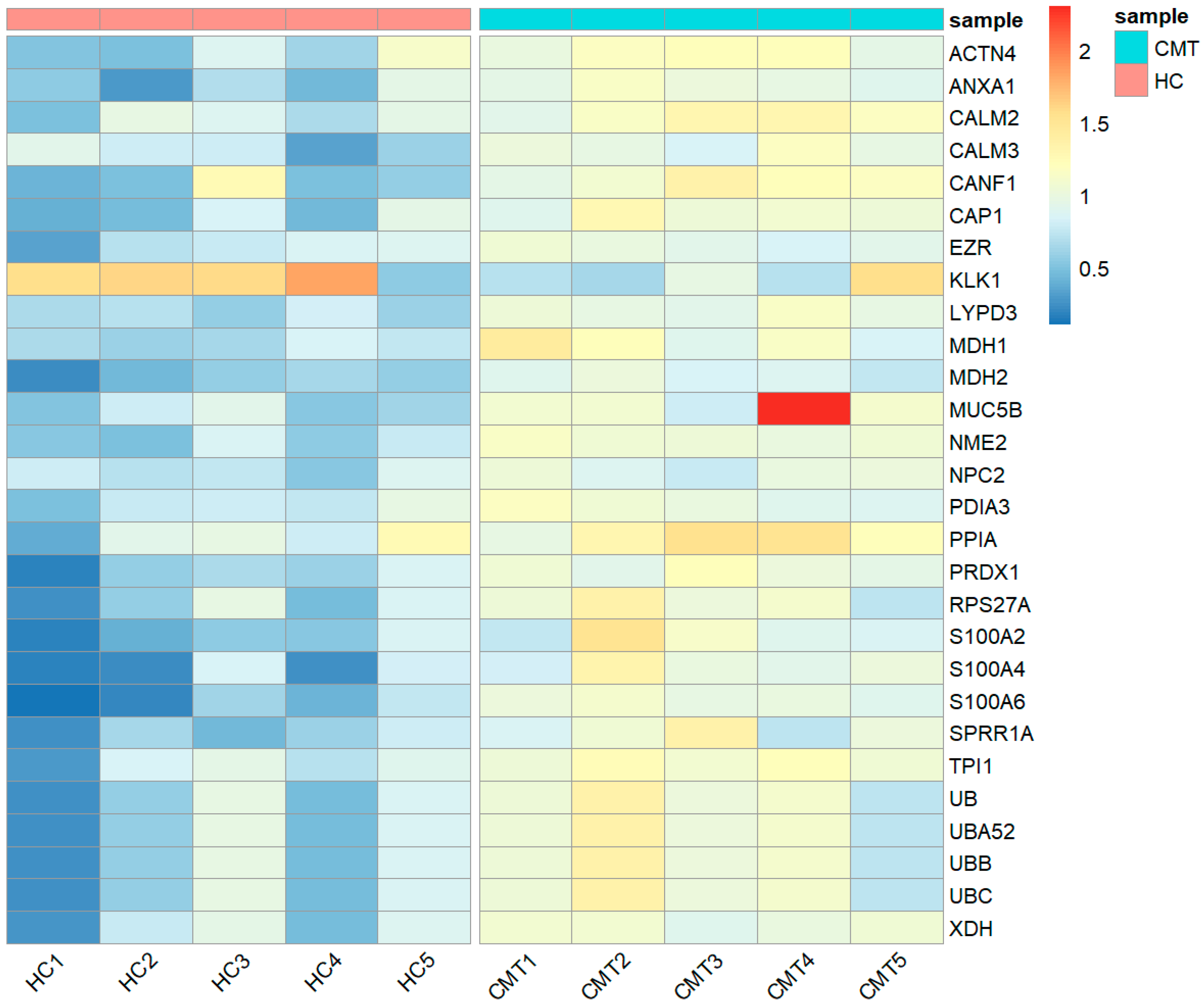
Table A1.
Serum proteins identified in dogs with canine mammary tumors (CMT) and healthy controls (HC).
Table A2.
Salivary proteins identified in dogs with canine mammary tumors (CMT) and healthy controls (HC).
References
- Fonseca, C.S.; Daleck, C.R. Neoplasias mamárias em cadelas: Influência hormonal e efeitos da ovario-histerectomia como terapia adjuvante. Ciênc. Rural 2000, 30, 731–735. [Google Scholar] [CrossRef] [Scilit]
- Benjamin, S.A.; Lee, A.C.; Saunders, W.J. Classification and behavior of canine mammary epithelial neoplasms based on life-span observations in Beagles. Vet. Pathol. 1999, 36, 423–436. [Google Scholar] [CrossRef] [Scilit] [PubMed]
- da Silva, A.E.; Serakides, R.; Cassali, G.D. Carcinogênese hormonal e neoplasias hormônio-dependentes. Ciênc. Rural 2004, 34, 625–633. [Google Scholar] [CrossRef] [Scilit]
- Zamani-Ahmadmahmudi, M.; Nassiri, S.M.; Rahbarghazi, R. Serological proteome analysis of dogs with breast cancer unveils common serum biomarkers with human counterparts. Electrophoresis 2014, 35, 901–910. [Google Scholar] [CrossRef] [Scilit] [PubMed]
- MacEwen, E.G. Spontaneous tumors in dogs and cats: Models for the study of cancer biology and treatment. Cancer Metastasis Rev. 1990, 9, 125–136. [Google Scholar] [CrossRef] [Scilit]
- Kycko, A.; Reichert, M. Proteomics in the search for biomarkers of animal cancer. Curr. Protein Pept. Sci. 2014, 15, 36–44. [Google Scholar] [CrossRef] [Scilit]
- Cerón, J.J. Acute phase proteins, saliva and education in laboratory science: An update and some reflections. BMC Vet. Res. 2019, 15, 197. [Google Scholar] [CrossRef] [Scilit]
- Sivadasan, P.; Gupta, M.K.; Sathe, G.J.; Balakrishnan, L.; Palit, P.; Gowda, H.; Suresh, A.; Kuriakose, M.A.; Sirdeshmukh, R. Human salivary proteome—A resource of potential biomarkers for oral cancer. J. Proteom. 2015, 127, 89–95. [Google Scholar] [CrossRef] [Scilit]
- Franco-Martínez, L.; Horvatić, A.; Gelemanović, A.; Samardžija, M.; Mrljak, V.; Contreras-Aguilar, M.D.; Martínez-Subiela, S.; And, R.D.; Tvarijonaviciute, A. Changes in the salivary proteome associated with canine pyometra. Front. Vet. Sci. 2020, in press. [Google Scholar]
- Franco-Martínez, L.; Tvarijonaviciute, A.; Horvatić, A.; Guillemin, N.; Cerón, J.J.; Escribano, D.; Eckersall, D.; Kocatürk, M.; Yilmaz, Z.; Lamy, E.; et al. Changes in salivary analytes in canine parvovirus: A high-resolution quantitative proteomic study. Comp. Immunol. Microbiol. Infect. Dis. 2018, 60, 1–10. [Google Scholar] [CrossRef] [Scilit]
- Martínez-Subiela, S.; Ceron, J.J.; Yilmaz, Z.; Martinez-Subiela, S.; Horvatic, A.; Escribano, D.; Pardo-Marin, L.; Kocaturk, M.; Mrljak, V.; Burchmore, R.; et al. Identification of novel biomarkers for treatment monitoring in canine leishmaniosis by high-resolution quantitative proteomic analysis. Vet. Immunol. Immunopathol. 2017, 191, 60–67. [Google Scholar] [CrossRef] [Scilit] [PubMed]
- Tvarijonaviciute, A.; Barić-Rafaj, R.; Horvatic, A.; Muñoz-Prieto, A.; Guillemin, N.; Lamy, E.; Tumpa, A.; Ceron, J.J.; Martinez-Subiela, S.; Mrljak, V. Identification of changes in serum analytes and possible metabolic pathways associated with canine obesity-related metabolic dysfunction. Vet. J. 2019, 244, 51–59. [Google Scholar] [CrossRef] [Scilit] [PubMed]
- Bera, A.; Russ, E.; Srinivasan, M.; Eidelman, O.; Eklund, M.; Hueman, M.; Pollard, H.B.; Hu, H.; Col, §.; Shriver, C.D.; et al. Proteomic Analysis of Inflammatory Biomarkers Associated With Breast Cancer Recurrence. Mil. Med. 2020, 185, 2020. [Google Scholar] [CrossRef] [Scilit] [PubMed]
- Delmonico, L.; Bravo, M.; Silvestre, R.T.; Ornellas, M.H.F.; De Azevedo, C.M.; Alves, G. Proteomic profile of saliva and plasma from women with impalpable breast lesions. Oncol. Lett. 2016, 12, 2145. [Google Scholar] [CrossRef] [Scilit]
- Giri, K.; Mehta, A.; Ambatipudi, K. In search of the altering salivary proteome in metastatic breast and ovarian cancers. FASEB Bioadv. 2019, 1, 191–207. [Google Scholar] [CrossRef] [Scilit]
- Klopfleisch, R.; Klose, P.; Weise, C.; Bondzio, A.; Multhaup, G.; Einspanier, R.; Gruber, A.D. Proteome of metastatic canine mammary carcinomas: Similarities to and differences from human breast cancer. J. Proteome Res. 2010, 9, 6380–6391. [Google Scholar] [CrossRef] [Scilit] [PubMed]
- Klose, P.; Weise, C.; Bondzio, A.; Multhaup, G.; Einspanier, R.; Gruber, A.D.; Klopfleisch, R. Is there a malignant progression associated with a linear change in protein expression levels from normal canine mammary gland to metastatic mammary tumors? J. Proteome Res. 2011, 10, 4405–4415. [Google Scholar] [CrossRef] [Scilit] [PubMed]
- Owen, L. TNM Classification of Tumours in Domestic Animals; World Health Organization: Geneva, Switzerland, 1980. [Google Scholar]
- Parra, M.; Tecles, F.; Martinez-Subiela, S.; Ceron, J. C-reactive protein measurement in canine saliva. J. Vet. Diagnostic Investig. 2005, 17, 139–144. [Google Scholar] [CrossRef] [Scilit] [PubMed]
- Contreras-Aguilar, M.D.; Tecles, F.; Martínez-Subiela, S.; Escribano, D.; Bernal, L.J.; Cerón, J.J. Detection and measurement of alpha-amylase in canine saliva and changes after an experimentally induced sympathetic activation. BMC Vet. Res. 2017, 13, 266. [Google Scholar] [CrossRef] [Scilit]
- Bradford, M.M. A rapid and sensitive method for the quantitation of microgram quantities of protein utilizing the principle of protein-dye binding. Anal. Biochem. 1976, 72, 248–254. [Google Scholar] [CrossRef]
- Horvatić, A.; Guillemin, N.; Kaab, H.; McKeegan, D.; O’Reilly, E.; Bain, M.; Kuleš, J.; Eckersall, P.D. Quantitative proteomics using tandem mass tags in relation to the acute phase protein response in chicken challenged with Escherichia coli lipopolysaccharide endotoxin. J. Proteom. 2019, 192, 64–77. [Google Scholar] [CrossRef] [Scilit] [PubMed]
- R Core Team. R: A Language and Environment for Statistical Computing; R Foundation for Statistical Computing: Vienna, Austria, 2013. [Google Scholar]
- Komsta, L. Package ‘outliers’. R Top. Doc. 2015. Available online: https://cran.r-project.org/web/packages/outliers/ (accessed on 15 January 2020).
- Doncheva, N.T.; Morris, J.H.; Gorodkin, J.; Jensen, L.J. Cytoscape StringApp: Network analysis and visualization of proteomics data. J. Proteome Res. 2019, 18, 623–632. [Google Scholar] [CrossRef] [Scilit] [PubMed]
- Kolde, R. Pheatmap: Pretty Heatmaps in R Package. Version 61. 2012, pp. 1–7. Available online: https://cran.r-project.org/web/packages/pheatmap/ (accessed on 15 January 2020).
- Kau, S.; Miller, I.; Tichy, A.; Gabriel, C. S100A4 (metastasin) positive mesenchymal canine mammary tumour spheroids reduce Tenascin C synthesis under DMSO exposure in vitro. Vet. Comp. Oncol. 2017, 15, 1428–1444. [Google Scholar] [CrossRef] [Scilit] [PubMed]
- Platt-Higgins, A.M.; Renshaw, C.A.; West, C.R.; Winstanley, J.H.R.; De Silva Rudland, S.; Barraclough, R.; Rudland, P.S. Comparison of the metastasis-inducing protein S100A4 (p9ka) with other prognostic markers in human breast cancer. Int. J. Cancer 2000, 89, 198–208. [Google Scholar] [CrossRef]
- Maděrka, M.; Pilka, R.; Neubert, D.; Hambálek, J. New serum tumor markers S100, TFF3 and AIF-1 and their possible use in oncogynecology. Ceska Gynekol. 2019, 84, 303–308. [Google Scholar]
- Davie, E.W.; Fujikawa, K. Basic mechanisms in blood coagulation. Annu. Rev. Biochem. 1975, 44, 799–829. [Google Scholar] [CrossRef] [Scilit] [PubMed]
- Wang, M.; Zhang, G.; Zhang, Y.; Cui, X.; Wang, S.; Gao, S.; Wang, Y.; Liu, Y.; Bae, J.H.; Yang, W.-H.; et al. Fibrinogen alpha chain knockout promotes tumor growth and metastasis through Integrin-AKT signaling pathway in lung cancer. Mol. Cancer Res. 2020. [Google Scholar] [CrossRef] [Scilit]
- Gerner, C.; Steinkellner, W.; Holzmann, K.; Gsur, A.; Grimm, R.; Ensinger, C.; Obrist, P.; Sauermann, G. Elevated plasma levels of crosslinked fibrinogen gamma-chain dimer indicate cancer-related fibrin deposition and fibrinolysis. Thromb. Haemost. 2001, 85, 494–501. [Google Scholar]
- Shi, Q.; Harris, L.N.; Lu, X.; Li, X.; Hwang, J.; Gentleman, R.; Iglehart, J.D.; Miron, A. Declining plasma fibrinogen alpha fragment identifies HER2-positive breast cancer patients and reverts to normal levels after surgery. J. Proteome Res. 2006, 5, 2947–2955. [Google Scholar] [CrossRef] [Scilit]
- Cheng, A.J.; Chen, L.C.; Chien, K.Y.; Chen, Y.J.; Chang, J.T.C.; Wang, H.M.; Liao, C.T.; Chen, I.H. Oral cancer plasma tumor marker identified with bead-based affinity-fractionated proteomic technology. Clin. Chem. 2005, 51, 2236–2244. [Google Scholar] [CrossRef] [Scilit] [PubMed]
- Britti, D.; Gaspari, M.; Massimini, G.; Casalinuovo, F.; Morittu, V.M.; Cuda, G. Proteomic analysis in canine leishmaniasis. Vet. Res. Commun. 2010, 34, 91–96. [Google Scholar] [CrossRef] [Scilit] [PubMed]
- Terabe, M.; Park, J.M.; Berzofsky, J.A. Role of IL-13 in regulation of anti-tumor immunity and tumor growth. Cancer Immunol. Immunother. 2004, 53, 79–85. [Google Scholar] [CrossRef] [Scilit] [PubMed]
- Król, M.; Pawłowski, K.M.; Majchrzak, K.; Gajewska, M.; Majewska, A.; Motyl, T. Global gene expression profiles of canine macrophages and canine mammary cancer cells grown as a co-culture in vitro. BMC Vet. Res. 2012, 8, 16. [Google Scholar] [CrossRef] [Scilit] [PubMed]
- Kwon, H.J.; Choi, J.E.; Bae, Y.K. Interleukin-13 receptor alpha 2 expression in tumor cells is associated with reduced disease-free survival in patients with luminal subtype invasive breast cancer. Tumor Biol. 2018, 40, 1–8. [Google Scholar] [CrossRef] [Scilit] [PubMed]
- Gallastegui, C.; Bernardez, B.; Regueira, A.; Dávila, C.; Leboreiro, B. Inmunología. In M. C. Gamundi Planas (Coordinadora), Farmacia Hospitalaria Tomo II; Sociedad Española de Farmacia Hospitalaria: Madrid, Spain, 2002; pp. 1077–1106. [Google Scholar]
- Cerón, J.J.; Eckersall, P.D.; Martínez-Subiela, S. Acute phase proteins in dogs and cats: Current knowledge and future perspectives. Vet. Clin. Pathol. 2005, 34, 85–99. [Google Scholar] [CrossRef] [Scilit]
- Planellas, M.; Bassols, A.; Siracusa, C.; Saco, Y.; Giménez, M.; Pato, R.; Pastor, J. Evaluation of serum haptoglobin and C-reactive protein in dogs with mammary tumors. Vet. Clin. Pathol. 2009, 38, 348–352. [Google Scholar] [CrossRef] [Scilit]
- Tecles, F.; Caldín, M.; Zanella, A.; Membiela, F.; Tvarijonaviciute, A.; Subiela, S.M.; Cerón, J.J. Serum acute phase protein concentrations in female dogs with mammary tumors. J. Vet. Diagn. Investig. 2009, 21, 214–219. [Google Scholar] [CrossRef] [Scilit] [PubMed]
- Merlo, A.; Rezende, B.C.G.; Franchini, M.L.; Simões, D.M.N.; Lucas, S.R.R. Serum C-reactive protein concentrations in dogs with multicentric lymphoma undergoing chemotherapy. J. Am. Vet. Med. Assoc. 2007, 230, 522–526. [Google Scholar] [CrossRef] [Scilit]
- Mischke, R.; Waterston, M.; Eckersall, P.D. Changes in C-reactive protein and haptoglobin in dogs with lymphatic neoplasia. Vet. J. 2007, 174, 188–192. [Google Scholar] [CrossRef] [Scilit]
- Salama, I.; Malone, P.S.; Mihaimeed, F.; Jones, J.L. A review of the S100 proteins in cancer. Eur. J. Surg. Oncol. 2008, 34, 357–364. [Google Scholar] [CrossRef] [Scilit] [PubMed]
- Rudland, P.S.; Platt-Higgins, A.; Renshaw, C.; West, C.R.; Winstanley, J.H.R.; Robertson, L.; Barraclough, R. Prognostic significance of the metastasis-inducing protein S100A4 (p9Ka) in human breast cancer. Cancer Res. 2000, 60, 1595–1603. [Google Scholar] [PubMed]
- Pedersen, K.B.; Nesland, J.M.; Fodstad, O.; Maelandsmo, G.M. Expression of S100A4, E-cadherin, alpha- and beta-catenin in breast cancer biopsies. Br. J. Cancer 2002, 87, 1281–1286. [Google Scholar] [CrossRef] [Scilit] [PubMed]
- Zha, C.; Jiang, X.H.; Peng, S.F. iTRAQ-Based Quantitative Proteomic Analysis on S100 Calcium Binding Protein A2 in Metastasis of Laryngeal Cancer. PLoS ONE 2015, 10, e0122322. [Google Scholar] [CrossRef] [Scilit] [PubMed]
- Naz, S.; Bashir, M.; Ranganathan, P.; Bodapati, P.; Santosh, V.; Kondaiah, P. Protumorigenic actions of S100A2 involve regulation of PI3/Akt signaling and functional interaction with Smad3. Carcinogenesis 2014, 35, 14–23. [Google Scholar] [CrossRef] [Scilit] [PubMed]
- Bai, Y.; Li, L.D.; Li, J.; Lu, X. Prognostic values of S100 family members in ovarian cancer patients. BMC Cancer 2018, 18, 1256. [Google Scholar] [CrossRef] [Scilit] [PubMed]
- Komatsu, K.; Andoh, A.; Ishiguro, S.; Suzuki, N.; Hunai, H.; Kobune-Fujiwara, Y.; Kameyama, M.; Miyoshi, J.; Akedo, H.; Nakamura, H. Increased expression of S100A6 (calcyclin), a calcium-binding protein of the S100 family, in human colorectal adenocarcinomas. Clin. Cancer Res. 2000, 6, 172–177. [Google Scholar]
- Wang, X.H.; Zhang, L.H.; Zhong, X.Y.; Xing, X.F.; Liu, Y.Q.; Niu, Z.J.; Peng, Y.; Du, H.; Zhang, G.G.; Hu, Y.; et al. S100A6 overexpression is associated with poor prognosis and is epigenetically up-regulated in gastric cancer. Am. J. Pathol. 2010, 177, 586–897. [Google Scholar] [CrossRef] [Scilit]
- Lyu, X.; Li, H.; Ma, X.; Li, X.; Gao, Y.; Ni, D.; Shen, D.; Gu, L.; Wang, B.; Zhang, Y.; et al. High-Level S100A6 promotes metastasis and predicts the outcome of T1–T2 stage in clear cell renal cell carcinoma. Cell Biochem. Biophys. 2014, 71, 279–290. [Google Scholar] [CrossRef] [Scilit]
- Zhang, S.; Wang, Z.; Liu, W.; Lei, R.; Shan, J.; Li, L.; Wang, X. Distinct prognostic values of S100 mRNA expression in breast cancer. Sci. Rep. 2017, 7, 39786. [Google Scholar] [CrossRef] [Scilit]
- Hait, W.N.; Lazo, J.S. Calmodulin: A potential target for cancer chemotherapeutic agents. J. Clin. Oncol. 1986, 4, 994–1012. [Google Scholar] [CrossRef] [Scilit] [PubMed]
- Fancy, R.M.; Kim, H.; Napier, T.; Buchsbaum, D.J.; Zinn, K.R.; Song, Y. Calmodulin antagonist enhances DR5-mediated apoptotic signaling in TRA-8 resistant triple negative breast cancer cells. J. Cell. Biochem. 2018, 119, 6216–6230. [Google Scholar] [CrossRef] [Scilit] [PubMed]
- Borgoño, C.A.; Diamandis, E.P. The emerging roles of human tissue kallikreins in cancer. Nat. Rev. Cancer 2004, 4, 876–890. [Google Scholar] [CrossRef] [Scilit] [PubMed]
- Wolf, W.C.; Evans, D.M.; Chao, L.; Chao, J. A synthetic tissue kallikrein inhibitor suppresses cancer cell invasiveness. Am. J. Pathol. 2001, 159, 1797–1805. [Google Scholar] [CrossRef] [Scilit]
- Avgeris, M.; Mavridis, K.; Scorilas, A. Kallikrein-related peptidases in prostate, breast, and ovarian cancers: From pathobiology to clinical relevance. Biol. Chem. 2012, 393, 301–317. [Google Scholar] [CrossRef] [Scilit] [PubMed]
- Yousef, G.M.; Polymeris, M.E.; Grass, L.; Soosaipillai, A.; Chan, P.C.; Scorilas, A.; Borgoño, C.; Harbeck, N.; Schmalfeldt, B.; Dorn, J.; et al. Human kallikrein 5: A potential novel serum biomarker for breast and ovarian cancer. Cancer Res. 2003, 63, 3958–3965. [Google Scholar]
- Ewan King, L.; Li, X.; Cheikh Saad Bouh, K.; Pedneault, M.; Chu, C.W. Human kallikrein 10 ELISA development and validation in breast cancer sera. Clin. Biochem. 2007, 40, 1057–1062. [Google Scholar] [CrossRef] [Scilit]
- Borgoño, C.A.; Grass, L.; Soosaipillai, A.; Yousef, G.M.; Petraki, C.D.; Howarth, D.H.C.; Fracchioli, S.; Katsaros, D.; Diamandis, E.P. Human Kallikrein 14: A new potential biomarker for ovarian and breast cancer. Cancer Res. 2003, 63, 9032–9041. [Google Scholar]
- Koh, S.C.L.; Razvi, K.; Chan, Y.H.; Narasimhan, K.; Ilancheran, A.; Low, J.J.; Choolani, M.; Sahil, F.; Lutan, D.; Siddik, D.; et al. The association with age, human tissue kallikreins 6 and 10 and hemostatic markers for survival outcome from epithelial ovarian cancer. Arch. Gynecol. Obstet. 2011, 284, 183–190. [Google Scholar] [CrossRef] [Scilit]
- Yousef, G.M.; Obiezu, C.V.; Luo, L.Y.; Black, M.H.; Diamandis, E.P. Prostase/KLK-L1 is a new member of the human kallikrein gene family, is expressed in prostate and breast tissues, and is hormonally regulated. Cancer Res. 1999, 59, 4252–4256. [Google Scholar]
- Loo, J.A.; Yan, W.; Ramachandran, P.; Wong, D.T. Comparative human salivary and plasma proteomes. J. Dent. Res. 2010, 89, 1016–1023. [Google Scholar] [CrossRef] [Scilit] [PubMed]
- Klopfleisch, R.; Gruber, A.D. Differential expression of cell cycle regulators p21, p27 and p53 in metastasizing canine mammary adenocarcinomas versus normal mammary glands. Res. Vet. Sci. 2009, 87, 91–96. [Google Scholar] [CrossRef] [Scilit] [PubMed]
- Spoerri, M.; Guscetti, F.; Hartnack, S.; Boos, A.; Oei, C.; Balogh, O.; Nowaczyk, R.M.; Michel, E.; Reichler, I.M.; Kowalewski, M.P. Endocrine control of canine mammary neoplasms: Serum reproductive hormone levels and tissue expression of steroid hormone, prolactin and growth hormone receptors. BMC Vet. Res. 2015, 11, 235. [Google Scholar] [CrossRef] [Scilit] [PubMed]
- Abadie, J.; Nguyen, F.; Loussouarn, D.; Peña, L.; Gama, A.; Rieder, N.; Belousov, A.; Bemelmans, I.; Jaillardon, L.; Ibisch, C.; et al. Canine invasive mammary carcinomas as models of human breast cancer. Part 2: Immunophenotypes and prognostic significance. Breast Cancer Res. Treat. 2018, 167, 459–468. [Google Scholar] [CrossRef] [Scilit] [PubMed]
- Westermeier, R.; Naven, T.; Höpker, H.-R. Wiley InterScience (Online service). In Proteomics in Practice: A Guide to Successful Experimental Design; Wiley-VCH: Weinheim, Germany, 2008; ISBN 3527622306. [Google Scholar]

© 2020 by the authors. Licensee MDPI, Basel, Switzerland. This article is an open access article distributed under the terms and conditions of the Creative Commons Attribution (CC BY) license (http://creativecommons.org/licenses/by/4.0/).