Review

An Updated List of Generic Names in the Thoracosphaeraceae

Department of Biology, Systematic Botany and Mycology, GeoBio-Center, University Munich, Menzinger Str. 67, D-80638 Munich, Germany
*
Author to whom correspondence should be addressed.
Microorganisms 2013, 1(1), 122-136; https://doi.org/10.3390/microorganisms1010122
Submission received: 28 August 2013 / Revised: 12 September 2013 / Accepted: 23 October 2013 / Published: 1 November 2013
(This article belongs to the Special Issue Biology of Dinoflagellates: Advances in the Last 25 Years (1987-2012))

Abstract

:
Calcareous dinophytes produce exoskeletal calcified structures during their life history (a unique character among the alveolates) and are subsumed under the Thoracosphaeraceae as part of the Peridiniales. We provide a brief synopsis about the taxonomic history of the group, from the first descriptions of fossils in the 19th century through to the results of molecular phylogenetics studies undertaken during the past two decades. Delimitation and circumscription of the Thoracosphaeraceae are challenging, as they comprise both phototrophic (presumably including endosymbiotic) as well as heterotrophic (and even parasitic) dinophytes from marine and freshwater environments, respectively. However, calcareous structures are not known from all members of the Thoracosphaeraceae, and the corresponding species and groups are considered to have lost the capacity to calcify. Five years ago, a taxonomic list of 99 generic names assigned to the Thoracosphaeraceae was published, and we update this compendium with 19 additional names based on recent studies.

1. Historical Survey

Many dinophytes develop two distinct stages during their life history, namely a motile thecate cell and a non-motile coccoid cell. During the coccoid stage, the production of exoskeletal calcified structures is a distinct character trait exclusively found in a subordinate collective of the Peridiniales Haeckel, notably in the calcareous dinophytes [1]. Their thecate cells exhibit a more or less conserved arrangement of cellulose plates (i.e., the tabulation) and are presumed to be haploid, while the coccoid cells are usually interpreted as hypnozygotes (i.e., diploid stage: [2]). The calcareous cells are morphologically highly diverse. Various degrees of expressed tabulation may be retained (formerly described as “paratabulation”), which is frequently restricted to the archeopyle (aperture for germination). Calcareous dinophytes are well documented in the fossil record, and their diversity assessment has a complex and uncompleted history.
From a paleontological perspective, the first descriptions of organisms with a calcified shell and today assigned to the dinophytes go back to F.J. Kaufmann, although he considered Cretaceous †Lagena sphaerica F.J.Kaufmann and †L. ovalis F.J.Kaufmann as members of the foraminifers [3]. At the beginning of the last century, T. Lorenz acknowledged the distinctiveness of those forms from †Lagena G.Walker & Boys, and subsequently introduced the generic name †Pithonella T.Lorenz, with the type species †Pithonella ovalis (F.J.Kaufmann) T.Lorenz and retained them in the foraminifers [4]. It took further decades until G. Deflandre recognized the true dinophyte nature of corresponding fossils, although he interpreted them as calcareous thecate cells [5]. Thereafter, the majority of the calcareous dinophyte diversity described was subsumed under the name Calciodinellaceae Deflandre (alternatively Calciodinelloideae Fensome, F.J.R.Taylor, G. Norris, Sarjeant, Wharton & G.L.Williams) and has since resulted in many studies [6,7,8,9,10,11,12,13,14,15,16,17,18].
Cultivation experiments with extant calcareous dinophytes showed that the immotile coccoid and not the motile cell is usually calcified [19]. Moreover, cultivation of calcareous dinophytes demonstrated that the thecate cells hatching from the coccoid cells can partly be assigned to species, which have long been known by neontologists [e.g., Scrippsiella trochoidea (F.Stein) A.R.Loebl.]. The pioneering work of D. Wall and B. Dale [19] was thus the start of numerous studies investigating in more detail the developmental link between thecate and coccoid cells in the life history of particular calcareous dinophyte species [20,21,22,23,24,25,26,27,28]. As a result, thecate cells of most calcareous dinophytes exhibit homogenously an ortho-hexa-tabulation pattern identifying them as members of the Peridiniales, whereas the morphology of coccoid cells is particularly diverse in calcareous dinophytes.
Similarly to the Calciodinelloideae, it took more than half a century until Thoracosphaera heimii (Lohmann) Kamptner (initially described under the coccolithophore Syracophaera Lohmann [29]) was recognized as a (calcareous) dinophyte [30,31,32]. Thoracosphaera Kamptner differs from the majority of calcareous dinophytes in several respects: the motile cells are athecate, and the calcareous coccoid cells are dividing vegetatively. These differences were considered so fundamental that the Thoracosphaerales Tangen were established at the same taxonomic level as the Peridiniales [32], implying that Thoracosphaera is only distantly related to the Calciodinelloideae. This classification was also followed in the epochal work of Fensome and colleagues [33] and by subsequent authors.
Since the onset of molecular studies, knowledge on the phylogenetic relationships and the constituent taxa of extant calcareous dinophytes has changed gradually but significantly overall. The early molecular studies identified two [23] and later three distinct evolutionary lineages [34] that include calcareous as part of peridinialean dinophytes (Figure 1), namely the E/Pe-clade (with species of Ensiculifera Balech and Pentapharsodinium Indel. & A.R.Loebl.), the T/Pf-clade (with species of Thoracosphaera and Pfiesteria Steid. & J.M.Burkh.), and Scrippsiella Balech sensu lato (s.l., also including fossil-taxa such as †Calciodinellum Deflandre and †Pernambugia Janofske & Karwath). An important result of the molecular studies showed that Thoracosphaera is not distinct from other calcareous dinophytes but in fact embedded within them. It was therefore proposed to unify the formerly segregated taxonomic units Calciodinelloideae and Thoracosphaerales and to treat the entirety of calcareous dinophytes under the Thoracosphaeraceae J.Schiller [1].
Opposing the view of Tangen [32], various authors (with a predominantly paleontological background) considered calcareous dinophytes as a monophyletic group based on the apomorphic calcified coccoid cells [10,35]. However, the first molecular studies challenged this simplistic circumscription of calcareous dinophytes. The Thoracosphaeraceae included not only calcareous but also non-calcareous dinophytes or at least those of which calcareous structures are not known so far (Figure 1). The lack of calcified structures in those members of the Thoracosphaeraceae has been considered a secondary loss [1,34]. However, as more molecular studies were published non-calcareous dinophytes included in the Thoracosphaeraceae became greater in number and more heterogeneous, as outlined below.
The pfiesterians are a group of heterotrophic dinophytes and versatile predators. Some of their species have been associated with harmful algal blooms and fish kills, but many aspects of their life histories and character traits (e.g., potential toxin activity) are still under debate (see [36] and literature therein). Since its first description [37], the systematic position of Pfiesteria in the dinophyte tree was unclear and was placed somewhere in the Gonyaulacales F.J.R.Taylor or Peridiniales. It was thus a great surprise when molecular phylogenies identified calcareous Leonella Janofske & Karwath and Thoracosphaera as the closest known relatives of the pfiesterians and that the latter may derive from calcareous dinophytes [34]. This scenario has been repeatedly supported by subsequent studies (partly investigating alternative loci [28,38,39,40]), and the molecular trees indicate a single loss event of the capacity for calcareous structures in the T/Pf-clade (Figure 1). The acceptance of the Pfiesteriaceae as a distinct systematic unit [41,42,43,44] would, anyhow, leave the remainders of the Thoracosphaeraceae paraphyletic.
Figure 1. Phylogeny of and molecular delimitations in the Thoracosphaeraceae (Bayesian tree) segregating into the three indicated clades E/Pe, T/Pf, and Scrippsiella s.l.(abbreviations: CAL, †Calciodinellum and relatives; E/Pe, Ensiculifera + Pentapharsodinium and relatives; PRE, Scrippsiella precaria Montresor & Zingone and relatives; STR, Scrippsiella trochoidea species complex; T/Pf, Thoracosphaera + Pfiesteria and relatives). Calcareous taxa are highlighted by bold branches. Branch lengths are drawn to scale, with the scale bar indicating the number of substitutions per site. Numbers on branches are statistical support values (above: Bayesian posterior probabilities, values under 0.90 are not shown; below: ML bootstrap support values, values under 50 are not shown), and maximal support values are indicated by asterisks.
Microorganisms 01 00122 g001
From an evolutionary perspective, the discovery of tintinnid parasites such as Duboscquodinium Grassé and Tintinnophagus Coats nested within the calcareous dinophytes of the Scrippsiella s.l. lineage based on molecular data [45] was presumably more unexpected than the Pfiesteria results. Moreover, K.D. Smith and colleagues reported that another ctenophoran parasite was closely related to †Calcicarpinum bivalvum G.Versteegh [= Pentapharsodinium tyrrhenicum (Balech) Montresor, Zingone & D.Marino] from the E/Pe-clade [46,47], highlighting the association between calcareous and parasitic dinophytes. However, it is presently unknown (and experimentally very difficult to investigate), whether calcareous dinophytes may exhibit also parasitic stages during their life history, in addition to the comparatively well investigated thecate and coccoid cells. The Scrippsiella s.l. lineage contains many species that are morphologically indistinguishable, but genetically differentiated (i.e., cryptic species), which refers particularly to the S. trochoidea species complex [26,48,49]. Linking this cryptic diversity with the hypothetical specificity of parasitic dinophytes (as inferred from inoculation experiments [47]) has triggered the idea that the species of the S. trochoidea species complex are neither differentiated based on morphology or spatial distribution, but based on tight interactions with particular host species (pers. comm. K.J.S Meier, Kiel).
The close relationship between the T/Pf-clade and the Scrippsiella lineage is undisputed today, and the vast majority of extant calcareous dinophytes known is reliably placed in one of the two clades. However, the E/Pe-clade challenges the assumption of a monophyletic calcareous dinophyte group, as its close relationship to the Scrippsiella- and T/Pf-clades is not shown, or at least not supported, in all molecular studies. The only calcareous member of this clade with published sequence data is †Calcicarpinum bivalvum, while calcified structures are not known from any other sequenced species out of this clade assigned to Ensiculifera or Pentapharsodinium. However, the group is considered to include a number of calcareous, mostly fossil-taxa such as †Follisdinellum G.Versteegh, †Melodomuncula G.Versteegh, and Pentadinellum Keupp, all of which were observed in Recent sediments, but have not been brought in culture so far [1]. Exploring the extant diversity, and exact phylogenetic placement, of the E/Pe-clade thus remains one of the major tasks in future research on calcareous dinophytes.
The ambiguity of the phylogenetic position regarding the E/Pe-clade refers in particular to other peridinalean dinophytes, of which sequences have been published in the past few years. The molecular studies suggest the monophyly of a highly disparate group [28,50] comprising endosymbionts (i.e., Zooxanthella), parasites (i.e., Blastodinium [51,52]), and dinophytes harboring a diatom as endosymbiont (i.e., the “dinotoms” [53,54,55]). In some molecular studies, this heterogeneous assemblage is closely related to the E/Pe-clade (i.e., are members of the Thoracosphaeraceae [38,50], Figure 1), in others they constitute the sister group of the Thoracosphaeracae, which then consist of the three clades E/Pe, T/Pf, and Scrippsiella s.l. [40]. Currently, it has to be emphasized that molecular phylogenies of dinophytes still have room for improvement because of various problems, including limited taxon sampling (less than a quarter of dinophytes at the generic level are currently known with respect to genetic sequence data), insufficient genetic data, and strong rate heterogeneity (see discussion in [28]).
In summary, the often puzzling diversity of the Thoracosphaeraceae in terms of nutrition modes (phototrophic→heterotrophic), habitat preferences (marine→freshwater), and coccoid cell morphologies (calcareous→non-calcareous) reflects to some degree the variation found throughout all dinophytes. This biological heterogeneity makes a morphological diagnosis of the Thoracosphaeraceae almost impossible, and their taxonomic delimitation relies mostly on molecular data at present. Also, the taxonomy of the Thoracosphaeraceae is further challenging, as they have been described under the rules of the International Code for Zoological Nomenclature (ICZN [56]) as well as the International Code of Nomenclature for algae, fungi and plants (ICN [57]) and based on thecate as well as coccoid (and parasitic) stages (for details, see [1]). In their current circumscription, the Thoracosphaeraceae (Peridiniales, Dinophyceae) comprise about 70 extant (morpho-)species, plus about 260 fossil species. Within the impressive diversity of the Alveolata, the capacity to produce calcareous structures is restricted to (i.e., has been considered apomorphic for) the Thoracosphaeraceae, arguing for the monophyly of the group [1,10,19]. The lack of calcified structures in those members of the Thoracosphaeraceae without known calcareous structures has then been considered a secondary loss [1,34].

2. Taxonomy

In the Agenda Calcareous Dinophytes from 2008, a list of 99 generic names in the Thoracosphaeraceae was published [1] based on the knowledge at that time. Since then, more taxa have been shown to be included in the Thoracosphaeraceae, and based on this work (Figure 1) and previous studies [45,58,59,60,61,62,63,64,65,66], the following 14 names are to be added to the list of generic names in the Thoracosphaeraceae (using the same reference format as in the Agenda Calcareous Dinophytes):
(1) Amyloodinium E.-M.Br. & HovasseZ, Proceedings of the Zoological Society of London 116: 45. 1916. Type: Amyloodinium ocellatum (E.-M.Br.) E.-M.Br. & HovasseZ, l.c.: 32–43, figs 1–9 ≡ Oodinium ocellatum E.-M.Br.Z, Proceedings of the Zoological Society of London 101: 345–346. 1931. Extant parasite in the gill mucosa of marine fish (without precise locality).
(2) †Calciconus Streng, Banasová, D.Reháková & H.WillemsB, Review of Palaeobotany and Palynology 153: 229. 2009 ≡ †Trigonus Banasová, Kopčáková & D.RehakováB, not validly published (ICN Art. 36.1b). Type: †Calciconus irregularis Streng, Banasová, D.Reháková & H.WillemsB, l.c.: 230, pl. II 1–10 ≡ †Trigonus conicus Banasová, Kopčáková & D.RehakováB, not validly published (ICN Art. 36.1b). Badenian (Slovak Republic: Bratislava).
(3) Chimonodinium Craveiro, Calado, Daugbjerg, Gert Hansen & MoestrupB, Protist 162: 604–605. 2011. Type: Chimonodinium lomnickii (Wołosz.) Craveiro, Calado, Daugbjerg, Gert Hansen & MoestrupB, l.c.: 605–606, figs 1–14 ≡ Peridinium lomnickii Wołosz.B, nom. corr. (ICN Arts 60.6, 60.12), Bulletin International de l’Académie des Sciences de Cracovie, Classe des Sciences Mathématiques et Naturelles. Série B 1915: 264, 267–268, pl. X 25–29. 1916 ≡ Glenodinium lomnickii (Wołosz.) Er.Lindem.B in Schoen., Einfachste Lebensformen des Tier- und Pflanzenreiches. Fünfte Auflage. Band 1 (Spaltpflanzen, Geißlinge, Algen, Pilze): 162, 168, 169. 1925. Extant (Ukraine: Lviv).
(4) †Cylindratus Banasová, Kopčáková & D.Rehaková ex Streng, Banasová, D.Reháková & H.WillemsB, Review of Palaeobotany and Palynology 153: 230. 2009. Type: †Cylindratus borzae Banasová, Kopčáková & D.Rehaková ex Streng, Banasová, D.Reháková & H.WillemsB, l.c.: 232, pl. III 1–9. Badenian (Slovak Republic: Bratislava).
(5) Duboscquodinium GrasséZ, Traité de zoologie 1: 358, 384. 1952. Type: Duboscquodinium collinii GrasséZ, nom. corr. (ICN Art. 60.12), l.c.: fig. 297A–B. Extant parasite (without precise locality).
Remark: If it can be reliably shown in future that Dubosquodinium and Scrippsiella are congeneric, then this would have dramatic consequences, as Dubosquodinium [67] is older than Scrippsiella [68] (even when A.R. Loeblich’s “validation” [69] is not considered) and would have taxonomic priority. However, further taxonomic activity should not be undertaken until the precise identity of the type species of Scrippsiella, S. sweeneyae Balech, is worked out.
(6) †Juergenella Banasová, Kopčáková & D.Rehaková ex Streng, Banasová, D.Reháková & H.WillemsB, Review of Palaeobotany and Palynology 153: 236. 2009. Type: †Juergenella ansata (Hildebrand-Habel & H.Willems) Streng, Banasová, D.Reháková & H.WillemsB, l.c.: 237 ≡ †Calcigonellum ansatum Hildebrand-Habel & H.WillemsB, Journal of Micropalaeontology 18: 93, pl. I 8–10. 1999. Upper Eocene (South Atlantic Ocean: Rio Grande Rise).
(7) Paulsenella ChattonZ, Archives de Zoologie Experimentale et Generale 59: 320. 1920. Type: Paulsenella chaetoceratis (Paulsen) ChattonZ, l.c.: fig. 139 ≡ Apodinium chaetoceratis PaulsenB, Meddelelser om Grønland [11. Marine Plankton from the East-Greenland Sea 3] 43: 316, fig. 17. 1910. Extant parasite (Atlantic Ocean: Greenland Sea).
(8) †Posoniella Streng, Banasová, D.Reháková & H.WillemsB, Review of Palaeobotany and Palynology 153: 233–234. 2009. Type: †Posoniella tricarinelloides (G.Versteegh) Streng, Banasová, D.Reháková & H.Willems B, l.c.: 234, fig. 5A,D,G ≡ †Bicarinellum tricarinelloides G.VersteeghB, Review of Palaeobotany and Palynology 78: 357, 359–360, pl. I 4–5. 1993. Pleistocene (Greece: Crete).
(9) Stoeckeria H.J.Jeong, Jae S.Kim, J.Y.Park, Jong H.Kim, Sang Kim, I.Lee, Seung H.Lee, J.H.Ha & W.H.YihZ, Journal of Eukaryotic Microbiology 52: 389. 2005. Type: Stoeckeria algicida H.J.Jeong, Jae S.Kim, J.Y.Park, Jong H.Kim, Sang Kim, I.Lee, Seung H.Lee, J.H.Ha & W.H.YihZ, l.c.: 384–385, figs 1–23. Extant (Pacific Ocean: East China Sea, off Korea).
Remark: There is some discussion whether Stoeckeria was validly published (using botanical “Dinophyceae” in the title, but lacked a Latin description or diagnosis). We agree with our colleagues [62] that it was not the authors’ intention to publish the new name under the rules of the ICN and therefore accept it pragmatically as validly published under the rules of the ICZN.
(10) Theleodinium Craveiro, Pandeirada, Daugbjerg, Moestrup & CaladoB, Phycologia 52. in press. Type: Theleodinium calcisporum Craveiro, Pandeirada, Daugbjerg, Moestrup & CaladoB, l.c. Extant (Portugal: Gafanha da Boavista).
(11) Tintinnophagus CoatsZ in Coats, Su.Kim, Bachvaroff, Handy & Delwiche, Journal of Eukaryotic Microbiology 57: 481. 2010. Type: Tintinnophagus acutus CoatsZ, l.c.: 471–473, figs 2–27. Extant parasite (USA–VA: Chesapeake Bay).
(12) †Trigonus Banasová, Kopčáková & D.RehakováB, Mineralia Slovaca 39: 111. 2007, not validly published (ICN Art. 36.1b). Type: †Trigonus conicus Banasová, Kopčáková & D.RehakováB, l.c.: 111–112, pl. I 9–12, not validly published (ICN Art. 36.1b). Badenian (Slovak Republic: Bratislava) ≡ †Calciconus Streng, Banasová, D.Reháková & H.Willems.
(13) Tyrannodinium Calado, Craveiro, Daugbjerg & MoestrupB, Journal of Phycology 45: 1202–1203. 2009. Type: Tyrannodinium berolinense (Lemmerm.) Calado, Craveiro, Daugbjerg & MoestrupB, l.c.: figs 1–6 ≡ Peridinium berolinense Lemmerm.B, Berichte der Deutschen Botanischen Gesellschaft 18: 308–309. 1900 ≡ Glenodinium berolinense (Lemmerm.) Er.Lindem.B in Schoen., Einfachste Lebensformen des Tier- und Pflanzenreiches. Fünfte Auflage. Band 1 (Spaltpflanzen, Geißlinge, Algen, Pilze): 162, 164. 1925 ≡ Peridiniopsis berolinense (Lemmerm.) Bourr.B, Protistologica 4: 9. 1968. Extant (Germany: Berlin).
(14) P†Zugelia ÖzdikmenZ, Munis Entomology & Zoology 4: 237. 2009. ≡ †Normandia ZügelB, not validly published (ICN Art. 53.1) (non: Normandia Hook.f.B, Icones plantarum 12: 20–21. 1872, nec: Normandia PicZ, Bulletin de la Société Entomologique de France 1900: 267. 1900). Type: †Zugelia circumperforata (Zügel) ÖzdikmenZ, l.c. ≡ †Normandia circumperforata ZügelB, Courier Forschungsinstitut Senckenberg 176: 32, 34, figs 12–13, pl. III 1–15. 1994, not validly published (ICN Art. 35.1). Turonian (France: Le Tilleul).
Remark: In the Agenda Calcareous Dinophytes [1], we overlooked that the name †Normandia Zügel had not been validly published neither under the ICN nor the ICZN because of the priority of earlier names. The affinity of the extinct pithonelloids to the calcareous dinophytes was debated in the past [1], but was recently corroborated based on exceptionally well preserved Cretaceous fossils [70].
The systematic position of a heterogeneous group comprising endosymbionts, dinophytes harboring endosymbionts, and parasites is not resolved at present with respect to the E/Pe-clade of the calcareous dinophytes. The following five names are therefore tentative candidates for being included in the Thoracosphaeraceae, but more research is necessary to determine their exact phylogenetic placement in the dinophyte tree:
(15) Blastodinium ChattonZ, Comptes Rendus Hebdomadaires des Séances de l’Académie des Sciences 143: 981. 1906. Type: Blastodinium pruvotii ChattonZ, nom. corr. (ICN Art. 60.12), l.c.: 981–983, figs 1–5. 1906. Extant (Mediterranean Sea, off France).
(16) Durinskia Carty & El.R.CoxB, Phycologia 25: 200. 1986. Type: Durinskia baltica (Levander) Carty & El.R.CoxB, l.c.: figs 7–14. 1986 ≡ Glenodinium balticum LevanderZ, Acta Societatis pro Fauna et Flora Fennica 12.2: 52. 1894 ≡ Peridinium balticum (Levander) Lemmerm.B, Kryptogamenflora der Mark Brandenburg. Dritter Band [Algen I (Schizophyceen, Flagellaten, Peridineen)]: 657. 1910. Extant (Finland).
Remark: Although sporadically used, the species’ name has never been validly published under Peridiniopsis [71].
(17) Galeidinium Tam. & T.Horig.B, Journal of Phycology 41: 661. 2005. Type: Galeidinium rugatum Tam. & T.Horig.B, l.c.: 661–667, figs 1A–G, 2A–B, 3A–F, 4A–E. 2005. Extant (Western Pacific Ocean, off Palau).
(18) Kryptoperidinium Er.Lindem.B, Botanisches Archiv 5: 116. 1924. Type: Kryptoperidinium foliaceum (F.Stein) Er.Lindem.B, l.c.: 116–117, figs 12–20. 1924 ≡ Glenodinium foliaceum F.SteinZ, Der Organismus der arthrodelen Flagellaten nach eigenen Forschungen in systematischer Reihenfolge bearbeitet 2: pl. III 22–26 (1883). Extant (Baltic Sea, off Germany).
(19) Zooxanthella K.BrandtB, Archiv für Anatomie und Physiologie/Physiologische Abteilung 1881: 572. 1881. Type: Zooxanthella nutricula K.BrandtB, l.c.Endodinium nutricula (K.Brandt) A.Hollande & Carré, nom. corr. (ICN Arts 23.5, 32.2, ICZN Art. 32), Protistologica 10: 573–601. 1974. Extant (Mediterranean Sea, off Italy).
Remark: The species has also been placed under the generic names Chrysidella Pascher [72], Amphidinium Clap. & J. Lachm. [73], and Scrippsiella [74], but the corresponding combinations have never been validly published. R.J. Blank and R.K. Trench [73] discuss the nomenclature of endosymbiotic dinophytes in detail. Their proposal to reject the name Zooxanthella under the botanical code, however, has been rejected by the Committee for Algae [75]. Moreover, Zooxanthella is in need of proper typification [50].
General Remark: If the systematic placement of this group has been correctly determined among the calcareous dinophytes, then the names Zooxanthellaceae G.A.Klebs [76,77] and Blastodiniaceae Cavers [78] would have priority over Thoracosphaeraceae [79].

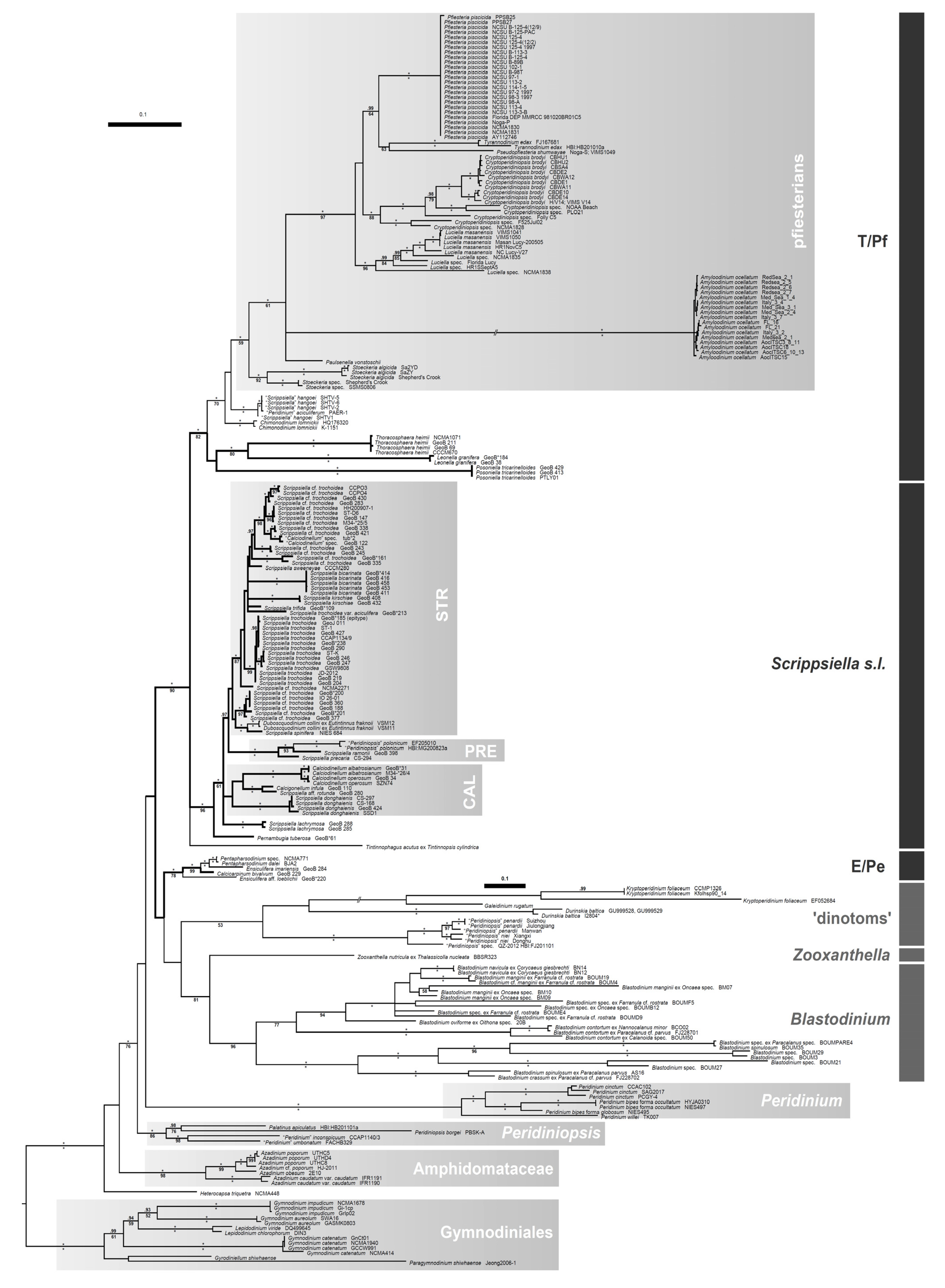
3. Brief Summary of Methods

The tree in Figure 1 is inferred from a “MAFFT” [80] generated nucleotide alignment (in total 2037 parsimony-informative positions). We defined the four regions of the ribosomal RNA (rRNA): SSU, ITS, LSU D1→D2, LSU D3→D10 and included all 199 Thoracosphaeraceae indicated by the bar on the right (plus 32 outgroup representatives), from which combinations of at least two loci were available. Additionally, we included sequences from Paulsenella Chatton that also show phylogenetic affinities to the Thoracosphaeraceae [59], but from which SSU data are only available. All outgroup taxa (other members of the Peridiniales, Amphidomataceae, Gymnodiniales) comprised the full sequence information. For the generation of new rRNA sequences from calcareous dinophyte strains out of our own culture collection (KF751921–KF751927), see the detailed descriptions in one of our previous studies [38]. Phylogenetic analyses were carried out using Maximum-Likelihood (ML) and Bayesian approaches, as described in detail previously [38]. The Bayesian analysis was performed using “MrBayes” v3.1.2 [81] under the GTR + Γ substitution model and the random-addition-sequence method with 10 replicates. We ran two independent analyses of four chains (one cold and three heated) with 15,000,000 cycles, sampled every 1000th cycle, with an appropriate burn-in (10%, after checking convergence). For the ML calculation, “RAxML” v7.2.6 [82] was applied by using the GTR + CAT substitution model to search for the best-scoring ML tree and a rapid bootstrap analysis of 1000 non-parametric replicates.

Acknowledgments

We thank John McNeill (Royal Ontario Museum, Toronto) and Vincent Demoulin (Université de Liège-Sart Tilman) for taxonomic discussion, James Byng (RBG Kew, University of Aberdeen) for improving the English text, and the Deutsche Forschungsgemeinschaft (grants KE 322/36, RI 1738/5, and WI 725/25) for financial support.

Conflicts of Interest

The authors declare no conflict of interest.

References

  1. Elbrächter, M.; Gottschling, M.; Hildebrand-Habel, T.; Keupp, H.; Kohring, R.; Lewis, J.; Meier, K.J.S.; Montresor, M.; Streng, M.; Versteegh, G.J.M.; et al. Establishing an Agenda for Calcareous Dinoflagellate Research (Thoracosphaeraceae, Dinophyceae) including a nomenclatural synopsis of generic names. Taxon 2008, 57, 1289–1303. [Google Scholar]
  2. Meier, K.J.S.; Young, J.R.; Kirsch, M.; Feist-Burkhardt, S. Evolution of different life-cycle strategies in oceanic calcareous dinoflagellates. Eur. J. Phycol. 2007, 42, 81–89. [Google Scholar] [CrossRef]
  3. Kaufmann, A. Polythalamien des Seewerkalkes. In Die Urwelt der Schweiz; (in German). Heer, O.V., Ed.; Schultheß: Zurich, Switzerland, 1865; pp. 194–199. [Google Scholar]
  4. Lorenz, T. Geologische Studien im Grenzgebiet zwischen helvetischer und ostalpiner Facies. II. Der südliche Rhaetikon. Ber. Naturf. Ges. Freiburg 1902, 12, 1–62. [Google Scholar]
  5. Deflandre, G. Micropaléontologie—Calciodinellum nov. gen., premier repésentant d’une famille nouvelle de dinoflagellés à thèque calcaire. Compt. Rend. Hebd. Séances Acad. Sci. 1947, 224, 1781–1782. (in French). [Google Scholar]
  6. Kamptner, E. Coccolithineen-Skelettreste aus Tiefseeablagerungen des Pazifischen Ozeans. Ann. Naturhistorischen Musums Wien 1963, 66, 139–204. (in German). [Google Scholar]
  7. Bolli, H.M. Jurassic and Cretaceous Calcisphaerulidae from DSDP Leg 27, eastern Indian Ocean. Init. Rep. 1974, 27, 843–907. [Google Scholar]
  8. Pflaumann, U.; Krasheninnikov, V.A. Cretaceous calcisphaerulids from DSDP Leg 41, eastern North Atlantic. Init. Rep. 1978, 41, 817–839. [Google Scholar]
  9. Keupp, H. Die kalkigen Dinoflagellaten-Zysten der borealen Unter-Kreide (Unter-Hauterivium bis Unter-Albium). Facies 1981, 5, 1–189. (in German). [Google Scholar] [CrossRef]
  10. Janofske, D. Kalkiges Nannoplankton, insbesondere Kalkige Dinoflagellaten-Zysten der alpinen Ober-Trias: Taxonomie, Biostratigraphie und Bedeutung für die Phylogenie der Peridiniales. Berliner Geowissenschaftliche Abhandlungen 1992, 4, 1–53. (in German). [Google Scholar]
  11. Willems, H. Kalkige Dinoflagellaten-Zysten aus der oberkretazischen Schreibkreide-Fazies N-Deutschlands (Coniac bis Maastricht). Senckenbergiana Lethaea 1988, 68, 433–477. (in German). [Google Scholar]
  12. Versteegh, G.J.M. New Pliocene and Pleistocene calcareous dinoflagellate cysts from southern Italy and Crete. Rev. Palaeobot. Palynol. 1993, 78, 353–380. [Google Scholar] [CrossRef]
  13. Kohring, R. Kalkdinoflagellaten aus dem Mittel- und Obereozän von Jütland (Dänemark) und dem Pariser Becken (Frankreich) im Vergleich mit anderen Tertiär-Vorkommen. Berliner Geowissenschaftliche Abhandlungen 1993, 6, 1–164. (in German). [Google Scholar]
  14. Kienel, U. Die Entwicklung der kalkigen Nannofossilien und der kalkigen Dinoflagellaten-Zysten an der Kreide/Tertiär-Grenze in Westbrandenburg im Vergleich mit Profilen in Nordjütland und Seeland (Dänemark). Berliner Geowissenschaftliche Abhandlungen 1994, 12, 1–87. (in German). [Google Scholar]
  15. Hildebrand-Habel, T. Die Entwicklung kalkiger Dinoflagellaten im Südatlantik seit der höheren Oberkreide. Berichte Fachbereich Geowissenschaften Universität Bremen 2002, 192, 1–152. (in German). [Google Scholar]
  16. Zügel, P. Verbreitung kalkiger Dinoflagellaten-Zysten im Cenomen/Turon von Westfrankreich und Norddeutschland. Courier Forschungsinstitut Senckenberg 1994, 176, 1–120. (in German). [Google Scholar]
  17. Streng, M. Phylogenetic aspects and taxonomy of calcareous dinoflagellates. Berichte Fachbereich Geowissenschaften Universität Bremen 2003, 210, 1–157. [Google Scholar]
  18. Meier, K.J.S. Calcareous dinoflagellates from the Mediterranean Sea: Taxonomy, ecology and palaeoenvironmental application. Berichte Fachbereich Geowissenschaften Universität Bremen 2003, 206, 1–126. [Google Scholar]
  19. Wall, D.; Dale, B. Quaternary calcareous dinoflagellates (Calciodinellideae) and their natural affinities. J. Paleontol. 1968, 42, 1395–1408. [Google Scholar]
  20. Lewis, J. Cyst-theca relationships in Scrippsiella (Dinophyceae) and related orthoperidinoid genera. Bot. Mar. 1991, 34, 91–106. [Google Scholar]
  21. Montresor, M.; Janofske, D.; Willems, H. The cyst-theca relationship in Calciodinellum operosum emend. (Peridiniales, Dinophyceae) and a new approach for the study of calcareous cysts. J. Phycol. 1997, 33, 122–131. [Google Scholar]
  22. Montresor, M.; Zingone, A.; Marino, D. The calcareous resting cyst of Pentapharsodinium tyrrhenicum comb. nov. (Dinophyceae). J. Phycol. 1993, 29, 223–230. [Google Scholar]
  23. D’Onofrio, G.; Marino, D.; Bianco, L.; Busico, E.; Montresor, M. Toward an assessment on the taxonomy of dinoflagellates that produce calcareous cysts (Calciodinelloideae, Dinophyceae): A morphological and molecular approach. J. Phycol. 1999, 35, 1063–1078. [Google Scholar]
  24. Karwath, B. Ecological studies on living and fossil calcareous dinoflagellates of the equatorial and tropical Atlantic Ocean. Berichte Fachbereich Geowissenschaften Universität Bremen 2000, 152, 1–175. [Google Scholar]
  25. Meier, K.J.S.; Janofske, D.; Willems, H. New calcareous dinoflagellates (Calciodinelloideae) from the Mediterranean Sea. J. Phycol. 2002, 38, 602–615. [Google Scholar]
  26. Gottschling, M.; Knop, R.; Plötner, J.; Kirsch, M.; Willems, H.; Keupp, H. A molecular phylogeny of Scrippsiella sensu lato (Calciodinellaceae, Dinophyta) with interpretations on morphology and distribution. Eur. J. Phycol. 2005, 40, 207–220. [Google Scholar] [CrossRef]
  27. Zinssmeister, C.; Soehner, S.; Kirsch, M.; Facher, E.; Meier, K.J.S.; Keupp, H.; Gottschling, M. Same but different: Two novel bicarinate species of extant calcareous dinophytes (Thoracosphaeraceae, Peridiniales) from the Mediterranean Sea. J. Phycol. 2012, 48, 1107–1118. [Google Scholar] [CrossRef]
  28. Gu, H.; Kirsch, M.; Zinssmeister, C.; Soehner, S.; Meier, K.J.; Liu, T.; Gottschling, M. Waking the dead: Morphological and molecular characterization of extant †Posoniella tricarinelloides (Thoracosphaeraceae, Dinophyceae). Protist 2013, 164, 583–597. [Google Scholar] [CrossRef]
  29. Lohmann, H. Die Bevölkerung des Ozeans mit Plankton. Nach den Ergebnissen der Zentrifugenfänge während der Ausreise der “Deutschland” 1911. Zugleich ein Beitrag zur Biologie des Atlantischen Ozeans. Arch. Biontologie 1920, 4, 1–617. (in German). [Google Scholar]
  30. Fütterer, D.K. Kalkige Dinoflagellaten (“Calciodinelloideae”) und die systematische Stellung der Thoracosphaeroideae. Neues Jahrbuch Geologie Paläontologie Abhandlungen 1976, 151, 119–141. (in German). [Google Scholar]
  31. Inouye, I.; Pienaar, R.N. Observations on the life cycle and microanatomy of Thoracosphaera heimii (Dinophyceae) with special reference to its systematic position. S. Afr. J. Bot. 1983, 2, 63–75. [Google Scholar]
  32. Tangen, K.; Brand, L.E.; Blackwelder, P.L.; Guillard, R.R.L. Thoracosphaera heimii (Lohmann) Kamptner is a dinophyte: Observations on its morphology and life cycle. Mar. Micropaleontol. 1982, 7, 193–212. [Google Scholar] [CrossRef]
  33. Fensome, R.A.; Taylor, F.J.R.; Norris, G.; Sarjeant, W.A.S.; Wharton, D.I.; Williams, G.L. A Classification of Living and Fossil Dinoflagellates. In Micropaleontology; Special Publication 7; American Museum of Natural History: : New York, NY, USA, 1993; pp. 1–245. [Google Scholar]
  34. Gottschling, M.; Keupp, H.; Plötner, J.; Knop, R.; Willems, H.; Kirsch, M. Phylogeny of calcareous dinoflagellates as inferred from ITS and ribosomal sequence data. Mol. Phylogenet. Evol. 2005, 36, 444–455. [Google Scholar] [CrossRef]
  35. Wall, D.; Dale, B. Modern dinoflagellate cysts and evolution of the Peridiniales. Micropaleontology 1968, 14, 265–304. [Google Scholar] [CrossRef]
  36. Place, A.R.; Saito, K.; Deeds, J.R.; Robledo, J.A.F.; Vasta, G.R. A Decade of Research on Pfiesteria spp. and Their Toxins: Unresolved Questions and an Alternative Hypothesis. In Seafood and Freshwater Toxins: Pharmacology, Physiology, and Detection, 2nd ed.; CRC Press: London, UK, 2008; pp. 717–751. [Google Scholar]
  37. Steidinger, K.A.; Burkholder, J.M.; Glasgow, H.B., Jr; Hobbs, C.W.; Garrett, J.K.; Truby, E.W.; Noga, E.J.; Smith, S.A. Pfiesteria piscicida gen. et sp. nov. (Pfiesteriaceae fam. nov.), a new toxic dinoflagellate with a complex life cycle and behavior. J. Phycol. 1996, 32, 157–164. [Google Scholar]
  38. Gottschling, M.; Soehner, S.; Zinssmeister, C.; John, U.; Plötner, J.; Schweikert, M.; Aligizaki, K.; Elbrächter, M. Delimitation of the Thoracosphaeraceae (Dinophyceae), including the calcareous dinoflagellates, based on large amounts of ribosomal RNA sequence data. Protist 2012, 163, 15–24. [Google Scholar] [CrossRef] [Green Version]
  39. Zhang, H.; Bhattacharya, D.; Lin, S.J. A three-gene dinoflagellate phylogeny suggests monophyly of Prorocentrales and a basal position for Amphidinium and Heterocapsa. J. Mol. Evol. 2007, 65, 463–474. [Google Scholar] [CrossRef]
  40. Tillmann, U.; Salas, R.; Gottschling, M.; Krock, B.; O’Driscol, D.; Elbrächter, M. Amphidoma languida sp. nov. (Dinophyceae) reveals a close relationship between Amphidoma and Azadinium. Protist 2012, 163, 701–719. [Google Scholar] [CrossRef]
  41. Steidinger, K.A.; Landsberg, J.H.; Mason, P.L.; Vogelbein, W.K.; Tester, P.A.; Litaker, R.W. Cryptoperidiniopsis brodyi gen. et sp. nov. (Dinophyceae), a small lightly armored dinoflagellate in the Pfiesteriaceae. J. Phycol. 2006, 42, 951–961. [Google Scholar] [CrossRef]
  42. Mason, P.L.; Litaker, R.W.; Jeong, H.J.; Ha, J.H.; Reece, K.S.; Stokes, N.A.; Park, J.Y.; Steidinger, K.A.; Vandersea, M.W.; Kibler, S.; Tester, P.A.; Vogelbein, W.K. Description of a new genus of Pfesteria-like dinoflagellate, Luciella gen. nov. (Dinophyceae), including two new species: Luciella masanensis sp. nov. and Luciella atlantis sp. nov. J. Phycol. 2007, 43, 799–810. [Google Scholar] [CrossRef]
  43. Calado, A.J. On the identity of the freshwater dinoflagellate Glenodinium edax, with a discussion on the genera Tyrannodinium and Katodinium, and the description of Opisthoaulax gen. nov. Phycologia 2011, 50, 641–649. [Google Scholar] [CrossRef]
  44. Stern, R.F.; Andersen, R.A.; Jameson, I.; Kuepper, F.C.; Coffroth, M.-A.; Vaulot, D.; le Gall, F.; Veron, B.; Brand, J.J.; Skelton, H.; et al. Evaluating the ribosomal Internal Transcribed Spacer (ITS) as a candidate dinoflagellate barcode marker. PLoS One 2012, 7, e42780. [Google Scholar] [CrossRef]
  45. Coats, D.W.; Kim, S.; Bachvaroff, T.R.; Handy, S.M.; Delwiche, C.F. Tintinnophagus acutus n. g., n. sp. (Phylum Dinoflagellata), an ectoparasite of the ciliate Tintinnopsis cylindrica Daday 1887, and its relationship to Duboscquodinium collini Grassé 1952. J. Eukaryot. Microbiol. 2010, 57, 468–482. [Google Scholar] [CrossRef]
  46. Smith, K.D.; Dodson, M.; Santos, S.; Gast, R.; Rogerson, A.; Sullivan, B.; Moss, A.G. Pentapharsodinium tyrrhenicum is a parasitic dinoflagellate of the ctenophore Mnemiopsis leidyi. J. Phycol. 2007, 43, 119. [Google Scholar]
  47. Smith, K.D. A Parasitic Dinoflagellate of the Ctenophore Mnemiopsis sp. Master’s Thesis, Auburn University, Auburn, AL, USA, 2011. [Google Scholar]
  48. Montresor, M.; Sgrosso, S.; Procaccini, G.; Kooistra, W.H.C.F. Intraspecific diversity in Scrippsiella trochoidea (Dinophyceae): Evidence for cryptic species. Phycologia 2003, 42, 56–70. [Google Scholar] [CrossRef]
  49. Soehner, S.; Zinssmeister, C.; Kirsch, M.; Gottschling, M. Who am I—And if so, how many? Species diversity of calcareous dinophytes (Thoracosphaeraceae, Peridiniales) in the Mediterranean Sea. Org. Divers. Evol. 2012, 12, 339–348. [Google Scholar] [CrossRef]
  50. Gottschling, M.; McLean, T.I. New home for tiny symbionts: Dinophytes determined as Zooxanthella are Peridiniales and distantly related to Symbiodinium. Mol. Phylogenet. Evol. 2013, 67, 217–222. [Google Scholar] [CrossRef]
  51. Skovgaard, A.; Massana, R.; Saiz, E. Parasitic species of the genus Blastodinium (Blastodiniphyceae) are peridinioid dinoflagellates. J. Phycol. 2007, 43, 553–560. [Google Scholar] [CrossRef]
  52. Coats, D.W.; Bachvaroff, T.; Handy, S.M.; Kim, S.; Gárate-Lizárraga, I.; Delwiche, C.F. Prevalence and phylogeny of parasitic dinoflagellates (genus Blastodinium) infecting copepods in the Gulf of California. CICIMAR Oceánides 2008, 23, 67–77. [Google Scholar]
  53. Tamura, M.; Shimada, S.; Horiguchi, T. Galeidiniium rugatum gen. et sp. nov. (Dinophyceae), a new coccoid dinoflagellate with a diatom endosymbiont. J. Phycol. 2005, 41, 658–671. [Google Scholar] [CrossRef]
  54. Horiguchi, T.; Takano, Y. Serial replacement of a diatom endosymbiont in the marine dinoflagellate Peridinium quinquecorne (Peridiniales, Dinophyceae). Phycol. Res. 2006, 54, 193–200. [Google Scholar] [CrossRef]
  55. Zhang, Q.; Liu, G.X.; Hu, Z.-Y. Durinskia baltica (Dinophyceae), a newly recorded species and genus from China, and its systematics. J. Syst. Evol. 2011, 49, 476–485. [Google Scholar] [CrossRef]
  56. Ride, W.D.L.; Cogger, H.G.; Dupuis, C.; Krauss, O.; Minelli, A.; Thompson, F.C.; Tubbs, P.K. International code of zoological nomenclature: Adopted by the International Union of Biological Sciences; International Trust for Zoological Nomenclature: London, UK, 1999. [Google Scholar]
  57. McNeill, J.; Barrie, F.R.; Buck, W.R.; Demoulin, V.; Greuter, W.; Hawksworth, D.L.; Herendeen, P.S.; Knapp, S.; Marhold, K.; Prado, J.; et al. International Code of Nomenclature for algae, fungi and plants (Melbourne Code) adopted by the Eighteenth International Botanical Congress Melbourne, Australia, July 2011; Koeltz: Königstein, Germany, 2012. [Google Scholar]
  58. Litaker, R.W.; Tester, P.A.; Colorni, A.; Levy, M.G.; Noga, E.J. The phylogenetic relationship of Pfiesteria piscicida, cryptoperidiniopsoid sp. Amyloodinoum ocellatum and a Pfiesteria-like dinoflagellate to other dinoflagellates and apicomplexans. J. Phycol. 1999, 35, 1379–1389. [Google Scholar] [CrossRef]
  59. Kühn, S.F.; Medlin, L.K. The systematic position of the parasitoid marine dinoflagellate Paulsenella vonstoschii (Dinophyceae) inferred from nuclear-encoded small subunit ribosomal DNA. Protist 2005, 156, 393–398. [Google Scholar] [CrossRef]
  60. Jeong, H.J.; Kim, J.S.; Park, J.Y.; Kim, J.H.; Kim, S.; Lee, I.; Lee, S.H.; Ha, J.H.; Yih, W.H. Stoeckeria algicida n. gen., n. sp. (Dinophyceae) from the coastal waters off southern Korea: Morphology and small subunit ribosomal DNA gene sequence. J. Eukaryot. Microbiol. 2005, 52, 382–390. [Google Scholar] [CrossRef]
  61. Banasová, M.; Kopčáková, J.; Reháková, D. Bádenské asociácie vápnitých dinoflagelát z vrtu Stupava HGP-3 a Malacky-101 (viedenská panva). Mineralia Slovaca 2007, 39, 107–122. (in Slovak). [Google Scholar]
  62. Calado, A.J.; Craveiro, S.C.; Daugbjerg, N.; Moestrup, Ø. Description of Tyrannodinium gen. nov., a freshwater dinoflagellate closely related to the marine Pfiesteria-like species. J. Phycol. 2009, 45, 1195–1205. [Google Scholar] [CrossRef]
  63. Özdikmen, H. Substitute names for some unicellular animal taxa (Protozoa). Munis Entomol. Zool. 2009, 4, 233–256. [Google Scholar]
  64. Streng, M.; Banasová, M.; Reháková, D.; Willems, H. An exceptional flora of calcareous dinoflagellates from the middle Miocene of the Vienna Basin, SW Slovakia. Rev. Palaeobot. Palynol. 2009, 153, 225–244. [Google Scholar] [CrossRef]
  65. Craveiro, S.C.; Calado, A.J.; Daugbjerg, N.; Hansen, G.; Moestrup, Ø. Ultrastructure and LSU rDNA-based phylogeny of Peridinium lomnickii and description of Chimonodinium gen. nov. (Dinophyceae). Protist 2011, 162, 590–615. [Google Scholar] [CrossRef]
  66. Craveiro, S.C.; Pandeirada, M.S.; Daugbjerg, N.; Moestrup, Ø.; Calado, A.J. Ultrastructure and phylogeny of Theleodinium calcisporum gen. et sp. nov., a freshwater dinoflagellate that produces calcareous cysts. Phycologia 2013, in press. [Google Scholar]
  67. Chatton, É.; Grassé, P.-P. Classe des Dinoflagelles ou Péridiniens. In Phylogénie. Protozoaires: Généralités. Flagellés 1; (in French). Grassé, P.-P., Ed.; Masson: Paris, France, 1952; pp. 309–406. [Google Scholar]
  68. Balech, E. Two new genera of dinoflagellates from California. Biol. Bull. 1959, 116, 195–203. [Google Scholar] [CrossRef]
  69. Loeblich, A.R. Dinoflagellate nomenclature. Taxon 1965, 14, 15–18. [Google Scholar] [CrossRef]
  70. Wendler, J.E.; Bown, P. Exceptionally well-preserved Cretaceous microfossils reveal new biomineralization styles. Nat. Commun. 2013, 4, 2052. [Google Scholar]
  71. Bourrelly, P. Notes sur les péridiniens d’eau douce. Protistologica 1968, 4, 5–13. (in French). [Google Scholar]
  72. Pascher, A. Über die Beziehungen der Cryptomonaden zu den Algen. Berichte Deutschen Botanischen Gesellschaft 1911, 29, 193–203. (in German). [Google Scholar]
  73. Blank, R.J.; Trench, R.K. Nomenclature of endosymbiotic dinoflagellates. Taxon 1986, 35, 286–294. [Google Scholar] [CrossRef]
  74. Banaszak, A.T.; Iglesias-Prieto, R.; Trench, R.K. Scrippsiella velellae sp. nov. (Peridiniales) and Gloeodinium viscum sp. nov. (Phytodiniales), dinoflagellate symbionts of two hydrozoans (Cnidaria). J. Phycol. 1993, 29, 517–528. [Google Scholar] [CrossRef]
  75. Silva, P.C. Report of the Committee for Algae: 2. Taxon 1994, 43, 257–264. [Google Scholar] [CrossRef]
  76. Klebs, G.A. Flagellatenstudien. Zeitschrift Wissenschaftliche Zoologie 1892, 5, 265–445. (in German). [Google Scholar]
  77. Loeblich, A.R. The Amphiesma or Dinoflagellate Cell Covering. In Proceedings of the North American Paleontological Convention 2; Yochelson, E.L., Ed.; Allen Press: Lawrence, KS, USA, 1971; pp. 867–929. [Google Scholar]
  78. Cavers, F. Recent work on flagellata and primitive algæ. New Phytol. 1913, 12, 225–232. [Google Scholar] [CrossRef]
  79. Schiller, J. Coccolithineae. In Dr. L. Rabenhorst’s Kryptogamen-Flora 10, Abt. 2; (in German). Kolkwitz, R., Ed.; Akademische Verlagsgesellschaft: Leipzig, Germany, 1930; pp. 89–273. [Google Scholar]
  80. Katoh, K.; Standley, D.M. MAFFT multiple sequence alignment software version 7: Improvements in performance and usability. Mol. Biol. Evol. 2013, 30, 772–780. [Google Scholar] [CrossRef]
  81. Ronquist, F.; Teslenko, M.; van der Mark, P.; Ayres, D.L.; Darling, A.; Höhna, S.; Larget, B.; Liu, L.; Suchard, M.A.; Huelsenbeck, J.P. MrBayes 3.2: Efficient Bayesian phylogenetic inference and model choice across a large model space. Syst. Biol. 2012, 61, 539–542. [Google Scholar] [CrossRef]
  82. Stamatakis, A. RAxML-VI-HPC: Maximum likelihood-based phylogenetic analyses with thousands of taxa and mixed models. Bioinformatics 2006, 22, 2688–2690. [Google Scholar] [CrossRef]

