Review

Prions in Variably Protease-Sensitive Prionopathy: An Update

1
Department of Pathology Case Western Reserve University School of Medicine, Cleveland, OH 44106, USA
2
Department of Neurology, Case Western Reserve University School of Medicine, Cleveland, OH 44106, USA
3
National Prion Disease Pathology Surveillance Center, Case Western Reserve University School of Medicine, Cleveland, OH 44106, USA
4
National Center for Regenerative Medicine, Case Western Reserve University School of Medicine, Cleveland, OH 44106, USA
5
The First Affiliated Hospital, Nanchang University, Nanchang 330006, Jiangxi Province, China
6
State Key Laboratory for Infectious Disease Prevention and Control, National Institute for Viral Disease Control and Prevention, Chinese Center for Disease Control and Prevention, Beijing 100050, China
7
Central Veterinary Institute of Wageningen UR, Lelystad 8200 AB, the Netherlands
8
Department of Veterinary Public Health and Food Safety, Istituto Superiore di Sanità, Viale Regina Elena 299 00161, Rome, Italy
*
Author to whom correspondence should be addressed.
Pathogens 2013, 2(3), 457-471; https://doi.org/10.3390/pathogens2030457
Submission received: 12 June 2013 / Revised: 28 June 2013 / Accepted: 2 July 2013 / Published: 5 July 2013
(This article belongs to the Special Issue Prions)

Abstract

:
Human prion diseases, including sporadic, familial, and acquired forms such as Creutzfeldt-Jakob disease (CJD), are caused by prions in which an abnormal prion protein (PrPSc) derived from its normal cellular isoform (PrPC) is the only known component. The recently-identified variably protease-sensitive prionopathy (VPSPr) is characterized not only by an atypical clinical phenotype and neuropathology but also by the deposition in the brain of a peculiar PrPSc. Like other forms of human prion disease, the pathogenesis of VPSPr also currently remains unclear. However, the findings of the peculiar features of prions from VPSPr and of the possible association of VPSPr with a known genetic prion disease linked with a valine to isoleucine mutation at residue 180 of PrP reported recently, may be of great importance in enhancing our understanding of not only this atypical human prion disease in particular, but also other prion diseases in general. In this review, we highlight the physicochemical and biological properties of prions from VPSPr and discuss the pathogenesis of VPSPr including the origin and formation of the peculiar prions.

1. Introduction

Prions are infectious pathogens that are associated with a group of fatal transmissible spongiform encephalopathies or prion diseases affecting both animals and humans. They are composed mainly, if not entirely, of the pathologic scrapie conformer (PrPSc) and originate from the cellular prion protein (PrPC) by means of a structural transition from a largely α-helical form to predominantly β-sheets [1]. Unlike other infectious agents, such as bacteria, viruses, and fungi, which contain genomes composed of either DNA or RNA, prions are the only known infectious pathogens that are devoid of nucleic acid, according to the “protein only” hypothesis [1]. Human prion diseases are highly heterogeneous: They can be familial, sporadic, or acquired by infection, and include Creutzfeldt-Jakob disease (CJD), Gerstmann-Sträussler-Scheinker (GSS) disease, fatal insomnia, kuru and variant CJD (vCJD) [2]. Atypical human and animal prion diseases have recently been identified including variably protease-sensitive prionopathy (VPSPr) in human and Nor98/atypical scrapie in sheep and goats [3,4,5,6,7]. The two atypical human and sheep prion diseases are characterized by the deposition of peculiar prions in the brain. No mutations have been found in the open reading frame of prion protein gene in the two diseases. While Nor98 scrapie is associated with polymorphisms at R154H and L141F, VPSPr is observed in all three genotypes of PrP polymorphism at residue 129 of PrP. PrPSc from the two diseases exhibited a small PK-resistant fragment similar to those observed in some of familial prion diseases [4,5,6,8].

2. Dominant Protease-Sensitive PrPSc Conformer

In the eleven cases first reported, they were all valine/valine homozygosity at residue 129 of PrP and more than half of them had a family history of dementia [4]. Although spongiform degeneration and PrP immunostaining were observed, surprisingly, no typical PK-resistant PrPSc (rPrPSc) was detectable in the brain of all cases by conventional Western blotting probing with the widely-used anti-PrP antibody 3F4. The 3F4 antibody that has an epitope between residues 106 and 112 [9] detected an abnormal PrP in PSPr only after enrichment with gene 5 protein (g5p) and sodium phosphotungstate (NaPTA) that are able to bind to abnormally-folded PrP molecules regardless of their PK resistance [10,11]. However, more than 70% of the abnormal PrP captured by g5p from these cases was sensitive to PK-digestion while only about 10% of captured PrPSc was PK-sensitive in sCJD. Therefore, this atypical human prion disease characterized by the deposition in the brain of dominant PK-sensitive PrPSc (sPrPSc) was initially termed as protease-sensitive prionopathy (PSPr) [4].
Figure 1. Detection of PrP from variably protease-sensitive prionopathy (VPSPr), Gerstmann-Sträussler-Scheinker (GSS), and sporadic Creutzfeldt-Jakob disease (sCJD) with 129 methionine/methionine (MM) polymorphism and PrPSc type 1 (sCJDMM1) with nine anti-PrP antibodies. A: Diagram of epitope locations of anti-PrP antibodies examined on human PrP. Antibodies and their epitopes are: 3F4 (PrP106-112), 1E4 (PrP97-105), 6D11 (PrP93-109), 8G8 (PrP95-110), Anti-C (PrP220-231), 6H4 (PrP145-152), 9A2 (PrP99-101), 12B2 (PrP89-93), and V14 (PrP185-196). B through J: Brain homogenates from VPSPr, GSS linked to PrPP1°2L mutation (GSS102), GSS linked to PrPA117V mutation, and sCJDMM1 were treated with PK or/PNGase F prior to SDS-PAGE and Western blotting with nine different anti-PrP antibodies, respectively. B: 3F4; C: 1E4; D: 6D11; E: 8G8; F: Anti-C; G: 6H4; H: 9A2; I: 12B2; J: V14. Of the nine antibodies used, 1E4 exhibits the highest affinity for rPrPSc from VPSPr. However, 1E4 has a lower affinity for rPrPSc from GSS102 compared to 3F4. It could be due to the PrPP102L mutation that is localized within the 1E4 epitope. Since VPSPr20 and VPSPr17 are detectable by 6D11 that is against human PrP93-109, their N-terminal domains may start at least from residue 93. VPSPr7 is recognized by 1E4 that is against human PrP97-105, suggesting that the N-terminus of VPSPr7 contains residue 97.
Pathogens 02 00457 g001

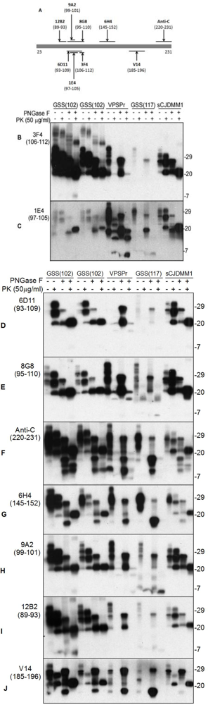
3. Pathognomonic Ladder-Like Electrophoretic Profile of Protease-Resistant PrPSc with a Peculiar Immunoreactivity Behavior

Although undetectable with 3F4 by conventional Western blotting, a ladder-like electrophoretic profile of PK-resistant PrPSc (rPrPSc) was readily detected with an antibody called 1E4, which is unprecedented in sporadic human prion diseases [4]. The 1E4 clone was derived from hybridization of SP2/0-Ag14 myeloma cells with spleen cells from a PrP-knockout mouse immunized with the peptide QWNKPSKPKTN that corresponds to the bovine PrP amino acid sequence 108–119 (http://www.cellsciences.com/content/p-detail.asp?rowid=8107). While this antibody was mostly used for detection of bovine and ovine PrPSc, we demonstrated that this antibody with an epitope localized between human PrP residues 97 and 105 (Yuan et al., 2008) exhibiting the highest affinity for PrPSc of PSPr among the nine anti-PrP antibodies we examined including 1E4 (against PrP97-105), 3F4 (PrP106-112), 6D11 (PrP93-109), 8G8 (PrP95-110), Anti-C (220-231), 6H4 (PrP145-152), 9A2 (PrP99-101), 12B2 (PrP89-93), and V14 (PrP185-196) (Figure 1). Since the 1E4-detected ladder-like electrophoretic profile of rPrPSc is very unique and was detected repeatedly in all eleven cases, it has been considered to be pathognomonic for PSPr.
The discovery of the pathognomonic molecular feature of PSPr has greatly facilitated the identification of this unique type of human prion disease. Bearing this feature of PSPr in mind, we reexamined retroprospectively suspected cases referred to the National Prion Disease Pathology Surveillance Center (NPDPSC, Cleveland, OH, USA) between 2002 and 2010 including two cases from Italy [5]. For newly-referred cases, it has become a routine procedure to re-do Western blot analysis with the 1E4 antibody in order to find out the possible cases of PSPr at NPDPSC if they are negative for rPrPSc by Western blotting with 3F4 but positive for H&E staining and immunohistochemistry with 3F4.

4. Polymorphism-Dependent PK-Sensitive and PK-Resistant PrPSc

In 2010, we first reported that PSPr affects not only subjects homozygous for valine at PrP residue 129 but also subjects homozygous for methionine (129 MM) or heterozygous for methionine/valine (129 MV) [5]. Of the fifteen cases we examined, one of the two Italian cases was previously reported by Giaccone et al [3]. Compared to the initially reported PSPr in valine homozygotes, the levels of sPrPSc were significantly decreased while the levels of rPrPSc were significantly increased in 129 MM or 129 MV cases. Interestingly, it seems that the levels of rPrPSc are dictated by methionine at residue 129. Vice versa, the levels of sPrPSc seem to be dictated by valine at residue 129. Although it has been well-documented that PrP polymorphism at residue 129 is implicated in mediating susceptibility to the disease, phenotypes of disease, and PrPSc types [2], to our knowledge, our study provided the first evidence that the polymorphism may also participate in medicating the amounts of sPrPSc or rPrPSc [5]. To more precisely reflect the polymorphism-dependent variation in the levels of rPrPSc or sPrPSc in this newly-identified disease, we revised the original designation as “variably protease-sensitive prionopathy” (VPSPr) [5].
Figure 2. Schematic diagram of electrophoretic profile of rPrPSc from VPSPr and sCJD probed with 1E4. Without PNGase F treatment, five rPrPSc fragments are detectable with Western blotting including VPSPr26, VPSPr23, VPSPr20, VPSPr17, and VPSPr7 from VPSPr while three rPrPSc fragments are detected including di-, mono-, and un-glycosylated PrP from classic sCJD. After PNGase F treatment, three core PrP fragments remain in VPSPr including VPSPr20, VPSPr17, and VPSPr7 while only one core PrP fragment remains in sCJD. VPSPr26 and VPSPr23 are monoglycosylated forms of VPSPr20 and VPSPr17, respectively.
Pathogens 02 00457 g002

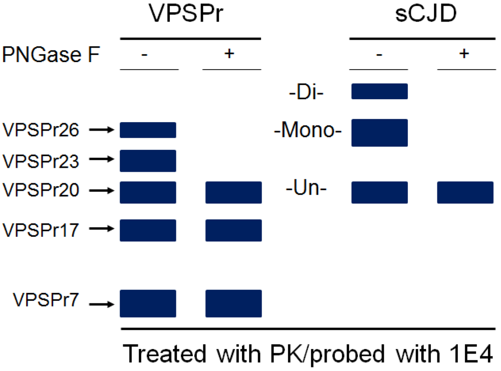
5. The Ladder-Like Electrophoretic Profile of 1E4-Detected rPrPSc Consisting of Five PrP Fragments

Some of the rPrPSc fragments became detectable in VPSPr129MM and VPSPr129MV with the 3F4 antibody, especially in the former. Notably, even though the amounts of rPrPSc in VPSPr129MM or VPSPr129MV were significantly increased compared to those of rPrPSc in VPSPr129VV, the profile of rPrPSc detected with 3F4 is different from that detected with 1E4. The most significant differences in the rPrPSc fragments detected with the two antibodies were the smallest fragment migrating at approximately 7 kDa called VPSPr7 that was detectable with 1E4 but not with 3F4 [5,6]. Except for VPSPr7, both 3F4 and 1E4 detected the other four rPrPSc fragments migrating at ~26 kDa, 23 kDa, 20 kDa, and 17 kDa, termed VPSPr26, VPSPr23, VPSPr20, and VPSPr 17, respectively, which is strikingly different from rPrPSc observed in classic sCJD (Figure 2). Based on gel migration, VPSPr26 corresponds to monoglycosylated rPrPSc of the classic sCJD, whereas VPSPr20 corresponds to unglycosylated rPrPSc of sCJD. Interestingly, no detectable diglycosylated rPrPSc was observed by both 3F4 and 1E4 in VPSPr, which was also observed in the first case reported [3]. On the other hand, three additional fragments VPSPr23, VPSPr17 and VPSPr7 were not detectable in sCJD. A PK-titration of PrPSc from VPSPr129MM or 129MV on the Western blots probed with 1E4 or 3F4 revealed that VPSPr26 and VPSPr20 gradually faded away while VPSPr23 and VPSPr17 became dominant upon increases in the PK concentrations, suggesting that VPSPr23 derives from VPSPr26 while VPSPr17 from VPSPr20. After deglycosylation, we convincingly showed that unglycosylated VPSPr20 decreased while VPSPr17 increased. As mentioned above, VPSPr20 may correspond to unglycosylated rPrPSc of sCJD and it may encompass PrP sequence from residue 90 to 231. So, it is most likely that the switch from VPSPr20 to VPSPr17 caused by PK-digestion may be due to cleavage of a C-terminal domain since both fragments were detected with antibodies against human PrP97-105 (1E4) and PrP106-112 (3F4). Moreover, the antibody Anti-C specific for human PrP220-231 detected four rPrPSc after deglycosylation with PNGase F. However, the migration of these fragments detected with Anti-C seemed to be different from that of 1E4-detected fragments except for VPSPr20. All these findings suggest that conformation of PrPSc in VPSPr is quite different from that of PrPSc in sCJD and it may have many more PK-cleavage sites of PrPSc in VPSPr than in sCJD. The cleavages may occur not only at the N-terminal domain but also at the C-terminal domain.

6. Glycoform-Selective Prion Formation in VPSPr and fCJDV180I

In general, the four glycoforms of PrPC variably glycosylated at the two N-linked glycosylation sites are usually all converted into their PrPSc counterparts in all human prion diseases [2]. However, there are exceptions. Familial CJD linked to either PrPT183A (fCJDT183A) or PrPV180I (fCJDV180I) mutation exhibits an rPrPSc that lacks the diglycosylated PrP species [12,13]. T183A mutation itself is known to completely eliminate the first glycosylation site at residue 181 [14], which is believed to account for the lack of diglycosylated rPrPSc. However, the molecular mechanism underlying alteration in glycosylation by V180I mutation is unknown. In addition to the two mutations, two cases of atypical sporadic CJD have also been reported to lack diglycosylated rPrPSc [3,15], one of which was subsequently proven to be a case of VPSPr [3,5].
To understand the molecular mechanism underlying the lack of diglycosylated rPrPSc and formation of ladder-like electrophoretic profile of rPrPSc as well as to investigate the potential association between sporadic VPSPr and familial CJD lacking diglycosylated PK-resistant PrP form, we compared their individual PrP glycoforms using antibodies that are able to distinguish the two glycosylation sites [6,16,17]. Using a combination of in vivo and in vitro assays, we demonstrated that the absence of the diglycosylated PrPSc in both VPSPr and fCJDV180I is associated with the inability of the di- and mono-glycosylated PrPC with the intact first glycosylation site (N181) to convert into PrPSc in the brain. Surprisingly, fCJDV180I was detected to have an rPrPSc that was markedly similar to that observed in VPSPr, with a five-step ladder-like electrophoretic profile, a pathognomonic molecular feature of VPSPr [6]. Therefore, although VPSPr is linked to wild-type PrP and fCJDV180I is linked to mutant PrP, both sporadic VPSPr and fCJDV180I may share a unique glycoform-selective prion formation pathway. Moreover, conformation of PrPSc in the two conditions also may be very similar, as evidenced by the generation of a virtually identical electrophoretic profile of rPrPSc upon PK-treatment.
Although the molecular mechanism underlying glycoform-selective prion formation is unclear at present, there are several clues that may be of significance in understanding this mystery. First, in contrast to fCJDT183A, both VPSPr and fCJDV180I exhibited an intact glycosylation prior to PK-digestion. Moreover, PrPV180I in cultured cells had a typical glycoform profile. It generated detectable typical rPrPSc with diglycosylated PrP form upon PK-treatment, in addition to mono-unglycosylated forms although they were detectable only with 1E4 but not with 3F4 [6]. Therefore, PrPV180I mutation itself does not eliminate any glycosylation sites and it can be converted into rPrPSc as does wild-type PrPC. Second, a decrease in the glycosylation potential value for the first glycosylation site was predicated by the N-linked glycosylation prediction algorithm [6], suggesting that PrPV180I may alter the composition of glycans at the first site. Third, diglycosylated PrP or monoglycosylated PrP carrying mono181 was not converted into rPrPSc, which was not observed in cultured cells but only observed in the brain in which there is an additional wild-type allele. Finally, compared to sCJD, the binding of ricinus communis agglutinin I (RCA-I) to monoglycosylated PrP decreased while the binding to diglycosylated PrP increased in VPSPr and fCJDV180I [6], suggesting that the two diseases have a changed composition of glycans. Therefore, it is possible that glycoform-selective prion formation observed in brain involves dominant-negative inhibition caused by the interaction between misfolded and normal PrP molecules. The changed composition of glycans at the first site by the mutation may alter local conformation around residue 181 that is close to the β-sheets 2/α-helixes 2 loop, the critical region implicated in dominant-negative inhibition [18]. Consistent with this hypothesis, it seems that there are significant differences in the effect of mutations occurring at either the first or the second glycosylation site on the conversion of PrPC into PrPSc. While most of mutations at the first site blocked the conversion, none of or a few, if any, mutations at the second site were found to block PrPC conversion in cell and animal models [19,20]. More specifically, interactions between different PrPC glycoforms mediate the efficiency of prion formation, which involves glycan-associated steric hindrance [21]. The same group also demonstrated that dominant negative inhibition of prion formation requires no protein X or any other accessory cofactor [22]. Although no PrP mutations have been observed in VPSPr, a similar aberrant glycosylation at N181 caused by a rare stochastic event has been proposed to trigger the processes as described for fCJDV180I [6].
Another possibility to cause glycoform-selective prion formation is that one or more co-factors may be operating in VPSPr and fCJDV180I and the co-factors may prevent conversion of diglycosylated PrP and mono181 into PrPSc. There may be some implications in the two diseases that other co-factors may be involved in the pathogenesis of the diseases. For instance, although linked to the PrPV180I mutation, no family history of neurodegenerative disorders has been reported in fCJDV180I cases [23]. On the other hand, while no mutations have been identified within the coding sequence (the open reading frame) of PrP gene in VPSPr to date, eight out of 26 reported VPSPr cases showed a familial history of dementia [5,24]. Indeed, several lines of evidence have indicated that other co-factors may be involved in the pathogenesis of prion diseases, including protein X and non-proteinaceous cofactors [25,26,27]. Whether protein X that was initially proposed to directly interact with PrPC is necessary for prion formation remains controversial [26,28]. However, genes or proteins that may indirectly trigger the conversion of PrPC into PrPSc may exist. It is possible that a mutation in a specific gene that participates in regulating PrP glycosylation may alter glycosylation at the first glycosylation site and then causes VPSPr or/and fCJDV180I. If this is the case, further investigation of the two diseases may provide an opportunity to find out about the existence of such a co-factor.

7. Transmissibility of VPSPr and fCJDV180I

It has been widely accepted that any prion diseases must be transmissible although there are some prion diseases that have not been transmitted yet [29,30]. Before Stanley Prusiner discovered prions and coined prion in 1982 [31], the term transmissible spongiform encephalopathy (TSE) had been widely used. As indicated by its striking name, a TSE must possess these two major characteristics: transmissibility and spongiform degeneration in the central nervous system (CNS). The discovery of prions as infectious protein pathogens, which are free of nuclear acids and which are the cause of transmissible spongiform encephalopathies, was a revolutionary development not only for the field in particular but also for the life science in general. PrPSc has in fact been observed in almost all TSE identified so far. Identification of PrPSc has become essential in the current diagnostic criteria for TSE. The designation, “prion diseases”, has largely replaced “transmissible spongiform encephalopathy”.
However, confusion results when it is observed that some prion diseases lack one or two characteristics of TSE. Approximately 10% of sporadic CJD and 32% of familial prion diseases were non-transmissible in nonhuman primates [32]. Moreover, all GSS, except one-third of GSSP102L cases, were difficult to transmit to rodents [33]. Further, the spongiform degeneration typical of TSE is not always present in all GSSP102L, although diffuse deposits of PrPSc plus PrP-amyloid plaques are present in the CNS [34]. In transgenic mice expressing murine PrP P101L (equivalent to human P102L) and challenged with GSS free of spongiform degeneration, neither symptoms nor spongiform degeneration was observed despite the presence of PrP-amyloid [35]. Obviously, conditions such as these, which do not manifest transmissibility or spongiform degeneration (singly or jointly), should not be considered as types of TSE. They do, however, constitute prion diseases. Based on a wealth of data gathered so far, one may wonder whether or not prion diseases should now be redefined. Under a reconsidered definition, they should include a group of disorders characterized by the accumulation of abnormal PrP including protease-sensitive and protease-resistant forms in the brain, regardless of the presence of transmissibility and spongiform degeneration. Most importantly, the spectrum of prion diseases must not be restrained by the definition of TSE.
As a prion disease, VPSPr should likewise meet Koch’s postulates as well [36]. So, it would be important to investigate the transmissibility of VPSPr that exhibits the peculiar PK-resistant behavior of PrP. Preliminary data recently reported by Nonno et al. indicate that transmissibility of VPSPr-129MM, -129MV and -129VV to bank voles is related to the bank vole methionine / isoleucine polymorphism at codon 109 [37]. Furthermore, studies with humanized mice suggest that transmissibility of VPSPr is much lower compared to classic sporadic CJD [38]. No clinical phenotypes were observed during the normal life span of transgenic mice expressing human PrP-129V at approximately 10 fold, following inoculation with brain homogenates from VPSPr-129VV cases. Less than 20% of the mice were found to have scattered PrP plaques with minimal or no spongiform degeneration, compared to the typical neuropathological changes found in 100% mice inoculated with the classic sCJD [38]. Similarly, using protein misfolding cyclic amplification (PMCA) assay, we found that the amplification efficiency of PrPSc from VPSPr is much lower compared to iatrogenic and sporadic CJD (Zou et al., unpublished data). To date, there are no reports available demonstrating that fCJDV180I is transmissible [39]. Therefore, prions in VPSPr and fCJDV180I exhibit striking similarities not only in physicochemical but also in biological properties. In collaboration with Drs. Yong-Sun Kim and Robert Petersen, we are generating humanized transgenic mice expressing human PrPV180I currently and will be testing transmissibility of VPSPr and fCJDV180I with this animal model.

8. Origin of Prions in VPSPr and fCJDV180I

As mentioned above, prions from VPSPr and fCJDV18°I are of unique physicochemical and biological properties. Remarkably, they exhibit a high immunoreactivity with the 1E4 antibody but a poor reactivity with 3F4 [5,6]. We have demonstrated that the two antibodies have adjacent epitopes and especially the 3F4 epitope (PrP106-112) is next to the C-terminus of the 1E4 epitope (PrP97-105) [9,40]. Because of the unique localization of the two epitopes, it is most likely that all five-step like rPrPSc fragments from the two diseases contain the 3F4 epitope. So, the poor affinity of 3F4 for rPrPSc from VPSPr and fCJDV180I may indicate that there might be some local structures or binding molecules that block the 3F4 epitope. We have noticed that the affinity of 3F4 for rPrPSc from VPSPr was increased in the preparations after purification steps compared to unpurified total brain homogenates (Zou et al., unpublished data). Thus, purification procedures may somehow remove the binding molecules or alter the local structures, which might make the 3F4 epitope exposed. On the other hand, all these findings may also suggest that PrPSc from VPSPr and fCJDV180I have an origin different from PrPSc detected in other human prion diseases. Using the same 1E4 antibody, we previously identified a PK-resistant PrP species termed insoluble PrPC (iPrPC) in uninfected human brains and cultured cells [6,9,40,41,42]. The small amount of PK-resistant PrP in uninfected brains and cells exhibited the same peculiar immunoreactivity behavior: higher affinity for 1E4 but lower affinity for 3F4. Remarkably, the resemblances of three PK-resistant PrP core fragments migrating at ~20 kDa, 17 kDa and 7 kDa observed in VPSPr were detected with 1E4 in uninfected human brains [43]. The same immunoreactivity behavior of iPrPC in uninfected brains and rPrPSc in VPSPr and fCJDV180I suggests that they may share a common molecular metabolic pathway or distribution and that VPSPr and fCJDV180I may result from an increase in the amount of iPrPC [43].

9. Association between VPSPr and Other Prion Diseases

In addition to V180I and T183A mutations, three other naturally occurring PrP mutations including D178N, F198S, and E200K linked to familial prion disease have reportedly been associated with altered ratios of the three PrP glycoforms. But all the three familial prion diseases do not have rPrPSc that lacks diglycosylated form [39]. Moreover, the 1E4-preferentially detectable rPrPSc has not been identified yet. The deposition in the brain of multiple small PK-resistant PrPSc, especially the 7-kDa fragment is the molecular hallmark of GSS [44]. Therefore, it is reasonable for us to anticipate some potential association between GSS and VPSPr. Indeed, because of the long disease duration, multiple PK-resistant PrP fragments, and variable PK-resistance of PrPSc, VPSPr was once suspected to be the sporadic form of GSS associated with PrPA117V mutation (GSSA117) [5]. However, we also observed different ratios and immunoreactivity of PrPSc between VPSPr and GSSA117V in the same study. It is known that GSS is frequently associated with a predominantly cerebellar dysfunction and is mainly characterized by the deposition of multicentric plaques in the cerebellum [39]. In contrast, VPSPr lacks typical multicentric plaques while it exhibits dot-like staining or small plaque-like formations in the cerebellum [5]. Whether VPSPr is the sporadic form of fCJDV180I or GSSA117V needs to be further determined. It is conceivable that cells and animals expressing human PrPV180I or PrPA117V will provide valid models for addressing the outstanding questions.
The fact that a small PK-resistant rPrPSc migrating at ~6–7 kDa that has been believed to be a molecular hallmark of various GSS is also detectable in both VPSPr and Nor98 [2,5,7,34,35,44] may imply a possible association among these diseases. To gain insights into their apparent similarity and difference and to investigate possible relationships among them, we further compared the small fragment from VPSPr, Nor98, various GSS linked to P102L, A117V, or F198S PrP mutation [8,45]. It was demonstrated that VPSPr and Nor98 share both similar and distinctive features. For instance, interestingly, they all have a core rPrPSc fragment encompassing PrP97-142 while the fragment can have varied N- and C-terminal cleavage sites (Table 1) [45].
Table 1. Antibody mapping of the 6–7 kDa small rPrPSc [45].
MAbsEpitopesHumanSheep
VPSPrA117F198S102sCJDNor98
SAF32Octarepeat+++
12B289–93+++++
9A299–101±+++++
6D1193–109++++++
8G895–110++++++
F89139–142++++++
L42145–150+±±+++
12F10143–152++
Repeat region amino acids: 59–65; 67–73; 75–81; 83–89–: No signal; +: strong signal; and +/–: week signal.

10. Conclusions

Prions found in sporadic VPSPr are clearly different from those of all other classic sporadic human prion diseases. Both VPSPr and fCJDV180I shares similar physicochemical properties of PrPSc and a glycoform-selective prion formation pathway. The finding of the effect of polymorphism at residue 129 on the levels of rPrPSc and sPrPSc further emphasizes the role of the polymorphism in the pathogenesis of human prion diseases. The two diseases specifically alter glycosylation at the first glycosylation site at residue 181 of PrP, which may involve a non-PrP protein that participates in regulating PrP glycosylation. Because of similar immunoreactivity and enzymatic fragmentation, PrPSc in VPSPr and fCJDV180I may have an origin similar to iPrPC. The possible correlation between human VPSPr and sheep Nor98 is interesting but remains to be further investigated. The low transmissibility of VPSPr and fCJDV180I may result from altered posttranslational modifications including not only glycosylation but also the glycophosphatidylinositol (GPI) anchor. Whether there are any changes in GPI anchor remains unknown. Our current protein sequencing study and glycan analysis of purified rPrPSc will provide insights into these issues. Future studies with the two diseases and with cell and animal models expressing PrPV180I mutation will help us understand the possible co-factors and molecular mechanisms underlying the formation of the unique prions identified in VPSPr and fCJDV180I.

Acknowledgements

The authors want to thank Hubert Laude and Mohammed Moudjon for kindly providing the V14 antibody. This study was supported by grants from the National Institutes of Health R01NS062787, the CJD Foundation, and the University Center on Aging and Health with the support of the McGregor Foundation and the President’s Discretionary Fund (Case Western Reserve University) to WQZ as well as grants from NIH P01 AG-14359, Charles S. Britton Fund, CDC UR8/CCU515004 to PG.

Conflict of Interest

The authors declare no conflict of interest.

References

  1. Prusiner, S.B. Prions. Proc. Natl. Acad. Sci. USA 1998, 95, 13363–13383. [Google Scholar] [CrossRef]
  2. Gambetti, P.; Kong, Q.; Zou, W.Q.; Parchi, P.; Chen, S.G. Sporadic and inherited CJD: Classification and characterisation. Br. Med. Bull. 2003, 66, 213–239. [Google Scholar]
  3. Giaccone, G.; Di Fede, G.; Mangieri, M.; Limido, L.; Capobianco, R.; Suardi, S.; Grisoli, M.; Binelli, S.; Fociani, P.; Bugiani, O.; Tagliavini, F. A novel phenotype of sporadic Creutzfeldt-Jakob disease. J. Neurol. Neurosurg. Psychiatry 2007, 78, 1379–1382. [Google Scholar] [CrossRef]
  4. Gambetti, P.; Dong, Z.; Yuan, J.; Xiao, X.; Zheng, M.; Alshekhlee, A.; Castellani, R.; Cohen, M.; Barria, M.A.; Gonzalez-Romero, D.; Belay, E.D.; Schonberger, L.B.; Marder, K.; Harris, C.; Burke, J.R.; Montine, T.; Wisniewski, T.; Dickson, D.W.; Soto, C.; Hulette, C.M.; Mastrianni, J.A.; Kong, Q.; Zou, W.Q. A novel human disease with abnormal prion protein sensitive to protease. Ann. Neurol. 2008, 63, 697–708. [Google Scholar] [CrossRef]
  5. Zou, W.Q; Puoti, G.; Xiao, X.; Yuan, J.; Qing, L.; Cali, I.; Shimoji, M.; Langeveld, J.P.; Castellani, R.; Notari, S.; Crain, B.; Schmidt, R.E.; Geschwind, M.; Dearmond, S.J.; Cairns, N.J.; Dickson, D.; Honig, L.; Torres, J.M.; Mastrianni, J.; Capellari, S.; Giaccone, G; Belay, E.D.; Schonberger, L.B.; Cohen, M.; Perry, G.; Kong, Q.; Parchi, P.; Tagliavini, F.; Gambetti, P. Variably protease-sensitive prionopathy: A new sporadic disease of the prion protein. Ann. Neurol. 2010, 68, 162–172. [Google Scholar] [CrossRef]
  6. Xiao, X.; Yuan, J.; Haïk, S.; Cali, I.; Zhan, Y.; Moudjou, M.; Li, B.; Laplanche, J.L.; Laude, H.; Langeveld, J.; Gambetti, P.; Kitamoto, T.; Kong, Q.; Brandel, J.P.; Cobb, B.A.; Petersen, R.B.; Zou, W.Q. Glycoform-selective prion formation in sporadic and familial forms of prion disease. PLoS One 2013, 8, e58786. [Google Scholar] [CrossRef]
  7. Benestad, S.L.; Sarradin, P.; Thu, B.; Schönheit, J.; Tranulis, M.A.; Bratberg, B. Cases of scrapie with unusual features in Norway and designation of a new type, Nor98. Vet Rec. 2003, 153, 202–208. [Google Scholar] [CrossRef]
  8. Pirisinu, L.; Nonno, R.; Gambetti, P.; Agrimi, U.; Zou, W.Q. Comparative study of sheep Nor98 with human variably protease-sensitive prionopathy and Gerstmann-Sträussler-Scheinker disease. Prion 5. 2011, 5, 76. [Google Scholar] [CrossRef]
  9. Zou, W.Q.; Langeveld, J.; Xiao, X.; Chen, S.; McGeer, P.L.; Yuan, J.; Payne, M.C.; Kang, H.E.; McGeehan, J.; Sy, M.S.; Greenspan, N.S.; Kaplan, D.; Wang, G.X.; Parchi, P.; Hoover, E.; Kneale, G.; Telling, G.; Surewicz, W.K.; Kong, Q.; Guo, J.P. PrP conformational transitions alter species preference of a PrP-specific antibody. J. Biol. Chem. 2010, 285, 13874–13884. [Google Scholar] [CrossRef]
  10. Zou, W.Q.; Zheng, J.; Gray, D.M.; Gambetti, P.; Chen, S.G. Antibody to DNA detects scrapie but not normal prion protein. Proc. Natl. Acad. Sci. USA 2004, 101, 1380–1385. [Google Scholar]
  11. Wadsworth, J.D.; Joiner, S.; Hill, A.F.; Campbell, T.A.; Desbruslais, M.; Luthert, P.J.; Collinge, J. Tissue distribution of protease resistant prion protein in variant Creutzfeldt-Jakob disease using a highly sensitive immunoblotting assay. Lancet 2001, 358, 171–180. [Google Scholar] [CrossRef]
  12. Grasbon-Frodl, E.; Lorenz, H.; Mann, U.; Nitsch, R.M.; Windl, O.; Kretzschmar, H.A. Loss of glycosylation associated with the T183A mutation in human prion disease. Acta Neuropathol. 2004, 108, 476–484. [Google Scholar] [CrossRef]
  13. Chasseigneaux, S.; Haïk, S.; Laffont-Proust, I.; De Marco, O.; Lenne, M.; Brandel, J.P.; Hauw, J.J.; Laplanche, J.L.; Peoc'h, K. V180I mutation of the prion protein gene associated with atypical PrPSc glycosylation. Neurosci. Lett. 2006, 408, 165–169. [Google Scholar] [CrossRef]
  14. Capellari, S.; Zaidi, S.I.; Long, A.C.; Kwon, E.E.; Petersen, R.B. The Thr183Ala mutation, not the loss of the first glycosylation site, alters the physical properties of the prion protein. J. Alzheimers Dis. 2000, 2, 27–35. [Google Scholar]
  15. Zanusso, G.; Polo, A.; Farinazzo, A.; Nonno, R.; Cardone, F.; Di Bari, M.; Ferrari, S.; Principe, S.; Gelati, M.; Fasoli, E.; Fiorini, M.; Prelli, F.; Frangione, B.; Tridente, G.; Bentivoglio, M.; Giorgi, A.; Schininà, M.E.; Maras, B.; Agrimi, U.; Rizzuto, N.; Pocchiari, M.; Monaco, S. Novel prion protein conformation and glycotype in Creutzfeldt-Jakob disease. Arch. Neurol. 2007, 64, 595–599. [Google Scholar] [CrossRef]
  16. Moudjou, M.; Treguer, E.; Rezaei, H.; Sabuncu, E.; Neuendorf, E.; Groschup, M.H.; Grosclaude, J.; Laude, H. Glycan-controlled epitopes of prion protein include a major determinant of susceptibility to sheep scrapie. J. Virol. 2004, 78, 9270–9276. [Google Scholar] [CrossRef]
  17. Féraudet, C.; Morel, N.; Simon, S.; Volland, H.; Frobert, Y.; Créminon, C.; Vilette, D.; Lehmann, S.; Grassi, J. Screening of 145 anti-PrP monoclonal antibodies for their capacity to inhibit PrPSc replication in infected cells. J. Biol. Chem. 2005, 280, 11247–11258. [Google Scholar] [CrossRef]
  18. Cong, X.; Bongarzone, S.; Giachin, G.; Rossetti, G.; Carloni, P.; Legname, G. Dominant-negative effects in prion diseases: insights from molecular dynamics simulations on mouse prion protein chimeras. J. Biomol. Struct. Dyn. 2012. [Google Scholar] [CrossRef]
  19. Salamat, M.K.; Dron, M.; Chapuis, J.; Langevin, C.; Laude, H. Prion propagation in cells expressing PrP glycosylation mutants. J. Virol. 2011, 85, 3077–3085. [Google Scholar] [CrossRef]
  20. Tuzi, N.L.; Cancellotti, E.; Baybutt, H.; Blackford, L.; Bradford, B.; Plinston, C.; Coghill, A.; Hart, P.; Piccardo, P.; Barron, R.M.; Manson, J.C. Host PrP glycosylation: A major factor determining the outcome of prion infection. PLoS Biol. 2008, 6, e100. [Google Scholar] [CrossRef]
  21. Nishina, K.A.; Deleault, N.R.; Mahal, S.P.; Baskakov, I.; Luhrs, T.; Riek, R.; Supattapone, S. The stoichiometry of host PrPC glycoforms modulates the efficiency of PrPSc formation in vitro. Biochemistry 2006, 45, 14129–14139. [Google Scholar]
  22. Geoghegan, J.C.; Miller, M.B.; Kwak, A.H.; Harris, B.T.; Supattapone, S. Trans-dominant inhibition of prion propagation in vitro is not mediated by an accessory cofactor. PLoS Pathog. 2009, 5, e1000535. [Google Scholar] [CrossRef]
  23. Mutsukura, K.; Satoh, K.; Shirabe, S.; Tomita, I.; Fukutome, T.; Morikawa, M.; Iseki, M.; Sasaki, K.; Shiaga, Y.; Kitamoto, T.; Eguchi, K. Familial Creutzfeldt-Jakob disease with a V180I mutation: Comparative analysis with pathological findings and diffusion-weighted images. Dement Geriatr. Cogn. Disord. 2009, 28, 550–557. [Google Scholar] [CrossRef]
  24. Jansen, C.; Head, M.W.; van Gool, W.A.; Baas, F.; Yull, H.; Ironside, J.W.; Rozemuller, A.J. The first case of protease-sensitive prionopathy (PSPr) in The Netherlands: A patient with an unusual GSS-like clinical phenotype. J. Neurol. Neurosurg. Psychiatry 2010, 81, 1052–1055. [Google Scholar] [CrossRef]
  25. Telling, G.C.; Scott, M.; Mastrianni, J.; Gabizon, R.; Torchia, M.; Cohen, F.E.; DeArmond, S.J.; Prusiner, S.B. Prion propagation in mice expressing human and chimeric PrP transgenes implicates the interaction of cellular PrP with another protein. Cell 1995, 83, 79–90. [Google Scholar] [CrossRef]
  26. Supattapone, S.; Miller, M.B. Cofactor Involvement in Prion Propagation. In Prions and Diseases: Physiology and Pathophysiology; Zou, W.Q., Gambetti, P., Eds.; Springer Science + Business Media: New York, NY, USA, 2013; Volume 1, pp. 93–105. [Google Scholar]
  27. Ma, J. Prion Protein Conversion and Lipids. In Prions and Diseases: Physiology and Pathophysiology; Zou, W.Q., Gambetti, P., Eds.; Springer Science + Business Media: New York, NY, USA, 2013; Volume 1, pp. 107–119. [Google Scholar]
  28. Colby, D.W.; Prusiner, S.B. Prions. Cold Spring Harb. Perspect. Biol. 2011, 3, a006833. [Google Scholar] [CrossRef]
  29. Zou, W.Q. Transmissible spongiform encephalopathy and beyond (E-letter). Science. Available online: http://www.sciencemag.org/content/308/5727/1420.long/reply#sci_el_10316 (accessed on 20 September 2007).
  30. Zou, W.Q.; Gambetti, P. Modeling of human prions and prion diseases in vitro and in vivo. Drug Disc. Today: Dis. Mod. 2004, 1, 157. [Google Scholar]
  31. Prusiner, S.B. Novel proteinaceous infectious particles cause scrapie. Science 1982, 216, 136–144. [Google Scholar]
  32. Brown, P.; Gibbs, C.J., Jr.; Rodgers-Johnson, P.; Asher, D.M.; Sulima, M.P.; Bacote, A.; Goldfarb, L.G.; Gajdusek, D.C. Human spongiform encephalopathy: The National Institutes of Health series of 300 cases of experimentally transmitted disease. Ann. Neurol. 1994, 35, 513–529. [Google Scholar] [CrossRef]
  33. Tateishi, J.; Kitamoto, T.; Hoque, M.Z.; Furukawa, H. Experimental transmission of Creutzfeldt-Jakob disease and related diseases to rodents. Neurology 1996, 46, 532–537. [Google Scholar] [CrossRef]
  34. Parchi, P.; Chen, S.G.; Brown, P.; Zou, W.; Capellari, S.; Budka, H.; Hainfellner, J.; Reyes, P.F.; Golden, G.T.; Hauw, J.J.; Gajdusek, D.C.; Gambetti, P. Different patterns of truncated prion protein fragments correlate with distinct phenotypes in P102L Gerstmann-Sträussler-Scheinker disease. Proc. Natl. Acad. Sci. USA 1998, 95, 8322–8327. [Google Scholar] [CrossRef]
  35. Piccardo, P.; Manson, J.C.; King, D.; Ghetti, B.; Barron, R.M. Accumulation of prion protein in the brain that is not associated with transmissible disease. Proc. Natl. Acad. Sci. USA 2007, 104, 4712–4717. [Google Scholar] [CrossRef]
  36. Zou, W.Q.; Gambetti, P. From microbes to prions: The final proof of the prion hypothesis. Cell 2005, 121, 155–157. [Google Scholar] [CrossRef]
  37. Nonno, R.; Di Bari, M.; Pirisinu, L.; D’Agostino, C.; Marcon, S.; Riccardi, G.; Vaccari, G.; Parchi, P.; Zou, W.Q.; Gambetti, P.; Agrimi, U. Variably protease-sensitive prionopathy is transmissible in bank voles. Prion 2012, 6, 6. [Google Scholar]
  38. Gambetti, P.; Xiao, X.; Yuan, J.; Cali, I.; Kong, Q.; Zou, W.Q. Variably protease-sensitive prionopathy: Transmissibility and PMCA studies. Prion 2011, 5, 14. [Google Scholar]
  39. Kong, Q.; Surewicz, W.K.; Petersen, R.B.; Zou, W.Q.; Chen, S.G.; Parchi, P.; Capellari, S.; Goldfarb, L.; Montagna, P.; Lugaresi, E.; Piccardo, P.; Ghetti, B.; Gambetti, P. Inherited Prion Diseases. In Prion Biology and Diseases; Prusiner, S.B., Ed.; Cold Spring Harbor Laboratory Press: New York, NY, USA, 2004; pp. 673–775. [Google Scholar]
  40. Yuan, J.; Dong, Z.; Guo, J.P.; McGeehan, J.; Xiao, X.; Wang, J.; Cali, I.; McGeer, P.L.; Cashman, N.R.; Bessen, R.; Surewicz, W.K.; Kneale, G.; Petersen, R.B.; Gambetti, P.; Zou, W.Q. Accessibility of a critical prion protein region involved in strain recognition and its implications for the early detection of prions. Cell Mol. Life Sci. 2008, 65, 631–643. [Google Scholar] [CrossRef]
  41. Yuan, J.; Xiao, X.; McGeehan, J.; Dong, Z.; Cali, I.; Fujioka, H.; Kong, Q.; Kneale, G.; Gambetti, P.; Zou, W.Q. Insoluble aggregates and protease-resistant conformers of prion protein in uninfected human brains. J. Biol. Chem. 2006, 281, 34848–34858. [Google Scholar] [CrossRef]
  42. Zou, W.Q. Insoluble Cellular Prion Protein. In Prions and Diseases: Physiology and Pathophysiology; Zou, W.Q., Gambetti, P., Eds.; Springer Science + Business Media: New York, NY, USA, 2013; Volume 1, pp. 67–82. [Google Scholar]
  43. Zou, W.Q.; Zhou, X.; Yuan, J.; Xiao, X. Insoluble cellular prion protein and its association with prion and Alzheimer diseases. Prion 2011, 5, 172–178. [Google Scholar] [CrossRef]
  44. Tagliavini, F.; Prelli, F.; Ghiso, J.; Bugiani, O.; Serban, D.; Prusiner, S.B.; Farlow, M.R.; Ghetti, B.; Frangione, B. Amyloid protein of Gerstmann-Sträussler-Scheinker disease (Indiana kindred) is an 11 kd fragment of prion protein with an N-terminal glycine at codon 58. EMBO J. 1991, 10, 513–519. [Google Scholar]
  45. Pirisinu, L.; Nonno, R.; Esposito, E.; Benestad, S.L.; Gambetti, P.; Agrimi, U.; Zou, W.Q. Small ruminant Nor98 prions share biochemical features with human Gerstmann-Sträussler-Scheinker disease and variably protease-sensitive prionopathy. PLoS ONE 2013, 8, e66405. [Google Scholar]

