Article

Experimental Investigations on the Influence of Adhesive Oxides on the Metal-Ceramic Bond

1
Department of Prosthetic Dentistry, Faculty of Medicine, Technische Universität Dresden, Fetscherstr, 74 D-01307 Dresden, Germany
2
Forschungszentrum Dresden-Rossendorf e.V., Institute of Ion Beam Physics and Materials Research, Bautzner Landstr, 400 D-01328 Dresden, Germany
*
Author to whom correspondence should be addressed.
Metals 2015, 5(1), 119-130; https://doi.org/10.3390/met5010119
Submission received: 23 September 2014 / Revised: 22 December 2014 / Accepted: 12 January 2015 / Published: 21 January 2015
(This article belongs to the Special Issue Advances in Cermets)

Abstract

:
The objective of this study was to test the influence of selected base metals, which act as oxide formers, on the metal-ceramic bond of dental veneer systems. Using ion implantation techniques, ions of Al, In and Cu were introduced into near-surface layers of a noble metal alloy containing no base metals. A noble metal alloy with base metals added for oxide formation was used as a reference. Both alloys were coated with a low-temperature fusing dental ceramic. Specimens without ion implantation or with Al2O3 air abrasion were used as controls. The test procedures comprised the Schwickerath shear bond strength test (ISO 9693-1), profile height (surface roughness) measurements (ISO 4287; ISO 4288; ISO 25178), scanning electron microscopy (SEM) imaging, auger electron spectroscopy (AES) and energy dispersive X-ray analysis (EDX). Ion implantation resulted in no increase in bond strength. The highest shear bond strengths were achieved after oxidation in air and air abrasion with Al2O3 (41.5 MPa and 47.8 MPa respectively). There was a positive correlation between shear bond strength and profile height. After air abrasion, a pronounced structuring of the surface occurred compared to ion implantation. The established concentration shifts in alloy and ceramic could be reproduced. However, their positive effects on shear bond strength were not confirmed. The mechanical bond appears to be of greater importance for metal-ceramic bonding.

1. Introduction

Dental ceramics are bioinert. Their biocompatibility in the oral environment is widely accepted. The metal-ceramic bond is a typical material bond used in practice for more than 40 years for dental crowns and bridges. There are numerous publications on in vitro and in vivo studies [1,2,3,4,5]. Metal-ceramic restorations combine the positive properties of ceramics with the high mechanical stability of the metallic framework. The advantages of cured ceramic masses are above all their good durability in the mouth with respect to food, their satisfactory aesthetics in the visible parts of dental prostheses, low thermal conductivity, neutrality toward the gingiva and mucous membranes and resistance to mechanical and other stresses. It was previously believed that the chemical bond was of primary importance in adhesion. In this respect, base metals, such as In, Sn and Fe, were particularly important for the formation of adhesive oxides. The clinical applicability of these alloys for dental prostheses is well documented [1,2]. Mechanical and adhesive bonding factors are considered to play only a secondary role in the metal-ceramic bond [6]. Base metals appear to be important for good bonding stability. However, they can also be cytotoxic and, in sensitive persons, can act as allergens [7,8,9,10]. In recent publications, there are evidences that the surface roughness of the metal substrate is important in the metal-ceramic bond. There are also reports that during the firing of the ceramic, enough oxides are formed on the surface of the alloy, so that those formed during oxidation in air should be abraded or stripped before applying the ceramic [11].
Various treatments/conditions have been tested to evaluate the adhesion of ceramics to metal with focus on the effect of base metals. To avoid negatively influencing the volumetric properties, such as the thermal expansion coefficient, the ion implantation method was chosen [12]. Mihoc and coworkers [13] reported positive results for the combination of Ti and Si ions.
Therefore, the goals of the present study were to evaluate statistically the bond strength of a noble metal alloy without base metals and with base metal ions introduced by ion implantation in order to explain the role of base metal implantation. Moreover, the effect of various surface treatments on the composition of the alloy surface was characterized in this study.

2. Materials and Methods

2.1. Materials

Two commercial dental alloys were used. One was a noble metal alloy without base metals (Primallor 3), the other one noble metal alloy with base metals added to form adhesive oxides (Degutan), which served as the reference alloy. Both alloys were coated with the same low-temperature fusing dental ceramic type, Symbio ceram (Ducera, Rosbach, Germany, Table 1). This is a hydrothermal ceramic designed for low firing temperatures [14].
Table 1. Materials used.
MaterialTrade NameManufacturerComposition as Percent Mass
AlloyPrimallor 3DeguDent Hanau, GermanyAu: 70.00; Pd 15.00; Pt: 7.50;
Ag: 7.68; Rh: 3.22; Ir: 0.43
DegutanDeguDent Hanau, GermanyAu: 80.20; Pd: 13.50; Pt: 4.00;
Ir: 0.20; Sn: 2.10
Dental ceramicSymbio ceramDucera Rosbach, GermanySiO2: 60–70; Al2O3: 10–15; K2O: 5–10;
Na2O: 10–15; CaO: 0–0.2; SnO2: 0–0.2;
F: 0–0.2; B2O3: 0–1; CeO2: 0–1;
Table 2. Summary of the various specimen treatments. The abbreviation “St” in Series 1 and 9 stands for standard treatment of the alloy surface, i.e., in accordance with the manufacturer’s specifications, before application of the ceramic. at% = atomic percent.
Series No.SeriesAlloyTreatment
1P StPrimallor 3—Air abrasion (110 µm Al2O3, 2 bar)
—Oxidation in air (980 °C, 10 min)
—Air abrasion (110 µm Al2O3, 2 bar)
2PPrimallor 3—Preparation with 1200 grit abrasive paper
3P Al IPrimallor 3—Preparation with 1200 grit abrasive paper
—Implantation of Al ions (~ 5 at%)
4P Al IIPrimallor 3—Preparation with 1200 grit abrasive paper
—Implantation of Al ions (~ 15 at%)
5P Cu IPrimallor 3—Preparation with 1200 grit abrasive paper
—Implantation of Cu ions (~ 5 at%)
6P Cu IIPrimallor 3—Preparation with 1200 grit abrasive paper
—Implantation of Cu ions (~ 5 at%)
—Oxidation in air (980 °C, 10 min)
7P InPrimallor 3—Preparation with 1200 grit abrasive paper
—Implantation of In ions (~ 5 at%)
8DgDegutan—Preparation with 1200 grit abrasive paper
9Dg StDegutan—Air abrasion (110 µm Al2O3, 2 bar)
—Oxidation in air (980 °C, 10 min)
—Air abrasion (110 µm Al2O3, 2 bar)
The models for the test specimens were made from the synthetic material, Erkodur (Erkodent, Pfalzgrafenweiler, Germany). This material leaves no traces after firing. The dimensions of synthetic material dies met the specifications of ISO 9693 and ISO 9693-1 [15,16]. Embedding was done with the embedding mass, Deguvest CF (DeguDent, Hanau, Germany). Pre-heating (burnout) of the muffles was done in a 5636 muffle furnace (KaVo-EWL, Leutkirch, Germany). Melting of the alloys was done with an open flame (single-orifice propane oxygen torch). The noble alloy specimens were prepared with a centrifugal casting machine (Multicast Compact; DeguDent, Hanau, Germany). After deflasking, the sprues were cut off and the specimens were cleaned with a spray of silica (50 µm particle size/diameter) at a pressure of 2 bar. Afterward, the specimens were subjected to various treatments (Table 2). Each group encompassed 8 specimens, which were prepared following the procedure recommended by the manufacturer:
-
Air abrasion with Al2O3 (particle size: 110 µm) at 2 bar pressure;
-
Oxidation in air at 980 °C for 10 min;
-
Air abrasion again (as above).
All specimens were subjected to wet abrasion in a Roto-Pol-22 (Struers, Rødovre, Denmark) using 1200 grit abrasive paper, analogous to the conditioning for corrosion testing according to ISO 1562 [17]. The elements Al, In and Cu were implanted into the specimens using a Danfys 1090 implanter (Danfysik, Jyllinge, Denmark) at the Institute for Ion Beam Physics and Materials Research at the Rossendorf Research Center. The implantation conditions are summarized in Table 3. Ceramic coating was performed using a ceramic furnace of model type Austromat 3001 (Dekema, Freilassing, Germany).
Table 3. Implantation conditions for the elements Al, Cu and In.
SeriesImplanted IonIon Energy (keV)Application Rate (ions/cm−2)
P Al IAl2004 × 1016
P Al IIAl2002 × 1017
P Cu I Cu2004 × 1016
P Cu II Cu2004 × 1016
P InIn2004 × 1016

2.2. Testing

The following test procedures were used:
-
Mechanical testing of the metal-ceramic bond was conducted using the Schwickerath method. The specimen preparation for this was in accordance with ISO 9693 and ISO 9693-1 [15,16]. Three-point bending tests to determine the shear bond strength were performed with a universal strength testing machine, TIRA test 2720 (Industriegerätewerk, Rauenstein, Germany). The testing speed was 1.5 mm/min. The evaluation of the results for the shear bond strength was done in accordance with ISO 9693 and ISO 9693-1 (Young’s modulus of Primallor 3: 97 kN/mm²; Degutan: 83 kN/mm2) [15,16].
-
Surface roughness measurement of the test specimens was performed with a profilometer of the type Hommel-Tester T6000 (Hommelwerke, Schwennigen, Germany) in roughness mode (TKE 100/17 probe). The direction of measurement was at a right angle to the direction of abrasion. The error of measurement was determined prior to each series with a surface roughness reference calibration standard (No. 230747/3511, Hommelwerke, Schwennigen, Germany). The mean error was 3.5%, which is within the allowed tolerances. Measurements were in accordance with ISO 4287, 4288 and 25178 [18,19,20].
-
Raster electron microscopy (REM) and SEM (XL 30 ESEM, Phillips, Eindhoven, The Netherlands) were used in order to analyze the surface microstructure combined with the chemical composition of specimens. One specimen surface was evaluated for each treatment condition. The effects of ion implantations were studied by comparing implanted and non-implanted surfaces.
-
The element distribution was analyzed using AES, applying low-energy electrons [21]. The depth profile of the implanted elements was produced with a microlab 310 F (Fissons Instruments, Uckfield, UK). The depth profiles of individual elements were plotted directly after implantation, after implantation and oxidation in air, as well as after application and firing of the dental ceramic. Using Profile-Codes™, a computer program (Implant Sciences Inc., Wilmington, WA, USA), the concentration profiles for each of the ions to implant was calculated beforehand, in order to derive the target implantation depth in relation to the initial implantation conditions mentioned.
-
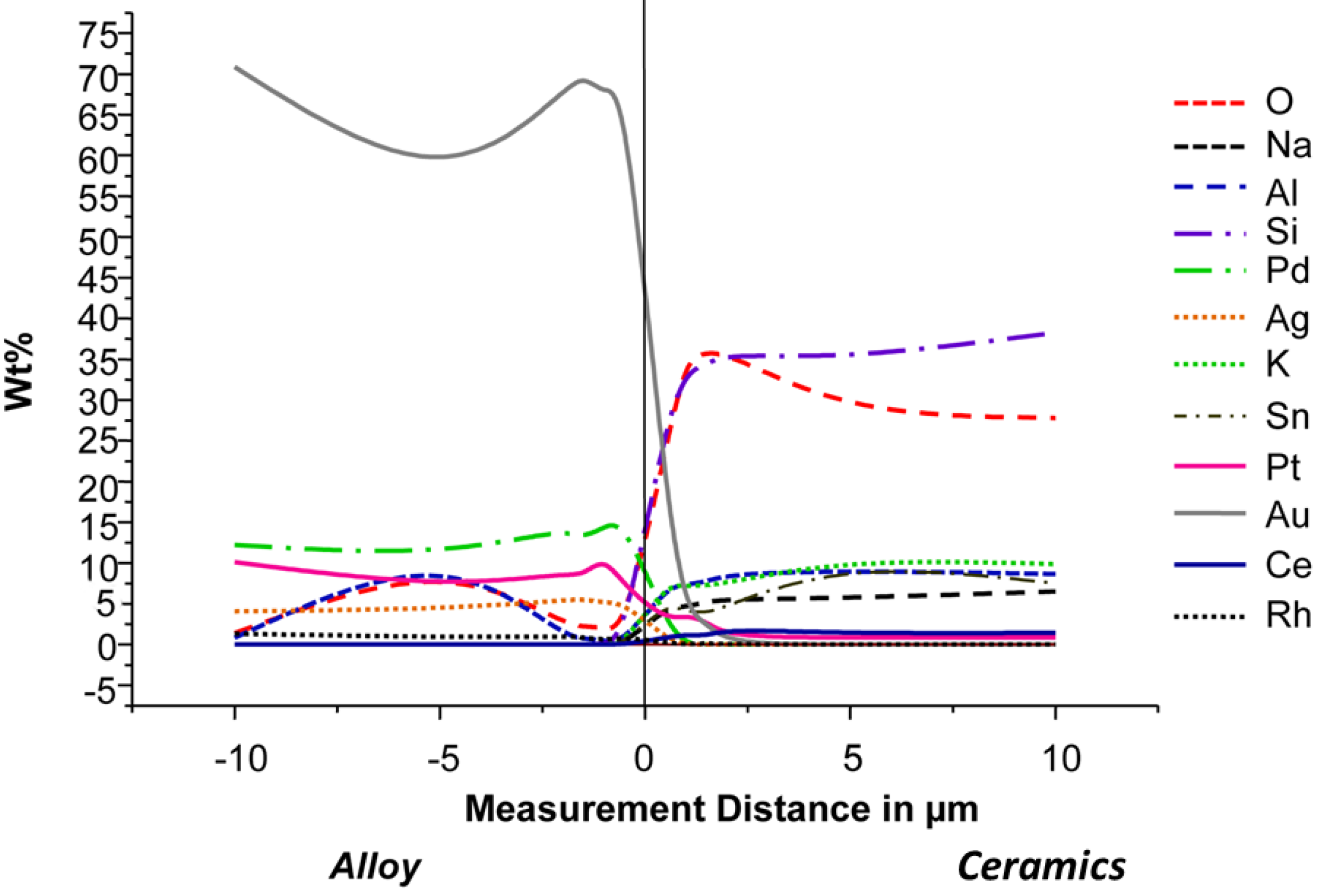
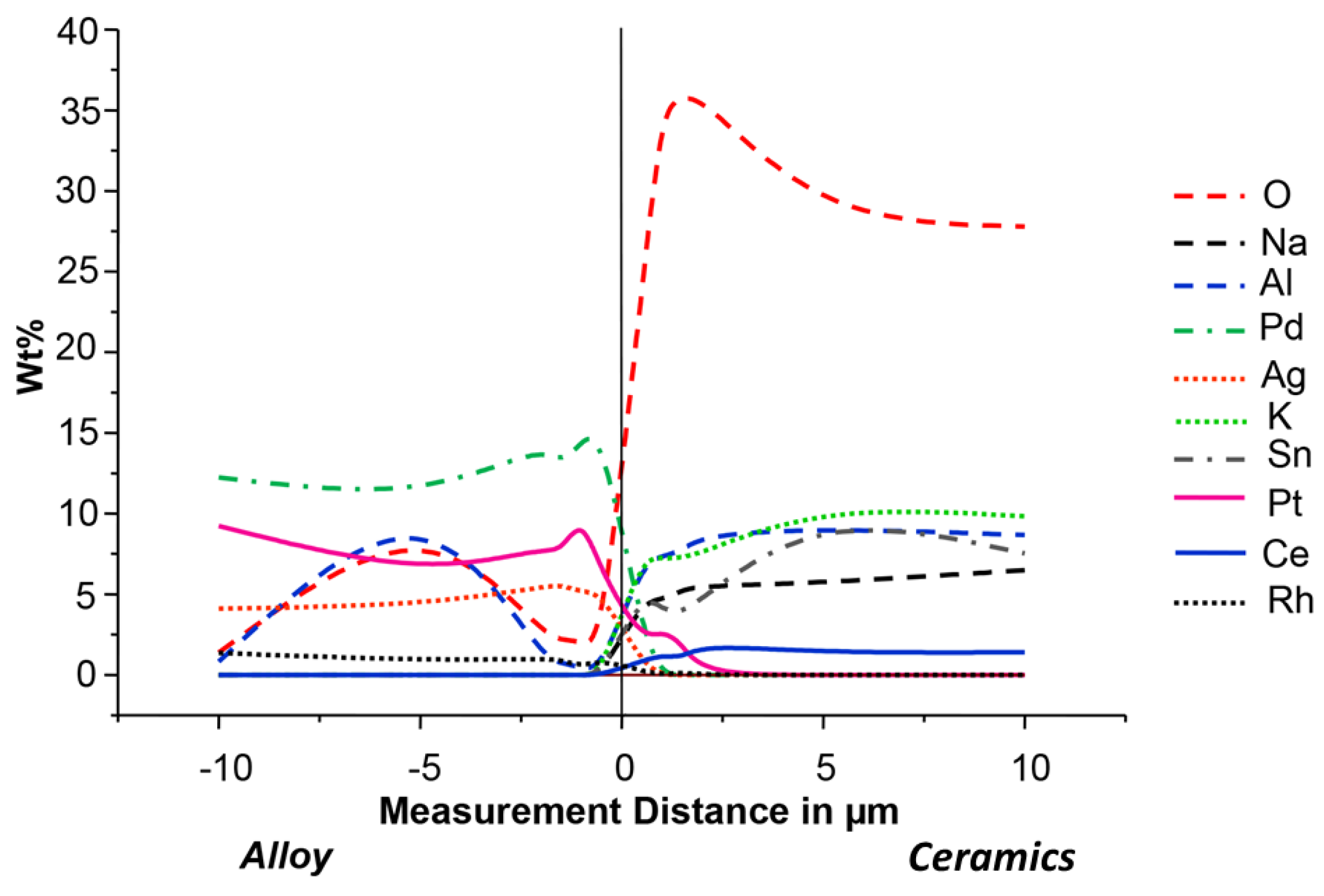
EDX was used to analyze the composition of the alloy surfaces. One specimen coated with ceramic was studied for each series. The specimen was embedded in a synthetic resin of the type Speci Fix 20 (Struers, Rødovre, Denmark). After hardening, the specimen resin block was sectioned using an Accutom 50 (Struers, Rødovre, Denmark) and prepared for EDX analysis. An Edwards Sputter Coater S 158 B (Edwards High Vacuum International, Crawby, West Sussex, UK) was used for carbon sputtering. The EDX analyses were performed with the aforementioned scanning electron microscope. In both the alloy and the ceramics, chemical composition was analyzed in 6 points situated on a line with various distance from the alloy-ceramic interface, 0.5, 1.0, 1.5, 2.0, 5.0 and 10 (mm) micrometers, respectively. An elemental analysis was derived from each measurement.

2.3. Statistics

For statistical testing of the results for shear bond strength and roughness, the Mann–Whitney U-test (α = 0.05) with an adjustment according to Bonferroni–Holm, as well as a correlation analysis (Spearman’s rank-order correlation, level of significance α = 0.01) were used. The method according to Bonferroni–Holm is an adapted fixation of the significance level [22,23].

3. Results

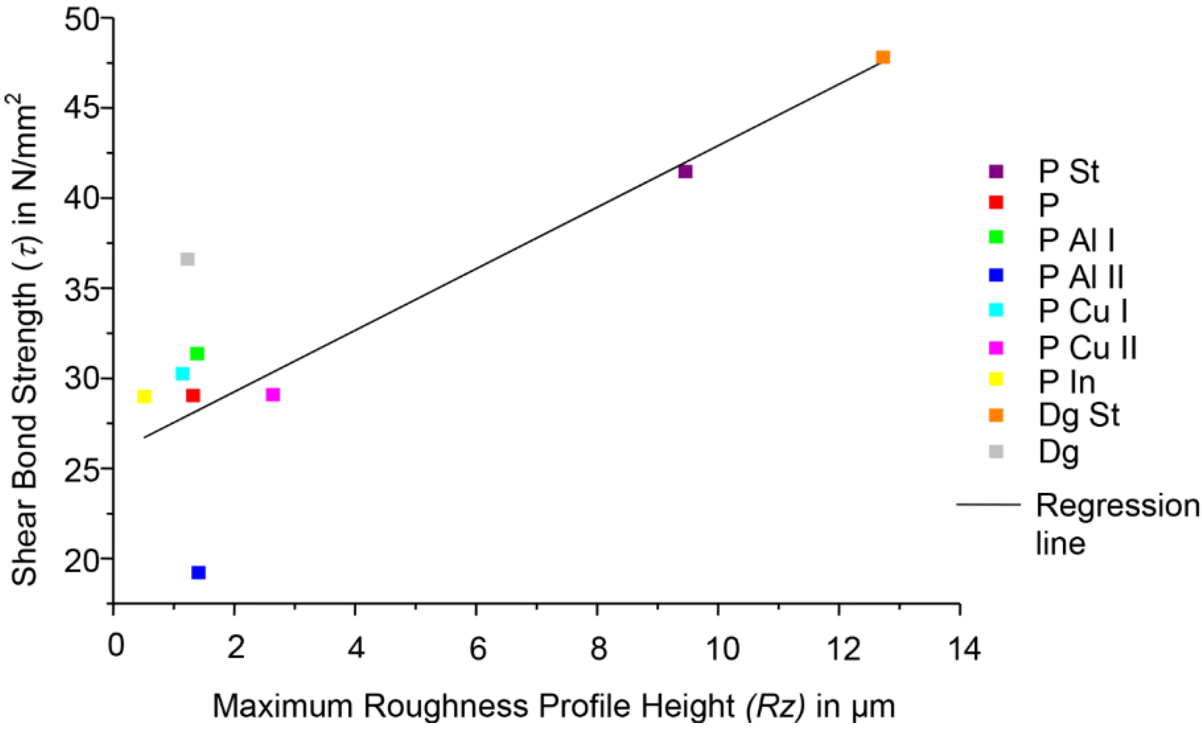
3.1. Shear Bond Strength

Table 4 and Table 5 summarize the results of all test series and their statistical relationships. Ion implantation of the base metals used resulted in no increase in bond strength. The highest values were achieved for both Degutan and Primallor 3 with the “standard” treatment conditions. Both of these series also had the best repeatability, expressed by the lowest standard deviations.
Table 4. Shear bond strength and maximum roughness profile height (Rz). All test series n = 8.
SeriesPrimallor 3Degutan
P StPP Al IP Al IIP Cu IP Cu IIP InDg StDg
Shear Bond Strength (MPa)Mean41.529.031.419.230.329.129.047.836.6
Standard Deviation2.65.04.63.24.63.25.21.73.8
Maximum Roughness Profile Height (Rz) (µm)Mean9.51.31.41.41.62.60.512.71.2
Standard Deviation0.90.20.10.10.31.10.10.60.2
Table 5. Statistical tests of the shear bond strengths of all implanted and non-implanted test series; p-values (s = significant, ns = not significant).
SeriesP Al IP Al IIP Cu IP Cu IIP In
P Sts. (0.002)s. (0.001)s. (0.001)s. (0.001)s. (0.001)
Pn.s. (0.172)s. (0.002)n.s. (0.600)n.s. (1.000)n.s. (0.834)
Dg Sts. (0.001)s. (0.001)s. (0.001)s. (0.001)s. (0.001)
Dgn.s. (0.003)s. (0.001)n.s. (0.003)n.s. (0.004)n.s. (0.004)

3.2. Roughness

The results of the profile depth measurements included the parameter currently found to be most significant, RZ (maximum roughness profile height). Table 4 and Table 6 summarize the data for the profile depth of the alloy surfaces of all test series and their statistical relationships. As expected, ion implantation results in minimal profile depth. With respect to this finding, the test series with standard conditioning differ in a statistically significant manner.
Table 6. Statistical tests of the RZ values of all implanted and non-implanted test series (s = significant, ns = not significant); p-values.
SeriesP Al IP Al IIP Cu IP Cu IIP In
P Sts. (0.002)s. (0.002)s. (0.002)s. (0.002)s. (0.001)
Pn.s. (0.010)n.s. (0.010)n.s. (0.008)s. (0.004)s. (0.001)
Dg Sts. (0.001)s. (0.002)s. (0.002)s. (0.002)s. (0.001)
Dgn.s. (0.007)n.s. (0.005)n.s. (0.012)s. (0.0019)s. (0.001)

3.3. Correlation Analysis Shear Bond Strength: Roughness

A correlation analysis was performed to test the relationship between roughness of the alloy surface and the shear bond strength values found. The Spearman’s rank-order correlation coefficient was +0.5614 (the level of significance was α = 0.01). This positive result means that shear bond strength increases with the roughness of the alloy surface (Figure 1).
Figure 1. Correlation plot of the maximum roughness profile height vs. shear bond strength.
Metals 05 00119 g001

3.4. Concentration Analyses

In the concentration analyses with AES and EDX, the concentration shifts in alloy and dental ceramic could be observed. Figure 2 and Figure 3 were included as examples. The vertical line, which represents the alloy ceramic interface, was defined through the clear differences in brightness between the two materials.
Figure 2. Concentration profiles of all elements near the metal-ceramic interface for the series P St, measured using EDX.
Metals 05 00119 g002
Figure 3. Concentration profiles near the metal-ceramic interface for the series P St, measured using EDX. For better representation of the chemical elements present at a lower concentration, the high concentration elements in Figure 2, Au and Si, were excluded.
Metals 05 00119 g003

4. Discussion

4.1. Shear Bond Strength and Roughness

The shear bond strength test by the Schwickerath method has been used for other low-fusing dental ceramics in previous studies [24]. In all series investigated in our study, except for one having specimens implanted with a high dose of Al ions, the values of shear bond strength are above 25 MPa, which is the minimum required in ISO standard [15,16]. The values measured for the reference alloy, Degutan, are similar to other metal-ceramic combinations with low fusing point dental ceramics. In addition to Sn and Fe, In was selected as an alloy component, because it ensures chemical bonding of the ceramic as a result of adhesive oxide formation in the firing process [25].
Shear bond strengths for the implanted series were found to be higher in this study than those described in a previous investigation [26]. In different metal-ceramic systems, Derfert found the highest shear bond strengths for noble metal alloys and low-temperature fusing dental ceramics with median values up to 51 MPa [27]. The Au content of the noble metal alloys had no significant influence in this material combination. Within the base metal alloys, higher values up to 42 MPa were found for Co-Cr alloys compared to Ni-Cr alloys [27].
The series blasted with Al2O3, which were characterized by the highest roughness, possessed superior shear bond strength values. This is consistent with the results from other studies [28,29]. They all came to the conclusion that the best adhesion could be achieved by oxidation and Al2O3 air abrasion.
The analyses resulted in a positive correlation between shear bond strength and roughness. Similar relationships were also described in the literature [30,31,32].
The investigations demonstrated that Al2O3 air abrasion of the alloy surface improves the shear bond strength for both alloys by approximately 30%. This means that air abrasion with Al2O3 is an important aspect in surface preparation prior to ceramic application, affecting the shear bond strength.

4.2. Chemical Bonding: Mechanical Bonding

The notion that chemical bonding, which originates from adhesive oxide formation and diffusion, contributes the major part of the metal-ceramic bond strength could not be supported. The standard surface treatment with air abrasion (110 µm Al2O3, 2 bar), oxidation in air (980 °C, 10 min) and repeated air abrasion (110 µm Al2O3, 2 bar) led to significantly higher shear bond strength values than with the ion implanted specimens. Therefore, ion implantation revealed not to be an effective method to improve adhesion and bond strength. The results presented indicate that the concentration of non-noble components in dental alloys can be minimized without deterioration of the shear bond strength. However, the expected resulting improvement of the biocompatibility of the respective alloys has to be verified experimentally and clinically. Long-term investigations and controlled clinical studies are necessary [33,34,35]. Additionally, it has to be noted that the incorporation of secondary base metal elements in the compositions of noble alloys for metal-ceramic applications also provides strengthening by the mechanisms of solid solution hardening and secondary phase formation (precipitates), in addition to their roles as potential oxide-formers.
Because a correlation between surface roughness and the degree of adhesion between alloy and ceramic could be determined, it is important to promote mechanical bonding by conditioning the surface with Al2O3 air abrasion. Based on experimental results, it can be assumed that an effective range of surface roughness resulting in high shear bond strength values was around Rz values from 9 to 13 µm. Since this was the highest value of roughness investigated, the effect of a further increase is unknown, so it is impossible to give an optimum roughness.
In reviewing published experimental results, also contradictory conclusions related to bonding mechanisms can be found. This suggests significant differences in the way the experiments were carried out, which were not described in the publications appraised [36]. It is to be supposed that these differences are secondary in nature and reflect influences, such as environmental conditions (temperature, humidity, etc.), temporal factors in the experiments, the manual skills of the experimenters and similar matters. Thus, it appears to be of significant importance that investigations of bonding strength are carried out according to simple, unified procedures that can be easily reproduced at any time and in each laboratory.
This study exhibits the typical limitations of an in vitro trial. Furthermore, no defects in the metal-ceramic interface were examined. However, these defects are frequently caused by flaws in the processing of dental restorations [37]. We tried to avoid respective problems by exclusively using one dental ceramic that was processed by only one experienced and specially-trained dental technician. Despite these limitations, our results are considered valid and may create the basis for further studies on metal-ceramic bonding of noble metal alloys without base metals for oxide formation. Additionally, prospective in vivo studies in the oral environment would be valuable [38].

5. Conclusions

The highest shear bond strength for a noble metal alloy without base metals was found for surfaces treated according to a standard protocol with air abrasion and oxidation in air. The implantation of ions and oxidation in air create a minimal increase in roughness. Implantation of base metal alloys leads to significantly lower bond strengths and does not positively influence and actually deteriorates bonding. The positive correlation between surface roughness and bond strength shows the necessity of mechanical retention through air abrasion with Al2O3.
We cannot support the hypothesis that the major part of the metal-ceramic bond originates from the chemical bond through adhesive base metal oxides. Instead, the results of the study suggest that the concentration of base metal components in dental alloys can be minimized without negatively affecting the material bond.

Acknowledgments

We would like to thank DeguDent GmbH for the company’s support of the investigations. The authors thank Ursula Range (Institute for Medical Information Technology and Biometrics, Medical Faculty, Technische Universität Dresden) for the cooperation.

Author Contributions

Susanne Enghardt: substantial contribution to the production and analysis of the results, preparation of the manuscript; Gert Richter: substantial contribution to the production and analysis of the results, preparation of the manuscript; Edgar Richter: substantial contribution to the production and analysis of the results (especially ion implantation), editing the manuscript; Bernd Reitemeier: substantial contribution to the design of the study, planning of the experiments, roughness measurements, organization of the interdisciplinary study, preparation of the manuscript; Michael Walter: substantial contribution to the design of the study, planning of the experiments, preparation of the manuscript.

Conflicts of Interest

The authors declare no conflict of interest.

References

  1. Coornaert, J.; Adriaens, P.; De Boever, J. Long-term clinical study of porcelain-fused-to-gold restorations. J. Prosthet. Dent. 1984, 51, 338–342. [Google Scholar]
  2. Walter, M.; Reppel, P.D.; Böning, K.; Freesmeyer, W.B. Six-year follow-up of titanium and high-gold porcelain-fused-to-metal fixed partial dentures. J. Oral. Rehabil. 1999, 26, 91–96. [Google Scholar] [CrossRef] [PubMed]
  3. Walton, T.R. An up to 15-year longitudinal study of 515 metal-ceramics FDPs: Part 2. Modes of failure and influence of various clinical characteristics. Int. J. Prosthodont. 2003, 16, 177–182. [Google Scholar] [PubMed]
  4. Holm, C.; Tidehag, P.; Tillberg, A.; Molin, M. Longevity and quality of FDPs: A retrospectivestudy of restorations 30, 20, and 10 years after insertion. Int. J. Prosthodont. 2003, 16, 283–289. [Google Scholar]
  5. Napankangas, R.; Raustia, A. An 18-year retrospective analysis of treatment outcomes with metal-ceramic fixed partial dentures. Int. J. Prosthodont. 2011, 24, 314–319. [Google Scholar] [PubMed]
  6. Eichner, K. Gegenwärtiger Stand der werkstoffkundlichen und klinischen Metallkeramik. ZWR 1997, 97, 477–485. [Google Scholar]
  7. Bumgardner, J.D.; Lucas, L.C. Corrosion and cell culture evaluations of nickel-chromium dental casting alloys. J. Appl. Biomater. 1994, 5, 203–213. [Google Scholar] [CrossRef] [PubMed]
  8. Wataha, J.C.; Hanks, C.T. Biological effects of palladium and risk of using palladium in dental casting alloys. J. Oral. Rehabil. 1996, 23, 309–320. [Google Scholar] [CrossRef] [PubMed]
  9. Wataha, J.C. Biocompatibility of dental casting alloys: A review. J. Prosthet. Dent. 2000, 83, 223–234. [Google Scholar] [CrossRef] [PubMed]
  10. Al-Hiyasat, A.S.; Bashabsheh, O.M.; Darmani, H. An investigation of the cytotoxic effects of dental casting alloys. Int. J. Prosthodont. 2003, 16, 8–12. [Google Scholar]
  11. Wataha, J.C. Alloys for prosthodontics restorations. J. Prosthet. Dent. 2002, 87, 351–363. [Google Scholar] [CrossRef] [PubMed]
  12. Möller, W.; Richter, E. Praktische Anwendungen der Ionenimplantation. Galvanotechnik 1989, 89, 3–11. [Google Scholar]
  13. Mihoc, R.; Hobkirk, J.A.; Armitage, D.; Jones, F.H. Influence of ion implantation on physicochemical processes at titanium surfaces. Int. J. Prosthodont. 2006, 19, 24–28. [Google Scholar]
  14. Hohmann, W. Dentalkeramik Auf Der Basis Hydrothermaler Gläser. Quintessenz: Berlin, Germany, 1993. [Google Scholar]
  15. Metal-Ceramic Dental Restorative Systems; Beuth: Berlin, Germany, 1999; ISO 9693.
  16. Dentistry—Compatibility Testing, Metal-Ceramic Systems; Beuth: Berlin, Germany, 2012; ISO 9693–1.
  17. Dentistry—Casting Gold Alloys; Beuth: Berlin, Germany, 2004; ISO 1562.
  18. Geometrical Product Specifications (GPS)—Surface Texture : Profile Method—Terms, Definitions and Surface Texture Parameters; Beuth: Berlin, Germany, 2010; ISO 4287.
  19. Geometrical Product Specifications (GPS)—Surface Texture: Profile Method—Rules and procedures for the assessment of surface texture; Beuth: Berlin, Germany, 1998; ISO 4288.
  20. Geometrical Product Specifications (GPS)—Surface Texture: Areal—Part 2: Terms, Definitions and Surface Texture Parameters; Surface Texture: Areal—Part 3: Specification Operators; Surface Texture: Areal—Part 6: Classification of Methods For Measuring Surface Texture; Beuth: Berlin, Germany, 2012; ISO 25178.
  21. Briggs, D.; Seah, M.P. Practical Surface Analysis: Vol. 1. In Auger and X-ray Photoelectron Spectroscopy; Wiley: Chichester, UK, 1990. [Google Scholar]
  22. Holm, S. A simple sequentially rejective multiple test procedure. Scand. J. Stat. 1979, 6, 65–70. [Google Scholar]
  23. Armitage, P.; Colton, T. Encyclopaedia of Biostatistics, 2nd ed.; Wiley: Chichester, UK, 2005; pp. 7872–7878. [Google Scholar]
  24. Gilbert, J.L.; Covey, D.A.; Lautenschläger, E.P. Bond characteristics of porcelain fused to milled titanium. Dent. Mater. 1994, 10, 134–140. [Google Scholar] [CrossRef] [PubMed]
  25. Hautaniemi, J.A. The effect of indium on porcelain bonding between porcelain and Au-Pd-In alloy. J. Sci. Mat. Med. 1995, 66, 46–50. [Google Scholar] [CrossRef]
  26. Petridis, H.; Garefis, P.; Hirayama, H.; Kafantaris, N.M; Koidis, P.T. Bonding indirect resin composites to metal: Part I. Comparison of shear bond strengths between different metal-resin bonding systems and a metal-ceramic system. Int. J. Prosthodont. 2003, 16, 635–639. [Google Scholar]
  27. Derfert, B. Vergleichende Untersuchungen zur Verbundfestigkeit von verschiedenen Metall-Keramik-Kombinationen auf der Basis von EM- und NEM-Legierungen in Verbindung mit herkömmlicher und niedrigschmelzender Keramik. Ph.D. Thesis, Freie Universität Berlin, Berlin, Germany, 12 December 2003. [Google Scholar]
  28. Mc Lean, J.W.; Sced, I.R. Bonding of Dental Porcelain to metal I. The Gold Alloy Porcelain Bond. Trans. J. Brit. Ceram. Soc. 1973, 72, 229–233. [Google Scholar]
  29. Charnay, R.; Guiraldenq, P.; Enriore, J.M.; Brugirard, J.; Heberard, X. Mechanical properties of the ceramic-metal bonding of a gold-platinum dental alloy and chemical characterization of the interface. Mem. Etud. Rev. Met. 1992, 12, 797–803. [Google Scholar]
  30. Mc Lean, J.W. The Science and Art of Dental Ceramics; Quintessence: Chicago, IL, USA, 1980; pp. 65–90. [Google Scholar]
  31. Anusavice, K.J. Phillip’s Science of Dental Materials, 12th ed.; Saunders: Philadelphia, PA, USA, 2012. [Google Scholar]
  32. Schwickerath, H. Das Festigkeitsverhalten von aufbrennfähigen Keramiken. Dtsch. Zahnärztl. Z 1985, 40, 996–1003. [Google Scholar] [PubMed]
  33. Karlsson, S. Failures and length of service in fixed Prosthodontics after Long-term function. A longitudinal clinical study. Swed. Dent. J. 1989, 13, 185–192. [Google Scholar] [PubMed]
  34. Jokstad, A.; Esposito, M.; Coulthard, P.; Worthington, H.V. The reporting of randomised controlled trials in prosthodontics. Int. J. Prosthodont. 2002, 15, 230–242. [Google Scholar] [PubMed]
  35. Glantz, P.O.; Nilner, K.; Jendresen, M.D.; Sundberg, H. Quality of fixed prosthodontics after twenty two years. Acta. Odontol. Scand. 2002, 60, 213–218. [Google Scholar] [CrossRef] [PubMed]
  36. Bullard, J.T.; Dill, R.E.; Marker, V.A.; Payne, E.V. Effects of sputtered metal oxide films on the ceramic-to-metal-bond. J. Prosthet. Dent. 1985, 54, 776–778. [Google Scholar] [CrossRef] [PubMed]
  37. Walter, M. Zur Porenbildung in der keramischen Verblendung von Palladium-Silber-Legierungen. Dtsch. Zahnärztl. Z 1988, 43, 145–149. [Google Scholar] [PubMed]
  38. Reitemeier, B.; Hänsel, K.; Kastner, C.; Weber, A.; Walter, M.H. A prospective 10-year study of metal ceramic single crowns and fixed dental prosthesis retainers in private practice settings. J. Prosthet. Dent. 2013, 109, 149–155. [Google Scholar] [CrossRef] [PubMed]

