Article

Improvement of the Insecticidal Capacity of Two Purpureocillium Lilacinum Strains against Tribolium Confusum

1
Consejo Nacional de Investigaciones Científicas y Técnicas (CONICET), Puerto Madryn, 9120, Argentina
2
Laboratorio de Ecología Microbiana, Departamento de Microbiología e Inmunología, Facultad de Ciencias Exactas Físico Químicas y Naturales, Universidad Nacional de Río Cuarto, Río Cuarto, Córdoba, 5800, Argentina
*
Author to whom correspondence should be addressed.
Insects 2015, 6(1), 206-223; https://doi.org/10.3390/insects6010206
Submission received: 9 December 2014 / Accepted: 10 March 2015 / Published: 18 March 2015

Abstract

:
Entomopathogenic fungi can regulate insect populations. They have extracellular enzymes that degrade cuticle components, mainly hydrocarbons, used as an energy source. The increase in insecticidal activity of fungi in a medium supplemented with cuticular hydrocarbons was assayed and the hydrolytic enzyme profiles of two strains of Purpureocillium lilacinum were evaluated. A spore suspension of P. lilacinum was inoculated in Petri plates with different values (0.99–0.97–0.95) of water activity (Aw) using the substrates gelatin, starch and tween-20. Growth rate on the different substrates and the enzymatic activity index for proteases, amylases and lipases at different incubation times, pH and Aw, was evaluated. Moreover, the insecticidal efficiency of strains grown in media supplemented with n-hexadecane and n-octacosane was analyzed. LT50 was calculated against adults of Tribolium confusum and showed that mortality increased about 15% when the strains grew in amended culture medium. High amylolytic activity was detected, but proteases were the main enzymes produced. Optimal protease production was observed in a range of acid and alkaline pH and lower Aw. The greatest growth rate was obtained in presence of gelatin. Lipase and amylase production was detected in small amounts. Fungal growth in media with hydrocarbon mixtures increased the pathogenicity of the two strains of P. lilacinum, with the strain JQ926223 being more virulent. The information obtained is important for achieving both an increase in insecticidal capacity and an understanding of physiological adaptation of the fungus.

1. Introduction

In Argentina about 6% of cereal production is lost at the post-harvest stage, due to flaws in the transportation and drying and mainly by insect damages [1,2]. Insect contamination in food commodities is an important quality control problem of concern for food industries [3].
Insects, such as Tribolium confusum (Jacquelin du Val) cause significant damage to stored maize [4]. Damage may be direct, such as weight loss, reduced germination and reduced nutritional value of grain or indirect such as heat and moisture migration, reservoir of disease and distribution of microorganisms [5]. Insects are involved in the colonization of grains, providing sites for fungal infection through the damage that can result in grains [6]. Also, previous studies have shown that certain insects that attack stored grains have the ability to disperse toxigenic Aspergillus flavus in those grains [7,8]. The interaction of these toxigenic fungi with substrate and abiotic predisposing factors may promote a moldy substrate and toxin accumulation in stored grains [9]. The main toxins produced by A. flavus are aflatoxins. The positive correlation between the consumption of food contaminated with aflatoxin B1 and increased incidence of liver cancer has led to the classification of this toxin as a carcinogen of the group 1 A by the International Agency for Research on Cancer [10]. For all these reasons, strategies to control insects are needed.
Currently, most of the management of post-harvest insect pests is carried out with synthetic chemical insecticides. However, the use of synthetic insecticides allowed in Argentine grains and in other countries is very limited [11]. It is known that these pesticides can adversely affect the environment. In addition, the continued use of these chemicals has resulted in serious problems such as resistance [12]. Hence, alternatives to conventional insecticides are needed. There are biologic agents with natural potential to protect crops. There is much interest in insect pathogenic fungi because they are considered to offer an environmentally friendly alternative to chemical pesticides [13,14,15]. Among these fungi, some belonging to the genus Purpureocillium have shown promising results for use as effective biocontrol agents [16,17,18,19]. In a previous study, all isolates of Purpureocillium lilacinum (Thom) Samson showed pathogenicity against insect vectors of aflatoxigenic A. flavus in stored maize [20].
Over the years, most of the studies for the development of a biocontrol agent have focused on the potential for inoculum production. Although these studies have been numerous, most of them have centered on optimizing the amount of propagules or mycelial fragments produced. However, little attention has been paid to the quality of the inoculum. Therefore, it is important to consider the existence of the real practical problem of the establishment and effectiveness of biocontrol agents in the natural environment. It is important that a biological control fungus has a tolerance for fluctuating abiotic factors like water activity and temperature [21].
All fungi need the physical presence of water for the diffusion of nutrients into the cell and the release of extracellular enzymes. However, water can be in the environment but not available. Under conditions of low water activity, fungi are able to accumulate polyols (sugar alcohols acyclic) at high concentrations. These polyols reduce the potential of intracellular water [22,23,24] producing a balance inside and outside the cell. Inhibition of enzymatic function by dehydration is also prevented [25]. Thus, water is available to maintain the turgor of the fungus. This allows the cell to continue operating efficiently. Therefore, physiological methods to improve tolerance to water stress are considered essential to enable the development of effective and consistent microbial biocontrol agents [26].
Early research has speculated that the mechanism of insect infection by fungi is through ingestion of the control agent [27]. However, over the years, it has been shown that entomopathogenic fungi infect the insect through the cuticle. For this reason, there is no need for these fungi to be ingested as they act as contact insecticides [28,29]. After binding the spore to the insect, the former germinate and penetrate the cuticle. This process occurs if factors, which influence the pathogenicity (water availability, temperature and nutrition) are favorable [30]. Also, it is necessary that a favorable environment is present for secretion of extracellular enzymes due to its hygroscopic nature [31]. Therefore, water availability is a critical factor for the germination of spores.
The entomopathogenic fungi produce a range of cuticle degrading enzymes corresponding to different polymers of the insect cuticle [32]. The insect cuticle is composed of a group of diverse constituents with variation in content and composition [33,34,35,36]. The cuticular composition has profound consequences on ecological and behavioral aspects of the insect. Hydrocarbons such as n-alkanes, alkenes and methyl-branched chains are common epicuticular lipids and have been widely studied [33,34,37]. Cuticular hydrocarbons can stimulate [38,39] or inhibit [40] the interaction between fungus and insect cuticle.
Fungal enzymes that hydrolyze proteins and chitin, which represent the main constituents of the insect cuticle, are considered vital to the infection process [32]. Many extracellular enzymes produced by fungi to degrade the outer cell layers of their hosts are regulated by carbon and nitrogen sources and pH [41]. The order of enzyme production is based on the constituents of the cuticle.
Starting from the biochemical mechanism of cuticular degradation and its potential as an alternative method of pest control, a hypothesis was proposed based on the probability that fungi with their mechanism of hydrocarbon assimilation enhanced could be directly related to increased virulence [42]. Therefore, hydrocarbons may induce germination besides functioning as catabolic substrates. Apart from this, the adaptation to different conditions like Aw, nutrients, pH and time could improve the pathogenicity and virulence of the fungus.
Therefore, the objective of this study was to stimulate the insecticidal efficacy of two P. lilacinum strains with proved efficacy against T. confusum. A first approximation of the enzyme profile of P. lilacinum was also evaluated.

2. Materials and Methods

2.1. Fungal Isolates

Two strains identified as P. lilacinum were used in these experiments. These strains were originally isolated from soil samples collected from the University of Río Cuarto Experimental Field Station in Río Cuarto, Córdoba, Argentina, and were identified and deposited in GenBank with the following accession number: JQ926212 and JQ926223. Both strains are pathogenic to T. confusum, an insect pest vector of aflatoxigenic fungi [20]. These strains are held in the Microbial Ecology Laboratory Collection, Microbiology and Immunology Department of the National University of Río Cuarto, Córdoba, Argentina.

2.2. Estimation of Enzymatic Activity of P. Lilacinum

Plate Assay Method

Protease, amylase and lipase activity was assayed by the plaque assay method [43]. The water activity (Aw) of the unmodified culture media was 0.99, and these were used as control treatments. The Aw was adjusted to 0.97 and 0.95 by addition of known amounts of non-ionic solute glycerol [44] and was determined using a water activity meter AquaLab (4TE model; AquaLab Technologies, Riverside, CA, USA). The activity of proteolytic and amylolytic enzymes of P. lilacinum was determined by using gelatin and starch, respectively, as substrate in a nutrient medium (0.5 g peptone, 0.3 g beef extract, 0.5 g NaCl, 1.5 g agar and 100 mL distilled water). For lipase activity, sorbitan monolaurate was added in nutrient medium (1 g peptone, 0.5 g NaCl, 0.1 g CaCl, 1.5 g agar and 100 mL distilled water) as a lipid substrate. Petri plates of different values of Aw and substrates were spot inoculated in the center of each plate, with P. lilacinum spores suspended in semi-solid agar [45]. Petri plates of the same values of Aw were sealed in polyethylene bags. The inoculated plates were incubated at 25 ± 1 °C for 15 days. At the end of the incubation period, the area of gelatin degradation was visible, flooding the plate with a saturated solution of mercury chloride reagent (15 g MgCl2 completely dissolved in 20 mL of 7 M HCl in 100 mL of distilled water). The mercury chloride solution reacts with the gelatin to produce a white precipitate that turns into a visible halo. To determine the starch degradation, the plates were flooded with iodine reagent (65 mg of iodine crystals and 130 mg of KI in 100 mL of distilled water). The lipolytic activity was evident by a precipitate, which was attributed to the production of fatty acids from lipids used as substrates by fungal enzymes. The diameter of each zone was recorded. Enzymatic activity (EA) was measured by the following formula [46]: EA = D−d; D = diameter of colony plus clearing precipitate zone; d = diameter of colony.

2.3. Growth Studies: Effect of Different Substrates on the Rate of P. Lilacinum Growth

The P. lilacinum strains grew, in each medium amended with different substrates for each enzyme, at 25 ± 1 °C during 15 days. The colony radius was measured daily during the incubation time. For each colony, two radii measured at right angles to one another, were averaged to find the mean radius for that colony. All colony radii were determined by using three replicates for each one. The radial growth rate (mm d−1) was subsequently calculated by linear regression of the linear phase for growth in relation to the carbon source of the culture medium and the water activity.

2.4. Quantitative Studies for Estimating the Production of Extracellular Enzymes by P. Lilacinum

2.4.1. Culture Media

Both strains of P. lilacinum were cultured in two minimal growth media, medium 1 (0.4 g yeast extract, 0.6 g K2HPO4, 0.6 g MgSO4, 0.6 g NaCl and 100 mL distilled water) and medium 2 (0.11 g FeSO4, 0.04 g ZnSO4, 0.05 g MgSO4, 0.2 g KNO3 and 100 mL distilled water). Medium 1 was used to stimulate the production of proteases and amylases, while lipases were produced in Medium 2. Each culture medium was supplemented with 1% gelatin, starch or tween-20, depending on the enzyme, which was to be evaluated. The Aw of the unmodified media was 0.99, and these were used as the control treatments. The Aw was adjusted to 0.97 and 0.95 as described above. The media were adjusted to pH 3, 7, 9 and 11 adding the necessary quantity of the following buffer solutions: citric acid 0.1 M + sodium hydroxide 1 M (5:1) (sol A)/hydrochloric acid 0.1 M (sol B); potassium monohydrogen phosphate 0.2 M (sol A)/potassium dihydrogen phosphate 0.2 M (sol B); glycine 1 M (sol A)/sodium hydroxide 1 N (sol B). Appropriate amounts of solutions A and B were used. To achieve pH 3, 40 mL sol A + 60 mL sol B of the first buffer solution were mixed. To achieve pH 7, 39 mL sol A + 61 mL sol B of the second buffer solution were used and to achieve pH 9 and 11, 85.03 mL sol A + 14.96 mL sol B and 52.3 mL sol A + 47.7 mL sol B of the third buffer solution were used, respectively.

2.4.2. Experimental Conditions and Fungal Inoculation

Hundred milliliters of each culture medium with different levels of Aw and pH were placed into flasks of 250 mL to estimate the production of each enzyme. Each flask was inoculated with 1 mL of spore suspension of P. lilacinum (107 spores mL−1). The inoculated culture media were incubated on a rotary shaker (150 rpm) at 25 ± 1 °C. After 3, 7, 10 and 15 days enzyme production was measured as follows:

2.4.2.1. Spectrophotometric Analysis of Proteases and Amylases

Total protein concentration was measured according to biuret protein assay [47]. A standard curve was calculated using bovine serum albumin (BSA) in a concentration range from 0.25 to 1 mg mL−1. Standard solution was prepared at a concentration of 1 mg mL−1. Absorbance was measured in a spectrophotometer at 545 nm. Aseptically collected aliquots from each flask were centrifuged at 13,000 rpm for 10 min. The supernatant was used as crude enzyme extract (CEE). It was mixed with 1 mL of biuret reagent (3.8 g CuSO4-5H2O, 6.7 g EDTA, 200 mL NaOH 5N, 1 g KI, and 700 mL distilled water) and vortexed. After 5 min, the absorbance was determined. One mL of buffer without inoculation was used as control. During the determination of protein in each sampling time, samples were kept at 4 °C to preserve the function of the enzyme.
The amylolytic enzyme concentration was determined according to Miller [48] with some modifications. Aseptically collected aliquots from each flask were centrifuged at 5000 rpm for 5 min. The supernatant was used as crude enzyme extract (CEE). Appropriately diluted enzyme was mixed with equal amounts (1mL) of starch solution (1 g starch and 100 mL distilled water). The reaction was allowed to proceed by incubating at 27 ± 1 °C for 15 min. At the end of the incubation period, the reaction was stopped by addition of 6 mL of water. Enzyme activity was detected as absorbance at 540 nm. Control tubes were prepared by addition of sterile distilled water instead of CEE.

2.4.2.2. Tritimetric Method

Lipase concentration was determined using a tritimetric assay [49] with NaOH 0.05 M, using emulsified oil as substrate. One milliliter of appropriately diluted enzyme was added to 5 mL of emulsion containing 25% (v/v) olive oil, 75% (v/v) arabic gum and 2 mL of 10 mM phosphate buffer solution at pH 7. Phenolphthalein in alcohol + water (1:1) was used as indicator. The assay was performed at 37 ± 1 °C for 30 min. The reaction was stopped by addition of 15 mL of acetone/ethanol (1:1 v/v). The amount of fatty acid released due to the enzyme activity was titrated against NaOH 0.05 M until the color of the reaction mixture changed to pink. A lipase unit was defined as the enzyme able of releasing 1 mol of fatty acid at 37 ± 1 °C and pH 7, per minute. One mL of titration volume is equal to 2.5 units of lipase.

2.5. Stimulation of Pathogenicity against T. Confusum

2.5.1. Fungal Growth on Synthetic Hydrocarbon-Enriched Media

The control culture medium (CM) of this study contained 0.4 g KH2PO4, 1.4 g NaHPO4, 0.6 g MgSO4, 1 g KCl, 1.4 g NH4NO3, 20 g glucose, 10 g yeast extract, 15 g agar and 1000 mL distilled water. The amended medium (SM) had a similar composition to the CM medium but glucose and yeast extract were replaced by 20 mL of hexane solution composed of 15% w/v of n-hexadecane and/or n-octacosane (Sigma Aldrich, St. Louis, MO, USA) [50]. The Aw of the unmodified media was 0.99, and these unmodified media were used as the control treatments. The Aw was adjusted to 0.97 and 0.95 as described above. A spore suspension (107 spores mL−1) of each entomopathogenic fungus strain was inoculated onto Petri plates containing CM and SM culture media with the purpose of stimulating their virulence. The Petri plates of the same Aw values were sealed in polyethylene bags and incubated at 25 ± 1 °C during 15 days.

2.5.2. Insect Rearing

Cultures of one strain of the confused flour beetle T. confusum (Jacquelin du Val) were obtained from the Department of Agricultural Zoology, Faculty of Agronomy, University of Buenos Aires, Argentina. Mixed-sex adults 1–3 weeks of age were used in the test. Insects were reared on a diet of wheat flour, cornstarch and yeast (10:10:1.5) in plastic containers with 200 g of the mixture. Insects were reared at 27 ± 1 °C, 70 ± 5% relative humidity and photoperiod of 12:12 h light:dark cycle.

2.5.3. Pathogenicity Bioassay

Twenty T. confusum adults were treated by immersion for 30 s in 600 µL of a spore suspension (107 spores mL−1) [51] of either of the two P. lilacinum fungal strains that were cultured using both CM and SM media. After treatment, beetles were placed in a plastic jar with 500 g of maize grains [52]. Jars were placed in a chamber under controlled conditions at 27 ± 1 °C, 70 ± 5% r.h, with a photoperiod of 12:12 h light:dark cycle [53]. Mortality was recorded daily for 20 days and analyzed by probit analysis to obtain lethal time (LT50) values for comparison with controls. Fungal mortality was confirmed by placing dead beetles on water agar (15 g l−1 agar and 1000 mL distilled water) and incubated at 25 ± 1 °C for 7 days.

2.6. Statistical Analysis

The analysis of variance in a completely randomized design was used to compare the effect of fungal strain, substrate, water activity, incubation time and pH on enzymes production, and the effect of the nutritional modification on insecticide efficiency of P. lilacinum against T. confusum. The statistical program, InfoStat, for Windows version 2008 (Group InfoStat, FCA, National University of Córdoba, Argentina) was used [54].

3. Results

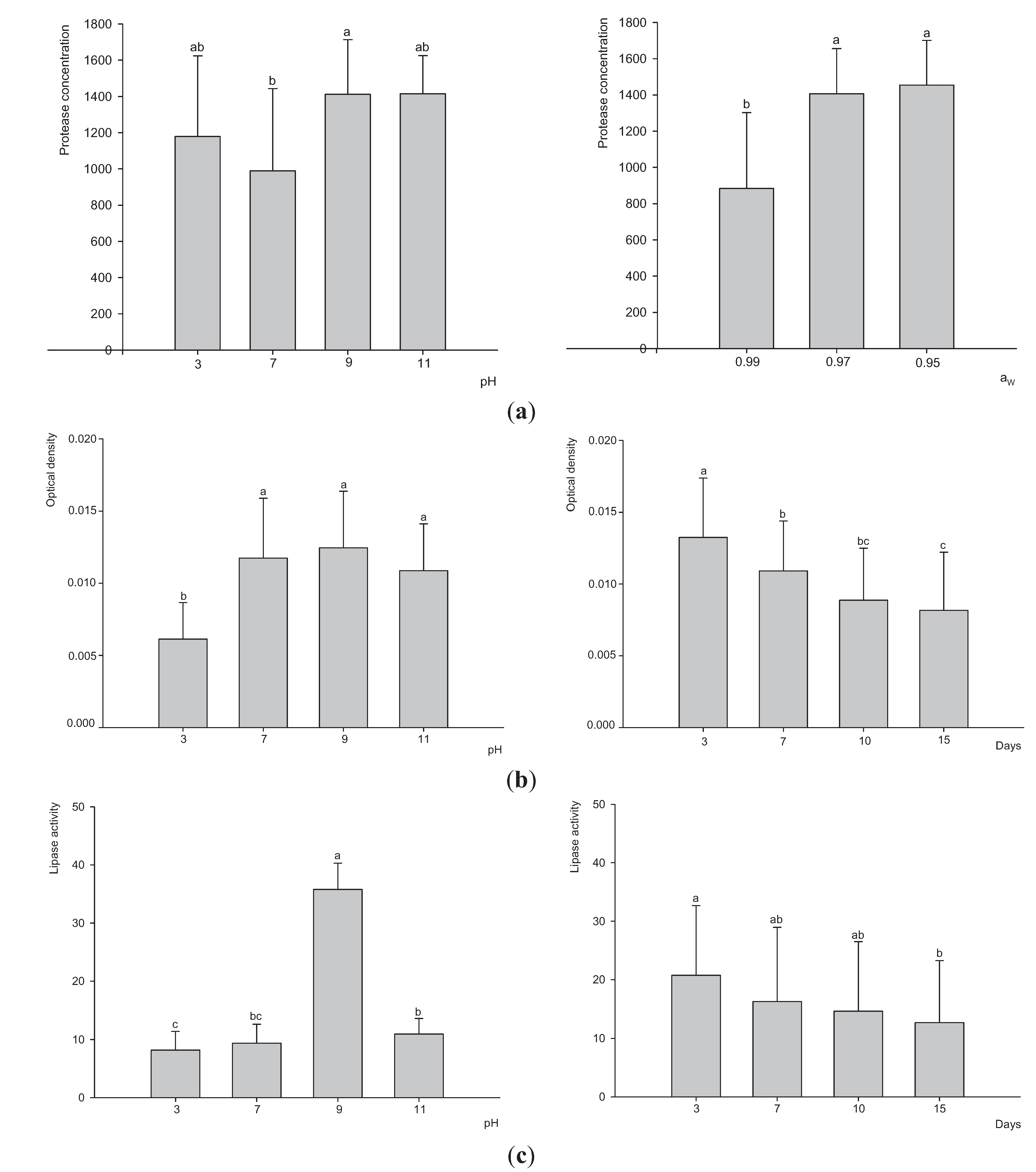
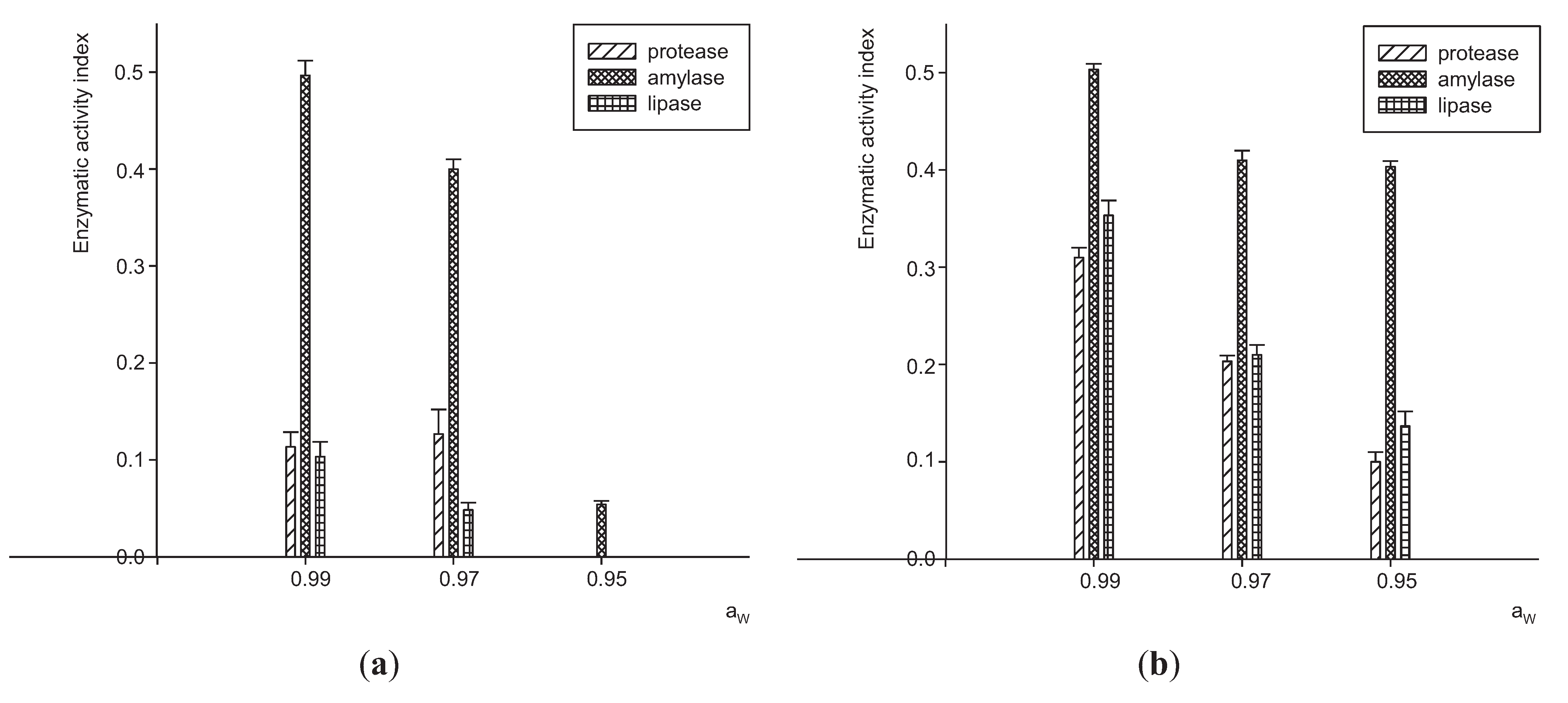
3.1. Extracellular Enzymes Activity by P. Lilacinum

An analysis of the influence of substrate in enzymatic activity was performed. The enzymes values produced varied by Aw and strain (P < 0.0001). Amylolytic activity was observed with both fungal strains at 3 Aw tested. The highest yield was obtained at 0.99 Aw. This activity was significantly greater than the other activities analyzed (P < 0.05). In general, all values of enzymatic activities were lower when Aw decreased. No protease and lipase activity for the JQ926212 strain at 0.95 Aw was observed. It was observed that the JQ926223 strain had higher activity than the JQ926212 strain for all enzymes assayed (Figure 1).
Figure 1. Extracellular enzyme activity index of two strains of P. lilacinum (a) JQ926212 and (b) JQ926223. Tuckey test (P < 0.05) was used to compare different enzymatic activity for the same Aw.
Insects 06 00206 g001

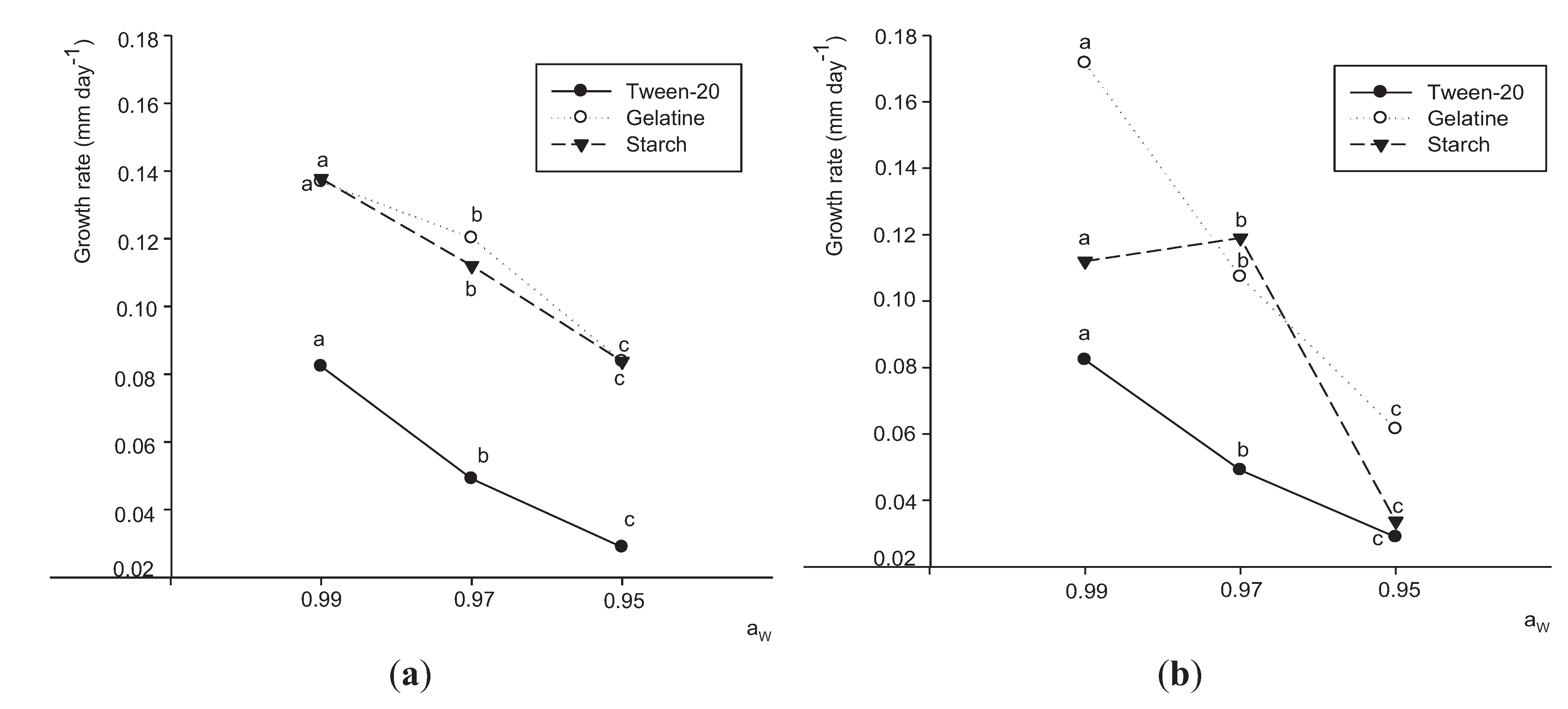
3.2. Effects of Aw and Substrates on Growth

Changes in growth rate of both strains in relation to Aw level and different substrates at 25 °C are shown in Figure 2. The major effect was that of Aw (F = 120.17; P < 0.0001). The growth rate of both strains was inhibited with decreasing Aw. Apart from this, both strains showed lower growth rates in the culture medium supplemented with tween-20. The strain JQ926223 showed a greater growth rate in presence of gelatine as substrate at the highest and lowest Aw tested. However, at 0.97 Aw, the growth rate was greater in presence of starch. The opposite was observed for strain JQ926212 in presence of gelatine and starch. It was observed that strain JQ926223 was more sensitive to changes of Aw for all substrates. A reduction of about 40% was observed when the fungus grew on the medium with gelatine in the Aw range of 0.99–0.97 and 0.97–0.95 Aw.

3.3. Extracellular Enzyme Production by P. Lilacinum

The determination of the optimal conditions for the production of higher yields of extracellular enzymes was carried out by analyzing the influence of factors, such as pH, Aw and incubation time. No significant differences between strains (F = 0.05; P = 0.8301) were observed; thus Figure 3 summarizes the effect of different factors on the two fungal strains. Moreover, no effect was observed for incubation time for protease activity or for Aw for amylase and lipase activities. Therefore, the effect of these two factors is not shown. The maximum protease concentration was achieved at alkaline and acidic pH and in the range of 0.97–0.95 Aw (Figure 3a). The pH (F = 23.29; P < 0.0001) and incubation time (F = 14.88; P < 0.0001) were the two factors that affected the amylolytic production the most. The optimum pH range for the production of amylases was alkaline. A good yield at neutral pH was also obtained. The maximum amylase production was obtained at the shortest incubation time (Figure 3b). Lipase production was affected by incubation time (F = 106.1; P < 0.0001) and pH (F = 343.1; P < 0.0001). The greatest amount of these enzymes was obtained after 3 days of incubation and pH 9 (Figure 3c).
Figure 2. Comparison of growth rates of P. lilacinum JQ926212 (a) and JQ926223 (b) with different substrates: ●: tween-20; ○: gelatine and ▼: starch at different water activities. Data with different letters for the same substrate at different Aw are significantly different based on the Tukey test (P < 0.05).
Insects 06 00206 g002

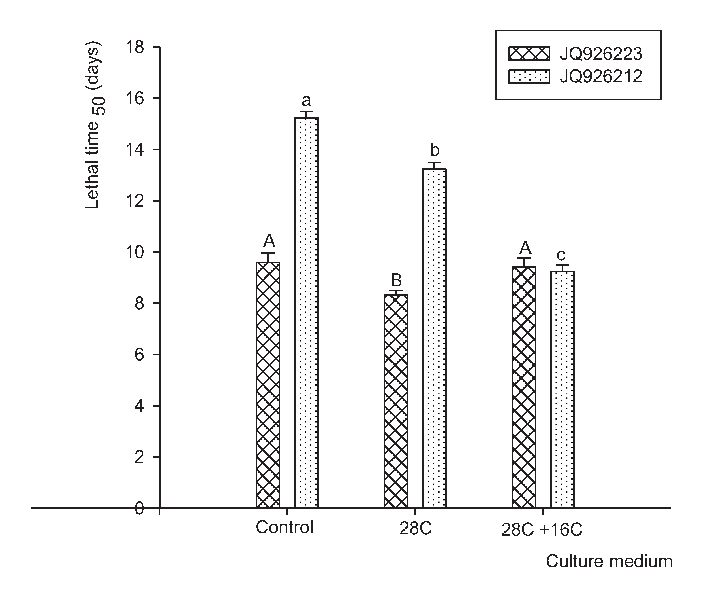
3.4. Pathogenicity Analysis

Statistical analyses of the effects of P. lilacinum strains, growth media and their interaction on insecticide efficiency against T. confusum showed significant differences (P < 0.0001). The major effect was that of strains (F = 242.08). Figure 4 shows the LT50 of both strains in the control medium and the medium amended with hydrocarbons. The LT50 for both strains of P. lilacinum after growth in the presence of C28 and C28 + C16 mixture was 10.9 and 9.6 days, respectively, while LT50 in the control medium was 12.5 days. The mortality in control treatments was low (<10%), and no mycosis was detected on any insects. Strains JQ926223 and JQ926212 showed a mortality rate of 70% and 50%, respectively, when these strains were grown in control medium. Furthermore, the mortality rate increased when both strains were grown in amended medium, showing a mortality rate of 80% and 70% for JQ926223 and JQ926212, respectively.
Figure 3. Production of proteases (a), amylases (b) and lipases (c) by both strains of P. lilacinum JQ926223 and JQ926212. Bars represent means and standard errors for each condition of Aw, pH and incubation time (days). Data with different letters for each Aw, pH or incubation time are significantly different based on LSD Fisher test (P < 0.05).
Insects 06 00206 g003
Figure 4. LT50 for T. confusum adults exposed during 15 days to two strains of P. lilacinum JQ926212 and JQ926223 grown in control medium and amended medium with 28C and 28C + 16C mixture. Data with different letters for each strain (JQ926223: capital letters, JQ926212: small letters) at different treatments are significantly different based on the Tukey test (P < 0.05).
Insects 06 00206 g004

4. Discussion

The assessment of an organism for microbial control in a pest management strategy requires basic studies such as isolation, culturing, biological testing and prediction of effects on the pest population and the environment [55].
Virulence of entomopathogenic fungi is often related to adhesion, rate of germination and growth of conidia on the cuticle of the insect hosts. This aspect reflects the metabolic advantages of degrading the cuticles of insects that can be seen in fungi stimulated by subculture in culture media amended with hydrocarbons [56,57,58]. This stimulation gives an initial advantage during the infective process. Among a variety of factors involved in the specificity of the interaction between the cuticles of insects and entomopathogenic fungi, the composition of the components of the host cuticle could stimulate or inhibit fungal growth [50]. Therefore, an increase of host mortality by inducing the synthesis of fungal degrading enzymes, suggests a possible approach for mycoinsecticide improvement. In our study, JQ926223 is more virulent than JQ926212 and both are pathogenic to T. confusum. However, mortality rate varied according to the culture medium used for fungal development. About 15% of increase in mortality was obtained when P. lilacinum strains grew in culture media supplemented with hydrocarbons.
Pedrini et al. [59] confirmed the results of Crespo et al. [50] regarding the increase of susceptibility of Acanthoscelides obtectus (60%–100% of mortality) to Beauveria bassiana after developing on media amended with hydrocarbons. A significant increase in mortality, reaching 90%, was also detected in Rhyzopertha dominica, therefore this method may increase the virulence favoring the early stages of the infection [50,60,61]. This increase is evidenced both by an increase in the percentage of mortality [50] as well as by a decrease in lethal time [61].
After the adhesion and germination of conidia occurred, penetration through the cuticle is the next stage of the infection cycle. The degradation of the hydrocarbons begins with the reaction of enzyme complexes. Information of fungal enzymatic activities involved in insect penetration would be required for the development of efficient mycotic agents for insect biological control [62]. Accordingly, in this study, we have analyzed the relationship of enzymatic activities with the fungus pathogenicity against T. confusum. Extracellular enzymes that degrade compounds in the cuticle of insects [63], such as proteases, chitinases and lipases are in some way related to the pathogenicity of entomopathogenic fungi [64]. However, other studies showed no correlation between enzyme activities and fungal pathogenicity [65].
In our study, high amylolytic activity was present in both strains. Protease and lipase activities were also present in lesser amounts. According to Esteves et al. [66], lipolytic activity of entomopathogenic fungi is low, although they suggest that lipases might have been synthesized later in time. They also conclude that lipase enzymes are secreted in little amounts when compared with the production of other extracellular enzymes. Moreover, the relatively low protease activity obtained in our study could be due to the fact that gelatin induced the production of chitinases and estearases, but surprisingly did not increase the production of proteases. The source of the substrate would favor the production of other enzymes than the proteases [66]. The use of high concentrations of gelatin repressed protease activity, such as albumin, whereas fibrous collagen enhanced protease activity [67].
It is known that enzymatic activity is directly related to fungal growth, which shows that the mycelial growth is crucial for an adequate production of different extracellular enzymes [68]. To determine the optimal conditions of physiological adaptation of the entomopathogenic fungus, it is important to find the best substrate to achieve the highest growth.
It is also important to compare if these optimum conditions match the increasing enzyme production. In this study, the greater growth rate was in presence of gelatin as substrate at the higher and lower Aw tested. On the other hand, both strains showed lower growth rates in the culture medium supplemented with tween-20 tested at 3 Aw. Both strains showed an acceptable growth rate with starch, probably because these strains are adapted to remain in the soil during the stages that not parasitize insects [69].
In this study, it was observed that the amount and type of enzymes secreted by P. lilacinum were different depending on the pH and Aw of the culture medium and the time of incubation. Therefore, cultural conditions appear to have an important effect on the results obtained.
Proteases secreted during the early stages of penetration [70] are considered the most important pathogenicity factor, because proteins are the major component in the exoskeleton (61%–70%) [71,72] and the cover of the eggs [73,74]. In our study, proteases were the main enzymes produced by these fungi. The mean values obtained were between 800 and 1400 mg/mL that were kept constant during the incubation time. The higher production of proteases was obtained in the range of acid and alkaline pH and lower Aw evaluated (0.97–0.95). The incubation time showed no statistically significant effect on the production. These results do not agree with those reported by Ghanem et al. [75] who showed that the highest protease production was recorded after 96 h of growth. But the production was not kept constant. Gomez Fernandez et al. [76] showed that Metarhizium anisopliae, B. bassiana and Paecilomyces sp. were good producers of proteases. Such production was enhanced when fungi were cultured in the absence of glucose, with gelatine as the only carbon source. Petlamul and Prasertsan [77] showed that M. anisopliae and B. bassiana had high protease activity during the first 4.5 to 6 days of incubation. While incubation time elapses, the protease activity decreases, probably for nutrient limitation or cell autolysis [78].
Different studies have shown that each entomopathogenic genus has a different enzymatic apparatus, with Paecilomyces sp. showing the greatest versatility for different enzymatic substrates. Other studies involving strains of the genus Pochonia have shown that this genus is also a producer of extracellular proteases in the presence of gelatine [79,80,81,82].
Macheronni Jr. et al. [83] showed production of lipase at neutral and alkaline pH, but no enzyme production was detected at acidic pH. Similar results were reported by Silva et al. [84], when they evaluated the production of lipase by M. anisopliae in presence of various carbonaceous sources. These authors suggest a relationship between these enzymes and the pathogenicity of the fungus. In our study, lipase production was low; the maximum lipolytic production was recorded after 3 days of incubation and alkaline pH (36 U/mL). Hasan et al. [85] showed that the maximum lipase production of Verticillium lecanii was found at pH 7 (55 U/mL). Results obtained by other authors and the results of this study are consistent with St. Leger et al. [86] who observed large variations in levels of enzymatic production among different entomopathogenic fungi.
In our study, the amylases were also detected but in small quantities. This could be due to a lack of production of enzymes or an intracellular location, because strains grew well in culture media supplemented with starch. The optimal amylolytic production was at 3 days of incubation and pH 9; however similar results were found at neutral pH. These results do not coincide with what Hasan et al. [85] observed. They reported the highest amylolytic production in strains of V. lecanii on the seventh day of incubation at pH 3. Lopez-Llorca and Carbonell [87] obtained similar results in the production of extracellular amylase by strains of V. lecanii isolated from soil and insect samples. According to Jackson et al. [64] there is an inverse relationship between the amylolytic activity and pathogenicity. There are at least two hypotheses for these results: (a) an absence of amylase production may be fortuitously associated with some virulence factor of the fungus (b) non-virulent isolates are predominantly saprophytes and are likely to require high amylolytic activity to achieve degradation of organic debris in soil.

5. Conclusions

The fungal growth with hydrocarbon mixtures increased the pathogenicity of the two strains of P. lilacinum, with the strain JQ926223 being more virulent. Both strains displayed mainly protease activity. The strains showed acceptable protease production in a range of pH values from acidic to alkaline and lower Aws.

Acknowledgments

This work was carried out thanks to grants from the Agencia Nacional de Promoción Científica y Tecnológica, PICT 2193/10 and in part by a grant of SECyT-UNRC 18/C401. We are grateful to the Magister Silvana Ponce for the English correction of the manuscript.

Author Contributions

All authors contributed to conducting, experimental design, writing and editing of the manuscript.

Conflicts of Interest

The authors declare no conflict of interest.

References

  1. Descamps, L.R.; Reviriego, M.E.; Suárez, A.A.; Ferrero, A.A. Reproducción de Sitophilus oryzae L. (Coleoptera: Curculionidae) y de Tribolium castaneum Herbst. (Coleoptera: Tenebrionidae) en cultivares de trigo argentinos. Bol. San. Veg. Plagas 2004, 30, 171–176. (in Spanish). [Google Scholar]
  2. Cassini, C.; Rodriguez, J.; Bartosik, R.; Peiretti, J.; Cabral, G. TRIGO Eficiencia de Cosecha y Postcosecha de granos. Manual Técnico N. 1. Sección Postcosecha; Ediciones INTA: Ciudad de Buenos Aires, Argentina, 2005; p. 120. (in Spanish) [Google Scholar]
  3. Fusé, C.B.; Villaverde, M.L.; Padín, S.B.; de Giusto, M.; Juarez, M.P. Evaluación de la actividad insecticida de tierras de diatomeas de yacimientos argentinos. Rev. Investig. Agropecuarias 2013, 2, 207–213. (in Spanish). [Google Scholar]
  4. Mejía, D. Food and Agriculture Organization of the United Nations (FAO). Available online: http://www.fao.org/inpho/content/compend/text/ch23 04.htm (accessed on 20 February 2015).
  5. White, N.D.G. Insects, mites and insecticides in stored-grain ecosystems. In Stored Grain Ecosystems; Jayas, P., White, N.D.G., Muir, W.E., Eds.; Marcel Dekker: New York, NY, USA, 1995; pp. 123–167. [Google Scholar]
  6. Wallace, H.A.H.; Sinha, R.N. Causal Factors Operative in Distributional Patterns and Abundance of Fungi: A Multivariate Study. In The Fungal Community. Its Organisation and Role in Ecosystems; Wicklow, D.T., Carroll, G.C., Eds.; Marcell Dekker Inc.: New York, NY, USA, 1981; pp. 233–247. [Google Scholar]
  7. Nesci, A.; Barra, P.; Etcheverry, M. Integrated management of insect vectors of Aspergillus flavus in stored maize using synthetic antioxidants and natural phytochemicals. J. Stored Prod. Res. 2011, 47, 231–237. [Google Scholar] [CrossRef]
  8. Nesci, A.; Montemarani, A.; Etcheverry, M. Assessment of mycoflora and infestation of insects, vector of Aspergillus section Flavi, in stored peanut from Argentina. Mycotoxin Res. 2011, 27, 5–12. [Google Scholar] [CrossRef] [PubMed]
  9. Barra, P.; Nesci, A.; Etcheverry, M. In vitro compatibility of natural and food grade fungicide and insecticide substances with Purpureocillium lilacinum and their effect against Aspergillus flavus. J. Stored Prod. Res. 2013, 54, 67–73. [Google Scholar] [CrossRef]
  10. International Agency for Research on Cancer (IARC). Monograph on the Evaluation of Carcinogenic Risk to Humans; IARC: Lyon, France, 1993. [Google Scholar]
  11. Cassini, C.; Santajuliana, M. Proyecto de Eficiencia de Cosecha, Postcosecha de Granos y Agroindustria en Origen; Instituto Nacional de Tecnología agropecuaria (INTA): Ciudad de Buenos Aires, Argentina, 2012. (in Spanish) [Google Scholar]
  12. Sartori, M.R.; Pacheco, I.A.; Vilar, R.M. Resistance to Phosphine in Stored Grain Insects in Brazil. In Proceedings of the Fifth International Working Conference on Stored-Product Protection, Bordeaux, France, 9–14 September 1990; Fleurat-Lessard, F., Ducom, P., Eds.; Imprimerie Medocaine: Blanquefort Cedex, France, 1991; pp. 1041–1050. [Google Scholar]
  13. Leemon, D.M.; Jonsson, N.N. Laboratory studies on Australian isolates of Metarhizium anisopliae as a biopesticide for the cattle tick Boophilus microplus. J. Invertebr. Pathol. 2008, 97, 40–49. [Google Scholar] [CrossRef] [PubMed]
  14. Ansari, M.A.; Pope, E.C.; Carpenter, S.; Scholte, E.J.; Butt, T.M. Entomopathogenic fungus as a biological control for an important vector of livestock disease: The Culicoides biting midge. PLOS ONE 2011, 6, e16108. [Google Scholar] [CrossRef] [PubMed]
  15. Bukhari, T.; Takken, W.; Koenraadt, C. Development of Metarhizium anisopliae and Beauveria bassiana formulations for control of malaria mosquito larvae. Parasit. Vectors 2011, 4, 23. [Google Scholar] [CrossRef] [PubMed]
  16. Morgan-Jones, G.; White, G.F.; Rodriguez-Kabana, R. Phytonematode pathology: Ultrastructural studies. II. Parasitism of Meloidogyne arenaria eggs and larvae by Paecilomyces lilacinus. Nematropica 1984, 14, 57–71. [Google Scholar]
  17. Jatala, P. Biological control of plant parasitic nematodes. Ann. Rev. Phytopathol. 1986, 24, 453–489. [Google Scholar] [CrossRef]
  18. Dube, B.; Smart, G.C. Biological control of Meloidogyne by Paecilomyces lilacinus and Pasteuria penetrans. J. Nematol. 1987, 19, 222–227. [Google Scholar] [PubMed]
  19. Khan, A.; Williams, K.L.; Nevalainen, H.K.M. Control of plant parasitic nematodes by Paecilomyces lilacinus and Monacrosporium lysipagum in pot trials. BioControl 2006, 51, 643–658. [Google Scholar] [CrossRef]
  20. Barra, P.; Rosso, L.; Nesci, A.; Etcheverry, M. Isolation and identification of entomopathogenic fungi and their evaluation against Tribolium confusum, Sitophilus zeamais, and Rhyzopertha dominica in stored maize. J. Pest Sci. 2013, 86, 217–226. [Google Scholar] [CrossRef]
  21. Magan, N. Physiological Approaches to Improving the Ecological Fitness of Fungal Biocontrol Agents. In Fungi as Biocontrol Agents; Butt, T.M., Jackson, C., Magan, N., Eds.; CAB International: Wallingford, UK, 2001. [Google Scholar]
  22. Hallsworth, J.E.; Magan, N. Effects of KCl concentration on accumulation of acyclic sugar alcohols and trehalose in conidia of three entomopathogenic fungi. Lett. Appl. Microbiol. 1994, 18, 8–11. [Google Scholar] [CrossRef]
  23. Hallsworth, J.E.; Magan, N. Effect of carbohydrate type and concentration on polyhydroxy alcohol and trehalose content of conidia of three entomopathogenic fungi. Microbiology 1994, 140, 2705–2713. [Google Scholar] [CrossRef]
  24. Frey, S.; Magan, N. Production of the fungal biocontrol agent Ulocladium atrum by submerged fermentation: Accumulation of endogenous reserves and shelf-life studies. Appl. Microbiol. Biotechnol. 2001, 56, 372–377. [Google Scholar] [CrossRef] [PubMed]
  25. Carpenter, J.F.; Crowe, J.H. The mechanism of cryoprotection of proteins by solutes. Cryobiology 1988, 25, 244–255. [Google Scholar] [CrossRef] [PubMed]
  26. Deacon, J. Significance of ecology in the development of biocontrol agents against soil-borne plant pathogens. Biocontrol Sci. Technol. 1991, 1, 5–20. [Google Scholar] [CrossRef]
  27. Samson, R.A.; Evans, H.C.; Latgé, J.P. Atlas of Entomopathogenic Fungi; Springer Verlag: New York, NY, USA, 1988; p. 187. [Google Scholar]
  28. Hajek, A.E.; St. Leger, R.J. Interactions between fungal pathogens and insect hosts. Annu. Rev. Entomol. 1994, 39, 293–322. [Google Scholar] [CrossRef]
  29. Charnley, A.K.; Collins, S.A. Entomopathogenic Fungi and Their Role in Pest Control. In The Mycota IV: Environmental and Microbial Relationships, 2nd ed.; Kubicek, C.P., Druzhinina, I.S., Eds.; Springer-Verlag: Berlin, Germany, 2007; pp. 159–187. [Google Scholar]
  30. Bidochka, M.J.; St. Leger, R.S.; Roberts, D.W. Mechanisms of Deuteromycete fungal infections in grasshoppers and locusts: An overview. Mem. Entomol. Soc. Can. 1997, 171, 213–224. [Google Scholar] [CrossRef]
  31. Boucias, D.; Pendland, J. Attachment of Mycopathogens to Cuticle. In The Fungal Spore and Disease Initiation in Plants and Animals; Cole, G.T., Hoch, H.C., Eds.; Plenum Press: New York, NY, USA, 1991; pp. 101–127. [Google Scholar]
  32. Fang, W.G.; Feng, J.; Fan, Y.H.; Zhang, Y.J.; Bidochka, M.J.; Leger, R.J.S.; Pei, Y. Expressing a fusion protein with protease and chitinase activities increases the virulence of the insect pathogen Beauveria bassiana. J. Invertebr. Pathol. 2009, 102, 155–159. [Google Scholar] [CrossRef] [PubMed]
  33. Blomquist, G.J.; Dillwith, J.W. Cuticular Lipids. In Comprehensive Insect Physiology, Biochemistry, and Pharmacology; Kerkutand, G.A., Gilbert, L.I., Eds.; Pergamon Press: Oxford, UK, 1985; pp. 117–154. [Google Scholar]
  34. Lockey, K.H. Lipids of the insect cuticle: Origin, composition and function. Comp. Biochem. Physiol. 1988, 89, 595–645. [Google Scholar] [CrossRef]
  35. Buckner, J.S. Cuticular Polar Lipids of Insects. In Insect Lipids: Chemistry, Biochemistry and Biology; Stanley-Samuelson, D.W., Nelson, D.R., Eds.; University of Nebraska Press: Lincoln, NE, USA, 1993; pp. 227–270. [Google Scholar]
  36. Nelson, D.R.; Blomquist, G.J. Insect Waxes. In Waxes Chemistry, Molecular Biology and Functions; Hamilton, R.J., Ed.; The Oily Press, Ltd.: Dundee, UK, 1995; pp. 1–90. [Google Scholar]
  37. Blomquist, G.J.; Nelson, D.R.; de Renobales, M. Chemistry, biochemistry and physiology of insect cuticular lipids. Arch. Insect Biochem. 1987, 6, 227–265. [Google Scholar] [CrossRef]
  38. Boucias, D.G.; Pendland, J.C. Nutritional requirements for conidial germination of several host range pathotypes of the entomopathogenic fungus Nomuraea rileyi. J. Invertebr. Pathol. 1984, 43, 288–292. [Google Scholar] [CrossRef]
  39. Boucias, D.G.; Pendland, J.C.; Latge, J.P. Nonspecific factors involved in attachment of entomopathogenic Deuteromycetes to host insect cuticle. Appl. Environ. Microbiol. 1988, 54, 1795–1805. [Google Scholar] [PubMed]
  40. Lord, J.C.; Howard, R.W. A proposed role for the cuticular fatty amides of Liposcelis bostrychophila (Psocoptera: Liposcelidae) in preventing adhesion of entomopathogenic fungi with dry-conidia. Mycopathologia 2004, 158, 211–217. [Google Scholar] [CrossRef] [PubMed]
  41. Aro, N.; Pakula, T.; Penttila, M. Transcriptional regulation of plant cell wall degradation by filamentous fungi. FEMS Microbiol. Rev. 2005, 29, 719–739. [Google Scholar] [CrossRef] [PubMed]
  42. Ortiz-Urquiza, A.; Keyhani, N.O. Action on the surface: Entomopathogenic fungi versus the insect cuticle. Insects 2013, 4, 357–374. [Google Scholar]
  43. Hankin, L.; Anagnostakis, S.L. The use of solid media for detection of enzyme production by fungi. Mycologia 1975, 67, 597–607. [Google Scholar] [CrossRef]
  44. Dallyn, H.; Fox, A. Spoliage of Material of Reduced Water Activity by Xerophilic Fungi. In Microbial Growth and Survival in Extreme Environments; Gould, G.H., Corry, E.L., Eds.; Academic Press: London, UK, 1980; pp. 129–139. [Google Scholar]
  45. Pitt, J.I. The Genus Penicillium and its Teleomorphic States Eupenicillium and Talaromyces; Academic Press: London, UK, 1979. [Google Scholar]
  46. Price, M.F.; Wilkinson, I.D.; Gentry, L.O. Plate method for detection of phospholipase activity in Candida albicans. Sabouraudia 1982, 20, 7–14. [Google Scholar] [CrossRef] [PubMed]
  47. Gornall, A.G.; Bardawill, C.J.; David, M.M. Determination of serum proteins by means of the biuret reaction. J. Biol. Chem. 1949, 177, 751–766. [Google Scholar]
  48. Miller, G.L. Use of dinitrosalicylic acid reagent for determination of reducing sugar. Anal. Chem. 1959, 31, 426–428. [Google Scholar] [CrossRef]
  49. Kamimura, E.S.; Medieta, O.; Rodrigues, M.L.; Maugeri, F. Studies on lipase-affinity adsorption using response-surface analysis. Biotech. Appl. Biochem. 2001, 33, 153–159. [Google Scholar] [CrossRef]
  50. Crespo, R.; Juárez, M.P.; dal Bello, G.M.; Padín, S.; Calderón Fernández, G.; Pedrini, N. Increased mortality of Acanthoscelides obtectus by alkane-grown Beauveria bassiana. BioControl 2002, 47, 685–696. [Google Scholar] [CrossRef]
  51. Goettel, M.S.; Inglis, G.D. Fungi: Hyphomycetes. In Manual of Techniques in Insect Pathology; Lacey, L.A., Ed.; Academic Press: San Diego, CA, USA, 1997; pp. 213–249. [Google Scholar]
  52. Padín, S.B.; dal Bello, G.M.; Vasicek, A.L. Pathogenicity of Beauveria bassiana for adults of Tribolium castaneum (Coleoptera: Tenebrionidae) in stored grains. Entomophaga 1997, 42, 569–574. [Google Scholar] [CrossRef]
  53. Wicklow, D.T.; Weaver, D.K.; Throne, J.E. Fungal colonists of maize grain conditioned at constant temperatures and humidities. J. Stored Prod. Res. 1998, 34, 355–361. [Google Scholar] [CrossRef]
  54. Di Rienzo, J.A.; Casanoves, F.; Balzarini, M.G.; Gonzalez, L.; Tablada, M.; Robledo, C.W. InfoStat, versión 2008; Grupo InfoStat, FCA: Universidad Nacional de Córdoba, Córdoba, Argentina, 2008. [Google Scholar]
  55. Lacey, L.A.; Frutos, R.; Kaya, H.K.; Vail, P. Insect pathogens as biological control agents: Do they have a future? Biol. Control. 2001, 21, 230–248. [Google Scholar] [CrossRef]
  56. Juárez, M.P.; Crespo, R.; Calderón Fernández, G.; Lecouna, R.; Cafferata, L.F.R. Characterization and carbon metabolism in fungi pathogenic to Triatoma infestans, a Chagas disease vector. J. Invertebr. Pathol. 2000, 76, 198–207. [Google Scholar] [CrossRef] [PubMed]
  57. Crespo, R.; Juárez, M.P.; Cafferatta, L.F.R. Biochemistry of the interaction between entomopathogenous fungi and their insect host-like hydrocarbons. Mycologia 2000, 92, 528–536. [Google Scholar] [CrossRef]
  58. Pedrini, N.; Juárez, M.; Crespo, R.; de Alaniz, M. Clues on the role of Beauveria bassiana catalases in alkane degradation events. Mycologia 2006, 98, 528–534. [Google Scholar]
  59. Pedrini, N.; dal Bello, G.; Padín, S.; Juárez, M. Capacidad insecticida de Beauveria bassiana cultivada en hidrocarburos para control de coleópteros en granos almacenados. Agrociencia (Uruguay) 2011, 1, 64–69. (in Portuguese). [Google Scholar]
  60. Pedrini, N.; Crespo, R.; Juarez, M.P. Biochemistry of insect epicuticle degradation by entomopathogenic fungi. Comp. Biochem. Physiol. C Pharmacol. Toxicol. Endocrinol. 2007, 146, 124–137. [Google Scholar] [CrossRef]
  61. Pedrini, N.; Mijailovsky, S.J.; Girotti, J.R.; Stariolo, R.; Cardozo, R.M.; Gentile, A.; Juárez, M.P. Control of pyrethroid-resistant chagas disease vectors with entomopathogenic fungi. PLOS Negl. Trop. Dis 2009, 3, e434. [Google Scholar] [CrossRef] [PubMed]
  62. Scorsetti, A.C.; Elíades, L.A.; Stenglein, S.A.; Cabello, M.N.; Pelizza, S.A.; Saparrat, M.C.N. Pathogenic and enzyme activities of the entomopathogenic fungus Tolypocladium cylindrosporum (Ascomycota: Hypocreales) from Tierra del Fuego, Argentina. Rev. Biol. Trop. 2011, 60, 833–841. [Google Scholar]
  63. Charnley, A.K. Fungal pathogens of insects: Cuticle degrading enzymes and toxins. Adv. Bot. Res. 2003, 40, 241–321. [Google Scholar]
  64. Jackson, C.W.; Heale, J.B.; Hall, R.A. Traits associated with virulence to the aphid Macrosiphoniella sanborni in eighteen isolates of Verticillium lecanii. Ann. Biol. 1985, 106, 39–48. [Google Scholar] [CrossRef]
  65. Rosato, Y.B.; Messias, C.L.; Azevedo, J.L. Production of extracellular enzymes by isolates of Metarhizium anisopliae. J. Invertebr. Pathol. 1981, 38, 1–3. [Google Scholar] [CrossRef]
  66. Esteves, I.; Peteira, B.; Atkins, S.; Magan, N.; Kerry, B. Production of extracellular enzymes by different isolates of Pochonia chlamydosporia. Mycol. Res. 2009, 113, 867–876. [Google Scholar] [CrossRef] [PubMed] [Green Version]
  67. Segers, R. The nematophagus fungus Verticillium Chlamydosporium: Aspects of phatogenicity. Ph.D. Thesis, Nottingham University, Nottingham, UK, 1996. [Google Scholar]
  68. Carlsen, M.; Spohr, A.B.; Nielsen, J.; Villadsen, J. Morpgology and physiology of an α amylase producing strain of Aspergillus oryzae during batch cultivations. Biotechnol. Bioeng. 1996, 49, 266–276. [Google Scholar] [CrossRef] [PubMed]
  69. Chandler, D.; Hay, D.; Reid, A.P. Sampling and occurrence of entomopathogenic fungi and nematodes in UK soils. Appl. Soil Ecol. 1997, 5, 133–141. [Google Scholar] [CrossRef]
  70. Bidochka, M.J.; Khachatourians, G.G. Identification of Beauveria bassiana extracellular protease as a virulence factor in pathogenicity toward the migratory grasshopper Melanoplus sanguinipes. J. Invertebr. Pathol. 1990, 59, 165–173. [Google Scholar] [CrossRef]
  71. Hepburn, H.R. Structure of the Integument. In Comprehensive Insect Physiology, Biochemistry and Pharmacology; Kerkut, G.A., Gilbert, L.I., Eds.; Pergamon: Oxford, UK, 1985; Volume 3, pp. 1–58. [Google Scholar]
  72. Bidochka, M.J. Interaction of the Entomopathogenic Fungus Beauveria Bassiana with the Migratory Grasshopper Melanoplus Sanguinipes a Systematic Study of Pathogenesis. Ph.D. Thesis, University of Saskatchewan, Saskatoon, SK, Canada, 1989. [Google Scholar]
  73. Andersen, S.O. Sclerotization and Tanning of the Cuticle. In Comprensive Insect Physiology, Biochemistry and Pharmacology; Kerkut, G.A., Gilbert, L.I., Eds.; Pergamon: Oxford, UK, 1985; pp. 59–74. [Google Scholar]
  74. Margaritis, L.H. Structure and Physiology of the Egg Shell. In Comprensive Insect Physiology, Biochemistry and Pharmacology; Kerkut, G.A., Gilbert, L.I., Eds.; Pergamon Press: Oxford, UK, 1985; Volume 1, pp. 153–230. [Google Scholar]
  75. Ghanem, K.M.; Al-Garni, S.M.; Al-Makishah, N.H. Statistical optimization of cultural conditions for chitinase production from fish scales waste by Aspergillus terreus. Afr. J. Biotechnol. 2010, 9, 5135–5146. [Google Scholar]
  76. Gómes Fernandes, E.; Maia Valério, H.; Feltrin, T.; van der Sand, S.T. Variability in the production of extracellular enzymes by entomopathogenic fungi grown on different substrates. Braz. J. Microbiol. 2012, 43, 827–833. [Google Scholar] [CrossRef] [PubMed]
  77. Petlamul, W.; Prasertsan, P. Evaluation of strains of Metarhizium anisopliae and Beauveria bassiana against Spodoptera litura on the basis of their virulence, germination rate, conidia production, radial growth and enzyme activity. Mycobiology 2012, 40, 111–116. [Google Scholar] [CrossRef] [PubMed]
  78. Dhar, P.; Gurvinder, K. Production of cuticle-degrading proteases by Beauveria bassiana and their induction in different media. Afr. J. Biochem. Res. 2010, 4, 65–72. [Google Scholar]
  79. Lopez-Llorca, L.V.; Olivares-Bernabeu, C.; Salinas, J.; Jansson, H.B.; Kolattukudy, P.E. Prepenetration events in fungal parasitism of nematode eggs. Mycol. Res. 2002, 106, 499–506. [Google Scholar] [CrossRef]
  80. Mendoza de Gives, P.; Behnke, J.M.; Davies, K.G. Extracellular enzyme production by nematophagous fungi in the presence and absence of nematodes. Int. J. Nemathol. 2003, 13, 27–36. [Google Scholar]
  81. Mortan, O.; Hirsch, P.R.; Peberdy, J.F.; Kerry, B.R. Cloning and genetic variation in protease VCP1 from the nematophagous fungus Pochonia chlamydosporia. Mycol. Res. 2003, 107, 38–46. [Google Scholar] [CrossRef] [PubMed]
  82. Segers, R.; Butt, T.M.; Kerry, BR.; Peberdy, J.F. The nematophagous fungus Verticillium chlamydosporium produces a chymoelastase-like protease which hydrolyses host nematode proteins in situ. Microbiology 1994, 140, 2715–2723. [Google Scholar] [CrossRef] [PubMed]
  83. Maccheroni Jr., W.; Welington, L.A.; Azevedo, J.L. Ambient pH-regulated enzime secretion in endophytic and pathogenic isolates of the fungal genus Colletotrichum. Sci. Agric. 2004, 61, 298–302. [Google Scholar] [CrossRef]
  84. Silva, W.O.B.; Mitidieri, S.; Schrank, A.; Vainstein, M.H. Productionand extraction of an extracellular lipase from the entomopathogenic fungus Metarhizium anisopliae. Process Biochem. 2005, 40, 321–326. [Google Scholar] [CrossRef]
  85. Hasan, S.; Ahmad, A.; Purwar, A.; Khan, N.; Kundan, R.; Gupta, G. Production of extracellular enzymes in the entomopathogenic fungus Verticillium lecanii. Bioinformation 2013, 9, 238–242. [Google Scholar] [CrossRef] [PubMed]
  86. St. Leger, R.J.; Charnley, A.K.; Cooper, R.M. Cuticle-degrading enzymes of entomopathogenic fungi: Synthesis in culture on cuticle. J. Invertebr. Pathol. 1986, 48, 85–95. [Google Scholar] [CrossRef]
  87. Lopez-Llorca, L.V.; Carbonell, T. Characterization of spanish strains of Verticillium lecanii. Rev. Iberoam. Micol. 1999, 16, 136–142. [Google Scholar] [PubMed]

