Article

Mamestra brassicae Multiple Nucleopolyhedroviruses Prevents Pupation of Helicoverpa armigera by Regulating Juvenile Hormone Titer

1
Institute of Microbial Application, Xinjiang Academy of Agricultural Sciences, Urumqi 830000, China
2
College of Life Sciences, Henan Agricultural University, Zhengzhou 450002, China
3
School of Biotechnology, Gautam, Buddha University, Greater Noida 201312, India
*
Authors to whom correspondence should be addressed.
Insects 2024, 15(3), 202; https://doi.org/10.3390/insects15030202
Submission received: 11 January 2024 / Revised: 26 February 2024 / Accepted: 6 March 2024 / Published: 18 March 2024
(This article belongs to the Section Insect Behavior and Pathology)

Abstract

:

Simple Summary

Baculovirus infection can prevent the pupation of insects. However, the molecular mechanism of baculovirus preventing the pupation of larvae by regulating the Juvenile hormone (JH) pathway is still unclear. In this study, we examined the interactive relationship of Mamestra brassicae multiple nucleopolyhedroviruses (MbMNPV) and Helicoverpa armigera (H. armigera). We found that MbMNPV infection caused an increased JH titer and inhibited the expression levels of juvenile hormone esterase (JHE) and juvenile hormone epoxide hydrolase (JHEH). Our studies further proved that the JH is mainly degraded by JHE in H. armigera larvae. Knocking down of HaJHE promoted MbMNPV replication. These findings suggest that the infection of H. armigera larvae by MbMNPV leads to an increased JH titer by inhibiting the expression of JHE, which prevents pupation of H. armigera and promotes MbMNPV replication.

Abstract

Baculovirus infection can prevent the pupation of insects. Juvenile hormone (JH) plays a vital role in regulating insect molting and metamorphosis. However, the molecular mechanism of baculovirus preventing the pupation of larvae by regulating the Juvenile hormone (JH) pathway is still unclear. In this study, we found that the Mamestra brassicae multiple nucleopolyhedroviruses (MbMNPV) infection prolonged the larval stage of fourth instar Helicoverpa armigera (H. armigera) by 0.52 d and caused an increase in JH titer. To identify the genes that contribute to the JH increase in H. armigera-MbMNPV interaction, we analyzed mRNA expression profiles of the fat bodies of H. armigera infected by MbMNPV. A total of 3637 differentially expressed mRNAs (DE-mRNAs) were filtered out through RNA-seq analysis. These DE-mRNAs were mainly enriched in Spliceosome, Ribosome biogenesis in eukaryotes, Aminoacyl-tRNA biosynthesis, Mismatch repair, and RNA degradation signaling pathway, which are related to the virus infection. Real-time PCR was used to verify the RNA sequencing results. To find out which genes caused the increase in JH titer, we analyzed all the DE-mRNAs in the transcriptome and found that the JHE and JHEH genes, which were related to JH degradation pathway, were down-regulated. JHE and JHEH genes in the larvae of MbMNPV-infected group were significantly down-regulated compared with the control group by RT-qPCR. We further proved that the JH is degraded by JHE in H. armigera larvae by RNAi, ELISA, RT-qPCR and bioassay, while the hydrolysis of JH by JHEH in H. armigera larvae can almost be ignored. Knocking down of HaJHE promoted the expression of the JH receptor gene Met and the downstream gene Kr-h1, and the replication of MbMNPV. This study clarified that JH is mainly degraded by JHE in H. armigera larvae. The MbMNPV infection of H. armigera larvae leads to the increase of JH titer by inhibiting the expression of JHE. The increase in JH titer promotes the expression of the JH receptor gene Met and the downstream gene Kr-h1, which prevents the pupation of H. armigera, and promotes MbMNPV replication. This study provides new insights into H. armigera and MbMNPV interaction mechanisms.

1. Introduction

Helicoverpa armigera (H. armigera) is an insect pest [1] that harms several crops, such as wheat, maize, tomato, pepper, and cotton, leading to heavy crop losses worldwide [2]. Control of the pest mainly relies on chemical pesticides, which has led to increasingly severe issues, such as food safety, ecological environment, and pest resistance. Hence, there is an urgent requirement to develop new, efficient, and safe bio-pesticides.
Baculoviruses have high host specificity and are deemed promising novel viral insecticides [3]. So far, only three kinds of broad-spectrum baculoviruses have been identified, including Mamestra brassicae multiple nucleopolyhedrovirus, Autographa californica multiple nucleopolyhedrovirus, and the Anagrapha falcifera multinucleocapsid [4,5,6,7]. Mamestra brassicae multiple nucleopolyhedrovirus (MbMNPV), a member of Baculoviridae, was isolated from the cabbage armyworm Mamestra brassicae belonging to the Noctuidae family, which is characterized by a wide host spectrum because it can infect 32 species of Lepidoptera pests [4]. MbMNPV has been developed as a commercial bio-pesticide in China [4].
Insect viruses, as the most important pathogens to Lepidoptera insects, can alter the development and physiological status of their hosts [8,9,10]. Insects infected with exogenous viruses typically exhibit developmental defects, such as prolonged larval stage and pupation failure [11,12]. Studies have found that the egt gene encoded by baculovirus can regulate the activity of ecdysone, and prevent infected larvae from molting or pupation [12,13]. Juvenile hormone (JH) can regulate the insect growth, development, reproduction, and metamorphosis [14]. Some of the genes in the JH signaling pathway have been reported, such as juvenile hormone esterase (JHE) [15], juvenile hormone epoxide hydrolase (JHEH) [16], juvenile hormone receptor Methoprene-tolerant (Met) [17], and Krüppel-homolog 1 (Kr-h1) [18]. However, the molecular mechanism of how the baculovirus prevents the pupation of larvae by juvenile hormone (JH) pathway is still unclear.
JHE plays a crucial role in controlling the JH titer in insects, especially during the late larval stage. Insect metamorphosis requires a low JH concentration [19]. The enzyme activity, distribution, and content of JHE vary depending on the type, developmental stage, and tissue organ of insects. JHEH is responsible for the irreversible hydrolysis of JH epoxides, and the metabolite JHA has been shown to have hormonal activity [20,21]. The functional study of the role and regulation of JHE and JHEH in H. armigera has not been reported yet. In insects, molting and metamorphosis can be regulated through the JH-Met-Kr-h1 signaling pathway. Kr-h1, an early response gene induced by JH, is also involved in insect metamorphosis and reproduction. JH can induce the expression of downstream gene Kr-h1 through the JH receptor Met, and thereby it plays a vital role in regulating insect metamorphosis [22,23]. The fat body of insects is a vital organ for biosynthesis, metabolism, detoxification, and control of insect ecdysis. It has become a significant research model for the study of metabolic disorders [24].
This study focuses on understanding the mechanism of how the baculovirus prevents the pupation of larvae by controlling the JH pathway. Our results provide a new insight into the role of JHE in H. armigera-MbMNPV interactions, which is helpful in understanding the host–baculovirus relation and can be a candidate to develop newer insecticides.

2. Materials and Methods

2.1. Insects and Viruses

The eggs of H. armigera were purchased from Jiyuan Baiyun Industry Co., Ltd. (Jiyuan, China). Insects were fed with an artificial diet and reared in the laboratory at 27 ± 2 °C, 60 ± 2% relative humidity, with a photoperiod of 16:8 (light:dark) [25].
MbMNPV culture was available in our laboratory and was propagated by feeding to third instar H. armigera larvae. Occlusion bodies (OBs) of MbMNPV were purified as described by Kyei-Poku & Kunimi [26]. The OBs were purified from MbMNPV-infected cadavers by differential centrifugation followed by sucrose density gradient ultracentrifugation.

2.2. Virus Infection and Juvenile Hormone Determination

The newly molted third instar larvae were fed with an artificial diet contaminated with MbMNPV occlusion bodies (30 OBs) after starving for 5 h. Upon consumption of the diets, the larvae were fed with fresh diets without exposure to any pathogens. Mock-infected larvae were fed with the same amounts of diet, but coated with distilled water. Thirty larvae per group were evaluated for larval weight, pupation rate, emergence rate, and larval mortality. The experiments were performed with three independent biological replicates.
In addition, the newly molted fourth instar larvae were fed with artificial diets contaminated with MbMNPV occlusion bodies (OBs, 1 × 10⁸ OBs/larva) after starving for 20 h. Thirty larvae per group were evaluated at fourth instar larval stage. JH titer in fat body and hemolymph at 24 h, 48 h, 72 h, and 96 h post-infection (hpi) was determined by ELISA using the JH ELISA kits from Jiangsu Meimian Industrial Co., Ltd. (Yancheng, Jiangsu, China) [27,28] according to the manufacturer’s instructions. The experiments were performed with three independent biological replicates and technical replicates.

2.3. RNA Extraction and Quality Control

The fat body of MbMNPV-infected and mock-infected larvae consisted of three repeats labeled as MbMNPV_1, MbMNPV_2, and MbMNPV_3, and CK_1, CK_2, and CK_3, respectively. At 72 h post-infection (hpi), total RNA was extracted using the TRIZOL reagent (Invitrogen, Carlsbad, CA, USA) according to the manufacturer’s protocol. The RNA quality and purity were evaluated using agarose gel electrophoresis, a Nanodrop microvolume spectrophotometer and an Agilent 2100 Bioanalyzer (Agilent Technologies, Santa Clara, CA, USA).

2.4. Library Preparation, mRNAs Sequencing, and Analysis

The libraries were sequenced on the Illumina Novaseq 6000 platform and 150 bp paired-end reads were generated. Raw reads were processed using fastp software (v 0.20.1) [29] and the low quality reads were removed to obtain the clean reads. The clean reads were mapped to the reference genome using HISAT2 software (v 2.1.0) [30] and the expression level of each gene was normalized by the FPKM (transcript per million) method [31]. Differential expression analysis was performed using the DESeq2 software (v 1.22.2) [32]. The differentially expressed mRNAs (DE-mRNAs) were selected with thresholds of q-value < 0.05 and |log2 (foldchange)| > 1. GO enrichment [33] and KEGG pathway enrichment analysis [34] of DE-mRNAs were respectively performed using R (v 3.2.0) based on the hypergeometric distribution. Top 30 GO terms and Top 20 of KEGG enrichment were considered to be enriched.

2.5. Validation of mRNA by Real-Time Quantitative PCR

To validate the RNA-seq data, three mRNAs were randomly selected for RT-qPCR confirmation with ChamQ™ Universal SYBR® RT-qPCR master mix (Vazyme, Nanjing, China). Total RNAs were extracted using RNA isolator Total RNA Extraction Reagent (Vazyme, China). For gene quantification, the cDNA was prepared using HiScript® III All-in-one RT SuperMix Perfect for RT-qPCR (Vazyme, Nanjing, China) with the gDNA wiper. The PCR was performed with a StepOnePlus™ Real-Time PCR Systems (Applied Biosystems, Thermo Fisher Scientific, Waltham, MA, USA) under the following conditions: 95 °C for 30 s, followed by 40 cycles of 95 °C for 10 s and 60 °C for 30 s. The specificity of each primer pair was ensured by analyzing the melting curve. The experiments were performed with three independent biological replicates and technical replicates. The relative quantity of mRNAs was analyzed by RT-qPCR, and β-actin was used as the reference gene. All the primers were synthesized by the Sangon Biotech Co., Ltd. (Shanghai, China) (Table S1). The data were analyzed by the 2−ΔΔCT method.

2.6. The Expression Patterns, Synthesis of dsRNA, RNA Interference

The expression patterns of JHE and JHEH in the fat bodies and midgut at different developmental stages of H. armigera larvae, viz. the last larval instar (fifth instar) molting stage (5L-M), the first day after last larval instar molting stage (5L-1), the second day after last larval instar molting stage (5L-2), the third day after last larval instar molting stage (5L-3), the fourth day after last larval instar molting stage (5L-4), and the fifth day after last larval instar molting stage (5L-5), were determined by RT-qPCR.
The synthesis and microinjection of dsRNA were performed as per the standard protocol (Ref). Briefly, target fragments of GFP (green fluorescent protein), HaJHE, and HaJHEH for dsRNA synthesis were amplified using specific primers. The PCR products were sub-cloned into the 5 min TA/Blunt-Zero Cloning Kit (Vazyme, China) and used as templates for target sequence amplification. dsRNAs were synthesized in vitro using T7 RNAi Transcription Kit (Vazyme, China), with specific primers fusing the T7 promoter at the 5′ end. The purity and yield of the dsRNA was checked on 1.0% agarose gel and spectrophotometer, respectively. The primers used for the synthesis of dsRNAs are shown in Table S2. In the knockdown experiment, newly molted last larval instar larvae were incubated for 30 min on ice, followed by injection of 10 μg single dsRNA solution (ds GFP, ds HaJHE, and ds HaJHEH) using a 10 µL microsyringe (Hamilton) in their last leg.
The expression level of JHE, JHEH, Met, and Kr-h1 gene, and JH titer in larvae injected with dsRNA (ds GFP, ds HaJHE, and ds HaJHEH) was detected by RT-qPCR and ELISA, respectively.
To study the effects of knocking down of HaJHE and HaJHEH in H. armigera larvae, the larval weight, feed intake, pupation rate, and pupation time of last larval instar H. armigera larvae injected with ds GFP, ds HaJHE, and ds HaJHEH respectively were measured by bioassay.

2.7. Viral Infection

We further investigated the impact of JHE knocking down on MbMNPV replication. Larvae on the first day after the last larval instar molting stage injected with dsRNA solution were fed with fresh artificial diets for 4 h. After overnight starvation, the larvae were fed with artificial diets coated with a MbMNPV suspension (OBs; 1 × 10⁷ OBs/larva) followed with feeding of fresh diets without exposure to any pathogens. Subsequently, three larvae infected with mock and MbMNPV were randomly selected at 48 h, and the tissues of the fat body were dissected. To reveal the impact of knocking down of HaJHE on MbMNPV infection, the relative expression of MbMNPV polyhedrin gene was performed by RT-qPCR for the mock-infected and MbMNPV-infected group.

2.8. Data Analysis

All statistical analyses in this study were performed with Prism software and the data were represented as means ± SEM. The statistical significance was determined by Student’s t-test for unpaired comparisons between two different groups, and p < 0.05 was regarded as statistically significant.

3. Results

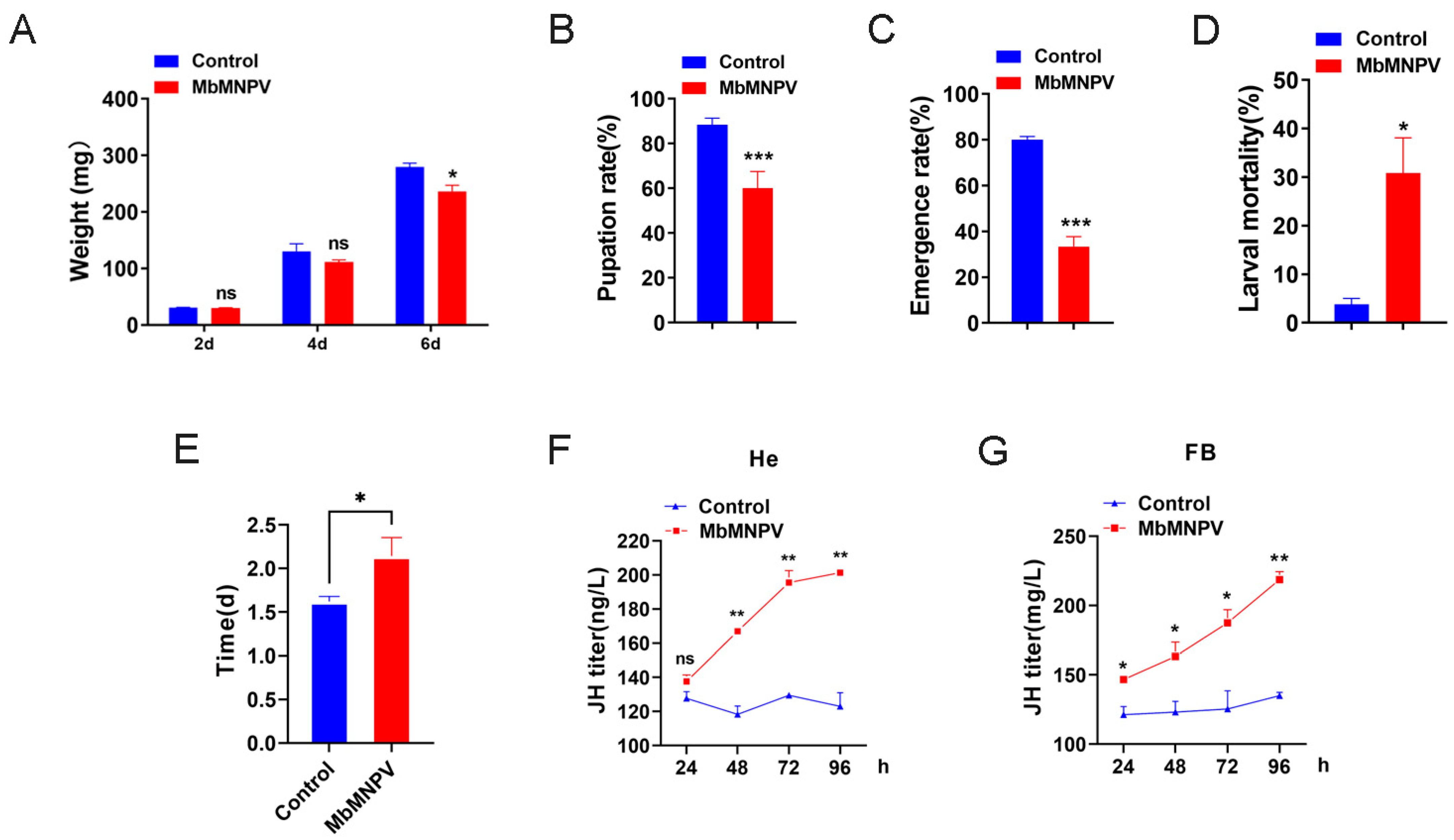
3.1. MbMNPV Infection Prolonged the Larval Stage and Caused the Increase of JH Titer

The larval weight, pupation rate, emergence rate, larval mortality, length of fourth instar larval stage, and JH titer of the mock-infected and MbMNPV-infected larvae were evaluated. Compared with the mock-infected group, the larval weight, pupation rate, and emergence rate of the MbMNPV-infected larvae had a significant reduction (Figure 1A–C). There was a significant increase in the larval mortality, length of the fourth instar larval stage, and JH titer of the MbMNPV-infected larvae (Figure 1D–F). The fourth instar larval stage of the MbMNPV-infected larvae was extended by 0.52 d (Figure 1E). This indicated that MbMNPV infection inhibited the growth of H. armigera, prolonged the larval stage, and caused the increase of JH titer.

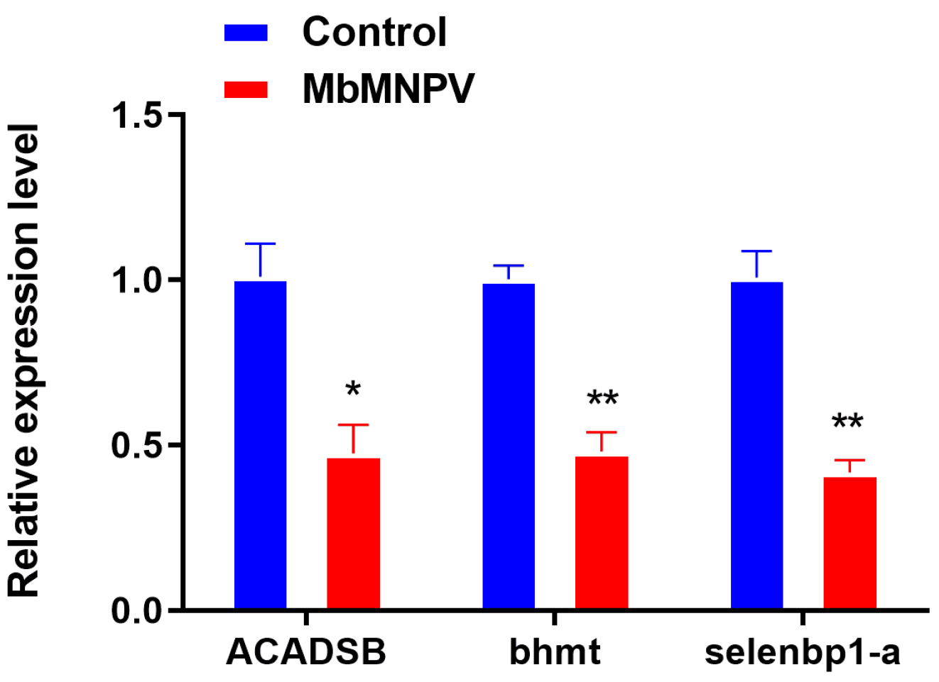
3.2. RNA-Seq Sequencing and Data Analysis

Six mRNA libraries (MbMNPV-infected and mock-infected larvae, three repeats labeled as MbMNPV_1, MbMNPV_2, and MbMNPV_3, and CK_1, CK_2, and CK_3, respectively) were sequenced and the quality of the sequencing data was analyzed (Table 1). Clean Reads were distributed at 47.24 M–49.58 M, with Q30 of 87.79–90.36%.
The DE-mRNAs between the MbMNPV-infected group and the control were screened based on FPKM values (q-value < 0.05 and |log2 (foldchange)| > 1). A total of 3637 mRNAs were differentially expressed, including 1634 up-regulated and 2003 down-regulated mRNAs (Figure 2A). The volcanic and heat maps (Figure 2C) of the normalized expression of mRNAs were generated and the expression patterns of the DE-mRNAs expressed during MbMNPV infection were analyzed.
GO and KEGG analyses were performed to investigate the function of DE-mRNAs (Figure 2B,D). GO analysis indicated that the DE-mRNAs were mainly enriched in the following terms: mitochondrial translation (GO: 0032543), mRNA splicing via Spliceosome (GO: 0000398), and rRNA processing (GO: 0006364), under biological process category; nucleolus (GO: 0005730), mitochondrial large ribosomal subunit (GO: 0005762), and mitochondrion (GO: 0005739), under cellular component category; RNA binding (GO: 0003723), unfolded protein binding (GO: 0051082), and ATP binding (GO: 0005524), under molecular function category (Figure 2B).
In the KEGG pathway analysis, Aminoacyl-tRNA biosynthesis (ID: haw00970), RNA polymerase (ID: haw03020), Ribosome biogenesis in eukaryotes (ID: haw03008), Spliceosome (ID: haw03040), Mismatch repair (ID: haw03430), and RNA degradation (ID: haw03018) were significantly enriched (Figure 2D).

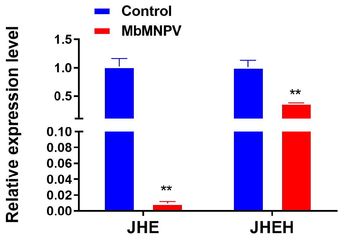
3.3. MbMNPV Infection Inhibited the Expression of JHE and JHEH

Three mRNAs (ACADSB, bhmt and selenbp1-a) were randomly selected from the DE-mRNAs to verify the sequencing data by RT-qPCR. The results showed that the expression of these three mRNAs in the MbMNPV-infected group was significantly down-regulated compared with the control group (Figure 3). The RT-qPCR results were consistent with that of the sequencing data, indicating that the sequencing data were reliable.
To identify the genes that caused the increase of JH titer, we analyzed all 3637 DE-mRNAs in the transcriptome and found that the JHE and JHEH genes, which were related to JH degradation pathway, were down-regulated. By RT-qPCR, it was confirmed that the JHE and JHEH genes in the larvae of MbMNPV-infected group were significantly down-regulated compared with the control (Figure 4). The RT-qPCR results were consistent with that of the sequencing results, indicating that MbMNPV infection might upregulate JH titer by inhibiting the expression of JHE and JHEH.

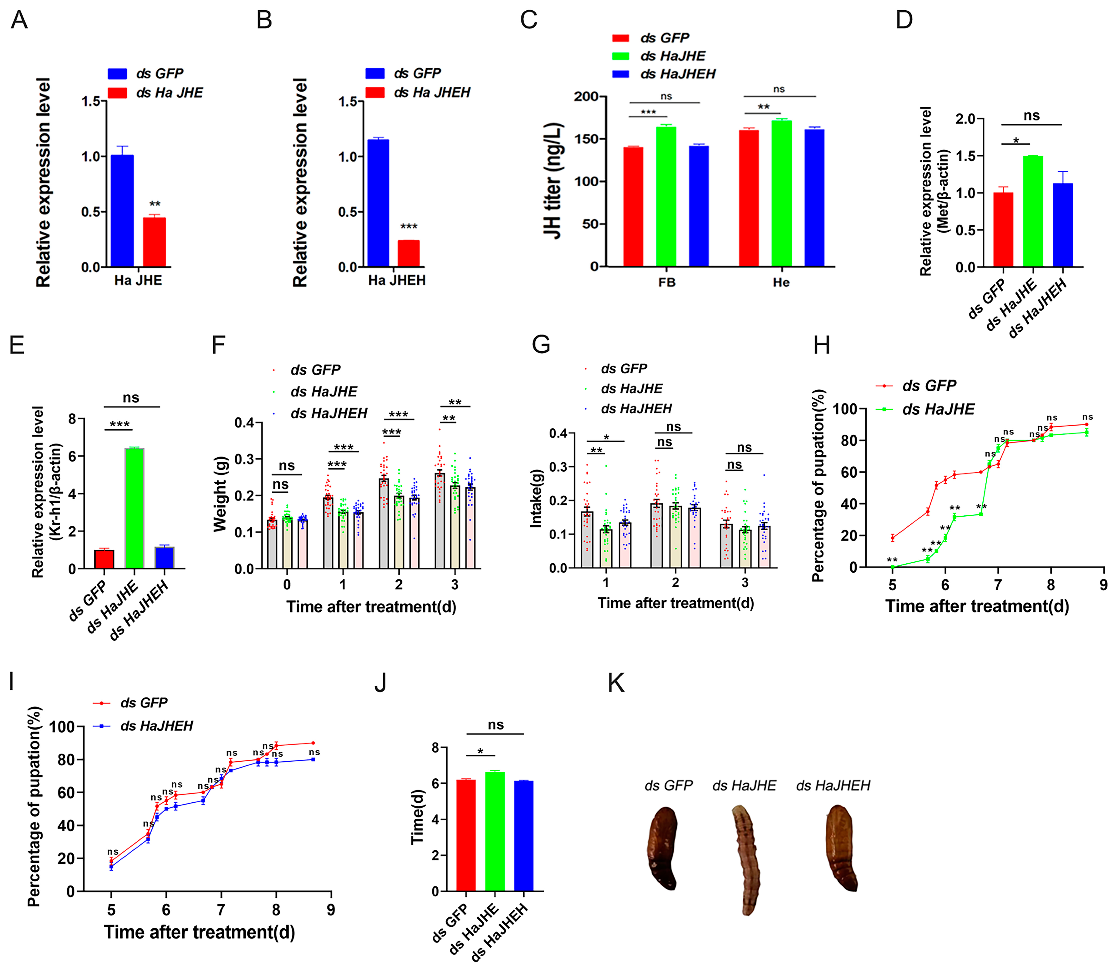
3.4. Effects of Knocking down of HaJHE and HaJHEH on the Pupation of H. armigera

To study the effects of HaJHE and HaJHEH knocking down in H. armigera larvae, dsHaJHE, and dsHaJHEH were injected into the H. armigera, respectively. After successful knockdown of HaJHE and HaJHEH (Figure 5A,B), we observed a significant increase of JH level at 48 h after injection of dsHaJHE, but there was no significant difference in the case of dsHaJHEH (Figure 5C). The relative expression levels of Met and Kr-h1 in H. armigera injected with dsHaJHE were significantly increased (Figure 5D,E), while those of H. armigera injected with dsHaJHEH were not different from the control group (Figure 5D,E). In addition, we measured the larval weight, feed intake, pupation rate, and pupation time of last larval instar H. armigera larvae injected with dsGFP, dsHaJHE, and ds HaJHEH, respectively. The results showed that in comparison with the control group, the larval weight and feed intake of larvae injected with dsHaJHE and dsHaJHEH decreased respectively (Figure 5F,G). The bioassay results showed that in comparison with the control group, the larvae injected with dsHaJHE had a delayed pupation of 0.43 d (Figure 5H,J,K), while the larvae injected with dsHaJHEH had a normal pupation without any delay (Figure 5I–K). In conclusion, these results show that the knockdown of HaJHE delayed the pupation of H. armigera, indicating that H. armigera larvae mainly degrade JH by JHE.

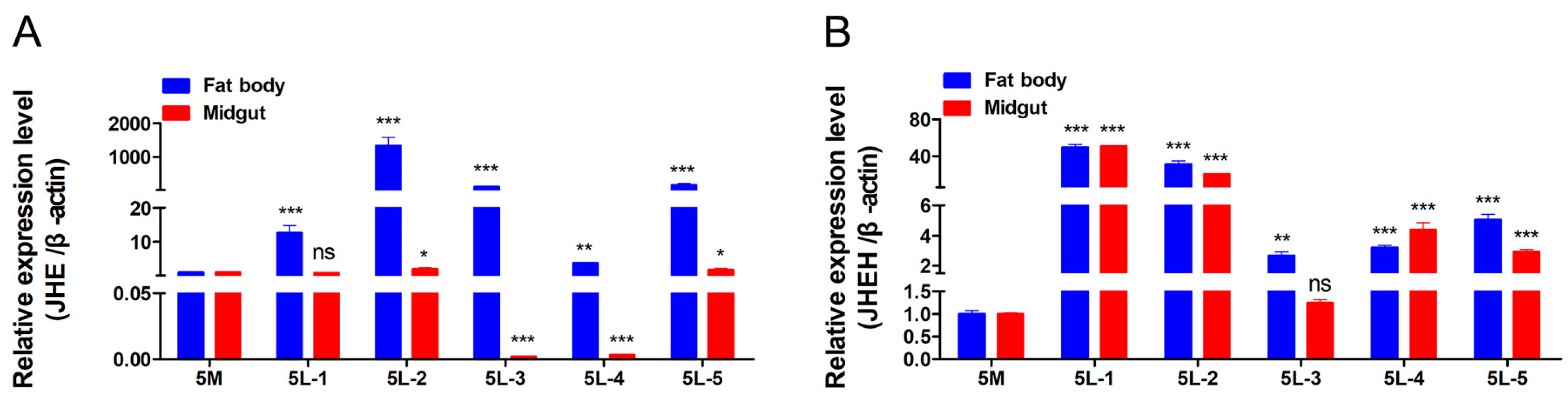
3.5. Detection of Expression Patterns of JHE and JHEH

In order to explore the expression patterns of JHE and JHEH in H. armigera, the temporal and spatial expression patterns of JHE and JHEH in the last larval instar H. armigera larvae were analyzed by RT-qPCR (Figure 6). The time expression pattern results showed that JHE and JHEH reached their peak expression levels at the second day and the first day after last larval instar molting stage, respectively.
The spatial expression pattern results showed that at the 5L-1, 5L-2, 5L-3, and 5L-5 stages, the expression levels of JHE and JHEH in fat bodies were higher than those in the midgut. The above results indicate that JHE and JHEH have a spatiotemporal expression specificity in H. armigera.

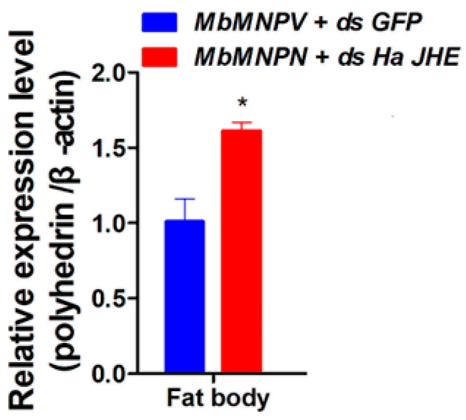
3.6. Knockdown of HaJHE Promoted the MbMNPV Infection

Since MbMNPV infection can inhibit the expression of HaJHE and prevent the pupation of H. armigera, we further investigated the impact of HaJHE knock down on MbMNPV replication. The relative expression of the MbMNPV polyhedrin gene was performed by RT-qPCR for the mock-infected and MbMNPV-infected group. The results showed that in comparison with the control, the expression level of the MbMNPV polyhedrin gene in the HaJHE-knocked down larvae increased by 61% (Figure 7), indicating that the knocking down of HaJHE promoted the MbMNPV infection.

4. Discussion

Baculoviruses, which are the effective biological insecticides for controlling various Lepidoptera pests, are characterized by vertical transmission and sustained control effects. In this study, the bioassays showed that MbMNPV infection had a significant inhibiting effect on H. armigera development, providing a theoretical basis for the application of MbMNPV (Figure 1A–D). The metabolism and synthesis of JH play a pivotal role in regulating insect molting and metamorphosis [35]. Studies have shown that insects infected with exogenous viruses typically exhibit developmental defects and prolonged larval stage [11,12]. It has been reported that Choristoneura fumiferana entomopoxvirus (CfEPV) infection can induce the change of JH level, and prolongs the larval stage in larval Mythimna separata [36], spruce budworm, and Choristoneura fumiferana [37], which is consistent with the results of the current study: MbMNPV infection prolonged the larval stage and induced the increase of JH titer in H. armigera (Figure 1E–G).
To identify the genes that contribute to the increase of JH titer in MbMNPV-infected H. armigera, we performed RNA sequencing of the fat bodies of MbMNPV-infected H. armigera. A total of 3637 DE-mRNAs were obtained, of which 1634 were up-regulated and 2003 were down-regulated. Some DE-mRNAs are enriched in pathways related to viral infection, such as Spliceosome [38], Ribosome biogenesis in eukaryotes [39], Aminoacyl-tRNA biosynthesis [40], and RNA degradation [41] (Figure 2). Research has shown that the dengue virus NS5 protein intrudes in the cellular Spliceosome and modulates splicing [42]. Hepatitis B virus X protein and c-Myc cooperate with promoting ribosome biogenesis [43]. HIV-1 utilizes dynamic aminoacyl-tRNA synthetase complexes to promote virus replication [44]. The RNA degradation pathway participates in PPARα-regulated anti-oral tumorigenesis [45]. The degradation and diffusion of viral RNA are bottlenecks in the efficiency of influenza A virus infection [46]. Transcriptome results indicate that the MbMNPV infection alters the host gene expression profile and triggers H. armigera-MbMNPV interactions.
In this study, bhmt, ACADSB, and selenbp1-a were identified as DE-mRNAs in MbMNPV-infected H. armigera (Figure 3), and the GO and KEGG pathways related to these three genes were further analyzed. bhmt is enriched into the zinc ion binding pathway (GO: 0008270) and the cysteine and methionine metabolism pathway (KEGG: haw00270). It has been reported that zinc ions can inhibit the main protease of SARS-CoV-2 in vitro and inhibit virus replication [47]. The increased concentration of homocysteine promotes HIV replication [48]. ACADSB is enriched in the branched chain amino acid catabolic process (GO: 0009083) and fatty acid degradation pathway (KEGG: haw00071). Branch chain amino acids reduce liver iron accumulation and oxidative stress in mice with hepatitis C virus multi protein expression [49]. Oncolytic avian reovirus σA regulates fatty acid metabolism to increase energy production for virus replication [50]. selenbp1-a is enriched in the nucleus (GO: 0005634) and sulfur metabolism pathway (KEGG: haw00920). In general, the nucleus is an important place for virus replication. The disorder of sulfur metabolism in AIDS patients is related to the depletion of cysteine and glutathione in white blood cells [51,52].
In this study, we also screened genes related to JH synthesis and metabolism pathways from 3637 DE-mRNAs and found that MbMNPV infection inhibited the relative expression levels of JHE and JHEH (Figure 4). JHE has been cloned or characterized in over 17 insect species, including Bombyx mori [53] and Trichoplusia ni [54] (Lepidoptera), Aedes aegypti [55] and Drosophila [56] (Diptera), Tenebrio molitor [53] and Leptinotarsa decemlineata (Say) [57] (Coleoptera). It has been confirmed that depletion of juvenile hormone esterase extends larval growth in Bombyx mori [58], which is consistent with the function of JHE in H. armigera in this study (Figure 5H,J,K). There are fewer studies on JHEH in comparison to JHE.
The hydrolysis function of JHEH to JH is different in different species. The effect of hydrolysis of JHEH on JH has been confirmed in tobacco hornworm, while its hydrolysis function in honeybee can almost be ignored [59]. This is consistent with our results that the larvae injected with dsHaJHEH had a normal pupation without any delay (Figure 5I–K), indicating the hydrolysis function of JH by JHEH in H. armigera larvae can be almost ignored and hence the JH in H. armigera is mainly degraded by JHE (Figure 5). Interestingly, we found that MbMNPV infection inhibited the weight gain of H. armigera (Figure 1A) and the relative expression levels of JHE and JHEH (Figure 4), while knocking down of HaJHE and HaJHEH also reduced the feed intake and weight of larvae (Figure 5F,G), indicating that MbMNPV infection inhibited the feed intake and weight gain of larvae by inhibiting the expression of HaJHE and HaJHEH. Knocking down of HaJHE promoted MbMNPV replication (Figure 7), indicating that deficiency of HaJHE reduced JH metabolism in H. armigera larvae, resulting in the host maintaining a longer larval state and ultimately promoting virus replication.

5. Conclusions

In conclusion, we profiled the differentially expressed mRNAs in the fat bodies of MbMNPV-infected H. armigera and clarified that JH is mainly degraded by JHE in H. armigera larvae. The MbMNPV infection of H. armigera larvae leads to the increase of JH titer by inhibiting the expression of JHE. The increase of JH titer promotes the expression of the JH receptor gene Met and the downstream gene Kr-h1, which prevents the pupation of H. armigera, and promotes MbMNPV replication. In the future, we will analyze the molecular mechanism of how the MbMNPV infection inhibits JHE expression. This research provides a new insight into the role of JHE in H. armigera and MbMNPV interactions, which is helpful in understanding the host–baculovirus correlation from the perspective baculovirus suppressing the insect development.

Supplementary Materials

The following supporting information can be downloaded at: This part has been added.https://www.mdpi.com/article/10.3390/insects15030202/s1, Table S1: Primers used for real-time PCR; Table S2: Primers used for synthesizing dsRNA.

Author Contributions

X.Z. and Z.L., designed and managed this research; Y.Y. performed the experiments; Y.Y., J.D., G.Z. and D.S., wrote and edited the manuscript; X.Z. and Z.L., finalized the manuscript. All authors have read and agreed to the published version of the manuscript.

Funding

This research was funded by the Special Project on Key Research and Development Tasks of Xinjiang Uygur Autonomous Region (No. 2022B02019) and the Support Program for Innovative Development of Nanjiang Key Industries (No. 2022DB026).

Data Availability Statement

All relevant data are within the article.

Conflicts of Interest

The authors declare no conflict of interests.

References

  1. Ahmad, M.; Rasool, B.; Ahmad, M.; Russell, D.A. Resistance and Synergism of Novel Insecticides in Field Populations of Cotton Bollworm Helicoverpa armigera (Lepidoptera: Noctuidae) in Pakistan. J. Econ. Entomol. 2019, 112, 859–871. [Google Scholar] [CrossRef]
  2. Dong, W.; Qiu, X.; Ren, X.; Fang, N.; Wang, K. Resistance selection and biochemical characterization of spinosad resistance in Helicoverpa armigera (Hübner) (Lepidoptera: Noctuidae). Pestic. Biochem. Physiol. 2009, 95, 90–94. [Google Scholar]
  3. Palli, S.R.; Retnakaran, A. Biological control of forest pests: A biotechnological perspective. In Forest Products Biotechnology; CRC Press: Boca Raton, FL, USA, 1997; pp. 277–296. [Google Scholar]
  4. Doyle, C.J.; Hirst, M.L.; Cory, J.S.; Entwistle, P.F. Risk Assessment Studies: Detailed Host Range Testing of Wild-Type Cabbage Moth, Mamestra brassicae (Lepidoptera: Noctuidae), Nuclear Polyhedrosis Virus. Appl. Environ. Microbiol. 1990, 56, 2704–2710. [Google Scholar] [CrossRef] [PubMed]
  5. Hostetter, D.L.; Puttler, B. A New Broad Host Spectrum Nuclear Polyhedrosis Virus Isolated from a Celery Looper, Anagrapha falcifera (Kirby), (Lepidoptera: Noctuidae). Environ. Entomol. 1991, 20, 1480–1488. [Google Scholar] [CrossRef]
  6. Ayres, M.D.; Howard, S.C.; Kuzio, J.; Lopez-Ferber, M.; Possee, R.D. The complete DNA sequence of Autographa californica nuclear polyhedrosis virus. Virology 1994, 202, 586–605. [Google Scholar] [CrossRef]
  7. Harrison, R.L.; Bonning, B.C. The nucleopolyhedroviruses of Rachiplusia ou and Anagrapha falcifera are isolates of the same virus. J. Gen. Virol. 1999, 80 Pt 10, 2793–2798. [Google Scholar] [CrossRef]
  8. Lawrence, P.O.; Lanzrein, B. Hormonal Interactions between Insect Endoparasites and Their Host Insects—ScienceDirect. Parasites Pathog. Insects 1993, 59–86. [Google Scholar] [CrossRef]
  9. Edwards, J.P.; Weaver, R.J. Endocrine changes in lepidopteran larvae—Potential challenges to parasitoid development and survival. Comp. Biochem. Physiol. Part B Biochem. Mol. Biol. 2000, 126, S31. [Google Scholar] [CrossRef]
  10. Kiuchi, M.; Yasui, H.; Hayasaka, S.; Kamimura, M. Entomogenous fungus Nomuraea rileyi inhibits host insect molting by C22-oxidizing inactivation of hemolymph ecdysteroids. Arch. Insect Biochem. Physiol. 2003, 52, 35–44. [Google Scholar] [CrossRef] [PubMed]
  11. Nakai, M.; Shiotsuki, T.; Kunimi, Y. An entomopoxvirus and a granulovirus use different mechanisms to prevent pupation of Adoxophyes honmai. Virus Res. 2004, 101, 185–191. [Google Scholar] [CrossRef]
  12. O’reilly, D.R. Baculovirus-encoded ecdysteroid UDP- glucosyl-transferase. Insect Biochem. Mol. Biol. 1995, 25, 541–550. [Google Scholar] [CrossRef]
  13. O’reilly, D.R.; Miller, L.K. A baculovirus blocks insect molting by producing ecdysteroid UDP-glucosyl transferase. Science 1989, 245, 1110–1112. [Google Scholar] [CrossRef]
  14. Wyatt, G.; Davey, K. Cellular and molecular actions of juvenile hormone. II. Roles of juvenile hormones in adult insects. Adv. Insect Physiol. 1996, 26, 1–55. [Google Scholar]
  15. Kamita, S.G.; Hammock, B.D. Juvenile hormone esterase: Biochemistry and structure. J. Pestic. Sci. 2010, 35, 265–274. [Google Scholar] [CrossRef]
  16. Tsubota, T.; Nakakura, T.; Shiotsuki, T. Molecular characterization and enzymatic analysis of juvenile hormone epoxide hydrolase genes in the red flour beetle Tribolium castaneum. Insect Mol. Biol. 2010, 19, 399–408. [Google Scholar] [CrossRef]
  17. Flatt, T.; Kawecki, T.J. Pleiotropic effects of methoprene-tolerant (Met), a gene involved in juvenile hormone metabolism, on life history traits in Drosophila melanogaster. Genetica 2004, 122, 141–160. [Google Scholar] [CrossRef]
  18. Kayukawa, T.; Minakuchi, C.; Namiki, T.; Togawa, T.; Yoshiyama, M.; Kamimura, M.; Mita, K.; Imanishi, S.; Kiuchi, M.; Ishikawa, Y.; et al. Transcriptional regulation of juvenile hormone-mediated induction of Krüppel homolog 1, a repressor of insect metamorphosis. Proc. Natl. Acad. Sci. USA 2012, 109, 11729–11734. [Google Scholar] [CrossRef]
  19. Wyatt, G.R. Comprehensive insect physiology, biochemistry, and pharmacology. Int. J. Biochem. 1985, 17, 1281–1282. [Google Scholar] [CrossRef]
  20. Ismail, S.; Goin, C.; Muthumani, K.; Kim, M.; Dahm, K.; Bhaskaran, G. Juvenile hormone acid and ecdysteroid together induce competence for metamorphosis of the Verson’s gland in Manduca sexta. J. Insect Physiol. 2000, 46, 59–68. [Google Scholar] [CrossRef]
  21. Ismail, S.M.; Satyanarayana, K.; Bradfield, J.Y.; Dahm, K.H.; Bhaskaran, G. Juvenile hormone acid: Evidence for a hormonal function in induction of vitellogenin in larvae of Manduca sexta. Arch. Insect Biochem. Physiol. 1998, 37, 305–314. [Google Scholar] [CrossRef]
  22. Minakuchi, C.; Namiki, T.; Shinoda, T. Krüppel homolog 1, an early juvenile hormone-response gene downstream of Methoprene-tolerant, mediates its anti-metamorphic action in the red flour beetle Tribolium castaneum. Dev. Biol. 2009, 325, 341–350. [Google Scholar] [CrossRef] [PubMed]
  23. Abdou, M.A.; He, Q.; Wen, D.; Zyaan, O.; Wang, J.; Xu, J.; Baumann, A.A.; Joseph, J.; Wilson, T.G.; Li, S.; et al. Drosophila Met and Gce are partially redundant in transducing juvenile hormone action. Insect Biochem. Mol. Biol. 2011, 41, 938–945. [Google Scholar] [CrossRef] [PubMed]
  24. Skowronek, P.; Wójcik, Ł.; Strachecka, A. Fat Body-Multifunctional Insect Tissue. Insects 2021, 12, 547. [Google Scholar] [CrossRef] [PubMed]
  25. Zhao, X.C.; Ma, B.W.; Berg, B.G.; Xie, G.Y.; Tang, Q.B.; Guo, X.R. A global-wide search for sexual dimorphism of glomeruli in the antennal lobe of female and male Helicoverpa armigera. Sci. Rep. 2016, 6, 35204. [Google Scholar] [CrossRef] [PubMed]
  26. Kyeiccoku, G.K.; Kunimi, Y. Effect of entomopoxvirus infection of Pseudaletia separata larvae on the oviposition behavior of Cotesia kariyai. Entomol. Exp. Appl. 2010, 83, 93–97. [Google Scholar]
  27. Yao, S.; Yang, Y.; Xue, Y.; Zhao, W.; Liu, X.; Du, M.; Yin, X.; Guan, R.; Wei, J.; An, S. New insights on the effects of spinosad on the development of Helicoverpa armigera. Ecotoxicol. Environ. Saf. 2021, 221, 112452. [Google Scholar] [CrossRef]
  28. Wang, S.A.-O.; Wang, L.L.; Pu, Y.X.; Liu, J.Y.; Wang, M.X.; Zhu, J.; Shen, Z.Y.; Shen, X.J.; Tang, S.A.-O. Exorista sorbillans (Diptera: Tachinidae) parasitism shortens host larvae growth duration by regulating ecdysone and juvenile hormone titers in Bombyx mori (Lepidoptera: Bombycidae). J. Insect Sci. 2023, 23, 6. [Google Scholar] [CrossRef]
  29. Chen, S.; Zhou, Y.; Chen, Y.; Gu, J. fastp: An ultra-fast all-in-one FASTQ preprocessor. Bioinformatics 2018, 34, i884–i890. [Google Scholar] [CrossRef]
  30. Kim, D.; Langmead, B.; Salzberg, S.L. HISAT: A fast spliced aligner with low memory requirements. Nat. Methods 2015, 12, 357–360. [Google Scholar] [CrossRef]
  31. Roberts, A.; Trapnell, C.; Donaghey, J.; Rinn, J.L.; Pachter, L. Improving RNA-Seq expression estimates by correcting for fragment bias. Genome Biol. 2011, 12, R22. [Google Scholar] [CrossRef]
  32. Love, M.I.; Huber, W.; Anders, S. Moderated estimation of fold change and dispersion for RNA-seq data with DESeq2. Genome Biol. 2014, 15, 550. [Google Scholar] [CrossRef] [PubMed]
  33. Gene Ontology Consortium. The Gene Ontology Resource: 20 years and still GOing strong. Nucleic Acids Res. 2019, 47, D330–D338. [Google Scholar] [CrossRef] [PubMed]
  34. Kanehisa, M.; Araki, M.; Goto, S.; Hattori, M.; Hirakawa, M.; Itoh, M.; Katayama, T.; Kawashima, S.; Okuda, S.; Tokimatsu, T.; et al. KEGG for linking genomes to life and the environment. Nucleic Acids Res. 2008, 36, D480–D484. [Google Scholar] [CrossRef] [PubMed]
  35. Truman, J.W.; Riddiford, L.M. Endocrine insights into the evolution of metamorphosis in insects. Annu. Rev. Entomol. 2002, 47, 467–500. [Google Scholar] [CrossRef] [PubMed]
  36. Nakai, M.; Kinjo, H.; Takatsuka, J.; Shiotsuki, T.; Kamita, S.G.; Kunimi, Y. Entomopoxvirus infection induces changes in both juvenile hormone and ecdysteroid levels in larval Mythimna separata. J. Gen. Virol. 2016, 97, 225–232. [Google Scholar] [CrossRef]
  37. Palli, S.R.; Ladd, T.R.; Tomkins, W.L.; Shu, S.; Ramaswamy, S.B.; Tanaka, Y.; Arif, B.; Retnakaran, A. Choristoneura fumiferana entomopoxvirus prevents metamorphosis and modulates juvenile hormone and ecdysteroid titers. Insect Biochem. Mol. Biol. 2000, 30, 869–876. [Google Scholar] [CrossRef]
  38. Tremblay, N.; Baril, M.; Chatel-Chaix, L.; Es-Saad, S.; Park, A.Y.; Koenekoop, R.K.; Lamarre, D. Spliceosome SNRNP200 Promotes Viral RNA Sensing and IRF3 Activation of Antiviral Response. PLoS Pathog. 2016, 12, e1005772. [Google Scholar] [CrossRef] [PubMed]
  39. Bianco, C.; Mohr, I. Ribosome biogenesis restricts innate immune responses to virus infection and DNA. eLife 2019, 8, e49551. [Google Scholar] [CrossRef]
  40. Lee, E.Y.; Kim, S.; Kim, M.H. Aminoacyl-tRNA synthetases, therapeutic targets for infectious diseases. Biochem. Pharmacol. 2018, 154, 424–434. [Google Scholar] [CrossRef]
  41. Abernathy, E.; Glaunsinger, B. Emerging roles for RNA degradation in viral replication and antiviral defense. Virology 2015, 479–480, 600–608. [Google Scholar] [CrossRef]
  42. De Maio, F.A.; Risso, G.; Iglesias, N.G.; Shah, P.; Pozzi, B.; Gebhard, L.G.; Mammi, P.; Mancini, E.; Yanovsky, M.J.; Andino, R.; et al. The Dengue Virus NS5 Protein Intrudes in the Cellular Spliceosome and Modulates Splicing. PLoS Pathog. 2016, 12, e1005841. [Google Scholar] [CrossRef]
  43. Shukla, S.K.; Kumar, V. Hepatitis B virus X protein and c-Myc cooperate in the upregulation of ribosome biogenesis and in cellular transformation. FEBS J. 2012, 279, 3859–3871. [Google Scholar] [CrossRef] [PubMed]
  44. Duchon, A.A.; St Gelais, C.; Titkemeier, N.; Hatterschide, J.; Wu, L.; Musier-Forsyth, K. HIV-1 Exploits a Dynamic Multi-aminoacyl-tRNA Synthetase Complex to Enhance Viral Replication. J. Virol. 2017, 91, 10–1128. [Google Scholar] [CrossRef] [PubMed]
  45. Chang, N.W.; Huang, Y.P. The RNA degradation pathway is involved in PPARα-modulated anti-oral tumorigenesis. BioMedicine 2019, 9, 27. [Google Scholar] [CrossRef] [PubMed]
  46. Schelker, M.; Mair, C.M.; Jolmes, F.; Welke, R.W.; Klipp, E.; Herrmann, A.; Flöttmann, M.; Sieben, C. Viral RNA Degradation and Diffusion Act as a Bottleneck for the Influenza a Virus Infection Efficiency. PLoS Comput. Biol. 2016, 12, e1005075. [Google Scholar] [CrossRef] [PubMed]
  47. Panchariya, L.; Khan, W.A.; Kuila, S.; Sonkar, K.; Sahoo, S.; Ghoshal, A.; Kumar, A.; Verma, D.K.; Hasan, A.; Khan, M.A.; et al. Zinc2+ ion inhibits SARS-CoV-2 main protease and viral replication in vitro. Chem. Commun. 2021, 57, 10083–10086. [Google Scholar] [CrossRef] [PubMed]
  48. MMüller, F.; Svardal, A.M.; Aukrust, P.; Berge, R.K.; Ueland, P.M.; Frøland, S.S. Elevated plasma concentration of reduced homocysteine in patients with human immunodeficiency virus infection. Am. J. Clin. Nutr. 1996, 63, 242–248. [Google Scholar] [CrossRef]
  49. Korenaga, M.; Nishina, S.; Korenaga, K.; Tomiyama, Y.; Yoshioka, N.; Hara, Y.; Sasaki, Y.; Shimonaka, Y.; Hino, K. Branched-chain amino acids reduce hepatic iron accumulation and oxidative stress in hepatitis C virus polyprotein-expressing mice. Liver Int. Off. J. Int. Assoc. Study Liver 2015, 35, 1303–1314. [Google Scholar] [CrossRef]
  50. Hsu, C.Y.; Chen, Y.H.; Huang, W.R.; Huang, J.W.; Chen, I.C.; Chang, Y.K.; Wang, C.Y.; Chang, C.D.; Liao, T.L.; Nielsen, B.L.; et al. Oncolytic avian reovirus σA-modulated fatty acid metabolism through the PSMB6/Akt/SREBP1/acetyl-CoA carboxylase pathway to increase energy production for virus replication. Vet. Microbiol. 2022, 273, 109545. [Google Scholar] [CrossRef]
  51. Toohey, J.I. Sulfur metabolism in AIDS: Cystamine as an anti-HIV agent. AIDS Res. Hum. Retroviruses 2009, 25, 1057–1060. [Google Scholar] [CrossRef]
  52. Breitkreutz, R.; Holm, S.; Pittack, N.; Beichert, M.; Babylon, A.; Yodoi, J.; Dröge, W. Massive loss of sulfur in HIV infection. AIDS Res. Hum. Retroviruses 2000, 16, 203–209. [Google Scholar] [CrossRef]
  53. Hinton, A.C.; Hammock, B.D. Juvenile hormone esterase (JHE) from Tenebrio molitor: Full-length cDNA sequence, in vitro expression, and characterization of the recombinant protein. Insect Biochem. Mol. Biol. 2003, 33, 477–487. [Google Scholar] [CrossRef]
  54. Jones, G.; Venkataraman, V.; Ridley, B.; O’Mahony, P.; Turner, H. Structure, expression and gene sequence of a juvenile hormone esterase-related protein from metamorphosing larvae of Trichoplusia ni. Biochem. J. 1994, 302 Pt 3, 827–835. [Google Scholar] [CrossRef]
  55. Bai, H.; Ramaseshadri, P.; Palli, S.R. Identification and characterization of juvenile hormone esterase gene from the yellow fever mosquito, Aedes aegypti. Insect Biochem. Mol. Biol. 2007, 37, 829–837. [Google Scholar] [CrossRef]
  56. Campbell, P.M.; Harcourt, R.L.; Crone, E.J.; Claudianos, C.; Hammock, B.D.; Russell, R.J.; Oakeshott, J.G. Identification of a juvenile hormone esterase gene by matching its peptide mass fingerprint with a sequence from the Drosophila genome project. Insect Biochem. Mol. Biol. 2001, 31, 513–520. [Google Scholar] [CrossRef]
  57. Vermunt, A.M.; Koopmanschap, A.B.; Vlak, J.M.; de Kort, C.A. Cloning and sequence analysis of cDNA encoding a putative juvenile hormone esterase from the Colorado potato beetle. Insect Biochem. Mol. Biol. 1997, 27, 919–928. [Google Scholar] [CrossRef] [PubMed]
  58. Zhang, Z.; Liu, X.; Shiotsuki, T.; Wang, Z.; Xu, X.; Huang, Y.; Li, M.; Li, K.; Tan, A. Depletion of juvenile hormone esterase extends larval growth in Bombyx mori. Insect Biochem. Mol. Biol. 2017, 81, 72–79. [Google Scholar] [CrossRef] [PubMed]
  59. Mackert, A.; Hartfelder, K.; Bitondi, M.M.; Simões, Z.L. The juvenile hormone (JH) epoxide hydrolase gene in the honey bee (Apis mellifera) genome encodes a protein which has negligible participation in JH degradation. J. Insect Physiol. 2010, 56, 1139–1146. [Google Scholar] [CrossRef] [PubMed]
Figure 1. The impact of MbMNPV infection in H. armigera larvae. The larval weight (A), pupation rate (B), emergence rate (C), larval mortality (D), fourth instar larval stage (E), and JH titer in hemolymph (He) (F) and fat body (FB) (G) in the mock-infected and MbMNPV-infected H. armigera larvae were evaluated. ns, p > 0.05; ***, p < 0.001; **, p < 0.01; *, p < 0.05; p < 0.05 indicated the significant difference (Student’s t-test). Each experiment was performed in three replicates, and the data are shown as mean ± s.e.m.
Insects 15 00202 g001
Figure 2. Screening and enrichment analysis of DE-mRNAs in the MbMNPV-infected group compared with control. (A) The volcano map of DE-mRNAs. (B) Top 30 significantly enriched GO terms of DE-mRNAs in molecular function, cellular component and biological process. (C) Hierarchical clustering analysis (heatmap) for DE-mRNAs. (D) Top 20 significantly enriched KEGG analyses of DE-mRNAs.
Insects 15 00202 g002
Figure 3. Relative expression of mRNAs in control and MbMNPV. *, p < 0.05; **, p < 0.01. p < 0.05 indicated the significant difference (Student’s t-test). Each experiment was performed in three replicates, and data are shown as mean ± s.e.m.
Insects 15 00202 g003
Figure 4. Relative expression of JHE and JHEH in control and MbMNPV. **, p < 0.01. p < 0.05 indicated the significant difference (Student’s t-test). Each experiment was performed in three replicates, and data are shown as mean ± s.e.m.
Insects 15 00202 g004
Figure 5. Effects of knocking down of HaJHE and HaJHEH on the pupation of H. armigera. Larvae on the first day after the last larval instar molting stage were injected with dsRNA of HaJHE (ds HaJHE), HaJHEH (ds HaJHEH), and dsRNA of Green fluorescent protein (ds GFP). (A) The expression levels of the JHE gene. (B) The expression levels of the JHEH gene. (C) The JH titer. (D) The expression levels of the Met gene. (E) The expression levels of the Kr-h1 gene. (F) The larval weight. (G) The feed intake of larvae. (H,I) The pupation rate. (J) The pupation time. (K) The larval phenotype after injection of dsRNA. The short vertical line represents a ruler. ns, p > 0.05; ***, p < 0.001; **, p < 0.01; *, p < 0.05; p < 0.05 indicated the significant difference (Student’s t-test). Each experiment was performed in three replicates, and data are shown as mean ± s.e.m.
Insects 15 00202 g005
Figure 6. Detection of the expression patterns of JHE and JHEH. (A) The expression patterns of JHE gene. (B) The expression patterns of JHEH gene. ns, p > 0.05; ***, p < 0.001; **, p < 0.01; *, p < 0.05; p < 0.05 indicated the significant difference (Student’s t-test). Each experiment was performed in three replicates, and data are shown as mean ± s.e.m.
Insects 15 00202 g006
Figure 7. Knockdown of HaJHE promoted MbMNPV infection. The relative expression of MbMNPV polyhedrin gene was performed by RT-qPCR for the mock-infected and MbMNPV-infected group. *, p < 0.05; p < 0.05 indicated the significant difference (Student’s t-test). Each experiment was performed in three replicates, and data are shown as mean ± s.e.m.
Insects 15 00202 g007
Table 1. Quality evaluation of sample sequencing data.
SampleRawReadsRawBasesCleanReadsValidBasesQ30GC
CK_148.33 M7.25G47.27 M94.87%87.79%49.76%
CK_248.26 M7.24G47.24 M95.23%90.05%49.65%
CK_348.54 M7.28G47.54 M95.64%90.36%49.31%
MbMNPV_150.64 M7.60G49.49 M95.07%89.39%49.47%
MbMNPV_249.58 M7.44G48.39 M95.04%90.02%48.86%
MbMNPV_350.75 M7.61G49.58 M94.99%89.25%49.82%
Disclaimer/Publisher’s Note: The statements, opinions and data contained in all publications are solely those of the individual author(s) and contributor(s) and not of MDPI and/or the editor(s). MDPI and/or the editor(s) disclaim responsibility for any injury to people or property resulting from any ideas, methods, instructions or products referred to in the content.

