Quantification of Right and Left Ventricular Function in Cardiac MR Imaging: Comparison of Semiautomatic and Manual Segmentation Algorithms

Abstract
1. Introduction
2. Materials and Methods
2.1. Study Population
2.2. MR Imaging Protocol
2.3. Image Analysis
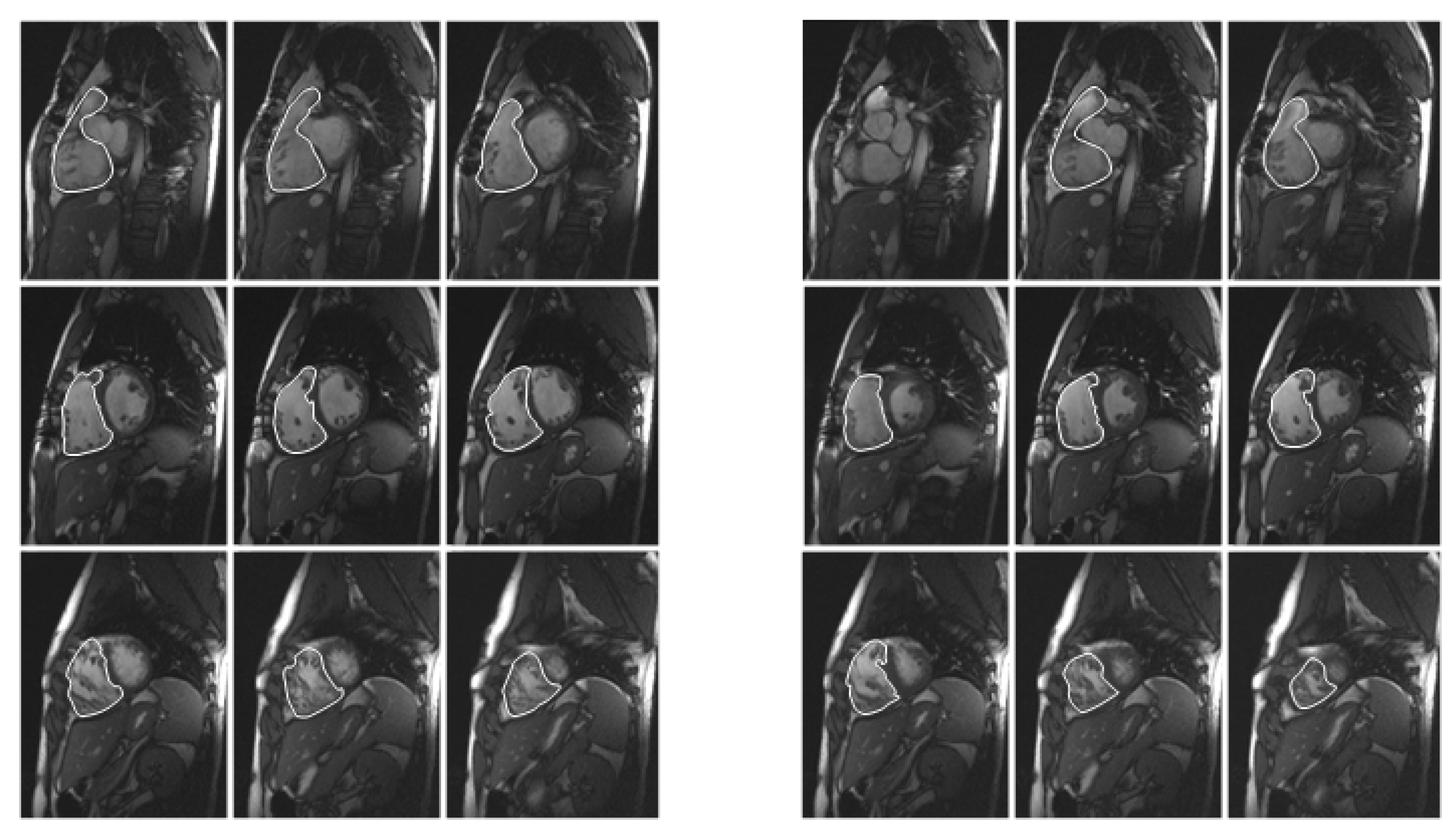
2.4. Manual Method
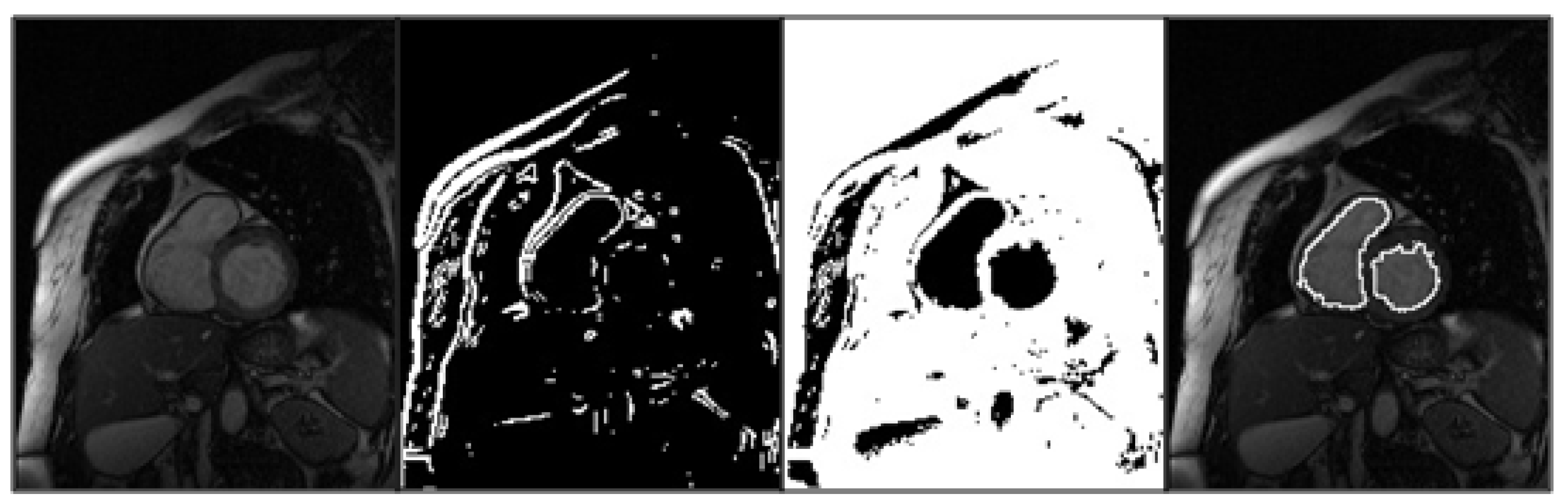
2.5. Semiautomatic Method
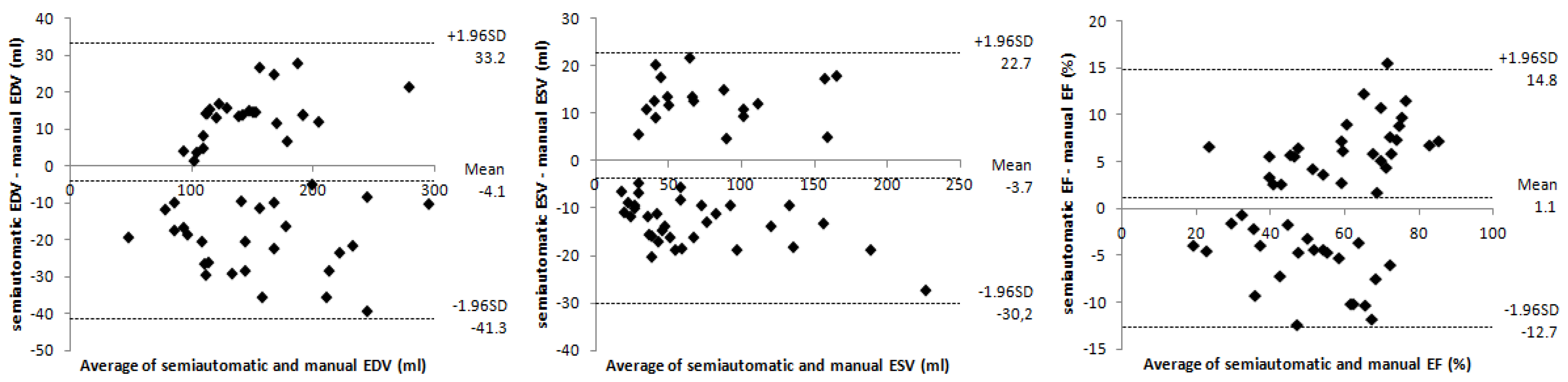
2.6. Statistical Analysis

| Semiautomatic Mean ± SD | Manual Mean ± SD | Semiautomatic vs. Manual r/p * | |
|---|---|---|---|
| EDV (mL) | 149.2 ± 53.4 | 153.2 ± 53.7 | 0.937/0.129 |
| ESV (mL) | 69.9 ± 48.4 | 73.7 ± 48.8 | 0.961/0.051 |
| EF (%) | 56.2 ± 17.7 | 55.2 ± 15.3 | 0.918/0.279 |
3. Results
3.1. Quantification of LV Parameters

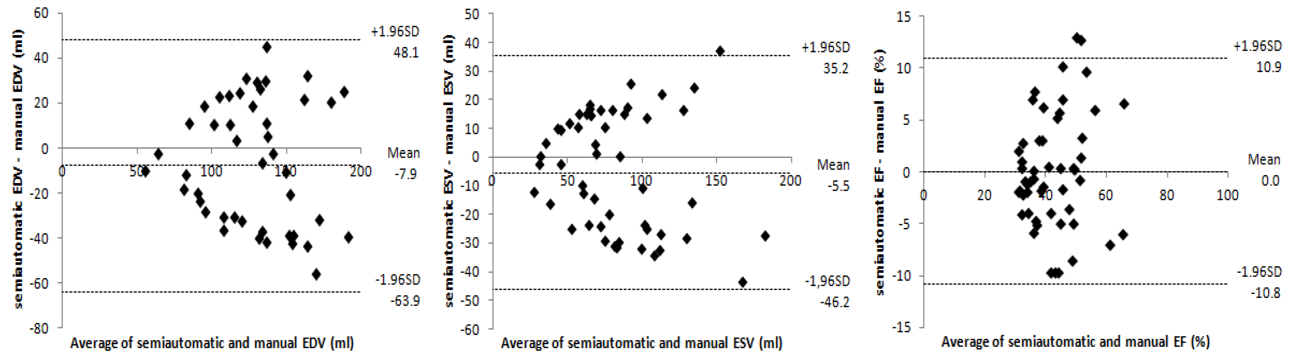
3.2. Quantification of RV Parameters
| Semiautomatic Mean ± SD | Manual Mean ± SD | Semiautomatic vs. Manual r/p * | |
|---|---|---|---|
| EDV (mL) | 142.5 ± 62.5 | 150.4 ± 67.8 | 0.907/0.050 |
| ESV (mL) | 83.5 ± 42.8 | 89.0 ± 48.2 | 0.902/0.061 |
| EF (%) | 42.6 ± 9.6 | 42.5 ± 8.9 | 0.823/0.952 |
Time Efficiency
4. Discussion
Acknowledgements
References
- Becker, M.; Frauenrath, T.; Hezel, F.; Krombach, G.A.; Kremer, U.; Koppers, B.; Butenweg, C.; Goemmel, A.; Utting, J.F.; Schulz-Menger, J.; Niendorf, T. Comparison of left ventricular function assessment using phonocardiogram- and electrocardiogram-triggered 2D SSFP CINE MR imaging at 1.5 T and 3.0 T. Eur. Radiol. 2010, 20, 1344–1355. [Google Scholar] [CrossRef]
- Baldy, C.; Douek, P.; Croisille, P.; Magnin, I.E.; Revel, D.; Amiel, M. Automated myocardial edge detection from breath-hold cine-MR images: Evaluation of left ventricular volumes and mass. Magn. Reson. Imag. 1994, 12, 589–598. [Google Scholar] [CrossRef]
- Bastarrika, G.; Domínguez, P.D.; Azcárate, P.M.; Castaño, S.; Fernández, M.E.; Gavira, J.J. Cuantificación de la función y masa ventricular con secuencias SSFP en tiempo real y respiración libre (in Spanish). Radiología 2008, 50, 67–74. [Google Scholar] [CrossRef]
- Lubbers, D.D.; Willems, T.P.; van der Vleuten, P.A.; Overbosch, J.; Götte, M.J.; van Veldhuisen, D.J.; Oudkerk, M. Assessment of global left ventricular functional parameters: Analysis of every second short-axis magnetic resonance imaging slices is as accurate as analysis of consecutive slices. Int. J. Cardiovasc. Imag. 2008, 24, 185–191. [Google Scholar] [CrossRef]
- Kunz, R.P.; Oellig, F.; Krummenauer, F.; Oberholzer, K.; Romaneehsen, B.; Vomweg, T.W.; Horstick, G.; Hayes, C.; Thelen, M.; Kreitner, K.F. Assessment of left ventricular function by breath-hold cine MR imaging: Comparison of different steady-state free precession sequences. J. Magn. Reson. Imag. 2005, 21, 140–148. [Google Scholar] [CrossRef]
- Holloway, C.J.; Edwards, L.M.; Rider, O.J.; Fast, A.; Clarke, K.; Francis, J.M.; Myerson, S.G.; Neubauer, S. A comparison of visual and quantitative assessment of left ventricular ejection fraction by cardiac magnetic resonance. Int. J. Cardiovasc. Imag. 2011, 27, 563–569. [Google Scholar] [CrossRef]
- Van der Geest, R.J.; Lelieveldt, B.P.; Angelié, E.; Danilouchkine, M.; Swingen, C.; Sonka, M.; Reiber, J.H. Evaluation of a new method for automated detection of left ventricular boundaries in time series of magnetic resonance imaging using an active appearance motion model. J. Cardiovasc. Magn. Reson. 2004, 6, 609–617. [Google Scholar] [CrossRef]
- Hudsmith, L.E.; Petersen, S.E.; Francis, J.M.; Robson, M.D.; Neubauer, S. Normal human left and right ventricular and left atrial dimensions using steady state free precession magnetic resonance imaging. J. Cardiovasc. Magn. Reson. 2005, 7, 775–782. [Google Scholar] [CrossRef]
- Maceira, A.M.; Prasad, S.K.; Khan, M.; Pennell, D.J. Reference right ventricular systolic and diastolic function normalized to age, gender and body surface area from steady-state free precession cardiovascular magnetic resonance. Eur. Heart J. 2006, 27, 2879–2888. [Google Scholar] [CrossRef]
- Geva, T. Repaired tetralogy of Fallot: The roles of cardiovascular magnetic resonance in evaluating pathophysiology and for pulmonary valve replacement decision support. J. Cardiovasc. Magn. Reson. 2011. [Google Scholar] [CrossRef]
- Morcos, P.; Vick, G.W., III; Sahn, D.J.; Jerosch-Herold, M.; Shurman, A.; Sheehan, F.H. Correlation of right ventricular ejection fraction and tricuspid annular plane systolic excursion in tetralogy of Fallot by magnetic resonance imaging. Int. J. Cardiovasc. Imag. 2009, 25, 263–270. [Google Scholar] [CrossRef]
- Sardanelli, F.; Quarenghi, M.; di Leo, G.; Boccaccini, L.; Schiavi, A. Segmentation of cardiac cine MR images of left and right ventricles: Interactive semiautomated methods and manual contouring by two readers with different education and experience. J. Magn. Reson. Imag. 2008, 27, 785–792. [Google Scholar] [CrossRef]
- Petitjean, C.; Dacher, J.N. A review of segmentation methods in short axis cardiac MR images. Med. Image Anal. 2011, 15, 169–184. [Google Scholar] [CrossRef]
- Alfakih, H.; Plein, S.; Thiele, H.; Jones, T.; Ridgway, J.P.; Sivananthan, N. Normal human left and right ventricular dimensions for MRI as assessed by turbo gradient echo and steady-state free precession imaging sequences. J. Magn. Reson. Imag. 2003, 17, 323–329. [Google Scholar] [CrossRef]
- Narayan, G.; Nayak, K.; Pauly, J.; Hu, B. Single-breathhold, four-dimensional, quantitative assessment of LV and RV function using triggered, real time, steady-state free precession MRI in heart failure patients. J. Magn. Reson. Imag. 2005, 22, 59–66. [Google Scholar] [CrossRef]
- Kim, T.H.; Ryu, Y.H.; Hur, J.; Kim, S.J.; Kim, H.S.; Choi, B.W.; Kim, Y.; Kim, H.J. Evaluation of right ventricular volume and mass using retrospective ECG-gated cardiac multidetector computed tomography: Comparison with first-pass radionuclide angiography. Eur. Radiol. 2005, 15, 1987–1993. [Google Scholar] [CrossRef]
- Lötjönen, J.M.; Järvinen, V.M.; Cheong, B.; Wu, E.; Kivistö, S.; Koikkalainen, J.R.; Mattila, J.J.; Kervinen, H.M.; Muthupillai, R.; Sheehan, F.H.; Lauerma, K. Evaluation of cardiac biventricular segmentation from multiaxis MRI data: A multicenter study. J. Magn. Reson. Imag. 2008, 28, 626–636. [Google Scholar] [CrossRef]
- Masip, L.R.; Tahoces, P.G.; Souto, M.; Martínez, A.; Vidal, J.J. Semiautomatic quantification of left and right ventricular functions in magnetic resonance imaging. Comput. Cardiol. 2010, 37, 797–800. [Google Scholar]
- Umbaugh, S.E. Computer Vision and Image Processing: A Practical Approach Using CVIP Tools; Prentice Hall PTR: Upper Saddle River, NJ, USA, 1998. [Google Scholar]
- Bellenger, N.G.; Davies, L.C.; Francis, J.M.; Coats, A.J.; Pennell, D.J. Reduction in sample size for studies of remodeling in heart failure by the use of cardiovascular magnetic resonance. J. Cardiovasc. Magn. Reson. 2000, 2, 271–278. [Google Scholar] [CrossRef]
- Bland, J.M.; Altman, D.G. Statistical methods for assessing agreement between two methods of clinical measurement. Lancet 1986, 1, 307–310. [Google Scholar] [CrossRef]
- Hendel, R.C.; Patel, M.R.; Kramer, C.M.; Poon, M.; Hendel, R.C.; Carr, J.C.; Gerstad, N.A.; Gillam, L.D.; Hodgson, J.M.; Kim, R.J.; et al. ACCF/ACR/SCCT/SCMR/ASNC/NASCI/SCAI/SIR 2006 appropriateness criteria for cardiac computed tomography and cardiac magnetic resonance imaging. J. Am. Coll. Cardiol. 2006, 48, 1475–1497. [Google Scholar] [CrossRef]
- Bastarrika, G.; Arraiza, M.; Pueyo, J.C.; Herraiz, M.J.; Zudaire, B.; Villanueva, A. Quantification of left ventricular function and mass in cardiac Dual-Source CT (DSCT) exams: Comparison of manual and semiautomatic segmentation algorithms. Eur. Radiol. 2008, 18, 939–946. [Google Scholar] [CrossRef]
- Mühlenbruch, G.; Das, M.; Hohl, C.; Wildberger, J.E.; Rinck, D.; Flohr, T.G.; Koos, R.; Knackstedt, C.; Günther, R.W.; Mahnken, A.H. Global left ventricular function in cardiac CT. Evaluation of an automated 3D region-growing segmentation algorithm. Eur. Radiol. 2006, 16, 1117–1123. [Google Scholar] [CrossRef]
- Vogel, M.; Gutberlet, M.; Dittrich, S.; Hosten, N.; Lange, P.E. Comparison of transthoracic three dimensional echocardiography with magnetic resonance imaging in the assessment of right ventricular volume and mass. Heart 1997, 78, 127–130. [Google Scholar] [CrossRef] [PubMed]
- Remy-Jardin, M.; Remy, J. Integrated Cardiothoracic Imaging with MDCT; Springer-Verlag: Berlin, Germany, 2009. [Google Scholar]
- Ho, S.Y.; Nihoyannopoulos, P. Anatomy, echocardiography, and normal right ventricular dimensions. Heart 2006, 92, i2–i13. [Google Scholar] [CrossRef]
- Childs, H.; Ma, L.; Ma, M.; Clarke, J.; Cocker, M.; Green, J.; Strohm, O.; Friedrich, M.G. Comparison of long and short axis quantification of left ventricular volume parameters by cardiovascular magnetic resonance, with ex vivo validation. J. Cardiovasc. Magn. Reson. 2011. [Google Scholar] [CrossRef]
- Catalano, O.; Corsi, C.; Antonaci, S.; Moro, G.; Mussida, M.; Frascaroli, M.; Baldi, M.; Caiani, E.; Lamberti, C.; Cobelli, F. Improved reproducibility of right ventricular volumes and function estimation from cardiac magnetic resonance images using level-set models. Magn. Reson. Med. 2007, 57, 600–605. [Google Scholar] [CrossRef]
- Von Knobelsdorff-Brenkenhoff, F.; Frauenrath, T.; Prothmann, M.; Dieringer, M.A.; Hezel, F.; Renz, W.; Kretschel, K.; Niendorf, T.; Schulz-Menger, J. Cardiac chamber quantification using magnetic resonance imaging at 7 Tesla—A pilot study. Eur. Radiol. 2010, 20, 2844–2852. [Google Scholar] [CrossRef]
- Nassenstein, K.; Eberle, H.; Maderwald, S.; Jensen, C.J.; Heilmaier, C.; Schlosser, T.; Bruder, O. Single breath-hold magnetic resonance cine imaging for fast assessment of global and regional left ventricular function in clinical routine. Eur. Radiol. 2010, 20, 2341–2347. [Google Scholar] [CrossRef]
© 2013 by the authors; licensee MDPI, Basel, Switzerland. This article is an open access article distributed under the terms and conditions of the Creative Commons Attribution license (http://creativecommons.org/licenses/by/3.0/).
Share and Cite
Souto, M.; Masip, L.R.; Couto, M.; Suárez-Cuenca, J.J.; Martínez, A.; Tahoces, P.G.; Carreira, J.M.; Croisille, P. Quantification of Right and Left Ventricular Function in Cardiac MR Imaging: Comparison of Semiautomatic and Manual Segmentation Algorithms. Diagnostics 2013, 3, 271-282. https://doi.org/10.3390/diagnostics3020271
Souto M, Masip LR, Couto M, Suárez-Cuenca JJ, Martínez A, Tahoces PG, Carreira JM, Croisille P. Quantification of Right and Left Ventricular Function in Cardiac MR Imaging: Comparison of Semiautomatic and Manual Segmentation Algorithms. Diagnostics. 2013; 3(2):271-282. https://doi.org/10.3390/diagnostics3020271
Chicago/Turabian StyleSouto, Miguel, Lambert Raul Masip, Miguel Couto, Jorge Juan Suárez-Cuenca, Amparo Martínez, Pablo G. Tahoces, Jose Martin Carreira, and Pierre Croisille. 2013. "Quantification of Right and Left Ventricular Function in Cardiac MR Imaging: Comparison of Semiautomatic and Manual Segmentation Algorithms" Diagnostics 3, no. 2: 271-282. https://doi.org/10.3390/diagnostics3020271
APA StyleSouto, M., Masip, L. R., Couto, M., Suárez-Cuenca, J. J., Martínez, A., Tahoces, P. G., Carreira, J. M., & Croisille, P. (2013). Quantification of Right and Left Ventricular Function in Cardiac MR Imaging: Comparison of Semiautomatic and Manual Segmentation Algorithms. Diagnostics, 3(2), 271-282. https://doi.org/10.3390/diagnostics3020271