MicroRNAs as Biomarkers in Cancer


1
Division of Basic and Translational Research, Department of Surgery, University of Minnesota, Minneapolis, MN 55455, USA
2
Department of Experimental and Clinical Pharmacology, University of Minnesota, Minneapolis, MN 55455, USA
3
Masonic Cancer Center, University of Minnesota, Minneapolis, MN 55455, USA
*
Author to whom correspondence should be addressed.
Diagnostics 2013, 3(1), 84-104; https://doi.org/10.3390/diagnostics3010084
Submission received: 15 November 2012
/
Revised: 28 December 2012
/
Accepted: 14 January 2013
/
Published: 16 January 2013
(This article belongs to the Special Issue Molecular Biomarkers for Cancer Targeted Therapeutics)
Abstract
:MicroRNAs (miRNAs) are small non-coding RNA molecules, which in recent years have emerged to have enormous potential as biomarkers. Recently, there have been significant developments in understanding miRNA biogenesis, their regulatory mechanisms and role in disease process, and their potential as effective therapies. The identification of miRNAs as biomarkers provides possibilities for development of less or non-invasive and more specific methods for monitoring tumor growth and progression. This review summarizes the recent developments in methods to detect and quantitate miRNAs in body fluids and their applications as biomarkers in cancers. The prospect of miRNAs as potential diagnostic and prognostic biomarkers with clinical applications is significant as more evidence points to their central role in cancer pathobiology.
1. Introduction
MicroRNAs (miRNAs) are a class of non-coding small RNAs (18–24 nucleotides), which are endogenously stable and evolutionarily conserved molecules. The major role of miRNAs appears to be in acting as crucial switches in regulating posttranscriptional gene expression. In recent years, miRNAs have been identified to play an important role in many physiological and pathological conditions [1,2,3,4]. Particularly, miRNA deregulation in various cancer types has been extensively studied. For example, deregulations in miRNAs expression have been identified to play a role not only in major cancers like lung [5], breast [6] and prostate [7] but also in rare cancers like waldenstrom macroglobulinemia [8] and cholangiocarcinoma [9]. In this review, we summarize the research advancements of using miRNAs as circulating biomarkers in cancers. The prospect of miRNAs as potential diagnostic and prognostic biomarkers in a clinical setting is significant as more evidence points towards a central role of miRNAs in cancer development and progression.
2. MicroRNAs and Cancer
microRNAs play a fundamental but significant role in cellular functions [10,11,12,13,14,15]. miRNA biogenesis and the mechanisms underlying miRNA mediated gene regulation are described elsewhere [16,17,18]. Since the discovery of miRNAs in C. elegans, about 1,500 miRNAs have been reported in humans [19]. Earlier studies established the function of miRNAs such as lin-4 and let-7 during embryogenesis and developmental control in C. elegans when complex gene regulations occur. Biochemical characterizations showed that these small RNAs block protein synthesis and/or regulate mRNA stability [20,21]. miRNAs have been found to regulate the expression of about 50% of the human coding gene transcripts [20]. The gene regulatory role of miRNAs suggests their important role in physiological functions [21] and pathological conditions [2,3]. Furthermore, single nucleotide polymorphisms in the 3’UTR of mRNAs may affect miRNA-mediated regulation and have been associated with certain cancer risk [22].
Pioneering studies by Calin et al. showed the biological relevance of miRNAs in cancer development; miR-15 and miR-16 were significantly downregulated in chronic lymphoid leukemia (CLL) through deletions in chromosome 13q14 locus. These earlier observations also revealed 52% of miRNA genes to be located in genomic regions, which are frequently altered in cancer. These results were further validated by mapping which revealed 186 miRNAs located at fragile sites and genomic regions [23]. Since this discovery, miRNAs have been implicated in many different types of cancers including the influence of miRNAs in neoplastic transformation, progression and patient outcomes [24,25,26,27,28,29]. miRNAs act as oncogenes or tumor suppressors taking the lead role in regulating cancer pathways. The target genes regulated by the miRNAs determine their role as oncogenes or tumor suppressors.
2.1. MicroRNAs as Oncogenes
The oncogenic potential of miRNAs has been well documented. An earlier study showed that B-cell integration cluster (BIC) accelerates MYC mediated growth of lymphomas [30]. Subsequent characterization of BIC gene revealed this region to harbor the primary transcript for miR-155. In pancreatic cancer, miR-155 is expressed at high levels and targets tumor suppressor TP53INP1, which is a stress induced-gene thus impairing stress response and resulting in decreased apoptosis [31]. Similarly, miR-17-92 cluster was identified as a potential oncogene since its expression along with cMYC accelerated the development of B-cell lymphoma in mouse models [32]. Likewise, miR-21 is significantly upregulated in many cancers and its validated targets includes tumor suppressor genes such as PDCD4, PTEN and TIMP3. miR-21 overexpression thereby inhibits apoptosis and promotes cell survival. Oncogenic miRNAs such as miR-221 and miR-222 inhibit CDKN1B and CDKN1C genes [25], which are important cell cycle regulators. Similarly, miR-182 targets FOXO1 and FOXO3 transcription factors and protects from oxidative stress and cell death [26,27].
2.2. MicroRNAs as Tumor Suppressors
miRNAs also function as tumor suppressors in different cancer types by regulating the oncogenic process and preventing tumor development. Kefas et al. documented the association of EGFR and AKT pathway activation with aggressiveness of glioblastoma [28]. The suppression of the AKT pathway and inhibition of EGFR by miR-7 resulted in decreased proliferation, survival and invasiveness providing evidence for the tumor suppressor effect of this miRNA. Similarly, in a study comparing primary melanoma and benign nevi, the authors showed the downregulation of let-7 family miRNAs in primary melanomas suggesting their role as tumor suppressors. Exogenous expression of let-7b in primary melanoma cells decreased the anchorage independent growth and inhibited cell cycle progression [29]. The increased expression of let-7b also suppressed CDK4 and cyclins (D1, D3 & A), which are implicated in the development of melanoma. In a recent study by Dong et al. [33] on uveal melanoma, which is the most common primary form of the intra ocular tumor in adults, the transfection of uveal melanoma cells by miR-34b/c caused cell cycle arrest resulting in significant reduction of melanoma cell growth and migration. The miR-34b/c has been identified to target the MET proto-oncogene in uveal melanoma cells. Interestingly, many studies have demonstrated miRNAs as oncogenes or tumor suppressors by involving multiple molecular mechanisms and different signaling pathways. There can be a single target or multiple functionally relevant targets for a miRNA. For example, miR-155 is upregulated in Hodgkin’s lymphoma and targets genes such as ZIC3, AGTR1, ZNF537, FGF7 and IKBKE [34], while miR-206 is downregulated in rhabdomyosarcoma and targets MET onco-protein [35]. The understanding that miRNAs can function as oncogenes and/or tumor suppressors in cancers allows us to explore them as therapeutic targets for clinical application. Extensive studies are underway to establish them as biomarkers for cancer detection and progression.
3. MicroRNAs as Potential Biomarkers in Cancer
With our increasing knowledge on gene regulatory pathways, we have achieved certain milestones in our understanding of cancer signaling. However, there is an immense need for fail proof biomarkers that can detect cancer in its earlier stages and/or function as prognostic indicators. Currently, most cancer diagnosis depends on imaging techniques like computerized tomogram (CT), magnetic resonance imaging (MRI), ultrasonography, positron emission tomography (PET), mammogram and invasive studies like colonoscopy, bronchoscopy and biopsy. Even though imaging and invasive studies are extensively relied on in order to generate valid information regarding the cancer, there is a high possibility of false positive or false negative results. The imaging methods are not cost effective and invasive studies cause unnecessary discomfort for the patients.
Further, the available biomarkers such as CA 125 for ovarian cancer, CA 19-9 for pancreatic cancer and CEA (carcino embryonic antigen) for colon cancer have poor sensitivity and specificity. Only 50% of patients with early stage ovarian cancer have elevated levels of CA125. In 2010, the National Institutes of Health advisory panel did not reissue the old protocol which advised women to begin yearly breast cancer screening once they attained the age of forty. Furthermore, the U.S. Preventive Services Task Force (USPSTF) recommendation on prostate cancer screenings (PSA) made available in October 2011 recommends discontinuing routine PSA tests due to poor sensitivity and specificity in distinguishing prostate cancer patients requiring surgery. Thus, there is a critical need for reliable biomarkers that allow precise monitoring of changes at the cellular level and the patient’s response to therapy. miRNAs have tremendous potential to be explored as diagnostic and prognostic biomarkers. miRNA expression is dynamic; many miRNAs are deregulated in early stages of tumor development and are upregulated during cancer progression exhibiting their potential for diagnostic utility [36,37]. The possibility of using combination of markers such as CA-19-9 and miRNAs significantly increase the diagnostic potential [38].
Resnick et al. compared levels of miRNAs (miR-21, -92 and -93) and the currently available biomarker for ovarian cancer, CA-125, and showed alteration in the level of these miRNAs in patients with ovarian cancer having normal levels of CA-125 [39]. With increasing implications of miRNAs in cancer development and progression, significant efforts are underway to use miRNAs as novel biomarkers with clinical applications [40] and as novel therapeutic targets for anti cancer drug resistance [41]. Another potential is in the diagnosis of pancreatic cancer, which still remains a cancer with poor prognosis with a median survival of 6 months to 1 year. Pancreatic cancer is asymptomatic in the early stages when their precursor lesions such as PanIN (pancreatic intraepithelial neoplasia) remain undetected. However, if these precursor lesions are detected, the potential for cure may be high. Several miRNAs have been identified in PanIN stages [42,43] of pancreatic cancer that allow us to investigate them as potential biomarkers.
4. Circulating MicroRNAs
Blood sampling from patients remain the least invasive method for identifying biomarkers. The presence of circulating miRNAs has been demonstrated in various disease conditions and the profiles vary with the degree of disease progression [44,45,46,47,48]. The presence of a specific set of circulating miRNAs in cancer condition is justified by passive diffusion due to high turnover of cells in cancer. Yu et al. showed differences between the metabolite profiles of plasma and serum. Concentrations of metabolite were generally higher in serum and provided higher sensitivity than plasma [49]. Initial studies to identify serum miRNAs in cancer were demonstrated by Lawrie et al., in which they observed that miR-155, miR-210, and miR-21 were upregulated in diffuse large B cell lymphoma when compared to healthy controls [50]. Further, miRNA expression profiles have been generated using serum/plasma from patients diagnosed with common cancers such as acute leukemia [51], breast cancer [52], colorectal cancer [53], gastric cancer [54], glioblastoma [55], hepatocellular cancer [56], lung cancer [57], oral and squamous cancer [58], ovarian [39] and prostate cancer [40]. Recently, miRNAs have also been detected in numerous other body fluids (see below).
With the increasing data about serum miRNAs as well the growing need for a good biomarker, which must also be easily accessible, the miRNAs in serum can be considered valid biomarkers for early detection, diagnosis and prognosis of various cancers [59,60,61,62,63,64]. As more interest has been drawn toward identifying circulating miRNAs in cancers, the biggest challenge currently encountered during the analysis of miRNAs in serum/plasma and other body fluids is the need for an internal normalization. Housekeeping genes like U6 or GAPDH, commonly used as internal normalization during miRNA analysis in cells and tissues, are not feasible in serum analysis since these controls are easily degraded and not detected in serum. Chen et al. [57] demonstrated a series of miRNAs existing in normal human serum and compared their levels to widely used U6, which was not stable in serum. They also showed the presence of miRNAs in the serum of rats, mice, calves, bovine fetuses and horse and the potential for using these miRNAs for normalization and comparative studies. In support of these observations, Mitchell et al. demonstrated the stability of miRNAs (miR-15b, miR-16, miR-19b and miR-24) occurring in normal human sera and their resistance to RNase digestion. The levels of these miRNAs are not altered during tumor growth and can possibly be considered as normalization controls [40]. Of these normally present miRNAs, miR-16 was widely present and was used as internal controls in serum miRNA studies [57]. However a recent study demonstrated high levels of miR-16 in red blood cells; thus it is possible that the presence of miR-16 in serum may in part be due to hemolysis of red blood cells [65]. This also raises the question of using miR-16 for normalization. As greater numbers of miRNAs are being detected in normal blood cells, to avoid the errors with internal control there is an essential need for a standard, normalization protocol when analyzing miRNAs from serum samples. This challenge is, for now, met by using spike-in synthetic C. elegans miRNA for internal normalization when using serum samples for miRNA analysis. Taken together, circulating miRNAs detected using validated methods can be valuable biomarkers for cancer diagnosis and prognosis. A comprehensive list of miRNAs identified as potential biomarkers in various cancer types are given in Table 1.

| Cancer | Sample type | Comparison criteria | Methods | miRNAs | Target/mechanisms | Ref. |
|---|---|---|---|---|---|---|
| Epithelial cancers | ||||||
| Breast | Tissue cancer cells | ER+, ER− tissues | Microarray, qPCR | miR-34b (↓) | miR-34b target cyclin D1 and JAG-1 | [66] |
| Whole blood | Tumor vs. normal | qPCR | miR-195 (↑)
let-7a (↑) miR-155 (↑) | let-7a target KRAS and miR-155 target RhoA transforming growth factor and induces EMT | [67] | |
| Cancer cells | Cancer cell growth | Microarray | miR-21 (↑) | PDCD4 | [68] | |
| Colorectal | Tissue & serum | Tumor vs. normal | qPCR microarray | miR-17-3p (↑)
miR-92 (↑) | miR-92 is elevated in plasma and can be used as a non-invasive molecular marker for screening | [53] |
| Plasma | Tumor vs. normal | qPCR | miR-29a (↑)
miR-92a (↑) | Promote cell proliferation, suppressed apoptosis, induce tumor angiogenesis and accelerated tumor progression | [69] | |
| Bladder | Tissue & cancer cell lines | Tumor vs. normal | qPCR | miR-145 (↓)
miR-30a-3p (↓) miR-133a (↓) miR-133b (↓) miR-195 (↓) miR-125b (↓) miR-199a (↓) | These miRNAs are downregulated and play the role of tumor suppressors by targeting KRT7, a common target with oncogenic function | [70] |
| Glioblastoma | Tissue | Tumor vs. normal, Different grades of malignancy | qPCR Northern blotting | miR-21 (↑)
miR-221 (↑) miR-128 (↓) miR-181b (↓) | Knockdown of miR-21 triggered the activation of caspases leading to apoptosis. miR-221 & miR-222 repress expression p27Kip1. miR-181b triggered growth inhibition, apoptosis, and inhibited invasion | [71] |
| Gastric | Tissue & cell lines | Tumor vs. normal | qPCR | miR-409 (↓) | miR-409 target RDX and suppresses cell invasion and metastasis | [72] |
| Lung | Serum | Overall survival in NSCLC | sequencingqPCR | miR-486 (↑)
miR-30d (↑) miR-1 (↓) miR-499 (↓) | miR-1 downregulates MET oncogene. Facilitates activation of Caspase 3, Caspase 7, and PARP-1 as well as depletion of MCL-1 | [73] |
| Plasma | Tumor vs. normal | qPCR | let-7f (↓)
miR-20b (↓) miR-30e-3p (↓) | let-7 targets cMyc. miR-30e regulates of Ubc9 | [74] | |
| Oral & squamous cell | Tissue | Tumor vs. normal | qPCR | miR-184 (↑) | miR-184 alters cMyc expression and affects anti-apoptosis and proliferation of tongue SCC cells | [58] |
| Tissue & saliva | Tumor vs. normal | qPCR | miR-31 (↑) | - | [75] | |
| Ovarian | Serum | Tumor vs. normal | qPCR | miR-21 (↑)
miR-29a (↑) miR-92 (↑) miR-93 (↑) miR-99b (↓) miR-126 (↓) miR-127 (↓)) miR-155 (↓)) | miR-21 regulates PDCD4 and maspin miR-92 and miR-93 regulates TGFβ miR-29a potentially targets PTEN miR-127 regulates BCL6 | [39] |
| Prostate | Plasma, serum, murine | Tumor vs. normal | qPCR | miR-141 (↑)
miR-375 (↑) miR-107 (↑) miR-574-3p (↑) | induces abnormal cell division and proliferation and the development of aggressive prostate cancer | [76] |
| Tissue & Serum | Metastatic, localized tumors vs. normal | qPCR | miR-141 (↑)
miR-375 (↑) | regulates genes controlling cellular growth and proliferation | [77] | |
| Pancreatic | Tissue | Tumor vs. normal | qPCR | miR-155 (↑)
miR-203 (↑) miR-210 (↑) miR-222 (↑) | - | [78] |
| Plasma | Tumor vs. normal | qPCR | miR-21 (↑)
miR-155 (↑) miR-196a (↑) miR-210 (↑) | miR-21 targets PTEN and PDCD4 miR-210 affect DNA repair and genomic instability miR-155 target TP53INP1 | [79] | |
| Hepatocellular | Tissue & cell cultures | Tumor vs. normal | qPCR | miR-519d (↑) | miR-519d has inhibitory effect on CDKN1A/p21, PTEN and TIMP2 expression | [80] |
| Endometrial | Tissue | Tumor vs. hyperplasia vs. normal | qPCR | miR-200 family (↑) | Negatively regulates ZEB1 and ZEB2 and implicated in EMT | [81] |
| Renal cell | Tissue | Tumor vs. normal | qPCR Microarray | miR-122 (↑)
miR-155 (↑) miR-210 (↑) miR-200c (↓) miR-335 (↓) miR-218 (↓) | - | [82] |
| Melanoma | Tissue & cell lines | Normal vs. cancer cell lines | Microarray | miR-193a (↓)
miR-338 (↓) miR-565 (↓) miR-191 (↓) miR-193b (↑) | miR-193 is regulated by HNF-1a and p53; predicted targets for miR-191 include FZD5 and BDNF | [83] |
| Thyroid | Tissue | Tumor vs. Normal | qPCR | miR-187 (↑)
miR-221 (↑) miR-222 (↑) miR-146b (↑) miR-155 (↑) miR-224 (↑) miR-197 (↑) | The oncogenic mutations in PCs, RET/PTC, BRAF, and RAS are all capable of activation of the MAPK pathway | [84] |
| SARCOMAS | ||||||
| Osteosarcoma | Tissue & cancer cell lines | Tumor vs. normal | qPCR | miR-135b (↑)
miR-150 (↑) miR-542-5p (↑) miR-652 (↑) | Pro-apoptotic EGR2 and P2X7 are targets of miR-150 | [85] |
| Tissue | Tumor vs. normal | qPCR Microarray | miR-17-92 (↓) | 14q32 miRNAs (miR-544, miR-369-3p, miR-134 and miR-382) act cooperatively to destabilize cMYC and in turn, control expression of miR-17-92 miRNAs | [86] | |
| Leiomyosarcoma | Tissue | Tumor vs. normal | qPCR Microarray CGH | miR-21 (↑)
let7 (↑) miR- 27a (↑) miR-30a (↑) miR-23b (↑) miR-29b (↓) miR-32 (↓) miR-144 (↓) miR-212 (↓) miR-197 (↓) | Targets MAPK pathway genes | [87] |
| Rhabdomyo-sarcoma | Cell lines, Tissue & Serum | Tumor vs. normal | qPCR | miR-206 (↑) | expression of miR-206 in RMS cells promoted myogenic differentiation and blocked tumor growth | [88] |
| Gastrointestinal Stromal Tumor | Tissue | Tumor vs. normal | qPCR | miR-221 (↓)
miR-222 (↓) | Regulates cKIT | [89] |
| Ewing's Sarcoma | Cell lines | Primary sarcoma vs. Progenitor cells | miRNA Profiling | miR-145 (↓) | miR-145 inhibits stem cell transcription factors Oct4, Sox2, Klf4 and Myc | [90] |
| Schwannoma | Tissue, cell lines | Tumor vs. Normal | Microarray, qPCR | miR-7 (↓) | Inhibited expression of Ack1, Pak1, and EGFR | [91] |
| MPNST | Tissue | MPNST vs. neurofibroma | Microarray, qPCR | miR-34a (↓) | Partly due to p53 inactivation | [93] |
| LEUKEMIA/ LYMPHOMA | ||||||
| Adult T Cell Leukemia | Cells | primary ATL cells vs. normal CD4+ T cells | Microarray | miR-31 (↓) | miR-31 is a suppressor of NIK and pathway involving polycomb-mediated miRNA silencing and NF-kB activation | [94] |
| Acute promyelocytic leukemia | Cells | Leukemia vs. Normal Promyelocytes | qPCR | miR-15b (↓)
miR-16 (↓) miR-107 (↓) miR-223 (↓) miR-342 (↓) and let-7c (↓) | PML/RARa binds the regulatory sequences of the intragenic miR-342 and let-7c | [95] |
| AML | Cell lines | AML, Human myeloid, CLL cell lines | qPCR microarray | miR-34b (↓) | Cyclic AMP-Responsive Element Binding Protein down-regulation | [96] |
| CLL | Peripheral blood mononuclear cells | Cancer cells vs. normal cells | qPCR | miR-92 (↑) | Abnormal elevation of HIF-1α, the key upstream regulator of VEGF | [97] |
| Peripheral Blood CD19+ cells | Prognostic factors | qPCR Western blot | miR-29c (↓)
miR-223 (↓) | Regulates the Tcl1 oncogene Down-regulation of miR-29 inversely correlates with DNMT expression | [98] | |
| Hodgkins lymphoma | Cancer cell lines | Hodgkins vs. B cell non Hodgkins | qPCR Microarray | miR-155 (↑) | IKBKE, ZNF537, ZIC3, FGF7, and AGTR1 are functional targets of miR-155 | [34] |
| Diffuse Large B-cell lymphoma | Serum | Tumor vs. Normal | qPCR | miR-15a (↑)
miR-16-1 (↑) miR-29c (↑) miR-155 (↑) miR-34a (↓) | miR-155 directly down regulates one of the MYC antagonists like MAD1, MXI1, ROX/MNT | [99] |
Upregulation and downregulation of microRNAs are denoted by (↑) and (↓) arrows respectively.
5. miRNA Detection in Body Fluids and Stability
In an attempt to establish miRNAs as biomarkers in cancer, considerable interest has been drawn to the ability of detecting these circulating biomarkers in different body fluids and the stability of miRNAs to resist the numerous extracellular enzymes present in human body. Weber et al., showed the presence of miRNAs in different body fluids such as plasma, saliva, tears, urine, amniotic fluid, colostrum, breast milk, bronchial lavage, cerebrospinal fluid, peritoneal fluid, pleural fluid, seminal fluid and serum [100]. There is also evidence of miRNAs being identified in different body fluids in various cancer types, some of them being serum miRNAs in prostate cancer [101], urine miRNAs in urothelial carcinoma [102], cerebrospinal fluid (CSF) miRNAs in glioma [103], plasma miRNAs in pre-eclampsia [104] and sputum miRNAs in non small cell lung cancer (NSCLC) [105]. These studies provide strong evidence to indicate the existence of miRNAs in different body fluids. The stability of endogenous miRNAs to RNase digestion was shown by adding synthetic miRNAs to the serum from subjects with prostate cancer. The added synthetic miRNAs were destroyed whereas the endogenous miRNAs were able to maintain their stability and were detected in plasma/serum [40]. In yet another study, Chen et al. elucidated the stability of miRNAs in various body fluids, after the miRNAs were subjected to a series of experimental conditions such as boiling, low/high pH, extended storage and multiple freeze thaw cycles [57].
Recent findings have demonstrated the presence of miRNAs in exosomes, microvesicles, lipoproteins, apoptotic bodies and large micro particles. Valadi et al. analyzed human and mouse mast cell lines and observed the synthesis of new proteins on transfer of exosomal RNA between the donor cells and the recipient cells. They proposed that the exosomes contained both mRNA and miRNA and a possible mechanism of genetic exchange between the cells [106]. It is probable that the stability of endogenous miRNAs is imparted by the protection of miRNAs in membrane bound exosome-like particles called microvesicles [107]. Gallo et al. recently observed that a majority of miRNAs in saliva and serum from healthy individuals and systemic lupus erythmatosus (SLE) patients were found to be concentrated in exosomes [108].
Evidence from recent studies has also proven the presence of miRNAs in high-density lipoproteins (HDL) [109]. The study by Vickers et al. showed that HDL has a major role in endogenous miRNA transport. They isolated HDL from normal human plasma and from patients with familial hypercholesterolemia by density gradient ultracentrifugation and fast-protein liquid chromatography followed by anti-apoA-1 immunoprecipitation. Total RNA was extracted from purified HDL and miRNA profiles analyzed using microarrays. They demonstrated occurrence of various miRNAs in HDL molecules, and one of the abundant miRNA found in both human and mouse HDL was miR-223 [110]. Furthermore, they evaluated dynamic response by subjecting various cell lines to stress. The results revealed a number of proteins involved in the exportation process and suggested nucleophosmin 1 (NPM1) as a protective protein guarding miRNA from degradation. These findings about the stability of endogenous miRNA and protection from degradation outside the cellular environment imparted by exosomes and protective proteins suggest possible reasons for their detection in plasma/serum.
5.1. miRNA Extraction and Quantifying Methods
Extensive studies have implicated miRNAs to be involved in initiation, progression, malignant transformation, prognosis and outcome of cancer. Since the extraction levels of miRNAs directly reflect tumor growth, metastatic potential and therapeutic response, the major challenge lies in determining the presence of miRNAs in different tissues, cell types and body fluids. The result is the development of innovative tools to detect the presence and demonstrate the expression of miRNAs.
Currently, there are various strategies for miRNA profiling. The widely followed methods for RNA extraction are using the mirVana miRNA extraction kit (Ambion), the Trizol reagent protocol (Invitrogen) and the miRNeasy Mini Kit (Qiagen). Several studies have used the above methods and have introduced few modifications such as double phenol-chloroform extraction in mirVana PARIS protocol [40] and phenol-chloroform extraction methods with or without proteinase K incubation in Trizol reagent protocol [111]. Once isolated as total RNA, miRNAs are further quantified by methods such as in situ hybridization, northern blotting, qRT-PCR [112], microarrays, high-throughput sequencing and bead-based arrays [113]. The northern blot analysis and in situ hybridization were the earlier methods used for miRNA analysis to elucidate details on miRNA maturation and to highlight the complementarity between a miRNA and its RNA target. However, it is labor-intensive and not amenable for large-scale experiments. The qRT-PCR is based on the quantitative relationship between the amount of starting target present in the assay and the amount of PCR product at any given cycle number. The higher the quantity of initial nucleic acid target present, the earlier the cycle number when the required PCR product amount is achieved.
A highly specific, sensitive and cost-effective qRT-PCR approach, called the miR-Q, has been developed which does not employ fluorochromic probes or locked nucleic acid (LNA)—modified oligonucleotides. Another convenient method of PCR is the poly (A)-tailed universal reverse transcription. However, all the above methods are low throughput miRNA profiling methods and more recently a high throughput method called Taqman low-density array cards, which allows simultaneous quantification of hundreds of miRNAs using megaplex stem-loop primer pools, have been developed.
Cummins et al. proved the existence of more miRNAs than those that were already known by developing a new approach called miRNA serial analysis of gene expression (miRAGE). The results of the study showed substantial evidence for the presence of miRNAs in colon cancer and many unrecognized forms of known miRNAs [114]. The bead-based arrays are based on impregnating glass beads with different concentrations of fluorescent dye. Different oligonucleotide sequences are attached to each bead, and thousands of beads can be self-assembled on the fiber bundle. Complementary oligonucleotides present in the sample bind to the beads, and bound oligonucleotides are measured by using a fluorescent label. Nelson et al. described the RNA-primed array-based Klenow enzyme (RAKE) assay [115]. This method involves the application of klenow fragments of DNA polymerase I to extend unmodified miRNAs hybridized to immobilized DNA probes. It allows rapid expression and simultaneous detection of all known miRNAs in samples. The microarray analysis is the most widely used technique for the genome-wide miRNA profiling and depends on hybridization between the target molecules and the complementary probes. This method is advantageous as it analyzes large numbers of miRNAs and their expression profile at the same time. More recently, nanotechnology based methods have been employed which are based on the electrical detection techniques. With the rapidly emerging role of miRNAs in cancers the expectations to detect miRNAs in tumor tissues with high specificity and sensitivity also keep escalating. Even though, there is no exclusive method available for miRNA extraction, which is rapid, involving easy protocols with minimal requirement of starting material that is also cost effective, the above-mentioned approaches all have great potential in detecting miRNAs in tumor tissues/cells with good amount of sensitivity and specificity.
With regard to miRNA isolation from body fluids, the miRCURY RNA isolation kits from Exiqon may overcome some of the common challenges encountered during isolation of RNA from bio-fluids. Nearly 50 ng RNA can be isolated from plasma separated from 10ml of whole blood sample and addition of carrier RNA such as MS2 phage RNA improves miRNA purification. The kit also suggests the use of RNA spike-in from Exiqon as a method of quality control. Other methods based on mirVana PARIS kit (Ambion) and miRNeasy Mini Kit (Qiagen) are also available, which involves sample disruption followed by an organic extraction process and washing steps to extract total RNA including small RNAs.
5.2. Potential Pitfalls in Developing miRNAs as Circulating Biomarkers
Although recent findings exemplify the major role of miRNAs in cancer diagnosis, their applications in clinical setting is still a work in progress. Even though there is good evidence suggesting the presence of miRNAs in various body fluids during cancer development, the current focus is in developing biomarkers using serum/plasma. However, there are a number of variables to be considered including sample volumes, quality and processing methods. Standardization of sample volume will directly affect the efficacy of miRNA extraction process, which will be the next pre-analytic variable. There are multiple kits available for miRNA extraction, and there has been a contradictions on which reagent gives the maximized miRNA yield. Since serum/plasma fluids are rich in protein there is the possibility of interference with the detection assay. Recent studies suggested the stability of circulating miRNAs from blood ribonucleases as miRNAs being part of exosomes, microvesicles or Ago2 protein [116]. Further analysis is required to see if there are considerable variations in miRNAs released from living cells, which are encapsulated in exosomes/microvesicles as compared to those released from dying or dead cells. Taken together, though there is potential to develop circulating miRNAs as biomarkers, further studies are required to overcome the above-mentioned limitations.
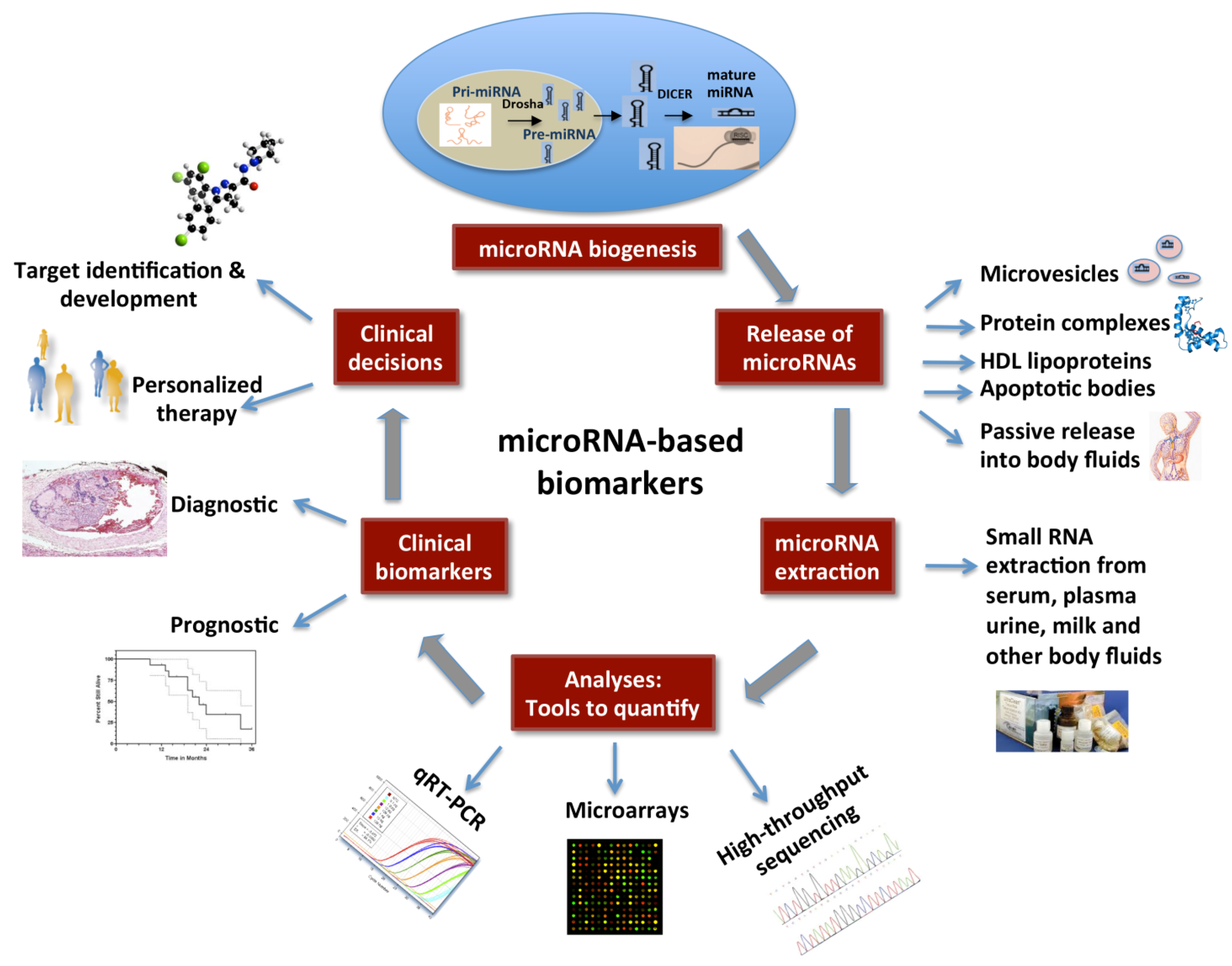
The workflow for development of miRNAs as biomarkers is schematically represented in Figure 1.
Figure 1.
Circulating microRNAs as biomarkers: A schematic diagram showing miRNA biogenesis, modes of their secretion into body fluids, RNA extraction and quantitative approaches. In future, clinical decisions may be made based on the expression levels of miRNAs. This figure is adapted from Steer and Subramanian [117].
6. Future Directions
miRNAs are revolutionizing the field of cancer research. These small RNAs are able to distinguish cancer types and subtypes and have the potential to be biomarkers in cancers. However, there is a need to address the level of correlation that exists between the miRNA identified in tumor tissue and circulating miRNAs. Further studies establishing miRNA expression patterns in relation to patient’s age, demography, common health conditions and lifestyles are needed. Before developing serum miRNA as a clinical biomarker, the techniques of miRNA extraction, quantification, data analysis and a normalization strategy needs to be standardized. Currently, one of the main challenges in analyzing miRNA from serum samples is the normalization factor. The effective use of animal models will significantly increase the success of developing suitable miRNA biomarkers in cases where the access to human patient samples is limited [118]. Effectively addressing these challenges will help us overcome some of the current pitfalls and establish miRNAs as fail proof biomarkers in cancer diagnosis and treatment.
Acknowledgments
This work is supported by grants from Academic Health Center, the Department of Defense (W81XwH10-1-0556), Minnesota Medical Foundation, University of Minnesota and The Karen Wykoff Sarcoma Foundation. We thank Jennie Walker Knoot for manuscript editing. Due to space restrictions we could not cite many significant contributions made by numerous other investigators in this important and rapidly progressing field.
References
- Cho, W.C. OncomiRs: The discovery and progress of microRNAs in cancers. Mol. Cancer 2007, 6. [Google Scholar] [CrossRef]
- Junn, E.; Mouradian, M.M. MicroRNAs in neurodegenerative diseases and their therapeutic potential. Pharmacol. Ther. 2012, 133, 142–150. [Google Scholar] [CrossRef]
- Maccani, M.A.; Padbury, J.F.; Marsit, C.J. miR-16 and miR-21 expression in the placenta is associated with fetal growth. PLoS ONE 2011, 6. [Google Scholar] [CrossRef]
- Zidar, N.; Bostjancic, E.; Glavac, D.; Stajer, D. MicroRNAs, innate immunity and ventricular rupture in human myocardial infarction. Dis. Markers 2011, 31, 259–265. [Google Scholar] [CrossRef] [PubMed]
- Rothschild, S.I.; Tschan, M.P.; Federzoni, E.A.; Jaggi, R.; Fey, M.F.; Gugger, M.; Gautschi, O. MicroRNA-29b is involved in the Src-ID1 signaling pathway and is dysregulated in human lung adenocarcinoma. Oncogene 2012, 31, 4221–4232. [Google Scholar] [CrossRef]
- Tavazoie, S.F.; Alarcon, C.; Oskarsson, T.; Padua, D.; Wang, Q.; Bos, P.D.; Gerald, W.L.; Massague, J. Endogenous human microRNAs that suppress breast cancer metastasis. Nature 2008, 451, 147–152. [Google Scholar] [CrossRef] [PubMed]
- Porkka, K.P.; Pfeiffer, M.J.; Waltering, K.K.; Vessella, R.L.; Tammela, T.L.; Visakorpi, T. MicroRNA expression profiling in prostate cancer. Cancer Res. 2007, 67, 6130–6135. [Google Scholar] [CrossRef] [PubMed]
- Roccaro, A.M.; Sacco, A.; Jia, X.; Azab, A.K.; Maiso, P.; Ngo, H.T.; Azab, F.; Runnels, J.; Quang, P.; Ghobrial, I.M. microRNA-dependent modulation of histone acetylation in Waldenstrom macroglobulinemia. Blood 2010, 116, 1506–1514. [Google Scholar] [CrossRef] [PubMed]
- Razumilava, N.; Bronk, S.F.; Smoot, R.L.; Fingas, C.D.; Werneburg, N.W.; Roberts, L.R.; Mott, J.L. miR-25 targets TNF-related apoptosis inducing ligand (TRAIL) death receptor-4 and promotes apoptosis resistance in cholangiocarcinoma. Hepatology 2012, 55, 465–475. [Google Scholar] [CrossRef]
- Bartel, D.P.; Chen, C.Z. Micromanagers of gene expression: the potentially widespread influence of metazoan microRNAs. Nat. Rev. Genet. 2004, 5, 396–400. [Google Scholar] [CrossRef]
- Hobert, O. miRNAs play a tune. Cell 2007, 131, 22–24. [Google Scholar] [CrossRef]
- Landgraf, P.; Rusu, M.; Sheridan, R.; Sewer, A.; Iovino, N.; Aravin, A.; Pfeffer, S.; Rice, A.; Kamphorst, A.O.; Landthaler, M.; et al. A mammalian microRNA expression atlas based on small RNA library sequencing. Cell 2007, 129, 1401–1414. [Google Scholar] [CrossRef]
- Li, X.; Cassidy, J.J.; Reinke, C.A.; Fischboeck, S.; Carthew, R.W. A microRNA imparts robustness against environmental fluctuation during development. Cell 2009, 137, 273–282. [Google Scholar] [CrossRef]
- Mendell, J.T. miRiad roles for the miR-17–92 cluster in development and disease. Cell 2008, 133, 217–222. [Google Scholar] [CrossRef]
- Croce, C.M. Causes and consequences of microRNA dysregulation in cancer. Nat. Rev. Genet. 2009, 10, 704–714. [Google Scholar] [CrossRef]
- Kim, V.N. Small RNAs: Classification, biogenesis, and function. Mol. Cells 2005, 19, 1–15. [Google Scholar] [CrossRef] [PubMed]
- Flynt, A.S.; Lai, E.C. Biological principles of microRNA-mediated regulation: Shared themes amid diversity. Nat. Rev. Genet. 2008, 9, 831–842. [Google Scholar] [CrossRef]
- Ambros, V. The functions of animal microRNAs. Nature 2004, 431, 350–355. [Google Scholar] [CrossRef] [PubMed]
- miRBase: The MicroRNA Database. Available online: http://www.mirbase.org (accessed on 15 November 2012).
- Lujambio, A.; Lowe, S.W. The microcosmos of cancer. Nature 2012, 482, 347–355. [Google Scholar] [CrossRef] [PubMed]
- Hwang, H.W.; Mendell, J.T. MicroRNAs in cell proliferation, cell death, and tumorigenesis. Br. J. Cancer 2006, 94, 776–780. [Google Scholar] [CrossRef]
- Conne, B.; Stutz, A.; Vassalli, J.D. The 3' untranslated region of messenger RNA: A molecular “hotspot” for pathology? Nat. Med. 2000, 6, 637–641. [Google Scholar] [CrossRef]
- Calin, G.A.; Dumitru, C.D.; Shimizu, M.; Bichi, R.; Zupo, S.; Noch, E.; Aldler, H.; Rattan, S.; Keating, M.; Rai, K.; et al. Frequent deletions and down-regulation of micro-RNA genes miR15 and miR16 at 13q14 in chronic lymphocytic leukemia. Proc. Natl. Acad. Sci. USA 2002, 99, 15524–15529. [Google Scholar] [CrossRef] [PubMed]
- Chan, E.; Patel, R.; Nallur, S.; Ratner, E.; Bacchiocchi, A.; Hoyt, K.; Szpakowski, S.; Godshalk, S.; Ariyan, S.; Sznol, M.; et al. MicroRNA signatures differentiate melanoma subtypes. Cell Cycle 2011, 10, 1845–1852. [Google Scholar] [CrossRef]
- Wurz, K.; Garcia, R.L.; Goff, B.A.; Mitchell, P.S.; Lee, J.H.; Tewari, M.; Swisher, E.M. MiR-221 and MiR-222 alterations in sporadic ovarian carcinoma: Relationship to CDKN1B, CDKNIC and overall survival. GCC (Genes Chromosomes Cancer) 2010, 49, 577–584. [Google Scholar]
- Guttilla, I.K.; White, B.A. Coordinate regulation of FOXO1 by miR-27a, miR-96, and miR-182 in breast cancer cells. J. Biol. Chem. 2009, 284, 23204–23216. [Google Scholar] [CrossRef] [PubMed]
- Van Der Heide, L.P.; Hoekman, M.F.; Smidt, M.P. The ins and outs of FoxO shuttling: Mechanisms of FoxO translocation and transcriptional regulation. Biochem. J. 2004, 380, 297–309. [Google Scholar] [CrossRef]
- Kefas, B.; Godlewski, J.; Comeau, L.; Li, Y.; Abounader, R.; Hawkinson, M.; Lee, J.; Fine, H.; Chiocca, E.A.; Lawler, S.; Purow, B. microRNA-7 inhibits the epidermal growth factor receptor and the Akt pathway and is down-regulated in glioblastoma. Cancer Res. 2008, 68, 3566–3572. [Google Scholar] [CrossRef] [PubMed]
- Schultz, J.; Lorenz, P.; Gross, G.; Ibrahim, S.; Kunz, M. MicroRNA let-7b targets important cell cycle molecules in malignant melanoma cells and interferes with anchorage-independent growth. Cancer Res. 2008, 18, 549–557. [Google Scholar]
- Tam, W.; Ben-Yehuda, D.; Hayward, W.S. Bic, a novel gene activated by proviral insertions in avian leukosis virus-induced lymphomas, is likely to function through its noncoding RNA. Mol. Cell Biol. 1997, 17, 1490–1502. [Google Scholar] [CrossRef] [PubMed]
- Cano, C.E.; Gommeaux, J.; Pietri, S.; Culcasi, M.; Garcia, S.; Seux, M.; Barelier, S.; Vasseur, S.; Spoto, R.P.; Pebusque, M.J.; Dusetti, N.J.; Iovanna, J.L.; Carrier, A. Tumor protein 53-induced nuclear protein 1 is a major mediator of p53 antioxidant function. Cancer Res. 2009, 69, 219–226. [Google Scholar] [CrossRef] [PubMed]
- He, L.; Thomson, J.M.; Hemann, M.T.; Hernando-Monge, E.; Mu, D.; Goodson, S.; Powers, S.; Cordon-Cardo, C.; Lowe, S.W.; Hannon, G.J.; Hammond, S.M. A microRNA polycistron as a potential human oncogene. Nature 2005, 435, 828–833. [Google Scholar] [CrossRef] [PubMed]
- Dong, F.; Lou, D. MicroRNA-34b/c suppresses uveal melanoma cell proliferation and migration through multiple targets. Mol. Vis. 2012, 18, 537–546. [Google Scholar] [PubMed]
- Gibcus, J.H.; Tan, L.P.; Harms, G.; Schakel, R.N.; de Jong, D.; Blokzijl, T.; Moller, P.; Poppema, S.; Kroesen, B.J.; van den Berg, A. Hodgkin lymphoma cell lines are characterized by a specific miRNA expression profile. Neoplasia 2009, 11, 167–176. [Google Scholar] [CrossRef] [PubMed]
- Taulli, R.; Bersani, F.; Foglizzo, V.; Linari, A.; Vigna, E.; Ladanyi, M.; Tuschl, T.; Ponzetto, C. The muscle-specific microRNA miR-206 blocks human rhabdomyosarcoma growth in xenotransplanted mice by promoting myogenic differentiation. J. Clin. Invest. 2009, 119, 2366–2378. [Google Scholar] [PubMed]
- Heneghan, H.M.; Miller, N.; Kelly, R.; Newell, J.; Kerin, M.J. Systemic miRNA-195 differentiates breast cancer from other malignancies and is a potential biomarker for detecting noninvasive and early stage disease. Oncologist 2010, 15, 673–682. [Google Scholar] [CrossRef]
- White, N.M.; Bao, T.T.; Grigull, J.; Youssef, Y.M.; Girgis, A.; Diamandis, M.; Fatoohi, E.; Metias, M.; Honey, R.J.; Stewart, R.; Pace, K.T.; Bjarnason, G.A.; Yousef, G.M. miRNA profiling for clear cell renal cell carcinoma: biomarker discovery and identification of potential controls and consequences of miRNA dysregulation. J. Urol. 2011, 186, 1077–1083. [Google Scholar] [CrossRef]
- Liu, J.; Gao, J.; Du, Y.; Li, Z.; Ren, Y.; Gu, J.; Wang, X.; Gong, Y.; Wang, W.; Kong, X. Combination of plasma microRNAs with serum CA19-9 for early detection of pancreatic cancer. Int. J. Cancer 2012, 131, 683–691. [Google Scholar] [CrossRef] [PubMed]
- Resnick, K.E.; Alder, H.; Hagan, J.P.; Richardson, D.L.; Croce, C.M.; Cohn, D.E. The detection of differentially expressed microRNAs from the serum of ovarian cancer patients using a novel real-time PCR platform. Gynecol. Oncol. 2009, 112, 55–59. [Google Scholar] [CrossRef]
- Mitchell, P.S.; Parkin, R.K.; Kroh, E.M.; Fritz, B.R.; Wyman, S.K.; Pogosova-Agadjanyan, E.L.; Peterson, A.; Noteboom, J.; O’Briant, K.C.; Allen, A.; et al. Circulating microRNAs as stable blood-based markers for cancer detection. Proc. Natl. Acad. Sci. USA 2008, 105, 10513–10518. [Google Scholar] [CrossRef] [PubMed]
- Zheng, T.; Wang, J.; Chen, X.; Liu, L. Role of microRNA in anticancer drug resistance. Int. J. Cancer 2010, 126, 2–10. [Google Scholar] [CrossRef]
- Ryu, J.K.; Matthaei, H.; Dal Molin, M.; Hong, S.M.; Canto, M.I.; Schulick, R.D.; Wolfgang, C.; Goggins, M.G.; Hruban, R.H.; Cope, L.; Maitra, A. Elevated microRNA miR-21 levels in pancreatic cyst fluid are predictive of mucinous precursor lesions of ductal adenocarcinoma. Pancreatology 2011, 11, 343–350. [Google Scholar] [CrossRef]
- Ryu, J.K.; Hong, S.M.; Karikari, C.A.; Hruban, R.H.; Goggins, M.G.; Maitra, A. Aberrant MicroRNA-155 expression is an early event in the multistep progression of pancreatic adenocarcinoma. Pancreatology 2010, 10, 66–73. [Google Scholar] [CrossRef]
- Russo, F.; Di Bella, S.; Nigita, G.; Macca, V.; Lagana, A.; Giugno, R.; Pulvirenti, A.; Ferro, A. miRandola: Extracellular circulating microRNAs database. PloS ONE 2012, 7. [Google Scholar] [CrossRef]
- Mo, M.H.; Chen, L.; Fu, Y.; Wang, W.; Fu, S.W. Cell-free Circulating miRNA Biomarkers in Cancer. Int. J. Cancer 2012, 3, 432–448. [Google Scholar] [CrossRef]
- Sun, Y.; Wang, M.; Lin, G.; Sun, S.; Li, X.; Qi, J.; Li, J. Serum microRNA-155 as a potential biomarker to track disease in breast cancer. PloS ONE 2012, 7. [Google Scholar] [CrossRef]
- Zhao, A.; Li, G.; Peoc’h, M.; Genin, C.; Gigante, M. Serum miR-210 as a novel biomarker for molecular diagnosis of clear cell renal cell carcinoma. Exp. Mol. Pathol. 2012, 94, 115–120. [Google Scholar] [PubMed]
- Dieckmann, K.P.; Spiekermann, M.; Balks, T.; Flor, I.; Loning, T.; Bullerdiek, J.; Belge, G. MicroRNAs miR-371–3 in serum as diagnostic tools in the management of testicular germ cell tumours. Br. J. Cancer 2012, 107, 1754–1760. [Google Scholar] [CrossRef]
- Yu, Z.; Kastenmuller, G.; He, Y.; Belcredi, P.; Moller, G.; Prehn, C.; Mendes, J.; Wahl, S.; Roemisch-Margl, W.; Ceglarek, U.; et al. Differences between human plasma and serum metabolite profiles. PLoS ONE 2011, 6. [Google Scholar] [CrossRef]
- Lawrie, C.H.; Gal, S.; Dunlop, H.M.; Pushkaran, B.; Liggins, A.P.; Pulford, K.; Banham, A.H.; Pezzella, F.; Boultwood, J.; Wainscoat, J.S.; Hatton, C.S.; Harris, A.L. Detection of elevated levels of tumour-associated microRNAs in serum of patients with diffuse large B-cell lymphoma. Br. J. Haematol. 2008, 141, 672–675. [Google Scholar] [CrossRef]
- Tanaka, M.; Oikawa, K.; Takanashi, M.; Kudo, M.; Ohyashiki, J.; Ohyashiki, K.; Kuroda, M. Down-regulation of miR-92 in human plasma is a novel marker for acute leukemia patients. PloS ONE 2009, 4. [Google Scholar] [CrossRef]
- Zhu, W.; Qin, W.; Atasoy, U.; Sauter, E.R. Circulating microRNAs in breast cancer and healthy subjects. BMC Res. Notes 2009, 2. [Google Scholar] [CrossRef]
- Ng, E.K.; Chong, W.W.; Jin, H.; Lam, E.K.; Shin, V.Y.; Yu, J.; Poon, T.C.; Ng, S.S.; Sung, J.J. Differential expression of microRNAs in plasma of patients with colorectal cancer: A potential marker for colorectal cancer screening. Gut 2009, 58, 1375–1381. [Google Scholar] [CrossRef] [Green Version]
- Tsujiura, M.; Ichikawa, D.; Komatsu, S.; Shiozaki, A.; Takeshita, H.; Kosuga, T.; Konishi, H.; Morimura, R.; Deguchi, K.; Fujiwara, H.; Okamoto, K.; Otsuji, E. Circulating microRNAs in plasma of patients with gastric cancers. Br. J. Cancer 2010, 102, 1174–1179. [Google Scholar] [CrossRef]
- Skog, J.; Wurdinger, T.; van Rijn, S.; Meijer, D.H.; Gainche, L.; Sena-Esteves, M.; Curry, W.T., Jr.; Carter, B.S.; Krichevsky, A.M.; Breakefield, X.O. Glioblastoma microvesicles transport RNA and proteins that promote tumour growth and provide diagnostic biomarkers. Nat. Cell Biol. 2008, 10, 1470–1476. [Google Scholar] [CrossRef]
- Yamamoto, Y.; Kosaka, N.; Tanaka, M.; Koizumi, F.; Kanai, Y.; Mizutani, T.; Murakami, Y.; Kuroda, M.; Miyajima, A.; Kato, T.; Ochiya, T. MicroRNA-500 as a potential diagnostic marker for hepatocellular carcinoma. Biomarkers 2009, 14, 529–538. [Google Scholar] [CrossRef]
- Chen, X.; Ba, Y.; Ma, L.; Cai, X.; Yin, Y.; Wang, K.; Guo, J.; Zhang, Y.; Chen, J.; Guo, X.; et al. Characterization of microRNAs in serum: a novel class of biomarkers for diagnosis of cancer and other diseases. Cell Res. 2008, 18, 997–1006. [Google Scholar] [CrossRef]
- Wong, T.S.; Liu, X.B.; Wong, B.Y.; Ng, R.W.; Yuen, A.P.; Wei, W.I. Mature miR-184 as potential oncogenic microRNA of squamous cell carcinoma of tongue. Clin. Cancer Res. 2008, 14, 2588–2592. [Google Scholar] [CrossRef]
- Baraniskin, A.; Nopel-Dunnebacke, S.; Ahrens, M.; Jensen, S.G.; Zollner, H.; Maghnouj, A.; Wos, A.; Mayerle, J.; Munding, J.; Kost, D.; et al. Circulating U2 small nuclear RNA fragments as a novel diagnostic biomarker for pancreatic and colorectal adenocarcinoma. Int. J. Cancer. 2012, 132, E48–E57. [Google Scholar] [PubMed]
- Bhat, K.; Wang, F.; Ma, Q.; Li, Q.; Mallik, S.; Hsieh, T.C.; Wu, E. Advances in biomarker research for pancreatic cancer. Curr. Pharm. Design 2012, 18, 2439–2451. [Google Scholar] [CrossRef]
- Liu, R.; Chen, X.; Du, Y.; Yao, W.; Shen, L.; Wang, C.; Hu, Z.; Zhuang, R.; Ning, G.; Zhang, C.; et al. Serum microRNA expression profile as a biomarker in the diagnosis and prognosis of pancreatic cancer. Clin. Chem. 2012, 58, 610–618. [Google Scholar] [CrossRef]
- Morimura, R.; Komatsu, S.; Ichikawa, D.; Takeshita, H.; Tsujiura, M.; Nagata, H.; Konishi, H.; Shiozaki, A.; Ikoma, H.; Okamoto, K.; Ochiai, T.; Taniguchi, H.; Otsuji, E. Novel diagnostic value of circulating miR-18a in plasma of patients with pancreatic cancer. Br. J. Cancer 2011, 105, 1733–1740. [Google Scholar] [CrossRef]
- Zheng, J.; Dong, P.; Gao, S.; Wang, N.; Yu, F. High expression of serum miR-17-5p associated with poor prognosis in patients with hepatocellular carcinoma. Hepato-Gastroenterology 2012, 60. [Google Scholar] [CrossRef]
- Qi, J.; Wang, J.; Katayama, H.; Sen, S.; Liu, S.M. Circulating microRNAs (cmiRNAs) as novel potential biomarkers for hepatocellular carcinoma. Neoplasma 2013, 60, 135–142. [Google Scholar] [PubMed]
- Kirschner, M.B.; Kao, S.C.; Edelman, J.J.; Armstrong, N.J.; Vallely, M.P.; van Zandwijk, N.; Reid, G. Haemolysis during sample preparation alters microRNA content of plasma. PLoS ONE 2011, 6, e24145. [Google Scholar] [CrossRef] [PubMed]
- Lee, Y.M.; Lee, J.Y.; Ho, C.C.; Hong, Q.S.; Yu, S.L.; Tzeng, C.R.; Yang, P.C.; Chen, H.W. miRNA-34b as a tumor suppressor in estrogen-dependent growth of breast cancer cells. Breast Cancer Res. 2011, 13, R166. [Google Scholar]
- Heneghan, H.M.; Miller, N.; Kelly, R.; Newell, J.; Kerin, M.J. Systemic miRNA-195 differentiates breast cancer from other malignancies and is a potential biomarker for detecting noninvasive and early stage disease. Oncologist 2010, 15, 673–682. [Google Scholar] [CrossRef]
- Frankel, L.B.; Christoffersen, N.R.; Jacobsen, A.; Lindow, M.; Krogh, A.; Lund, A.H. Programmed cell death 4 (PDCD4) is an important functional target of the microRNA miR-21 in breast cancer cells. J. Biol. Chem. 2008, 283, 1026–1033. [Google Scholar] [CrossRef] [PubMed]
- Huang, Z.; Huang, D.; Ni, S.; Peng, Z.; Sheng, W.; Du, X. Plasma microRNAs are promising novel biomarkers for early detection of colorectal cancer. Int. J. Cancer 2010, 127, 118–126. [Google Scholar] [CrossRef] [PubMed]
- Ichimi, T.; Enokida, H.; Okuno, Y.; Kunimoto, R.; Chiyomaru, T.; Kawamoto, K.; Kawahara, K.; Toki, K.; Kawakami, K.; Nishiyama, K.; Tsujimoto, G.; Nakagawa, M.; Seki, N. Identification of novel microRNA targets based on microRNA signatures in bladder cancer. Int. J. Cancer 2009, 125, 345–352. [Google Scholar] [CrossRef] [PubMed]
- Conti, A.; Aguennouz, M.; La Torre, D.; Tomasello, C.; Cardali, S.; Angileri, F.F.; Maio, F.; Cama, A.; Germano, A.; Vita, G.; Tomasello, F. miR-21 and 221 upregulation and miR-181b downregulation in human grade II-IV astrocytic tumors. J. Neuro-Oncology 2009, 93, 325–332. [Google Scholar] [CrossRef]
- Zheng, B.; Liang, L.; Huang, S.; Zha, R.; Liu, L.; Jia, D.; Tian, Q.; Wang, Q.; Wang, C.; Long, Z.; et al. MicroRNA-409 suppresses tumour cell invasion and metastasis by directly targeting radixin in gastric cancers. Oncogene 2012, 31, 4509–4516. [Google Scholar] [CrossRef]
- Hu, Z.; Chen, X.; Zhao, Y.; Tian, T.; Jin, G.; Shu, Y.; Chen, Y.; Xu, L.; Zen, K.; Zhang, C.; Shen, H. Serum microRNA signatures identified in a genome-wide serum microRNA expression profiling predict survival of non-small-cell lung cancer. J. Clin. Ooncol. 2010, 28, 1721–1726. [Google Scholar] [CrossRef]
- Silva, J.; Garcia, V.; Zaballos, A.; Provencio, M.; Lombardia, L.; Almonacid, L.; Garcia, J.M.; Dominguez, G.; Pena, C.; Diaz, R.; et al. Vesicle-related microRNAs in plasma of nonsmall cell lung cancer patients and correlation with survival. Eur. Respir. J. 2011, 37, 617–623. [Google Scholar] [CrossRef] [PubMed]
- Liu, C.J.; Kao, S.Y.; Tu, H.F.; Tsai, M.M.; Chang, K.W.; Lin, S.C. Increase of microRNA miR-31 level in plasma could be a potential marker of oral cancer. Oral Dis. 2010, 16, 360–364. [Google Scholar] [CrossRef] [PubMed]
- Bryant, R.J.; Pawlowski, T.; Catto, J.W.; Marsden, G.; Vessella, R.L.; Rhees, B.; Kuslich, C.; Visakorpi, T.; Hamdy, F.C. Changes in circulating microRNA levels associated with prostate cancer. Br. J. Cancer 2012, 106, 768–774. [Google Scholar] [CrossRef] [PubMed]
- Brase, J.C.; Johannes, M.; Schlomm, T.; Falth, M.; Haese, A.; Steuber, T.; Beissbarth, T.; Kuner, R.; Sultmann, H. Circulating miRNAs are correlated with tumor progression in prostate cancer. Int. J. Cancer 2011, 128, 608–616. [Google Scholar] [CrossRef] [PubMed]
- Greither, T.; Grochola, L.F.; Udelnow, A.; Lautenschlager, C.; Wurl, P.; Taubert, H. Elevated expression of microRNAs 155, 203, 210 and 222 in pancreatic tumors is associated with poorer survival. Int. J. Cancer 2010, 126, 73–80. [Google Scholar] [CrossRef]
- Wang, J.; Chen, J.; Chang, P.; LeBlanc, A.; Li, D.; Abbruzzesse, J.L.; Frazier, M.L.; Killary, A.M.; Sen, S. MicroRNAs in plasma of pancreatic ductal adenocarcinoma patients as novel blood-based biomarkers of disease. Cancer Prev. Res. (Phila) 2009, 2, 807–813. [Google Scholar] [CrossRef]
- Fornari, F.; Milazzo, M.; Chieco, P.; Negrini, M.; Marasco, E.; Capranico, G.; Mantovani, V.; Marinello, J.; Sabbioni, S.; Callegari, E.; et al. In hepatocellular carcinoma miR-519d is up-regulated by p53 and DNA hypomethylation and targets CDKN1A/p21, PTEN, AKT3 and TIMP2. J. Pathol. 2012, 227, 275–285. [Google Scholar] [CrossRef]
- Snowdon, J.; Zhang, X.; Childs, T.; Tron, V.A.; Feilotter, H. The microRNA-200 family is upregulated in endometrial carcinoma. PLoS ONE 2011, 6. [Google Scholar] [CrossRef]
- White, N.M.; Bao, T.T.; Grigull, J.; Youssef, Y.M.; Girgis, A.; Diamandis, M.; Fatoohi, E.; Metias, M.; Honey, R.J.; Stewart, R.; et al. miRNA profiling for clear cell renal cell carcinoma: Biomarker discovery and identification of potential controls and consequences of miRNA dysregulation. J. Urol. 2011, 186, 1077–1083. [Google Scholar] [CrossRef] [PubMed]
- Caramuta, S.; Egyhazi, S.; Rodolfo, M.; Witten, D.; Hansson, J.; Larsson, C.; Lui, W.O. MicroRNA expression profiles associated with mutational status and survival in malignant melanoma. J. Investig. Dermatol. 2010, 130, 2062–2070. [Google Scholar] [CrossRef]
- Nikiforova, M.N.; Tseng, G.C.; Steward, D.; Diorio, D.; Nikiforov, Y.E. MicroRNA expression profiling of thyroid tumors: biological significance and diagnostic utility. J. Clin. Endocrinol. Metabol. 2008, 93, 1600–1608. [Google Scholar] [CrossRef]
- Lulla, R.R.; Costa, F.F.; Bischof, J.M.; Chou, P.M.; de, F.B.M.; Vanin, E.F.; Soares, M.B. Identification of differentially expressed microRNAs in osteosarcoma. Sarcoma 2011, 2011. [Google Scholar] [CrossRef]
- Thayanithy, V.; Sarver, A.L.; Kartha, R.V.; Li, L.; Angstadt, A.Y.; Breen, M.; Steer, C.J.; Modiano, J.F.; Subramanian, S. Perturbation of 14q32 miRNAs-cMYC gene network in osteosarcoma. Bone 2012, 50, 171–181. [Google Scholar] [CrossRef] [PubMed]
- Zavadil, J.; Ye, H.; Liu, Z.; Wu, J.; Lee, P.; Hernando, E.; Soteropoulos, P.; Toruner, G.A.; Wei, J.J. Profiling and functional analyses of microRNAs and their target gene products in human uterine leiomyomas. PloS ONE 2010, 5. [Google Scholar] [CrossRef]
- Miyachi, M.; Tsuchiya, K.; Yoshida, H.; Yagyu, S.; Kikuchi, K.; Misawa, A.; Iehara, T.; Hosoi, H. Circulating muscle-specific microRNA, miR-206, as a potential diagnostic marker for rhabdomyosarcoma. Biochem. Biophys. Res. Commun. 2010, 400, 89–93. [Google Scholar] [CrossRef]
- Koelz, M.; Lense, J.; Wrba, F.; Scheffler, M.; Dienes, H.P.; Odenthal, M. Down-regulation of miR-221 and miR-222 correlates with pronounced Kit expression in gastrointestinal stromal tumors. Int. J. Oncology 2011, 38, 503–511. [Google Scholar]
- Ban, J.; Jug, G.; Mestdagh, P.; Schwentner, R.; Kauer, M.; Aryee, D.N.; Schaefer, K.L.; Nakatani, F.; Scotlandi, K.; Reiter, M.; Strunk, D.; Speleman, F.; Vandesompele, J.; Kovar, H. Hsa-mir-145 is the top EWS-FLI1-repressed microRNA involved in a positive feedback loop in Ewing's sarcoma. Oncogene 2011, 30, 2173–2180. [Google Scholar] [CrossRef]
- Saydam, O.; Senol, O.; Wurdinger, T.; Mizrak, A.; Ozdener, G.B.; Stemmer-Rachamimov, A.O.; Yi, M.; Stephens, R.M.; Krichevsky, A.M.; Saydam, N.; Brenner, G.J.; Breakefield, X.O. miRNA-7 attenuation in Schwannoma tumors stimulates growth by upregulating three oncogenic signaling pathways. Cancer Res. 2011, 71, 852–861. [Google Scholar] [CrossRef] [PubMed]
- Sarver, A.L.; Li, L.; Subramanian, S. MicroRNA miR-183 functions as an oncogene by targeting the transcription factor EGR1 and promoting tumor cell migration. Cancer Res. 2010, 70, 9570–9580. [Google Scholar] [CrossRef]
- Subramanian, S.; Thayanithy, V.; West, R.B.; Lee, C.H.; Beck, A.H.; Zhu, S.; Downs-Kelly, E.; Montgomery, K.; Goldblum, J.R.; Hogendoorn, P.C.; et al. Genome-wide transcriptome analyses reveal p53 inactivation mediated loss of miR-34a expression in malignant peripheral nerve sheath tumours. J. Pathol. 2010, 220, 58–70. [Google Scholar] [CrossRef]
- Yamagishi, M.; Nakano, K.; Miyake, A.; Yamochi, T.; Kagami, Y.; Tsutsumi, A.; Matsuda, Y.; Sato-Otsubo, A.; Muto, S.; Utsunomiya, A.; et al. Polycomb-mediated loss of miR-31 activates NIK-dependent NF-kappaB pathway in adult T cell leukemia and other cancers. Cancer Cell. 2012, 21, 121–135. [Google Scholar] [CrossRef]
- Careccia, S.; Mainardi, S.; Pelosi, A.; Gurtner, A.; Diverio, D.; Riccioni, R.; Testa, U.; Pelosi, E.; Piaggio, G.; Sacchi, A.; et al. A restricted signature of miRNAs distinguishes APL blasts from normal promyelocytes. Oncogene 2009, 28, 4034–4040. [Google Scholar] [CrossRef] [PubMed]
- Pigazzi, M.; Manara, E.; Baron, E.; Basso, G. miR-34b targets cyclic AMP-responsive element binding protein in acute myeloid leukemia. Cancer Res. 2009, 69, 2471–2478. [Google Scholar] [CrossRef]
- Ghosh, A.K.; Shanafelt, T.D.; Cimmino, A.; Taccioli, C.; Volinia, S.; Liu, C.G.; Calin, G.A.; Croce, C.M.; Chan, D.A.; Giaccia, A.J.; et al. Aberrant regulation of pVHL levels by microRNA promotes the HIF/VEGF axis in CLL B cells. Blood 2009, 113, 5568–5574. [Google Scholar] [CrossRef] [PubMed]
- Stamatopoulos, B.; Meuleman, N.; Haibe-Kains, B.; Saussoy, P.; Van Den Neste, E.; Michaux, L.; Heimann, P.; Martiat, P.; Bron, D.; Lagneaux, L. microRNA-29c and microRNA-223 down-regulation has in vivo significance in chronic lymphocytic leukemia and improves disease risk stratification. Blood 2009, 113, 5237–5245. [Google Scholar] [CrossRef]
- Fang, C.; Zhu, D.X.; Dong, H.J.; Zhou, Z.J.; Wang, Y.H.; Liu, L.; Fan, L.; Miao, K.R.; Liu, P.; Xu, W.; Li, J.Y. Serum microRNAs are promising novel biomarkers for diffuse large B cell lymphoma. Ann. Hematol. 2012, 91, 553–559. [Google Scholar] [CrossRef]
- Weber, J.A.; Baxter, D.H.; Zhang, S.; Huang, D.Y.; Huang, K.H.; Lee, M.J.; Galas, D.J.; Wang, K. The microRNA spectrum in 12 body fluids. Clin. Chem. 2010, 56, 1733–1741. [Google Scholar] [CrossRef]
- Mahn, R.; Heukamp, L.C.; Rogenhofer, S.; von Ruecker, A.; Muller, S.C.; Ellinger, J. Circulating microRNAs (miRNA) in serum of patients with prostate cancer. Urology 2011, 77, 1265.e9–1265.e16. [Google Scholar] [CrossRef]
- Yamada, Y.; Enokida, H.; Kojima, S.; Kawakami, K.; Chiyomaru, T.; Tatarano, S.; Yoshino, H.; Kawahara, K.; Nishiyama, K.; Seki, N.; Nakagawa, M. MiR-96 and miR-183 detection in urine serve as potential tumor markers of urothelial carcinoma: Correlation with stage and grade, and comparison with urinary cytology. Cancer Sci. 2011, 102, 522–529. [Google Scholar] [CrossRef]
- Baraniskin, A.; Kuhnhenn, J.; Schlegel, U.; Maghnouj, A.; Zollner, H.; Schmiegel, W.; Hahn, S.; Schroers, R. Identification of microRNAs in the cerebrospinal fluid as biomarker for the diagnosis of glioma. J. Neuro-Oncology 2012, 14, 29–33. [Google Scholar] [CrossRef]
- Wu, L.; Zhou, H.; Lin, H.; Qi, J.; Zhu, C.; Gao, Z.; Wang, H. Circulating microRNAs are elevated in plasma from severe preeclamptic pregnancies. Reproduction 2012, 143, 389–397. [Google Scholar] [CrossRef] [PubMed]
- Xie, Y.; Todd, N.W.; Liu, Z.; Zhan, M.; Fang, H.; Peng, H.; Alattar, M.; Deepak, J.; Stass, S.A.; Jiang, F. Altered miRNA expression in sputum for diagnosis of non-small cell lung cancer. Lung Cancer 2010, 67, 170–176. [Google Scholar] [CrossRef]
- Valadi, H.; Ekstrom, K.; Bossios, A.; Sjostrand, M.; Lee, J.J.; Lotvall, J.O. Exosome-mediated transfer of mRNAs and microRNAs is a novel mechanism of genetic exchange between cells. Nat. Cell Biol. 2007, 9, 654–659. [Google Scholar] [CrossRef]
- Hunter, M.P.; Ismail, N.; Zhang, X.; Aguda, B.D.; Lee, E.J.; Yu, L.; Xiao, T.; Schafer, J.; Lee, M.L.; Schmittgen, T.D.; Nana-Sinkam, S.P.; Jarjoura, D.; Marsh, C.B. Detection of microRNA expression in human peripheral blood microvesicles. PLoS ONE 2008, 3. [Google Scholar] [CrossRef]
- Gallo, A.; Tandon, M.; Alevizos, I.; Illei, G.G. The majority of microRNAs detectable in serum and saliva is concentrated in exosomes. PLoS ONE 2012, 7. [Google Scholar] [CrossRef]
- Fichtlscherer, S.; Zeiher, A.M.; Dimmeler, S. Circulating microRNAs: biomarkers or mediators of cardiovascular diseases? Arterioscler. Thromb. Vasc. Biol. 2011, 31, 2383–2390. [Google Scholar] [CrossRef]
- Vickers, K.C.; Palmisano, B.T.; Shoucri, B.M.; Shamburek, R.D.; Remaley, A.T. MicroRNAs are transported in plasma and delivered to recipient cells by high-density lipoproteins. Nat. Cell Biol. 2011, 13, 423–433. [Google Scholar] [CrossRef]
- Gilad, S.; Meiri, E.; Yogev, Y.; Benjamin, S.; Lebanony, D.; Yerushalmi, N.; Benjamin, H.; Kushnir, M.; Cholakh, H.; Melamed, N.; Bentwich, Z.; Hod, M.; Goren, Y.; Chajut, A. Serum microRNAs are promising novel biomarkers. PloS ONE 2008, 3. [Google Scholar] [CrossRef]
- Kroh, E.M.; Parkin, R.K.; Mitchell, P.S.; Tewari, M. Analysis of circulating microRNA biomarkers in plasma and serum using quantitative reverse transcription-PCR (qRT-PCR). Methods 2010, 50, 298–301. [Google Scholar] [CrossRef]
- Hu, Z.; Chen, X.; Zhao, Y.; Tian, T.; Jin, G.; Shu, Y.; Chen, Y.; Xu, L.; Zen, K.; Zhang, C.; Shen, H. Serum microRNA signatures identified in a genome-wide serum microRNA expression profiling predict survival of non-small-cell lung cancer. J. Clin. Oncol. 2010, 28, 1721–1726. [Google Scholar] [CrossRef]
- Cummins, J.M.; He, Y.; Leary, R.J.; Pagliarini, R.; Diaz, L.A., Jr.; Sjoblom, T.; Barad, O.; Bentwich, Z.; Szafranska, A.E.; Labourier, E.; et al. The colorectal microRNAome. Proc. Natl. Acad. Sci. USA 2006, 103, 3687–3692. [Google Scholar] [CrossRef] [PubMed]
- Nelson, P.T.; Baldwin, D.A.; Scearce, L.M.; Oberholtzer, J.C.; Tobias, J.W.; Mourelatos, Z. Microarray-based, high-throughput gene expression profiling of microRNAs. Nat. Methods 2004, 1, 155–161. [Google Scholar] [CrossRef]
- Ajit, S.K. Circulating microRNAs as biomarkers, therapeutic targets, and signaling molecules. Sensors 2012, 12, 3359–3369. [Google Scholar] [CrossRef]
- Steer, C.J.; Subramanian, S. Circulating microRNAs as biomarkers: a new frontier in diagnostics. Liver Transplant. 2012, 18, 265–269. [Google Scholar] [CrossRef]
- LaConti, J.J.; Shivapurkar, N.; Preet, A.; Deslattes Mays, A.; Peran, I.; Kim, S.E.; Marshall, J.L.; Riegel, A.T.; Wellstein, A. Tissue and serum microRNAs in the Kras(G12D) transgenic animal model and in patients with pancreatic cancer. PloS ONE 2011, 6. [Google Scholar] [CrossRef]
© 2013 by the authors; licensee MDPI, Basel, Switzerland. This article is an open access article distributed under the terms and conditions of the Creative Commons Attribution license (http://creativecommons.org/licenses/by/3.0/).
Share and Cite
MDPI and ACS Style
Sundarbose, K.; Kartha, R.V.; Subramanian, S. MicroRNAs as Biomarkers in Cancer. Diagnostics 2013, 3, 84-104. https://doi.org/10.3390/diagnostics3010084
AMA Style
Sundarbose K, Kartha RV, Subramanian S. MicroRNAs as Biomarkers in Cancer. Diagnostics. 2013; 3(1):84-104. https://doi.org/10.3390/diagnostics3010084
Chicago/Turabian StyleSundarbose, Kamini, Reena V. Kartha, and Subbaya Subramanian. 2013. "MicroRNAs as Biomarkers in Cancer" Diagnostics 3, no. 1: 84-104. https://doi.org/10.3390/diagnostics3010084