Next Issue
Volume 14, May-1
Previous Issue
Volume 14, April-1


Diagnostics, Volume 14, Issue 8 (April-2 2024) – 78 articles

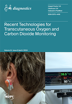
Cover Story (view full-size image): Remote patient monitoring enables improved care quality, patient autonomy, personalized treatment, and cost reduction. The evaluation of human well-being places crucial importance on respiratory parameters such as oxygen and carbon dioxide partial pressures. The transcutaneous monitoring of these gases overcomes the limitations of the reference technique, arterial blood gas analysis, which is intermittent and painful. It offers a non-invasive and potentially remote continuous monitoring solution, enabling the early detection of respiratory issues, as evidenced in COVID-19 patients. Recent research has focused on technologies for a wearable transcutaneous monitoring device including luminescence, electronic paramagnetic resonance, and photoacoustic sensors, with optical sensors emerging as the most promising option. View this paper
  • Issues are regarded as officially published after their release is announced to the table of contents alert mailing list.
  • You may sign up for e-mail alerts to receive table of contents of newly released issues.
  • PDF is the official format for papers published in both, html and pdf forms. To view the papers in pdf format, click on the "PDF Full-text" link, and use the free Adobe Reader to open them.
Order results
Result details
Section
Select all
Export citation of selected articles as:
14 pages, 3776 KiB  
Review
Diabetes Mellitus and Pneumococcal Pneumonia
by Catia Cilloniz and Antoni Torres
Diagnostics 2024, 14(8), 859; https://doi.org/10.3390/diagnostics14080859 - 22 Apr 2024
Viewed by 306
Abstract
Currently, there are more than 500 million people suffering from diabetes around the world. People aged 65 years or older are the most affected by this disease, and it is estimated that approximately 96% of diabetes cases worldwide are type 2 diabetes. People [...] Read more.
Currently, there are more than 500 million people suffering from diabetes around the world. People aged 65 years or older are the most affected by this disease, and it is estimated that approximately 96% of diabetes cases worldwide are type 2 diabetes. People with diabetes mellitus are at an increased risk of infections such as pneumonia, due to a series of factors that may contribute to immune dysfunction, including hyperglycemia, inhibition of neutrophil chemotaxis, impaired cytokine production, phagocytic cell dysfunction, altered T cell-mediated immune responses and the co-existence of chronic comorbidities. Rates of infection, hospitalization and mortality in diabetic patients are reported to be higher than in the general population. Research into the risk of infectious diseases such as pneumonia in these patients is very important because it will help improve their management and treatment. Full article
(This article belongs to the Special Issue Hyperglycemia in Respiratory Diseases—Impact and Challenges)
Show Figures

Figure 1

32 pages, 5799 KiB  
Review
The Dual Lens of Endoscopy and Histology in the Diagnosis and Management of Eosinophilic Gastrointestinal Disorders—A Comprehensive Review
by Alberto Barchi, Edoardo Vespa, Sandro Passaretti, Giuseppe Dell’Anna, Ernesto Fasulo, Mona-Rita Yacoub, Luca Albarello, Emanuele Sinagra, Luca Massimino, Federica Ungaro, Silvio Danese and Francesco Vito Mandarino
Diagnostics 2024, 14(8), 858; https://doi.org/10.3390/diagnostics14080858 - 22 Apr 2024
Viewed by 503
Abstract
Eosinophilic Gastrointestinal Disorders (EGIDs) are a group of conditions characterized by abnormal eosinophil accumulation in the gastrointestinal tract. Among these EGIDs, Eosinophilic Esophagitis (EoE) is the most well documented, while less is known about Eosinophilic Gastritis (EoG), Eosinophilic Enteritis (EoN), and Eosinophilic Colitis [...] Read more.
Eosinophilic Gastrointestinal Disorders (EGIDs) are a group of conditions characterized by abnormal eosinophil accumulation in the gastrointestinal tract. Among these EGIDs, Eosinophilic Esophagitis (EoE) is the most well documented, while less is known about Eosinophilic Gastritis (EoG), Eosinophilic Enteritis (EoN), and Eosinophilic Colitis (EoC). The role of endoscopy in EGIDs is pivotal, with applications in diagnosis, disease monitoring, and therapeutic intervention. In EoE, the endoscopic reference score (EREFS) has been shown to be accurate in raising diagnostic suspicion and effective in monitoring therapeutic responses. Additionally, endoscopic dilation is the first-line treatment for esophageal strictures. For EoG and EoN, while the literature is more limited, common endoscopic findings include erythema, nodules, and ulcerations. Histology remains the gold standard for diagnosing EGIDs, as it quantifies eosinophilic infiltration. In recent years, there have been significant advancements in the histological understanding of EoE, leading to the development of diagnostic scores and the identification of specific microscopic features associated with the disease. However, for EoG, EoN, and EoC, precise eosinophil count thresholds for diagnosis have not yet been established. This review aims to elucidate the role of endoscopy and histology in the diagnosis and management of the three main EGIDs and to analyze their strengths and limitations, their interconnection, and future research directions. Full article
(This article belongs to the Special Issue Advances in Endoscopy)
Show Figures

Figure 1

3 pages, 500 KiB  
Interesting Images
Myxofibrosarcoma of the Chest Wall Detected on 99mTc-MDP Whole-Body Bone Scan
by Chia-Hsuan Lee, Hueng-Yuan (Daniel) Shen, Yow-Ling (Shirley) Shiue, Hung-Yen Chan and Hung-Pin Chan
Diagnostics 2024, 14(8), 857; https://doi.org/10.3390/diagnostics14080857 - 22 Apr 2024
Viewed by 269
Abstract
Myxofibrosarcoma is a type of soft tissue sarcoma, predominantly characterized by a high propensity for local recurrence, albeit demonstrating a relatively diminished risk for distant metastasis. Its prevalence is notably higher in elderly patients. Here, we present a case of a 73-year-old woman [...] Read more.
Myxofibrosarcoma is a type of soft tissue sarcoma, predominantly characterized by a high propensity for local recurrence, albeit demonstrating a relatively diminished risk for distant metastasis. Its prevalence is notably higher in elderly patients. Here, we present a case of a 73-year-old woman diagnosed with Myxofibrosarcoma. She was subjected to a whole-body bone scan using 99mTc-methylene diphosphonate (MDP) to survey potential bony metastasis. It revealed marked MDP accumulation with peripheral soft tissue uptake in the right lateral chest region of this patient. This imaging phenotype could potentially be attributed to the augmented vascularity within the tumor, a finding that was prominently displayed in this particular case. Full article
(This article belongs to the Special Issue Imaging Diagnosis in Musculoskeletal Medicine)
Show Figures

Figure 1

18 pages, 294 KiB  
Review
Breaking Sound Barriers: Exploring Tele-Audiology’s Impact on Hearing Healthcare
by Mien-Jen Lin and Chin-Kuo Chen
Diagnostics 2024, 14(8), 856; https://doi.org/10.3390/diagnostics14080856 - 22 Apr 2024
Viewed by 363
Abstract
Hearing impairment is a global issue, affecting billions of people; however, there is a gap between the population affected by hearing loss and those able to access hearing healthcare. Tele-audiology, the application of telemedicine in audiology, serves as a new form of technology [...] Read more.
Hearing impairment is a global issue, affecting billions of people; however, there is a gap between the population affected by hearing loss and those able to access hearing healthcare. Tele-audiology, the application of telemedicine in audiology, serves as a new form of technology which aims to provide synchronous or asynchronous hearing healthcare. In this article, we reviewed some recent studies of tele-audiology-related topics to have a glimpse of the current development, associated challenges, and future advancement. Through the utilization of tele-audiology, patients can conveniently access hearing healthcare, and thus save travel costs and time. Recent studies indicate that remote hearing screening and intervention are non-inferior to the performance of traditional clinical pathways. However, despite its potential benefits, the implementation of tele-audiology faces numerous challenges, and audiologists have varying attitudes on this technology. Overcoming obstacles such as high infrastructure costs, limited reimbursement, and the lack of quality standards calls for concerted efforts to develop effective strategies. Ethical concerns, reimbursement, and patient privacy are all crucial aspects requiring in-depth discussion. Enhancing the education and training of students and healthcare workers, along with providing relevant resources, will contribute to a more efficient, systematic hearing healthcare. Future research will aim to develop integrated models with evidence-based protocols and incorporating AI to enhance the affordability and accessibility of hearing healthcare. Full article
(This article belongs to the Special Issue Advanced Diagnosis of Hearing Loss)
4 pages, 2461 KiB  
Interesting Images
Endoscopic Retrograde Cholangiopancreatography (ERCP) for Suspected Mirizzi Syndrome Type IV as Both a Diagnostic and Bridge-to-Surgery Procedure
by Giacomo Emanuele Maria Rizzo, Settimo Caruso and Ilaria Tarantino
Diagnostics 2024, 14(8), 855; https://doi.org/10.3390/diagnostics14080855 - 22 Apr 2024
Viewed by 280
Abstract
Mirizzi syndrome (MS) is a challenging diagnosis due to its similar presentation with other biliary diseases; thus, the role of endoscopy is sometimes unclear, especially in altered anatomy. Radiological examinations may usually suspect it, but deeper examinations could be necessary to confirm it. [...] Read more.
Mirizzi syndrome (MS) is a challenging diagnosis due to its similar presentation with other biliary diseases; thus, the role of endoscopy is sometimes unclear, especially in altered anatomy. Radiological examinations may usually suspect it, but deeper examinations could be necessary to confirm it. Endoscopic retrograde cholangiopancreatography (ERCP) certainly has a therapeutic role in cases of jaundice, cholangitis or concurrent choledocolithiasis, although surgery is without doubt the definitive treatment in most of the cases. Therefore, surgeons may have a clearer picture of the condition of the biliary tree with respect to fistulas thanks to ERCP, particularly in patients with a higher grade of MS (type higher than 2 in the Csendes classification). Therefore, a complete removal of biliary stones is sometimes not possible due to size and location, so biliary stenting becomes the only option, even if transitory. Our brief report is a further demonstration of the fundamental role of ERCP in managing MS, even when it has no long-term therapeutic aim but is performed as bridge-to-surgery, especially in cases with a more difficult biliary anatomy due to the type of fistula. Moreover, we truly suggest discussing patients affected with MS in a multidisciplinary board, preferably in tertiary hepatobiliary centers. Full article
(This article belongs to the Special Issue Advances in Endoscopy)
Show Figures

Figure 1

12 pages, 2439 KiB  
Article
Fecal Calprotectin in Patients with Crohn’s Disease: A Study Based on the History of Bowel Resection and Location of Disease
by Jeongkuk Seo, Subin Song, Seung Hwan Shin, Suhyun Park, Seung Wook Hong, Sang Hyoung Park, Dong-Hoon Yang, Byong Duk Ye, Jeong-Sik Byeon, Seung-Jae Myung, Suk-Kyun Yang and Sung Wook Hwang
Diagnostics 2024, 14(8), 854; https://doi.org/10.3390/diagnostics14080854 - 22 Apr 2024
Viewed by 344
Abstract
Fecal calprotectin (FC) is commonly used to assess Crohn’s disease (CD) activity. However, standardized cut-off values accounting for bowel resection history and disease location are lacking. In this study, we analyzed data from patients with CD who underwent magnetic resonance enterography, ileocolonoscopy, and [...] Read more.
Fecal calprotectin (FC) is commonly used to assess Crohn’s disease (CD) activity. However, standardized cut-off values accounting for bowel resection history and disease location are lacking. In this study, we analyzed data from patients with CD who underwent magnetic resonance enterography, ileocolonoscopy, and FC measurements from January 2017 to December 2018. In 267 cases from 254 patients, the FC levels in the ‘operated’ patients were higher when the disease was active compared with those who were in the remission group (178 vs. 54.7 μg/g; p < 0.001), and similar findings were obtained for the ‘non-operated’ patients (449.5 vs. 40.95 μg/g; p < 0.001). The FC levels differed significantly according to the location of inflammation, with lower levels in the small bowel compared to those in the colon. The FC cut-off levels of 70.8 μg/g and 142.0 μg/g were considered optimal for predicting active disease for operated and non-operated patients, respectively. The corresponding FC cut-off levels of 70.8 μg/g and 65.0 μg/g were observed for patients with disease only in the small bowel. In conclusion, different FC cut-off values would be applicable to patients with CD based on their bowel resection history and disease location. Tight control with a lower FC target may benefit those with a history of bowel resection or small-bowel-only disease. Full article
(This article belongs to the Special Issue Advances in the Diagnosis of Gastrointestinal Diseases—Volume 2)
Show Figures

Figure 1

11 pages, 521 KiB  
Article
Predictors of Early Continence Recovery Following Radical Prostatectomy, Including Transperineal Ultrasound to Evaluate the Membranous Urethra Length (CHECK-MUL Study)
by Bara Barakat, Mustapha Addali, Boris Hadaschik, Christian Rehme, Sameh Hijazi and Samy Zaqout
Diagnostics 2024, 14(8), 853; https://doi.org/10.3390/diagnostics14080853 - 21 Apr 2024
Viewed by 293
Abstract
Introduction: To predict early continence recovery following radical prostatectomy (RP) using baseline demographic and clinical data, as well as dynamic transperineal ultrasound (TPUS) parameters of membranous urethral length (MUL). Patients and Methods: A retrospective CHECK-MUL (check of membranous urethral length) study was conducted. [...] Read more.
Introduction: To predict early continence recovery following radical prostatectomy (RP) using baseline demographic and clinical data, as well as dynamic transperineal ultrasound (TPUS) parameters of membranous urethral length (MUL). Patients and Methods: A retrospective CHECK-MUL (check of membranous urethral length) study was conducted. We evaluated 154 patients who underwent RP between August 2018 and April 2023. All patients underwent pre- and postoperative dynamic TPUS to measure MUL. Urinary continence was defined as the use of one safety pad or less 3 months post surgery. The International Consultation on Incontinence Questionnaire-Short Form (ICIQ-SF) was used to assess urinary incontinence (UI). We used logistic regression to assess the association between MUL and early continence recovery. A multivariable logistic regression model was then constructed for the prediction of early continence recovery based on the MUL. Results: The median MUL observed pre- and postoperatively in this study were similar (14.6 mm and 12.9 mm). In the univariable logistic regression analysis, the pre- and postoperative MUL measured by TPUS (odds ratio (OR): 1.12; 95%-CI: 1.02–1.79; p = 0.05 and OR: 1.01; 95%-CI: 1.02–1.12; p < 0.01) directions were independent predictors of early continence recovery 3 months post surgery. In addition, age (OR: 1.23; 95%-CI: 1.11–1.42; p = 0.03), BMI (OR: 1.44; 95%-CI: 1.18–2.92; p = 0.05), and bilateral nerve sparing (OR: 1.24; 95%-CI: 1.02–1.9; p = 0.05) were independent predictors of urinary continence in univariable logistic regression models. Preoperative MUL >15 mm (95% CI 1.28–1.33; p = 0.03) and postoperative MUL >14 mm (95% CI 1.2–1.16; p = 0.05) were significantly associated with early continence recovery at 3 months post surgery. Conclusions: The likelihood of continence recovery increases with membranous urethral length and decreases with age, BMI, and lack of nerve sparing. Preoperative MUL >15 mm and postoperative MUL >14 mm were significantly associated with early continence recovery at 3 months post surgery. Full article
(This article belongs to the Special Issue Ultrasound Elastography in Diagnosis)
Show Figures

Figure 1

3 pages, 185 KiB  
Editorial
Multidisciplinarity and Trandisciplinarity in the Diagnosis and Treatment of Pediatric Gastrointestinal Diseases
by Cristina Oana Mărginean
Diagnostics 2024, 14(8), 852; https://doi.org/10.3390/diagnostics14080852 - 20 Apr 2024
Viewed by 330
Abstract
It is an honor and a privilege to have helped bring this Special Issue titled “Multidisciplinarity and Trandisciplinarity in the Diagnosis and Treatment of Pediatric Gastrointestinal Diseases” to you [...] Full article
(This article belongs to the Special Issue Pediatric Gastrointestinal Diseases: Diagnosis and Management)
11 pages, 1188 KiB  
Article
Advanced Respiratory Failure Requiring Tracheostomy—A Marker of Unfavourable Prognosis after Heart Transplantation
by Marta Załęska-Kocięcka, Marco Morosin, Jonathan Dutton, Rita Fernandez Garda, Katarzyna Piotrowska, Nicholas Lees, Tuan-Chen Aw, Diana Garcia Saez and Ana Hurtado Doce
Diagnostics 2024, 14(8), 851; https://doi.org/10.3390/diagnostics14080851 - 20 Apr 2024
Viewed by 395
Abstract
Advanced respiratory failure with tracheostomy requirement is common in heart recipients. The aim of the study is to assess the tracheostomy rate after orthotopic heart transplantation and identify the subgroups of patients with the highest need for tracheostomy and these groups’ association with [...] Read more.
Advanced respiratory failure with tracheostomy requirement is common in heart recipients. The aim of the study is to assess the tracheostomy rate after orthotopic heart transplantation and identify the subgroups of patients with the highest need for tracheostomy and these groups’ association with mortality at a single centre through a retrospective analysis of 140 consecutive patients transplanted between December 2012 and July 2018. As many as 28.6% heart recipients suffered from advanced respiratory failure with a need for tracheostomy that was performed after a median time of 11.5 days post-transplant. Tracheostomy was associated with a history of stroke (OR 3.4; 95% CI) 1.32–8.86; p = 0.012), previous sternotomy (OR 2.5; 95% CI 1.18–5.32; p = 0.017), longer cardiopulmonary bypass time (OR 1.01; 95% CI 1.00–1.01; p = 0.007) as well as primary graft failure (OR 6.79; 95% CI2.93–15.71; p < 0.001), need of renal replacement therapy (OR 19.2; 95% 2.53–146; p = 0.004) and daily mean SOFA score up to 72 h (OR 1.50; 95% 1.23–1.71; p < 0.01). One-year mortality was significantly higher in patients requiring a tracheostomy vs. those not requiring one during their hospital stay (50% vs. 16%, p < 0.001). The need for tracheostomy in heart transplant recipients was 30% in our study. Advanced respiratory failure was associated with over 3-fold greater 1-year mortality. Thus, tracheostomy placement may be regarded as a marker of unfavourable prognosis. Full article
Show Figures

Figure 1

4 pages, 1972 KiB  
Interesting Images
Hyperacute Radiation Pneumonitis after Severe irAE
by Yang Chou and Wei-Kai Chuang
Diagnostics 2024, 14(8), 850; https://doi.org/10.3390/diagnostics14080850 - 19 Apr 2024
Viewed by 285
Abstract
A 54-year-old woman presented to an outpatient clinic with a recurrence of triple-negative breast cancer and multiple bone metastases. The patient had a large mass lesion of 10 cm on the sternum. She received the immune checkpoint inhibitors pembrolizumab and taxane. Initially, the [...] Read more.
A 54-year-old woman presented to an outpatient clinic with a recurrence of triple-negative breast cancer and multiple bone metastases. The patient had a large mass lesion of 10 cm on the sternum. She received the immune checkpoint inhibitors pembrolizumab and taxane. Initially, the patient responded excellently to treatment, but stopped pembrolizumab for grade IV skin toxicity with multiple ulcerative wounds over the bilateral leg and trunk. The lesions abated following administration of antibiotics and oral prednisolone for two months. After that, she was referred to the radiation oncology department for further treatment. She received radiotherapy for the sternum mass but stopped radiation at 42Gy/21 fractions for severe dyspnea and fever. Blood sampling found leukocytosis with neutrophil predominance. Chest radiography showed bilateral lung infiltration. Pulmonary CT scan yielded bilateral lung patchy consolidation compatible with radiation isodose-line. Bronchial lavage showed positive Pneumocystis jiroveci PCR. Dyspnea improved after titrating methylprednisolone within two days. The patient recovered well with TMP-SMX and glucocorticoids after the initiation of therapy. Full article
(This article belongs to the Section Medical Imaging and Theranostics)
Show Figures

Figure 1

14 pages, 5496 KiB  
Article
Comprehensive Investigation of Angiogenesis, PASS Score and Immunohistochemical Factors in Risk Assessment of Malignancy for Paraganglioma and Pheochromocytoma
by Marija Milinkovic, Ivan Soldatovic, Vladan Zivaljevic, Vesna Bozic, Maja Zivotic, Svetislav Tatic and Dusko Dundjerovic
Diagnostics 2024, 14(8), 849; https://doi.org/10.3390/diagnostics14080849 - 19 Apr 2024
Viewed by 300
Abstract
A challenging task in routine practice is finding the distinction between benign and malignant paragangliomas and pheochromocytomas. The aim of this study is to conduct a comparative analysis of angiogenesis by assessing intratumoral microvascular density (MVD) with immunohistochemical (IHC) markers (CD31, CD34, CD105, [...] Read more.
A challenging task in routine practice is finding the distinction between benign and malignant paragangliomas and pheochromocytomas. The aim of this study is to conduct a comparative analysis of angiogenesis by assessing intratumoral microvascular density (MVD) with immunohistochemical (IHC) markers (CD31, CD34, CD105, ERG), and S100 immunoreactivity, Ki67 proliferative index, succinate dehydrogenase B (SDHB) expressiveness, tumor size with one the most utilized score Pheochromocytoma of Adrenal Gland Scales Score (PASS), using tissue microarray (TMA) with 115 tumor samples, 61 benign (PASS < 4) and 54 potentially malignant (PASS ≥ 4). We found no notable difference between intratumoral MVD and potentially malignant behavior. The group of potentially malignant tumors is significantly larger in size, has lower intratumoral MVD, and a decreased number of S100 labeled sustentacular cells. Both groups have low proliferative activity (mean Ki67 is 1.02 and 1.22, respectively). Most tumors maintain SDHB expression, only 6 cases (5.2%) showed a loss of expression (4 of them in PASS < 4 group and 2 in PASS ≥ 4). PASS score is easily available for assessment and complemented with markers of biological behavior to complete the risk stratification algorithm. Size is directly related to PASS score and malignancy. Intratumoral MVD is extensively developed but it is not crucial in evaluating the malignant potential. Full article
(This article belongs to the Section Pathology and Molecular Diagnostics)
Show Figures

Figure 1

36 pages, 3382 KiB  
Review
Deep Learning in Breast Cancer Imaging: State of the Art and Recent Advancements in Early 2024
by Alessandro Carriero, Léon Groenhoff, Elizaveta Vologina, Paola Basile and Marco Albera
Diagnostics 2024, 14(8), 848; https://doi.org/10.3390/diagnostics14080848 - 19 Apr 2024
Viewed by 531
Abstract
The rapid advancement of artificial intelligence (AI) has significantly impacted various aspects of healthcare, particularly in the medical imaging field. This review focuses on recent developments in the application of deep learning (DL) techniques to breast cancer imaging. DL models, a subset of [...] Read more.
The rapid advancement of artificial intelligence (AI) has significantly impacted various aspects of healthcare, particularly in the medical imaging field. This review focuses on recent developments in the application of deep learning (DL) techniques to breast cancer imaging. DL models, a subset of AI algorithms inspired by human brain architecture, have demonstrated remarkable success in analyzing complex medical images, enhancing diagnostic precision, and streamlining workflows. DL models have been applied to breast cancer diagnosis via mammography, ultrasonography, and magnetic resonance imaging. Furthermore, DL-based radiomic approaches may play a role in breast cancer risk assessment, prognosis prediction, and therapeutic response monitoring. Nevertheless, several challenges have limited the widespread adoption of AI techniques in clinical practice, emphasizing the importance of rigorous validation, interpretability, and technical considerations when implementing DL solutions. By examining fundamental concepts in DL techniques applied to medical imaging and synthesizing the latest advancements and trends, this narrative review aims to provide valuable and up-to-date insights for radiologists seeking to harness the power of AI in breast cancer care. Full article
Show Figures

Figure 1

13 pages, 1940 KiB  
Article
A Lateral-Flow Device for the Rapid Detection of Scedosporium Species
by Genna E. Davies and Christopher R. Thornton
Diagnostics 2024, 14(8), 847; https://doi.org/10.3390/diagnostics14080847 - 19 Apr 2024
Viewed by 392
Abstract
Scedosporium species are human pathogenic fungi, responsible for chronic, localised, and life-threatening disseminated infections in both immunocompetent and immunocompromised individuals. The diagnosis of Scedosporium infections currently relies on non-specific CT, lengthy and insensitive culture from invasive biopsy, and the time-consuming histopathology of tissue [...] Read more.
Scedosporium species are human pathogenic fungi, responsible for chronic, localised, and life-threatening disseminated infections in both immunocompetent and immunocompromised individuals. The diagnosis of Scedosporium infections currently relies on non-specific CT, lengthy and insensitive culture from invasive biopsy, and the time-consuming histopathology of tissue samples. At present, there are no rapid antigen tests that detect Scedosporium-specific biomarkers. Here, we report the development of a rapid (30 min) and sensitive (pmol/L sensitivity) lateral-flow device (LFD) test, incorporating a Scedosporium-specific IgG1 monoclonal antibody (mAb), HG12, which binds to extracellular polysaccharide (EPS) antigens between ~15 kDa and 250 kDa secreted during the hyphal growth of the pathogens. The test is compatible with human serum and allows for the detection of the Scedosporium species most frequently reported as agents of human disease (Scedosporium apiospermum, Scedosporium aurantiacum, and Scedosporium boydii), with limits of detection (LODs) of the EPS biomarkers in human serum of ~0.81 ng/mL (S. apiospermum), ~0.94 ng/mL (S. aurantiacum), and ~1.95 ng/mL (S. boydii). The Scedosporium-specific LFD (ScedLFD) test therefore provides a potential novel opportunity for the detection of infections caused by different Scedosporium species. Full article
(This article belongs to the Special Issue Microbiology Laboratory: Sample Collection and Diagnosis Advances)
Show Figures

Figure 1

11 pages, 1112 KiB  
Brief Report
Illuminating the Genetic Basis of Congenital Heart Disease in Patients with Kabuki Syndrome
by Chung-Lin Lee, Chih-Kuang Chuang, Ming-Ren Chen, Ju-Li Lin, Huei-Ching Chiu, Ya-Hui Chang, Yuan-Rong Tu, Yun-Ting Lo, Hsiang-Yu Lin and Shuan-Pei Lin
Diagnostics 2024, 14(8), 846; https://doi.org/10.3390/diagnostics14080846 - 19 Apr 2024
Viewed by 824
Abstract
Congenital heart defects (CHDs) affect a substantial proportion of patients with Kabuki syndrome. However, the prevalence and type of CHD and the genotype–phenotype correlations in Asian populations are not fully elucidated. This study performed a retrospective analysis of 23 Taiwanese patients with molecularly [...] Read more.
Congenital heart defects (CHDs) affect a substantial proportion of patients with Kabuki syndrome. However, the prevalence and type of CHD and the genotype–phenotype correlations in Asian populations are not fully elucidated. This study performed a retrospective analysis of 23 Taiwanese patients with molecularly confirmed Kabuki syndrome. Twenty-two patients presented with pathogenic variants in the KMT2D gene. Comprehensive clinical assessments were performed. A literature review was conducted to summarize the spectrum of CHDs in patients with Kabuki syndrome. In total, 16 (73.9%) of 22 patients with pathogenic KMT2D variants had CHDs. The most common types of CHD were atrial septal defects (37.5%), ventricular septal defects (18.8%), coarctation of the aorta (18.8%), bicuspid aortic valve (12.5%), persistent left superior vena cava (12.5%), mitral valve prolapse (12.5%), mitral regurgitation (12.5%), and patent ductus arteriosus (12.5%). Other cardiac abnormalities were less common. Further, there were no clear genotype–phenotype correlations found. A literature review revealed similar patterns of CHDs, with a predominance of left-sided obstructive lesions and septal defects. In conclusion, the most common types of CHDs in Taiwanese patients with Kabuki syndrome who presented with KMT2D mutations are left-sided obstructive lesions and septal defects. Full article
(This article belongs to the Section Pathology and Molecular Diagnostics)
Show Figures

Figure 1

10 pages, 931 KiB  
Article
CT Perfusion Derived rCBV < 42% Lesion Volume Is Independently Associated with Followup FLAIR Infarct Volume in Anterior Circulation Large Vessel Occlusion
by Dhairya A. Lakhani, Aneri B. Balar, Hamza Salim, Manisha Koneru, Sijin Wen, Burak Ozkara, Hanzhang Lu, Richard Wang, Meisam Hoseinyazdi, Risheng Xu, Mehreen Nabi, Ishan Mazumdar, Andrew Cho, Kevin Chen, Sadra Sepehri, Nathan Hyson, Victor Urrutia, Licia Luna, Argye E. Hillis, Jeremy J. Heit, Greg W. Albers, Ansaar T. Rai, Adam A. Dmytriw, Tobias D. Faizy, Max Wintermark, Kambiz Nael and Vivek S. Yedavalliadd Show full author list remove Hide full author list
Diagnostics 2024, 14(8), 845; https://doi.org/10.3390/diagnostics14080845 - 19 Apr 2024
Viewed by 382
Abstract
Pretreatment CT Perfusion (CTP) parameter rCBV < 42% lesion volume has recently been shown to predict 90-day mRS. In this study, we aim to assess the relationship between rCBV < 42% and a radiographic follow-up infarct volume delineated on FLAIR images. In this [...] Read more.
Pretreatment CT Perfusion (CTP) parameter rCBV < 42% lesion volume has recently been shown to predict 90-day mRS. In this study, we aim to assess the relationship between rCBV < 42% and a radiographic follow-up infarct volume delineated on FLAIR images. In this retrospective evaluation of our prospectively collected database, we included acute stroke patients triaged by multimodal CT imaging, including CT angiography and perfusion imaging, with confirmed anterior circulation large vessel occlusion between 9 January 2017 and 10 January 2023. Follow-up FLAIR imaging was used to determine the final infarct volume. Student t, Mann-Whitney-U, and Chi-Square tests were used to assess differences. Spearman’s rank correlation and linear regression analysis were used to assess associations between rCBV < 42% and follow-up infarct volume on FLAIR. In total, 158 patients (median age: 68 years, 52.5% female) met our inclusion criteria. rCBV < 42% (ρ = 0.56, p < 0.001) significantly correlated with follow-up-FLAIR infarct volume. On multivariable linear regression analysis, rCBV < 42% lesion volume (beta = 0.60, p < 0.001), ASPECTS (beta = −0.214, p < 0.01), mTICI (beta = −0.277, p < 0.001), and diabetes (beta = 0.16, p < 0.05) were independently associated with follow-up infarct volume. The rCBV < 42% lesion volume is independently associated with FLAIR follow-up infarct volume. Full article
(This article belongs to the Special Issue Digital Imaging in Acute Ischemic Stroke)
Show Figures

Figure 1

13 pages, 3279 KiB  
Article
Opportunistic CT for Prediction of Adverse Postoperative Events in Patients with Spinal Metastases
by Neal D. Kapoor, Olivier Q. Groot, Colleen G. Buckless, Peter K. Twining, Michiel E. R. Bongers, Stein J. Janssen, Joseph H. Schwab, Martin Torriani and Miriam A. Bredella
Diagnostics 2024, 14(8), 844; https://doi.org/10.3390/diagnostics14080844 - 19 Apr 2024
Viewed by 358
Abstract
The purpose of this study was to assess the value of body composition measures obtained from opportunistic abdominal computed tomography (CT) in order to predict hospital length of stay (LOS), 30-day postoperative complications, and reoperations in patients undergoing surgery for spinal metastases. 196 [...] Read more.
The purpose of this study was to assess the value of body composition measures obtained from opportunistic abdominal computed tomography (CT) in order to predict hospital length of stay (LOS), 30-day postoperative complications, and reoperations in patients undergoing surgery for spinal metastases. 196 patients underwent CT of the abdomen within three months of surgery for spinal metastases. Automated body composition segmentation and quantifications of the cross-sectional areas (CSA) of abdominal visceral and subcutaneous adipose tissue and abdominal skeletal muscle was performed. From this, 31% (61) of patients had postoperative complications within 30 days, and 16% (31) of patients underwent reoperation. Lower muscle CSA was associated with increased postoperative complications within 30 days (OR [95% CI] = 0.99 [0.98–0.99], p = 0.03). Through multivariate analysis, it was found that lower muscle CSA was also associated with an increased postoperative complication rate after controlling for the albumin, ASIA score, previous systemic therapy, and thoracic metastases (OR [95% CI] = 0.99 [0.98–0.99], p = 0.047). LOS and reoperations were not associated with any body composition measures. Low muscle mass may serve as a biomarker for the prediction of complications in patients with spinal metastases. The routine assessment of muscle mass on opportunistic CTs may help to predict outcomes in these patients. Full article
(This article belongs to the Special Issue Artificial Intelligence in Orthopedic Oncology)
Show Figures

Figure 1

10 pages, 726 KiB  
Article
Association between Pityriasis Rosea (PR) and HHV-6/HHV-7 Infection: Importance of Sample Selection and Diagnostic Techniques
by Mine Aydin Kurc, Gamze Erfan, Ayse Demet Kaya, Dumrul Gülen, Meltem Oznur and Mehmet Emin Yanik
Diagnostics 2024, 14(8), 843; https://doi.org/10.3390/diagnostics14080843 - 18 Apr 2024
Viewed by 380
Abstract
Recent studies have focused on the role of human herpesvirus 6 (HHV-6) and human herpesvirus 7 (HHV-7) in PR etiology with varying results. In our study, with the approach that the discrepancy between the results may be related to the different samples and [...] Read more.
Recent studies have focused on the role of human herpesvirus 6 (HHV-6) and human herpesvirus 7 (HHV-7) in PR etiology with varying results. In our study, with the approach that the discrepancy between the results may be related to the different samples and techniques used, we aimed to clarify the etiology by examining tissue and plasma samples using molecular methods and evaluating the results together with serological parameters. Skin biopsies and plasma samples of twenty-five PR patients were tested to detect HHV-6 and HHV-7 DNA using calibrated quantitative real-time polymerase chain reaction (CQ RT-PCR). IgG and IgM antibodies against HHV-6 and HHV-7 were tested by enzyme-linked immunosorbent assay and indirect immunofluorescence. Of the patient group, 64% were positive for HHV-6 IgG without IgM positivity. HHV-6 DNA was present in seven tissue and ten plasma samples. HHV-7 positivity was 100% and 12% for IgG and IgM antibodies, respectively. HHV-7 DNA was detected in four tissue samples and one plasma sample. Patients with HHV-7 DNA-positive plasma and tissue samples had also HHV-7 IgM antibodies. In conclusion, our results seem to support the role of HHV-6/HHV-7 in the etiology of PR. To clarify the etiology of PR and avoid confusion, the collection of different biological materials simultaneously and the usage of CQ RT-PCR as a diagnostic technique are recommended. Full article
(This article belongs to the Section Pathology and Molecular Diagnostics)
Show Figures

Figure 1

16 pages, 2410 KiB  
Article
A Cost-Effective Model for Predicting Recurrent Gastric Cancer Using Clinical Features
by Chun-Chia Chen, Wen-Chien Ting, Hsi-Chieh Lee, Chi-Chang Chang, Tsung-Chieh Lin and Shun-Fa Yang
Diagnostics 2024, 14(8), 842; https://doi.org/10.3390/diagnostics14080842 - 18 Apr 2024
Viewed by 352
Abstract
This study used artificial intelligence techniques to identify clinical cancer biomarkers for recurrent gastric cancer survivors. From a hospital-based cancer registry database in Taiwan, the datasets of the incidence of recurrence and clinical risk features were included in 2476 gastric cancer survivors. We [...] Read more.
This study used artificial intelligence techniques to identify clinical cancer biomarkers for recurrent gastric cancer survivors. From a hospital-based cancer registry database in Taiwan, the datasets of the incidence of recurrence and clinical risk features were included in 2476 gastric cancer survivors. We benchmarked Random Forest using MLP, C4.5, AdaBoost, and Bagging algorithms on metrics and leveraged the synthetic minority oversampling technique (SMOTE) for imbalanced dataset issues, cost-sensitive learning for risk assessment, and SHapley Additive exPlanations (SHAPs) for feature importance analysis in this study. Our proposed Random Forest outperformed the other models with an accuracy of 87.9%, a recall rate of 90.5%, an accuracy rate of 86%, and an F1 of 88.2% on the recurrent category by a 10-fold cross-validation in a balanced dataset. We identified clinical features of recurrent gastric cancer, which are the top five features, stage, number of regional lymph node involvement, Helicobacter pylori, BMI (body mass index), and gender; these features significantly affect the prediction model’s output and are worth paying attention to in the following causal effect analysis. Using an artificial intelligence model, the risk factors for recurrent gastric cancer could be identified and cost-effectively ranked according to their feature importance. In addition, they should be crucial clinical features to provide physicians with the knowledge to screen high-risk patients in gastric cancer survivors as well. Full article
(This article belongs to the Special Issue Advances in the Diagnosis of Gastrointestinal Diseases—Volume 2)
Show Figures

Figure 1

12 pages, 836 KiB  
Article
Could Phosphorous MR Spectroscopy Help Predict the Severity of Vasospasm? A Pilot Study
by Malik Galijasevic, Ruth Steiger, Stephanie Alice Treichl, Wing Man Ho, Stephanie Mangesius, Valentin Ladenhauf, Johannes Deeg, Leonhard Gruber, Miar Ouaret, Milovan Regodic, Lukas Lenhart, Bettina Pfausler, Astrid Ellen Grams, Ondra Petr, Claudius Thomé and Elke Ruth Gizewski
Diagnostics 2024, 14(8), 841; https://doi.org/10.3390/diagnostics14080841 - 18 Apr 2024
Viewed by 367
Abstract
One of the main causes of the dismal prognosis in patients who survive the initial bleeding after aneurysmal subarachnoidal hemorrhage is the delayed cerebral ischaemia caused by vasospasm. Studies suggest that cerebral magnesium and pH may potentially play a role in the pathophysiology [...] Read more.
One of the main causes of the dismal prognosis in patients who survive the initial bleeding after aneurysmal subarachnoidal hemorrhage is the delayed cerebral ischaemia caused by vasospasm. Studies suggest that cerebral magnesium and pH may potentially play a role in the pathophysiology of this adverse event. Using phosphorous magnetic resonance spectrocopy (31P-MRS), we calculated the cerebral magnesium (Mg) and pH levels in 13 patients who suffered from aSAH. The values between the group that developed clinically significant vasospasm (n = 7) and the group that did not (n = 6) were compared. The results of this study show significantly lower cerebral Mg levels (p = 0.019) and higher pH levels (p < 0.001) in the cumulative group (all brain voxels together) in patients who developed clinically significant vasospasm. Further clinical studies on a larger group of carefully selected patients are needed in order to predict clinically significant vasospasm. Full article
(This article belongs to the Special Issue Advances in Cerebrovascular Imaging and Interventions)
Show Figures

Figure 1

5 pages, 4013 KiB  
Interesting Images
The Role of CT Imaging in a Fractured Coronary Stent with Pseudoaneurysm Formation
by Radu Octavian Baz, George Gherghescu, Adnan Mustafa, Mihaly Enyedi, Cristian Scheau and Radu Andrei Baz
Diagnostics 2024, 14(8), 840; https://doi.org/10.3390/diagnostics14080840 - 18 Apr 2024
Viewed by 345
Abstract
We report a case of a 63-year-old male patient with multiple cardiovascular risk factors and previous myocardial infarction who was referred to the emergency department on September 2023 with symptoms and clinical and biological data consistent with an acute coronary event. A coronary [...] Read more.
We report a case of a 63-year-old male patient with multiple cardiovascular risk factors and previous myocardial infarction who was referred to the emergency department on September 2023 with symptoms and clinical and biological data consistent with an acute coronary event. A coronary angiography revealed severe ostial stenosis of the left anterior descending artery (LAD) and intrastent thrombotic occlusion in the first two segments of the LAD. Two drug-eluting stents were implanted and the patient was discharged when hemodynamically stable; however, three weeks later, he returned to the emergency department complaining of fever, anterior chest pain, dyspnea at rest, and high blood pressure values at home. High levels of troponin T, C-reactive protein, and NT-proBNP were detected and blood cultures showed methicillin-resistant Staphylococcus aureus. The computed tomography (CT) examination showed a saccular dilatation had developed between two fragments of a stent mounted at the level of the LAD, surrounded by a hematic pericardial accumulation. LAD pseudoaneurysm ablation and a double aortocoronary bypass with inverted saphenous vein autograft were performed and the patient showed a favorable postoperative evolution. In this case, surgical revascularization was proven to be the appropriate treatment strategy, demonstrating the need to choose an individualized therapeutic option depending on case-specific factors. Full article
(This article belongs to the Special Issue Diagnosis, Prognosis, and Management of Cardiovascular Disease)
Show Figures

Figure 1

11 pages, 849 KiB  
Article
The Role of Large Language Models (LLMs) in Providing Triage for Maxillofacial Trauma Cases: A Preliminary Study
by Andrea Frosolini, Lisa Catarzi, Simone Benedetti, Linda Latini, Glauco Chisci, Leonardo Franz, Paolo Gennaro and Guido Gabriele
Diagnostics 2024, 14(8), 839; https://doi.org/10.3390/diagnostics14080839 - 18 Apr 2024
Viewed by 378
Abstract
Background: In the evolving field of maxillofacial surgery, integrating advanced technologies like Large Language Models (LLMs) into medical practices, especially for trauma triage, presents a promising yet largely unexplored potential. This study aimed to evaluate the feasibility of using LLMs for triaging complex [...] Read more.
Background: In the evolving field of maxillofacial surgery, integrating advanced technologies like Large Language Models (LLMs) into medical practices, especially for trauma triage, presents a promising yet largely unexplored potential. This study aimed to evaluate the feasibility of using LLMs for triaging complex maxillofacial trauma cases by comparing their performance against the expertise of a tertiary referral center. Methods: Utilizing a comprehensive review of patient records in a tertiary referral center over a year-long period, standardized prompts detailing patient demographics, injury characteristics, and medical histories were created. These prompts were used to assess the triage suggestions of ChatGPT 4.0 and Google GEMINI against the center’s recommendations, supplemented by evaluating the AI’s performance using the QAMAI and AIPI questionnaires. Results: The results in 10 cases of major maxillofacial trauma indicated moderate agreement rates between LLM recommendations and the referral center, with some variances in the suggestion of appropriate examinations (70% ChatGPT and 50% GEMINI) and treatment plans (60% ChatGPT and 45% GEMINI). Notably, the study found no statistically significant differences in several areas of the questionnaires, except in the diagnosis accuracy (GEMINI: 3.30, ChatGPT: 2.30; p = 0.032) and relevance of the recommendations (GEMINI: 2.90, ChatGPT: 3.50; p = 0.021). A Spearman correlation analysis highlighted significant correlations within the two questionnaires, specifically between the QAMAI total score and AIPI treatment scores (rho = 0.767, p = 0.010). Conclusions: This exploratory investigation underscores the potential of LLMs in enhancing clinical decision making for maxillofacial trauma cases, indicating a need for further research to refine their application in healthcare settings. Full article
Show Figures

Figure 1

20 pages, 5248 KiB  
Review
Multi-Modality Imaging in Vasculitis
by Mohamed N. Allam, Nima Baba Ali, Ahmed K. Mahmoud, Isabel G. Scalia, Juan M. Farina, Mohammed Tiseer Abbas, Milagros Pereyra, Moaz A. Kamel, Kamal A. Awad, Yuxiang Wang, Timothy Barry, Steve S. Huang, Ba D. Nguyen, Ming Yang, Clinton E. Jokerst, Felipe Martinez, Chadi Ayoub and Reza Arsanjani
Diagnostics 2024, 14(8), 838; https://doi.org/10.3390/diagnostics14080838 - 18 Apr 2024
Viewed by 460
Abstract
Systemic vasculitides are a rare and complex group of diseases that can affect multiple organ systems. Clinically, presentation may be vague and non-specific and as such, diagnosis and subsequent management are challenging. These entities are typically classified by the size of vessel involved, [...] Read more.
Systemic vasculitides are a rare and complex group of diseases that can affect multiple organ systems. Clinically, presentation may be vague and non-specific and as such, diagnosis and subsequent management are challenging. These entities are typically classified by the size of vessel involved, including large-vessel vasculitis (giant cell arteritis, Takayasu’s arteritis, and clinically isolated aortitis), medium-vessel vasculitis (including polyarteritis nodosa and Kawasaki disease), and small-vessel vasculitis (granulomatosis with polyangiitis and eosinophilic granulomatosis with polyangiitis). There are also other systemic vasculitides that do not fit in to these categories, such as Behcet’s disease, Cogan syndrome, and IgG4-related disease. Advances in medical imaging modalities have revolutionized the approach to diagnosis of these diseases. Specifically, color Doppler ultrasound, computed tomography and angiography, magnetic resonance imaging, positron emission tomography, or invasive catheterization as indicated have become fundamental in the work up of any patient with suspected systemic or localized vasculitis. This review presents the key diagnostic imaging modalities and their clinical utility in the evaluation of systemic vasculitis. Full article
(This article belongs to the Special Issue Recent Advances in the Diagnosis and Treatment of Vascular Diseases)
Show Figures

Figure 1

8 pages, 1455 KiB  
Interesting Images
Challenges of Secondary Glaucoma Management Following Congenital Cataract Surgery, Penetrating Keratoplasty and Vitreoretinal Surgery
by Valeria Coviltir, Maria Cristina Marinescu, Miruna Gabriela Burcel, Maria-Emilia Cerghedean-Florea, Adrian Hașegan, Ciprian Tănăsescu, Mihaela Laura Vică and Horațiu Dura
Diagnostics 2024, 14(8), 837; https://doi.org/10.3390/diagnostics14080837 - 18 Apr 2024
Viewed by 351
Abstract
Glaucoma is one of the world’s leading causes of irreversible vision loss. It is often asymptomatic until it reaches an advanced stage, which can have a significant impact on patients’ daily lives. This paper describes the case of a 50-year-old female patient who [...] Read more.
Glaucoma is one of the world’s leading causes of irreversible vision loss. It is often asymptomatic until it reaches an advanced stage, which can have a significant impact on patients’ daily lives. This paper describes the case of a 50-year-old female patient who presented with acute onset of ocular pain, photophobia, and loss of visual acuity in her right eye (RE). The patient’s medical history includes congenital cataracts, surgical aphakia, nystagmus, strabismus, amblyopia, and secondary glaucoma. Ophthalmological examination showed BCVA RE-hand movement, left eye (LE)—0.08 with an intraocular pressure (IOP) of 30 mmHg in RE and 16 mmHg in LE. Biomicroscopic examination of RE showed corneal graft, epithelial and endothelial edema, endothelial precipitates, corneal neovascularization, aphakia, and Ahmed valve superotemporally. Despite maximal topical and systemic treatment, Ahmed valve, and trabeculectomy, secondary glaucoma in the right eye remained refractory. Reimplantation of an Ahmed valve was performed. This resulted in a favorable outcome with increased visual acuity and controlled intraocular pressure. The combination of aphakia, penetrating keratoplasty, and secondary glaucoma is a challenge for any surgeon. It is important that both the perioperative risks and the possible complications are carefully assessed in each patient, especially if associated pathology is present. Full article
(This article belongs to the Section Medical Imaging and Theranostics)
Show Figures

Figure 1

12 pages, 5175 KiB  
Article
Unselective Measurement of Tumor-to-Stroma Proportion in Colon Cancer at the Invasion Front—An Elusive Prognostic Factor: Original Patient Data and Review of the Literature
by Zsolt Fekete, Patricia Ignat, Amelia Cristina Resiga, Nicolae Todor, Alina-Simona Muntean, Liliana Resiga, Sebastian Curcean, Gabriel Lazar, Alexandra Gherman and Dan Eniu
Diagnostics 2024, 14(8), 836; https://doi.org/10.3390/diagnostics14080836 - 18 Apr 2024
Viewed by 398
Abstract
The tumor-to-stroma ratio is a highly debated prognostic factor in the management of several solid tumors and there is no universal agreement on its practicality. In our study, we proposed confirming or dismissing the hypothesis that a simple measurement of stroma quantity is [...] Read more.
The tumor-to-stroma ratio is a highly debated prognostic factor in the management of several solid tumors and there is no universal agreement on its practicality. In our study, we proposed confirming or dismissing the hypothesis that a simple measurement of stroma quantity is an easy-to-use and strong prognostic tool. We have included 74 consecutive patients with colorectal cancer who underwent primary curative abdominal surgery. The tumors have been grouped into stroma-poor (stroma < 10%), medium-stroma (between 10 and 50%) and stroma-rich (over 50%). The proportion of tumor stroma ranged from 5% to 70% with a median of 25%. Very few, only 6.8% of patients, had stroma-rich tumors, 4% had stroma-poor tumors and 89.2% had tumors with a medium quantity of stroma. The proportion of stroma, at any cut-off, had no statistically significant influence on the disease-specific survival. This can be explained by the low proportion of stroma-rich tumors in our patient group and the inverse correlation between stroma proportion and tumor grade. The real-life proportion of stroma-rich tumors and the complex nature of the stroma–tumor interaction has to be further elucidated. Full article
(This article belongs to the Special Issue Advances in the Diagnosis of Gastrointestinal Diseases—Volume 2)
Show Figures

Figure 1

12 pages, 1473 KiB  
Communication
Can Plasma Volume Determination in Cirrhosis Be Replaced by an Algorithm Using Body Weight and Hematocrit?
by Martine Prütz Nørskov, Thormod Mønsted, Nina Kimer, Morten Damgaard and Søren Møller
Diagnostics 2024, 14(8), 835; https://doi.org/10.3390/diagnostics14080835 - 17 Apr 2024
Viewed by 314
Abstract
Background: Patients with cirrhosis often develop hyperdynamic circulation with increased cardiac output, heart rate, and redistribution of the circulating volume with expanded plasma volume (PV). PV determination is part of the evaluation of patients with cirrhosis, but gold-standard methods are invasive, expensive, and [...] Read more.
Background: Patients with cirrhosis often develop hyperdynamic circulation with increased cardiac output, heart rate, and redistribution of the circulating volume with expanded plasma volume (PV). PV determination is part of the evaluation of patients with cirrhosis, but gold-standard methods are invasive, expensive, and time-consuming. Therefore, other estimations of PV would be preferable, and the aim of this study was therefore to study if PV, as assessed by a simplified algorithm based on hematocrit and weight, can replace the gold-standard method. Methods: We included 328 patients with cirrhosis who had their PV assessed by the indicator dilution technique as the gold-standard method (PVI-125). Actual PV was estimated as PVa = (1 − hematocrit)·(a + (b·body weight)). Ideal PV was estimated as PVi = c · body weight, where a, b, and c are constants. Results: PVI-125, PVa, and PVi were 3.99 ± 1.01, 3.09 ± 0.54, and 3.01 ± 0.65 (Mean ± SD), respectively. Although PVI-125 correlated significantly with PVa (r = 0.72, p < 0.001), a Bland–Altman plot revealed wide limits of confidence. Conclusions: The use of simplified algorithms does not sufficiently estimate PV and cannot replace the indicator dilution technique. Full article
(This article belongs to the Special Issue Advances and Novelties in Hepatobiliary and Pancreatic Imaging 2.0)
Show Figures

Figure 1

11 pages, 262 KiB  
Article
Non-Invasive Assessment of Micro- and Macrovascular Function after Initiation of JAK Inhibitors in Patients with Rheumatoid Arthritis
by Panagiota Anyfanti, Elena Angeloudi, Athanasia Dara, Eleni Pagkopoulou, Georgia-Savina Moysidou, Kleopatra Deuteraiou, Maria Boutel, Eleni Bekiari, Michael Doumas, George D. Kitas and Theodoros Dimitroulas
Diagnostics 2024, 14(8), 834; https://doi.org/10.3390/diagnostics14080834 - 17 Apr 2024
Viewed by 325
Abstract
Background: Janus kinase (JAK) inhibitors constitute a novel class of oral biologic disease-modifying antirheumatic drugs for patients with rheumatoid arthritis (RA). However, their use has been associated with increased risk of major cardiovascular events. We investigated whether treatment with JAK inhibitors exerts significant [...] Read more.
Background: Janus kinase (JAK) inhibitors constitute a novel class of oral biologic disease-modifying antirheumatic drugs for patients with rheumatoid arthritis (RA). However, their use has been associated with increased risk of major cardiovascular events. We investigated whether treatment with JAK inhibitors exerts significant alterations in the micro- and microvasculature in RA patients. Methods: Thirteen patients with RA initiating treatment with JAK inhibitors were prospectively studied. Eventually, data from 11 patients who completed the study were analyzed. Procedures were performed at baseline and 3 months after treatment. Nailfold videocapillaroscopy was applied to detect alterations of the dermal capillary network. Participants underwent 24 h ambulatory blood pressure monitoring (Mobil-O-Graph device) for the assessment of blood pressure (both brachial and aortic) and markers of large artery stiffening [pulse wave velocity (PWV), augmentation index] throughout the whole 24 h and the respective day- and nighttime periods. Carotid intima–media thickness was assessed with ultrasound. Results: Three-month treatment with JAK inhibitors was not associated with any differences in brachial and aortic blood pressure, arterial stiffness, and carotid atherosclerosis, with the only exception of nighttime PWV, which was significantly elevated at follow-up. However, three-month treatment with JAK inhibitors induced significant microvascular alterations and increased the total number of capillaroscopic abnormalities. Conclusions: Three-month treatment with JAK inhibitors may exert significant effects on microcirculation as assessed with nailfold videocapillaroscopy, whereas macrovascular structure and function appears largely unaffected. Further research toward this direction may add substantial information to the available literature regarding cardiovascular aspects of JAK inhibitors in RA. Full article
(This article belongs to the Special Issue Vascular Malformations: Diagnosis and Management)
11 pages, 1137 KiB  
Article
Referable Diabetic Retinopathy Prediction Algorithm Applied to a Population of 120,389 Type 2 Diabetics over 11 Years Follow-Up
by Pedro Romero-Aroca, Raquel Verges, Jordi Pascual-Fontanilles, Aida Valls, Josep Franch-Nadal, Xavier Mundet, Antonio Moreno, Josep Basora, Eugeni Garcia-Curto and Marc Baget-Bernaldiz
Diagnostics 2024, 14(8), 833; https://doi.org/10.3390/diagnostics14080833 - 17 Apr 2024
Viewed by 298
Abstract
(1) Background: Although DR screening is effective, one of its most significant problems is a lack of attendance. The aim of the present study was to demonstrate the effectiveness of our algorithm in predicting the development of any type of DR and referable [...] Read more.
(1) Background: Although DR screening is effective, one of its most significant problems is a lack of attendance. The aim of the present study was to demonstrate the effectiveness of our algorithm in predicting the development of any type of DR and referable DR. (2) Methods: A retrospective study with an 11-year follow-up of a population of 120,389 T2DM patients was undertaken. (3) Results: Applying the results of the algorithm showed an AUC of 0.93 (95% CI, 0.92–0.94) for any DR and 0.90 (95% CI, 0.89–0.91) for referable DR. Therefore, we achieved a promising level of agreement when applying our algorithm. (4) Conclusions: The algorithm is useful for predicting which patients may develop referable forms of DR and also any type of DR. This would allow a personalized screening plan to be drawn up for each patient. Full article
(This article belongs to the Section Machine Learning and Artificial Intelligence in Diagnostics)
Show Figures

Figure 1

14 pages, 1229 KiB  
Systematic Review
Advancing Pediatric Sarcomas through Radiomics: A Systematic Review and Prospective Assessment Using Radiomics Quality Score (RQS) and Methodological Radiomics Score (METRICS)
by Gayane Aghakhanyan, Tommaso Filidei, Maria Febi, Salvatore C. Fanni, Andrea Marciano, Roberto Francischello, Francesca Pia Caputo, Lorenzo Tumminello, Dania Cioni, Emanuele Neri and Duccio Volterrani
Diagnostics 2024, 14(8), 832; https://doi.org/10.3390/diagnostics14080832 - 17 Apr 2024
Viewed by 385
Abstract
Pediatric sarcomas, rare malignancies of mesenchymal origin, pose diagnostic and therapeutic challenges. In this review, we explore the role of radiomics in reshaping our understanding of pediatric sarcomas, emphasizing methodological considerations and applications such as diagnostics and predictive modeling. A systematic review conducted [...] Read more.
Pediatric sarcomas, rare malignancies of mesenchymal origin, pose diagnostic and therapeutic challenges. In this review, we explore the role of radiomics in reshaping our understanding of pediatric sarcomas, emphasizing methodological considerations and applications such as diagnostics and predictive modeling. A systematic review conducted up to November 2023 identified 72 papers on radiomics analysis in pediatric sarcoma from PubMed/MEDLINE, Web of Knowledge, and Scopus. Following inclusion and exclusion criteria, 10 reports were included in this review. The studies, predominantly retrospective, focus on Ewing sarcoma and osteosarcoma, utilizing diverse imaging modalities, including CT, MRI, PET/CT, and PET/MRI. Manual segmentation is common, with a median of 35 features extracted. Radiomics Quality Score (RQS) and Methodological Radiomics Score (METRICS) assessments reveal a consistent emphasis on non-radiomic features, validation criteria, and improved methodological rigor in recent publications. Diagnostic applications dominate, with innovative studies exploring prognostic and treatment response aspects. Challenges include feature heterogeneity and sample size variations. The evolving landscape underscores the need for standardized methodologies. Despite challenges, the diagnostic and predictive potential of radiomics in pediatric oncology is evident, paving the way for precision medicine advancements. Full article
(This article belongs to the Special Issue Diagnostic and Clinical Application of Magnetic Resonance Imaging)
Show Figures

Figure 1

15 pages, 3315 KiB  
Article
Point-of-Care Diagnostic System for Viable Salmonella Species via Improved Propidium Monoazide and Recombinase Polymerase Amplification Based Nucleic Acid Lateral Flow
by So-Young Lee and Se-Wook Oh
Diagnostics 2024, 14(8), 831; https://doi.org/10.3390/diagnostics14080831 - 17 Apr 2024
Viewed by 364
Abstract
Salmonella species are prominent foodborne microbial pathogens transmitted through contaminated food or water and pose a significant threat to human health. Accurate and rapid point-of-care (POC) diagnosis is gaining attention in effectively preventing outbreaks of foodborne disease. However, the presence of dead bacteria [...] Read more.
Salmonella species are prominent foodborne microbial pathogens transmitted through contaminated food or water and pose a significant threat to human health. Accurate and rapid point-of-care (POC) diagnosis is gaining attention in effectively preventing outbreaks of foodborne disease. However, the presence of dead bacteria can interfere with an accurate diagnosis, necessitating the development of methods for the rapid, simple, and efficient detection of viable bacteria only. Herein, we used an improved propidium monoazide (PMAxx) to develop a nucleic acid lateral flow (NALF) assay based on recombinase polymerase amplification (RPA) to differentiate viable Salmonella Typhimurium. We selected an RPA primer set targeting the invA gene and designed a probe for NALF. RPA-based NALF was optimized for temperature (30–43 °C), time (1–25 min), and endonuclease IV concentration (0.025–0.15 unit/µL). PMAxx successfully eliminated false-positive results from dead S. Typhimurium, enabling the accurate detection of viable S. Typhimurium with a detection limit of 1.11 × 10² CFU/mL in pure culture. The developed method was evaluated with spiked raw chicken breast and milk with analysis completed within 25 min at 39 °C. This study has potential as a tool for the POC diagnostics of viable foodborne pathogens with high specificity, sensitivity, rapidity, and cost-effectiveness. Full article
(This article belongs to the Special Issue Visualization Technology in Point-of-Care Diagnostics)
Show Figures

Figure 1

14 pages, 1548 KiB  
Article
A Machine Learning System to Indicate Diagnosis of Idiopathic Pulmonary Fibrosis Non-Invasively in Challenging Cases
by Yousef Ahmad, Joshua Mooney, Isabel E. Allen, Julia Seaman, Angad Kalra, Michael Muelly and Joshua Reicher
Diagnostics 2024, 14(8), 830; https://doi.org/10.3390/diagnostics14080830 - 17 Apr 2024
Viewed by 365
Abstract
Radiologic usual interstitial pneumonia (UIP) patterns and concordant clinical characteristics define a diagnosis of idiopathic pulmonary fibrosis (IPF). However, limited expert access and high inter-clinician variability challenge early and pre-invasive diagnostic sensitivity and differentiation of IPF from other interstitial lung diseases (ILDs). We [...] Read more.
Radiologic usual interstitial pneumonia (UIP) patterns and concordant clinical characteristics define a diagnosis of idiopathic pulmonary fibrosis (IPF). However, limited expert access and high inter-clinician variability challenge early and pre-invasive diagnostic sensitivity and differentiation of IPF from other interstitial lung diseases (ILDs). We investigated a machine learning-driven software system, Fibresolve, to indicate IPF diagnosis in a heterogeneous group of 300 patients with interstitial lung disease work-up in a retrospective analysis of previously and prospectively collected registry data from two US clinical sites. Fibresolve analyzed cases at the initial pre-invasive assessment. An Expert Clinical Panel (ECP) and three panels of clinicians with varying experience analyzed the cases for comparison. Ground Truth was defined by separate multi-disciplinary discussion (MDD) with the benefit of surgical pathology results and follow-up. Fibresolve met both pre-specified co-primary endpoints of sensitivity superior to ECP and significantly greater specificity (p = 0.0007) than the non-inferior boundary of 80.0%. In the key subgroup of cases with thin-slice CT and atypical UIP patterns (n = 124), Fibresolve’s diagnostic yield was 53.1% [CI: 41.3–64.9] (versus 0% pre-invasive clinician diagnostic yield in this group), and its specificity was 85.9% [CI: 76.7–92.6%]. Overall, Fibresolve was found to increase the sensitivity and diagnostic yield for IPF among cases of patients undergoing ILD work-up. These results demonstrate that in combination with standard clinical assessment, Fibresolve may serve as an adjunct in the diagnosis of IPF in a pre-invasive setting. Full article
(This article belongs to the Section Machine Learning and Artificial Intelligence in Diagnostics)
Show Figures

Figure 1

Previous Issue
Next Issue
Back to TopTop