Article

Regulation of Expression and Evolution of Genes in Plastids of Rhodophytic Branch

by
Oleg Anatolyevich Zverkov
*,
Alexandr Vladislavovich Seliverstov
and
Vassily Alexandrovich Lyubetsky
Institute for Information Transmission Problems of the Russian Academy of Sciences (Kharkevich Institute), Bolshoy Karetny per. 19, Build. 1, Moscow 127051, Russia
*
Author to whom correspondence should be addressed.
Submission received: 17 December 2015 / Revised: 20 January 2016 / Accepted: 25 January 2016 / Published: 29 January 2016
(This article belongs to the Special Issue Structure and Evolution of Genome)

Abstract

:
A novel algorithm and original software were used to cluster all proteins encoded in plastids of 72 species of the rhodophytic branch. The results are publicly available at http://lab6.iitp.ru/ppc/redline72/ in a database that allows fast identification of clusters (protein families) both by a fragment of an amino acid sequence and by a phylogenetic profile of a protein. No such integral clustering with the corresponding functions can be found in the public domain. The putative regulons of the transcription factors Ycf28 and Ycf29 encoded in the plastids were identified using the clustering and the database. A regulation of translation initiation was proposed for the ycf24 gene in plastids of certain red algae and apicomplexans as well as a regulation of a putative gene in apicoplasts of Babesia spp. and Theileria parva. The conserved regulation of the ycf24 gene expression and specificity alternation of the transcription factor Ycf28 were shown in the plastids. A phylogenetic tree of plastids was generated for the rhodophytic branch. The hypothesis of the origin of apicoplasts from the common ancestor of all apicomplexans from plastids of red algae was confirmed.

Graphical Abstract

1. Introduction

The rapid growth of the number of sequenced plastid genomes gives rise to assumptions concerning their evolution and regulation not only in algae but also in plastid-bearing non-photosynthetic protists. The latter include the agents of dangerous protozoan infections, malaria and toxoplasmosis. Namely the phylum Apicomplexa includes many parasitic genera. For example, malaria is caused by Plasmodium spp.; Toxoplasma gondii is one of the most common parasites and can cause toxoplasmosis; Babesia microti is the primary cause of human babesiosis. In HIV patients, Toxoplasma gondii as well as Cryptosporidium spp. can cause serious and often fatal illness. Apicomlexan parasites also cause diseases in animals including cattle, chickens, dogs, and cats.
Apicoplasts are relict nonphotosynthetic plastids found in many species of the supergroup Chromalveolata. They originated from red algae through secondary endosymbiosis. The apicoplast is surrounded by four membranes that could emerge during endosymbiosis. The ancestral genome was reduced by deletions and rearrangements to its present 35 kb size.
Apicoplasts are among the efficient targets for therapeutic intervention and generation of non-virulent strains for rapid vaccine production [1].
All known plastids originate from cyanobacteria [2]. Three branches of primary plastids of independent origin are recognized; they are represented in GenBank by green algae and plants, glaucophyte Cyanophora paradoxa, and red algae. At the same time, many species distant from those mentioned above have secondary or tertiary plastids derived from the primary ones. This study is focused on plastids of the rhodophytic branch, which have a common origin with red algal plastids. These comprise apicomplexan apicoplasts [3] as well as plastids of various algae including photosynthetic alveolates [4,5]. The latter include Durinskia baltica and Kryptoperidinium foliaceum with tertiary plastids originating from the plastids of diatoms, which consequently originate from those of red algae.
All plastid genomes are examples of reductive evolution. The identification of apicoplast origin in non-photosynthetic species is often problematic due to a significant reduction of their genomes. This explains the controversy concerning the origin of apicoplasts [6,7]. Indeed, early reports suggested green algae as the source of apicoplasts. Recent studies confirm that apicoplasts belong to the rhodophytic branch of plastids [3,5]. The identified putative common regulation of gene expression preserved in some apicoplasts is an important argument for the red algal origin of apicoplasts [3]. The coral endosymbiotic algae Chromera velia and Vitrella brassicaformis share a common ancestry with apicomplexan parasites [8]. A common ancestry of their plastids and apicoplasts can also be anticipated.
Some plastids have no genes of the photosystems and are incapable of photosynthesis but synthesize amino acids and isoprenoids and carry out fatty acid oxidation as well as other chemical reactions. For instance, such plastids are found in red algae Choreocolax polysiphoniae (GenBank: NC_026522) [9] or cryptomonad Cryptomonas paramecium (GenBank: NC_013703.1), and such apicoplasts are found in many apicomplexan parasites. Comparative analysis of proteomes of photosynthetic and non-photosynthetic species exposes the relationships between different proteins and makes it possible to identify putative regulons of transcription factors encoded in plastids.
Certain apicomplexan species lack apicoplasts, for instance Cryptosporidium parvum [10] and Gregarina niphandrodes [11,12]. This raises the question of the origin of apicoplasts: do they have a common origin and were lost in some species or were they independently acquired by different groups?

2. Materials and Methods

2.1. Materials

Plastid genomes were retrieved from GenBank. Table 1 presents the complete list of species and accession numbers of their plastids. These include apicoplasts in Piroplasmida: Babesia orientalis strain Wuhan (NC_028029.1) [13], Babesia microti strain RI (LK028575.1) [14], Babesia bovis T2Bo (NC_011395.1) [15], and Theileria parva strain Muguga (NC_007758.1) [16]; in Coccidia: Eimeria tenella (NC_004823.1) [17], Cyclospora cayetanensis (KP866208.1) [18], and Toxoplasma gondii RH (NC_001799.1) [6]; and in Haemosporida: Leucocytozoon caulleryi (NC_022667.1) [19] and Plasmodium chabaudi [20]. The plastid genome of Porphyra purpurea (NC_000925.1) [21] was used as the reference. Note that two variants of plastid genomes in Plasmodium chabaudi code for the same proteins but have a different order of genes on the chromosome: “The DNA is present in two different forms A and B that share identical sequence except for the opposite direction of the rRNA/tRNA gene cluster between rps4 and sufB” [20].
Table 1. Numbers of proteins (P), clusters (C), and singletons (S) per species.
LocusSpeciesPCSLocusSpeciesPCS
NC_024079.1Asterionella formosa1341290NC_024084.1Leptocylindrus danicus1321300
NC_024080.1Asterionellopsis glacialis1451381NC_022667.1Leucocytozoon caulleryi30300
NC_012898.1Aureococcus anophagefferens1051050NC_024085.1Lithodesmium undulatum1381290
NC_012903.1Aureoumbra lagunensis1101100NC_020014.1Nannochloropsis gaditana1191163
NC_011395.1Babesia bovis32263NC_022259.1N. granulata1251230
LK028575.1B. microti31227NC_022262.1N. limnetica1241230
NC_028029.1B. orientalis38287NC_022263.1N. oceanica1261231
NC_021075.1Calliarthron tuberculosum2012001NC_022260.1N. oculata1261230
NC_025313.1Cerataulina daemon1321300NC_022261.1N. salina1231230
NC_025310.1Chaetoceros simplex1311280NC_001713.1Odontella sinensis1401289
NC_020795.1Chondrus crispus2042040NC_020371.1Pavlova lutheri1111038
NC_026522.1Choreocolax polysiphoniae71710NC_016703.2Phaeocystis antarctica1081080
NC_014340.2Chromera velia785124NC_021637.1P. globosa1081080
NC_014345.1Chromerida sp. RM1181695NC_008588.1Phaeodactylum tricornutum1321300
NC_024081.1Coscinodiscus radiatus1391300NC_023293.1Plasmodium chabaudi31310
NC_013703.1Cryptomonas paramecium82793NC_017932.1P. vivax31310
NC_004799.1Cyanidioschyzon merolae20718918NC_000925.1Porphyra purpurea2092090
NC_001840.1Cyanidium caldarium19718611NC_023133.1Porphyridium purpureum22418340
KP866208.1Cyclospora cayetanensis28271NC_027721.1Pseudo-nitzschia multiseries1041031
NC_024082.1Cylindrotheca closterium16114212NC_021189.1Pyropia haitanensis2112101
NC_024083.1Didymosphenia geminata1301280NC_024050.1P. perforata2092072
NC_014287.1Durinskia baltica1291270NC_007932.1P. yezoensis2092063
NC_013498.1Ectocarpus siliculosus1481431NC_025311.1Rhizosolenia imbricata1351231
NC_004823.1Eimeria tenella28262NC_009573.1Rhodomonas salina1461451
NC_007288.1Emiliania huxleyi1191127NC_025312.1Roundia cardiophora1401260
NC_024928.1Eunotia naegelii1601362NC_018523.1Saccharina japonica1391390
NC_015403.1Fistulifera solaris1351301NC_027589.1Teleaulax amphioxeia1431430
NC_016735.1Fucus vesiculosus1391390NC_014808.1Thalassiosira oceanica1421261
NC_024665.1Galdieria sulphuraria1821811NC_008589.1T. pseudonana1411270
NC_023785.1Gracilaria salicornia2022002NC_025314.1T. weissflogii1411270
NC_006137.1G. tenuistipitata2032012NC_007758.1Theileria parva442712
NC_021618.1Grateloupia taiwanensis23320132NC_001799.1Toxoplasma gondii26215
NC_000926.1Guillardia theta1471425NC_026851.1Trachydiscus minutus1371248
NC_010772.1Heterosigma akashiwo1561393NC_027746.1Triparma laevis1411354
NC_014267.1Kryptoperidinium foliaceum1391326NC_016731.1Ulnaria acus1301280
NC_027093.1Lepidodinium chlorophorum62527NC_026523.1Vertebrata lanosa1921911

2.2. Methods

Bacterial-type promoters were identified using the method described elsewhere [22,23] based on the data relating nucleotide substitutions with the intensity of binding of bacterial-type RNA polymerase to the promoter upstream of the psbA gene in mustard plastids [24]. On the whole this method relies on comparison of genome regions with known promoters. The sfdp program of the Graphviz package [25] was used to visualize the clusters (protein families). The sequence Logos were prepared with WebLogo tool [26]. The phylogenetic trees were visualized using the MEGA 6 [27] and TreeView 1.6.6 [28] software. Conserved protein domains were identified using the Pfam database [29]. Amino acid sequences were aligned using the MUSCLE algorithm [30]. Trees were generated from multiple alignments of protein sequences using the RAxML software [31].
Protein clustering was done with the method from [32] and successfully tested in a series of works [33,34,35]. Let us note that MCL [36] is commonly used to define clusters in a graph. However, our method performs well as confirmed by correct clusterings obtained by this method for reference data [33,34,35]; at the same time, it requires essentially less computation time.
The representation of proteins as points in Euclidean space makes it possible to apply clustering methods described in [37,38,39,40,41]. However, the real data on proteins are inconsistent with the Euclidean metric. Our approach to clustering does not require even the triangle inequality to hold.
In mathematical terms, the following problem is solved. We are given a set of protein sequences. It is required to generate a clustering, i.e., to partition this set into pairwise disjoint subsets so that a cluster includes proteins with similar sequences from different proteomes, and proteins from the same proteome are included in the same cluster as rarely as possible.

2.3. Description of the Clustering Algorithm

We are given a set of proteomes Si and sets of component proteins Pij for each proteome. The BLAST raw score was used to compute the similarity so(P1,P2) between proteins; so(Pij,Pkl) is evaluated for all pairs of proteins (Pij,Pkl) from all pairs of proteomes, so that the normalized similarity can be computed:
s ( P i j , P k l ) = 2 s 0 ( P i j , P k l ) ( s 0 ( P i j , P i j ) + s 0 ( P k l , P k l ) ) 1
It peaks for identical proteins. Let us consider an undirected graph Go with a set of nodes {Pij}, which are connected by an edge if the BLAST E-value for the corresponding pair of proteins is no less than the expect threshold. Each edge (Pij,Pkl) is given the value s(Pij,Pkl), which will be referred to as the edge weight; loops are not allowed. Go is used to generate a sparse graph G which only includes edges meeting the following requirements:
s ( P i j , P k l ) = max m   s ( P i m , P k l ) = max m   s ( P i j , P k m )   and   s ( P i j , P k l ) L
where the maximums are taken for all proteins of the corresponding plastids i and k, and L is the algorithm parameter. The case when i = k imposes the constraint that ml and the second equality is not considered.
Our algorithm implements Kruskal’s procedure [42] for the graph G to generate a forest F (an acyclic subgraph with trees as the connected components) that includes all nodes from G. Specifically, edges in G are searched in descending order of their weight (in the case of equal weights, the edges connecting proteins of the same proteome are considered first), and the edges from G whose addition to F do not introduce a cycle in F are called edges of the constructed forest F. Total weight of all edges in the forest is called its weight. The weight of the resulting forest is the highest among all other forests in G.
The following procedure of forest partition generating a set C of desired protein clusters is applied to the forest F. Let T be a tree from F and e be the edge in T with the minimum weight s among all edges in T. If s < H, where H is the algorithm parameter, and T does not meet the criterion of tree preservation stated below, then T is replaced in F with two new trees F’ and F” by removing the edge e from T; otherwise (when the criterion is met or sH) the tree T is transposed to the set C.
The criterion of tree T preservation is that two conditions are satisfied: (1) the edge (Pij, Pkl) with the minimum weight in T connects proteins Pij and Pkl, where ik; and (2) any pair of nodes Pij and Pil in the tree T corresponding to proteins of plastid i is connected in T by a path composed of nodes that correspond to proteins of this plastid.
If there are trees remaining in F, the next tree T in F is considered; otherwise the algorithm terminates. The resulting set of trees C represents clusters of initial proteins: each cluster consists of sequences assigned to all nodes of the same tree.
The following algorithm parameters were used: H = 0.60, E = 0.001, and L = 0.

3. Results

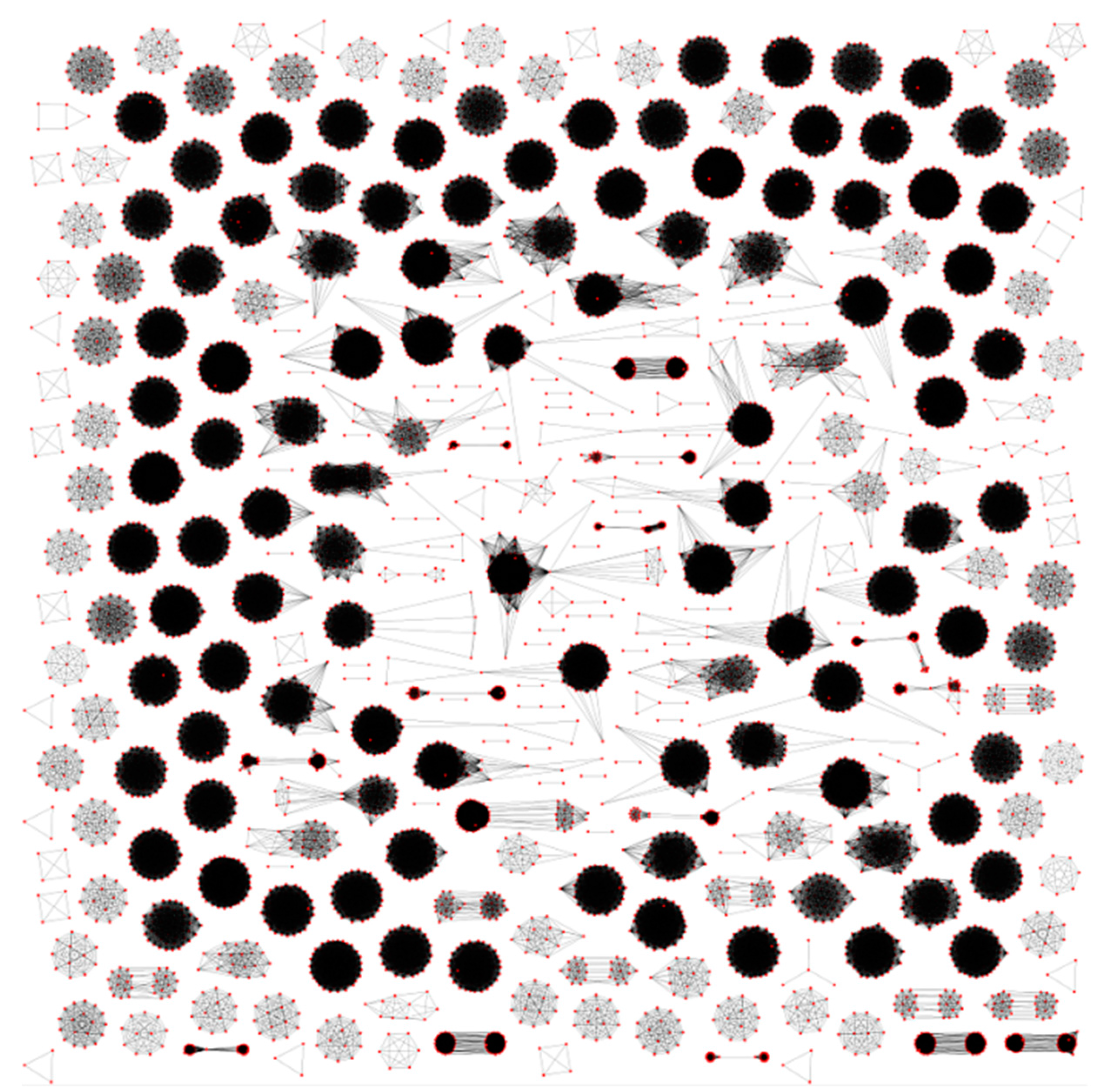
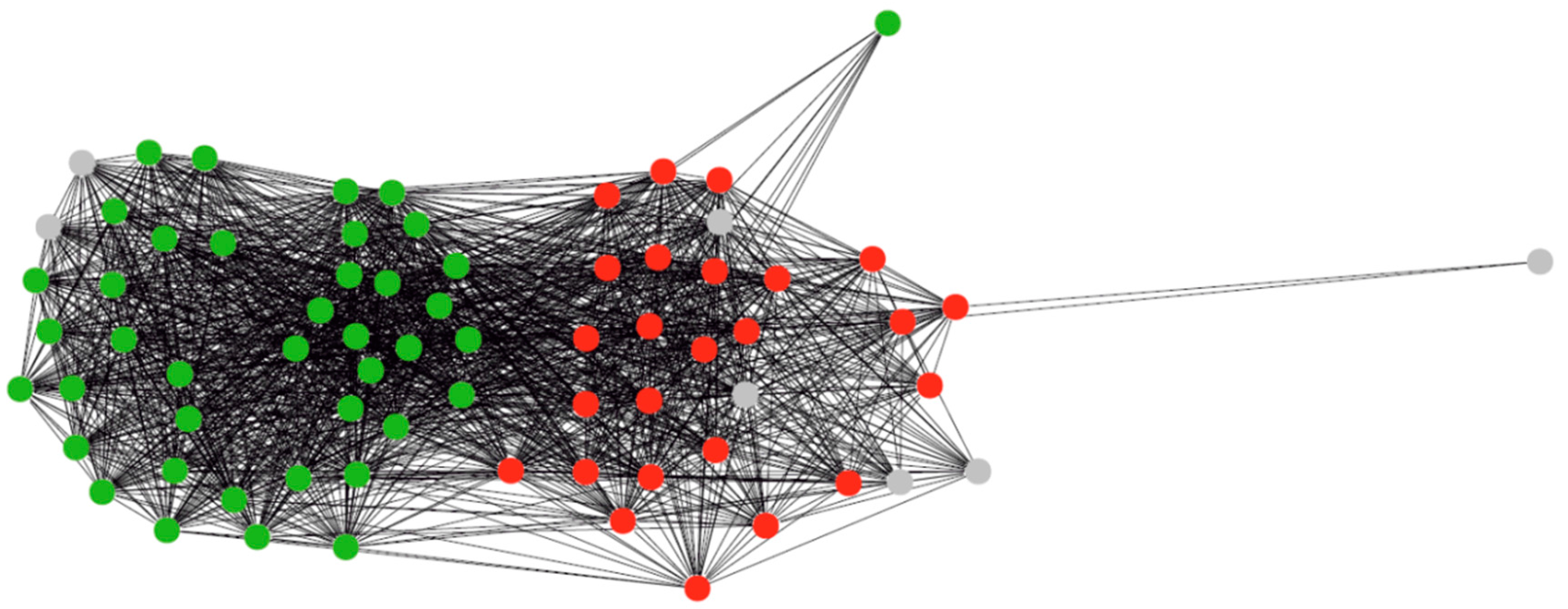
3.1. Clustering of Proteins

We have clustered proteins encoded in the plastids of the rhodophytic branch. The results are publicly available at http://lab6.iitp.ru/ppc/redline72/. The database functions allow rapid cluster identification by either a fragment of a protein amino acid sequence or by a protein phylogenetic profile.
The total number of proteins is 9286; the number of singletons is 265; and the number of clusters is 305. The number of clusters including exactly n proteins in a particular species and no more than n proteins in any species is referred to as PC(n). For this clustering, PC(1) = 223, PC(2) = 79, PC(3) = 2, and PC(4) = 1. Some general data about the clusters are given in Table 1.
The relationship between the number of clusters and the number of species in them is shown in Figure 1. Only seven clusters are represented in all considered plastids: six ribosomal proteins L2, L14, L16, S3, S11, and S12 as well as RNA polymerase beta subunit (RpoB). The genes odpA (pdhA), odpB (pdhB), trpA, trpG, tilS (ycf62), and infC are specific for all plastids in the considered Rhodophyta species. The tree of apicoplasts and plastids of photosynthetic Chromera velia and Chromerida sp. generated from the concatenated multiple alignment of proteins is shown in Figure 2.
Figure 1. Dependence of the number of clusters on the number of species represented in it.
Life 06 00007 g001
Figure 2. Tree of apicoplasts. Chromera velia and Chromerida sp. RM11 plastids were used as the outgroup.
Life 06 00007 g002
Figure 3 exemplifies a sparse graph G for our data. Many connected components are high density or even cliques. The graph contains 9072 non-isolated nodes and 223,377 edges; 245 isolated nodes (singletons) in it are due to the absence of bidirectional best hits for the corresponding proteins, and only 20 singletons were added by the algorithm during tree partitioning. The number of connected components of the sparse graph excluding singletons is 33 less than the ultimate number of clusters generated by the algorithm. Finding them is a non-trivial result of our algorithm.
Figure 3. Connectivity components of the sparse graph of proteins. Red dots represent proteins and lines represent bidirectional BLAST hits.
Life 06 00007 g003
Let us specify the following connected components of the sparse graph that are partitioned into smaller clusters by our algorithm: ApcA+ApcD, ApcB+ApcF, ApcE+CpcG, AtpA+AtpB, CarA+TrpG, CbbX(CfxQ)+FtsH+Ycf46, ChlB+ChlN, CpcB+CpeB, InfB+TufA, OdpA+IlvB, PetJ+PsbV, PsaA+PsaB, PsbA+PsbD, PsbB+PsbC, PsbK+Rpl20, RpoC1+RpoC2+RpoC2B, Rps4+Ycf24(SufB), Rpl22+Ycf88, Ycf3+Ycf37, Ycf27(OmpR)+Ycf29, and Ycf60+Ycf90. Each connected component here is denoted by a typical protein name; non-orthologous proteins are separated by the plus sign.

3.2. Regulons of Transcription Factors Encoded by Plastids

As compared to our previous data [35], the clusters of the MoeB and Ycf28 proteins were both supplemented by proteins encoded in the plastids of Vertebrata lanosa; neither of these proteins is encoded in plastids of Choreocolax polysiphoniae or any species beyond Rhodophyta. The profile identical to that of MoeB and Ycf28 was found in the proteins encoded by the apcA, apcB, apcD, apcE, apcF, carA, cpcA, cpcB, cpcG, gltB, nblA (ycf18), preA, and rpl28 genes; however, their 5’-leader sequences lack the conserved site found upstream of the moeB genes instead of the typical -35 promoter box.
The transcription factor Ycf29 is encoded in plastids of cryptomonads and rhodophytic algae except Porphyridium purpureum. The Ycf29 proteins are listed in Table 2. In the sparse graph, the Ycf29 and Ycf27 (OmpR) proteins belonged to the same connected component but were separated after clustering by our algorithm, which corresponds to the NCBI annotation. No other proteins with such phylogenetic profile have been identified. A similar profile was observed for the CemA protein found in Porphyridium purpureum but not in Choreocolax polysiphoniae. CemA includes the PF03040 domain and was localized to the inner face of the outer membrane in chloroplasts but not to the thylakoid membrane. Cyanobacterial proteins orthologous to CemA are involved in carbon dioxide transport but are not transporters [43]. The membrane protein Ycf19 also has a similar phylogenetic profile. A sequence close to the consensus of the conserved bacterial-type promoter was found upstream of the ycf19 gene. Since Ycf29 is a part of the two-component signaling system, its regulon is linked to the response to environmental rather than intraplastid changes. The Ycf19 and Ycf89 proteins are not partitioned with the clustering parameters used. At the same time, the proteins listed in Ycf19 annotations together with several related proteins constitute a dense subgraph. The graph of proteins Ycf19 and Ycf89 generated by the algorithm is shown in Figure 4.
Figure 4. Graph of Ycf19 and Ycf89 proteins. Colors indicate the proteins annotation: red = Ycf19, green = Ycf89, gray = no name specified.
Life 06 00007 g004
Table 2. Ycf29 proteins encoded in the plastids of the rhodophytic branch.
AccessionSourceProtein Description
YP_007878178.1Calliarthron tuberculosumconserved hypothetical plastid protein
YP_007627336.1Chondrus crispusconserved hypothetical plastid protein
YP_009122074.1Choreocolax polysiphoniaehypothetical protein
YP_003359295.1Cryptomonas parameciumTctD-like protein
NP_849011.1Cyanidioschyzon merolaeompR-like transcriptional regulator
NP_045122.1Cyanidium caldariumregulatory component of sensory transduction system
YP_009051025.1Galdieria sulphurariaputative transcriptional regulator LuxR
YP_009019567.1Gracilaria salicorniatctD transcriptional regulator
YP_063559.1Gracilaria tenuistipitatatctD transcriptional regulator
YP_008144796.1Grateloupia taiwanensisputative transcriptional regulator Ycf29
NP_050668.1Guillardia thetatctD homolog
NP_053953.1Porphyra purpureaORF29
YP_007947873.1Pyropia haitanensishypothetical chloroplast protein 29
YP_009027627.1Pyropia perforatahypothetical chloroplast protein 29
YP_537024.1Pyropia yezoensishypothetical chloroplast protein 29
YP_001293481.1Rhodomonas salinaTctD-like protein
YP_009159161.1Teleaulax amphioxeiaTctD-like protein
YP_009122313.1Vertebrata lanosahypothetical protein

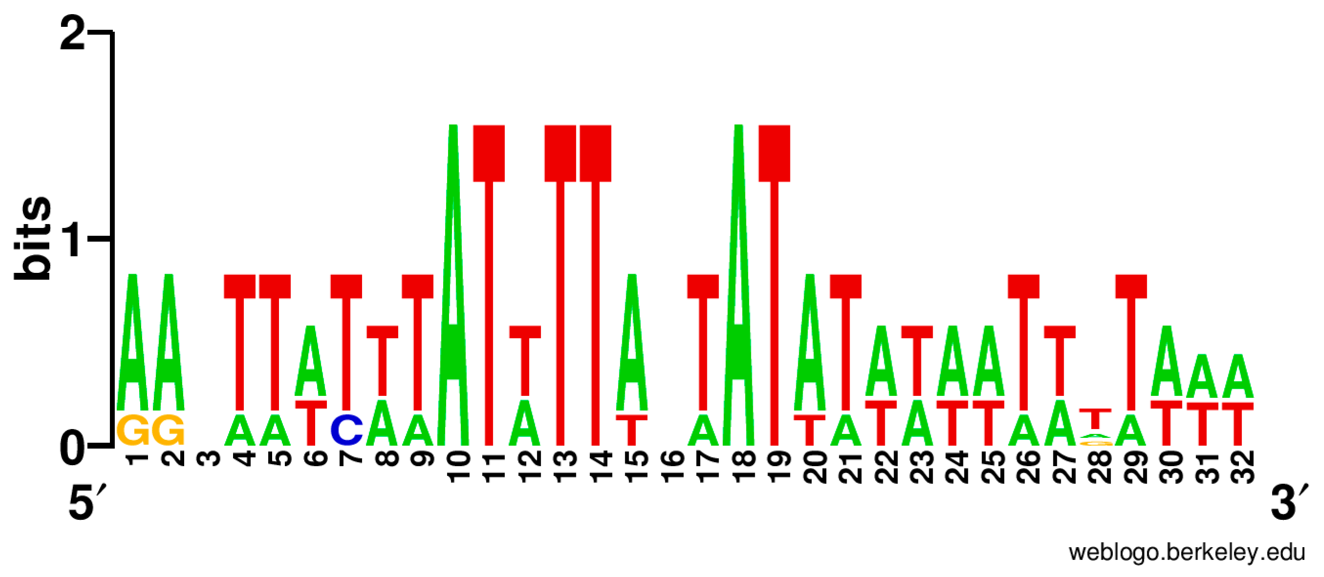
3.3. Regulation of Ycf24 (SufB) Translation Initiation

A conserved site was found in the 5’-untranslated region of ycf24 (sufB) in Eimeria tenella, Cyclospora cayetanensis, Toxoplasma gondii RH, Leucocytozoon caulleryi, Plasmodium chabaudi, and Porphyra purpurea. The sequence logo of this site is shown in Figure 5.
Figure 5. Sequence LOGO of the putative site in the 5’-untranslated region of ycf24 (sufB).
Life 06 00007 g005

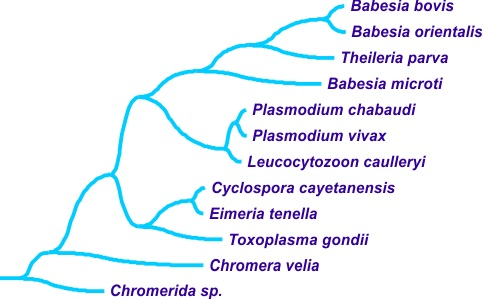
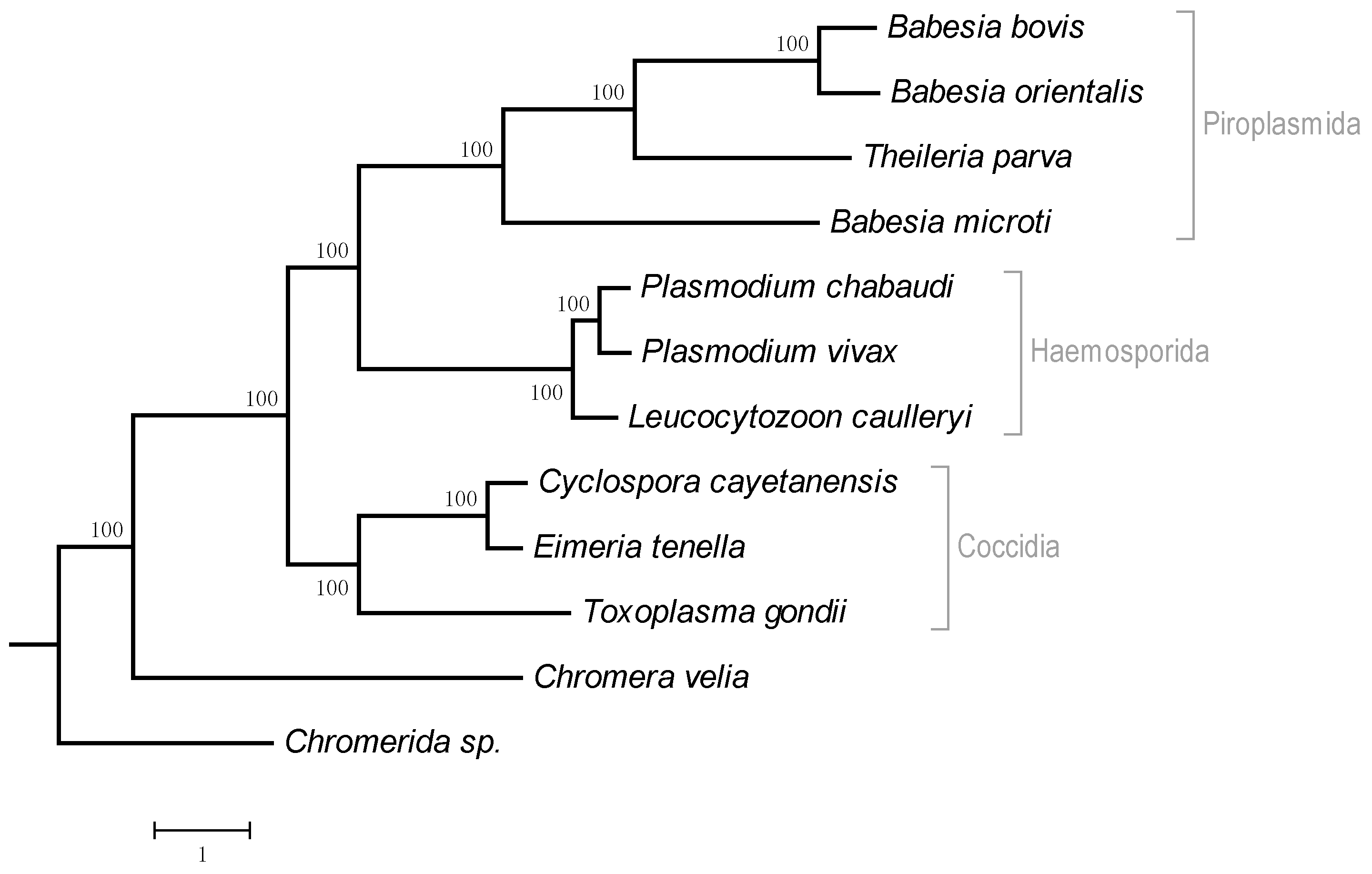
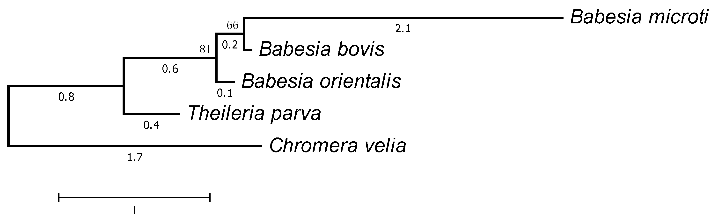
3.4. Regulation of Translation Initiation in Babesia spp. and Theileria parva

The genes from plastids of the Piroplasmida order lying between the rpl14 and rps8 are of particular interest. Although one such gene codes for the ribosomal protein L5 in many plastid-bearing algae of the rhodophytic branch, rpl14 and rps8 are neighboring genes in Coccidia and Haemosporida. The functional identification of the protein encoded by the gene lying between rpl14 and rps8 is questionable. Our clustering in Babesia bovis, Babesia orientalis, and Theileria parva suggests that this gene codes for the ribosomal protein L5, which belongs to a large cluster. In Babesia microti, this protein forms a singleton but is also annotated as L5. At the same time, it is only marginally similar to ribosomal proteins according to Pfam. The tree of these proteins is shown in Figure 6, and the proteins are listed in Table 3.
Conserved sites were identified in the leader regions 170–100 nt upstream of such genes in Piroplasmida. In Babesia spp., such sites reside within the coding sequence of rpl14. However, there was an insertion near the site, which is missing in orthologous L14 proteins. The sequence of this insertion is TSYSIDDRNRFKD in Babesia bovis. In Theileria parva, the site is not overlapped by the coding sequences. The corresponding transcription factor remains unknown in this case.
Figure 6. The tree of proteins encoded by the plastid genes located between the rpl14 and rps8 genes in Babesia spp. and Theileria parva. The plastid protein L5 from Chromera velia was used as the outgroup.
Life 06 00007 g006
Table 3. The plastid proteins in Piroplasmida with a marginal similarity with the ribosomal protein L5 discussed in Section 3.4 and Section 4.4 are shown.
AccessionSourceProtein Description
YP_002290869.1Babesia bovishypothetical protein
CDR32594.1Babesia microtiribosomal protein L5
YP_009170371.1Babesia orientalisribosomal protein L5
XP_762679.1Theileria parvahypothetical protein

4. Discussion

4.1. Protein Clustering

Overall, the data obtained indicate a good agreement between the clustering of plastid-encoded proteins performed by our algorithm and published data on the protein and species evolution. The proposed clustering algorithm and its software implementation are applicable to a wide range of problems related to graphs.
The clustering pattern of proteins encoded in red algal plastids demonstrate a substantial distance of Porphyridium purpureum from other species, which is accompanied by multiple DNA rearrangements in Rhodophyta plastids [44]; in addition, it demonstrates the separation of the Cyanidiaceae family including Galdieria sulphuraria, Cyanidium caldarium, and Cyanidioschyzon merolae.

4.2. Regulons of Plastid-Encoded Transcription Factors Ycf28, Ycf29, and Ycf30

The coincidence of the phylogenetic profiles of Ycf28 and MoeB reported previously [35] has been confirmed. The Ycf28 protein demonstrates a significant similarity with the cyanobacterial transcription factor NtcA. Consequently, we propose that Ycf28 is the factor that controls the transcription of the moeB gene by binding the DNA region near the promoter where the conserved motif was identified. There are no grounds to believe that Ycf28 is related to nitrogen metabolism, which assumes a change of the transcription factor specificity relative to cyanobacteria contrary to the previous proposal [45]. The absence of the typical -35 promoter box upstream of the moeB gene indicates that Ycf28 is a transcription activator.
The presence of Ycf29 in the plastid genomes of non-photosynthetic Cryptomonas paramecium and Choreocolax polysiphoniae indicates that this protein regulates processes related to photosynthesis. One can assume that Ycf19 orthologs include proteins in the large cluster combining Ycf19 and Ycf89 that are encoded in plastids together with the Ycf29 factor. This allows us to refine protein clustering and, at the same time, to identify the putative photosynthesis-independent regulation.
Plastids of many algal species are known to encode the transcription factor Ycf30, which controls the expression of the rbcLS genes coding for subunits of ribulose-bisphosphate carboxylase (EC 4.1.1.39) as well as of the cbbX gene. Light-induced transcriptional activation was experimentally demonstrated and the Ycf30-binding motif was identified in these genes in plastids isolated from Cyanidioschyzon merolae [46]. Our phylogenetic profiles of these proteins agree with these data. However, the variability of Ycf30-binding site complicates its unambiguous identification in the DNA sequence. The sequence variability of experimentally confirmed Ycf30-binding site suggests that the factor binding to DNA largely depends on the DNA curvature [47] or electrostatic potential along the DNA [48] rather than on the nucleotide context.

4.3. Regulation of Ycf24 (SufB) Translation Initiation

The same regulation found in red algae, Coccidia, and Haemosporida supports the common origin of all apicoplasts from red algal plastids. Moreover, early separation of these apicomplexan groups naturally suggests that Cryptosporidium spp. and Gregarina niphandrodes lost their apicoplasts in the course of evolution but the common ancestor of apicomplexans had apicoplasts.
Moreover, the site identical to that upstream of ycf24 was found in the 5’-untranslated region of rps4 of Toxoplasma gondii [3]. This indicates possible the common regulation of translation in the apicoplast.

4.4. Regulation of Translation Initiation in Babesia spp. and Theileria parva

We believe that the gene coding for the ribosomal protein L5 was eliminated from the apicoplast in the ancestor of apicomplexan parasites, and a new gene was inserted into this chromosomal locus in the ancestor of Piroplasmida. The recognition of a new type of proteins is confirmed by the analysis of their 5’-leader regions, where conserved sites were identified. Indeed, it is natural to assume that a conserved site is involved in the regulation of gene expression, and the same expression pattern indicates a common functional significance of the corresponding proteins.

5. Conclusions

We have made a publicly available web service for protein identification by their phylogenetic profile. To our knowledge, no other services for the identification of plastid-encoded proteins by their phylogenetic profile (the two lists of species) are available. Our method allowed us to confirm the previous assumption concerning the regulation of plastid gene expression in the rhodophytic branch. In particular, our results confirm the hypothesis that apicoplasts in the common ancestor of apicomplexans descend from red algal plastids.

Acknowledgments

The research has been carried out at the Institute for Information Transmission Problems of The Russian Academy of Sciences at the expense of the Russian Science Foundation, project no. 14-50-00150.

Author Contributions

Vassily Alexandrovich Lyubetsky, Oleg Anatolyevich Zverkov, and Alexandr Vladislavovich Seliverstov conceived and designed this research; Oleg Anatolyevich Zverkov and Alexandr Vladislavovich Seliverstov contributed algorithm design and analyzed the data; Alexandr Vladislavovich Seliverstov and Vassily Alexandrovich Lyubetsky wrote the paper. They all have read and approved the final manuscript.

Conflicts of Interest

The authors declare no conflict of interest.

References

  1. Aboulaila, M.; Munkhjargal, T.; Sivakumar, T.; Ueno, A.; Nakano, Y.; Yokoyama, M.; Yoshinari, T.; Nagano, D.; Katayama, K.; El-Bahy, N.; et al. Apicoplast-targeting antibacterials inhibit the growth of Babesia parasites. Antimicrob. Agents Chemother. 2012, 56, 3196–3206. [Google Scholar] [CrossRef] [PubMed]
  2. Li, B.; Lopes, J.S.; Foster, P.G.; Embley, T.M.; Cox, C.J. Compositional Biases among Synonymous Substitutions Cause Conflict between Gene and Protein Trees for Plastid Origins. Mol. Biol. Evol. 2014, 31, 1697–1709. [Google Scholar] [CrossRef] [PubMed]
  3. Sadovskaya, T.A.; Seliverstov, A.V. Analysis of the 5′-leader regions of several plastid genes in Protozoa of the phylum Apicomplexa and red algae. Mol. Biol. 2009, 43, 552–556. [Google Scholar] [CrossRef]
  4. Janouškovec, J.; Horak, A.; Oborník, M.; Lukeš, J.; Keeling, P.J. A common red algal origin of the apicomplexan, dinoflagellate, and heterokont plastids. Proc. Natl. Acad. Sci. USA 2010, 107, 10949–10954. [Google Scholar] [CrossRef] [PubMed]
  5. Imanian, B.; Pombert, J.F.; Keeling, P.J. The complete plastid genomes of the two “dinotoms” Durinskia baltica and Kryptoperidinium foliaceum. PLoS ONE 2010, 5, e10711. [Google Scholar] [CrossRef] [PubMed]
  6. Kohler, S.; Delwiche, C.F.; Denny, P.W.; Tilney, L.G.; Webster, P.; Wilson, R.J.; Palmer, J.D.; Roos, D.S. A plastid of probable green algal origin in Apicomplexan parasites. Science 1997, 275, 1485–1489. [Google Scholar] [CrossRef] [PubMed]
  7. Lau, A.O.; McElwain, T.F.; Brayton, K.A.; Knowles, D.P.; Roalson, E.H. Babesia bovis: A comprehensive phylogenetic analysis of plastid-encoded genes supports green algal origin of apicoplasts. Exp. Parasitol. 2009, 123, 236–243. [Google Scholar] [CrossRef] [PubMed]
  8. Oborník, M.; Lukeš, J. The Organellar Genomes of Chromera and Vitrella, the Phototrophic Relatives of Apicomplexan Parasites. Annu. Rev. Microbiol. 2015, 69, 129–144. [Google Scholar] [CrossRef] [PubMed]
  9. Salomaki, E.D.; Nickles, K.R.; Lane, C.E. The ghost plastid of Choreocolax polysiphoniae. J. Phycoly 2015, 51, 217–221. [Google Scholar] [CrossRef]
  10. Zhu, G.; Marchewka, M.J.; Keithly, J.S. Cryptosporidium parvum appears to lack a plastid genome. Microbiology 2000, 146, 315–321. [Google Scholar] [CrossRef] [PubMed]
  11. Toso, M.A.; Omoto, C.K. Gregarina niphandrodes may lack both a plastid genome and organelle. J. Eukaryot. Microbiol. 2007, 54, 66–72. [Google Scholar] [CrossRef] [PubMed]
  12. Simdyanov, T.G.; Diakin, A.Y.; Aleoshin, V.V. Ultrastructure and 28S rDNA phylogeny of two gregarines: Cephaloidophora cf. communis and Heliospora cf. longissima with remarks on gregarine morphology and phylogenetic analysis. Acta Protozool. 2015, 54, 241–263. [Google Scholar]
  13. Huang, Y.; He, L.; Wu, W.; He, P.; He, W.J.; Yu, L.; Malobi, N.; Zhou, Q.Y.; Shen, B.; Zhao, L.J. Characterization and annotation of Babesia orientalis apicoplast genome. Parasit. Vectors 2015, 8. [Google Scholar] [CrossRef] [PubMed]
  14. Garg, A.; Stein, A.; Zhao, W.; Dwivedi, A.; Frutos, R.; Cornillot, E.; ben Mamoun, C. Sequence and annotation of the apicoplast genome of the human pathogen Babesia microti. PLoS ONE 2014, 9. [Google Scholar] [CrossRef]
  15. Brayton, K.A.; Lau, A.O.; Herndon, D.R.; Hannick, L.; Kappmeyer, L.S.; Berens, S.J.; Bidwell, S.L.; Brown, W.C.; Crabtree, J.; Fadrosh, D.; et al. Genome sequence of Babesia bovis and comparative analysis of apicomplexan hemoprotozoa. PLoS Pathog. 2007, 3, 1401–1413. [Google Scholar] [CrossRef] [PubMed]
  16. Gardner, M.J.; Bishop, R.; Shah, T.; de Villiers, E.P.; Carlton, J.M.; Hall, N.; Ren, Q.; Paulsen, I.T.; Pain, A.; Berriman, M.; et al. Genome sequence of Theileria parva, a bovine pathogen that transforms lymphocytes. Science 2005, 309, 134–137. [Google Scholar] [CrossRef] [PubMed]
  17. Cai, X.; Fuller, A.L.; McDougald, L.R.; Zhu, G. Apicoplast genome of the coccidian Eimeria tenella. Gene 2003, 321, 39–46. [Google Scholar] [CrossRef] [PubMed]
  18. Tang, K.; Guo, Y.; Zhang, L.; Rowe, L.A.; Roellig, D.M.; Frace, M.A.; Li, N.; Liu, S.; Feng, Y.; Xiao, L. Genetic similarities between Cyclospora cayetanensis and cecum-infecting avian Eimeria spp. in apicoplast and mitochondrial genomes. Parasit. Vectors 2015, 8. [Google Scholar] [CrossRef] [PubMed]
  19. Imura, T.; Sato, S.; Sato, Y.; Sakamoto, D.; Isobe, T.; Murata, K.; Holder, A.A.; Yukawa, M. The apicoplast genome of Leucocytozoon caulleryi, a pathogenic apicomplexan parasite of the chicken. Parasitol. Res. 2014, 113, 823–828. [Google Scholar] [CrossRef] [PubMed]
  20. Sato, S.; Sesay, A.K.; Holder, A.A. The unique structure of the apicoplast genome of the rodent malaria parasite Plasmodium chabaudi chabaudi. PLoS ONE 2013, 8. [Google Scholar] [CrossRef]
  21. Reith, M.E.; Munholland, J. Complete nucleotide sequence of the Porphyra purpurea chloroplast. Plant Mol. Biol. Rep. 1995, 13, 333–335. [Google Scholar] [CrossRef]
  22. Seliverstov, A.V.; Lysenko, E.A.; Lyubetsky, V.A. Rapid evolution of promoters for the plastome gene ndhF in flowering plants. Russ. J. Plant Physiol. 2009, 56, 838–845. [Google Scholar] [CrossRef]
  23. Lyubetsky, V.A.; Rubanov, L.I.; Seliverstov, A.V. Lack of conservation of bacterial type promoters in plastids of Streptophyta. Biol. Direct. 2010, 5. [Google Scholar] [CrossRef] [PubMed]
  24. Homann, A.; Link, G. DNA-binding and transcription characteristics of three cloned sigma factors from mustard (Sinapis alba L.) suggest overlapping and distinct roles in plastid gene expression. Eur. J. Biochem. 2003, 270, 1288–1300. [Google Scholar] [CrossRef] [PubMed]
  25. Hu, Y. Efficient high-quality force-directed graph drawing. Math. J. 2006, 10, 37–71. [Google Scholar]
  26. Crooks, G.E.; Hon, G.; Chandonia, J.M.; Brenner, S.E. WebLogo: A sequence logo generator. Genome Res. 2004, 14, 1188–1190. [Google Scholar] [CrossRef] [PubMed]
  27. Tamura, K.; Stecher, G.; Peterson, D.; Filipski, A.; Kumar, S. MEGA6: Molecular Evolutionary Genetics Analysis Version 6.0. Mol. Bio.l Evol. 2013, 30, 2725–2729. [Google Scholar] [CrossRef] [PubMed]
  28. Page, R.D.M. TreeView: An application to display phylogenetic trees on personal computers. Comput. Appl. Biosci. 1996, 12, 357–358. [Google Scholar] [PubMed]
  29. Finn, R.D.; Bateman, A.; Clements, J.; Coggill, P.; Eberhardt, R.Y.; Eddy, S.R.; Heger, A.; Hetherington, K.; Holm, L.; Mistry, J.; et al. The Pfam protein families database. Nucleic Acids Res. 2014, 42, D222–D230. [Google Scholar] [CrossRef] [PubMed]
  30. Edgar, R.C. MUSCLE: Multiple sequence alignment with high accuracy and high throughput. Nucleic Acids Res. 2014, 32, 1792–1797. [Google Scholar] [CrossRef] [PubMed]
  31. Stamatakis, A. RAxML version 8: A tool for phylogenetic analysis and post-analysis of large phylogenies. Bioinformatics 2014, 30, 1312–1313. [Google Scholar] [CrossRef] [PubMed]
  32. Lyubetsky, V.A.; Seliverstov, A.V.; Zverkov, O.A. Elaboration of the homologous plastid-encoded protein families that separate paralogs in Magnoliophytes. Math. Biol. Bioinform. 2013, 8, 225–233. (in Russian). [Google Scholar] [CrossRef]
  33. Zverkov, O.A.; Seliverstov, A.V.; Lyubetsky, V.A. Plastid-encoded protein families specific for narrow taxonomic groups of algae and protozoa. Mol. Biol. 2012, 46, 717–726. [Google Scholar] [CrossRef]
  34. Lyubetsky, V.; Seliverstov, A.; Zverkov, O. Transcription regulation of plastid genes involved in sulfate transport in Viridiplantae. BioMed Res. Int. 2013, 2013. [Google Scholar] [CrossRef] [PubMed]
  35. Zverkov, O.A.; Seliverstov, A.V.; Lyubetsky, V.A. A database of plastid protein families from red algae and Apicomplexa and expression regulation of the moeB gene. BioMed Res. Int. 2015, 2015. [Google Scholar] [CrossRef] [PubMed]
  36. Van Dongen, S. Graph clustering via a discrete uncoupling process. SIAM J. Matrix Anal. Appl. 2008, 30, 121–141. [Google Scholar] [CrossRef]
  37. Galashov, A.E.; Kel’manov, A.V. A 2-approximate algorithm to solve one problem of the family of disjoint vector subsets. Autom. Remote Control. 2014, 75, 595–606. [Google Scholar] [CrossRef]
  38. Kel'manov, A.V.; Khamidullin, S.A. An approximation polynomial-time algorithm for a sequence bi-clustering problem. Comp. Math. Math. Phys. 2015, 55, 1068–1076. [Google Scholar] [CrossRef]
  39. Kel'manov, A.V.; Khandeev, V.I. A randomized algorithm for two-cluster partition of a set of vectors. Comp. Math. Math. Phys. 2015, 55, 330–339. [Google Scholar] [CrossRef]
  40. Kel’manov, A.V.; Romanchenko, S.M. An FPTAS for a vector subset search problem. J. Appl. Ind. Math. 2014, 8, 329–336. [Google Scholar] [CrossRef]
  41. Kel’manov, A.V.; Khamidullin, S.A. An approximating polynomial algorithm for a sequence partitioning problem. J. Appl. Ind. Math. 2014, 8, 236–244. [Google Scholar] [CrossRef]
  42. Kruskal, J.B. On the Shortest Spanning Subtree of a Graph and the Traveling Salesman Problem. Proc. Am. Math. Soc. 1956, 7, 48–50. [Google Scholar] [CrossRef]
  43. Katoh, A.; Lee, K.S.; Fukuzawa, H.; Ohyama, K.; Ogawa, T. cemA homologue essential to CO2 transport in the cyanobacterium Synechocystis PCC6803. Proc. Natl. Acad. Sci. USA 1996, 93, 4006–4010. [Google Scholar] [CrossRef] [PubMed]
  44. Bhattacharya, D.; Price, D.C.; Chan, C.X.; Qiu, H.; Rose, N.; Ball, S.; Weber, A.P.; Arias, M.C.; Henrissat, B.; Coutinho, P.M.; et al. Genome of the red alga Porphyridium purpureum. Nat. Commun. 2013, 4. [Google Scholar] [CrossRef] [PubMed]
  45. Lopatovskaya, K.V.; Seliverstov, A.V.; Lyubetsky, V.A. NtcA and NtcB regulons in cyanobacteria and rhodophyta chloroplasts. Mol. Biol. 2011, 45, 522–526. [Google Scholar] [CrossRef]
  46. Minoda, A.; Weber, A.P.; Tanaka, K.; Miyagishima, S.Y. Nucleus-independent control of the rubisco operon by the plastid-encoded transcription factor Ycf30 in the red alga Cyanidioschyzon merolae. Plant Physiol. 2010, 154, 1532–1540. [Google Scholar] [CrossRef] [PubMed]
  47. Kozobay-Avraham, L.; Hosid, S.; Bolshoy, A. Involvement of DNA curvature in intergenic regions of prokaryotes. Nucleic Acids Res. 2006, 34, 2316–2327. [Google Scholar] [CrossRef] [PubMed]
  48. Kamzolova, S.G.; Sorokin, A.A.; Dzhelyadin, T.R.; Beskaravainy, P.M.; Osypov, A.A. Electrostatic potentials of E. coli genome DNA. J. Biomol. Struct. Dyn. 2005, 23, 341–346. [Google Scholar] [PubMed]

