Article

Selection and Characterization of Single Domain Antibodies Specific for Bacillus anthracis Spore Proteins

1
Naval Research Laboratory, Center for Bio/Molecular Science and Engineering, Washington, DC 20375, USA
2
Nova Research Inc., Alexandria, VA 22308, USA
*
Authors to whom correspondence should be addressed.
Antibodies 2013, 2(1), 152-167; https://doi.org/10.3390/antib2010152
Submission received: 30 January 2013 / Revised: 28 February 2013 / Accepted: 7 March 2013 / Published: 15 March 2013
(This article belongs to the Special Issue Single-Domain Antibody)

Abstract

:
To obtain thermostable immunoreagents specific for the spore form of Bacillus anthracis two llamas were immunized with a combination of six different recombinant proteins. These proteins BclA, gerQ, SODA1, SOD15, BxpB and the protein p5303 have all been shown as components of the B. anthracis spore and could potentially serve as targets for the detection of spores in multiplexed biosensors. Peripheral blood lymphocytes were used to construct a phage display library from which single domain antibodies (sdAbs) targeting each of the proteins were isolated. Unique sdAbs exhibiting nanomolar or better affinities for the recombinant proteins were obtained and most of the isolated sdAbs retained their ability to bind antigen after cycles of heating as determined by enzyme linked immunosorbent assay (ELISA). SdAbs targeting the BclA and gerQ proteins were able to successfully detect bacterial spores, whether broken or intact, using a direct ELISA; the sdAbs were specific, showing binding only to B. anthracis spores and not to other Bacillus species. Additionally, SODA1 and p5303 binding sdAbs detected spores in sandwich assays serving as both captures and tracers. Used in combination, sdAbs targeting B. anthracis proteins could be integrated into emerging biosensors to improve specificity in multiplex assays.

1. Introduction

Bacillus anthracis, the causative agent of anthrax, has been considered a bioweapon of grave concern since long before the letter-based attacks of 2001. The ease of production, dissemination, environmental persistence, and resultant lethality of the bacterial spore has led to anthrax’s significant historical past with bioweapons programs of several nations and an all too likely future with terrorist organizations [1,2]. The spores of B. anthracis, once aerosolized and dispersed, can remain dormant and viable for extended periods. Additionally, cleanup of contaminated areas currently requires harsh chemical agents and repeated treatments to ensure complete inactivation of the bacterial spores.
Bacillus anthracis is highly similar, morphologically, to other members of the Bacillus cereus group which complicates the development of highly specific assays for detection of pathogenic B. anthracis spores and vegetative cells [3]. Though discrimination of the closely related bacteria of the B. cereus group is possible with assays that utilize PCR [4,5,6], these techniques frequently require laboratory based equipment and skilled technicians. Immunoassays, such as lateral flow assays, offer advantages over PCR-based techniques in their simplicity, portability, and low cost allowing them to readily be deployed to the field. Immunoassays, however, must utilize antibodies that target proteins unique to B. anthracis spores and vegetative cells to be useful in detection assays.
Single domain antibodies (sdAbs) are recombinant antibodies derived from a novel class of immunoglobulins found in camelids referred to as heavy-chain only antibodies [7,8]. As the name implies, the parental antibody and its antigen binding domain is formed from only the heavy chain; a product of several mutations that eliminate interaction with the light chain subunit [9]. Sharks also possess heavy chain antibodies termed IgNAR from which a type of sdAb, often referred to as VNAR, can be derived [10,11]. To obtain high affinity sdAbs, usually one starts with an immunized host animal to permit in vivo affinity maturation prior to isolating the animal’s immune repertoire for further selection. Still, one typically requires a high-throughput screening method such as phage display to isolate target binding elements. SdAbs isolated following rounds of selection typically demonstrate high affinity and target specificity [12,13]. Additionally, though not a universal characteristic, most sdAbs exhibit the ability to refold into active detection elements following denaturation [14]. This is advantageous for their application in assays designed for use in more austere environments where cold chains may be limited or completely absent.
There are several reports of recombinantly expressed antibody binding domains for the specific detection of B. anthracis [15,16]; however, these binding elements target EA1, a vegetative protein that is often found as a contaminant in spore preparations. In this work sdAbs were isolated from immunized llama libraries against five proteins of the B. anthracis spore: BclA, gerQ (YwdL), SODA1, SOD15, and the formerly putative protein p5303 to develop thermal stable detection elements that could be used for the detection of bacterial spores. The sdAbs characterized in this study could be used in combination with one another or antibodies targeting other proteins of the spore or vegetative cell to improve discrimination of target and non-target bacterium while potentially eliminating false negatives that may arise from loss or modification of a single epitope.

2. Results and Discussion

2.1. Selection of sdAbs Targeting Spore Proteins

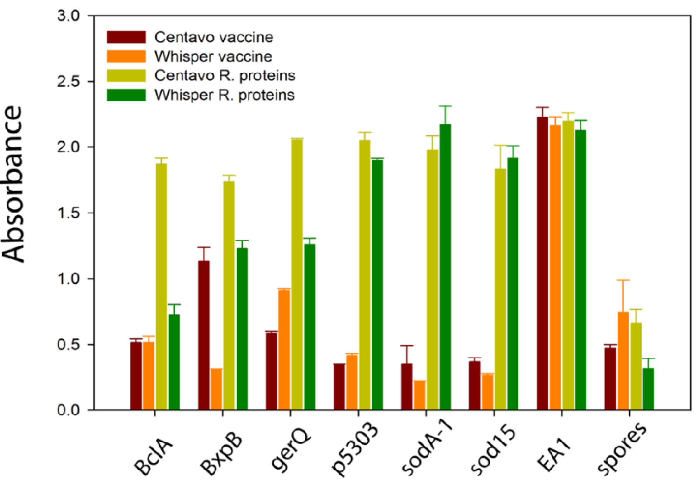
Two llamas, Whisper and Centavo, had been previously immunized with the standard veterinary vaccine for Bacillus anthracis (Sterne 34F2 strain; Colorado Serum Company). Binding sdAbs selected from the initial library constructed following immunization with commercial vaccine primarily recognized the S-layer protein EA1, as described by Walper et al. [15]. Although the isolated sdAbs were specific for B. anthracis, EA1 has been shown to be a spore contaminant and not an integral component of the bacterial spore [17]. The two llamas were subjected to a second series of immunizations with a combination of recombinant spore-specific proteins: BclA, gerQ, SODA1, SOD15, BxpB, and p5303 [18,19]. Centavo received a mix of all six proteins; while Whisper was immunized with a cocktail that did not include the BclA protein in an attempt to allow a stronger immune response toward the other spore proteins that may not be as immunodominant as the BclA antigen. Plasma retained during library construction was tested from both llamas against the recombinant spore proteins as well as the EA1 protein and bacterial spores in a direct binding ELISA to assess the host llama’s immune response to the antigens, Figure 1. As anticipated, both animals demonstrated a significantly stronger immune response to the spore specific proteins following immunization with the recombinant spore proteins. Also as expected, Centavo showed a stronger response to BclA than Whisper.
Figure 1. Binding of polyclonal antibodies from the plasma of two llamas, Centavo and Whisper, after immunization with the veterinary anthrax vaccine (red and orange) and after additional immunizations with recombinant B. anthracis spore proteins (yellow and green). Binding is shown for the six recombinant spore proteins, the EA1 S-layer protein, and B. anthracis spores.
Antibodies 02 00152 g001
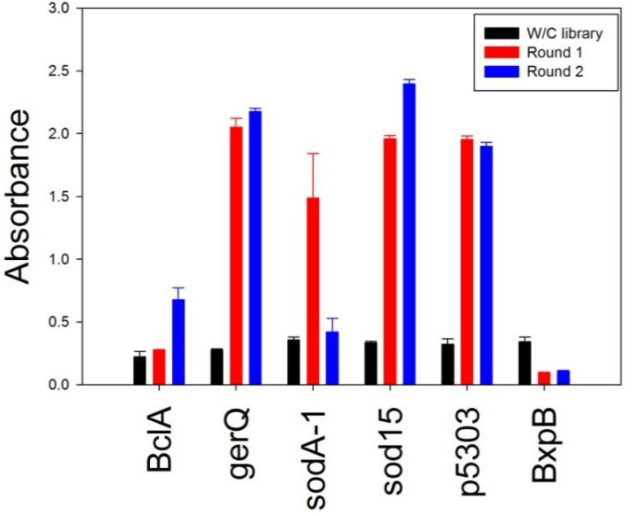
The sdAb library was constructed from RNA extracted from the peripheral blood lymphocytes using a two stage PCR as outlined by Gharhroudi et al. [20]. Selection of target binding sdAbs was conducted using a standard biopanning protocol; two rounds of selection proved sufficient to enrich for target-binding sdAbs in all cases save BxpB, as judged by polyclonal phage ELISA, Figure 2. Though analysis of the llama plasma suggested BxpB was immunogenic, biopanning did not enrich for binders to the target protein. Biopanning was repeated for BxpB and conducted for three rounds but with similar results (data not shown). The failure to isolate BxpB binding sdAbs, despite the fact that analysis of the plasma showed binding to BxpB could be due to low diversity of the library itself. Alternatively, it is possible that the llama’s immune response may be primarily conventional antibodies rather than heavy-chain only antibodies from which the sdAb are derived. Although not done for this work, purification of the heavy-chain only antibodies is possible and can be used to assess the potential of isolating sdAbs from the library [21].
Figure 2. Polyclonal phage ELISA using phage prepared from the initial library and after the first and second round of biopanning. Recombinant spore proteins were passively immobilized and an HRP-conjugated anti-M13 antibody was used to assess binding.
Antibodies 02 00152 g002
Individual clones from the second round of biopanning for each of the six target antigens were transferred to microtiter plates and assessed for their ability to bind target using a monoclonal phage ELISA. Clones were defined as positive if the signal generated for target antigen was at least twice background. Positive clones were sequenced and divided into families based on amino acid alignments of their complementarity determining regions (CDRs). The BclA binding sdAbs exhibited the greatest diversity of sequence with eleven unique families identified. There were seven families of sdAbs selected for binding to both the SODA1 and p5303 antigens, while sdAb selected towards the SOD15 and gerQ antigens had three and four families respectively. Representative sequences from each of the binding families are shown in Supplemental Figure 1. Though the response was minimal in the monoclonal phage ELISA (absorbance values less than two-fold greater than background), ten clones from the second round of biopanning against BxpB were also sequenced, however none were close enough in amino acid composition of the CDRs to be categorized within a defined family. These sdAbs also failed to express using a variety of vectors and expression strategies and therefore were not further characterized.

2.2. Characterization of sdAbs Targeting Spore Proteins

Representative clones from each sequence family were cloned into the pECAN45 expression vector and purified from the bacterial periplasm as detailed in Walper et al. [15]. Purified proteins were first tested via surface plasmon resonance (SPR) to identify those clones that recognize different epitopes on their target antigen. Despite differences in amino acid composition of the CDRs, a single epitope for each of the target antigens; or one of such proximity as to inhibit binding; was observed in all instances (data not shown). Although it is important to identify binding reagents that recognize different epitopes when performing sandwich assays for simple targets, bacterial spores are likely to have multiple copies of each of the target proteins so identifying proteins that recognize different regions is not as critical.
Following epitope evaluation, representative clones from each family were evaluated to determine binding kinetics. The majority of the clones tested showed nanomolar or better affinities for the recombinant proteins. Clones representing the two families with the highest binding affinities for each target protein were selected for further study; the binding kinetics for each is shown in Table 1.
Table 1. SdAb binding kinetics for recombinant proteins.
sdAb cloneKa (1/Ms)Kd (1/s)KD (M)
BclA A52.1E +051.2E -055.9E -11
BclA B73.1E +052.3E -048.7E -10
BclA H33.3E +055.4E -041.6E -9
gerQ B21.2E+068.7E-037.6E-09
gerQ G4 (to BclA)1.4E+052.8E+041.9E-09
SODA1 F34.5E +051.4E -033.1E -09
SODA1 G45.2E +051.5E -043.0E -10
SOD15 A63.4E +051.7E -035.1E -09
SOD15 D21.2E +051.8E -031.6E -08
p5303 A31.6E +065.6E -043.6E -10
p5303 B65.9E +055.5E -059.3E -11
p5303 F13.2E +068.2E -032.5E -09
Each of the sdAbs chosen for more complete evaluation was further characterized by circular dichroism (CD) to determine its melting temperature and ability to refold following thermal denaturation. Melting temperatures (Tm) ranged from 46–72 °C with the mean Tm calculated to be 62.4 °C for the sdAbs examined, Table 2. Though many sdAbs are able to regain their native structure following thermal denaturation, most of the sdAbs characterized via circular dichroism in this study appeared to demonstrate a propensity to misfold and aggregate during the cooling cycles. An example of an sdAb that shows good refolding on heat cycling, one that loses its ability to refold as it is heated and cooled, and one that failed to refold is shown in Figure 3, the data from all the sdAbs evaluated for this study is shown in Supplemental Figure 2. Several of the sdAbs did not re-fold following the initial thermal denaturation cycle as seen for all the p5303 and SOD15 binding sdAbs that were evaluated. One of the gerQ binders also showed a complete loss of structure (gerQ B2), one refolded with approximately 50% efficiency which decreases with each additional cycle (gerQ H2), and the third clone (gerQ G4) showed a 40% decrease in ellipticity following the initial cycle but appeared to refold with the same efficiency in subsequent rounds. Both of the SODA1 binders displayed an initial and consistent decrease in ellipticity with each successive round of thermal denaturation. Though BclA H3 was not able to refold after heating, both BclA A5 and BclA B7 showed significant thermal stability.
Table 2. SdAb melting temperatures determined using circular dichroism.
SdAb cloneTemperature (°C)
BclA A569
BclA B769
BclA H358
gerQ B249
gerQ G446
SODA1 F370
SODA1 G465
SOD15 A670
SOD15 D265
p5303 A364
p5303 B672
p5303 F162
Figure 3. Circular dichroism of representative sdAb showing the three refolding patterns observed. BclA B7 refolds after cycles of heating and cooling; SODA1 F3 loses ability to refold during cycles of heating, and BclA H3 loses all its secondary structure after the first heating cycle.
Antibodies 02 00152 g003
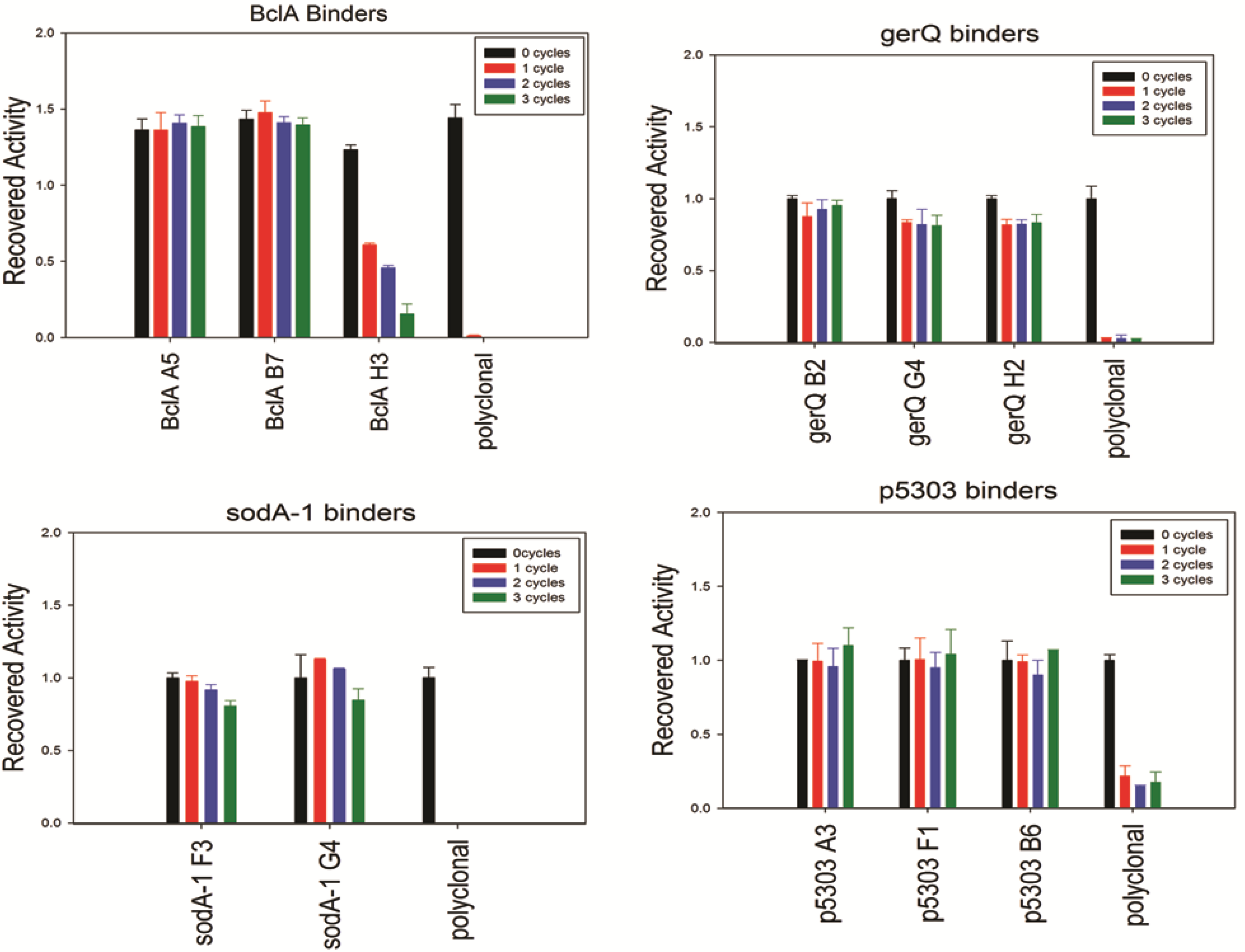
Though the CD analysis of each of the sdAbs showed significant variability in the capacity to regain secondary structure, each was tested for antigen binding capability following 0 to 3 rounds of thermal denaturation using a direct ELISA. Results are shown in Figure 4; the sdAbs specific for SODA1, GerQ, and both BclA A5 and BclA B7 all retained an undiminished ability to bind their target antigen. Thus, while CD analysis suggests that many of these sdAb permanently lose native structure upon thermal denaturation, the direct ELISA suggests that nonetheless a sufficient percentage remains active and is able to bind the target antigen. Due to the quantity of sdAb utilized in the ELISA assay and their high affinities greater than 90% inactivation may be required before an impact is noted. In exception to the other sdAbs examined, BclA H3 showed diminished signal after each heating cycle and both of the SOD15 sdAbs were unable to reproducibly bind antigen following heating (data not shown). In all assays, the conventional polyclonal antibody that was subject to identical heating shows a near-complete loss of its ability to bind antigen following a single cycle of heating and cooling.
Figure 4. Binding activity after iterative cycles of heating and cooling was assayed using a direct ELISA and biotinylated sdAbs. An HRP-conjugated streptavidin served as the secondary detection element.
Antibodies 02 00152 g004

2.3. Detection of Spores (Broken and Intact)

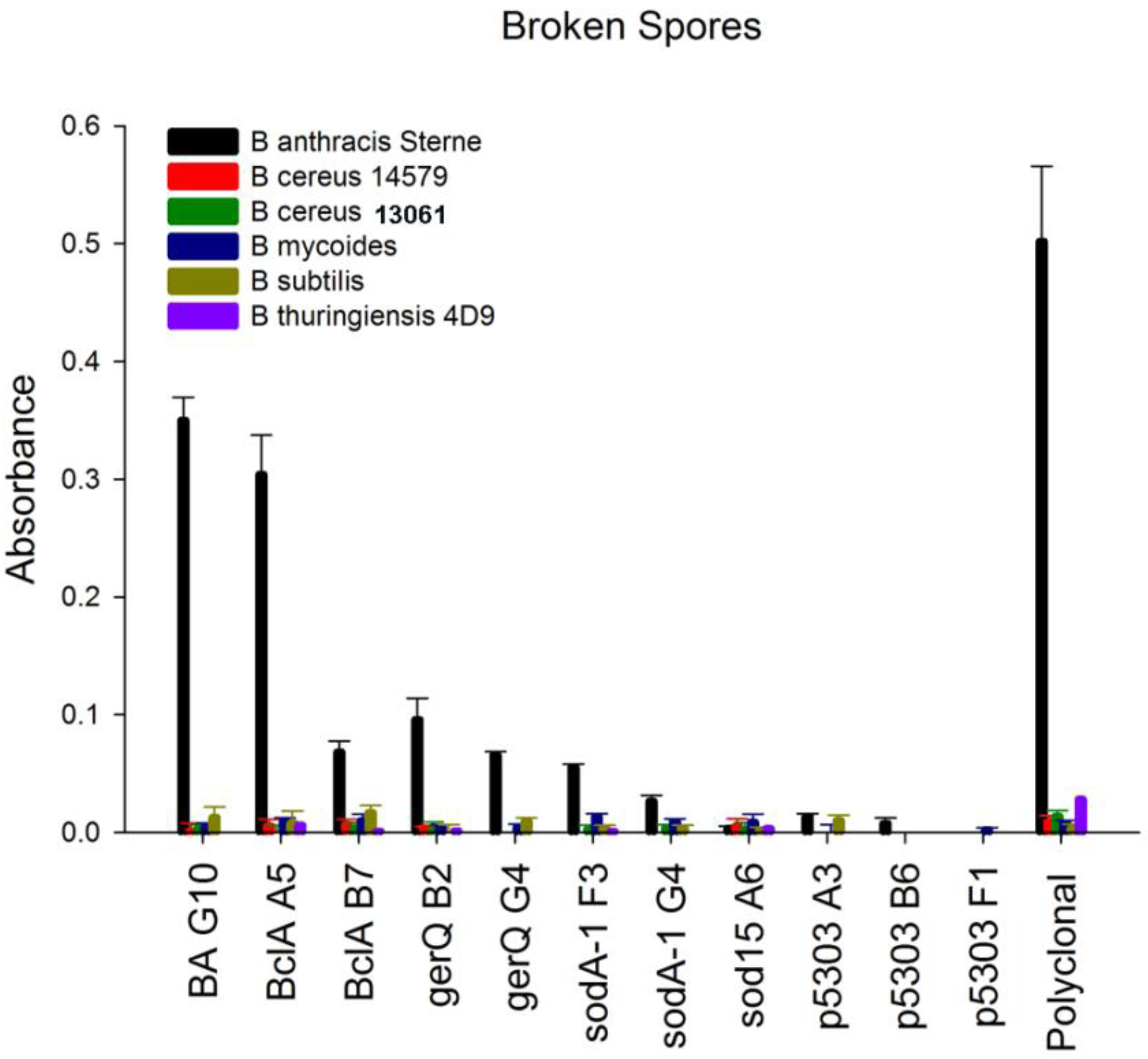
The ability to discriminate between the spores of Bacillus anthracis and other Bacillus species is of significant importance for the development of any assay or sensor. As an initial test of target specificity we evaluated the ability of the sdAb to recognize spores from B. anthracis Sterne and a limited number of closely related Bacillus strains. Spores were harvested from B. anthracis Sterne strain 34F2, B.cereus 13061, B. mycoides, B. subtilis, and B. thuringiensis Kurstaki 4D9 as described in Walper et al. [15]. Purified spores were mechanically sheared using 1.0 mm glass beads, a BioSpec Products Mini-Bead Beater, and a protocol modified from that described by Vandeventer et al. [22]. Protein concentrations obtained for several of the spore samples, particularly B. mycoides and B. subtilis, were significantly lower (10-fold) than those obtained from the other spore samples. Rather than immobilize spore material based on original spore optical density, equivalent protein concentrations were immobilized to microtiter plates. This was done to ensure that a negative result was not a result of low protein concentration rather an indicator of specificity. The sdAbs targeting SOD15 and p5303 failed to generate a positive signal on any of the spore material using a direct ELISA method. In contrast, the sdAbs selected for binding spore proteins BclA, gerQ, and SODA1 all showed the ability to bind immobilized spore material and to discriminate between the spores of B. anthracis and the spore material of non-target Bacillus species, Figure 5 and Supplemental Figure 3. While the signal generated by most of the spore-specific sdAbs was minimal, a positive signal was evident above background. This could be observed within five minutes of substrate addition. In contrast to other spore-specific protein binders, the BclA binder A5 generated a significant signal for target spore material, comparable to the signal generated by the EA1 binding sdAb BA G10.
Figure 5. Specificity assay. Direct binding ELISA of the isolated sdAb to broken spores from B. anthracis Sterne and other Bacillus species. Binding of biotinylated sdAbs was detected with an HRP-conjugated streptavidin.
Antibodies 02 00152 g005
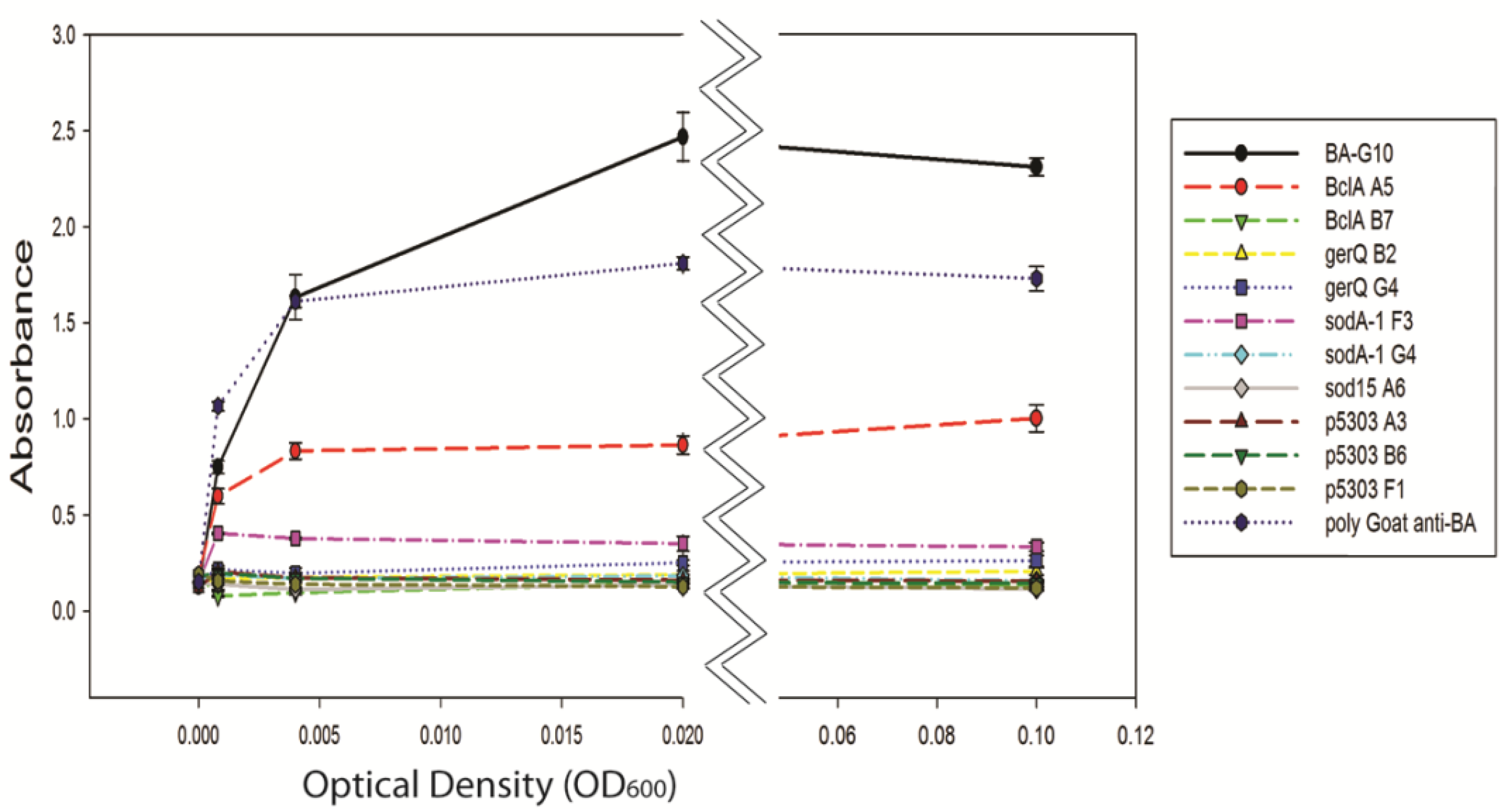
It has been shown previously that antibodies can readily recognize broken spore material; difficulties, however, can be encountered when attempting to detect intact bacterial spores [15,23]. As an intact structure, the proteins of the exosporium and coat likely occlude antibody epitopes that are readily exposed once the spore is broken. Intact B. anthracis Sterne strain spores were washed repeatedly to remove debris then immobilized to microtiter plates as described above. With the exception of the BclA A5 and BA G10 sdAbs, intact spore detection was unsuccessful using this assay format, Figure 6. The absorbance measured for BclA A5 was threefold lower at the highest concentration compared to BA G10 and twofold lower than the polyclonal antibody.
Figure 6. Limit of detection for intact B. anthracis Sterne strain spores assessed using a direct binding ELISA assay.
Antibodies 02 00152 g006
A series of bead-based, sandwich assays were conducted using the MAGPIX system to assess limits of detection for both broken and intact spores. Prior to developing the sandwich assays, an initial assay was conducted in which recombinant target protein was immobilized to magnetic beads to better evaluate specificity of individual sdAbs, Supplemental Figure 4. In this assay, many of the sdAbs showed significant levels of cross reactivity in particular with the recombinant BclA antigen which has consistently exhibited a characteristic “stickiness”. This is typified by BA G10, which showed low affinity binding to BclA, but high affinity binding to its target EA1 (Supplemental Figure 4, panel B). Though unexpected, these results were confirmed using a direct binding ELISA (data not shown). In the direct binding MAGPIX assays, the critical factor is the antibody titer more than absolute signal level; the amount of active antigen on each type of bead set may vary. However it is encouraging when both high titer and high signal are observed. Examining this data, it appears that at least in one case the sdAb’s target was misidentified, as the gerQ binder G4 binds BclA with higher affinity than it does gerQ, while the gerQ binder B2 had a similar affinity for both gerQ and BclA. These results were confirmed using an SPR chip that had all spore coat protein immobilized. The sdAb showing the best affinity and specificity were the BclA binders A5 and B7, and the p5303 binders A3 and F1.
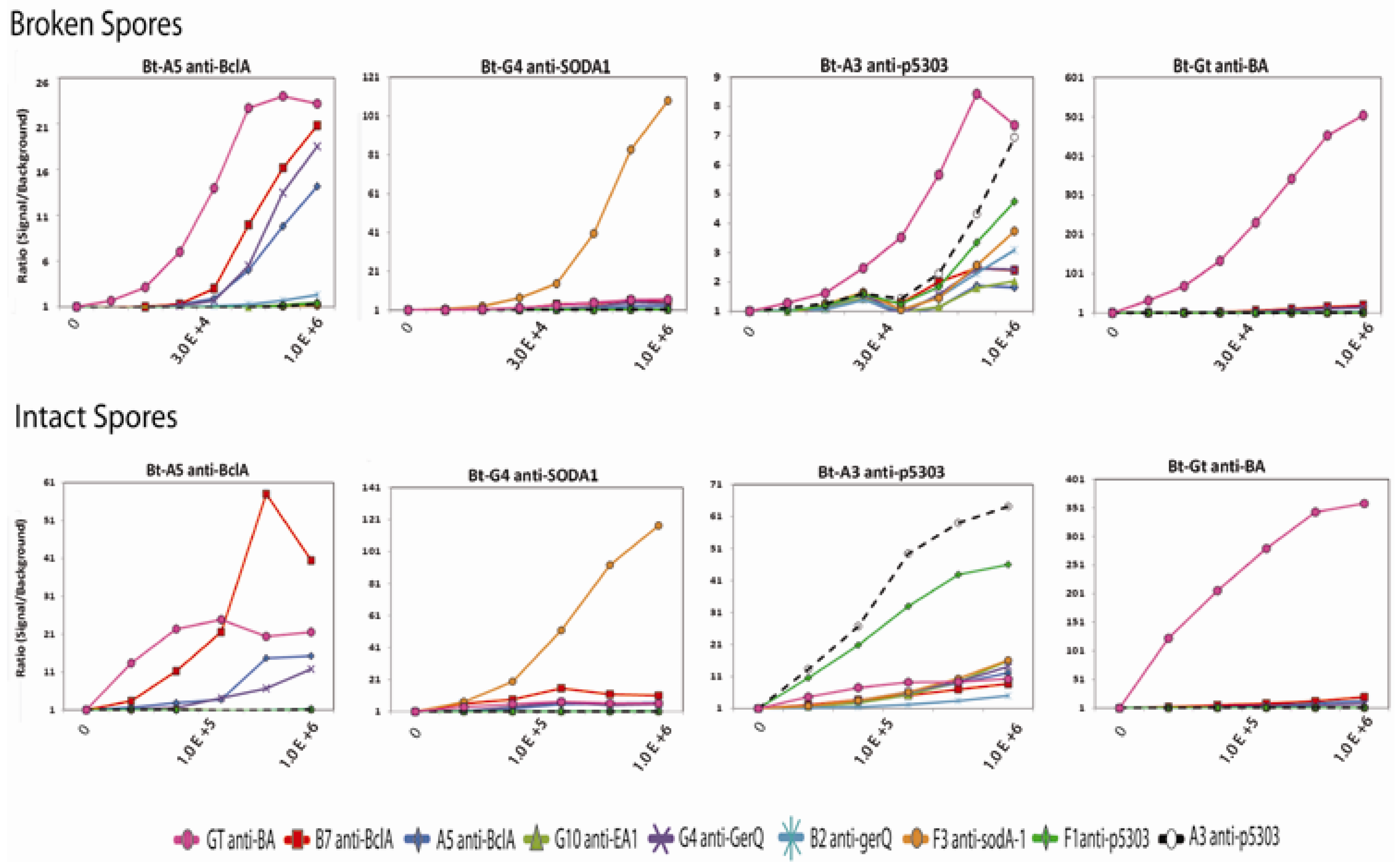
Seven of the newly isolated sdAbs, along with BA G10 and polyclonal goat anti-B. anthracis antibody, were directly immobilized to magnetic bead sets to serve as captures in sandwich assays. The biotinylated sdAbs used in the ELISA and MAGPIX specificity assays served as detection antibodies to assess limits of detection for both broken and intact spore samples. Purified B. anthracis spores were diluted in PBS then either broken using glass beads or used intact in sandwich MAGPIX assays. Figure 7 shows four representative assays for intact BA spores using the sdAbs BclA A5, SODA1 G4, p5303 A3, and polyclonal goat anti-anthracis as the biotinylated detection antibodies in conjunction with all nine capture reagents; the data for all the sandwich assays are shown in Supplemental Figure 5.
Figure 7. Sandwich immunoassays using four tracers in combination with nine capture reagents for the detection of broken (top) and intact (bottom) B. anthracis spores. Seven of the spore specific sdAbs, BA G10 (a previously isolated anti-B. anthracis sdAb), and a polyclonal goat anti-anthrax antibody were used as captures. The X-axis defines the approximate spore concentration (cfu/mL). The highest concentration (1 × 10⁶ spores/mL), an intermediate concentration, and the lowest concentration (no spores) is shown for each data set. Other data points represent a 1:5 serial dilution. Labels were omitted to improve clarity of the axis.
Antibodies 02 00152 g007
Surprisingly, the anti-p5303 sdAbs, which had not shown binding to spores in direct ELISA assays provided detection of intact spores down to at least 1.23 × 10⁴ spores/mL in the MAGPIX sandwich assay. As anticipated, the most successful tracer varied with each capture molecule for the detection of both broken and intact spore samples. Regardless of the combination of capture and tracer, intact spores were reliably detected with the best capture: tracer pairs to approximately 1.23 × 10⁴ spores/mL and broken spores to about 4.12 × 10³ spores/mL as shown in Supplemental Figure 4. The lowest limit of detection was observed when the affinity purified polyclonal goat anti-B. anthracis antibody was used as the capture and polyclonal goat anti-B. anthracis antibody as the tracer antibody. Several of the sdAb tracers gave the best detection when paired with the polyclonal capture. However, effective sdAb pairs for the detection of both broken and intact spores were also observed. For example, the sdAb BclA B7 capture showed good detection of intact spores when it was paired with the SODA1 binding sdAb F3 or the gerQ binding sdAb B2 as the tracers. Similarly, the sdAbs binding the putative spore protein p5303 (A3, B6, and F1) provided good detection when used in combination as capture and tracer pairs. An order of magnitude improvement in limit of detection was observed when broken spore material was used in these assays, Supplemental Figure 5.
As detailed above, the sdAbs were not able to achieve the limits of detection shown by the conventional antibodies when used as either captures or tracers in MAGPIX assays. This is likely not the result of poor binding, rather the small size of the sdAb can result in a degree of inactivation upon immobilization when utilized as the capture reagent and a reduced level of attached biotin when utilized as the recognition reagent, both these factors can contribute to reduced signal amplification. While not evaluated for these particular clones, the sdAb platform is amenable to the addition of fusion proteins that allow for not only an increase in available biotinylation sites but also the development of assays in which the sdAb directly serves as the reporter [24]. Future development of such assays may better showcase the functionality of sdAbs in spore detection assays.
3. Experimental Section

3.1. Antigen and Antibody Reagents

The following reagents were obtained through BEI Resources, NIAID, NIH: B. anthracis Collagen-like Protein BclA (Locus_Tag: BA_1222) with N-terminal Histidine Tag, Recombinant from Escherichia coli, NR-9577; B. anthracis Spore Coat Protein GerQ/YwdL (Locus Tag: BA_5641) with N-terminal Histidine Tag, Recombinant from E. coli, NR-10435; B. anthracis Hypothetical Protein p5303 (Locus_Tag: BA_5699) with N-terminal Histidine Tag, Recombinant from E. coli, NR-12130. The following reagents were obtained through the NIH Biodefense and Emerging Infections Research Resources Repository, NIAID, NIH: B. anthracis Exosporium Basal Layer Protein BxpB (Locus_Tag: BA_1237) with N-terminal Histidine Tag, Recombinant from E. coli, NR-12132; B. anthracis Superoxide Dismutase SODA1 (Locus_Tag: BA_4499) with N-terminal Histidine Tag, Recombinant from E. coli, NR-10505; B. anthracis Superoxide Dismutase SOD15 (Locus_Tag: BA_1489) with N-terminal Histidine Tag, Recombinant from E. coli, NR-12128. Aliquots of recombinant spore proteins (BclA, gerQ (YwdL), SODA1, SOD15, BxpB and p5303) were also provided as a kind gift from James Carney at Edgewood Chemical Biological Center (ECBC).
The affinity purified polyclonal goat anti-B. anthracis antibody was a kind gift from Dr. Jill Czarnecki, Naval Medical Research Center (NMRC). The polyclonal goat anti-B. anthracis antibody was the kind gift of Dr. Thomas O’Brien, Tetracore, Rockville MD. Both polyclonal antibodies were isolated from animals following immunization with B. anthracis Sterne spores. The BA-G10 sdAb had been previously described and shown to bind the EA1 protein of B. anthracis [15].

3.2. Library Construction and Selection

Llamas were immunized and maintained by Triple J. Farms (Bellingham, WA). Initial immunization with standard veterinary anthrax vaccine has been previously described [15]. A total of five subsequent immunizations were performed with a combination of the spore proteins. Each immunization contained 100 µg of pooled recombinant spore proteins. One of the llamas (Centavo) was immunized with all of the spore proteins, while the other llama (Whisper) was immunized with a mix lacking BclA. Approval of immunization protocol was obtained from the Institutional Animal Care and Use Committee (IACUC) through Triple J. Farms. Peripheral blood lymphocytes were isolated from whole blood following five immunizations with the recombinant spore proteins. The sdAb library was constructed as previously described following RNA extraction and cDNA synthesis [15,25]. Polyclonal antibody was purified from plasma from each of the immunized animals.
Enrichment of target binding sdAbs was accomplished using a modified selection protocol as described by Griffiths et al. [26]. The recombinant antigens were immobilized to Nunc Maxisorb microtiter plates at a concentration of 1–3 µg/mL in phosphate buffered saline (pH 7.4, PBS). Immobilization proceeded overnight at 4 °C.
Following two rounds of selection amplified phage preparations from the initial library and both rounds of selection were tested using a polyclonal phage ELISA as described by Bradbury and Marks [27] to assess enrichment of target binding clones. Subsequent identification of individual clones producing target binding sdAbs was accomplished via a monoclonal phage ELISA using protocols described by the aforementioned authors [27]. Plasmid DNA from select clones was isolated using a QIAgen Plasmid Mini kit and sequenced by Eurofins MWG Operon (Huntsville, AL) to identify unique clones and antibody families based on amino acid composition of the complementary determining regions (CDRs).

3.3. Protein Production

Expression and purification of target-binding sdAbs was performed as previously described [15]. Briefly, the sdAb gene was cloned to the bacterial expression vector pECAN45 which was transformed to the Rosetta Codon Plus Escherichia coli bacterial strain (Merck Millipore) for protein expression. A two-step chromatography protocol was employed for purification consisting of an initial immobilized metal affinity chromatography (IMAC) step and subsequent FPLC using a Superdex 75 10/300 GL column. Purified proteins were quantitated using a Nanodrop 1000 spectrophotometer (ThermoFisher, USA) and calculation based on absorbance at 280 nm.

3.4. Determining Thermal Stability

Circular dichroism (CD) using a Jasco J-815 CD spectrometer was used to determine both the melting temperature and the ability of individual sdAbs to regain secondary structure following thermal denaturation. Purified sdAbs (200–300 µg) were dialyzed overnight at 4 °C in deionized water (DI) (18 MΩ cm) then diluted in DI water to a final concentration of 45 µg/mL. Ellipticity was measured at 208 nm as temperature was cycled from 25–95 °C. The melting temperature correlated to the inflection point between the folded and unfolded protein and ability to renature was calculated from the recovery of ellipticity as the protein was subjected to iterative rounds of heating and cooling.
In addition to CD measurements, thermal stability was also assessed as the ability to maintain antigen recognition. For these experiments, biotinylated sdAbs were tested in a direct binding ELISA against the target antigen to examine if antigen binding capabilities were maintained through iterative rounds of heating and cooling. SdAbs were biotinylated with a Sulfo-NHS –LC-LC-biotin reagent (Thermo Fisher Scientific) and separated from excess biotin using a Bio-Gel P10 column. Biotinylated sdAbs were diluted in phosphate buffered saline (PBS, pH 7.4) to a final concentration of 10 µg/mL then heated and cooled to 95 °C and 25 °C in a Eppendorf Mastercycler at a rate of 0.5 °C/min. Antigen was immobilized to high binding capacity microtiter plates at a concentration of 3 µg/mL overnight at 4 °C. The following morning the plate was washed twice with PBS containing 0.05% (v/v) Tween 20 (PBST) then blocked with PBST and 5% milk proteins (PBSTM). Following thermal cycling for 0, 1, 2, or 3 rounds; biotinylated sdAbs were diluted in PBSTM to a final concentration of 1 µg/mL. Polyclonal antibodies were used at a concentration of 3 µg/mL to ensure an equivalent molar concentration. A streptavidin conjugated horseradish peroxidase (Prozyme, Hayward CA) at 1:3,000 dilution in PBSTM served as the secondary antibody for detection of sdAb binding. The SigmaFast OPD substrate (Sigma-Aldrich) served as the peroxidase substrate. The reaction was terminated after 10–15 min with 4 M H2SO4 then quantitated using a Tecan Infinite M1000 spectrophotometer measuring absorbance at 492 nm.

3.5. Binding Kinetics

Binding kinetics for each sdAb were determined using a BioRad ProteOn XPR36 system. Recombinant spore proteins were diluted in 10 mM sodium acetate buffer (pH 4.0) to a concentration of 5 µg/mL. The antigen was immobilized to a GLC sensor chip using a standard EDC/NHS facilitated conjugation reaction. Non-reacted binding sites were inactivated with 1 mM ethanolamine. Kinetic parameters (ka, kd, KD) were determined by flowing six concentrations of purified sdAb targeting the antigen at a flow rate of 50 µL/min for 3 min to determine association kinetics. Dissociation was monitored for 10 min. Kinetic values were calculated from accumulated data using the ProteOn Manager software provided by the manufacturer. The surface was regenerated with a 50 mM glycine (pH 2.5) solution between experiments.

3.6. Spore Detection

Spores from Bacillus anthracis Sterne strain (34F2) and other non-target bacillus species were produced using protocols previously described [15]. Purified bacterial spores were used either intact or broken for specificity assays. Bacterial spores were broken using 1.0 mm glass beads and a Biospec Products Mini-bead beater. Glass beads were washed first with 4M H2SO4 and then numerous times with DI water prior to use. Bacterial spores were diluted in PBS to an optical density at 600 nm of 0.1 (OD600) to which 0.1 grams of cleaned glass beads were added. Samples were agitated two times for 15 min in the bead beater. Beads were allowed to settle in the tube before the soluble material was transferred to a new microfuge tube. Protein concentration was approximated based on absorbance at 280 nm and the basic calculation of Abs280 1 = 1.0 mg/mL using a Nanodrop 1000 (Thermo Fisher Scientific).
For both the broken and intact spore direct ELISAs, spore material was passively immobilized to microtiter plates at 30 °C for 2 h. Broken spore material was immobilized at a concentration of 10 µg/mL. Intact spores were immobilized at an initial OD600 = 0.1 then serially (1:5) diluted for the remaining wells. Washing and blocking steps, sdAb and secondary antibody concentration, and substrate for these assays were consistent with those described above.
The bead-based MAGPIX system (Luminex) was used to assess both specificity for the target spore protein and for sandwich-type detection assays with broken and intact spore material. SdAbs and recombinant spore proteins were immobilized to individual magnetic bead sets following the manufacturer’s suggested EDC/NHS protocol [28]. Each of the biotinylated sdAbs was tested for binding to both target and non-target proteins immobilized to the magnetic beads. For detection assays, bacterial spores were diluted to equivalent optical densities in PBS. Spore material was broken prior to analysis using glass beads as described above. Polyclonal antibody (NMRC) and sdAb coated magnetic beads were incubated with spore material for 1 hour then washed with PBST. As with the cross-reactivity assays, biotinylated polyclonal (Tetracore) or sdAbs were used in combination with each of the capture bead sets. A phycoerythrin-conjugated streptavidin was diluted in PBST to a concentration of 5 ug/mL to serve as the reporter for all experiments.

4. Conclusions

SdAbs targeting the proteins of the B. anthracis Sterne strain spores were isolated from an immune llama library using phage display and characterized in terms of target binding kinetics, thermal stability, specificity, and utility in sandwich assays for the detection of both intact and broken spores. Many of the isolated sdAbs possessed excellent affinity for their targets and were able to bind after cycles of heating to 95 °C. Several combinations of sdAbs could be used as pairs in sandwich assays for the detection of B. anthracis spores, both broken and intact. Though the single domain antibodies did not demonstrate the same limits of detection as the conventional antibodies, this is likely not a product of low affinity binding. Although the small size of sdAbs can be advantageous, it can lead to a greater degree of inactivation when these reagents are immobilized or biotinylated using non-directional EDC/NHS coupling chemistries. Because they are produced recombinantly, sdAbs can be engineered for directional immobilization, site-specific biotinylation, or produced with a variety of fusion tags that can serve as reporters of aid in immobilization [24] any of which may further improve detection limits. The sdAbs characterized in this work provide the basis for recognition reagents for the sensitive and specific detection of B. anthracis spores, though prior to integration into any existing or future assay these sdAbs would require additional specificity assays with other non-target bacillus species; particularly those that demonstrate high sequence homology with the target proteins.

Supplementary Materials

Supplementary materials can be accessed at: https://www.mdpi.com/2073-4468/2/1/152/s1.

Supplementary Files

Supplementary File 1

Acknowledgments

This work was supported by the Defense Threat Reduction Agency, project # CBM.DIAGB.01.10.NRL.011 and the Office of Naval Research NRL base funds. Scott Walper was a supported as a National Research Council Post-doctoral Fellow for a portion of this research.

References

  1. Inglesby, T.V. Anthrax as a biological weapon, 2002: Updated recommendations for management. JAMA 2002, 287, 2236–2252. [Google Scholar] [CrossRef]
  2. Schmid, G.; Kaufmann, A. Anthrax in europe: It’s epidemiology, clinical characteristics, and role in bioterrorism. Clin. Microbiol. Infect. 2002, 8, 479–488. [Google Scholar] [CrossRef]
  3. Drobniewski, F. Bacillus cereus and related species. Clin. Microbiol. Rev. 1993, 6, 324–338. [Google Scholar]
  4. Taitt, C.R.; Malanoski, A.P.; Lin, B.; Stenger, D.A.; Ligler, F.S.; Kusterbeck, A.W.; Anderson, G.P.; Harmon, S.E.; Shriver-Lake, L.C.; Pollack, S.K.; et al. Discrimination between biothreat agents and a near neighbor species using a resequencing array. FEMS Immunol. Med. Microbiol. 2008, 54, 356–364. [Google Scholar] [CrossRef]
  5. Ellerbrok, H.; Nattermann, H.; Ozel, M.; Beutin, L.; Appel, B.; Pauli, G. Rapid and sensitive identification of pathogenic and apathogenic bacillus anthracis by real-time pcr. FEMS Microbiol. Lett. 2002, 214, 51–59. [Google Scholar] [CrossRef]
  6. Qi, Y.; Patra, G.; Liang, X.; Williams, L.E.; Rose, S.; Redkar, R.J.; DelVecchio, V.G. Utilization of the rpob gene as a specific chromosomal marker for real-time pcr detection of bacillus anthracis. Appl. Environ. Microbiol. 2001, 67, 3720–3727. [Google Scholar] [CrossRef]
  7. Hamers-Casterman, C.; Atarhouch, T.; Muyldermans, S.; Robinson, G.; Hamers, C.; Songa, E.B.; Bendahman, N.; Hamers, R. Naturally occurring antibodies devoid of light chains. Nature 1993, 363, 446–448. [Google Scholar] [CrossRef]
  8. Ghahroudi, M.A.; Desmyter, A.; Wyns, L.; Hamers, R.; Muyldermans, S. Selection and identification of single domain antibody fragments from camel heavy-chain antibodies. FEBS Lett. 1997, 414, 521–526. [Google Scholar] [CrossRef]
  9. Vu, K.B.; Ghahroudi, M.A.; Wyns, L.; Muyldermans, S. Comparison of llama vh sequences from conventional and heavy chain antibodies. Mol. Immunol. 1997, 34, 1121–1131. [Google Scholar] [CrossRef]
  10. Greenberg, A.S.; Avila, D.; Hughes, M.; Hughes, A.; McKineey, E.C.; Flajnik, M.F. A new antigen recepetor gene family that undergoes rearrangement and extensive somatic diversification in sharks. Nature 1995, 374, 168–173. [Google Scholar]
  11. Nuttall, S.D.; Krishnan, U.V.; Hattarki, M.; Gori, R.D.; Irving, R.A.; Hudson, P.J. Isolation of the new antigen receptor from wobbegong sharks, and use as a scaffold for the display of protein loop libraries. Mol. Immunol. 2001, 38, 313–326. [Google Scholar] [CrossRef]
  12. Eyer, L.; Hruska, K. Single-domain antibody fragments derived from heavy-chain antibodies: A review. Vet. Med.-Czech 2012, 57, 439–513. [Google Scholar]
  13. Wesolowski, J.; Alzogaray, V.; Reyelt, J.; Unger, M.; Juarez, K.; Urrutia, M.; Cauerhff, A.; Danquah, W.; Rissiek, B.; Scheuplein, F.; et al. Single domain antibodies: Promising experimental and therapeutic tools in infection and immunity. Med. Microbiol. Immunol. 2009, 198, 157–174. [Google Scholar] [CrossRef]
  14. Dumoulin, M.; Conrath, K.; Van Meirhaeghe, A.; Meersman, F.; Heremans, K.; Frenken, L.G.J.; Muyldermans, S.; Wyns, L.; Matagne, A. Single-domain antibody fragments with high conformational stability. Protein Sci. 2002, 11, 500–515. [Google Scholar]
  15. Walper, S.A.; Anderson, G.P.; Brozozog Lee, P.A.; Glaven, R.H.; Liu, J.L.; Bernstein, R.D.; Zabetakis, D.; Johnson, L.; Czarnecki, J.M.; Goldman, E.R. Rugged single domain antibody detection elements for bacillus anthracis spores and vegetative cells. PLoS One 2012, 7, e32801. [Google Scholar]
  16. Love, T.; Redmond, C.; Mayers, C. Real time detection of anthrax spores using highly specific anti-ea1 recombinant antibodies produced by competitive panning. J. Immunol. Methods 2008, 334, 1–10. [Google Scholar] [CrossRef]
  17. Williams, D.D.; Turnbough, C.L. Surface layer protein ea1 is not a component of bacillus anthracis spores but is a persistent contaminant in spore preparations. J. Bacteriol. 2003, 186, 566–569. [Google Scholar] [CrossRef]
  18. Cybulski, R.J., Jr.; Sanz, P.; McDaniel, D.; Darnell, S.; Bull, R.L.; O’Brien, A.D. Recombinant bacillus anthracis spore proteins enhance protection of mice primed with suboptimal amounts of protective antigen. Vaccine 2008, 26, 4927–4939. [Google Scholar] [CrossRef]
  19. Cybulski, R.J., Jr.; Sanz, P.; Alem, F.; Stibitz, S.; Bull, R.L.; O'Brien, A.D. Four superoxide dismutases contribute to bacillus anthracis virulence and provide spores with redundant protection from oxidative stress. Infect. Immun. 2008, 77, 274–285. [Google Scholar]
  20. Ghahroudi, M.A.; Desmyter, A.; Wyns, L.; Hamers, R.; Muyldermans, S. Selection and identification of single domain antibody fragments from camel heavy-chain antibodies. FEBS Lett. 1997, 414, 521–526. [Google Scholar] [CrossRef]
  21. Anderson, G.; Ortiz-Vera, Y.A.; Hayhurst, A.; Czarnecki, J.; Dabbs, J.; Vo, B.; Goldman, E. Evaluation of llama anti-botulinum toxin heavy chain antibody. Botulinum J. 2008, 1, 100–116. [Google Scholar] [CrossRef]
  22. Vandeventer, P.E.; Weigel, K.M.; Salazar, J.; Erwin, B.; Irvine, B.; Doebler, R.; Nadim, A.; Cangelosi, G.A.; Niemz, A. Mechanical disruption of lysis-resistant bacterial cells by use of a miniature, low-power, disposable device. J. Clin. Microbiol. 2011, 49, 2533–2539. [Google Scholar] [CrossRef]
  23. Nuttall, S.D.; Wilkins, M.L.; Streltsov, V.A.; Pontes-Braz, L.; Dolezal, O.; Tran, H.; Liu, C.-Q. Isolation, kinetic analysis, and structural characterization of an antibody targeting the bacillus anthracis major spore surface protein bcla. Proteins 2011, 79, 1306–1317. [Google Scholar] [CrossRef]
  24. Walper, S.A.; Brozozog Lee, P.A.; Goldman, E.R.; Anderson, G.P. Comparison of single domain antibody immobilization strategies evaluated by surface plasmon resonance. J. Immunol. Methods 2013, 388, 68–77. [Google Scholar] [CrossRef]
  25. Goldman, E.R.; Anderson, G.; Conway, J.O.; Sherwood, L.J.; Fech, M.; Vo, B.; Liu, J.L.; Hayhurst, A. Thermostable llama single domain antibodies for detection of botulinum a neurotoxin complex. Anal. Chem. 2008, 80, 8583–8591. [Google Scholar] [CrossRef]
  26. Griffiths, A.; Malmqvist, M.; Marks, J.; Bye, J.; Embleton, M.J.; McCafferty, J.; Baier, M.; Holliger, K.P.; Gorick, B.; Hughes-Jones, N.; et al. Human anti-self antibodies with high specificity from phage display libraries. EMBO J. 1993, 12, 725–734. [Google Scholar]
  27. Clackson, T.; Lowman, H.B. Phage Display; Oxford University Press: New York, NY, USA, 2004; p. 331. [Google Scholar]
  28. Anderson, G.P.; Legler, P.M.; Zabetakis, D.; Goldman, E.R. Comparison of immunoreactivity of staphylococcal enterotoxin b mutants for use as toxin surrogates. Anal. Chem. 2012, 84, 5198–5203. [Google Scholar] [CrossRef]