Article

The Effects of Aniline-Promoted Electron Shuttle-Mediated Goethite Reduction by Shewanella oneidensis MR-1 and theDegradation of Aniline

1
School of Resources and Geosciences, China University of Mining and Technology, Xuzhou 221116, China
2
Key Laboratory of Coalbed Methane Resources & Reseryoir Formation Process, Ministry of Education, Xuzhou 221116, China
3
School of Mines, Liaoning Technology University, Huludao 125105, China
*
Author to whom correspondence should be addressed.
Water 2023, 15(20), 3686; https://doi.org/10.3390/w15203686
Submission received: 26 September 2023 / Revised: 18 October 2023 / Accepted: 19 October 2023 / Published: 21 October 2023

Abstract

:
The biological reduction of Fe (III) is common in underground environments. This process not only affects the biogeochemical cycle of iron but also influences the migration and transformation of pollutants. Humic substances are considered effective strategies for improving the migration and transformation of toxic substances and enhancing the bioavailability of Fe (III). In this study, the electron shuttle anthraquinone-2-sulfonate (AQS) significantly promoted the bio-reduction of Fe (III). On this basis, different concentrations of aniline were added. The research results indicate that at an aniline concentration of 3 μM, the production of Fe (II) in the reaction system was 2.51 times higher compared to the microbial reaction group alone. Furthermore, the degradation of aniline was most effective in this group. The increased consumption of sodium lactate suggests that aniline, under the mediation of AQS, promoted the metabolism of Shewanella oneidensis MR-1 cells and facilitated the involvement of more electrons in the reduction process. After the reaction, the solid mineral Fe (II)-O content increased to 41.32%. This study provides insights into the reduction mechanism of Fe (III) in the complex environment of microorganisms, iron minerals, electron shuttles, and pollutants. It aims to offer a theoretical basis for the biodegradation of aromatic hydrocarbon pollutants.

1. Introduction

Iron ranks as the fourth most common element in the Earth’s crust [1] and is a widespread transition metal element that exhibits variable oxidation states. Fe (II) tends to have higher solubility and is more easily utilized by living organisms [2]. Microbial iron reduction is a primary pathway used to reduce Fe (III) minerals in the natural environment [3]. Most microorganisms are capable of metabolizing and utilizing Fe (III) and Fe (II), facilitating electron transfer between various electron donors and acceptors [4]. The process not only significantly impacts the cycling of elements such as C, N, S, and P in the natural environment but also plays a crucial role in the transformation and breakdown of both organic and inorganic contaminants [5,6,7,8,9,10], including those released from industrial and mining areas [11,12].
Aromatic compounds are widely distributed in subsurface environments and have been detected in many industrial areas [13]. Aniline is a typical aromatic compound, serving as a significant chemical raw material, and is annually discharged into the environment in large quantities, leading to subsurface contamination [14]. Aniline exhibits strong toxicity and can have detrimental effects on both animals and plants [15,16]. This toxicity may also impact Fe (III)-reducing microbes. These bacteria are common in natural environments such as soil, river sediments, estuarine sediments, petroleum reservoirs, groundwater, and hot springs [2]. Currently, research on the impact of aromatic compounds on microbial Fe (III) reduction is limited.
Anaerobic environments are rich in Fe (III) oxides, which include ferrihydrite, goethite, lepidocrocite, and hematite t. Microorganisms have ability to utilize extracellular iron oxides as terminal electron acceptors, facilitating the reduction of Fe (III) by oxidizing electron donors. As a result, microbial iron reduction is widely present in underground environments [17,18,19,20,21,22,23,24]. However, these oxides exhibit and possess higher thermodynamically stable and stronger crystal structures, resulting in low bioavailability [25,26]. The addition of exogenous electron shuttles can significantly promote the reduction of Fe (III) and enhance the bioavailability of Fe (III) oxides [27]. Electron shuttles refer to a class of chemical substances that can mediate electron transfer through their own redox reactions [28]. Humic substances are known to be exogenous extracellular electron shuttles, primarily composed of oxidized forms of electron shuttling moieties that accept electrons from organic compounds and transfer them to reduced forms of electron shuttles. The reduced electron shuttles transfer carrier electrons to the surface of the Fe (III) oxides, converting them into oxidized forms of electron shuttles [29]. In sediment and aquatic systems, organic matter with redox activity is commonly present [30], and it contains quinone functional groups that can act as electron shuttles. Anthraquinone-2-sulfonate sodium (AQS) is a typical synthesized quinone compound that has been widely used as a model substance for studying the redox properties of quinone moieties in humic substances. It can act as an electron shuttle between Fe (III) and microorganisms, thereby accelerating the extracellular electron transfer rate between them [31,32,33]. Aromatic hydrocarbons, microorganisms, Fe (III) iron oxides, and quinone electron shuttles commonly coexist in underground environments. However, there is little research on their mutual interactions and their influence on the pollutant aniline.
This study focuses on the iron-reducing model bacterium Shewanella oneidensis MR-1, Fe (III) oxide (goethite, GT) as the iron mineral, and anthraquinone-2-sulfonate sodium (AQS) as the electron shuttle. The organic pollutant represented by aniline is investigated. The objectives of this work were (1) to determine the key effects of AQS on the reduction and release of Fe (III) by Shewanella oneidensis MR-1; (2) to determine the impact of aniline on the bio-reduction of Fe (III) mediated by AQS; and (3) to illustrate the concentration changes of aniline during the iron reduction process.

2. Materials and Methods

2.1. Culture Conditions

Shewanella oneidensis MR-1 strain (hereafter referred to as MR-1) was obtained from the Marine Culture Collection of China (MCCC 1A00048). The bacterium is a facultative anaerobic. Prior to inoculation, MR-1 strain is placed on aseptic operation table. Using an inoculation loop, the bacterial culture was transferred into Luria–Bertani (LB) medium (yeast extract 5 g/L, peptone 10 g/L, sodium chloride 10 g/L) [34] broth at 30 °C until logarithmic growth phase and subjected to aerobic activation until reaching the logarithmic growth phase. MR-1 strain was placed in a 50 mL centrifuge tube and centrifuged for 10 min at 8000 rpm. After centrifugation, the supernatant is carefully discarded, and the pellet is resuspended in sterile water until the optical density (OD600) reaches approximately 1.0.

2.2. Goethite Synthesis

Goethite was prepared following the method described by Schwertmann and Cornell [35]. In a polyethylene bottle containing 100 mL of 1 M Fe(NO3)3 solution, 180 mL of 5 M KOH solution was rapidly added and stirred. Dilute the mixture to 2 L with deionized water and age at 70 °C in a water bath for 60 h. The solid precipitate was collected, and the solid precipitate was washed with ultrapure water until neutral and dried at 70 °C. It was then ground to a particle size below 200 mesh for further use.

2.3. Effects of Different Concentrations of AQS on the Fe (III) Bio-Reduction

Batch experiments were carried out in an anaerobic incubator to explore the impact of anthraquinone-2,6-disulfonate sodium salt (AQS) on the Fe (III)-reducing by Shewanella oneidensis MR-1. Sodium lactate (15 mM) was used as the sole source of carbon, and goethite (hereafter referred to as GT) was an electron acceptor. Different concentrations of AQS (0.0, 0.1, 0.3, and 0.5 mM) were added to experimental groups. A control group without MR-1 was also included. The Fe (III)-reduction culture medium per liter contains the following components: NaHCO3 2.5 g, NH4Cl 0.25 g, KCl 0.5 g, NaCl 0.1 g, KH2PO4·7H2O 0.04 g, MgCl2·6H2O 0.2 g, MgSO4·7H2O 0.05 g. Each experimental condition was conducted in triplicate. Samples of the suspension were collected at specific time intervals (0, 12, 20, 32, 48, 72 h) from the anaerobic glove box to measure Fe (II) and sodium lactate concentrations. After the reaction was completed, goethite mineral samples were characterized by scanning electron microscope (SEM), X-ray diffractometer (XRD), and X-ray photoelectron spectroscopy (XPS).

2.4. Effects of Different Aniline Concentrations on AQS-Mediated Fe (III) Bio-reduction

To investigate the impact of different concentrations of aniline on AQS-mediated Fe (III) bio-reduction, different concentrations of aniline (0.0, 1, 3, and 7 μM) were added to the iron-reducing medium containing 0.5 mM AQS with a control group without aniline. Anaerobic glove box was used for all experiments, and other experimental conditions were consistent with those described in Section 2.3. Samples of the suspension were collected at specific time intervals to measure Fe (II), sodium lactate, and aniline concentrations. After the reaction was completed, goethite mineral samples were characterized by (SEM), XRD, and XPS.

2.5. Chemical Analysis

The 1,10-phenanthroline spectrophotometric method was used to determine the concentration of Fe (II) in the reduction system [36]. The samples were acidified with 0.5 mM hydrochloric acid for 15 min; supernatant was then collected by centrifugation for 10 min. The color reagent 0.5% ortho-phenanthroline was then added, and the mixture was allowed to stain for 15 to 20 min. Afterward, acetic acid–acetate buffer solution was added, and the measurement was conducted at 510 nm using a UV–visible spectrophotometer.
High-performance liquid chromatography (HPLC) (Agilent 1260, Agilent Technologies (Inc., La Jolla, CA, USA)) with a C18 column was used to detect the content of sodium lactate in the supernatant during the reduction of Fe (III). The supernatant was filtered through a 0.22 µM organic filter tip and detected by HPLC with an injection volume of 20 µL after sampling with a sterile syringe. The mobile phase was 95% phosphate (0.1%, pH 3.5) and 5% acetonitrile at a flow rate of 1 mL min−1.
Aniline concentration was determined using purge and trap gas chromatography-mass spectrometry (GC: Atomx, Teledyne Tekmar LLC. (Mason, OH, USA); GC-MS: Thermo Fisher Scientific Inc. (Waltham, MA, USA)) with the following operating conditions: Gas flow rate of 40 mL/min was used to sweep for 11 min; 2 min at 190 °C; 6 min at 200 °C. Inlet temperature: 220 °C. Carrier gas: high-purity helium gas. Initial column temperature: 35 °C for 2 min. Temperature ramp: 5 °C/min from 35 °C to 100 °C. Temperature ramp: 10 °C/min from 100 °C to 200 °C, followed by a 1 min hold at 200 °C. Ionization source: electron impact (EI). Full scan range: 35 to 270. External standard combined with internal standard for quantitative analysis.

2.6. Solid Mineral Characterization

An X-ray diffractometer (XRD) (Rigaku Ultima IV, Rigaku Corporation (Tokyo, Japan)) was used for the qualitative analysis of the minerals. After centrifugation and drying, the test samples were ground to powder using an agate mortar and then passed through 200-mesh sieve. The powder was then tested using the pellet press method. The test conditions were as follows: test voltage 40 kV, test current 40 mA, Cu Kα ray, test angle range 5°~90°, scanning speed 5°/min, step scanning, step size 0.02°, and experimental data analysis in MDI Jade 6.0 software.
To determine the changes in surface iron oxidation states before and after the experiment, X-ray photoelectron spectroscopy (XPS) (Thermo Fisher Escalab Xi+, Thermo Fisher Scientific Inc. (Waltham, MA, USA)) was used on the samples. XPS was performed using a monochromatic aluminum card X-ray source. Test conditions: vacuum 3 × 10⁻⁷ Pa. The binding energy of the C (1 s) level at 284.6 eV was used as reference. Surface full-spectrum scanning of the samples was performed to collect high-resolution spectra of Fe2p, which were analyzed by subtracting the baseline and performing a least-squares fit using the Gauss–Lorentz equation in Avantage software(5.99-31).
The morphology of iron minerals after the Fe (III) reduction experiment was observed using a scanning electron microscope (SEM) (Oxford Ultim Max65, Oxford Instruments plc. (Oxford, UK)). Test conditions: mineral powder is adhered to a conductive binder for mounting, followed by a gold coating treatment for SEM topography analysis. During observation, the accelerating voltage is set in the range of 10–15 kV, with a beam of electron excitation set at 30–40 μA.

2.7. Kinetic Analyses

The consumption rate of sodium lactate and aniline in the medium over time was fitted according to a quasi-primary kinetic model.
l n C t = l n C 0 K t
where C0 is the initial concentration of aniline or sodium lactate at t = 0, Ct (mM) is the concentration of aniline or sodium lactate at t (h), and K is the first-order reaction rate constant h−1 of sodium lactate or the first-order reaction rate constant h−1 of aniline.

2.8. Statistical Analysis

Statistical analysis involves performing analysis of variance (ANOVA) using the SPSS software(26) and conducting a Duncan test at a significance level of p < 0.05. and the figures were drawn using Origin. Detailed analysis is provided in the supplementary date.

3. Results

3.1. Characterizations of Goethite

Synthesized goethite was characterized by XRD and SEM. The XRD pattern of the prepared goethite (Figure 1a) was compared with the standard mineral (Goethite, Card No. 29-0713), and the comparison results confirmed that the synthesized yellow mineral was pure goethite without any other crystalline phases. Under SEM observation (Figure 1b), the morphology of the prepared goethite appeared as long needle-like structures.

3.2. Bio-reductive Transformation of Fe (III)

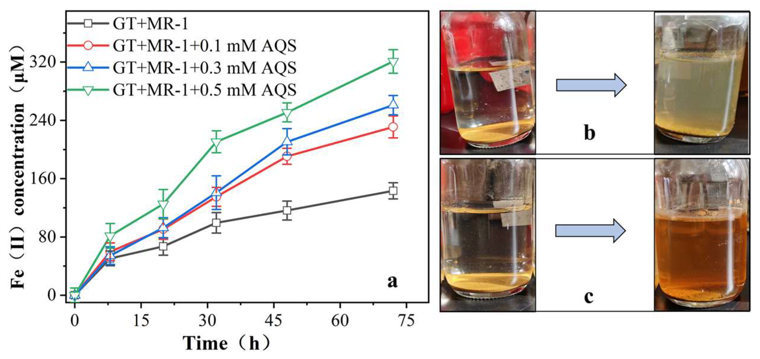
3.2.1. Effect of AQS on the Reduction of Fe (III) by MR-1

To study the impact of different concentrations of aniline on AQS-mediated Fe (III) bio-reduction, varying concentrations of AQS were added during the MR-1-mediated reduction of goethite. Fe (II) produced during goethite bio-reduction mediated by AQS is shown in Figure 2a. Consistent with previous research results, MR-1 is capable of individually reducing Fe (III), and the addition of the electron shuttle AQS significantly accelerated the reduction process [37,38,39]. When MR-1 was solely responsible for reducing Fe (III), the detected Fe (II) released into the reaction system during goethite reduction was 143.3 μM. As Fe (II) continued to be released, the color of the entire reaction system gradually changed to light green (Figure 2b).
In the reaction groups with the addition of 0.1, 0.3, and 0.5 mM AQS, the generated Fe (II) was 1.61, 1.82, and 2.23 times higher than that of the microbial reaction group alone, respectively. This indicates that AQS can serve as an electron shuttle, enhancing the transfer efficiency of electrons from the microbial strain MR-1 to goethite, leading to the release of a significant amount of Fe (II) into the reaction medium. The conversion of the quinone structure of AQS to the phenolic structure of AH2QS during the reaction enhances the molecular conjugation effect [40,41]. Therefore, during the experimental process, the reaction system can be observed to change from colorless to reddish brown (Figure 2c).

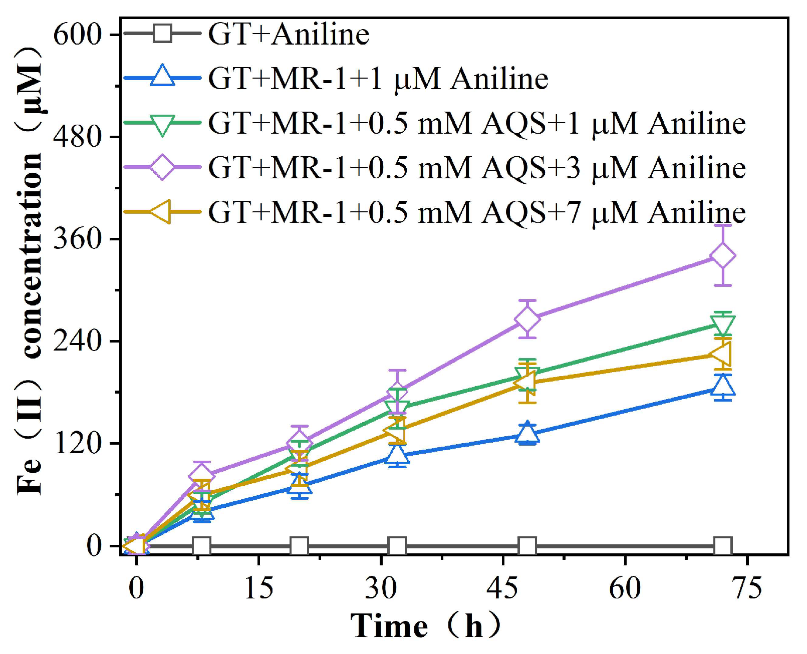
3.2.2. Effect of Aniline on AQS-Mediated Reduction of Fe (III) by MR-1

Figure 3 shows the effect of different concentrations of aniline on the AQS-mediated bio-reduction of goethite. Different concentrations of aniline exhibit noticeable variations in their promotion of Fe (III) reduction. Among them, the addition of 3 μM aniline had the most significant effect on the AQS-mediated promotion of acicular ferrite bio-reduction, with Fe (II) production being 1.07 times higher than that of the reaction system containing 0.5 mM AQS. However, as the aniline concentration increases to 7 μM, the promotion intensity starts to decrease. In the absence of the MR-1 reaction system, no detectable generation of Fe (II) is observed during the reaction period. This suggests that the pollutant aniline cannot directly induce the reduction of Fe (III) oxide, and the promotion of Fe (III) reduction by aniline may be a result of its interaction with the iron-reducing bacteria.

3.2.3. Consumption and Kinetic Analysis of Sodium Lactate

The use of sodium lactate as the sole carbon source and energy for MR-1 metabolism is a key feature [42,43]. The concentration of sodium lactate in the reaction system was measured to explore whether the presence of AQS and aniline could accelerate the consumption of the electron donor sodium lactate, thereby promoting the bio-reduction of Fe (III) by MR-1 (Figure 4a, c and Table 1). After the addition of AQS and aniline, there was a significant increase in the consumption of sodium lactate, with the highest consumption observed when 3 μM aniline was added. The first-order kinetic constants of sodium lactate consumption were calculated for quantitative analysis (Figure 4b, d and Table 1). When MR-1 alone reduced goethite, the first-order kinetic constant (K value) for sodium lactate consumption was only 0.0023 ± 0.002 h−1. However, with the addition of 0.1, 0.3, and 0.5 mM AQS, the K values increased to 0.0097 ± 0.002, 0.0132 ± 0.001, and 0.0181 ± 0.002 h−1, respectively. In this experiment, the reduction of Fe (III) by MR-1 was also associated with aniline. When 1, 3, and 7 μM aniline were added under AQS mediation, the K values increased to 0.0144 ± 7.51 h−1, 0.0191 ± 0.001 h−1 and 0.0125 ± 6.39 h−1, respectively. These data indicate that the addition of AQS and aniline indeed accelerates the consumption of lactate. Previous research has indicated that the addition of aniline increases lactate consumption, promotes cellular metabolism, and enhances cell membrane permeability, which is beneficial for cellular metabolism and electron donor consumption. Moreover, it provides additional electrons for Fe (III) reduction [33,44,45]. Consequently, the enhanced bio-reduction of Fe (III) by MR-1, resulting from the addition of aniline, can be attributed to increased cell membrane permeability and faster consumption of sodium lactate, allowing more electrons to participate in the reaction.

3.2.4. Degradation and Kinetic Analysis of Aniline

The concentration of aniline varied, as shown in Figure 5a, showing a slow decreasing trend with time. The change in aniline concentration was small at a concentration of 1 μM; then, it is possible that the concentration of the pollutant was low, and the microorganisms were not able to fully utilize it. By fitting the first-order kinetics to the aniline measured in the iron-reduced reaction system (Figure 5b and Table 2), the degradation rates of aniline were 0.0139 ± 0.001 h−1, 0.0143 ± 0.002 h−1, 0.0232 ± 0.001 h−1, 0.0098 ± 0.001 h−1 (corresponding to reaction groups GT + 1 μM aniline, aniline 1 μM, 3 μM, and 7 μM, respectively), where the addition of 3 μM aniline showed the fastest degradation rate, while 7 μM aniline degradation rate was slower, probably due to fact that high concentration inhibits microbial reproduction and reduces the bioavailability of aniline to microbial MR-1. However, the reduction was sustained, which also indicated that the iron-reducing bacterium MR-1 had a better tolerance to aniline. According to previous research, electron shuttles can catalyze the degradation of organic pollutants, including aromatic hydrocarbons, halogenated hydrocarbons, dyes, and long-chain fatty acids during dissimilatory iron reduction, but the extent of utilization by microorganisms varies for different pollutants. Substances with functional groups like –NH2, –OH, and –COOH are more easily utilized by microorganisms under anaerobic conditions compared to those with –CH3 groups [46,47,48,49,50]. In this experiment, aniline is a substance with an –NH2 functional group, and therefore, it can be used by microorganisms as a carbon source for degradation.

3.3. Solid Mineral Characterisation

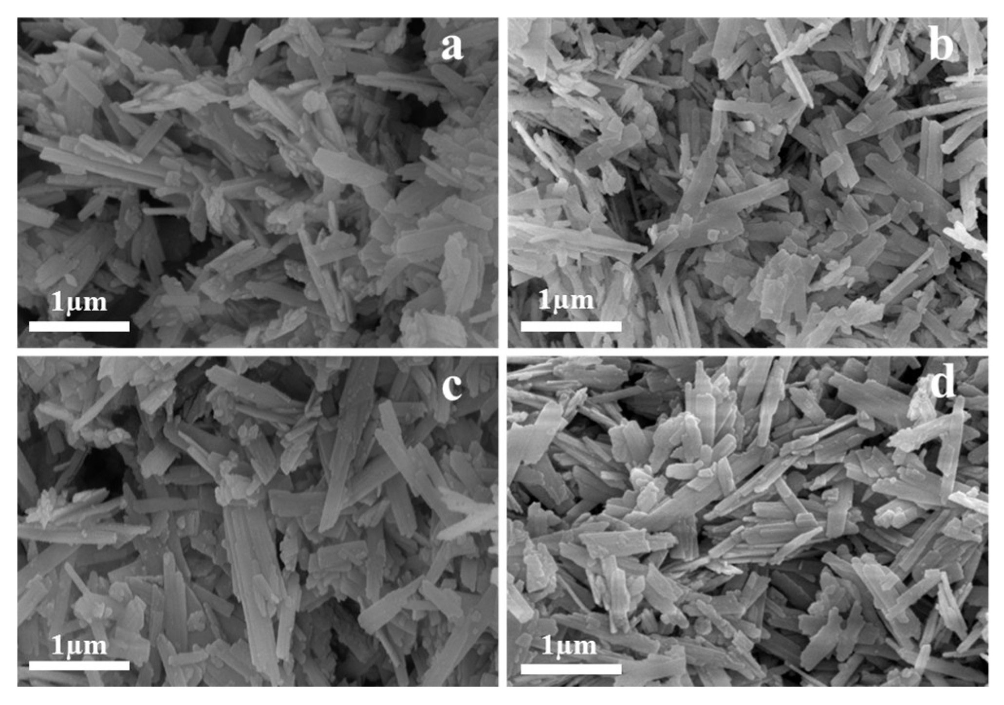
3.3.1. Scanning Electron Microscopy Analysis

The morphology of goethite minerals after bio-reduction under different conditions was observed using scanning electron microscopy (SEM). From Figure 6, it can be observed that the mineral morphology of the four groups of goethite, labeled as (a) GT + MR-1, (b) GT + MR-1 + 0.5 mM AQS, (c) GT + MR-1 + aniline, and (d) GT + MR-1 + 0.5 mM AQS + aniline, still exhibits the typical characteristics of goethite after the reaction, which is consistent with their pre-reaction morphology. This demonstrates the inherent stability of goethite. However, the mineral morphology of group (d) after bio-reduction of goethite is more “mottled” than the other three groups, and the tail end is broken and uneven. A common phenomenon can be observed from the SEM images of (a) and (c): solid particles are attached to the surface of goethite after bio-reduction. This is due to the opposite surface charges between microorganisms and iron oxides in most environments [51,52]. Therefore, in the presence of microorganisms alone, MR-1 adheres to the surface of iron oxides and directly reduces Fe (III). This process is mainly facilitated by the attachment of MR-1’s self-secreted polysaccharide polymers to the surface of goethite, forming a dense biofilm, and the extracellular membrane-bound reductase transfers electrons to Fe (III), thereby promoting its reduction [53,54]. However, this phenomenon is not observed in (b) and (d), indicating the existence of other electron transfer mechanisms in addition to direct contact [55]. In this reaction process, AQS functions as an extracellular electron shuttle, accelerating the transfer of electrons from the outer membrane of microorganisms to insoluble iron minerals, thus promoting the bio-reduction of Fe (III) [56,57]. This further illustrates that AQS, as an electron shuttle, significantly enhances the bio-reduction of Fe (III) by MR-1 in this experiment.

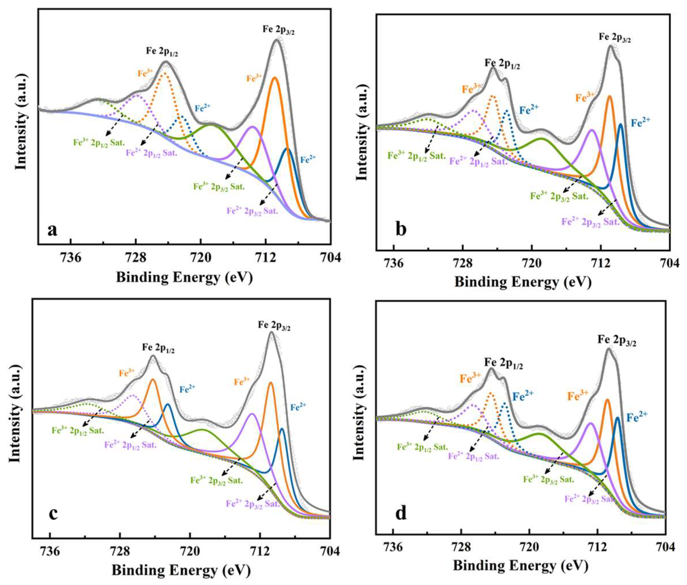
3.3.2. X-ray Photoelectron Spectral Analysis

To further investigate the effect of MR-1 under varying conditions on goethite reduction, XPS was used to detect the chemical state changes of iron elements on the surface of goethite after the completion of the reaction (Figure 7 and Table 3). Under the action of MR-1 alone, the Fe2p spectrum of goethite exhibited two peaks at 709.2 eV and 722.25 eV, belonging to Fe (II) oxides, with a content of 32.2%. Peaks at 710.75 eV and 724.33 eV belonged to Fe (III) oxides or hydroxides, with a content of 67.8%. After the addition of the electron shuttle AQS, the surface Fe (II)-O oxides increased to 40.81%, indicating that the electron shuttle revealed electron transfer from bacteria to goethite, thereby reducing Fe (III) to Fe (II). Furthermore, after goethite bio-reduction in the presence of aniline under the mediation of AQS, the content of Fe (II) characteristic peaks increased to 41.32%, which was 1.28 times higher than under the action of MR-1 alone. The specific values are provided in Table 1. Moreover, there was a shift in the peak positions of Fe (II)-O and Fe (III)-O, suggesting a change in electron density, and iron elements may have been involved in chemical reactions [58]. According to the results of Notini et al. [59], goethite with a higher surface defect density was more than twice that of goethite with fewer defects. Based on the XPS results mentioned above, aniline stimulated increased surface defects in goethite when present in the company of the electron shuttle AQS mediated by MR-1, increased surface defects in goethite, facilitating the aggregation of more electrons by the electron shuttle. As a result, more Fe (II) was released into the reaction system. This demonstrates that the synergistic action of adding aniline and the electron shuttle can significantly promote the Fe (III)-reducing by MR-1.

3.3.3. X-ray Diffraction Analysis

In this experiment, it was observed that the addition of aniline led to a significant increase in the reduction of Fe (III) oxide by MR-1 with the mediation of the electron shuttle AQS. The amount of Fe (II) produced and the surface Fe (II)-O detected in the reaction system were much higher compared to the reduction of Fe (III) by microorganisms alone. X-ray diffraction (XRD) analysis was performed on the reacted mineral to investigate the mineral phase of goethite after bio-reduction (Figure 8). Upon comparison with the standard XRD pattern of pure goethite (JCPC29-0713), the diffraction peaks identified at 21.22°, 26.32°, 33.24°, 34.70°, 36.65°, 39.98°, 41.19°, 53.24°, and 59.02° correspond to the (110), (120), (130), (021), (111), (121), (140), (221), and (151) crystallographic planes of goethite, respectively. Based on the comparison, it can be concluded that the solid minerals in the system after bio-reduction are mainly composed of goethite, and no characteristic peaks of other minerals were observed. To create secondary minerals like siderite (FeCO3), vivianite (Fe3(PO4)2), magnetite, or green rust, a longer bio-reduction period and sufficient Fe (II) generation are required. This is necessary to produce secondary Fe (II) or Fe (II)/Fe (III) mixed minerals [60,61]. Due to the relatively short cultivation period in this experiment, although aniline enhanced the reduction rate of Fe (III) by MR-1 mediated by the electron shuttle, the accumulation of Fe (II) in the reaction system was insufficient to generate new secondary Fe (II) or Fe (II)/Fe (III) mixed minerals. Therefore, the mineral phase remained Fe (III) dominant in this study.

4. Discussion

4.1. AQS Enhance the Fe (III) Reduction by Shewanella Oneidensi MR-1

This experimental study reveals that the reduction of Fe (III) oxide goethite by the microorganism MR-1 predominantly occurs through direct contact interactions. When Fe (III) oxide is the only electron acceptor, the addition of 0.1, 0.3, and 0.5 mM AQS significantly increased the production of Fe (II) in the reaction groups, which were 1.61, 1.82, and 2.39-fold, respectively, above those of microbial-only. This is mainly due to the presence of quinones in the electron shuttle compound AQS, which serves as the primary electron carrier in dissimilatory iron reduction [62]. Thus, a higher concentration of AQS is more favorable for the Fe (III) biological reduction.
Furthermore, the addition of the electron shuttle compound AQS also accelerated the consumption of sodium lactate. Sodium lactate stimulates cellular metabolism and provides more electrons for the Fe (III) biological reduction [43,63,64]. Zhu Weihuang et al. found that the interaction between microorganisms and iron oxides became more favorable in the presence of AQS in the reaction system. AQS can enhance the affinity between the electron donor (lactate) and the electron acceptor (Fe (III) oxide minerals) [65]. As an electron transfer carrier, the electron shuttle facilitates the utilization of lactate by microorganisms, significantly enhancing the bio-reduction of Fe (III) oxide minerals.

4.2. Aniline Promotes AQS-Mediated Reduction of Fe (III) Oxides by MR-1

Our initial hypothesis was that aniline, as a common toxic substance, might have a detrimental effect on microorganisms, thereby inhibiting the Fe (III) oxides bio-reduction. However, the results of this study demonstrate that the addition of aniline noticeably enhances the reduction of Fe (III) by MR-1 under the mediation of AQS. In particular, the introduction of 3 μM aniline resulted in a 2.51-fold increase in Fe (II) production compared to solely microbial activity. But the promoting effect started to diminish when the concentration of aniline increased to 7 μM. XPS results demonstrated an increase in Fe (II)-O content on the solid mineral surface to 41.32% after the reaction, and SEM analysis indicated that aniline did not affect the morphology of Fe (III) minerals. Additionally, no secondary mineral formation was observed through XRD analysis.
Simultaneously, aniline exhibited varying degrees of degradation during the reduction process. Previous research has shown that iron-reducing bacteria with strong metabolic capabilities in anaerobic environments can significantly affect the degradation of organic pollutants (such as sugars, organic acids, and aromatic as a common toxic substance, may have a toxic effect on microorganisms, thereby inhibiting the biological reduction of Fe (III) oxides. However, the results of this experiment showed that hydrocarbons) can promote Fe (III) bio-reduction [66,67,68]. Furthermore, the addition of redox-active substances capable of forming an electron shuttle system can effectively promote this bio-reduction reaction, and the electron shuttle can catalyze the degradation of various aromatic hydrocarbon pollutants [38,69,70,71,72]. In previous studies, aromatic compounds such as aniline, toluene, and phenol have been utilized and degraded by microorganisms as carbon sources under anaerobic conditions [42,47,73]. Therefore, in this experiment, it is possible that aniline was utilized by MR-1, leading to a gradual decrease in its concentration.

5. Conclusions

In this study, we not only emphasized the significant promotion of the electron shuttle AQS on Shewanella oneidensis MR-1’s reduction of goethite but also demonstrated that aniline, as a typical aromatic compound, can enhance MR-1’s reduction of Fe (III) while undergoing degradation during the process. After the reduction, the Fe (II)-O content on the surface of the goethite solid mineral significantly increased. This research provides novel observations into the roles of organic pollutants like aniline and electron shuttles in iron bio-reduction, which is crucial for understanding the biogeochemical cycling of iron in polluted environments. Moreover, this method can be considered a favorable technique for environmental bio-remediation in contaminated underground environments, such as soil and sediments. However, this experiment primarily focused on changes in the solid-phase minerals after reduction. Further exploration of microbial changes at the Fe (III)-reducing is necessary to gain better insight into the mechanisms of microbial reduction of iron (hydr)oxide minerals. Additionally, this study was conducted in a controlled laboratory setting, and real-world environments may contain various potential influencing factors. Therefore, it is necessary to apply this research to geological environments rich in iron oxides to gain practical environmental insights.

Supplementary Materials

The following supporting information can be downloaded at: https://www.mdpi.com/article/10.3390/w15203686/s1, Table S1 Effect of different concentrations of AQS on Fe (II) production in the bio-reduction process of goethite. (a) Fe (II) content in the reaction system. (Data for Figure 2a); Table S2 Effect of different concentrations of aniline on the production of Fe (II) during AQS-mediated goethite bio-reduction. (Data for Figure 3); Table S3 Consumption of sodium lactate during AQS-mediated Fe (III) bio-reduction. (Data for Figure 4a); Table S4 Impact of aniline on sodium lactate consumption during AQS-mediated Fe (III) bio-reduction. (Data for Figure 4c); Table S5 Change in aniline concentration. (Data for Figure 5a).

Author Contributions

Conceptualization, M.T. and C.W.; methodology, M.T. and Z.D.; software, M.T.; validation, C.W., Z.D. and Q.C.; formal analysis, M.T.; investigation, M.T., Z.W. and Y.Z.; resources, C.W.; data curation, M.T.; writing—original draft preparation, M.T.; writing—review and editing, M.T.; project administration, C.W.; funding acquisition, C.W. All authors have read and agreed to the published version of the manuscript.

Funding

This study was funded by the National Natural Science Foundation of China (No. 41772129).

Data Availability Statement

The data are available from the corresponding authors.

Conflicts of Interest

The authors declare no conflict of interest.

References

  1. Li, H.J.; Peng, J.J. Recent advances in studies on dissimilatory Fe(III)-reducing microorganisms. Acta Ecol. Sin. 2012, 32, 1633–1642. [Google Scholar]
  2. Chen, L.; Zhang, H.X.; Li, Y.; Zheng, S.L.; Liu, F.H. The role of microorganisms in the geochemical iron cycle. Sci. Sin. 2016, 46, 1069–1078. [Google Scholar]
  3. Zhang, Y.X.; Wang, Y.P.; Wang, H.P. Research Advances in the Mechanisms of Fe (III) Dissimilatory Reduction and the Reduction Products; The Chemical Industry and Engerring Soceiety of China: Beijing, China, 2015; p. 5. [Google Scholar]
  4. Schuth, S.; Hurraß, J.; Münker, C.; Mansfeldt, T. Redox-dependent fractionation of iron isotopes in suspensions of a groundwater-influenced soil. Chem. Geol. 2015, 392, 74–86. [Google Scholar] [CrossRef]
  5. Li, Y.; Yu, S.; Strong, J.; Wang, H. Are the biogeochemical cycles of carbon, nitrogen, sulfur, and phosphorus driven by the "FeIII - FeII redox wheel" in dynamic redox environments? J. Soils Sediments 2012, 12, 683–693. [Google Scholar] [CrossRef]
  6. Kappler, A.; Haderlein, S.B. Natural organic matter as reductant for chlorinated aliphatic pollutants Environ. Sci. Technol. 2003, 37, 2714–2719. [Google Scholar] [CrossRef] [PubMed]
  7. Jiang, Y.; Xi, B.D.; Li, R.; Li, M.X.; Xu, Z.; Yang, Y.N.; Gao, S.B. Advances in Fe (III) bioreduction and its application prospect for groundwater remediation: A review. Front. Environ. Sci. Eng. 2019, 13, 89. [Google Scholar] [CrossRef]
  8. Lalonde, K.; Mucci, A.; Ouellet, A.; Mucci, A.; Ouellet, A.; Gélinas, Y. Preservation of organic matter in sediments promoted by iron. Nature 2012, 483, 198–200. [Google Scholar] [CrossRef] [PubMed]
  9. Melton, E.D.; Swanner, E.D.; Behrens, S.; Behrens, S.; Schmidt, C.; Kappler, A. The interplay of microbially mediated and abiotic reactions in the biogeochemical Fe cycle. Nat. Rev. Microbiol. 2014, 12, 797–808. [Google Scholar] [CrossRef]
  10. Tobler, N.B.; Hofstetter, T.B.; Straub, K.L.; Fontana, D.; Schwarzenbach, R.P. Iron-Mediated Microbial Oxidation and Abiotic Reduction of Organic Contaminants under Anoxic Conditions. Environ. Sci. Technol. 2007, 41, 7765–7772. [Google Scholar] [CrossRef]
  11. Straub, K.L.; Benz, M.; Schink, B. Iron metabolism in anoxic environments at near neutral pH. FEMS Microbiol. Ecol. 2001, 34, 181–186. [Google Scholar] [CrossRef]
  12. Thomsen, U.; Thamdrup, B.; Stahl, D.A.; Canfield, D.E. Pathways of organic carbon oxidation in a deep lacustrine sediment, Lake Michigan. Limnol. Oceanogr. 2004, 49, 2046–2057. [Google Scholar] [CrossRef]
  13. Li, H.; Zhang, Q.; Wang, X.-L.; Ma, X.-Y.; Lin, K.-F.; Liu, Y.-D.; Gu, J.-D.; Lu, S.-G.; Shi, L.; Lu, Q.; et al. Biodegradation of benzene homologues in contaminated sediment of the East China Sea. Bioresour. Technol. 2012, 124, 129–136. [Google Scholar] [CrossRef] [PubMed]
  14. Wu, Y.; Guo, J.; Han, Y.; Zhu, J.Y.; Zhou, L.X.; Lan, Y.Q. Insights into the mechanism of persulfate activated by rice straw biochar for the degradation of aniline. Chemosphere 2018, 200, 373–379. [Google Scholar] [CrossRef] [PubMed]
  15. United States Environmental Protection Agency. OPPT Chemical Fact Sheets, Aniline Fact Sheet, Support Document (CAS No. 62-53-3); United States Environmental Protection Agency: Washington, DC, USA, 1994.
  16. Li, J.; Jin, Z. Effect of hypersaline aniline-containing pharmaceutical wastewater on the structure of activated sludge-derived bacterial community. J. Hazard. Mater. 2009, 172, 432–438. [Google Scholar] [CrossRef] [PubMed]
  17. Lovley, D.R.; Phillips, E.J.; Lonergan, D.J. Lonergan. Enzymic versus nonenzymic mechanisms for iron (III) reduction in aquatic sediments. Environ. Sci. Technol. 1991, 25, 1062–1067. [Google Scholar] [CrossRef]
  18. Zhu, X.Y.; Yuan, Y.X.; Song, C.C.; Jiang, M. Advance in Dissimilatory Iron Reduction in Wetland Soils and Sediments. Wetl. Sci. 2020, 18, 122–128. [Google Scholar]
  19. Bae, S.; Lee, W. Biotransformation of lepidocrocite in the presence of quinones and flavins. Geochim. Cosmochim. Acta 2013, 114, 144–155. [Google Scholar] [CrossRef]
  20. Orsetti, S.; Laskov, C.; Haderlein, S.B. Electron Transfer between Iron Minerals and Quinones: Estimating the Reduction Potential of the Fe(II)-Goethite Surface from AQDS Speciation. Environ. Sci. Technol. 2013, 47, 14161–14168. [Google Scholar] [CrossRef]
  21. Roh, Y.; Liu, S.V.; Li, G.; Huang, H.; Phelps, T.J.; Zhou, J. Isolation and characterization of metal-reducing Thermoanaerobacter strains from deep subsurface environments of the Piceance Basin, Colorado. Appl. Environ. Microbiol. 2002, 68, 6013–6020. [Google Scholar] [CrossRef]
  22. Yang, L.; Steefel, C.I.; Marcus, M.A.; Bargar, J.R. Kinetics of Fe (II)-Catalyzed Transformation of 6-line Ferrihydrite under Anaerobic Flow Conditions. Environ. Sci. Technol. 2010, 44, 5469–5475. [Google Scholar] [CrossRef]
  23. Till, J.L.; Guyodo, Y.; Lagroix, F.; Ona-Nguema, G.; Brest, J. Magnetic comparison of abiogenic and biogenic alteration products of lepidocrocite. Earth Planet. Sci. Lett. 2014, 395, 149–158. [Google Scholar] [CrossRef]
  24. Usman, M.; Abdelmoula, M.; Faure, P.; Ruby, C.; Hanna, K. Transformation of various kinds of goethite into magnetite: Effect of chemical and surface properties. Geoderma 2013, 197–198, 9–16. [Google Scholar] [CrossRef]
  25. Lee, S.H.; Lee, I.; Roh, Y. Biomineralization of a poorly crystalline Fe(III) oxide, akaganeite, by an anaerobic Fe(III)-reducing bacterium (Shewanella alga) isolated from marine environment. Geosci. J. 2003, 7, 217–222. [Google Scholar] [CrossRef]
  26. Li, X.; Liu, L.; Liu, T.; Yuan, T.; Zhang, W.; Li, F.; Zhou, S.; Li, Y. Electron transfer capacity dependence of quinone-mediated Fe (III) reduction and current generation by Klebsiella pneumoniae L17. Chemosphere 2013, 92, 218–224. [Google Scholar] [CrossRef] [PubMed]
  27. Nevin, K.P.; Lovley, D.R. Potential for nonenzymatic reduction of Fe (III) via electron shuttling in subsurface sediments. Environ. Sci. Technol. 2000, 34, 2472–2478. [Google Scholar] [CrossRef]
  28. Zhang, Y.; Chen, X.; Wu, Y. Electron shuttle-mediated microbial extracellular electron transfer: Mechanisms and geochemical implications. Ecol. Environ. 2021, 30, 213–222. [Google Scholar]
  29. Liu, G.F.; Zhu, J.Q.; Yu, H.L.; Jin, R.; Wang, J.; Zhou, J. Review on electron-shuttle-mediated microbial reduction of iron oxides minerals. Earth Sci. 2018, 43 (Suppl. S1), 157–170. [Google Scholar]
  30. Shi, L.; Dong, H.; Reguera, G.; Beyenal, H.; Lu, A.; Liu, J.; Yu, H.-Q.; Fredrickson, J.K. Extracellular electron transfer mechanisms between microorganisms and minerals. Nat. Rev. Microbiol. 2016, 14, 651–662. [Google Scholar] [CrossRef] [PubMed]
  31. Yang, Y.; Chen, L.; Jiang, Y.; Xu, Z.; Gao, S.; Li, M.; Li, R.; Xi, B. Promoting mechanism of electronic shuttle for bioavailability of Fe (III) oxide and its environmental significance. Water Supply 2020, 20, 1157–1166. [Google Scholar] [CrossRef]
  32. Liu, H.; Yuan, Y.; Zhang, S.; Li, K. Characteristics of Fe (III) reduction by Fe (III)-reducing bacterium Clostridium butyricum LQ25 under the condition of electron shuttles. Acta Microbiol. Sin. 2021, 61, 1496–1506. [Google Scholar]
  33. Liu, W.; Wu, Y.; Liu, T.; Li, F.; Dong, H.; Jing, M. Influence of incubation temperature on 9,10-anthraquinone-2-sulfonate (AQS)-mediated extracellular electron transfer. Front. Microbiol. 2019, 10, 464. [Google Scholar] [CrossRef] [PubMed]
  34. Mohamed, A.; Sun, B.; Yu, C.; Gu, X.; Ashry, N.; Riahi, Y.; Dai, K.; Huang, Q. Size effect of hematite particles on the Cr (VI) reduction by Shewanella oneidensis MR-1. J. Environ. Chem. Eng. 2021, 9, 105096. [Google Scholar] [CrossRef]
  35. Schwertmann, U. The Iron Oxides: Structure, Properties, Reactions, Occurrences and Uses; John Wiley & Sons: Hoboken, NJ, USA, 2007. [Google Scholar]
  36. Tamura, H.; Goto, K.; Yotsuyanagi, T.; Nagayama, M. Spectrophotometric determination of iron (II) with 1,10-phenanthroline in the presence of large amounts of iron (III). Talanta 1974, 21, 314–318. [Google Scholar] [CrossRef] [PubMed]
  37. Shi, Z.; Zachara, J.M.; Shi, L.; Wang, Z.; Moore, D.A.; Kennedy, D.W.; Fredrickson, J.K. Redox Reactions of Reduced Flavin Mononucleotide (FMN), Riboflavin (RBF), and Anthraquinone-2,6-disulfonate (AQDS) with Ferrihydrite and Lepidocrocite. Environ. Sci. Technol. 2012, 46, 11644–11652. [Google Scholar] [CrossRef]
  38. Liu, C.; Zachara, J.M.; Foster, N.S.; Strickland, J. Kinetics of Reductive Dissolution of Hematite by Bioreduced Anthraquinone-2,6-disulfonate. Environ. Sci. Technol. 2007, 41, 7730–7735. [Google Scholar] [CrossRef]
  39. Yu, L.; Wang, S.; Tang, Q.-W.; Cao, M.-Y.; Li, J.; Yuan, K.; Wang, P.; Li, W.-W. Enhanced reduction of Fe (III) oxides and methyl orange by Klebsiella oxytoca in presence of anthraquinone-2-disulfonate. Appl. Microbiol. Biotechnol. 2016, 100, 4617–4625. [Google Scholar] [CrossRef] [PubMed]
  40. Rau, J.; Knackmuss, H.; Stolz, A. Effects of different quinoid redox mediators on the anaerobic reduction of azo dyes by bacteria. Environ. Sci. Technol. 2002, 36, 1497–1504. [Google Scholar] [CrossRef]
  41. Zhu, W.; Yao, T.; Huang, T. The kinetic characteristics of the microbial dissimilatory reduction of lepidocrocite. Huanjing Kexue Xuebao 2012, 32, 1348–1356. [Google Scholar]
  42. Okamoto, A.; Hashimoto, K.; Nealson, K.H.; Nakamura, R. Rate enhancement of bacterial extracellular electron transport involves bound flavin semiquinones. Proc. Natl. Acad. Sci. USA 2013, 110, 7856–7861. [Google Scholar] [CrossRef]
  43. Pinchuk, G.E.; Rodionov, D.A.; Yang, C.; Li, X.; Osterman, A.L.; Dervyn, E.; Geydebrekht, O.V.; Reed, S.B.; Romine, M.F.; Collart, F.R.; et al. Genomic reconstruction of Shewanella oneidensis MR-1 metabolism reveals a previously uncharacterized machinery for lactate utilization. Proc. Natl. Acad. Sci. USA 2009, 106, 2874–2879. [Google Scholar] [CrossRef]
  44. Wang, J.; Xie, Z.; Wang, Y.; Chen, M. Synergy between indigenous bacteria and extracellular electron shuttles enhances transformation and mobilization of Fe (III)/As (V). Sci. Total Environ. 2021, 783, 147002. [Google Scholar] [CrossRef]
  45. Zheng, Y.; Dai, X.; Wang, L.; Xu, W.; He, Z.; Ma, M. Arsenate reduces copper phytotoxicity in gametophytes of Pteris vittata. J. Plant Physiol. 2008, 165, 1906–1916. [Google Scholar] [CrossRef]
  46. Rooney-Varga, J.N.; Anderson, R.T.; Fraga, J.L.; Ringelberg, D.; Lovley, D.R. Microbial communities associated with anaerobic benzene degradation in a petroleum-contaminated aquifer. Appl. Environ. Microbiol. 1999, 65, 3056–3063. [Google Scholar] [CrossRef]
  47. Shuo, L.; Dong, Q. Effects of benzene analogs as sole carbon source on dissimilatory iron reduction. J. Northwest Sci.-Tech. Univ. Agric. For. 2006, 10, 101–106+112. [Google Scholar]
  48. Anderson, R.T.; Lovley, D.R. Naphthalene and benzene degradation under Fe (III)-reducing conditions in petroleum-contaminated aquifers. Bioremediat. J. 1999, 3, 121–135. [Google Scholar] [CrossRef] [PubMed]
  49. Lovley, D.R.; Woodward, J.C.; Chapelle, F.H. Stimulated anoxic biodegradation of aromatic hydrocarbons using Fe (III) ligands. Nature 1994, 370, 128–131. [Google Scholar] [CrossRef]
  50. Tor, J.M.; Lovley, D.R. Anaerobic degradation of aromatic compounds coupled to Fe (III) reduction by Ferroglobus placidus. Environ. Microbiol. 2001, 3, 281–287. [Google Scholar] [CrossRef]
  51. Cornell, R.M.; Schwertmann, U. The Iron Oxides: Structure, Properties, Reactions, Occurences and Uses, 2nd ed.; Wiley: Weinheim, Germany, 2004. [Google Scholar]
  52. Collins, Y.E.; Stotzky, G. Heavy metals alter the electrokinetic properties of bacteria, yeasts, and clay minerals. Appl. Environ. Microbiol. 1992, 58, 1592–1600. [Google Scholar] [CrossRef]
  53. Franks, A.E.; Nevin, K.P.; Glaven, R.H.; Lovley, D.R. Microtoming coupled to microarray analysis to evaluate the spatial metabolic status of Geobacter sulfurreducens biofilms. ISME J. 2010, 4, 509–519. [Google Scholar] [CrossRef]
  54. Gagen, E.J.; Zaugg, J.; Tyson, G.W.; Southam, G. Goethite Reduction by a Neutrophilic Member of the Alphaproteobacterial Genus Telmatospirillum. Front. Microbiol. 2019, 10, 2938. [Google Scholar] [CrossRef] [PubMed]
  55. Luu, Y.S.; Ramsay, J.A. Review: Microbial mechanisms of accessing insoluble Fe (III) as an energy source. World J. Microbiol. Biotechnol. 2003, 19, 215–225. [Google Scholar] [CrossRef]
  56. Shi, L.; Squier, T.C.; Zachara, J.M.; James, K. Respiration of metal (hydr)oxides by Shewanella and Geobacter: A key role for multihaem c-type cytochromes. Mol. Microbiol. 2010, 65, 12–20. [Google Scholar] [CrossRef] [PubMed]
  57. O’Loughlin, E.J. Effects of Electron Transfer Mediators on the Bioreduction of Lepidocrocite (γ-FeOOH) by Shewanella putrefaciens CN132. Environ. Sci. Technol. 2008, 42, 6876–6882. [Google Scholar] [CrossRef]
  58. Lu, M.C. Oxidation of chlorophenols with hydrogen peroxide in the presence of goethite. Chemosphere 2000, 40, 125–130. [Google Scholar] [CrossRef] [PubMed]
  59. Notini, L.; Byrne, J.M.; Tomaszewski, E.J.; Latta, D.E.; Zhou, Z.; Scherer, M.M.; Kappler, A. Mineral Defects Enhance Bioavailability of Goethite toward Microbial Fe (III) Reduction. Environ. Sci. Technol. 2019, 53, 8883–8891. [Google Scholar] [CrossRef]
  60. Zhou, X.; Liu, D.; Bu, H.; Deng, L.; Liu, H.; Yuan, P.; Du, P.; Song, H. XRD-based quantitative analysis of clay minerals using reference intensity ratios, mineral intensity factors, Rietveld, and full pattern summation methods: A critical review. Solid Earth Sci. 2018, 3, 16–29. [Google Scholar] [CrossRef]
  61. Yamamura, S.; Sudo, T.; Watanabe, M.; Tsuboi, S.; Soda, S.; Ike, M.; Amachi, S. Effect of extracellular electron shuttles on arsenic-mobilizing activities in soil microbial communities. J. Hazard. Mater. 2017, 342, 571–578. [Google Scholar] [CrossRef]
  62. Struyk, Z.; Sposito, G. Redox properties of standard humic acids. Geoderma 2001, 102, 329–346. [Google Scholar] [CrossRef]
  63. Liu, Y.; Liu, S.; Liu, C.; Wang, X.; Yan, L.; Zhu, G.; Lu, X. Benzene promotes microbial Fe (III) reduction and flavins secretion. Geochim. Cosmochim. Acta 2019, 264, 92–104. [Google Scholar] [CrossRef]
  64. Finke, N.; Vandieken, V.; Jørgensen, B.B. Acetate, lactate, propionate, and isobutyrate as electron donors for iron and sulfate reduction in Arctic marine sediments, Svalbard. FEMS Microbiol. Ecol. 2007, 59, 10–22. [Google Scholar] [CrossRef]
  65. Zhu, W.; Nan, Y.; Huang, T.; Wu, F. The mechanism, thermodynamic and kinetic characteristics of the microbial reduction of goethite mediated by anthraquinone-2-sulfonate. Geomicrobiol. J. 2013, 30, 928–940. [Google Scholar] [CrossRef]
  66. Lovley, D.R.; Lonergan, D.J. Anaerobic Oxidation of Toluene, Phenol, and p-Cresol by the Dissimilatory Iron-Reducing Organism, GS-15. Appl. Environ. Microbiol. 1990, 56, 1858–1864. [Google Scholar] [CrossRef] [PubMed]
  67. Borch, T.; Kretzschmar, R.; Kappler, A.; Van Cappellen, P. Biogeochemical redox processes and their impact on contaminant dynamics. Environ. Sci. Technol. 2010, 44, 15–23. [Google Scholar] [CrossRef]
  68. Lovley, D.R.; Baedecker, M.J.; Lonergan, D.J.; Cozzarelli, I.M.; Elizabeth J., P.; Siegel, D.I. Oxidation of aromatic contaminants coupled to microbial iron reduction. Nature 1989, 339, 297–300. [Google Scholar] [CrossRef]
  69. Wolf, M.; Kappler, A.; Jiang, J.; Meckenstock, R.U. Effects of humic substances and quinones at low concentrations on ferrihydrite reduction by Geobacter metallireducens. Environ. Sci. Technol. 2009, 43, 5679–5685. [Google Scholar] [CrossRef] [PubMed]
  70. Cervantes, F.J.; Dijksma, W.; Duong-Dac, T.; Ivanova, A.; Lettinga, G.; Field, J.A. Anaerobic mineralization of toluene by enriched sediments with quinones and humus as terminal electron acceptors. Appl. Environ. Microbiol. 2001, 67, 4471–4478. [Google Scholar] [CrossRef] [PubMed]
  71. Chakraborty, R.; Coates, J.D. Anaerobic degradation of monoaromatic hydrocarbons. Appl. Microbiol. Biotechnol. 2004, 64, 437–446. [Google Scholar] [CrossRef] [PubMed]
  72. Lovley, D.R.; Woodward, J.C.; Chapelle, F.H. Rapid anaerobic benzene oxidation with a variety of chelated Fe (III) forms. Appl. Environ. Microbiol. 1996, 62, 288–291. [Google Scholar] [CrossRef] [PubMed]
  73. Borch, T.; Inskeep, W.P.; Harwood, J.A.; Gerlach, R. Impact of ferrihydrite and anthraquinone-2, 6-disulfonate on the reductive transformation of 2, 4, 6-trinitrotoluene by a gram-positive fermenting bacterium. Environ. Sci. Technol. 2005, 39, 7126–7133. [Google Scholar] [CrossRef]
Figure 1. Characterizations of synthesized goethite: (a) XRD pattern and (b) SEM image.
Water 15 03686 g001
Figure 2. Effect of different concentrations of AQS on Fe (II) production in the bio-reduction process of goethite. (a) Fe (II) content in the reaction system. Data are presented as mean ± SD (n = 3); at certain time points, the error bar is smaller than the size of the top line. (b) Color change of GT + MR-1 culture medium. (c) Color change of GT + MR-1 + 0.5 mM AQS culture medium.
Water 15 03686 g002
Figure 3. Effect of different concentrations of aniline on the production of Fe (II) during AQS-mediated goethite bio-reduction. Data are presented as mean ± SD (n = 3). At certain time points, the error bar is smaller than the size of the top line.
Water 15 03686 g003
Figure 4. Sodium lactate consumption and first-order kinetic fitting: (a) Consumption of sodium lactate during AQS-mediated Fe (III) bio-reduction. (b) AQS-mediated first-order kinetic fitting of sodium lactate in Fe (III) bio-reduction processes. (c) Impact of aniline on sodium lactate consumption during AQS-mediated Fe (III) bio-reduction. (d) First-order kinetic depletion of sodium lactate consumption during AQS-mediated Fe (III) bio-reduction by aniline. Data are presented as mean ± SD (n = 3). At certain time points, the error bar is smaller than the size of the top line.
Water 15 03686 g004
Figure 5. (a) Change in aniline concentration. (b) First-order kinetic fitting of aniline degradation. Data are presented as mean ± SD (n = 3). At certain time points, the error bar is smaller than the size of the top line.
Water 15 03686 g005
Figure 6. SEM characterization of minerals after bio-reduction of goethite: (a) GT + MR-1; (b) GT + MR-1 + 0.5 mM AQS; (c) GT + MR-1 + 1 μM aniline; (d) GT + MR-1 + 0.5 mM AQS + 3 μM aniline.
Water 15 03686 g006
Figure 7. XPS characterization of minerals after bio-reduction of goethite: (a) GT + MR-1; (b) GT + 0.5 mM AQS; (c) GT + MR-1 + 1 μM aniline; (d) GT + MR-1 + 0.5 mM AQS + 3 μM aniline.
Water 15 03686 g007
Figure 8. XRD patterns of minerals after bio-reduction of goethite under different conditions.
Water 15 03686 g008
Table 1. First-order rate constants for sodium lactate consumption during Fe (III) bio-reduction under different experimental conditions.
Experimental ConditionsK for Sodium Lactate Consumption (h−1)R2 for Sodium Lactate Consumption
GT + MR-10.0023 ± 0.0020.87
GT + MR-1 + 0.1 mM AQS0.0097 ± 0.0020.99
GT + MR-1 + 0.3 mM AQS0.0132 ± 0.0010.98
GT + MR-1 + 0.5 mM AQS0.0181 ± 0.0020.98
GT + MR-1 + 1 μM Aniline0.0052 ± 0.0030.97
GT + MR-1 + 0.3 mM AQS + 1 μM Aniline0.0144 ± 0.0050.94
GT + MR-1 + 0.5 mM AQS + 3 μM Aniline0.0191 ± 0.0010.96
GT + MR-1 + 0.5 mM AQS + 7 μM Aniline0.0125 ± 0.0020.97
Table 2. First-order rate constants for aniline degradation during AQS-mediated Fe (III) bio-reduction.
Experimental ConditionsK for Aniline
Consumption (h−1)
R2 for Aniline
Consumption
GT + MR-1 + 1 μM Aniline0.0139 ± 0.0010.97
GT + MR-1 + 0.5 mM AQS + 1 μM Aniline0.0143 ± 0.0020.95
GT + MR-1 + 0.5 mM AQS + 1 μM Aniline0.0232 ± 0.0010.98
GT + MR-1 + 0.5 mM AQS + 1 μM Aniline0.0098 ± 0.0010.99
Table 3. Percentage of Fe valence in Fe 2p spectrum of solid minerals after biological reduction of goethite.
FeGT + MR-1GT + MR-1 + 0.5 mM AQSGT + MR-1 + 1 μM
Aniline
GT + MR-1 + 0.5 mM AQS + 3 μM Aniline
B.E./eVat%B.E./eVat%B.E./eVat%B.E./eVat%
Fe2+2p3/2709.232.2709.6740.81709.2437.3709.741.32
Fe2+2p1/2722.25722.89722.48722.9
Fe3+2p3/2710.7567.8710.9259.19710.762.27710.8558.68
Fe3+2p1/2724.33724.46724.18724.6
Disclaimer/Publisher’s Note: The statements, opinions and data contained in all publications are solely those of the individual author(s) and contributor(s) and not of MDPI and/or the editor(s). MDPI and/or the editor(s) disclaim responsibility for any injury to people or property resulting from any ideas, methods, instructions or products referred to in the content.

