Review

Non-Neuronal Functions of the M2 Muscarinic Acetylcholine Receptor

Institute of Biochemistry, Medical Faculty, University of Giessen, Friedrichstrasse 24, 35392 Giessen, Germany
*
Author to whom correspondence should be addressed.
Genes 2013, 4(2), 171-197; https://doi.org/10.3390/genes4020171
Submission received: 31 January 2013 / Revised: 10 March 2013 / Accepted: 25 March 2013 / Published: 2 April 2013
(This article belongs to the Special Issue Signal Transduction)

Abstract

:
Acetylcholine is an important neurotransmitter whose effects are mediated by two classes of receptors. The nicotinic acetylcholine receptors are ion channels, whereas the muscarinic receptors belong to the large family of G protein coupled seven transmembrane helix receptors. Beyond its function in neuronal systems, it has become evident that acetylcholine also plays an important role in non-neuronal cells such as epithelial and immune cells. Furthermore, many cell types in the periphery are capable of synthesizing acetylcholine and express at least some of the receptors. In this review, we summarize the non-neuronal functions of the muscarinic acetylcholine receptors, especially those of the M2 muscarinic receptor in epithelial cells. We will review the mechanisms of signaling by the M2 receptor but also the cellular trafficking and ARF6 mediated endocytosis of this receptor, which play an important role in the regulation of signaling events. In addition, we provide an overview of the M2 receptor in human pathological conditions such as autoimmune diseases and cancer.

1. Introduction to Acetylcholine and Its Receptors

In the early 1920s, Otto Loewi identified the neurotransmitter acetylcholine as a substance that displays an inhibitory effect on heart functions. Due to its apparent release by the vagus nerve, acetylcholine was named “Vagusstoff”. In the 1960s, this discovery led to the detection of the respective acetylcholine binding nicotinic and muscarinic receptors [1,2,3]. Besides their function in the central nervous system, acetylcholine receptors have been shown to display important functions in non-neuronal cells. In fact, many peripheral cells of our body are capable of synthesizing acetylcholine and express at least some of the receptors. The nicotinic acetylcholine receptors are ion channels. Represented by five subtypes, namely M1, M2, M3, M4 and M5, the muscarinic acetylcholine receptors (mAChRs) belong to the seven transmembrane helix receptor superfamily which activate trimeric guanine nucleotide binding proteins (G proteins) [4,5]. The genes encoding the five mAChRs are found in different genetic loci in humans: 11q12-13 (CHRM1), 7q35-36 (CHRM2), 1q43-44 (CHRM3), 11p12-p11.2 (CHRM4) and 15q26 (CHRM5). The mAChR genes are highly compact in that they do not contain any introns, and the resulting gene products are highly homologous among mammalian species [5,6].
In general, mAChR subtypes are differentially expressed in various cell lines and tissues, which all seem to express several receptor types. The expression pattern results in functional differences, which may sometimes be hard to assign to a specific receptor type. The non-neuronal functions of muscarinic receptors are summarized in Section 2.
On the basis of their signaling mechanism, mAChRs can be divided into two major classes [7]. The stimulation of M1, M3 and M5 receptor subtypes increases the intracellular calcium concentration upon binding of pertussis toxin insensitive G proteins (Gq/11) and mediates the activation of the membrane bound enzyme phospholipase C (PLC) [8,9]. In contrast, M2 and M4 receptors mainly regulate the inhibition of adenylyl cyclase and the activation of potassium conductivity via pertussis toxin sensitive G proteins of the Gi and Go classes. However, all mAChRs can in principle affect adenylyl cyclase activity and thus lead to an increase or decrease of cAMP by means of non-canonical signaling [10,11]. The mechanisms of muscarinic receptor signaling are more closely reviewed in Section 3.
In structural terms, the muscarinic receptors consist of seven relatively well conserved transmembrane domains (TM). These domains are connected through three extracellular loops, including the extracellular amino terminus, which exhibits a high variability among the five mAChR subtypes, and three intracellular loops. In addition to the TMs, it has been reported that the C-terminal tail of the M2 receptor is associated with membranes by palmitoylation [12]. The acetylcholine binding cavity is formed by the extracellular face of the TMs, with TM3 providing the bottom of the cavity, whereas TM2 and TM7 narrow it down. In an activated state, the mAChRs bind G proteins within their third intracellular loop and catalyze GDP-GTP exchange, leading to downstream signaling events [10,13]. In this review, we have focused on the muscarinic receptor subtype M2 whose structure, signaling and cellular trafficking are the subject of Section 4. In this section, we also summarize the importance of the small GTPase ARF6 for the endocytosis and trafficking of the M2 receptor and G protein coupled receptors (GPCRs) in general.
Muscarinic AChRs frequently are targets of autoantibodies, resulting in autoimmune diseases. Due to their function in muscle contraction, they have also been suggested to participate in the pathogenesis of e.g., lung diseases. Importantly, it is known that mAChR subtypes play a key role in various cancers. Section 5 provides a short summary of the role of muscarinic receptors, especially that of the M2 receptor, in human diseases. Concluding remarks and future directions are addressed in Section 6.

2. Functions of Muscarinic Acetylcholine Receptors in Non-neuronal Cells

The functions of the different muscarinic receptor subtypes in the non-neuronal cholinergic system are diverse, depending on the distribution of single receptor subtypes in different tissues. Cells expressing mAChRs can be found virtually everywhere in the body. They are present e.g., in the epithelial layer of the airways, the skin, the immune system, the urinary bladder, reproductive organs, vascular endothelial cells, connective tissue, muscles and even tendons. Genetically ablated mice have been generated for all five muscarinic receptor subtypes [14,15,16,17,18]. These mice are viable and show no major developmental defects. However, some specific functions of receptor subtypes have been conclusively demonstrated in these mice (Summarized in [19]). In this paper, we are not aiming at providing an exhaustive review of the non-neuronal mAChR functions. For this, the reader should refer to recent excellent reviews in this topic [20,21,22].
Keratinocytes of the skin display the mRNA for all five muscarinic subtypes, and acetylcholine apparently plays a role in migration, differentiation, proliferation and adhesion of the cells [23,24,25]. In keratinocytes, it was shown that, in conjunction with nicotinic AChRs, muscarinic receptors are also necessary e.g., for the development of epithelial architecture and thus barrier formation. M1 and M4 receptors are well expressed in the suprabasal layer of the human skin, whereas M2, M3 and M4 receptors are found especially in the basal layer, as shown by in situ hybridization and antibody staining of skin sections [3,20]. As keratinocytes synthesize and secrete acetylcholine for autocrine stimulation, it is plausible that the secreted acetylcholine also affects melanocytes which express M1 through M5 receptors, detectable both at mRNA and protein level [26]. The migration of human keratinocytes is regulated in a reciprocal manner by two different muscarinic receptors. On the one hand, the M4 receptor induces migration and wound re-epithelialization, whereas M3 inhibits these processes. Accordingly, the M4 receptor upregulates the expression of the integrins α5β1, αVβ5, and αVβ6, which are connected to the movement of the cell, and the M3 receptor causes the expression of the integrins α2β1 and α3β1 that promote a sedentary state of the cell [27]. In fact, an acetylcholine gradient has even been shown to induce the migration of skin keratinocytes upwards in the epidermis [28].
The urothelium of the urinary bladder also expresses all of the muscarinic receptor subtypes of which M2 is the most prominently expressed one. Quantitative PCR and immunohistochemistry experiments showed that M1 and M2 are predominantly expressed by specific cell types in the urothelium. The M1 receptor is found in basal cells, and the M2 receptor shows a high expression in umbrella cells. M3 and M4 receptors are more homogenously distributed, and the M5 receptor content decreases from luminal to the basal cells [29]. Furthermore, the M3 receptor seems to be important for the contraction of the detrusor muscle [30] and generally appears to be the major regulator of smooth muscle contraction, as evidenced by knockout mouse studies [19].
In the epithelium of the airways, the M1 receptor plays a vital role. Together with nicotinic acetylcholine receptors, it stimulates the proliferation of the epithelial cells [31]. Moreover, ciliar activity, chloride secretion and thus the mucociliary clearance are mediated by muscarinic receptors [32]. Furthermore, M3 receptors stimulate while M2 receptors inhibit the cilia driven particle transport in the airways [33]. In addition to these receptor subtypes, the M4 receptor is highly abundant in peripheral lung tissues and has been suggested to play a role in autoinhibition of acetylcholine release in the trachea [34].
In regard to mediation of parasympathetic control, the M2 receptor is so far considered as the only muscarinic receptor subtype able to influence cardiac functions, e.g., heart rate or contractility. However, also M1 and M3 receptor proteins are expressed in the membrane of human cardiac cells [35] and participate in the control of the blood circulation system. Some major vasoactive mediators are nitric oxide, endothelium derived hyperpolarizing factor and prostanoids. All these substances are supplied by endothelial cells upon stimulation of their muscarinic receptors. Acetylcholine release itself might be regulated by the blood flow, shearing forces and blood pressure and may therefore modulate the release of the vasoactive mediators [22]. In line with this, release of acetylcholine has been shown as a consequence of increased blood flow in cultured human umbilical vein endothelial cells [36].
The non-neuronal cholinergic system also plays an important role in the immune system. The mRNAs for all five muscarinic receptors are present in most human mononuclear leukocytes, human leukemic cell lines and also in animal immune cells [20,37,38]. During immunological reactions, the autocrine and paracrine stimulation of macrophages, T cells or dendritic cells induces immune responses such as the expression of interleukin-2 and its receptor [8,38]. Depending on the immune status, the expression of muscarinic receptors varies in T cells. In non activated CD4 and CD8 cells, M1, M3, M4 and M5 receptors are present. This pattern changes upon activation of the cells via T cell receptor crosslinking, in that M1 and M5 receptors become upregulated in CD4 cells. In contrast, CD8 cells show an increased expression of M1 and M4 receptors, while the M3 receptor is downregulated [39]. Nevertheless, the M5 receptor seems to be the most important muscarinic receptor subtype in B and T cells, as the mRNA of this receptor subtype is upregulated upon activation in many of these cell types. The immunological activation therefore requires the cholinergic transmission for correct cell differentiation [40]. M5, together with M1 receptors, is also involved in modulation of the immune system as it is important for the regulation of cytokine production [41]. Another fundamental mechanism in the human body that is regulated by the non-neuronal cholinergic system is the release of insulin from β-pancreatic cells, which serves to maintain glucose homeostasis. It is mainly induced by glucose but at the same time, it can also be facilitated by muscarinic receptors. In this context, it is the M3 receptor delivering the stimulatory effects on the insulin release and potentiating it [42].

3. Signaling by the Muscarinic Acetylcholine Receptors

The muscarinic acetylcholine receptors belong to the family of GPCRs, comprising seven transmembrane domains. They initiate the signal transduction through their coupling with heterotrimeric guanine nucleotide binding proteins, the so called trimeric G proteins. These G proteins consist of α-, β- and γ-subunits which couple to the muscarinic receptors mainly at the receptor’s third intracellular (i3) loop. However, the second intracellular loop and the C-terminus have also been suggested to be involved [43]. Upon binding of a ligand, GPCRs are conformationally rearranged. This results in an exchange of bound GDP to GTP in the α-subunit, which then leaves the complex, whereas the β- and γ-subunits stay together to facilitate signal transduction. There are several isoforms of the G proteins, and due to their preferential coupling to a different subset, muscarinic receptors can be divided into two distinct groups. The M1, M3 and M5 receptor subtypes preferentially couple to Gq/11, whereas M2 and M4 receptors couple to Gi/o. Depending on the class of the G protein involved, different downstream effectors are activated in a cell upon receptor stimulation. The typical signaling pathways of mAChRs are summarized in Figure 1. Through their linkage to Gq/11, M1, M3 and M5 receptors predominantly activate PLC via the α-subunit. The activation of PLC results in production of diacylglycerol (DAG), which is generated together with inositol trisphosphate (IP3) upon the hydrolysis of phosphatidylinositol 4,5-bisphosphate (PIP2) by PLC. This path facilitates the mobilization of intracellular Ca2+ and activation of the protein kinase C (PKC). On the other hand, M2 and M4 receptors mainly inhibit the adenylyl cyclase through their corresponding G proteins, leading to a decrease in cAMP levels. However, this provides only a simplified view of the basal signaling events, and there are a lot more proteins involved in the fine tuning and regulation. In addition, the βγ-dimer is also able to participate in the signal transduction.
In accordance with the fact that there are 27 α-, five β- and 14 γ-subunit isoforms in the human genome for the heterotrimeric G proteins [44], several different signaling pathways can be stimulated by the muscarinic receptors. For example, phospholipase D (PLD) can be activated via G12 coupled to M3 receptors [45], and M2 as well as M3 receptors can activate the sphingosine kinase to mobilize Ca2+ via sphingosine-1-phosphate [46]. Furthermore, the signaling can be influenced by the Regulator of G protein Signaling (RGS) proteins which can accelerate the inactivation of G proteins and thus reduce their signaling [47]. Muscarinic receptors are also able to act on different ion channels in the cell membrane. Whereas M2 and M4 receptors can directly activate inward rectifying potassium channels via their βγ-subunits [48], the M1 receptor can inhibit this conductance [49]. In addition, M1, M3 and M5 receptors have the ability to activate calcium channels, which has been shown to be dependent on G protein activation [50,51].
A large scale approach to identify interaction partners of muscarinic receptors was done by Borroto-Escuela et al. by means of a tandem affinity purification method and subsequent mass spectrometry [52,53]. A wide number of proteins were selectively recruited upon carbachol stimulation of a specific receptor. For the M1 receptor, these included PLC β1, G protein coupled kinases GRK2 and GRK6, as well as the focal adhesion kinase. Proteins that were detected together with M1 receptors even in unstimulated neuroblastoma cells were caveolin-1 and -2, β-tubulin and ADP ribosylation factor 6 (ARF6). The trimeric G proteins that were found to be receptor associated were the Gα isoforms q, 11, 12 and 13 for M1, M3 and M5 receptors, and Gα i1, i2 and i3 for the M2 and M4 receptors. Furthermore, association of Gβ isoforms 1 and 2 as well as Gγ isoforms 2, 7 and 11 was also identified.
A downstream signaling cascade that is accessed by all subtypes of muscarinic receptors is the mitogen activated protein (MAP) kinase pathway. For M1, M3 and M5 receptors, the activation of the MAP kinase pathway typically involves the activation of PKC. This cascade includes the Gαq/11 mediated activation of PLC and the resulting production of DAG and IP3, as described above. In human neuroblastoma SK-N-BE2(C) cells, the MAP kinase pathway proceeds via a sequential activation of Ras, Raf and MEK [54], whereas in the case of M1, M3 and M5 receptors, the involvement of Raf but not Ras is more typical [55,56]. Beside these, there are pathways leading to the phosphorylation and therefore activation of the extracellular signal regulated kinases ERK1/2, which are PKC independent. In some cases, the signaling is mediated by the Gβγ dimer and Src or phosphatidylinositol 3-kinases (PI3K) [57,58]. Other alternative pathways for these receptors to activate MAP kinases, in a PKC dependent or independent manner, involve a subset of G proteins different from Gq/11 [59,60]. Even though all muscarinic receptor subtypes can accomplish the activation of ERK1/2, not all of them significantly contribute to ERK activation in a single cell type. In PC12D cells, M1, M4 and M5 receptor mRNAs are present but only the M1 receptor appears to contribute to the phosphorylation of ERK1/2, although M1 represents only a small proportion of the population of muscarinic receptors at the protein level in these cells [59]. M2 and M4 receptors preferentially utilize the βγ subunits of Gi for the signaling to the MAP kinases. In COS-7 cells, this pathway involves the recruitment of PI3Kγ to the plasma membrane and the activation of a tyrosine kinase, as well as Shc, Grb2, Sos, Ras and Raf, which at the end results in the phosphorylation of ERK1/2 [61].
Figure 1. Canonical signaling of muscarinic receptors. The muscarinic acetylcholine receptors (mAChRs) are coupled to trimeric G proteins that consist of α-, β- and γ-subunits. M2 and M4 receptors couple preferentially to Gi/0, whereas M1, M3 and M5 receptors mainly couple to Gq/11. Upon stimulation with acetylcholine or a related agonist, M2 and M4 receptors inhibit the activity of the adenylyl cyclase (AC), leading to a decrease in intracellular cAMP levels. In addition, both receptor subgroups can activate ion channels. M1, M3 and M5 receptors activate PKC by means of upstream PLC activation and increase in IP3 and Ca2+ levels. PKC can activate Raf kinase, leading to the activation of the MAP kinase cascade and ERK1/2. Pathways that are common for all receptor subtypes are the activation of ERK1/2 via a Src/PI3K pathway or transactivation of the EGF receptor. Transactivation of EGFR by M2 and M4 receptors occurs in a Fyn dependent mechanism, whereas M1, M3 and M5 utilize matrix metalloproteases (MMP) that release extracellular, EGF like peptides.
Genes 04 00171 g001
Beside ERK1/2, the c-Jun N-terminal kinases (JNKs) are another group of MAP kinases that can be targets of muscarinic signaling. In COS-7 cells, they are brought into action via M1 and M2 receptors in a Ras and Rac1 dependent manner, induced by the βγ-subunits of the receptors [62]. However, JNKs can also be activated via the α-subunit of a GPCR, as is the case for the α-subunit of G11 coupled to the M1 receptor in human embryonic kidney (HEK)-293 cells [63]. The activation of JNKs can occur simultaneously with ERK1/2, as ERK1/2 and JNK are both activated by overexpressed M3 receptors in Chinese hamster ovary (CHO) cells. On the contrary, ligand binding to M2 receptors expressed at a comparable level only results in the phosphorylation of ERK1/2. Activation of ERK1/2 through these pathways has been shown to be independent of changes in the intracellular Ca2+ concentration [64]. Further members of the MAP kinase family, the p38 mitogen-activated protein kinases, can also be stimulated by the muscarinic receptors. The activation of p38 by M1 receptors involves the α- and βγ-subunits, whereas the M2 receptor only utilizes the βγ-subunits in HEK 293 cells [65]. This pathway is also dependent on PKC and Src kinase signaling [63].
After ligand binding and stimulation, muscarinic receptors are desensitized in a process that is dependent on receptor phosphorylation at serine and threonine residues by members of the GRK family [66]. These GRKs only work on agonist bound receptors, thus leading to a specific desensitization after stimulation. Phosphorylation of the receptor allows β-arrestins to bind, which suppresses the G protein interactions and therefore terminates the signal. In unstimulated cells, GRKs are located in the cytoplasm and only translocate to the receptors at the plasma membrane upon stimulation. They bind to the released βγ-subunits and PIP2 [67]. This desensitization process underlies a negative feedback loop, as ERK1/2 can phosphorylate GRK2 and decrease its activity [68,69]. Besides its role in desensitization, the phosphorylation of muscarinic receptors is also important for their internalization. Again, β-arrestins are involved in most cases, and their binding is necessary for an accurate endocytosis of the muscarinic receptors. However, the dominant negative β-arrestin mutant V35D only displayed a significant effect on M1, M3 and M4 receptor sequestration, indicating that the receptor subtypes may differ in their requirement for β-arrestin [70]. Upon internalization, the receptors can either be resensitized or degraded. Resensitization can be blocked by applying protein phosphatase inhibitors, likely through inhibition of receptor dephosphorylation in the endosomal compartments [71].

4. The M2 Muscarinic Acetylcholine Receptor

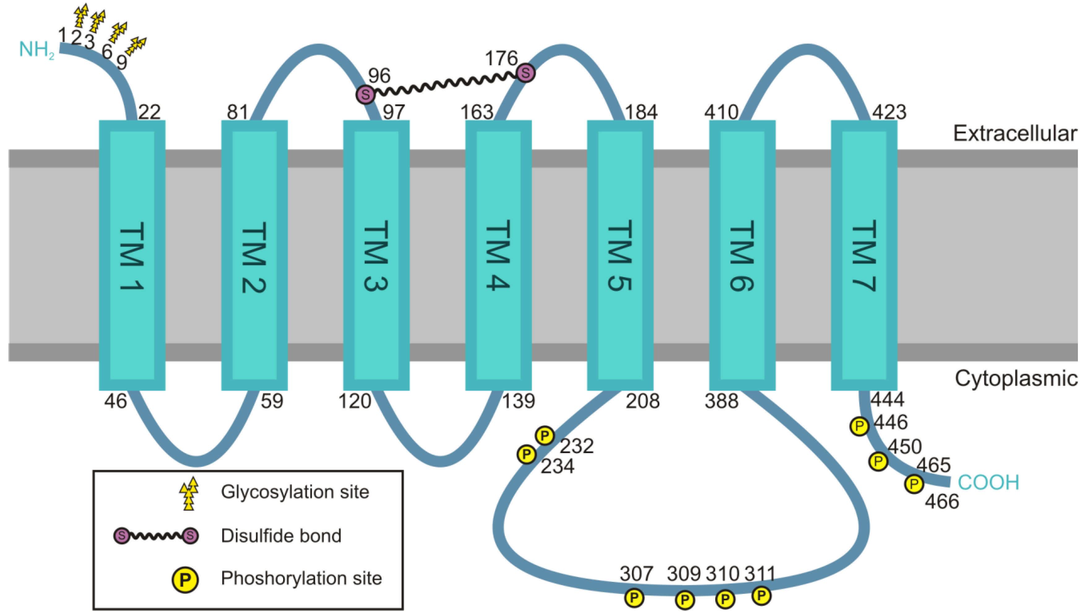
4.1. Structural Features of the M2 Receptor

The M2 receptor subtype is apparently different from all the other muscarinic receptors in several respects. Even though the M2 receptor is coupled to Gi/o, similarly to M4 receptor, it exhibits some properties that are characteristic only for this receptor subtype. Whereas all other muscarinic receptors are resensitized and recycled back to the plasma membrane in different cell types, the M2 receptor is usually downregulated by degradation [72]. Furthermore, its internalization appears to differ in the requirement for β-arrestin and dynamin [70,73].
The most important structural part of the M2 receptor that facilitates the signaling process is, as with the other subtypes, the i3 loop of the receptor. The structure of the M2 receptor is depicted in Figure 2. A recent study has described the atomic structure of the M2 receptor, revealing interesting common features but also differences to other muscarinic receptors [74]. Unfortunately, this structure does not contain the i3 loop which turned out to be too flexible to allow for a structure determination at a sufficiently high resolution. The i3 loops of the muscarinic receptors are relatively long and consist of 160 to 240 amino acid residues. They represent the part of the receptor that is responsible for most of the differences between the single subtypes. The first and second intracellular loops show sequence similarities of approximately 90% between receptor subtypes, whereas the i3 loops merely feature 40% identity in their amino acid sequence, accompanied by a high variability in their length [52]. The i3 loop of the M2 receptor exhibits a flexible structure without specific secondary structure elements, except for the region at the C- and N-terminal part that may form α-helices [75]. A short sequence of four amino acids at the C-terminal end of the i3 loop, close to the sixth TM, mediates the coupling of the M2 receptor to the α-subunit of Gi/0 [76]. The i3 loop is also the most important section where the βγ-subunits of the heterotrimeric G proteins bind. Furthermore, it is phosphorylated by GRK2 upon stimulation and plays an important role in receptor internalization [77,78,79].
Figure 2. Structure of the M2 receptor. The M2 muscarinic acetylcholine receptor belongs to the family of G protein coupled receptors. The seven transmembrane domains (TM1-7) are connected by intracellular or extracellular loops, and the receptor exhibits four glycosylation sites at its extracellular N-terminal domain. The serine residues 96 and 176 in the first and second extracellular loop are connected to each other by a disulfide bond. Except for the third intracellular (i3) loop, other loops are relatively small. The i3 loop consists of 181 amino acids and has no specific secondary structure. The C-terminal domain and the i3 loop can be phosphorylated at several residues.
Genes 04 00171 g002
The M2 receptor’s affinity for its ligand, the internalization and efficiency of signal transduction can be influenced by the cholesterol content of the plasma membrane, and depletion of cholesterol leads to a reduced ligand affinity, lower rates of receptor internalization but an increased inhibition of the adenylyl cyclase [80]. However, although this would suggest the involvement of the cholesterol rich, lipid raft like microdomains, M2 receptors are not localized in rafts at the plasma membrane of Madin-Darby Canine Kidney (MDCK) cells, and rafts are not involved in their apical targeting [81]. In polarized MDCK cells, the apical sorting signal for the M2 receptor was found to reside in the i3 loop [81]. However, the apical targeting of M2 receptors in polarized MDCK cells is independent of glycosylation, and the expression and correct function of the receptor do not require glycosylation [81,82].
It has become clear that the M2 receptor is not found as a monomer but that it homo-oligomerizes to a certain degree [83]. Using fluorescence resonance energy transfer approaches, the M2 receptor was identified to reside in the membrane of CHO cells as a tetramer [84]. Beyond that, the existence of hetero-oligomers was discussed, and analysis using bioluminescence resonance energy transfer could demonstrate the formation of such oligomers between M1, M2 and M3 receptors [85]. The interaction between M2 and M3 receptors is likely to be mediated by electrostatic interactions between the i3 loops [86]. Heterodimerization between M2 and M3 receptors has also been suggested by the data showing that coexpression of both receptors in JEG-3 cells results in increased lysosomal degradation of the M3 receptor which normally displays robust recycling rather than degradation [85]. This was attributed to a more dominant effect of the M2 receptor, which is predominantly sorted in the lysosomal pathway instead of being recycled.

4.2. Signaling of the M2 Muscarinic Acetylcholine Receptor

As a Gi/o coupled receptor, the main effect of M2 receptor stimulation could be expected to be the inhibition of the adenylyl cyclase and a decrease in cellular cAMP levels. However, this appears not always to be the case. In CHO cells expressing M2 receptors, for example, also the net synthesis of cAMP can be stimulated upon agonist binding to the M2 receptor. In these cells, the M2 receptor also seems to couple to Gs or Gq/11, thus changing the outcome of the receptor signaling [87].
Very important downstream targets of the M2 receptor signaling are the MAP kinases ERK1/2, which can be activated by different signaling pathways. In M2 receptor expressing COS-7 cells, the G protein βγ-subunits coupled to the M2 receptor are able to induce the phosphorylation of ERK1/2 via the PI3Kγ as a part of a pathway that involves a tyrosine kinase, Shc, Grb2, Sos, Ras and Raf [61], but the α-subunit can also be involved in ERK1/2 activation [88]. An isoform of Rap1 GTPase-activating protein, rap1GAPII, can bind specifically to the α-subunits of the Gi family. Upon stimulation of the M2 receptor, rap1GAPII decreases the amount of GTP bound Rap1. As active Rap1 antagonizes Ras function, the resulting inactivation of Rap1 represents a way of the α-subunit of the Gi coupled M2 receptor to facilitate ERK1/2 activation [88]. The transactivation of the epidermal growth factor receptor (EGFR) can also contribute to MAP kinase signaling following the stimulation of GPCRs. Upon extracellular ligand stimulus, EGFR dimerizes, its tyrosine kinase activity is promoted, and it becomes autophosphorylated. Thus, its downstream signaling pathways are activated and contribute to ERK1/2 phosphorylation. In fact, our unpublished data show that in HaCaT keratinocytes, most of the ERK activation upon carbachol stimulation can be attributed to EGFR transactivation. The transactivation has been shown in several cell lines [89], and it may involve the activation of matrix metalloproteases (MMP) that lead to the proteolytic release of EGF like peptides from the cell surface [90,91], as already demonstrated for M1 and M3 receptors [92,93]. However, for the M2 receptor an MMP independent mechanism dependent on the protein tyrosine kinase Fyn was established. In COS-7 cells transiently overexpressing M2 receptor, the M2 receptor mediated transactivation upon carbachol stimulation leads to an incomplete EGFR downstream signaling towards ERK1/2 and Akt, whereas PLCγ is not activated [94]. The authors postulated that differences in the tyrosine phosphorylation profiles of the EGFR might be generated depending on the activation mechanism, and the transactivation thus can result in an incomplete signaling.
The interaction of β-arrestin with ERK1/2 is thought to inhibit its translocation to the nucleus, thus restricting the ERK signaling of GPCRs to the cytoplasm and changing the outcome of the ERK signaling [95,96]. Importantly, β-arrestins do not only inhibit the nuclear translocation but also function as scaffolds that bring together ERK2, MEK1 and Raf-1 [95]. An endosomal localization was observed for the overexpressed M2 receptor, ERK1/2 and β-arrestins in HeLa cells upon carbachol stimulation, whereas a more diffuse cytoplasmic distribution of β-arrestin was detected after stimulation upon overexpression of the other muscarinic receptors [97]. As the association of β-arrestin with the M2 receptor is stable only after stimulation, it is possible that the mechanisms of ERK1/2 activation differ to a great extent between the M2 receptor and the other muscarinic receptors. It can be hypothesized that the prolonged interaction of M2 receptor with β-arrestin might play a role to restrict the signal of this receptor subtype to the cytoplasm. Together with the G protein mediated ERK activation targeted to the nucleus, the β-arrestin mediated cytoplasmic signaling provides additional possibilities for the control of MAP kinase activity and substrates that become phosphorylated, applying a high level of complexity to the modulation of signaling.
Upon agonist stimulation, the M2 receptor is rapidly phosphorylated on serine/threonine residues by second messenger kinases such as protein kinase A (PKA) or by specific G protein receptor kinases, e.g., GRK2 which appears to be the main kinase for the M2 receptor [98]. The targets of GRK2 are the serine and threonine residues in the central part of the i3 loop [99], and overexpression of a dominant negative GRK2 mutant together with the M2 receptor in HEK cells leads to a complete loss of M2 receptor desensitization [100]. Phosphorylation of the serines and threonines within the amino acid residues 307 to 311 upon receptor stimulation allows β-arrestin binding [101] which is required for the termination of M2 receptor signaling by disrupting the interaction of the receptor and its coupled G protein. It has been suggested that the phosphorylation dependent process of M2 receptor internalization is independent of β-arrestins and does not require clathrindependent endocytosis [101]. However, studies in mouse embryonic fibroblast derived from β-arrestin deficient mice showed that β-arrestin is indeed required for M2 receptor endocytosis [97]. The phosphorylation of M2 receptor in the i3 loop upon stimulation seems to act as a signal for desensitization and may help the β-arrestins to overcome a putative inhibitory signal that precludes inappropriate receptor binding [102]. Some aspartic and glutamic acid residues in close proximity to the phosphorylation sites are essential for an adequate response to receptor phosphorylation. However, they rather appear to be involved in β-arrestin binding than directly in phosphorylation, as mutating them into asparagines and glutamines had no effect on receptor phosphorylation but impaired β-arrestin binding [103].

4.3. Receptor Internalization and Its Connection to Signaling

Resensitization of GPCRs involves their dephosphorylation after endocytosis [104]. However, different from all the other muscarinic receptor subtypes, the M2 receptor is not resensitized in most cases and does not recycle back to the plasma membrane [72]. Figure 3 summarizes the main features of the M2 receptor endocytosis. While the internalization of the M2 receptor seems to be important for signal termination by means of receptor degradation, it does not seem to play a major role in the temporal regulation of the signaling. A comparative study of the extent and kinetics of receptor internalization for the different muscarinic receptor subtypes in CHO cells showed that the M2 receptor internalizes faster and more extensively than all the other receptors. Its degree of downregulation was comparable to those of M3, M4 and M5 receptors, and only the M1 receptor displayed a more intense degradation. The M1, M3 and M5 receptor downregulation exhibits a rapid component after a short term treatment with carbachol and a slower component for further downregulation upon long term agonist treatment, whereas this slower component was not apparent for the M2 and M4 receptors [105]. However, these results are not completely consistent with earlier studies. Roseberry and Hosey showed in HEK-293 cells that the ectopically expressed M2 receptor was rapidly and to a great extent internalized but only minimally degraded after 24 h of carbachol treatment. They even observed some degree of recycling of the receptor, albeit very slow, back to the plasma membrane [106]. Nevertheless, it is evident that there are differences in the mechanisms involved in receptor internalization and downregulation of the M2 receptor as compared to those of the other muscarinic receptors. However, special care needs to be taken when comparing studies addressing the trafficking and signaling of the receptors as these may be very much dependent on the cell type used.
M1, M3 and M4 all undergo endocytosis in a clathrin and dynamin dependent manner. Even though it is evident that clathrin is not involved in M2 receptor endocytosis, the involvement of caveolin, β-arrestins and the GTPase dynamin in this process has remained less conclusive. Although an interaction between the M2 receptor and caveolin has been shown in cardiac myocytes [107], M2 receptor endocytosis takes place independently of caveolin in HEK-293 cells [108]. Endocytosis of many other GPCRs can be inhibited by means of expression of a dominant negative K44A mutant of dynamin2. Earlier studies concluded that dynamin would not be required for M2 receptor endocytosis [73], since the K44A dynamin2 mutant did not show any effect on M2 receptor endocytosis. More recent studies have used other mutants of dynamin2, such as the PIP2 independent K535M and a deletion mutant lacking the GTP binding domain. These studies could indeed show that dynamin is involved in M2 receptor endocytosis [109,110]. These discrepancies may be explained by unspecific effects of overexpression of the K44A and other dynamin mutants on cellular trafficking (Reviewed in [111]). Furthermore, it has recently been suggested that some dynamin functions may be independent of its GTPase activity, and thus different dynamin2 mutants may impair different steps during trafficking. Unfortunately, the dynamin dependency of M2 receptor trafficking has not yet been addressed using the specific chemical inhibitors of dynamin, such as Dynasore [112] that have recently become available.
The stimulation of M1 or M2 receptors leads to a stable ubiquitination of β-arrestin in mouse embryonic fibroblasts [113]. The internalization of the receptors is not affected by proteasome inhibitors but they completely block receptor downregulation which is increased upon expression of a constitutively ubiquitinated β-arrestin. However, the M2 receptor does not seem to undergo proteasomal degradation but is rather directed to lysosomes, indicating a role for β-arrestin ubiquitination in the correct lysosomal targeting of the receptor. Upon β-arrestin ubiquitination, the M2 receptor and β-arrestin stably colocalize in intracellular compartments, which was not observed for the M1 receptor [113]. It can be suggested that the pattern of ubiquitination on β-arrestin is important for the receptor targeting and the stable association of β-arrestin and the receptor.
Figure 3. Agonist stimulated internalization of the M2 receptor. The M2 receptor is internalized by a clathrin-independent, ARF6-dependent pathway. Upon agonist stimulation, the M2 receptor is phosphorylated on serine/threonine residues by GRK, which facilitates the binding of β-arrestins and receptor desensitization. In addition, the β-arrestin activates ARF6, resulting in M2 receptor internalization in the early endosome. A phosphatase dephosphorylates the M2 receptor, causing β-arrestin dissociation from the receptor. The small G protein Rab22 has been implicated in the recycling of the M2 receptor. However, recycling appears to be a minor route for the M2 receptor which is mainly targeted to lysosomes and degraded.
Genes 04 00171 g003
A protein connected to the M2 receptor whose role is not completely understood so far is the Receptor for Activated C Kinase 1 (RACK1), a protein enriched in M2 receptor immunoprecipitates, whose interaction with the receptor is disrupted upon treatment with the muscarinic agonist carbachol. RACK1 seems to prevent endocytosis, as a decreased expression of RACK1 increases the agonist mediated internalization of the receptor. However, at the same time, both overexpression and a decrease in RACK1 expression level inhibit receptor downregulation [114]. As RACK1 and β-arrestins have overlapping binding sites in the receptors [115], RACK1 might prevent β-arrestin binding to unstimulated receptors.

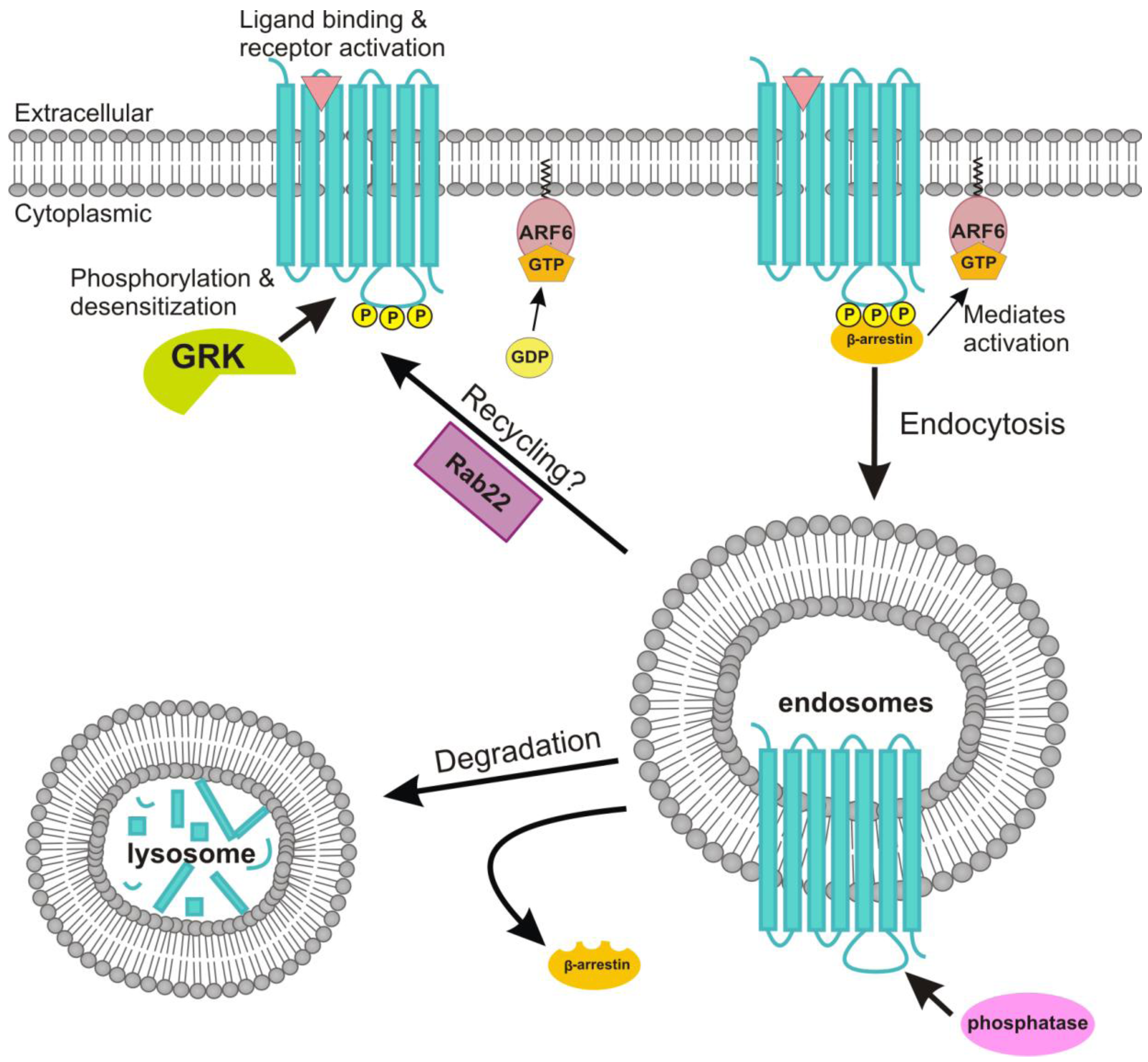
4.4. Role of ADP-Ribosylation Factor 6 in the Endocytosis of the M2 Receptor

The ADP ribosylation factor (ARF) family belongs to the Ras superfamily of GTP binding proteins which contains five major subclasses, namely Rab, Rho, Ras, Ran and ARF GTPases [116]. The six closely related mammalian ARF proteins are divided in three classes based on their sequence homologies. Accordingly, ARF1, ARF2 and ARF3 belong to class I and ARF4 and ARF5 to class II, whereas the sole member of class III is ARF6 [117,118]. As with other GTPases, ARF proteins switch between a GTP bound active and a GDP bound inactive form. The cycle of binding and hydrolysis of GTP is important for the localization of ARF proteins, and their activation is also connected with the membrane localization [118,119]. ARFs are able to interact with a plethora of regulatory proteins such as guanine nucleotide exchange factors (GEFs) and GTPase activating proteins (GAPs). Each ARF protein exhibits a preference for specialized GAPs and GEFs, and ARFs are specifically recruited to cellular compartments in which these regulatory proteins reside [119].
ARF6, together with ARF1, is the best studied family member of the ARF proteins, and its homologues have been found in many eukaryotes, including Xenopus laevis and Drosophila melanogaster, as well as in metazoans and fungi, whereas none appear to exist in plants. Similar to all members of the ARF family, ARF6 is myristoylated on the N-terminal glycine residue, which is important for the membrane association and thus influences the subcellular distribution of ARF6. This lipid modification is also essential for the activity of ARF6 in membrane trafficking [120,121,122,123] and for its membrane localization, as a non-myristoylated ARF6 (Gly2Ala mutation) was not able to bind to membranes and remained cytosolic [124].
ARF6 has been shown to be localized both at the plasma membrane and in endosomal compartments, and it evidently plays an important role in the regulation of peripheral membrane dynamics and the cortical actin cytoskeleton at the plasma membrane [117,125]. In line with this, ARF6 is a mediator of cytoskeletal rearrangements such as the formation of actin-rich plasma membrane protrusions [126,127]. ARF6 is also involved in Rac mediated membrane ruffling [126,127,128,129,130], phagocytosis [130] and regulation of secretion [131]. Furthermore, activated ARF6 has been indicated to promote the internalization of E-cadherin to early endosomes, inducing a disassembly of adherens junctions [132,133,134]. ARF6 mediated activation of PLD leads to the activation of phosphatidylinositol 4-phosphate 5-kinase (PIP5K), resulting in an increase of PIP2 at the cell periphery [135] where it is involved in clathrin dependent endocytic processes [136].
The requirement of ARF6 in membrane trafficking processes such as endosomal recycling is evidenced by the fact that the expression of a dominant negative ARF6 blocks recycling from endosomes. Thereafter, several studies have demonstrated the vital role of ARF6 in endosomal membrane trafficking [117,124,131,137,138]. Furthermore, ARF6 can regulate both the clathrin dependent as well as the clathrin independent endocytosis, but the ARF6 dependent endocytic pathway appears to be dynamin independent [133,139]. This is also suggested by the fact that depletion of ARF6 affects the endocytosis of several GPCRs, irrespective of their dependency on clathrin [140]. During clathrin independent endocytosis, activated ARF6 associates with the adaptor protein complex AP2 which mediates the post-endocytic sorting of ARF6 cargos, such as the major histocompatibility complex I (MHC1), β1-Integrin and the M2 receptor [141].
Stimulation of GPCRs not only activates downstream signaling cascades but also leads to internalization and desensitization of the respective receptor. In this process, the internalization is important for both the resensitization and degradation pathway [142,143,144]. It has been shown that ARF6 is important for the endocytosis of e.g., endothelin type B receptor, vasopressin type 2 receptor and the angiotensin type 1 receptor [140]. Stimulation of some GPCRs, such as β2-adrenergic and luteinizing receptors, results in the activation of ARF6, and overexpression of an ARF6 GAP reduces β2-adrenergic receptor internalization [145]. Furthermore, Claing et al. could show that β-arrestin regulates receptor endocytosis through the activation of and interaction with ARF6, thereby providing a link between the GPCR and endocytic proteins, such as clathrin and AP2 [146,147].
The M2 receptor has been demonstrated to be a cargo protein of ARF6 dependent endocytosis. Constitutively active ARF6 expression diminished the endocytosis but enhanced the rate and extent of recycling of the M2 receptor, implicating that the agonist stimulated internalization and perhaps also recycling of the M2 receptor is regulated by ARF6 [109,110,140]. However, a role for ARF6 in the ER-Golgi trafficking of GPCRs has also been suggested [148]. Reiner and Nathanson studied the involvement of several small G proteins in the regulation of the agonist induced endocytosis of the M2 receptor [109,110]. The dominant negative T22N mutant ARF6 and Rab22, a GTPase implicated in ARF dependent recycling [149], were able to substantially increase the M2 receptor endocytosis. M2 and M4 receptors initially utilize different endocytic pathways. Only later, they appear to end up in the same compartment which is positive for markers of clathrin dependent endocytosis [110]. Thus, the M2 receptor is initially internalized via a clathrin independent and ARF6 associated pathway, during which the M2 receptor is first observed in structures that lack the markers for clathrin dependent endocytosis. Thereafter, the M2 receptor internalization pathway quickly merges with the clathrin dependent pathway at the level of early endosomes, which contain the early endosome autoantigen 1 (EEA-1) [109].

5. Muscarinic Acetylcholine Receptors in Human Diseases

As with most GPCRs, muscarinic receptors are involved in human diseases by distinct mechanisms. In various autoimmune diseases, mAChRs frequently are targets of autoantibodies, which bind to the receptors and lead either to their activation or inactivation. Initially caused by the parasite Trypanosoma cruzi, the Chagas disease may lead to a heart failure and a sudden death upon disease progression. The involvement of the heart is possibly connected to the existence of autoantibodies in patients with Chagas disease against the M2 receptor, as these receptors display a negative chronotropic effect in cultured cardiomyocytes [150,151]. These autoantibodies against M2 receptor have been shown to induce receptor crosslinking and internalization due to activation of the receptor [152,153]. The Sjögren’s syndrome is a highly common autoimmune rheumatic disease, which is also connected to autoantibodies directed against the muscarinic receptors, among other antigens. Upon progression of the disease, lymphocytes infiltrate and attack the lacrimal and salivary glands, leading to dryness of the mouth and the eyes [154]. Autoantibodies recognizing the M3 receptor have been shown to be involved, leading to its inhibition and internalization. Due to the reduced signaling and impairment of secretion, the salivary flow rate is reduced [155,156]. In line with this, knockout mouse studies have shown that M3 receptor, together with M1 and M4 receptors, plays a major role in regulating salivary excretion [19].
Not only autoimmune diseases are related to the muscarinic receptors. In the Achilles tendon from patients with tendinosis exhibiting hypercellularity and hypervascularity, a higher level of M2 receptor can be observed in the endothelial cells of the vessels as compared to the tendons of healthy patients, suggesting the occurrence of acetylcholine and M2 receptor mediated vasodilatory mechanisms in the Achilles tendon [157]. Both M2 and M3 receptors affect the bronchial and tracheal smooth muscle contraction and are therefore likely to play a role in asthma and chronic obstructive pulmonary disease that are accompanied by an increased smooth muscle tone [158].
The expression of muscarinic receptors is described for a variety of cancer cells in which they appear to be highly involved in proliferation and migration. In the breast cancer cell line MCF-7, stimulation of muscarinic receptors leads to an activation of the ERK pathway in a manner dependent on PKC-ζ, phosphoinositide 3-kinase and a kinase of the Src family, increasing protein biosynthesis and cell proliferation [159]. A stimulatory effect on cell migration was shown for HeLa cells in an ERK1/2 dependent mechanism [160]. In addition, muscarinic receptors are thought to play a role in angiogenesis in breast cancer [161]. A cancer type that overexpresses muscarinic receptors is colon cancer, in which high levels of M3 receptors have been found to mediate cancer cell proliferation [162], and the M3 receptor is involved in cell migration of these cells via the release of MMPs and transactivation of EGF receptors [163,164]. This is clearly different from the influence of muscarinic receptors on the migration of keratinocytes, where the M3 receptor is rather inhibiting cell migration than promoting it [27].
Whereas M3 receptors stimulate cell proliferation of tumor cells, M2 receptors seem to have a contrary effect. A specific cancer type that is influenced by this muscarinic receptor is glioblastoma, a common brain tumor. In glioblastoma cell lines, addition of the M2 receptor agonist arecaidine decreased cell growth in a time and dose dependent manner and was counteracted by the M2 receptor antagonist gallamine. In contrast, muscarine had no significant influence on proliferation [165]. The effect of cholinergic ligands on small cell lung carcinoma cells is more controversial. On the one hand, cholinergic stimulation of the small cell lung cancer cells H69 enhances cell proliferation [166], but on the other hand, the SCC-9 cell line is inhibited in cell cycle progression upon cholinergic stimulation and shows a diminished DNA synthesis [167]. Another cell type that uses acetylcholine as a growth factor is hepatocellular carcinoma. In cancer tissues, the acetylcholinesterase is often downregulated and its decrease correlates with tumor aggressiveness [168]. Due to the diminished expression of acetylcholinesterase, acetylcholine cannot be hydrolyzed. It is likely that the elevated level of acetylcholine acts on muscarinic receptors of the tumor cells and therefore increases their proliferation. The above observations reveal how different the influence of muscarinic receptors on specific cells is, depending on the cellular environment and the cellular proteome. However, although muscarinic receptors sometimes exhibit unpredictable or unexpected effects on cancer progression, they still might be an interesting target for therapeutic drugs in the future.

6. Conclusions and Future Challenges

In tissues, the effects of acetylcholine are mediated by the repertoire of the receptor subtypes expressed, and both nicotinic and muscarinic receptors contribute to the cellular response observed. Since a certain cell type normally expresses more than one type of acetylcholine receptor, the cellular response is often a compiled one, with a contribution of several subtypes. Furthermore, the observed effects on the cell physiology are also dependent on the class of the G protein subunits and their modulators expressed by the specific cell type. Thus, the interpretation of the data resulting from studies in various cell types may be a complicated task. Some specific physiological functions of muscarinic receptor subtypes have been revealed by the studies on knockout mouse models (Reviewed in [169]). However, even in this system, one needs to be cautious since loss of one subtype may result in aberrant desensitization of another one due to a hypercholinergic status produced by the lack of the major receptor subtype in a certain tissue [170].
Pharmacological substances that result in the activation of only either of the receptor groups are available. However, the activation or even inhibition of a specific receptor subtype is notoriously difficult, as the substances used are rarely fully specific for a single receptor subtype. With increasing knowledge about the structural differences of the receptors, it may become possible to develop novel compounds that specifically affect the functions of a single subtype. Thereby, special interest has been paid on ligands that bind to allosteric regulatory sites. Intriguingly, the structure of the M2 receptor was recently solved [74] and revealed some important structural features specific for this subtype that may facilitate the generation of more selective drugs in the future.
Older studies have frequently been performed using overexpression of a specific receptor subtype. However, if the receptor expression level is too high, this may even result in saturation of signaling or trafficking partners (“dominant negative effect”) and thus misinterpretation of the results. Furthermore, other receptors expressed in the cell type still contribute to the signaling response although the overexpressed one may be predominant. The discovery of specific gene knockdown strategies in cell culture, e.g., RNA interference, has provided better options to modulate the signaling in the target cells by depleting a specific receptor subtype. However, even this is not completely secure, as the other receptor subtypes expressed may compensate for the loss of another one. Interestingly, mouse embryonic fibroblast have been described not to contain mRNA or protein for any of the muscarinic receptors [97]. Thus, this cell type may be especially suitable for studies addressing the subtype specific signaling, provided that the required partners are also expressed.
Future challenges in the studies of non-neuronal functions of muscarinic receptors will certainly include revisiting the data obtained in various cell systems and by means of receptor overexpression. It will be of special importance to compile these data in a model that needs to be tested in a single system that allows definite conclusions on the function of a single receptor type. Special effort should also be dedicated to novel pharmaceutical compounds, both inhibitors and activators, which affect the function of only a specific muscarinic receptor. With these tools at hand, it will be possible to obtain a more conclusive picture about the non-neuronal functions of this receptor class.

Acknowledgments

The work in the Tikkanen lab is financially supported by the LOEWE Research Network “Non-neuronal Cholinergic Systems”, financed by the state of Hessia, to RT & AB. We thank the members of the Tikkanen group, especially Melanie Meister, for comments and discussions.

Conflict of Interest

The authors declare no conflict of interest.

References and Notes

  1. Bennett, M.R. The concept of transmitter receptors: 100 years on. Neuropharmacology 2000, 39, 523–546. [Google Scholar] [CrossRef]
  2. Curtis, D.R.; Ryall, R.W. Nicotinic and Muscarinic Receptors of Renshaw Cells. Nature 1964, 203, 652–653. [Google Scholar] [CrossRef]
  3. Kurzen, H.; Wessler, I.; Kirkpatrick, C.J.; Kawashima, K.; Grando, S.A. The non-neuronal cholinergic system of human skin. Horm. Metab. Res. 2007, 39, 125–135. [Google Scholar] [CrossRef]
  4. Bonner, T.I.; Buckley, N.J.; Young, A.C.; Brann, M.R. Identification of a family of muscarinic acetylcholine receptor genes. Science 1987, 237, 527–532. [Google Scholar]
  5. Caulfield, M.P.; Birdsall, N.J. International Union of Pharmacology. XVII. Classification of muscarinic acetylcholine receptors. Pharmacol. Rev. 1998, 50, 279–290. [Google Scholar]
  6. Hall, J.M.; Caulfield, M.P.; Watson, S.P.; Guard, S. Receptor subtypes or species homologues: relevance to drug discovery. Trends Pharmacol. Sci. 1993, 14, 376–383. [Google Scholar] [CrossRef]
  7. Jones, S.V.; Heilman, C.J.; Brann, M.R. Functional responses of cloned muscarinic receptors expressed in CHO-K1 cells. Mol. Pharmacol. 1991, 40, 242–247. [Google Scholar]
  8. Alea, M.P.; Borroto-Escuela, D.O.; Romero-Fernandez, W.; Fuxe, K.; Garriga, P. Differential expression of muscarinic acetylcholine receptor subtypes in Jurkat cells and their signaling. J. Neuroimmunol. 2011, 237, 13–22. [Google Scholar] [CrossRef]
  9. Nahorski, S.R.; Tobin, A.B.; Willars, G.B. Muscarinic M3 receptor coupling and regulation. Life Sci. 1997, 60, 1039–1045. [Google Scholar] [CrossRef]
  10. Hulme, E.C.; Lu, Z.L.; Saldanha, J.W.; Bee, M.S. Structure and activation of muscarinic acetylcholine receptors. Biochem. Soc. Trans. 2003, 31, 29–34. [Google Scholar]
  11. Nathanson, N.M. A multiplicity of muscarinic mechanisms: Enough signaling pathways to take your breath away. Proc. Natl. Acad. Sci. USA 2000, 97, 6245–6247. [Google Scholar] [CrossRef]
  12. Hayashi, M.K.; Haga, T. Palmitoylation of muscarinic acetylcholine receptor m2 subtypes: Reduction in their ability to activate G proteins by mutation of a putative palmitoylation site, cysteine 457, in the carboxyl-terminal tail. Arch. Biochem. Biophys. 1997, 340, 376–382. [Google Scholar] [CrossRef]
  13. Shah, N.; Khurana, S.; Cheng, K.; Raufman, J.P. Muscarinic receptors and ligands in cancer. Am. J. Physiol. Cell. Physiol. 2009, 296, C221–C232. [Google Scholar]
  14. Gomeza, J.; Shannon, H.; Kostenis, E.; Felder, C.; Zhang, L.; Brodkin, J.; Grinberg, A.; Sheng, H.; Wess, J. Pronounced pharmacologic deficits in M2 muscarinic acetylcholine receptor knockout mice. Proc. Natl. Acad. Sci. USA 1999, 96, 1692–1697. [Google Scholar] [CrossRef]
  15. Gomeza, J.; Zhang, L.; Kostenis, E.; Felder, C.; Bymaster, F.; Brodkin, J.; Shannon, H.; Xia, B.; Deng, C.; Wess, J. Enhancement of D1 dopamine receptor-mediated locomotor stimulation in M(4) muscarinic acetylcholine receptor knockout mice. Proc. Natl. Acad. Sci. USA 1999, 96, 10483–10488. [Google Scholar] [CrossRef]
  16. Miyakawa, T.; Yamada, M.; Duttaroy, A.; Wess, J. Hyperactivity and intact hippocampus-dependent learning in mice lacking the M1 muscarinic acetylcholine receptor. J. Neurosci. 2001, 21, 5239–5250. [Google Scholar]
  17. Yamada, M.; Lamping, K.G.; Duttaroy, A.; Zhang, W.; Cui, Y.; Bymaster, F.P.; McKinzie, D.L.; Felder, C.C.; Deng, C.X.; Faraci, F.M.; Wess, J. Cholinergic dilation of cerebral blood vessels is abolished in M(5) muscarinic acetylcholine receptor knockout mice. Proc. Natl. Acad. Sci. USA 2001, 98, 14096–14101. [Google Scholar] [CrossRef]
  18. Yamada, M.; Miyakawa, T.; Duttaroy, A.; Yamanaka, A.; Moriguchi, T.; Makita, R.; Ogawa, M.; Chou, C.J.; Xia, B.; Crawley, J.N.; Felder, C.C.; Deng, C.X.; Wess, J. Mice lacking the M3 muscarinic acetylcholine receptor are hypophagic and lean. Nature 2001, 410, 207–212. [Google Scholar] [CrossRef]
  19. Bymaster, F.P.; McKinzie, D.L.; Felder, C.C.; Wess, J. Use of M1-M5 muscarinic receptor knockout mice as novel tools to delineate the physiological roles of the muscarinic cholinergic system. Neurochem. Res. 2003, 28, 437–442. [Google Scholar] [CrossRef]
  20. Kawashima, K.; Fujii, T. Basic and clinical aspects of non-neuronal acetylcholine: Overview of non-neuronal cholinergic systems and their biological significance. J. Pharmacol. Sci. 2008, 106, 167–173. [Google Scholar] [CrossRef]
  21. Kummer, W.; Lips, K.S.; Pfeil, U. The epithelial cholinergic system of the airways. Histochem. Cell Biol. 2008, 130, 219–234. [Google Scholar] [CrossRef]
  22. Wessler, I.; Kirkpatrick, C.J. Acetylcholine beyond neurons: The non-neuronal cholinergic system in humans. Br. J. Pharmacol. 2008, 154, 1558–1571. [Google Scholar] [CrossRef]
  23. Grando, S.A.; Crosby, A.M.; Zelickson, B.D.; Dahl, M.V. Agarose gel keratinocyte outgrowth system as a model of skin re-epithelization: requirement of endogenous acetylcholine for outgrowth initiation. J. Invest. Dermatol. 1993, 101, 804–810. [Google Scholar]
  24. Grando, S.A.; Kist, D.A.; Qi, M.; Dahl, M.V. Human keratinocytes synthesize, secrete, and degrade acetylcholine. J. Invest. Dermatol. 1993, 101, 32–36. [Google Scholar]
  25. Ndoye, A.; Buchli, R.; Greenberg, B.; Nguyen, V.T.; Zia, S.; Rodriguez, J.G.; Webber, R.J.; Lawry, M.A.; Grando, S.A. Identification and mapping of keratinocyte muscarinic acetylcholine receptor subtypes in human epidermis. J. Invest. Dermatol. 1998, 111, 410–416. [Google Scholar] [CrossRef]
  26. Buchli, R.; Ndoye, A.; Arredondo, J.; Webber, R.J.; Grando, S.A. Identification and characterization of muscarinic acetylcholine receptor subtypes expressed in human skin melanocytes. Mol. Cell. Biochem. 2001, 228, 57–72. [Google Scholar] [CrossRef]
  27. Chernyavsky, A.I.; Arredondo, J.; Wess, J.; Karlsson, E.; Grando, S.A. Novel signaling pathways mediating reciprocal control of keratinocyte migration and wound epithelialization through M3 and M4 muscarinic receptors. J. Cell. Biol. 2004, 166, 261–272. [Google Scholar] [CrossRef]
  28. Nguyen, V.T.; Ndoye, A.; Hall, L.L.; Zia, S.; Arredondo, J.; Chernyavsky, A.I.; Kist, D.A.; Zelickson, B.D.; Lawry, M.A.; Grando, S.A. Programmed cell death of keratinocytes culminates in apoptotic secretion of a humectant upon secretagogue action of acetylcholine. J. Cell. Sci. 2001, 114, 1189–1204. [Google Scholar]
  29. Bschleipfer, T.; Schukowski, K.; Weidner, W.; Grando, S.A.; Schwantes, U.; Kummer, W.; Lips, K.S. Expression and distribution of cholinergic receptors in the human urothelium. Life Sci. 2007, 80, 2303–2307. [Google Scholar] [CrossRef]
  30. Chess-Williams, R.; Chapple, C.R.; Yamanishi, T.; Yasuda, K.; Sellers, D.J. The minor population of M3-receptors mediate contraction of human detrusor muscle in vitro. J. Auton. Pharmacol. 2001, 21, 243–248. [Google Scholar] [CrossRef]
  31. Metzen, J.; Bittinger, F.; Kirkpatrick, C.J.; Kilbinger, H.; Wessler, I. Proliferative effect of acetylcholine on rat trachea epithelial cells is mediated by nicotinic receptors and muscarinic receptors of the M1-subtype. Life Sci. 2003, 72, 2075–2080. [Google Scholar] [CrossRef]
  32. Acevedo, M. Effect of acetyl choline on ion transport in sheep tracheal epithelium. Pflugers Arch. 1994, 427, 543–546. [Google Scholar]
  33. Klein, M.K.; Haberberger, R.V.; Hartmann, P.; Faulhammer, P.; Lips, K.S.; Krain, B.; Wess, J.; Kummer, W.; Konig, P. Muscarinic receptor subtypes in cilia-driven transport and airway epithelial development. Eur. Respir. J. 2009, 33, 1113–1121. [Google Scholar] [CrossRef]
  34. Kilbinger, H.; von Bardeleben, R.S.; Siefken, H.; Wolf, D. Prejunctional muscarinic receptors regulating neurotransmitter release in airways. Life Sci. 1995, 56, 981–987. [Google Scholar] [CrossRef]
  35. Wang, H.; Han, H.; Zhang, L.; Shi, H.; Schram, G.; Nattel, S.; Wang, Z. Expression of multiple subtypes of muscarinic receptors and cellular distribution in the human heart. Mol. Pharmacol. 2001, 59, 1029–1036. [Google Scholar]
  36. Milner, P.; Kirkpatrick, K.A.; Ralevic, V.; Toothill, V.; Pearson, J.; Burnstock, G. Endothelial cells cultured from human umbilical vein release ATP, substance P and acetylcholine in response to increased flow. Proc. Biol. Sci. 1990, 241, 245–248. [Google Scholar] [CrossRef]
  37. Kawashima, K.; Fujii, T. Expression of non-neuronal acetylcholine in lymphocytes and its contribution to the regulation of immune function. Front. Biosci. 2004, 9, 2063–2085. [Google Scholar] [CrossRef]
  38. Kawashima, K.; Yoshikawa, K.; Fujii, Y.X.; Moriwaki, Y.; Misawa, H. Expression and function of genes encoding cholinergic components in murine immune cells. Life Sci. 2007, 80, 2314–2319. [Google Scholar] [CrossRef]
  39. Qian, J.; Galitovskiy, V.; Chernyavsky, A.I.; Marchenko, S.; Grando, S.A. Plasticity of the murine spleen T-cell cholinergic receptors and their role in in vitro differentiation of naive CD4 T cells toward the Th1, Th2 and Th17 lineages. Genes Immun. 2011, 12, 222–230. [Google Scholar] [CrossRef]
  40. Fujii, T.; Watanabe, Y.; Inoue, T.; Kawashima, K. Upregulation of mRNA encoding the M5 muscarinic acetylcholine receptor in human T- and B-lymphocytes during immunological responses. Neurochemical. Res. 2003, 28, 423–429. [Google Scholar] [CrossRef]
  41. Kawashima, K.; Fujii, T.; Moriwaki, Y.; Misawa, H. Critical roles of acetylcholine and the muscarinic and nicotinic acetylcholine receptors in the regulation of immune function. Life Sci. 2012, 91, 1027–1032. [Google Scholar] [CrossRef]
  42. Duttaroy, A.; Zimliki, C.L.; Gautam, D.; Cui, Y.; Mears, D.; Wess, J. Muscarinic stimulation of pancreatic insulin and glucagon release is abolished in m3 muscarinic acetylcholine receptor-deficient mice. Diabetes 2004, 53, 1714–1720. [Google Scholar] [CrossRef]
  43. Horn, F.; van der Wenden, E.M.; Oliveira, L.; AP, I.J.; Vriend, G. Receptors coupling to G proteins: Is there a signal behind the sequence? Proteins 2000, 41, 448–459. [Google Scholar] [CrossRef]
  44. Albert, P.R.; Robillard, L. G protein specificity: traffic direction required. Cell Signal. 2002, 14, 407–418. [Google Scholar] [CrossRef]
  45. Rumenapp, U.; Asmus, M.; Schablowski, H.; Woznicki, M.; Han, L.; Jakobs, K.H.; Fahimi-Vahid, M.; Michalek, C.; Wieland, T.; Schmidt, M. The M3 muscarinic acetylcholine receptor expressed in HEK-293 cells signals to phospholipase D via G12 but not Gq-type G proteins: regulators of G proteins as tools to dissect pertussis toxin-resistant G proteins in receptor-effector coupling. J. Biol. Chem. 2001, 276, 2474–2479. [Google Scholar] [CrossRef]
  46. Meyer zu Heringdorf, D.; Lass, H.; Alemany, R.; Laser, K.T.; Neumann, E.; Zhang, C.; Schmidt, M.; Rauen, U.; Jakobs, K.H.; van Koppen, C.J. Sphingosine kinase-mediated Ca2+ signalling by G-protein-coupled receptors. EMBO J. 1998, 17, 2830–2837. [Google Scholar] [CrossRef]
  47. Neubig, R.R.; Siderovski, D.P. Regulators of G-protein signalling as new central nervous system drug targets. Nat. Rev. Drug Discov. 2002, 1, 187–197. [Google Scholar] [CrossRef]
  48. Logothetis, D.E.; Kurachi, Y.; Galper, J.; Neer, E.J.; Clapham, D.E. The beta gamma subunits of GTP-binding proteins activate the muscarinic K+ channel in heart. Nature 1987, 325, 321–326. [Google Scholar] [CrossRef]
  49. Jones, S.V. Modulation of the inwardly rectifying potassium channel IRK1 by the m1 muscarinic receptor. Mol. Pharmacol. 1996, 49, 662–667. [Google Scholar]
  50. Jones, S.V. Muscarinic receptor subtypes: modulation of ion channels. Life Sci. 1993, 52, 457–464. [Google Scholar] [CrossRef]
  51. Wickman, K.; Clapham, D.E. Ion channel regulation by G proteins. Physiol. Rev. 1995, 75, 865–885. [Google Scholar]
  52. Borroto-Escuela, D.O.; Agnati, L.F.; Fuxe, K.; Ciruela, F. Muscarinic acetylcholine receptor-interacting proteins (mAChRIPs): Targeting the receptorsome. Curr. Drug Targets 2012, 13, 53–71. [Google Scholar] [CrossRef]
  53. Borroto-Escuela, D.O.; Correia, P.A.; Romero-Fernandez, W.; Narvaez, M.; Fuxe, K.; Ciruela, F.; Garriga, P. Muscarinic receptor family interacting proteins: role in receptor function. J. Neurosci. Meth. 2011, 195, 161–169. [Google Scholar] [CrossRef]
  54. Kim, J.Y.; Yang, M.S.; Oh, C.D.; Kim, K.T.; Ha, M.J.; Kang, S.S.; Chun, J.S. Signalling pathway leading to an activation of mitogen-activated protein kinase by stimulating M3 muscarinic receptor. Biochem. J. 1999, 337, 275–280. [Google Scholar] [CrossRef]
  55. Crespo, P.; Xu, N.; Daniotti, J.L.; Troppmair, J.; Rapp, U.R.; Gutkind, J.S. Signaling through transforming G protein-coupled receptors in NIH 3T3 cells involves c-Raf activation. Evidence for a protein kinase C-independent pathway. J. Biol. Chem. 1994, 269, 21103–21109. [Google Scholar]
  56. Hawes, B.E.; van Biesen, T.; Koch, W.J.; Luttrell, L.M.; Lefkowitz, R.J. Distinct pathways of Gi- and Gq-mediated mitogen-activated protein kinase activation. J. Biol. Chem. 1995, 270, 17148–17153. [Google Scholar]
  57. Crespo, P.; Xu, N.; Simonds, W.F.; Gutkind, J.S. Ras-dependent activation of MAP kinase pathway mediated by G-protein beta gamma subunits. Nature 1994, 369, 418–420. [Google Scholar] [CrossRef]
  58. Rosenblum, K.; Futter, M.; Jones, M.; Hulme, E.C.; Bliss, T.V. ERKI/II regulation by the muscarinic acetylcholine receptors in neurons. J. Neurosci. 2000, 20, 977–985. [Google Scholar]
  59. Berkeley, J.L.; Levey, A.I. Muscarinic activation of mitogen-activated protein kinase in PC12 cells. J. Neurochem. 2000, 75, 487–493. [Google Scholar] [CrossRef]
  60. van Biesen, T.; Hawes, B.E.; Raymond, J.R.; Luttrell, L.M.; Koch, W.J.; Lefkowitz, R.J. G(o)-protein alpha-subunits activate mitogen-activated protein kinase via a novel protein kinase C-dependent mechanism. J. Biol. Chem. 1996, 271, 1266–1269. [Google Scholar]
  61. Lopez-Ilasaca, M.; Crespo, P.; Pellici, P.G.; Gutkind, J.S.; Wetzker, R. Linkage of G protein-coupled receptors to the MAPK signaling pathway through PI 3-kinase gamma. Science 1997, 275, 394–397. [Google Scholar] [CrossRef]
  62. Coso, O.A.; Teramoto, H.; Simonds, W.F.; Gutkind, J.S. Signaling from G protein-coupled receptors to c-Jun kinase involves beta gamma subunits of heterotrimeric G proteins acting on a Ras and Rac1-dependent pathway. J. Biol. Chem. 1996, 271, 3963–3966. [Google Scholar]
  63. Nagao, M.; Yamauchi, J.; Kaziro, Y.; Itoh, H. Involvement of protein kinase C and Src family tyrosine kinase in Galphaq/11-induced activation of c-Jun N-terminal kinase and p38 mitogen-activated protein kinase. J. Biol. Chem. 1998, 273, 22892–22898. [Google Scholar] [CrossRef]
  64. Wylie, P.G.; Challiss, R.A.; Blank, J.L. Regulation of extracellular-signal regulated kinase and c-Jun N-terminal kinase by G-protein-linked muscarinic acetylcholine receptors. Biochem. J. 1999, 338, 619–628. [Google Scholar] [CrossRef]
  65. Yamauchi, J.; Nagao, M.; Kaziro, Y.; Itoh, H. Activation of p38 mitogen-activated protein kinase by signaling through G protein-coupled receptors. Involvement of Gbetagamma and Galphaq/11 subunits. J. Biol. Chem. 1997, 272, 27771–27777. [Google Scholar] [CrossRef]
  66. Hosey, M.M.; Benovic, J.L.; DebBurman, S.K.; Richardson, R.M. Multiple mechanisms involving protein phosphorylation are linked to desensitization of muscarinic receptors. Life Sci. 1995, 56, 951–955. [Google Scholar] [CrossRef]
  67. Pitcher, J.A.; Touhara, K.; Payne, E.S.; Lefkowitz, R.J. Pleckstrin homology domain-mediated membrane association and activation of the beta-adrenergic receptor kinase requires coordinate interaction with G beta gamma subunits and lipid. J. Biol. Chem. 1995, 270, 11707–11710. [Google Scholar] [CrossRef]
  68. Elorza, A.; Sarnago, S.; Mayor, F., Jr. Agonist-dependent modulation of G protein-coupled receptor kinase 2 by mitogen-activated protein kinases. Mol. Pharmacol. 2000, 57, 778–783. [Google Scholar]
  69. Pitcher, J.A.; Tesmer, J.J.; Freeman, J.L.; Capel, W.D.; Stone, W.C.; Lefkowitz, R.J. Feedback inhibition of G protein-coupled receptor kinase 2 (GRK2) activity by extracellular signal-regulated kinases. J. Biol. Chem. 1999, 274, 34531–34534. [Google Scholar]
  70. Vogler, O.; Nolte, B.; Voss, M.; Schmidt, M.; Jakobs, K.H.; van Koppen, C.J. Regulation of muscarinic acetylcholine receptor sequestration and function by beta-arrestin. J. Biol. Chem. 1999, 274, 12333–12338. [Google Scholar]
  71. Edwardson, J.M.; Szekeres, P.G. Endocytosis and recycling of muscarinic receptors. Life Sci. 1999, 64, 487–494. [Google Scholar] [CrossRef]
  72. van Koppen, C.J. Multiple pathways for the dynamin-regulated internalization of muscarinic acetylcholine receptors. Biochem. Soc. Trans. 2001, 29, 505–508. [Google Scholar] [CrossRef]
  73. Vogler, O.; Bogatkewitsch, G.S.; Wriske, C.; Krummenerl, P.; Jakobs, K.H.; van Koppen, C.J. Receptor subtype-specific regulation of muscarinic acetylcholine receptor sequestration by dynamin. Distinct sequestration of m2 receptors. J. Biol. Chem. 1998, 273, 12155–12160. [Google Scholar]
  74. Haga, K.; Kruse, A.C.; Asada, H.; Yurugi-Kobayashi, T.; Shiroishi, M.; Zhang, C.; Weis, W.I.; Okada, T.; Kobilka, B.K.; Haga, T.; Kobayashi, T. Structure of the human M2 muscarinic acetylcholine receptor bound to an antagonist. Nature 2012, 482, 547–551. [Google Scholar] [CrossRef] [Green Version]
  75. Ichiyama, S.; Oka, Y.; Haga, K.; Kojima, S.; Tateishi, Y.; Shirakawa, M.; Haga, T. The structure of the third intracellular loop of the muscarinic acetylcholine receptor M2 subtype. FEBS Lett. 2006, 580, 23–26. [Google Scholar] [CrossRef]
  76. Hawes, B.E.; Luttrell, L.M.; Exum, S.T.; Lefkowitz, R.J. Inhibition of G protein-coupled receptor signaling by expression of cytoplasmic domains of the receptor. J. Biol. Chem. 1994, 269, 15776–15785. [Google Scholar]
  77. Schlador, M.L.; Grubbs, R.D.; Nathanson, N.M. Multiple topological domains mediate subtype-specific internalization of the M2 muscarinic acetylcholine receptor. J. Biol. Chem. 2000, 275, 23295–23302. [Google Scholar] [CrossRef]
  78. Tsuga, H.; Kameyama, K.; Haga, T.; Honma, T.; Lameh, J.; Sadee, W. Internalization and down-regulation of human muscarinic acetylcholine receptor m2 subtypes. Role of third intracellular m2 loop and G protein-coupled receptor kinase 2. J. Biol. Chem. 1998, 273, 5323–5330. [Google Scholar]
  79. Wu, G.; Bogatkevich, G.S.; Mukhin, Y.V.; Benovic, J.L.; Hildebrandt, J.D.; Lanier, S.M. Identification of Gbetagamma binding sites in the third intracellular loop of the M(3)-muscarinic receptor and their role in receptor regulation. J. Biol. Chem. 2000, 275, 9026–9034. [Google Scholar]
  80. Michal, P.; Rudajev, V.; El-Fakahany, E.E.; Dolezal, V. Membrane cholesterol content influences binding properties of muscarinic M2 receptors and differentially impacts activation of second messenger pathways. Eur. J. Pharmacol. 2009, 606, 50–60. [Google Scholar] [CrossRef]
  81. Chmelar, R.S.; Nathanson, N.M. Identification of a novel apical sorting motif and mechanism of targeting of the M2 muscarinic acetylcholine receptor. J. Biol. Chem. 2006, 281, 35381–35396. [Google Scholar] [CrossRef]
  82. van Koppen, C.J.; Nathanson, N.M. Site-directed mutagenesis of the m2 muscarinic acetylcholine receptor. Analysis of the role of N-glycosylation in receptor expression and function. J. Biol. Chem. 1990, 265, 20887–20892. [Google Scholar]
  83. Park, P.; Sum, C.S.; Hampson, D.R.; Van Tol, H.H.; Wells, J.W. Nature of the oligomers formed by muscarinic m2 acetylcholine receptors in Sf9 cells. Eur. J. Pharmacol. 2001, 421, 11–22. [Google Scholar] [CrossRef]
  84. Pisterzi, L.F.; Jansma, D.B.; Georgiou, J.; Woodside, M.J.; Chou, J.T.; Angers, S.; Raicu, V.; Wells, J.W. Oligomeric size of the m2 muscarinic receptor in live cells as determined by quantitative fluorescence resonance energy transfer. J. Biol. Chem. 2010, 285, 16723–16738. [Google Scholar] [CrossRef]
  85. Goin, J.C.; Nathanson, N.M. Quantitative analysis of muscarinic acetylcholine receptor homo- and heterodimerization in live cells: regulation of receptor down-regulation by heterodimerization. J. Biol. Chem. 2006, 281, 5416–5425. [Google Scholar] [CrossRef]
  86. Maggio, R.; Barbier, P.; Fornai, F.; Corsini, G.U. Functional role of the third cytoplasmic loop in muscarinic receptor dimerization. J. Biol. Chem. 1996, 271, 31055–31060. [Google Scholar] [CrossRef]
  87. Michal, P.; El-Fakahany, E.E.; Dolezal, V. Muscarinic M2 receptors directly activate Gq/11 and Gs G-proteins. J. Pharmacol. Exp. Ther. 2007, 320, 607–614. [Google Scholar]
  88. Mochizuki, N.; Ohba, Y.; Kiyokawa, E.; Kurata, T.; Murakami, T.; Ozaki, T.; Kitabatake, A.; Nagashima, K.; Matsuda, M. Activation of the ERK/MAPK pathway by an isoform of rap1GAP associated with G alpha(i). Nature 1999, 400, 891–894. [Google Scholar] [CrossRef]
  89. Daub, H.; Wallasch, C.; Lankenau, A.; Herrlich, A.; Ullrich, A. Signal characteristics of G protein-transactivated EGF receptor. EMBO J. 1997, 16, 7032–7044. [Google Scholar] [CrossRef]
  90. Gschwind, A.; Zwick, E.; Prenzel, N.; Leserer, M.; Ullrich, A. Cell communication networks: epidermal growth factor receptor transactivation as the paradigm for interreceptor signal transmission. Oncogene 2001, 20, 1594–1600. [Google Scholar] [CrossRef]
  91. Prenzel, N.; Zwick, E.; Daub, H.; Leserer, M.; Abraham, R.; Wallasch, C.; Ullrich, A. EGF receptor transactivation by G-protein-coupled receptors requires metalloproteinase cleavage of proHB-EGF. Nature 1999, 402, 884–888. [Google Scholar]
  92. McCole, D.F.; Keely, S.J.; Coffey, R.J.; Barrett, K.E. Transactivation of the epidermal growth factor receptor in colonic epithelial cells by carbachol requires extracellular release of transforming growth factor-alpha. J. Biol. Chem. 2002, 277, 42603–42612. [Google Scholar]
  93. Tsai, W.; Morielli, A.D.; Peralta, E.G. The m1 muscarinic acetylcholine receptor transactivates the EGF receptor to modulate ion channel activity. EMBO J. 1997, 16, 4597–4605. [Google Scholar] [CrossRef]
  94. Stirnweiss, J.; Valkova, C.; Ziesche, E.; Drube, S.; Liebmann, C. Muscarinic M2 receptors mediate transactivation of EGF receptor through Fyn kinase and without matrix metalloproteases. Cell. Signal. 2006, 18, 1338–1349. [Google Scholar] [CrossRef]
  95. DeFea, K.A.; Zalevsky, J.; Thoma, M.S.; Dery, O.; Mullins, R.D.; Bunnett, N.W. beta-arrestin-dependent endocytosis of proteinase-activated receptor 2 is required for intracellular targeting of activated ERK1/2. J. Cell. Biol 2000, 148, 1267–1281. [Google Scholar] [CrossRef]
  96. Tohgo, A.; Pierce, K.L.; Choy, E.W.; Lefkowitz, R.J.; Luttrell, L.M. beta-Arrestin scaffolding of the ERK cascade enhances cytosolic ERK activity but inhibits ERK-mediated transcription following angiotensin AT1a receptor stimulation. J. Biol. Chem. 2002, 277, 9429–9436. [Google Scholar]
  97. Jones, K.T.; Echeverry, M.; Mosser, V.A.; Gates, A.; Jackson, D.A. Agonist mediated internalization of M2 mAChR is beta-arrestin-dependent. J. Mol. Signal. 2006, 1, 7. [Google Scholar] [CrossRef]
  98. Pitcher, J.A.; Freedman, N.J.; Lefkowitz, R.J. G protein-coupled receptor kinases. Annu. Rev. Biochem. 1998, 67, 653–692. [Google Scholar] [CrossRef]
  99. Nakata, H.; Kameyama, K.; Haga, K.; Haga, T. Location of agonist-dependent-phosphorylation sites in the third intracellular loop of muscarinic acetylcholine receptors (m2 subtype). Eur. J. Biochem. 1994, 220, 29–36. [Google Scholar] [CrossRef]
  100. Pals-Rylaarsdam, R.; Xu, Y.; Witt-Enderby, P.; Benovic, J.L.; Hosey, M.M. Desensitization and internalization of the m2 muscarinic acetylcholine receptor are directed by independent mechanisms. J. Biol. Chem. 1995, 270, 29004–29011. [Google Scholar]
  101. Pals-Rylaarsdam, R.; Gurevich, V.V.; Lee, K.B.; Ptasienski, J.A.; Benovic, J.L.; Hosey, M.M. Internalization of the m2 muscarinic acetylcholine receptor. Arrestin-independent and -dependent pathways. J. Biol. Chem. 1997, 272, 23682–23689. [Google Scholar]
  102. Lee, K.B.; Ptasienski, J.A.; Bunemann, M.; Hosey, M.M. Acidic amino acids flanking phosphorylation sites in the M2 muscarinic receptor regulate receptor phosphorylation, internalization, and interaction with arrestins. J. Biol. Chem. 2000, 275, 35767–35777. [Google Scholar]
  103. Lee, K.B.; Ptasienski, J.A.; Pals-Rylaarsdam, R.; Gurevich, V.V.; Hosey, M.M. Arrestin binding to the M(2) muscarinic acetylcholine receptor is precluded by an inhibitory element in the third intracellular loop of the receptor. J. Biol. Chem. 2000, 275, 9284–9289. [Google Scholar]
  104. Krueger, K.M.; Daaka, Y.; Pitcher, J.A.; Lefkowitz, R.J. The role of sequestration in G protein-coupled receptor resensitization. Regulation of beta2-adrenergic receptor dephosphorylation by vesicular acidification. J. Biol. Chem. 1997, 272, 5–8. [Google Scholar] [CrossRef]
  105. Thangaraju, A.; Sawyer, G.W. Comparison of the kinetics and extent of muscarinic M1-M5 receptor internalization, recycling and downregulation in Chinese hamster ovary cells. Eur. J. Pharmacol. 2011, 650, 534–543. [Google Scholar] [CrossRef]
  106. Roseberry, A.G.; Hosey, M.M. Trafficking of M(2) muscarinic acetylcholine receptors. J. Biol. Chem. 1999, 274, 33671–33676. [Google Scholar] [CrossRef]
  107. Feron, O.; Smith, T.W.; Michel, T.; Kelly, R.A. Dynamic targeting of the agonist-stimulated m2 muscarinic acetylcholine receptor to caveolae in cardiac myocytes. J. Biol. Chem. 1997, 272, 17744–17748. [Google Scholar] [CrossRef]
  108. Roseberry, A.G.; Hosey, M.M. Internalization of the M2 muscarinic acetylcholine receptor proceeds through an atypical pathway in HEK293 cells that is independent of clathrin and caveolae. J. Cell. Sci 2001, 114, 739–746. [Google Scholar]
  109. Delaney, K.A.; Murph, M.M.; Brown, L.M.; Radhakrishna, H. Transfer of M2 muscarinic acetylcholine receptors to clathrin-derived early endosomes following clathrin-independent endocytosis. J. Biol. Chem. 2002, 277, 33439–33446. [Google Scholar]
  110. Reiner, C.; Nathanson, N.M. The internalization of the M2 and M4 muscarinic acetylcholine receptors involves distinct subsets of small G-proteins. Life Sci. 2008, 82, 718–727. [Google Scholar] [CrossRef]
  111. Ferguson, S.M.; De Camilli, P. Dynamin, a membrane-remodelling GTPase. Nat. Rev. Mol. Cell Biol. 2012, 13, 75–88. [Google Scholar]
  112. Macia, E.; Ehrlich, M.; Massol, R.; Boucrot, E.; Brunner, C.; Kirchhausen, T. Dynasore, a cell-permeable inhibitor of dynamin. Dev. Cell 2006, 10, 839–850. [Google Scholar] [CrossRef]
  113. Mosser, V.A.; Jones, K.T.; Hoffman, K.M.; McCarty, N.A.; Jackson, D.A. Differential role of beta-arrestin ubiquitination in agonist-promoted down-regulation of M1 vs M2 muscarinic acetylcholine receptors. J. Mol. Signal. 2008, 3, 20. [Google Scholar] [CrossRef]
  114. Reiner, C.L.; McCullar, J.S.; Kow, R.L.; Le, J.H.; Goodlett, D.R.; Nathanson, N.M. RACK1 associates with muscarinic receptors and regulates M(2) receptor trafficking. PLoS One 2010, 5, e13517. [Google Scholar]
  115. Bolger, G.B.; Baillie, G.S.; Li, X.; Lynch, M.J.; Herzyk, P.; Mohamed, A.; Mitchell, L.H.; McCahill, A.; Hundsrucker, C.; Klussmann, E.; Adams, D.R.; Houslay, M.D. Scanning peptide array analyses identify overlapping binding sites for the signalling scaffold proteins, beta-arrestin and RACK1, in cAMP-specific phosphodiesterase PDE4D5. Biochem. J. 2006, 398, 23–36. [Google Scholar] [CrossRef]
  116. Wennerberg, K.; Rossman, K.L.; Der, C.J. The Ras superfamily at a glance. J. Cell. Sci. 2005, 118, 843–846. [Google Scholar] [CrossRef]
  117. Donaldson, J.G. Multiple roles for Arf6: Sorting, structuring, and signaling at the plasma membrane. J. Biol. Chem. 2003, 278, 41573–41576. [Google Scholar] [CrossRef]
  118. Donaldson, J.G.; Jackson, C.L. ARF family G proteins and their regulators: roles in membrane transport, development and disease. Nat. Rev. Mol. Cell Biol. 2010, 12, 362–375. [Google Scholar] [CrossRef]
  119. Donaldson, J.G.; Honda, A. Localization and function of Arf family GTPases. Biochem. Soc. Trans. 2005, 33, 639–642. [Google Scholar] [CrossRef]
  120. Boman, A.L.; Taylor, T.C.; Melancon, P.; Wilson, K.L. A role for ADP-ribosylation factor in nuclear vesicle dynamics. Nature 1992, 358, 512–514. [Google Scholar]
  121. Dascher, C.; Balch, W.E. Dominant inhibitory mutants of ARF1 block endoplasmic reticulum to Golgi transport and trigger disassembly of the Golgi apparatus. J. Biol. Chem. 1994, 269, 1437–1448. [Google Scholar]
  122. Lenhard, J.M.; Kahn, R.A.; Stahl, P.D. Evidence for ADP-ribosylation factor (ARF) as a regulator ofin vitro endosome-endosome fusion . J. Biol. Chem. 1992, 267, 13047–13052. [Google Scholar]
  123. Taylor, T.C.; Kahn, R.A.; Melancon, P. Two distinct members of the ADP-ribosylation factor family of GTP-binding proteins regulate cell-free intra-Golgi transport. Cell 1992, 70, 69–79. [Google Scholar]
  124. D'Souza-Schorey, C.; Li, G.; Colombo, M.I.; Stahl, P.D. A regulatory role for ARF6 in receptor-mediated endocytosis. Science 1995, 267, 1175–1178. [Google Scholar] [CrossRef]
  125. Sabe, H. Requirement for Arf6 in cell adhesion, migration, and cancer cell invasion. J. Biochem. 2003, 134, 485–489. [Google Scholar] [CrossRef]
  126. D'Souza-Schorey, C.; Boshans, R.L.; McDonough, M.; Stahl, P.D.; Van Aelst, L. A role for POR1, a Rac1-interacting protein, in ARF6-mediated cytoskeletal rearrangements. Embo. J. 1997, 16, 5445–5454. [Google Scholar] [CrossRef]
  127. Radhakrishna, H.; Klausner, R.D.; Donaldson, J.G. Aluminum fluoride stimulates surface protrusions in cells overexpressing the ARF6 GTPase. J. Cell. Biol. 1996, 134, 935–947. [Google Scholar] [CrossRef]
  128. Brown, F.D.; Rozelle, A.L.; Yin, H.L.; Balla, T.; Donaldson, J.G. Phosphatidylinositol 4,5-bisphosphate and Arf6-regulated membrane traffic. J. Cell Biol 2001, 154, 1007–1017. [Google Scholar] [CrossRef]
  129. Franco, M.; Chardin, P.; Chabre, M.; Paris, S. Myristoylation of ADP-ribosylation factor 1 facilitates nucleotide exchange at physiological Mg2+ levels. J. Biol. Chem. 1995, 270, 1337–1341. [Google Scholar]
  130. Zhang, Q.; Cox, D.; Tseng, C.C.; Donaldson, J.G.; Greenberg, S. A requirement for ARF6 in Fcgamma receptor-mediated phagocytosis in macrophages. J. Biol. Chem. 1998, 273, 19977–19981. [Google Scholar]
  131. Caumont, A.S.; Galas, M.C.; Vitale, N.; Aunis, D.; Bader, M.F. Regulated exocytosis in chromaffin cells. Translocation of ARF6 stimulates a plasma membrane-associated phospholipase D. J. Biol. Chem. 1998, 273, 1373–1379. [Google Scholar]
  132. D'Souza-Schorey, C. Disassembling adherens junctions: breaking up is hard to do. Trends Cell. Biol 2005, 15, 19–26. [Google Scholar] [CrossRef]
  133. Palacios, F.; D'Souza-Schorey, C. Modulation of Rac1 and ARF6 activation during epithelial cell scattering. J. Biol. Chem. 2003, 278, 17395–17400. [Google Scholar] [CrossRef]
  134. Palacios, F.; Schweitzer, J.K.; Boshans, R.L.; D'Souza-Schorey, C. ARF6-GTP recruits Nm23-H1 to facilitate dynamin-mediated endocytosis during adherens junctions disassembly. Nat. Cell Biol. 2002, 4, 929–936. [Google Scholar] [CrossRef]
  135. Martin, T.F. PI(4,5)P(2) regulation of surface membrane traffic. Curr. Opin. Cell Biol. 2001, 13, 493–499. [Google Scholar] [CrossRef]
  136. Wenk, M.R.; De Camilli, P. Protein-lipid interactions and phosphoinositide metabolism in membrane traffic: insights from vesicle recycling in nerve terminals. Proc. Natl. Acad. Sci. USA 2004, 101, 8262–8269. [Google Scholar] [CrossRef]
  137. Powelka, A.M.; Sun, J.; Li, J.; Gao, M.; Shaw, L.M.; Sonnenberg, A.; Hsu, V.W. Stimulation-dependent recycling of integrin beta1 regulated by ARF6 and Rab11. Traffic 2004, 5, 20–36. [Google Scholar] [CrossRef]
  138. Radhakrishna, H.; Donaldson, J.G. ADP-ribosylation factor 6 regulates a novel plasma membrane recycling pathway. J. Cell. Biol. 1997, 139, 49–61. [Google Scholar] [CrossRef]
  139. Franco, M.; Peters, P.J.; Boretto, J.; van Donselaar, E.; Neri, A.; D'Souza-Schorey, C.; Chavrier, P. EFA6, a sec7 domain-containing exchange factor for ARF6, coordinates membrane recycling and actin cytoskeleton organization. Embo. J. 1999, 18, 1480–1491. [Google Scholar] [CrossRef]
  140. Houndolo, T.; Boulay, P.L.; Claing, A. G protein-coupled receptor endocytosis in ADP-ribosylation factor 6-depleted cells. J. Biol. Chem. 2005, 280, 5598–5604. [Google Scholar] [CrossRef]
  141. Lau, A.W.; Chou, M.M. The adaptor complex AP-2 regulates post-endocytic trafficking through the non-clathrin Arf6-dependent endocytic pathway. J. Cell. Sci. 2008, 121, 4008–4017. [Google Scholar] [CrossRef]
  142. Hein, L.; Ishii, K.; Coughlin, S.R.; Kobilka, B.K. Intracellular targeting and trafficking of thrombin receptors. A novel mechanism for resensitization of a G protein-coupled receptor. J. Biol. Chem. 1994, 269, 27719–27726. [Google Scholar]
  143. Pierce, K.L.; Luttrell, L.M.; Lefkowitz, R.J. New mechanisms in heptahelical receptor signaling to mitogen activated protein kinase cascades. Oncogene 2001, 20, 1532–1539. [Google Scholar] [CrossRef]
  144. Tong, H.; Rockman, H.A.; Koch, W.J.; Steenbergen, C.; Murphy, E. G protein-coupled receptor internalization signaling is required for cardioprotection in ischemic preconditioning. Circ. Res. 2004, 94, 1133–1141. [Google Scholar] [CrossRef]
  145. Premont, R.T.; Claing, A.; Vitale, N.; Freeman, J.L.; Pitcher, J.A.; Patton, W.A.; Moss, J.; Vaughan, M.; Lefkowitz, R.J. beta2-Adrenergic receptor regulation by GIT1, a G protein-coupled receptor kinase-associated ADP ribosylation factor GTPase-activating protein. Proc. Natl. Acad. Sci. USA 1998, 95, 14082–14087. [Google Scholar] [CrossRef]
  146. Claing, A.; Chen, W.; Miller, W.E.; Vitale, N.; Moss, J.; Premont, R.T.; Lefkowitz, R.J. beta-Arrestin-mediated ADP-ribosylation factor 6 activation and beta 2-adrenergic receptor endocytosis. J. Biol. Chem. 2001, 276, 42509–42513. [Google Scholar]
  147. Claing, A.; Laporte, S.A.; Caron, M.G.; Lefkowitz, R.J. Endocytosis of G protein-coupled receptors: roles of G protein-coupled receptor kinases and beta-arrestin proteins. Prog. Neurobiol. 2002, 66, 61–79. [Google Scholar] [CrossRef]
  148. Madziva, M.T.; Birnbaumer, M. A role for ADP-ribosylation factor 6 in the processing of G-protein-coupled receptors. J. Biol. Chem. 2006, 281, 12178–12186. [Google Scholar] [CrossRef]
  149. Weigert, R.; Yeung, A.C.; Li, J.; Donaldson, J.G. Rab22a regulates the recycling of membrane proteins internalized independently of clathrin. Mol. Biol. Cell. 2004, 15, 3758–3770. [Google Scholar] [CrossRef]
  150. Wallukat, G.; Nissen, E.; Morwinski, R.; Muller, J. Autoantibodies against the beta- and muscarinic receptors in cardiomyopathy. Herz 2000, 25, 261–266. [Google Scholar] [CrossRef]
  151. Nussinovitch, U.; Shoenfeld, Y. The diagnostic and clinical significance of anti-muscarinic receptor autoantibodies. Clin. Rev. Allergy Immunol. 2012, 42, 298–308. [Google Scholar] [CrossRef]
  152. Beltrame, S.P.; Auger, S.R.; Bilder, C.R.; Waldner, C.I.; Goin, J.C. Modulation of M(2) muscarinic receptor-receptor interaction by immunoglobulin G antibodies from Chagas' disease patients. Clin. Exp. Immunol. 2011, 164, 170–179. [Google Scholar] [CrossRef]
  153. Leiros, C.P.; Sterin-Borda, L.; Borda, E.S.; Goin, J.C.; Hosey, M.M. Desensitization and sequestration of human m2 muscarinic acetylcholine receptors by autoantibodies from patients with Chagas' disease. J. Biol. Chem. 1997, 272, 12989–12993. [Google Scholar]
  154. Fox, R.I.; Stern, M.; Michelson, P. Update in Sjogren syndrome. Curr. Opin. Rheumatol. 2000, 12, 391–398. [Google Scholar] [CrossRef]
  155. He, J.; Qiang, L.; Ding, Y.; Wei, P.; Li, Y.N.; Hua, H.; Li, Z.G. The role of muscarinic acetylcholine receptor type 3 polypeptide (M3RP205–220) antibody in the saliva of patients with primary Sjogren's syndrome. Clin. Exp. Rheumatol. 2012, 30, 322–326. [Google Scholar]
  156. Jin, M.; Hwang, S.M.; Davies, A.J.; Shin, Y.; Bae, J.S.; Lee, J.H.; Lee, E.B.; Song, Y.W.; Park, K. Autoantibodies in primary Sjogren's syndrome patients induce internalization of muscarinic type 3 receptors. Biochimica et biophysica acta 2012, 1822, 161–167. [Google Scholar]
  157. Bjur, D.; Danielson, P.; Alfredson, H.; Forsgren, S. Presence of a non-neuronal cholinergic system and occurrence of up- and down-regulation in expression of M2 muscarinic acetylcholine receptors: new aspects of importance regarding Achilles tendon tendinosis (tendinopathy). Cell Tissue Res. 2008, 331, 385–400. [Google Scholar] [CrossRef]
  158. Fisher, J.T.; Vincent, S.G.; Gomeza, J.; Yamada, M.; Wess, J. Loss of vagally mediated bradycardia and bronchoconstriction in mice lacking M2 or M3 muscarinic acetylcholine receptors. FASEB J. 2004, 18, 711–713. [Google Scholar]
  159. Jimenez, E.; Montiel, M. Activation of MAP kinase by muscarinic cholinergic receptors induces cell proliferation and protein synthesis in human breast cancer cells. J. Cell. Physiol. 2005, 204, 678–686. [Google Scholar] [CrossRef]
  160. Parnell, E.A.; Calleja-Macias, I.E.; Kalantari, M.; Grando, S.A.; Bernard, H.U. Muscarinic cholinergic signaling in cervical cancer cells affects cell motility via ERK1/2 signaling. Life Sci. 2012, 91, 1093–1098. [Google Scholar] [CrossRef]
  161. Fiszman, G.L.; Middonno, M.C.; de la Torre, E.; Farina, M.; Espanol, A.J.; Sales, M.E. Activation of muscarinic cholinergic receptors induces MCF-7 cells proliferation and angiogenesis by stimulating nitric oxide synthase activity. Cancer Biol. Ther. 2007, 6, 1106–1113. [Google Scholar] [CrossRef]
  162. Frucht, H.; Jensen, R.T.; Dexter, D.; Yang, W.L.; Xiao, Y. Human colon cancer cell proliferation mediated by the M3 muscarinic cholinergic receptor. Clin. Cancer Res. 1999, 5, 2532–2539. [Google Scholar]
  163. Cheng, K.; Zimniak, P.; Raufman, J.P. Transactivation of the epidermal growth factor receptor mediates cholinergic agonist-induced proliferation of H508 human colon cancer cells. Cancer Res. 2003, 63, 6744–6750. [Google Scholar]
  164. Xie, G.; Cheng, K.; Shant, J.; Raufman, J.P. Acetylcholine-induced activation of M3 muscarinic receptors stimulates robust matrix metalloproteinase gene expression in human colon cancer cells. Am. J. Physiol. Gastrointest Liver Physiol. 2009, 296, G755–G763. [Google Scholar] [CrossRef]
  165. Ferretti, M.; Fabbiano, C.; Di Bari, M.; Ponti, D.; Calogero, A.; Tata, A.M. M2 muscarinic receptors inhibit cell proliferation in human glioblastoma cell lines. Life Sci. 2012, 91, 1134–1137. [Google Scholar] [CrossRef]
  166. Fucile, S.; Napolitano, M.; Mattei, E. Cholinergic stimulation of human microcytoma cell line H69. Biochem. Biophys Res. Commun. 1997, 230, 501–504. [Google Scholar] [CrossRef]
  167. Williams, C.L.; Lennon, V.A. Activation of muscarinic acetylcholine receptors inhibits cell cycle progression of small cell lung carcinoma. Cell Regul. 1991, 2, 373–381. [Google Scholar]
  168. Zhao, Y.; Wang, X.; Wang, T.; Hu, X.; Hui, X.; Yan, M.; Gao, Q.; Chen, T.; Li, J.; Yao, M.; Wan, D.; Gu, J.; Fan, J.; He, X. Acetylcholinesterase, a key prognostic predictor for hepatocellular carcinoma, suppresses cell growth and induces chemosensitization. Hepatology 2011, 53, 493–503. [Google Scholar] [CrossRef]
  169. Wess, J.; Duttaroy, A.; Zhang, W.; Gomeza, J.; Cui, Y.; Miyakawa, T.; Bymaster, F.P.; McKinzie, L.; Felder, C.C.; Lamping, K.G.; Faraci, F.M.; Deng, C.; Yamada, M. M1-M5 muscarinic receptor knockout mice as novel tools to study the physiological roles of the muscarinic cholinergic system. Recept. Channels 2003, 9, 279–290. [Google Scholar] [CrossRef]
  170. Ito, Y.; Oyunzul, L.; Seki, M.; Fujino Oki, T.; Matsui, M.; Yamada, S. Quantitative analysis of the loss of muscarinic receptors in various peripheral tissues in M1-M5 receptor single knockout mice. Br. J. Pharmacol. 2009, 156, 1147–1153. [Google Scholar] [CrossRef]

