Article

Critical Evaluation of Biocontrol Ability of Bayoud Infected Date Palm Phyllospheric Bacillus spp. Suggests That In Vitro Selection Does Not Guarantee Success in Planta

1
Laboratory of Applied Microbiology, Department of Microbiology, Faculty of Natural and Life Sciences, University Ferhat Abbas of Setif, Setif 19000, Algeria
2
East Azerbaijan Agricultural and Natural Resources Research and Education Centre, Plant Protection Research Department, Agricultural Research, Education and Extension Organization (AREEO), Tabriz 5355179854, Iran
3
Department of Biology and Genetics, University of Veterinary Medicine and Pharmacy in Košice, 04181 Kosice, Slovakia
4
Marine Biodiscovery Centre, Department of Chemistry, University of Aberdeen, Old Aberdeen, Aberdeen AB24 3UE, UK
5
Laboratory of Soil Biology, University of Neuchatel, 11 Rue Emile Argand, CH-2000 Neuchatel, Switzerland
*
Author to whom correspondence should be addressed.
Agronomy 2022, 12(10), 2403; https://doi.org/10.3390/agronomy12102403
Submission received: 22 August 2022 / Revised: 27 September 2022 / Accepted: 30 September 2022 / Published: 4 October 2022

Abstract

:
The vascular wilt of date palm (Phoenix dactylifera L.), also known as Bayoud and caused by Fusarium oxysporum f.sp. albedinis (FOA), is the most destructive disease in North Africa. The disease has resulted in huge economic losses due to declining crop yield and quality. The use of potential biocontrol agents is a sustainable and environmentally friendly strategy compared to synthetic fungicides. The use of date palm-associated microflora for the screening of native antagonistic bacteria with potential applications is the most promising way to control this disease. Thus, the epidemic palm groves (in the valley of M’zab-Ghardaia-Algeria) were chosen for the isolation of rhizospheric bacteria and endophytes from the soil and roots of healthy or infected plants. A total of 8 bacterial isolates (83, 84, 300, 333, 322, 260, 249, and 227) selected from 75 FOA-active strains, showed strong activity against several strains of FOA and other major plant pathogens. Their phylogenetic identification proved they belong to the genus Bacillus (Bacillus sp., B. subtilis, B. atrophaeus, B. halotolerans, B. amyloliquefasiens, and B. paralicheformis). Bacterial cultures and a cell-free culture supernatant tested alone or in co-culture showed FOA inhibitory activity. However, the effect of the co-culture did not show any synergy in both cases. Unlike diffusible compounds, volatile organic compounds did not show a significant antifungal ability. The concept of selecting biocontrol agents in vitro does not always guarantee in vivo performance. In addition to antibiosis, other strategies such as competition and resistance induction are required for biocontrol agent efficacy. To evaluate the biocontrol effect in vivo, germinated seeds of date palm were treated with bacteria, infected with the pathogen, and then incubated for 45 days at room temperature in the dark. The majority of the strains (84, 300, and 333) tested showed moderate fungistatic effects and the protection rate reached an average of 60%. In particular, promising results were obtained with B. paralicheniformis strain 260 which significantly protected palm seeds up to 85%, compared to its in vitro test where a low inhibition rate (27.77%) was recorded. Screening methods should be equipped with multifactorial or plant-mediated control mechanisms. Furthermore, these bacteria have shown other potentialities to improve plant growth and resist stressful conditions. Thus, an effective biocontrol agent must combine several beneficial characteristics to be used successfully in situ. In this respect, Bacillus remains the best candidate for biological control.

1. Introduction

Date palm farming plays a very important role in the agriculture and economy of desert regions. The date palm (Phoenix dactylifera L.) is the main desert fruit crop in arid regions and is the mainstay of oasis ecosystems [1,2]. On the socio-economic level, the date constitutes the principal production of the Saharan regions. Indeed, it contributes to the agricultural income of the populations of these regions [3] and constitutes the basis of human and animal food. They are also a vital part of the culture and agrobiodiversity of the region. Furthermore, dates are not only a staple food but also an important export cash crop [4]. The highly appreciated Deglet Nour variety, both on the national and international markets, constitutes 51.8% of total Algerian date palm production and places Algeria fourth among the producing countries in the world [5].
Nevertheless, date palms are susceptible to several diseases and pests that limit date production. Many serious date palm diseases have been reported in the association of different Fusarium species [6,7]. Fusarium oxysporum (Fo) is a soil-borne plant pathogen causing a devastating effect on agricultural crops worldwide. It is one of the fungal species that causes a significant reduction in crop yield and quality and exhibits a high level of host specificity. This pathogen contains several special forms diverse in morphology and physiology, representing different levels of virulence depending on the host range [8]. Fusarium oxysporum f.sp. albedinis (FOA) infects the date palm and results in the vascular wilt called Bayoud which is, without a doubt, the most serious cryptogamic disease of the date palm. It is a real scourge of date palm areas in part of North Africa and poses a threat to neighboring countries [9]. It is classified on the EPPO “A2” list of quarantine organisms and on the “A” list of harmful organisms against which monitoring and control are compulsory in Algeria [10,11]. Since its first appearance at the end of the 19th century in Morocco, the disease has spread to all date-producing areas of Morocco, part of Algeria, and Mauritania [12,13]. The number of date palms destroyed by the disease is more than 12 million in Morocco and three million in Algeria [14,15]. Bayoud was a disease originating from the Draa Valley in Morocco, first observed around 1870 [15]. Since then, the epidemic progressed towards the east and reached the west of Algeria in 1898 [16]. Its extension continued towards the southwest and the central Sahara of Algeria in 1941. Then, it propagated in the palm groves of central Algeria, reaching Metlili in 1950, the region of M’zab (Ghardaia) in 1965, El Atteuf in 1967 [11], and El Ménéa in 1978 but the disease was eradicated from this oasis [17] (Figure 1).
FOA attacks palm trees by penetrating the roots and progressing through the entire vascular system to colonize it by migrating into the libero-ligneous vessels [18]. Specific symptoms appear as a brown color along the conductive vessels and side of the rachis. Symptoms of the affected leaves progress through the appearance of a palm in the middle crown which dries up and turns white, hence the Arabic name of Bayoud, deriving from Abied, meaning white [11]. The leaflets dry out from bottom to top and fold towards the rachis. The palm takes on the characteristic appearance of a wet feather. Affected palms eventually dry out, leading to the death of the tree in a few weeks to several months [9].
To date, no curative treatment exists against this fungus [19]. The means recommended against the disease are chemical control (use of fungicides, eradication, and incineration of the palm, soil disinfestation by solarization, and fumigation). Unfortunately, chemical control affects beneficial soil microbiota and can accumulate in the food chain [20]. Fungicides have negative effects on the environment and human health and can promote the development of new resistant strains of fungi [21]. In addition, the use of resistant cultivars does not provide a high food quality in dates [22]. Most commercial varieties, such as Deglet-Nour, are very susceptible and require intensive management to protect it against the pathogen.
Biological control remains the most effective method. The use of microorganisms is one of the most promising biological control methods to suppress plant diseases. Novel approaches involve the use of plant growth promoting bacteria (PGPB) which are categorized as those associated with the surface of plant roots, called rhizobacteria, and those that live inside plant tissues, called endophytes [23,24]. These bacteria are known for their abilities to promote and stimulate plant growth and protect host plants against phytopathogens [25]. Their biocontrol activities are attributed to intra-rhizospheric competition such as antagonism, competition for nutrients and resources, production of antibiotics or diffusible and volatile secondary metabolites, and activation of the plant defense by inducing systemic resistance (ISR) [26,27,28].
To undertake biological control of these diseases, the choice of microorganisms antagonistic to the pathogen is a very important criterion. The antagonists must be effective in situ by showing a significant aptitude for competition or antibiosis. In addition, they must persist in soils and be able to rapidly colonize plant roots. Therefore, it is very important to develop a reliable and practical strategy for screening effective inoculants with broad-spectrum resistance against several environmental stresses [29].
Selection procedures for appropriate bacterial inoculants are a limiting step to successful biocontrol. In vitro selection procedures used as a screening method, such as the dual culture of fungal pathogens on agar plates, may be inappropriate as they exclude host-antagonist-pathogen interaction factors and do not allow for the selection of biocontrol agents using other mechanisms such as root colonization, induction of systemic resistance, and/or niche competition [30]. Although the majority of studies use metabolite production on culture media for screening tests, only a few studies have compared the results of different screening strategies and concluded that whole plant testing was the most effective strategy [31,32,33]. Screening methods must have multifactorial or plant-mediated control mechanisms [29].
The most studied bacteria in biocontrol are members of the genera Bacillus, Burkholderia, Enterobacter, and Pseudomonas. Bacillus is one of the most abundant genera in the soil and the most studied as a biological control agent. It has been isolated from the rhizosphere of various plants [23,34]. These particular biocontrol agents are used, not only, to substitute chemical pesticides but to improve plant growth and mitigate various natural environmental stresses (salinity, aridity, extreme temperature, and pH) [35,36]. In addition to its various plant health benefits, Bacillus, with its ability to form resilient spores, is actually a very promising target for agricultural applications [37]. The spores are highly resistant to various abiotic stresses which facilitates their formulation for agricultural application [38]. The existence of several desirable traits in a single inoculant or a consortium of bacteria could, therefore, be successfully used in such studies [29,35].
The present study aims to propose the use of potential antagonistic bacteria indigenous to the palm rhizosphere to control a serious palm disease. To achieve this objective, bacterial strains (rhizospheric and endophytic) with antifungal activity against FOA are isolated from the roots of healthy and infected date palms from the Ghardaïa region. The most efficient antagonistic bacteria are tested for their antagonism towards other phytopathogens, their PGP and enzymatic potential, and their tolerance to abiotic stress. Finally, to evaluate their performance in the biocontrol of FOA, these bacterial strains are tested in vivo on the roots of a Deglet Noor date palm infected by the pathogen.

2. Materials and Methods

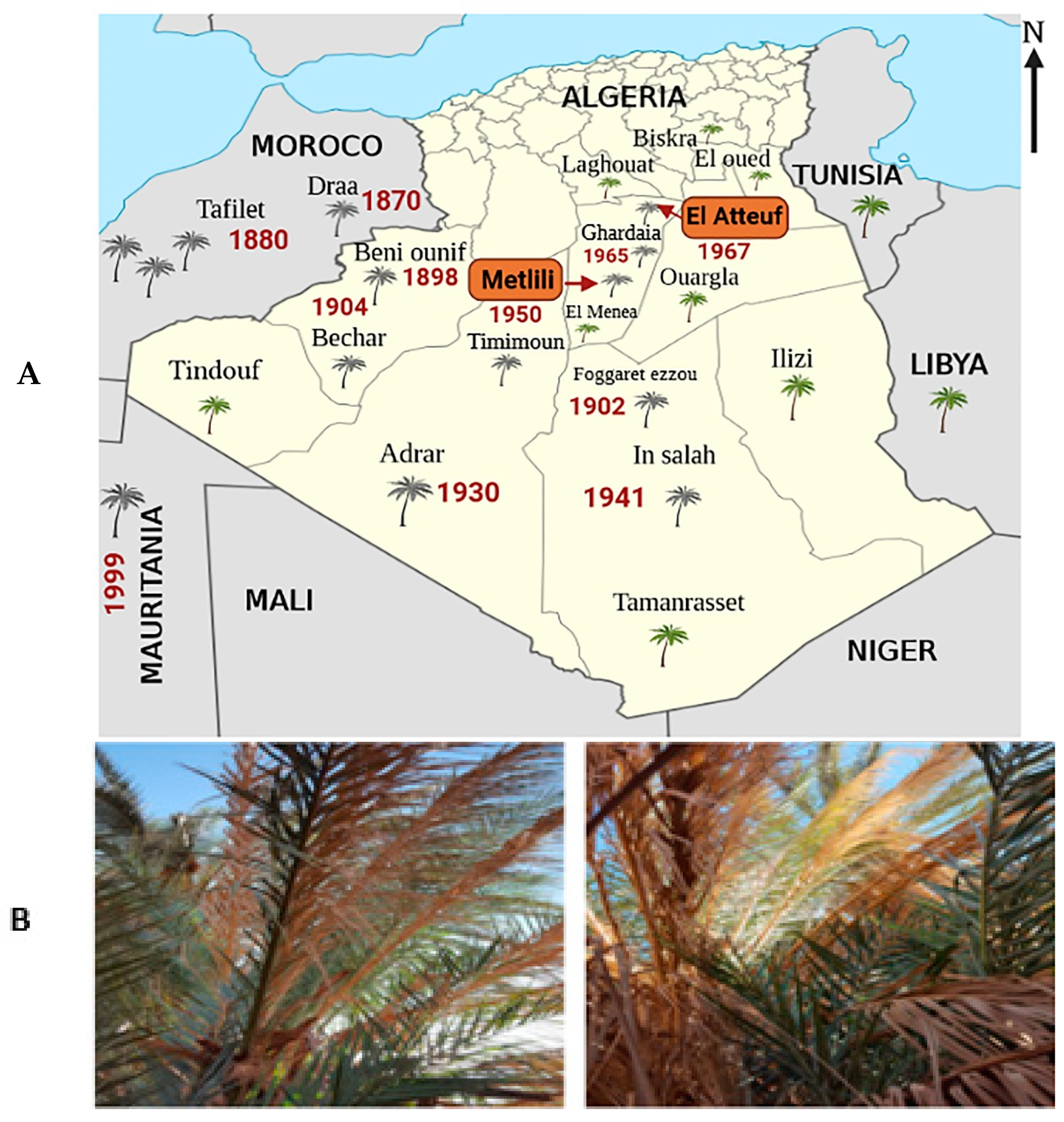
2.1. Description and Location of Sampling Sites

Sampling was from two sites; El Atteuf located in the M’zab region at 9 km east of Ghardaïa city (32°28′49.0″ N, 3°44′55.2″ E), and Metlili, also located in the M’zab region at 40 km south of Ghardaïa city (32°20′12.1″ N, 3°36′35.4″ E) (Figure 1A,B). The samples consisted of rhizospheric soil from healthy (HRh) and infected (IRh) palm trees, non-rhizospheric soil (SNRh) and roots from healthy (HR) and infected (IR) palm trees. The samples were taken randomly from two palm trees, at least six meters apart, and the samples were then mixed to obtain a representative sample. Each sample was collected in a sterile container kept cool and transported to the laboratory. The samples were achieved in triplicate.

2.2. Isolation and Screening of Antagonistic Bacterial Strains

2.2.1. Bacteria Isolation

Bacteria were isolated from different samples using three culture media: Trypticase Soja Agar (TSA) (containing g L−1: casein peptone, 15; soya peptone, 5; sodium chloride, 5; agar, 15; and 1 L distilled water; final pH 7 ± 0.2) [39], King B agar (containing g L−1: peptone, 20; glycerol, 10; K2HPO4, 1.5; MgSO4. 7 H2O, 1.5; agar, 12; and 1 L distilled water; final pH 7 ± 0.2) [40], and GYM Streptomyces (Glucose Yeast Malt) agar (containing g L−1: glucose, 4; yeast extract, 4; malt extract, 10; CaCO3, 2; Agar 12; and 1 L distilled water; final pH 7 ± 0.2). A total of 1 g of soil was added to 10 mL of sterile physiological water (0.85% NaCl). The mixture was stirred for 30 min then a decimal dilution series was made. In total, 0.1 mL of each dilution was plated onto different agar media. For endophyte isolation, 1 g of roots were rinsed with water then disinfected with 70% ethanol for 3 min and sterilized with 3% sodium hypochlorite for 15 min. The roots were then rinsed several times with sterile distilled water and the last rinse was incubated at 30 °C/24 h on TSA agar to check disinfection. The sterilized roots were crushed in a sterile mortar and then added to 10 mL of sterile physiological water. Decimal dilutions were made, and 0.1 mL of each dilution was then plated onto different agar media; plating Petri dishes were conducted in duplicate. Plates were incubated at 30 °C for 48 h. The number of CFU (N) was counted and the result was expressed in log CFU/g of soil or roots according to the following formula:
N = Σ C/(n1 + 0.1n2) dv (C: number of colonies counted on all plates having a number between 15 and 300 colonies, n1: number of plates retained at the first dilution, n2: number of plates retained at the second dilution, V: volume of the inoculum expressed in mL, and d: dilution rate corresponding to the first dilution retained).
Colonies with a different morphology were sub cultured in order to obtain bacterial pure cultures. The bacterial strains obtained were stored for a short period in slants agar at 4 °C, and for a long period at −20 °C in a medium supplemented with 30% glycerol.

2.2.2. Fungal Isolates

Fusarium oxysporum f.sp. albedinis isolates (FOA1, FOA2, FOA3, FOA4, and FOA5) were kindly provided by the National Institute for Plant Protection-Algeria. The pathogenic fungi were isolated from the Ghardaia region and identified by this institute. Other phytopathogenic fungi (Alternaria alternata Aa, Phytophthora infestans Pi, Fusarium solani Fs, and Fusarium graminearum Fg) were obtained from the collection of Laboratory of Applied Microbiology (Faculty of Nature and Life Sciences, University Ferhat Abbas of Setif) as described by Slama et al. [23].

2.2.3. Screening for In Vitro Antagonism

The in vitro antifungal activity was assessed by a dual culture assay on the basis of the hyphal growth rate of filamentous fungi, comparing the presence and absence of the isolated bacterial cultures [41]. A total of 335 isolated bacterial strains from different samples (HRh = 126, IRh = 91, SNRh = 62, HR = 33, and IR = 23) were screened for their activity against FOA1. The antifungal activity was performed on PDA (Potato Dextrose Agar). The Agar disc of a fresh fungal culture was placed in the center of the PDA plate. A total of 2 μL of each fresh bacterial culture was spotted at 3 cm from the fungal strain. A negative control containing plates without antagonistic bacteria was also conducted. Plates were incubated at 28 °C for 7 days. The percentage inhibition was calculated according to the following formula:
PI% (Inhibition percentage) = 1 − (a/b) × 100%. Where a is the distance of the fungus growth in the Petri dish inoculated with the bacterial isolate and b is the distance of the fungus growth in the control Petri dish without bacteria.

2.3. Antifungal Potential of Selected Isolates

Eight selected bacterial strains from different samples named 83, 84 (HRh), 300 (IRh), 333, 322 (SNRh), 260, 249 (IR), and 227 (HR) showing better inhibition rate against FOA, were examined for their ability to inhibit the growth of different strains of FOA (FOA1, FOA2, FOA3, FOA4, and FOA5) and other phytopathogenic fungi (Aa, Pi, Fs, and Fg). The antagonism of bacterial isolates toward phytopathogenic fungi was performed in vitro using PDA plates by the dual culture method. Each bacterial culture (108 cells/mL) was inoculated on the PDA by a horizontal streak at the end of the Petri plate. An agar disc of each fresh fungal culture was placed on the other end of the Petri plate. Plates were incubated for 7 days at 28 °C. The plates not inoculated with the antagonistic bacteria were considered a negative control. The percentage inhibition was calculated according to the previous formula.

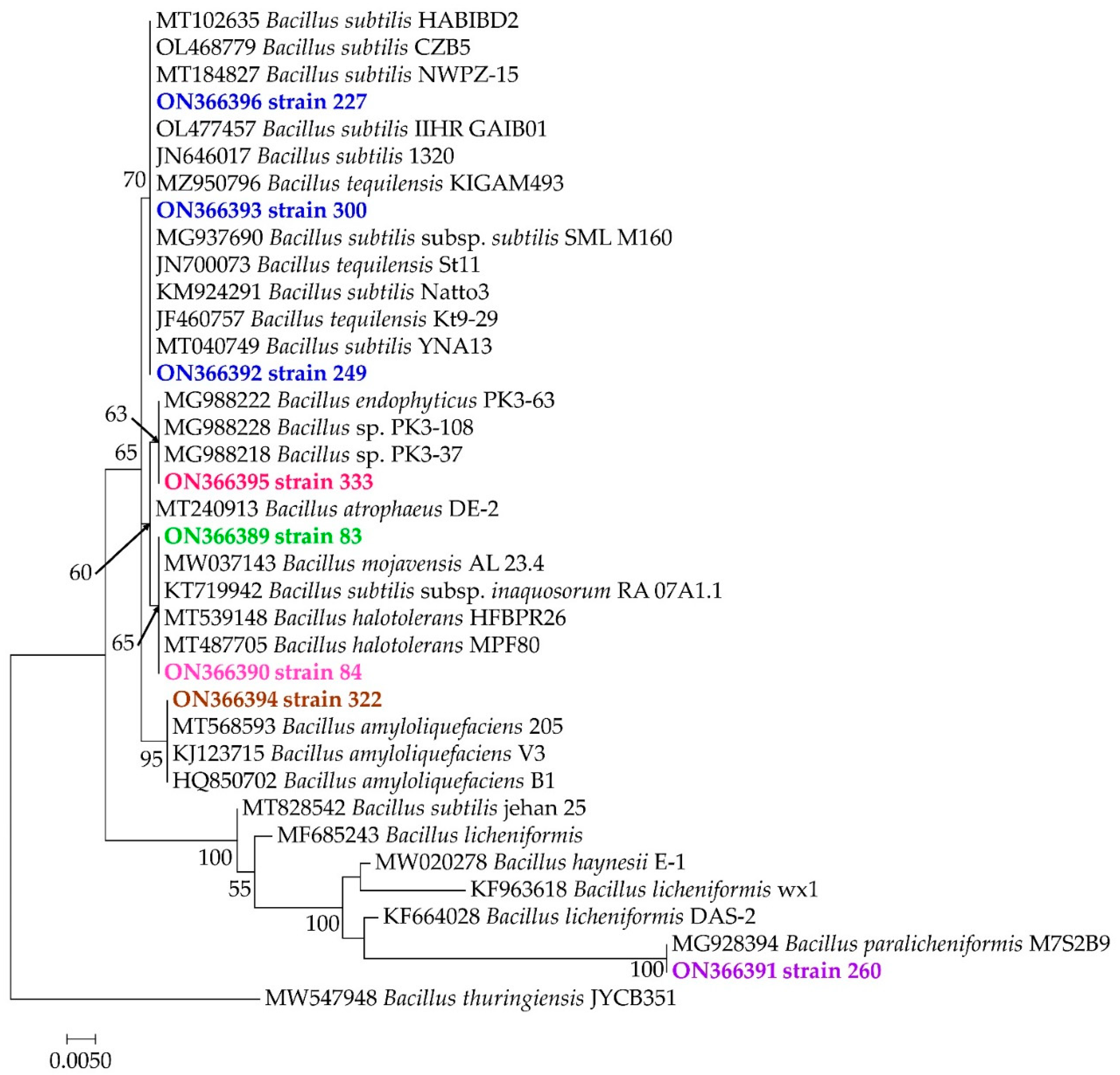
2.4. Molecular Characterization of Selected Bacterial Strains

DNA extraction from the eight selected bacteria (83, 84, 227, 249, 260, 300, 322, and 333) was performed according to Luchi et al. [42]. Estimation of DNA quality and quantity was also assessed. Amplification and sequencing were performed as previously. Phylogenetic analyses were performed according to Mefteh et al. [43].

2.5. Antagonistic Effect of Bacterial Co-Culture

This test was performed to detect a possible synergy of bacterial co-culture to improve the antagonistic activity [44]. Bacterial suspensions were obtained by growing the bacterial strains (83, 84, 227, 249, 260, 300, 322, and 333) with different combinations of two bacteria inoculated together in Tryptic Soy Broth (TSB) that resulted in 28 combinations. The bacterial culture was incubated for 24 h at 30 °C. After incubation, the antagonism was conducted by the dual-culture method. Each bacterial culture containing each combination (108 cells/mL) was inoculated on the PDA by a horizontal streak at the end of the Petri plate. An agar disc of each fresh fungal culture was placed on the other end of the Petri plate. Plates were incubated for 7 days at 28 °C. The plates not inoculated with the antagonistic bacteria were considered a negative control. The percentage inhibition was calculated according to the previous formula.

2.6. Antagonistic Effect of Cell-Free Supernatant

To test the antifungal activity of the cell-free supernatant, the bacterial isolates were inoculated individually or in pairs in 20 mL of TSB medium and incubated at 30 °C for 48 h with stirring. The bacterial suspensions (108 cells/mL) were then centrifuged at 5000 rpm for 15 min. The bacterial supernatants were incorporated aseptically into a supercooled PDA medium (at 25%) supplemented with oxytetracycline at 100 μg/mL and poured into Petri dishes. An agar disc of fungal culture was placed in the center of the PDA medium. Petri dishes inoculated with the fungus without bacterial supernatant were considered negative controls [45]. The percentage of inhibition was calculated after incubation for 7 days at 28 °C according to the previous formula.

2.7. Antifungal Activity by Volatile Organic Compounds (VOCs)

The antagonistic effect of the volatile substances produced by the bacterial strains was tested on the mycelial growth of FOA. Petri plates split into two compartments were used. Bacterial cultures (108 cells/mL) were inoculated by streaking on a TSA medium contained in one compartment of the Petri plate and a fungal agar disk from an FOA culture was inoculated on a PDA medium contained in the other compartment [46]. A negative control consisted of a plate inoculated only with the fungus. The plates were then sealed and incubated at 28 °C for 7 days. The percentage inhibition was calculated using the previous formula.

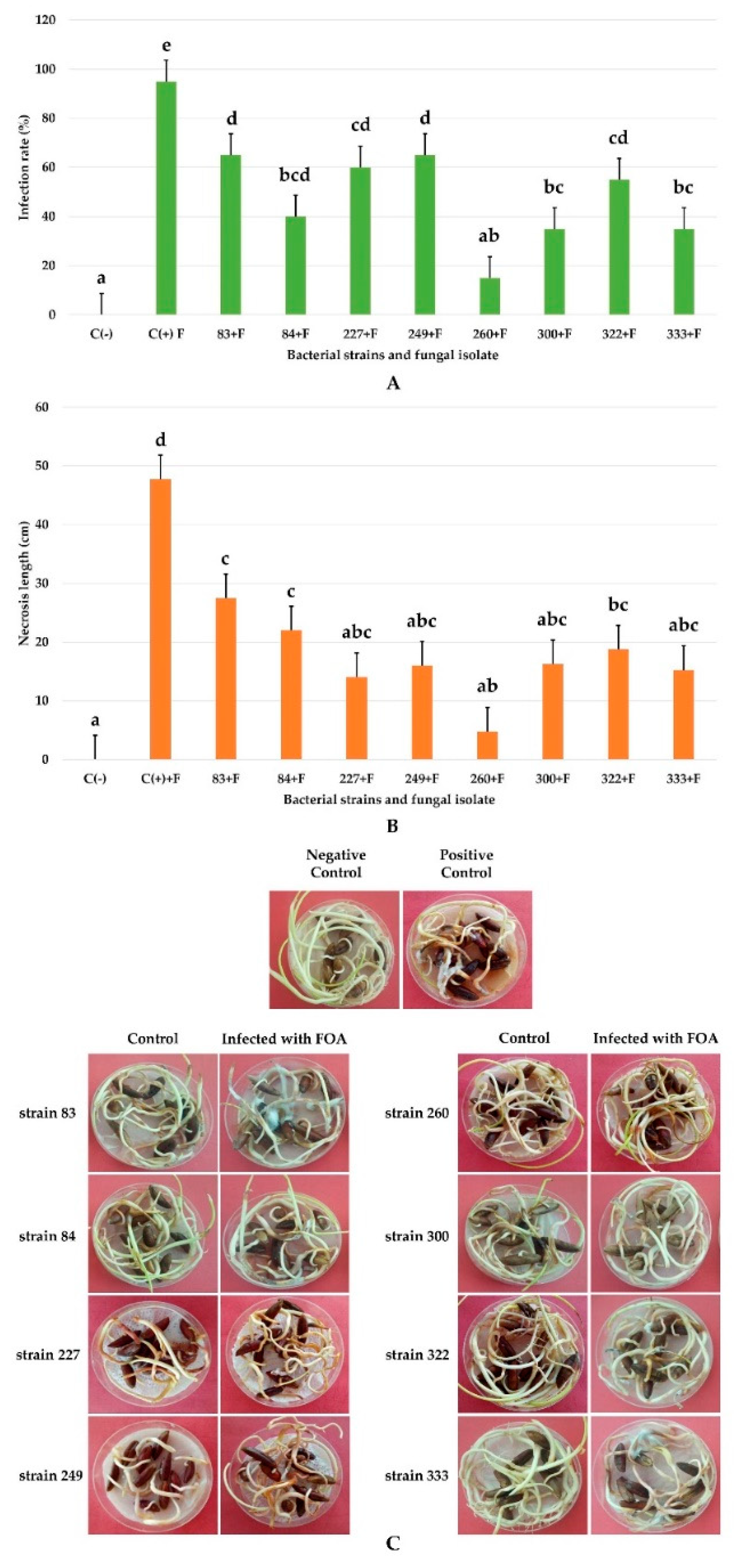
2.8. In Vivo Biocontrol Assay for the Reduction in Date Palm Root Disease Using Selected Bacteria

The selected bacteria were tested for their ability to inhibit the growth of the fungus in vivo. The strains were cultured in 5 mL of LB broth (Luria–Bertani) and incubated at 30 °C for 24 h with stirring. A total of 1 mL of each bacterial culture was introduced into 50 mL of LB medium. After incubation for 48 h at 30 °C with stirring, the cultures were centrifuged at 5000 rpm for 15 min and the bacterial pellets were suspended in 50 mL of sterile distilled water. The cultures were adjusted to a concentration of 108 cells/mL. Date palm seeds of the Deglet Nour variety, previously disinfected, were germinated aseptically in Petri plates lined with absorbent paper soaked in sterile distilled water for 15 days. Then, 10 germinated seeds were soaked in each bacterial suspension for one hour and subsequently infected after 24 h with 1 mL of an FOA spore suspension adjusted to 105 spores/mL. Seeds were placed in Petri plates lined with absorbent paper soaked in water, sealed, and incubated at room temperature in the dark for 45 days. The experiment was repeated four times. Seeds infected only with FOA in the absence of bacteria were considered a positive control and seeds which were neither infected nor treated were considered a negative control. The degree of protection was assessed by calculating the percentage of infection expressed by the number of diseased seeds/total number of seeds × 100 and by the size of the necrosis of diseased roots measured (cm) by a graduate ruler.

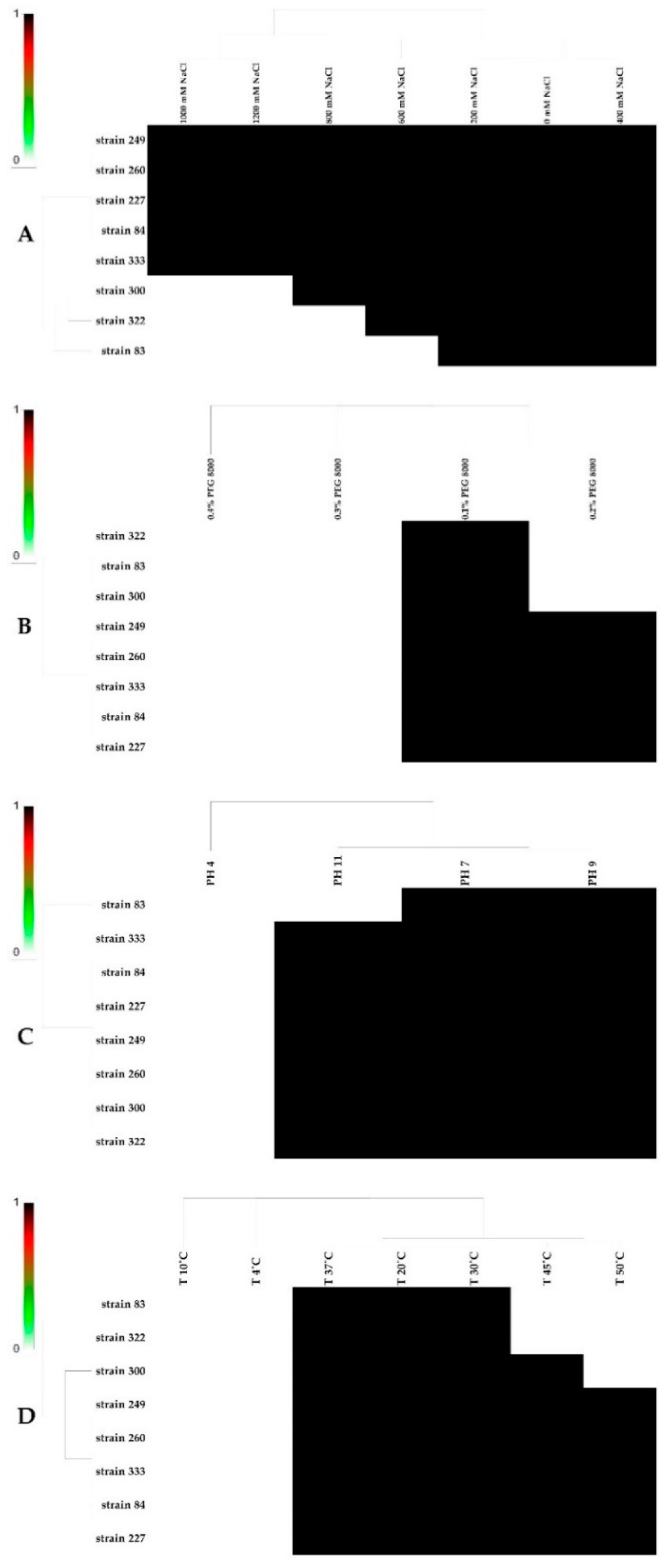
2.9. Effect of Salt, pH, PEG, and Temperature on the Growth of Bacterial Strains

The ability of bacterial strains to tolerate salt stress, water stress, and different pH values was carried out on TSA agar containing increasing concentrations of NaCl (0, 200, 400, 600, 800, 1000, and 1200 mM), PEG% (10, 20, 30, and 40), and different pH values (4, 7, 9, and 10). A total of 2 μL of each bacterial culture was inoculated by spots on the agar media and incubated at 30 °C for 48 h. The ability of the strains to grow under different temperatures was carried out on TSA agar incubated at 4, 10, 20, 30, 37, 45, and 50 °C for 48 h. Any bacterial growth indicated the ability of the strains to tolerate different abiotic stresses [25].

2.10. Determination of Resistance to Heavy Metals

The resistance of bacterial strains to lead, cadmium, cobalt, and mercury was evaluated on a solid LB medium supplemented with the various metallic salts PbCl2, Cd(NO3)2, CoCl2, and HgCl2, at concentrations of 100, 250, 500, and 1000 ppm. A total of 10 μL of each bacterial suspension were spotted on agar media and incubated for 24 h. Any bacterial growth after incubation at 30 °C for 72 h indicated the ability of the isolates to resist heavy metals [47].

2.11. PGP Potentialities of Bacterial Isolates

The production of the phytohormone indole acetic acid (IAA) was detected on LB broth supplemented with tryptophan (2 g/L). The absorbance of the bacterial culture supernatants supplemented with the Salkowski reagent were recorded at 535 nm [48]. IAA concentrations were determined by comparison with a standard curve generated from different IAA concentrations. The ability of bacterial strains to solubilize phosphates was detected in the Pikovskaya medium. The soluble phosphate was measured in the supernatant according to Olsen and Sommers [49]. The phosphate concentration was determined by measuring the optical density at 610 nm. A calibration curve was made to determine the phosphate concentration. The production of siderophores was determined using the Chrom azurol S (CAS) test [50]. Siderophore production was assessed by inoculating the King B medium. The absorbance of culture supernatants supplemented with CAS solution was measured at 630 nm. The percentage of siderophore production was expressed by calculating the following formula expressed as a percentage: SP(%) = OD of the sample/OD of the control (CAS solution) [51].
Other fertilization and biocontrol tests of bacterial isolates were determined qualitatively. The ability to fix atmospheric nitrogen was detected by the growth of bacteria on a nitrogen-free medium after incubation at 30 °C for 3 days. The production of NH3 was performed on a peptone water medium [52]. The cultures were incubated at 30 °C for 2 days. After the addition of Nessler’s reagent (0.5 mL), the appearance of a yellowish to brownish color indicated NH3 production. The HCN production test was performed on agar supplemented with glycine (4.4 g/L) [53]. A disc (9 cm) of paper impregnated with a solution of sodium picrate was placed at the bottom of the Petri plate lid. The plates were sealed and incubated at 30 °C for 2 days. A change in the color of the paper from yellow to orange indicated a positive result.

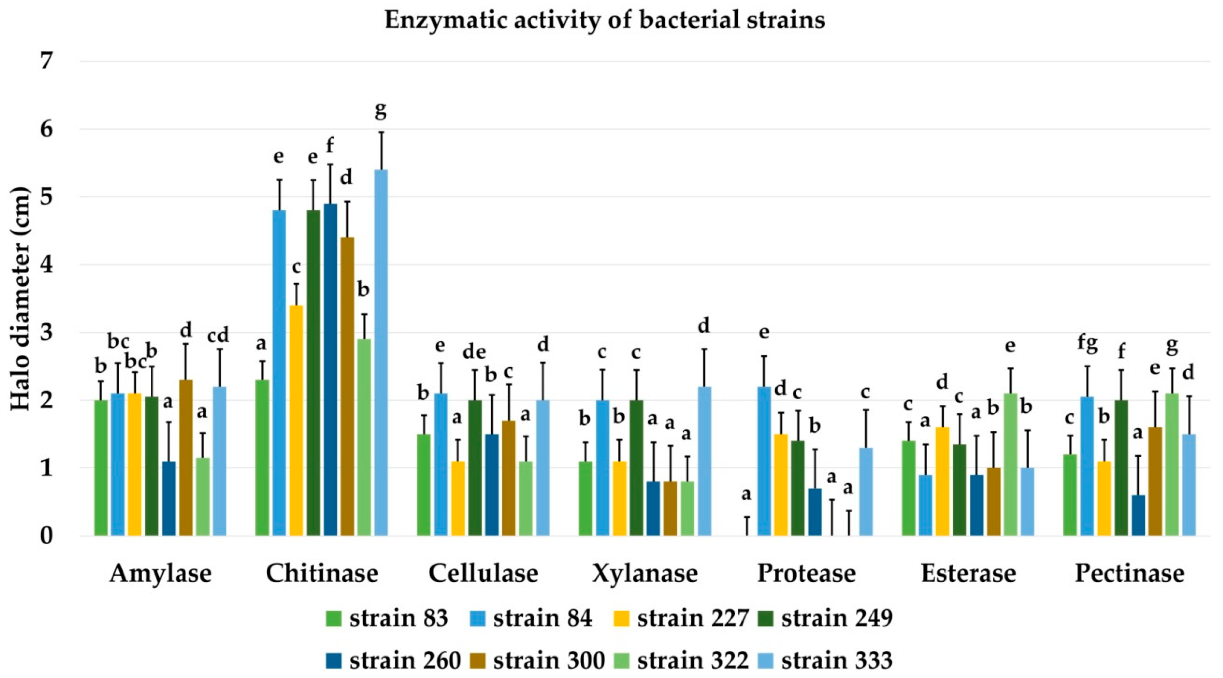
2.12. Screening for Bacterial Hydrolytic Enzymes

The capacity of the isolates to produce hydrolytic enzymes was tested on an agar medium supplemented with the corresponding substrates allowing the release of the enzymes. The production of cellulase, chitinase, amylase, xylanase, and pectinase were tested on an agar medium containing, respectively, carboxymethylcellulose (1% w/v), chitin (1% w/v), starch (0.5% w/v), xylan (1% w/v), and pectin (1% w/v). The media were inoculated by spotting bacterial cultures. The Petri dishes were incubated for 3 days at 30 °C. The degradation of the substrates resulted in the appearance of a clear halo around the colony after exposure by pouring an iodine solution (0.3 g iodine and 0.6 g KI/L) on the Petri plate’s surface. The lipase activity was tested on an agar medium supplemented with tween 20 (sorbitol oleate) [54] inoculated by spotting and incubated at 30 °C for 3 days. The degradation of the tween resulted in the appearance of an opaque halo around the colony due to the precipitation of calcium oleate. Protease production was performed on skimmed milk agar media [55]. The plates were inoculated by spotting and then incubated at 30 °C for 3 days and the protease activity was recorded by the development of a clear zone (halo) around the colonies. The results of the different enzymatic activities were expressed by measuring the diameter of the enzyme production halos.

2.13. Statistical Analyses

All the experiments were repeated three times and the results were expressed as mean ± standard error of the mean. The data were analyzed using GraphPad Prism 8. One-way ANOVA and two-way ANOVA were used to analyze the data to find whether there was a significant effect of the treatment compared to the control sample. A significant level of 5% (p < 0.05) was used, and Tukey’s multiple comparison tests were performed when a significant difference was encountered.

3. Results

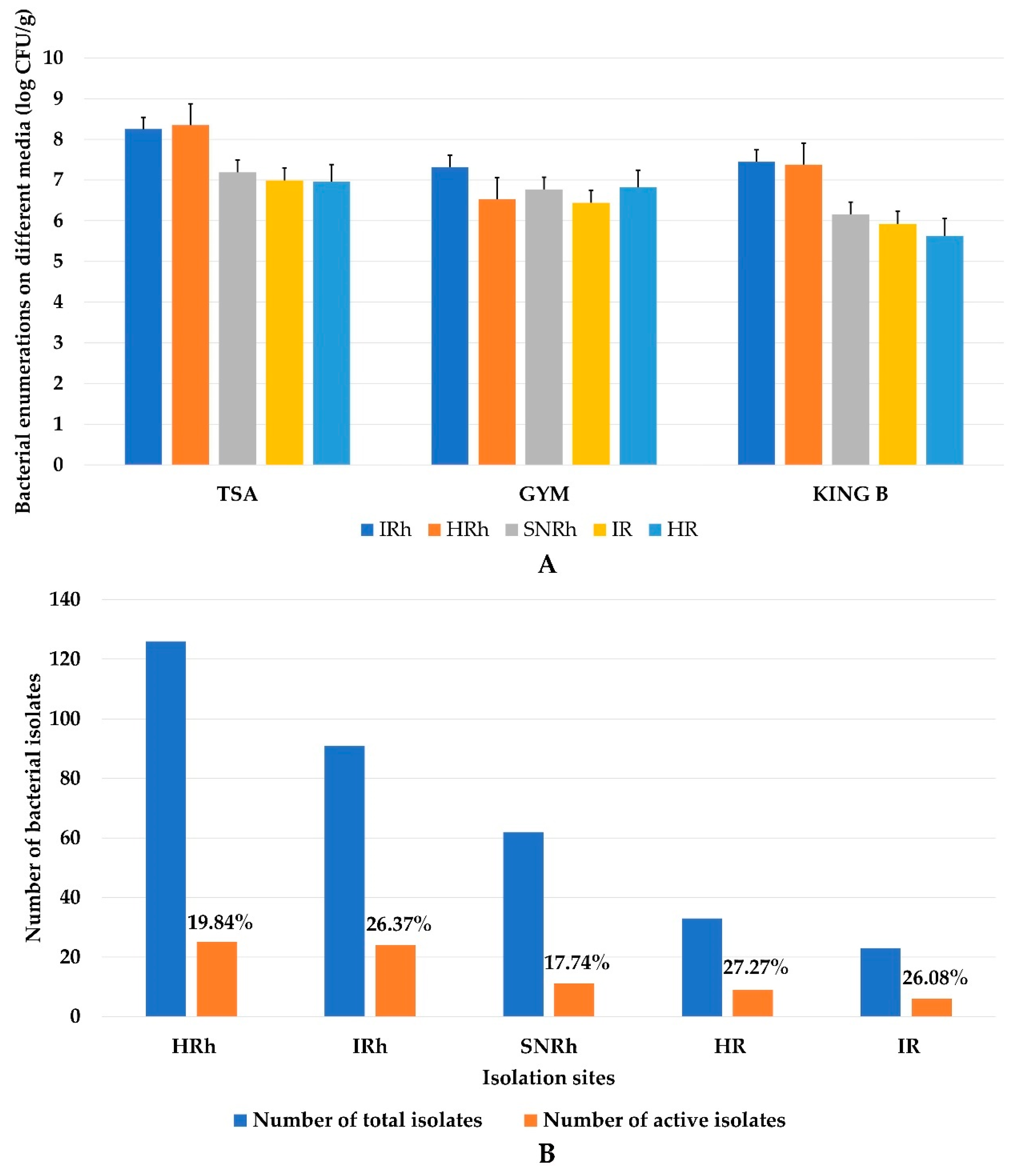
3.1. Enumeration, Isolation, and Selection of Antagonistic Bacteria

A total of 335 bacterial strains forming different characteristic colonies were isolated from healthy (126) and infected (91) rhizospheric soil, non-rhizospheric soil (62) and healthy (33) and infected (23) roots. All isolates grew on TSA, King B, and GYM media. The bacterial count (log CFU/g) revealed that the rhizospheric bacterial density was higher than those of non-rhizospheric bacteria and endophytic bacteria (Figure 2A).
The rate of isolates showing antifungal activity against FOA1 is shown in Figure 2B. Of the 335 isolates tested, 75 were able to inhibit FOA growth in vitro (Table S1). The results showed that 19.84%, 26.37%, 17.74%, 27.27%, and 26.08% of active strains were isolated, respectively, from healthy rhizospheric soil, infected soil, non-rhizospheric soil, and healthy and infected roots. Indeed, the number of antagonistic endophytic and rhizospheric bacteria was higher than that of non-rhizospheric bacteria.

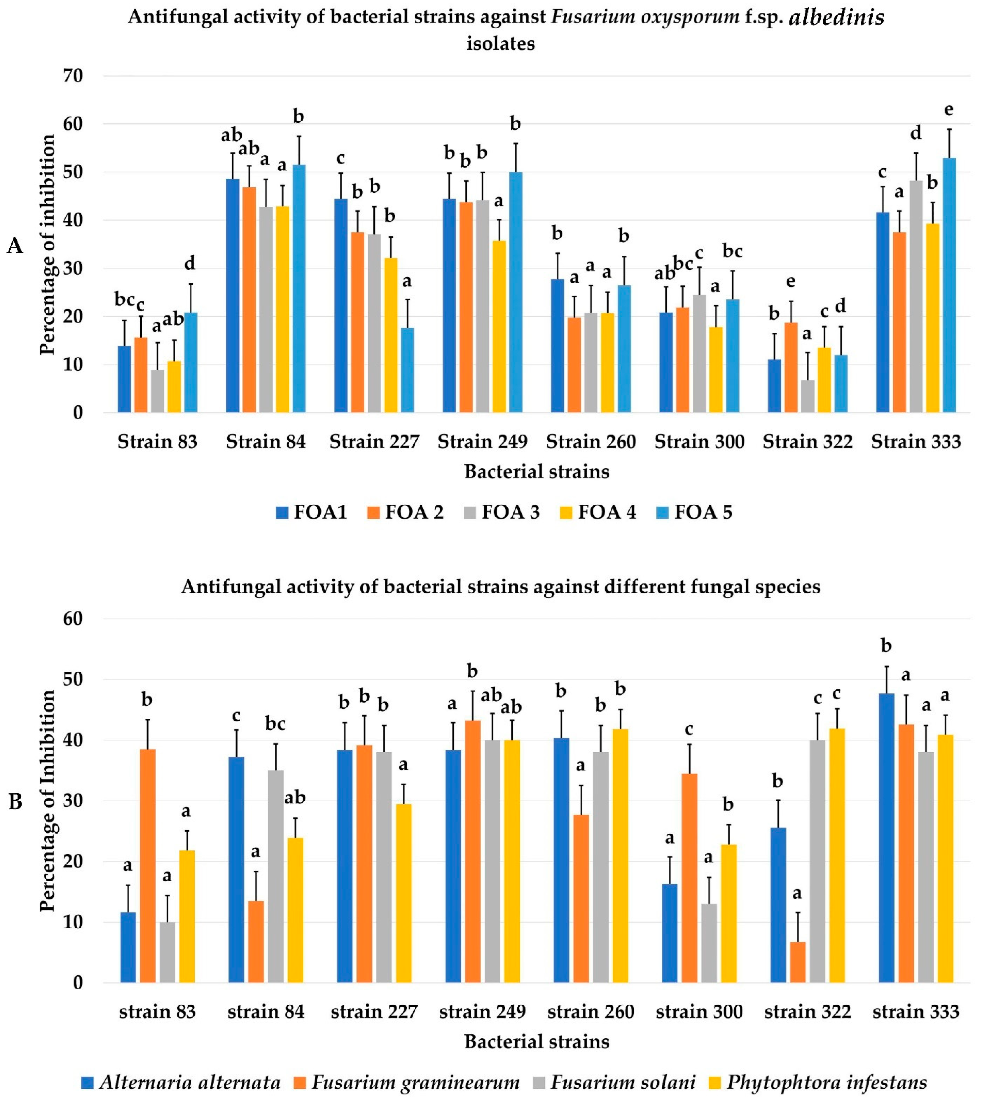
3.2. Biocontrol Ability of Bacterial Isolates towards FOA and Other Phytopathogenic Fungi

Among the 75 FOA antagonistic bacteria, 8 strains from different sampling compartments (83, 84, 300, 333, 322, 260, 249, and 227) having the best inhibition rates were chosen. The bacteria also inhibited additional strains of FOA (FOA1, FOA2, FOA3, FOA4, and FOA5). Strains 333 and 84 showed high inhibitory potential towards FOA strains. A maximum inhibition rate of 52.94% was observed for strain 333 towards FOA5 (Figure 3A and Figure 4). Strain 84 was found to be effective in significantly inhibiting (p < 0.05) the growth of FOA1, FOA2, and FOA4 strains with inhibition rates of 48.61%, 46.88%, and 42.85%, respectively. Moreover, weak antagonistic activity was marked by strains 322 and 83 towards all the strains of FOA. The bacterial isolates also showed strong activities against four other phytopathogenic fungi. Isolates 333 and 249 proved to be very effective against the development of all four fungi (Figure 3B and Figure S1). However, the inhibition rates reached 47.67% against Aa (strain 333) and 43.24% against Fg (strain 249), while a weak antagonistic potential was observed for strains 322 and 83 towards Fg (6.72%) and Fs (10%), respectively.

3.3. Molecular Identification of Bacterial Strains and Their Phylogenetic Positions

The phylogenetic tree based on the comparison of the 16S-rDNA sequences of the strains (227, 300, 249, 333, 83, 84, 322, and 260) with the similar sequences strains available in the databases (GenBank) confirmed that all the strains belonged to Bacillus genus (Figure 5). Strains 227, 300, and 249 exhibited homologies with B. subtilis. Strains 333, 83, and 84 were identified as Bacillus sp., B. atrophaeus, and B. halotolerans, respectively. In contrast, 322 and 260 were related to B. amyloliquefasiens and B. paralicheniformis species, respectively. Their sequences were submitted to GenBank and accession numbers were implied in the tree. The Bacillus thuringiensis strain JYCB351was used as an outgroup.

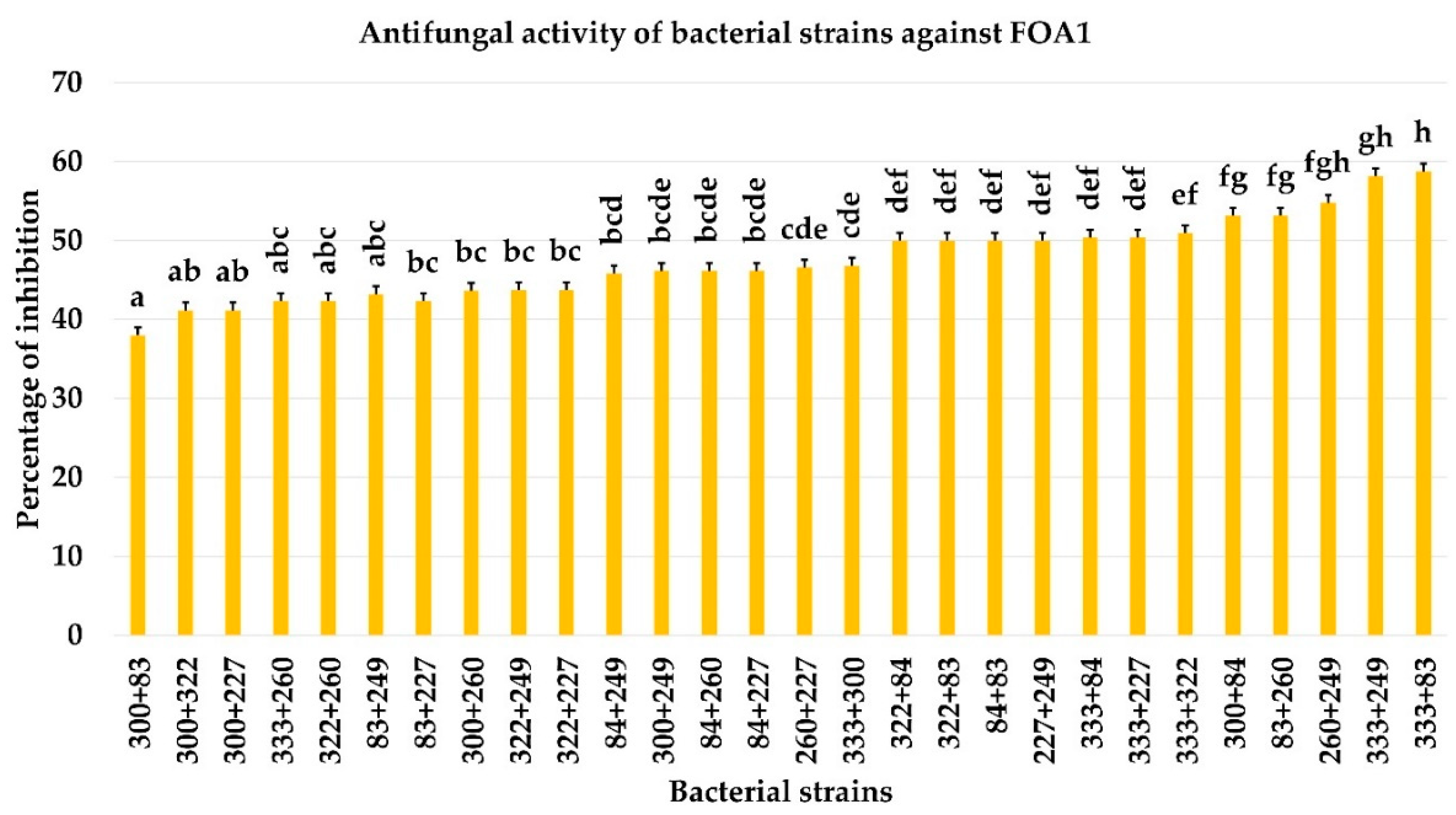
3.4. Effect of Bacterial Co-Culture on FOA Growth

The antifungal activity of the bacterial strains in combined form towards FOA1 is represented in Figure 6 and Figure S2. The antagonistic effect of the bacterial strains co-culture showed inhibition rates of 58.77%, 58.17%, 54.77 %, 53.17%, and 53.12% (p < 0.05) for the combinations 333 + 83, 333 + 249, 260 + 249, 83 + 260, and 300 + 84, respectively. However, a low inhibition rate of 37.98% was seen in strains 300 + 83. According to the results obtained, the antagonistic effect of the bacterial co-culture did not show an important difference compared to the activity of bacterial strains alone mentioned above.

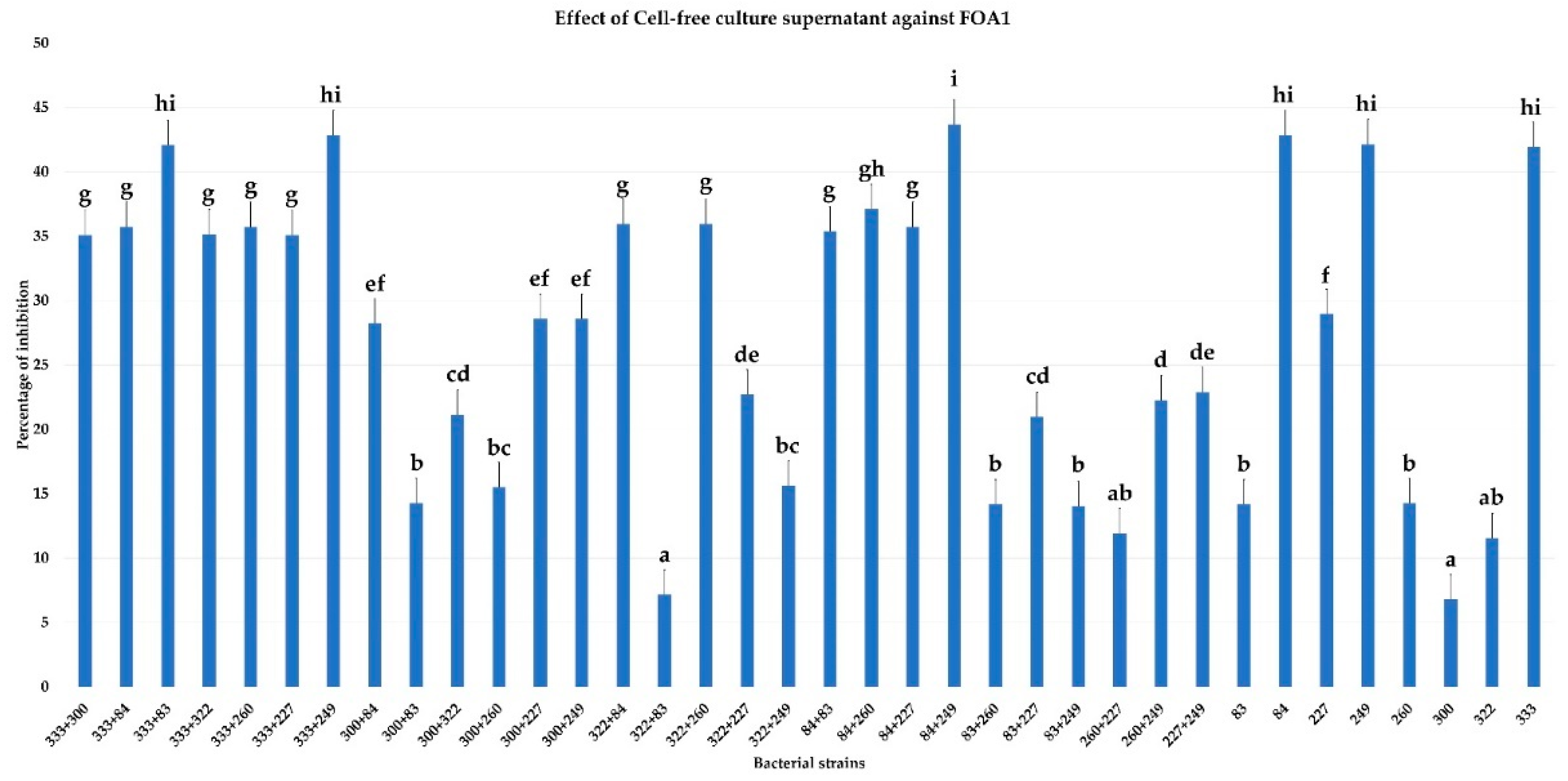
3.5. Effect of Cell-Free Culture Supernatant on FOA Growth

The results showed an inhibitory effect of cell-free culture supernatant alone or in combination on the growth of FOA1. As indicated in Figure 7 and Figure S3, better inhibitory activity was demonstrated both for the supernatants of the combined bacterial strains 84 + 249 (43.69%), 333 + 249 (42.85%), and 333 + 83 (42.10%) than those of single bacterial strains 84 (42.85%), 249 (42.12%), and 333 (41.92%) (p < 0.05). According to the inhibition rates, there was no synergistic or additive effect of the different bacterial supernatants combined on the growth inhibition of FOA1.

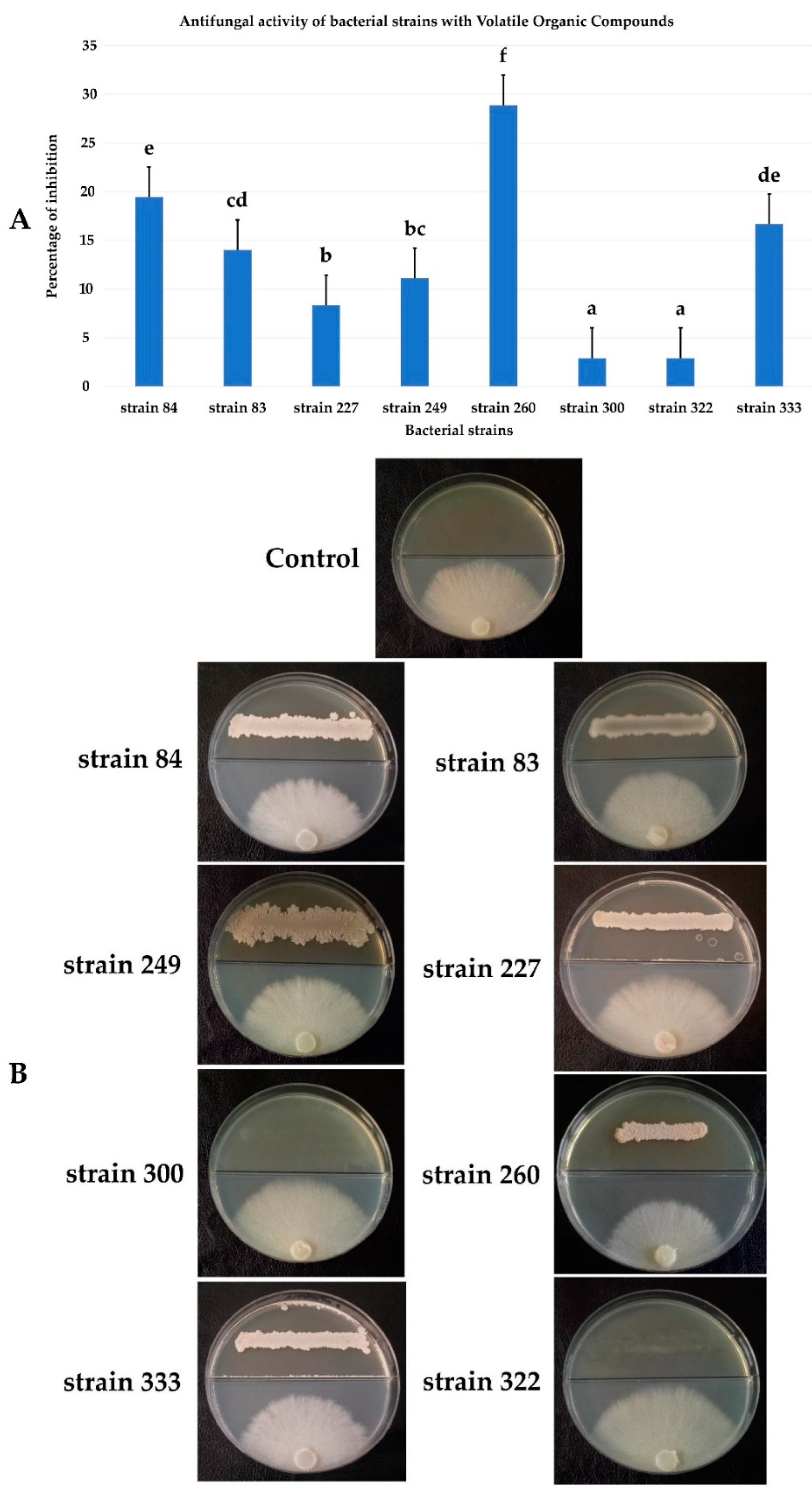
3.6. Antifungal Activity by Volatile Organic Compounds (VOCs)

The effect of VOCs produced by bacterial strains on the mycelial growth of FOA1 showed a weak inhibition. However, the volatile substances secreted by strain 260 showed a good inhibition rate of 28.89% compared to those of other bacterial strains (Figure 8A,B). Moreover, a very weak inhibition of 2.40% was noted by the VOCs produced by strains 300 and 322.

3.7. In Vivo Biocontrol Test on Germinated Date Palm Seeds

The ability of bacterial strains to inhibit FOA1 growth was assessed in vivo on the germinated seeds of the date palm variety Deglet Nour. The degree of protection evaluated by the infection rate and the size of root necrosis showed that the majority of strains provided weak protection against FOA infection (Figure 9). However, seeds treated with strain 260 significantly (p < 0.05) reduced the infection rate to 15% (Figure 9A) and the size of necrosis to 4.7 cm (Figure 9B). According to the results, strains 84, 300, and 333 protected the germinated seeds by about 60% but the other strains 83, 227, 249, and 322 displayed a lower degree of protection (Figure 9B).

3.8. Effect of Salt, pH, PEG, and Temperature on the Bacterial Growth of Strains

Bacterial strains were tested for their ability to tolerate different abiotic stresses. The majority of strains grew at salt concentrations of 0–400 mM (Figure 10A), however, strains 249, 260, 227, 84, and 333 tolerated concentrations of NaCl up to 1200 mM. Thus, the osmotic effect of different concentrations of PEG on bacterial growth was documented in Figure 10B. Most of the strains were osmotolerant at concentrations of 10% PEG. Strains 249, 260, 333, 84, and 227 grew at concentrations of 20% PEG. All the strains showed an ability to grow at high pH values (pH 9 and 11) except 83 which did not tolerate a pH greater than 9. A negative effect of acidity was observed on the growth of bacterial strains (Figure 10C). In addition, bacterial growth was observed at temperatures of 20 to 37 °C. Certain strains (249, 260, 333, 84, and 227) tolerated high temperatures of up to 50 °C but low temperatures negatively affected the bacterial growth (Figure 10D).

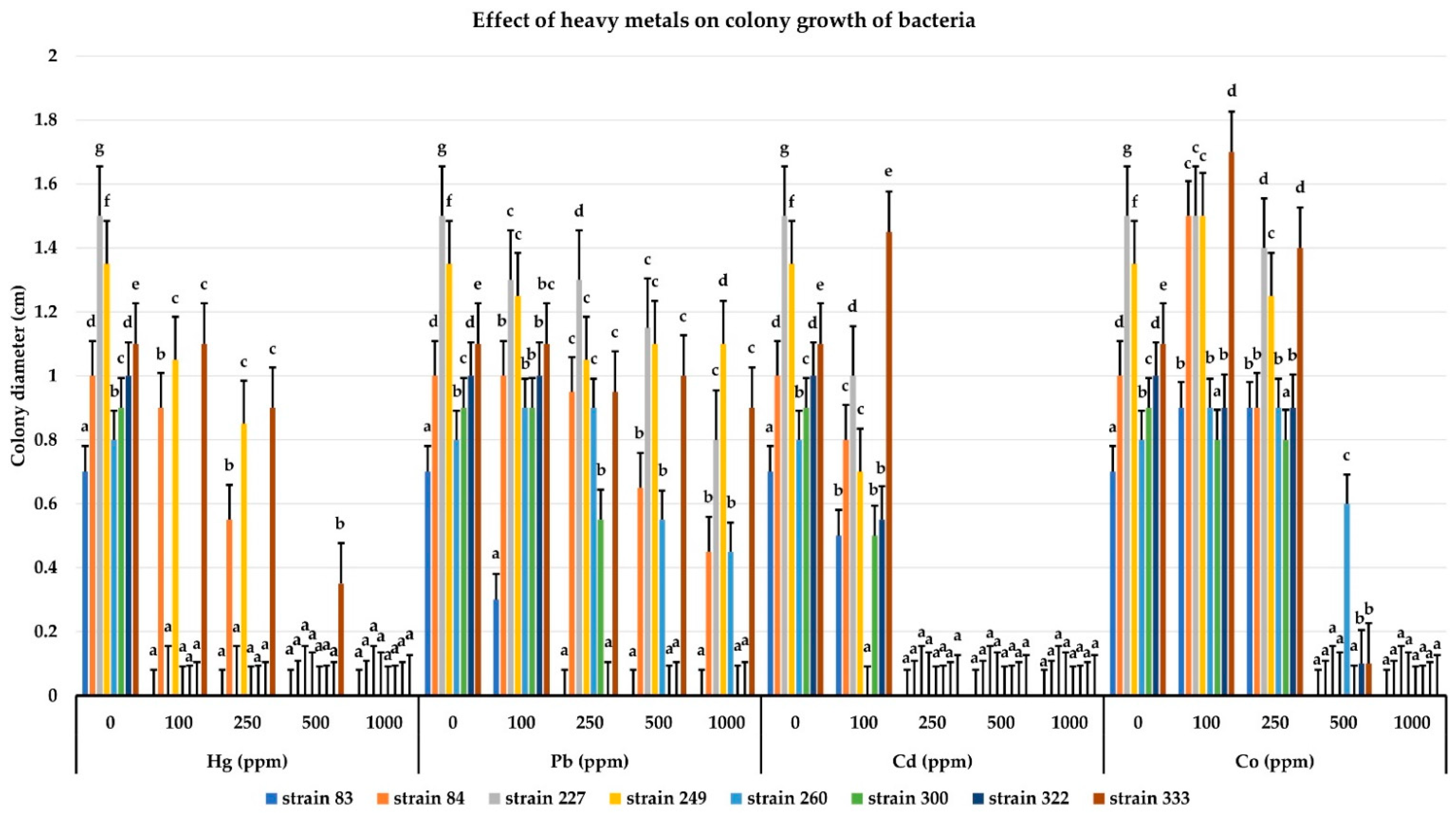
3.9. Determination of Resistance to Heavy Metals

The effect of different concentrations of heavy metals (Hg, Pb, Cd, and Co) on the bacterial growth of strains expressed by the size of the colonies is illustrated in Figure 11. According to the results, the strains seemed to tolerate Pb well. The majority of them were able to grow up to 1000 ppm with strain 249 showing better growth. In addition, the majority of the strains could not grow beyond 250 ppm of Hg and Co, with the exception of strains 260 and 333 which were able to resist up to 500 ppm of Co and Hg, respectively. In contrast, Cd was the most toxic for bacterial growth and the majority of strains hardly tolerated more than 100 ppm.

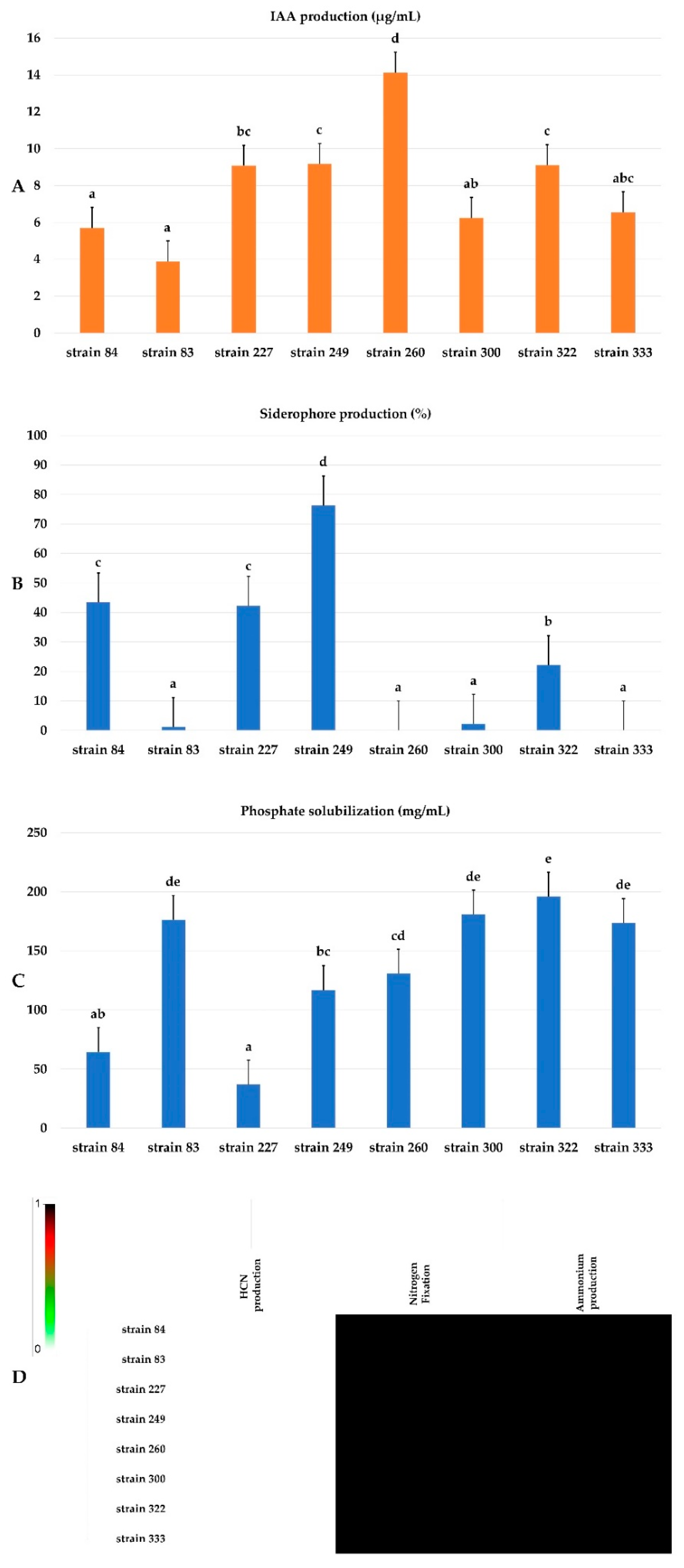
3.10. PGP Potentialities of Selected Strains

The selected bacterial isolates possessed several PGP activities (Figure 12). All strains produced IAA at varying rates and the maximum production was observed for strain 260 (14.12 μg/mL) (Figure 12A). The majority of strains produced siderophores, with the exception of strains 333 and 83. The most efficient strain was 249 (76.23%) (Figure 12B). It was also noted that the strains were efficient in phosphate solubilization. The amount of P2O5 produced by the strains varied from 36.86 to 195.88 mg/mL (Figure 12C). The maximum concentration of soluble P2O5 was observed in strain 322 (195.88 mg/mL), 300 (180.75 mg/mL), 83 (176.31 mg/mL), and 333 (173.55 mg/mL). Thus, all strains were able to fix nitrogen and produce ammonia. However, they failed to produce HCN (Figure 12D).

3.11. Production of Hydrolytic Enzymes

The enzymatic activity was detected by measuring the production of halos around the bacterial colonies (Figure 13). The results showed that the enzyme produced by all the strains was chitinase. The ability to produce other enzymes, amylase, cellulase, xylanase, esterase, and pectinase, was a property common to all isolates. Nevertheless, some strains (83, 300, and 322) failed to produce protease.

4. Discussion

The vascular wilt of date palm, also known as Bayoud, caused by FOA is the most destructive disease in North Africa and has no effective control strategy. Since its appearance, this disease has caused enormous economic losses due to the decline in crop yield and quality. Given the disadvantages of chemical control, biological control remains the most effective means. The use of microorganisms is one of the most promising biological control methods for suppressing plant diseases. In any ecosystem, the host-microbiota interaction is an established concept. This microbial symbiotic relationship plays an important role in plant growth control and disease resistance. Therefore, the study of microflora associated with date palms is necessary to identify the beneficial agents important in preventing pathogen infections and that contribute to the growth of high-quality disease-free cultivars.
This study aimed to screen the antagonistic bacterial species of FOA. Therefore, the exploration of epidemic foci in the palm groves of the M’zab valley, being the front of the progression of Bayoud for more than 40 years, constituted the sampling sites. Two sites were chosen, El-Atteuf and Metlili, for the isolation of bacterial strains from healthy and infected rhizospheric soil, non-rhizospheric soil (between palm trees), and the roots of healthy and infected palm trees. A significant number of bacteria were bound to the rhizosphere which represents the main location for nutrient uptake [56]. In this environment, plant roots provide soil microorganisms with root exudates that are used as substrates and signaling molecules. These molecules mediate interactions between plant roots and microbial communities in the rhizosphere [57]. Isolates (n = 335) recovered from these two sites were screened for their inhibitory effect against FOA using in vitro confrontation tests. The rates of active isolates were 19.84%, 26.37%, 17.74%, 27.27%, and 26.08% coming, respectively, from healthy rhizospheric soil, infected, non-rhizospheric soil, and healthy and infected roots. These levels indicated that the number of antagonistic endophytic and rhizospheric bacteria was higher than that of non-rhizospheric bacteria. This can be explained by the root surface and the root system being colonized by a wide range of soil bacteria capable of stimulating plant growth and health [58]. Certain endophytic bacteria colonizing an ecological niche make them suitable as biological control agents [56]. Biological control agents may have a better chance of becoming established and effectively controlling pathogens if they originate from the soil, compared to exotic microorganisms, because these type of native microorganisms are already adapted to the local climate and edaphic conditions as well as to the soil microbiota [59]. It has also been suggested that diseased date palm endophytes represent an interesting niche of endophytes capable of controlling a wide range of plant pathogens [60].
The screening yielded eight bacterial isolates with strong activity against FOAs. In our study, we selected the bacterial strains according to the sampling compartment. These strains tested against other strains of FOA have shown that they have inhibitory action against the fungi tested. This makes it possible to confirm their antagonistic character towards the strains of FOA. Similar results were observed by Slama et al. [23] where the screening of four bacterial isolates designated BFOA1, BFOA2, BFOA3, and BFOA4, were found to be highly active against FOA LMA1. These bacteria were found to be effective in inhibiting other strains of FOA [61]. The percentage of inhibition of FOA was approximately 55%. A similar result was found in our experiments and the maximum inhibition rate of 52.94% was observed for strain 333 against FOA5. Other authors have also proven the antifungal efficacy of two strains of Bacillus amyloliquefaciens Ag1 and Burkholderia cepacia Cs5 against FOA with maximum inhibition rates of 75% and 83%, respectively [62]. It has been proven that certain microorganisms such as Bacillus spp., Pseudomonas spp., species of actinomycetes, and Aspergillus spp. Isolated from the rhizosphere of the date palm are antagonists of the FOA growth, thus offering protection to date palms against the Bayoud disease [63]. In addition, the bacterial isolates showed inhibitory activity against four other major plant pathogens (Aa, Fg, Fs, and Pi) also infecting other crops planted in the same oasis ecosystems exploited in this study. Due to their biocontrol potential, these strains can be used as biological control agents against many phytopathogens [64,65,66].
The phylogenetic identification of bacterial strains by 16S-DNA sequencing revealed that all strains phylogenetically belong to the Bacillus species (Bacillus sp.; B. subtilis; B. atrophaeus; B. halotolerans; B. amyloliquefasiens, and B. paralicheniformis). Bacillus species have a wide range of biocontrol activities against various plant pathogens [23,67,68,69] through the formation of endospores [70,71] which allows them to survive in stressful conditions [35,72]. Currently, several species of the Bacillus genus (B. subtilis, B. amyloliquefaciens, B. licheniformis, and B. pumilus) have been widely studied to mitigate the incidence of agricultural diseases [73,74,75]. They have been identified as biocontrol agents and are considered good candidates for biological control [76] and prevent the establishment and development of plant pathogens by different mechanisms [77,78].
In order to obtain an eventual synergistic antagonism towards FOA, the co-culture of the bacterial strains was carried out. The association of biocontrol agents is sometimes necessary to increase their ability to suppress multiple pathogens [79]. Inhibition rates of the combined strains recorded a maximum of 58.77%. In comparison to the antifungal activity of bacteria in monoculture and co-culture, no significant difference was reported. However, the mixture of biocontrol agents may result in increased, decreased, or similar effects of pathogen suppression [80]. Moreover, new studies have proven that compatibility and taxonomic diversity are the two main factors responsible for the success of the combination between several strains of biocontrol agents [81,82]. Compatibility is defined as the possibility of growing in co-culture in vitro without one inhibiting the growth of the other [83]. Moreover, combining biocontrol agents with taxonomic distance seems desirable because the amount and number of secondary metabolites that inhibit pathogen growth increase with increasing taxonomic diversity [81]. The bacterial strains of the present study are compatible, but all belong to the same genus Bacillus. Indeed, the lack of taxonomic diversity between these bacterial strains during the combination may explain the lack of improvement in inhibition rates.
The production of metabolites by antagonistic bacteria is essential to help the plant fight against fungal diseases by interfering with the growth and activities of pathogens. The suppression of pathogens is based on the synthesis of antibiotics, antimicrobial peptides, bacteriocins, toxins, and lytic enzymes [84,85]. The antifungal effect of bacterial supernatants, alone or combined in vitro, showed inhibitory activity on the growth of FOA. Several supernatants obtained from Bacillus species have demonstrated activity against pathogens. However, our results showed that the inhibition percentages of the bacterial supernatants alone (strains 333, 84, and 249) and their combinations were close. This proves that there is no synergistic or additive effect between the different bacterial supernatants. Indeed, the inhibition capacity of supernatants with their metabolites is linked to the presence of lipopeptides, including iturins, fengycins, surfactins, and sphingofungins [65,86]. The success of the combination of bacteria in biocontrol is conditioned when the individual strains exert complementary mechanisms of disease suppression [85]; when one mechanism is ineffective in particular conditions, the others can compensate for it. Thus, the combination of strains with different modes of action can increase the possibility of building an effective and consistent defense against plant pathogens [87]. Nevertheless, the combination of biocontrol agents may give undesirable results in increased plant pathogen inhibition compared to the use of the most effective component by a biocontrol strain alone [88].
In addition to diffusible metabolites, volatile organic compounds (VOCs) are also used in the suppression of many plant pathogens [89]. The antifungal effect of the VOCs emitted by the bacterial strains towards FOA revealed very low inhibition rates. Only strain 260 displayed an average inhibition rate of 28.89%. Studies have found that the inhibition by VOCs varies widely between fungal species. According to the results obtained by Che et al. [90], the volatiles of Lysinibacillus sp. FJAT-4748 did not affect the growth of all pathogenic fungi in the same way. The observed differences in fungal sensitivities to bacterial VOCs may be explained by differences in physiological and pathogenicity properties. VOCs produced by the Bacillus genus depend on the growth of the fungi and also on the nature of the pathogenic fungi against which the VOCs were tested [91]. However, the nature of the VOCs also varies according to the biocontrol strains. Four volatile substances identified (methyl isobutyl ketone, ethanol, 5-methyl-2-heptanone, and S-(-)-2-methylbutylamine) produced by B. pumilus TM-R are responsible for the inhibition of F. oxysporum and other phytopathogenic fungi [92]. Among the VOCs detected in B. licheniformis, 3-methyl-1-butanol was the most abundant compound responsible for the inhibition of mycotoxic fungi [33]. Ngo et al. [93] described that B. amyloliquefaciens NJN-6 was able to produce 11 VOCs and inhibit the growth of F. oxysporum f.sp. cubense.
To evaluate the biocontrol effect in vivo, the bacterial strains were tested on germinated date palm seeds infected with FOA. The majority of strains tested showed mild to moderate fungistatic effects. However, treatment with strain 260 significantly reduced the infection rate to 15% and the size of the necrosis. Several authors have wondered whether the in vitro selection of biocontrol agents guarantees success in planta. However, in planta experiments have shown that strains with better antifungal activities in vitro do not always perform well in vivo and vice versa [94]. Indeed, some strains such as 260 showed satisfactory biocontrol results on palm seeds but its antagonism towards FOA in vitro was limited (low inhibition rate). Bacterial strains that show better inhibitory activity in vitro are not always effective in vivo. As in our case, strain 333 isolated from non-rhizospheric soil showed significant inhibition rates in vitro but a low ability to protect palm seeds against FOA. This can be explained by insufficient colonization of the roots of the palm unlike the endophyte strain 260 characterized by its competitive capacity of colonization, thus presenting better results of biocontrol in vivo. The results of the present study are in agreement with those obtained by Dihazi et al. [63] who proved that the pre-treatment of date palm roots with the two strains of B. amyloliquefaciens Ag and Burkholderia cepacia Cs significantly reduced the size of the browning zone around the FOA inoculation site, leading to localized necrosis. When necrosis formed disease symptoms were reduced, plants were more resistant to Bayoud disease. Biocontrol is mainly explained by several mechanisms of action that act alone or together to give the plant better resistance to disease: competition for space and nutrients, production of inhibiting substances (lipopeptides, antibiotics, volatile compounds, lytic enzymes, etc.), and induced systemic resistance.
Competition for niches and nutrients is an aspect involved by bacteria in biological control [95]. Indeed, their ability to colonize roots is the first fundamental step in protecting plants against plant pathogens. The root system is a nutrient-rich niche that serves as a chemoattractant for microbial colonization [96]. However, the competence and/or colonization of bacteria is mainly linked to their ability to take advantage of and adapt to a specific environment [77]. The process of colonization is based on the attachment and establishment of the bacteria on the root and the use of these plant nutrients [97]. Studies have proven that the protection of the date palm against Fusarium wilt by endo-rhizospheric strains is linked much more to the capacity of root colonization than to a phenomenon of antibiosis. The performance of bacterial strains is linked to their rapid growth and their fairly high root colonization power, which allows them to be competitive for nutrient sources and thus occupy root sites before the pathogen [98].
In addition, Bacillus spp. are among the most studied rhizobacteria that trigger ISR in plants [99]. Through the induction of hormone regulatory networks, bacteria can rapidly trigger plant defense responses when attacked by a pathogen [33]. Both rhizospheric and endophytic bacteria promote ISR in plants through the production of various metabolites such as antibiotics, siderophores, volatile organic compounds (VOCs), and others considered as ISR elicitors [38]. Bacteria-triggered ISR has been reported to start at the root and then spread to other parts of the plant [100]. ISR caused by B. subtilis results in changes in the host cell wall composition, production of pathogenesis-related (PR) proteins, such as chitinases and glucanases, and synthesis of phytoalexins host resistance against pathogens. In addition, ISR activates antioxidant enzymes that are responsible for the elimination of ROS [101]. Recent studies have clearly shown the potential of B. paralicheniformis in activating antioxidant defense enzymes in tomato plants infected with F. oxysporum and preventing oxidative damage through hydroxyl radical scavenging activities by suppressing the appearance of vascular wilt disease [67]. Furthermore, the transcriptome sequencing performed by Jiang et al. [102] revealed that nearly a thousand differentially expressed genes were linked to the process of triggering ISR by B. velezensis F21 against F. oxysporum f.sp. niveum (Fon), the causative agent of the vascular wilt of watermelon.
These particular biocontrol agents are used not only to replace chemical pesticides but to improve plant growth and attenuate various natural environmental stresses (salinity, aridity, extreme temperatures and pH, et or stresses related to environmental contamination. Members of the Bacillus genus are the most important PGPBs that have unique characteristics as biopesticides, biofertilizers, and bio-remediators.
The PGP potential of the selected strains was evaluated in vitro. All isolates showed nitrogen-fixing abilities. The production of IAA and the solubilization of phosphates were common traits in all bacterial strains. IAA is one of the most important phytohormones and is considered a signal molecule in the regulation of plant development [103]. On the other hand, the ability of PGPRs to solubilize phosphate (P) has various physiological functions related to plant health and resistance to biotic and abiotic stresses [104]. Our study indicated that the majority of strains produced siderophores, with the exception of strains 333 and 83. Siderophores act as specific ferric iron chelating agents. Some PGPR improve plant growth by producing extracellular siderophores which help control several plant diseases [83]. Thus, low levels of iron prevent plant pathogens from proliferating in the root zone of the plant. In contrast, plants can use this siderophore-iron complex for their nutrition [105,106]. All isolates showed nitrogen fixation and NH3 production and failed to produce HCN. Ammonia production can satisfy the nitrogen demand of host plants and promote root elongation and root biomass. When produced in excess, it can also provide defense against plant pathogens by reducing their colonization in host plants [107]. On the other hand, hydrocyanic acid (HCN) is known for its toxicity against plant pathogens and the chelation of metal ions [108]. All these PGP activities are characteristic of biocontrol bacteria [59].
Hydrolytic enzymes play a very important role in the biocontrol of plant pathogens. Many rhizobacteria and biological control agents synthesize extracellular hydrolytic enzymes involved in the hydrolysis of fungal cell wall components such as chitin, proteins, and cellulose [109]. These enzymes also prevent fungal spore germination and contribute to protein breakdown [106]. According to the results, the different enzymes amylase, chitinase, cellulase, xylanase, protease, esterase, and pectinase were produced, with chitinase as the most abundant. Microbial chitinase deteriorates and destroys the cell walls of many parasites and pathogens [110]. It has been reported that the antifungal effect of B. subtilis on the thinning of fungal hyphae is related to the secretion of chitinase and glucanase [21]. Several authors have demonstrated that Bacillus sp. inhibits the mycelial growth of phytopathogenic fungi by attaching to their cell walls and releasing lytic enzymes (chitinase, protease, and cellulase), siderophores, and HCN. Therefore, these antifungal compounds cause cracks and deformation in hyphae, which leads to impaired cellular structure and functions due to vacuolation and leakage of protoplasts [111].
The survival of a strain introduced into the rhizosphere is affected by a number of abiotic factors. In contrast, stress-tolerant strains can be effectively involved in extreme environments where they can show better competence in the rhizosphere and better competitive ability [112,113,114]. The tolerance of bacterial strains to different abiotic stresses was also examined in our study. The results showed that the majority of strains grew at concentrations of 400 mM NaCl and 10% PEG. Additionally, some strains tolerate up to 1200 mM NaCl and 20% PEG. Thus, all strains have shown growth capacity at high pH values (pH 9 and 11) and can withstand temperatures up to 50 °C [22,115]. The bacterial strains also exhibited resistance to different concentrations of heavy metals. Some strains were able to withstand up to 500 ppm mercury, 1000 ppm lead, and 500 ppm cobalt. Nevertheless, most managed to grow at 100 ppm cadmium. This tolerance is explained by several resistance mechanisms observed by PGPBs [116]. PGPBs reduce heavy metal toxicity by improving crop growth in polluted soil conditions [117] or when irrigating with waters containing high levels of heavy metals [23]. Bacillus licheniformis has been shown to enhance the accumulation and distribution of Cd, Cr, Pb, Cu, and Zn in plants grown in soil contaminated with heavy metals, resulting in reduced levels of toxic metals in soil [111].

5. Conclusions

The use of antagonistic bacteria to control the Fusarium wilt of the date palm is currently a major challenge. The choice of bacteria antagonists of the pathogenic agent is a very important criterion. Eight rhizospheric and endophytes Bacillus species showed their innate potential for biocontrol and biofertilization. These strains have shown the ability to produce a range of diffusible or volatile antifungal metabolites, cell wall degrading enzymes, and plant growth-promoting compounds. The in vivo study of the bio-control assay showed that the in vitro selection of biocontrol agents is not correlated with the disease-suppressing activity in vivo. The antagonistic activity and efficacy of any bio-control agent depend critically on the combination of different competitive and antibiosis mechanisms inducing systemic resistance in plants. Therefore, these isolates are worthy of further evaluation under field conditions. The genus Bacillus, according to its physiological and metabolic properties, meets these requirements to become an effective commercial biological control product. This is supported by various studies conducted to assess the performance of Bacillus in controlling plant diseases. Further work will be carried out to validate the results in the field and future research will target the possibility of developing appropriate formulation and application techniques to ensure or enhance date palm resistance against FOA.

Supplementary Materials

The following supporting information can be downloaded at: https://www.mdpi.com/article/10.3390/agronomy12102403/s1, Table S1: Bacterial strains activity against Fusarium albedinis FOA.

Author Contributions

Conceptualization, S.B., H.C.-S., N.S. and L.B.; methodology, S.B., A.S., H.C.-S., N.S. and L.B.; software, A.C.B., A.S., H.C.-S. and L.B.; validation, H.C.-S., A.S., L.L. and L.B.; formal analysis, A.C.B. and L.B.; investigation, S.B., H.C.-S., A.S., L.L. and L.B.; resources, H.C.-S., A.S., L.L. and L.B.; data curation, S.B., A.C.B., N.S. and L.B.; writing—original draft preparation, S.B., H.C.-S., A.S., N.S. and L.B.; writing—review and editing, A.C.B., L.L. and F.N.A.; visualization, L.L., F.N.A., A.C.B. and L.B.; supervision, H.C.-S., A.S. and L.B.; project administration, H.C.-S., A.S. and L.B.; and funding acquisition, H.C.-S., A.S., L.L. and L.B. All authors have read and agreed to the published version of the manuscript.

Funding

This research received no external funding.

Institutional Review Board Statement

Not applicable.

Informed Consent Statement

Not applicable.

Conflicts of Interest

The authors declare no conflict of interest.

References

  1. Djerbi, M. Précis de Phéniciculture. ED; FAO: Rome, Italy, 1994; p. 192. [Google Scholar]
  2. Hassan, M.M.; Allam, M.A.; Shams El-Din, I.M.; Malhat, M.H.; Taha, R.A. High-frequency direct somatic embryogenesis and plantlet regeneration from date palm immature inflorescences using picloram. J. Genet. Eng. Biotechnol. 2021, 19, 1–11. [Google Scholar] [CrossRef]
  3. De la Perrière, R.B.; Amir, H.; Bounaga, N. Prospects for integrated control of ‘bayoud’ (Fusarium wilt of the date palm) in Algerian plantations. Crop Protect. 1995, 14, 227–235. [Google Scholar] [CrossRef]
  4. Sidky, R. Optimized direct organogenesis from shoot tip explants of date palm. In Date Palm Biotechnology Protocols; Humana Press: New York, NY, USA, 2017; Volume 1, pp. 37–45. [Google Scholar]
  5. FAOSTAT. Available online: http://www.fao.org/faostat/en/#data/QC (accessed on 20 February 2020).
  6. Al-Sharidah, A. Report of Black Scorch Disease on Date Palm Trees in the State of Kuwait. Am. Acad. Sci. Res. J. Eng. Technol. Sci. 2017, 28, 14–17. [Google Scholar]
  7. Abedalred, E.M.; Ismail, W.M.; Abdulmoohsin, R.G.; Al-Karhi, M.A. First molecular identification of Fusarium fujikuroi causing pollen rot of palm trees (Phoenix dactylifera L.) in Iraq and evaluation efficacy of some nanoparticles against it. In Proceedings of the IOP Conference Series: Earth and Environmental Science, Bristol, UK, 1 November 2019. [Google Scholar]
  8. Armstrong, G.M.; Armstrong, J.R. Forms specials and races of Fusarium oxysporum causing wilt diseases. In Fusarium: Diseases, Biology and Taxonomy; Nelson, P.E., Toussoun, T.A., Cook, R.J., Eds.; The Pennsylvania States University Press: University Park, PA, USA, 1981; pp. 391–393. [Google Scholar]
  9. Ahmed, Y.; Hussien, A.; El-badry, N.; Soliman, M.S. Validation of a Diagnostic Protocol for the Detection of Fusarium oxysporum f.sp. albedinis, the Causal Agent of Bayoud Disease of Date Palm. Egypt. J. Phytopathol. 2019, 47, 297–312. [Google Scholar] [CrossRef]
  10. OEPP/EPP. Fiches informatives sur les organismes de quarantaine n 70, Fusarium oxysporum f.sp. albedinis. Bulletin 1982, 12. Available online: https://gd.eppo.int/taxon/FUSAAL/datasheet (accessed on 6 December 2017).
  11. Bahriz, H.; Bouras, N. Etude de la Maladie du Bayoud, le Comportement Variétal du Palmier Dattier vis-à-vis du Fusarium oxysporum f.sp. albedinis dans la Vallée du M’Zab. Afr. Rev. Sci. Technol. Develop. 2020, 5, 41–60. [Google Scholar]
  12. EPPO (European and Mediterranean Plant Protection Organization). EPPO Global Database. Available online: https://gd.eppo.int (accessed on 6 December 2017).
  13. Bouhlali, E.D.T.; Derouich, M.; Ben-Amar, H.; Meziani, R.; Essarioui, A. Exploring the potential of using bioactive plant products in the management of Fusarium oxysporum f.sp. albedinis: The causal agent of Bayoud disease on date palm (Phoenix dactylifera L.). Beni-Seuf. Univ. J. Appl. Sci. 2020, 9, 1–9. [Google Scholar] [CrossRef]
  14. Djerbi, M. Diseases of the Palm Phoenix Dactylifera; FAO: Baghdad, Iraq, 1983; p. 45. [Google Scholar]
  15. Benzohra, I.E.; Megateli, M.; Elayachi, B.A.; Zekraoui, M.; Djillali, K.; Bouafia, A.; Benouis, S.; Benaziza, A.; Rekis, A. Integrated management of Bayoud disease on date palm (Phoenix dactylifera L.) caused by Fusarium oxysporum f.sp. albedinis in Algeria. JARA 2017, 14, 93–100. [Google Scholar]
  16. Djerbi, M. Les Maladies du Palmier Dattier. Projet Régional de Lutte Contre le Bayoud; FAO: Alger, Algeria, 1988; p. 127. [Google Scholar]
  17. EPPO. Diagnostic protocols for regulated pests PM 7/16 Fusarium oxysporum f.sp. albedinis. Bull. OEPP 2003, 33, 245–247. [Google Scholar]
  18. EFSA Panel on Plant Health (PLH); Jeger, M.; Bragard, C.; Caffier, D.; Candresse, T.; Chatzivassiliou, E.; Dehnen-Schmutz, K.; Gilioli, G.; Grégoire, J.C.; Jaques Miret, J.A.; et al. Pest categorisation of Fusarium oxysporum f.sp. albedinis. EFSA J. 2018, 16, e05183. [Google Scholar]
  19. Kaddouri, Y.; Abrigach, F.; Ouahhoud, S.; Benabbes, R.; El Kodadi, M.; Alsalme, A.; Al-Zaqri, N.; Warad, I.; Touzani, R. Mono-Alkylated Ligands Based on Pyrazole and Triazole Derivatives Tested Against Fusarium oxysporum f.sp. albedinis: Synthesis, Characterization, DFT, and Phytase Binding Site Identification Using Blind Docking/Virtual Screening for Potent Fophy Inhibitors. Front. Chem. 2020, 8, 559262. [Google Scholar] [CrossRef]
  20. De Lamo, F.J.; Takken, F.L. Biocontrol by Fusarium oxysporum using endophyte-mediated resistance. Front. Plant Sci. 2020, 11, 37. [Google Scholar] [CrossRef] [Green Version]
  21. Khan, N.; Martínez-Hidalgo, P.; Ice, T.A.; Maymon, M.; Humm, E.A.; Nejat, N.; Sanders, E.R.; Kaplan, D.; Hirsch, A.M. Antifungal activity of Bacillus species against Fusarium and analysis of the potential mechanisms used in biocontrol. Front. Microbiol. 2018, 9, 2363. [Google Scholar] [CrossRef] [PubMed] [Green Version]
  22. Sedra, M.H. La Maladie du Bayoud du Palmier Dattier en Afrique du Nord: Diagnostic et Caractérisation; Actes du Symposium International sur le Développement durable des Systèmes Oasiens: Erfoud, Marocco, 2005. [Google Scholar]
  23. Slama, H.B.; Cherif-Silini, H.; Chenari Bouket, A.; Qader, M.; Silini, A.; Yahiaoui, B.; Alenezi, F.N.; Luptakova, L.; Triki, M.A.; Vallat, A.; et al. Screening for Fusarium antagonistic bacteria from contrasting niches designated the endophyte Bacillus halotolerans as plant warden against Fusarium. Front. Microbiol. 2019, 9, 3236. [Google Scholar] [CrossRef] [Green Version]
  24. Slama, H.B.; Cherif-Silini, H.; Chenari Bouket, A.; Silini, A.; Alenezi, F.N.; Luptakova, L.; Vallat, A.; Belbahri, L. Biotechnology and bioinformatics of endophytes in biocontrol, Bioremediation, and plant growth promotion. In Endophytes: Mineral Nutrient Management; Maheshwari, D.K., Dheeman, S., Eds.; Springer: Cham, Switzerland, 2021; Volume 3, pp. 181–205. [Google Scholar] [CrossRef]
  25. Cherif-Silini, H.; Thissera, B.; Chenari Bouket, A.; Saadaoui, N.; Silini, A.; Eshelli, M.; Alenezi, F.N.; Vallat, A.; Luptakova, L.; Yahiaoui, B.; et al. Durum wheat stress tolerance induced by endophyte Pantoea agglomerans with genes contributing to plant functions and secondary metabolite arsenal. Int. J. Mol. Sci. 2019, 20, 3989. [Google Scholar] [CrossRef]
  26. Mefteh, F.B.; Daoud, A.; Chenari Bouket, A.; Thissera, B.; Kadri, Y.; Cherif-Silini, H.; Eshelli, M.; Alenezi, F.N.; Vallat, A.; Oszako, T.; et al. Date Palm Trees Root-Derived Endophytes as Fungal Cell Factories for Diverse Bioactive Metabolites. Int. J. Mol. Sci. 2018, 19, 1986. [Google Scholar] [CrossRef] [Green Version]
  27. Díaz-Valle, A.; López-Calleja, A.C.; Alvarez-Venegas, R. Enhancement of pathogen resistance in common bean plants by inoculation with Rhizobium etli. Front. Plant Sci. 2019, 10, 1317. [Google Scholar] [CrossRef]
  28. Alenezi, F.N.; Chenari Bouket, A.; Cherif-Silini, H.; Silini, A.; Jaspars, M.; Oszako, T.; Belbahri, L. Loss of Gramicidin Biosynthesis in Gram-Positive Biocontrol Bacterium Aneurinibacillus migulanus (Takagi et al., 1993) Shida et al. 1996 Emend Heyndrickx et al., 1997 Nagano Impairs Its Biological Control Ability of Phytophthora. Forests 2022, 13, 535. [Google Scholar] [CrossRef]
  29. Cherif-Silini, H.; Silini, A.; Chenari Bouket, A.; Alenezi, F.N.; Luptakova, L.; Bouremani, N.; Nowakowska, J.A.; Oszako, T.; Belbahri, L. Tailoring next generation plant growth promoting microorganisms as versatile tools beyond soil desalinization: A road map towards field application. Sustainability 2021, 13, 4422. [Google Scholar] [CrossRef]
  30. Pliego, C.; Ramos, C.; de Vicente, A.; Cazorla, F.M. Screening for candidate bacterial biocontrol agents against soilborne fungal plant pathogens. Plant Soil. 2011, 340, 505–520. [Google Scholar] [CrossRef] [Green Version]
  31. Knudsen, I.M.B.; Hockenhull, J.; Jensen, D.F.; Gerhardson, B.; Hökeberg, M.; Tahvonen, R.; Teperi, E.; Sundheim, L.; Henriksen, B. Selection of biological control agents for controlling soil and seed-borne diseases in the field. Eur. J. Plant Pathol. 1997, 103, 775–784. [Google Scholar] [CrossRef]
  32. Daayf, F.; Adam, L.; Fernando, W.G.D. Comparative screening of bacteria for biocontrol of potato late blight (strain US-8), using in-vitro, detached-leaves, and whole plant testing systems. Can. J. Plant Pathol. 2003, 25, 276–284. [Google Scholar] [CrossRef]
  33. Besset-Manzoni, Y.; Joly, P.; Brutel, A.; Gerin, F.; Soudiere, O.; Langin, T.; Prigent-Combaret, C. Does in vitro selection of biocontrol agents guarantee success in planta? A study case of wheat protection against Fusarium seedling blight by soil bacteria. PLoS ONE 2019, 14, e0225655. [Google Scholar] [CrossRef] [Green Version]
  34. Cherif-Silini, H.; Silini, A.; Yahiaoui, B.; Ouzari, I.; Boudabous, A. Phylogenetic and plant-growth-promoting characteristics of Bacillus isolated from the wheat rhizosphere. Ann. Microbiol. 2016, 66, 1087–1097. [Google Scholar] [CrossRef]
  35. Kerbab, S.; Silini, A.; Chenari Bouket, A.; Cherif-Silini, H.; Eshelli, M.; El Houda Rabhi, N.; Belbahri, L. Mitigation of NaCl stress in wheat by rhizosphere engineering using salt habitat adapted PGPR halotolerant bacteria. Appl. Sci. 2021, 11, 1034. [Google Scholar] [CrossRef]
  36. Rabhi, N.E.H.; Cherif-Silini, H.; Silini, A.; Alenezi, F.N.; Chenari Bouket, A.; Oszako, T.; Belbahri, L. Alleviation of Salt Stress via Habitat-Adapted Symbiosis. Forests 2022, 13, 586. [Google Scholar] [CrossRef]
  37. Shao, J.; Liu, Y.; Xie, J.; Stefanic, P.; Lv, Y.; Fan, B.; Mandic-Mulec, I.; Zhang, R.; Shen, Q.; Xu, Z. Annulment of Bacterial Antagonism Improves Plant Beneficial Activity of a Bacillus velezensis Consortium. Appl. Environ. Microbiol. 2022, 88, e0024022. [Google Scholar] [CrossRef]
  38. Borriss, R. Bacillus, A Plant-Beneficial Bacterium. In Principles of Plant-Microbe Interactions; Lugtenberg, B., Ed.; Springer: Cham, Switzerland, 2015. [Google Scholar] [CrossRef]
  39. Mac Faddin, J.F. Media for Isolation-Cultivation-Identification-Maintenance of Medical Bacteria; Williams and Wilkins: Baltimore, MD, USA, 1985; Volume 1. [Google Scholar]
  40. King, E.O.; Ward, M.; Raney, D.E.J. Two simple media for the demonstration of pyocyanin and fluorescein. Lab. Clin. Méd. 1954, 44, 301. [Google Scholar]
  41. Palumbo, J.D.; O’Keeffe, T.L. Method for high-throughput antifungal activity screening of bacterial strain libraries. J. Microbiol. Methods. 2021, 189, 106311. [Google Scholar] [CrossRef]
  42. Luchi, N.; Ghelardini, L.; Belbahri, L.; Quartier, M.; Santini, A. Rapid detection of Ceratocystis platani inoculum by quantitative Real-Time PCR assay. Appl. Environ. Microbiol. 2013, 79, 5394–5404. [Google Scholar] [CrossRef] [Green Version]
  43. Mefteh, F.B.; Chenari Bouket, A.; Daoud, A.; Luptakova, L.; Alenezi, F.N.; Gharsallah, N.; Belbahri, L. Metagenomic insights and genomic analysis of phosphogypsum and its associated plant endophytic microbiomes reveals valuable actors for waste bioremediation. Microorganisms 2019, 7, 382. [Google Scholar] [CrossRef] [Green Version]
  44. Li, T.; Tang, J.; Karuppiah, V.; Li, Y.; Xu, N.; Chen, J. Co-culture of Trichoderma atroviride SG3403 and Bacillus subtilis 22 improves the production of antifungal secondary metabolites. Biol. Control 2019, 140, 104122. [Google Scholar] [CrossRef]
  45. Dutta, S.; Woo, E.E.; Yu, S.M.; Nagendran, R.; Yun, B.S.; Lee, Y.H. Control of anthracnose and gray mold in pepper plants using culture extract of white-rot fungus and active compound schizostatin. Mycobiology 2019, 47, 87–96. [Google Scholar] [CrossRef] [Green Version]
  46. He, C.N.; Ye, W.Q.; Zhu, Y.Y.; Zhou, W.W. Antifungal activity of volatile organic compounds produced by Bacillus methylotrophicus and Bacillus thuringiensis against five common spoilage fungi on loquats. Molecules 2020, 25, 3360. [Google Scholar] [CrossRef] [PubMed]
  47. Yu, X.; Li, Y.; Zhang, C.; Liu, H.; Liu, J.; Zheng, W.; Kang, X.; Leng, X.; Zhao, K.; Gu, Y. Culturable heavy metal-resistant and plant growth promoting bacteria in V-Ti magnetite mine tailing soil from Panzhihua, China. PLoS ONE 2014, 9, e106618. [Google Scholar] [CrossRef] [PubMed]
  48. Patten, C.L.; Glick, B.R. Role of Pseudomonas putida indoleacetic acid in development of the host plant root system. Appl. Environ. Microbiol. 2002, 68, 3795–3801. [Google Scholar] [CrossRef] [Green Version]
  49. Olsen, R.S.; Sommers, L.E. Phosphorus. In Methods of Soil Analysis, Part 2: Chemical and Microbiological Properties, 2nd ed.; Pages, A.L., Miller, R.H., Keeney, D.R., Eds.; American Society of Agronomy: Madison, WI, USA, 1982; pp. 403–430. [Google Scholar]
  50. Schwyn, B.; Neilands, J.B. Universal chemical assay for the detection and determination of siderophores. Anal. Biochem. 1987, 160, 47–56. [Google Scholar] [CrossRef]
  51. Gokarn, K. Comments: Siderophore production by uropathogenic Escherichia coli. Indian J. Pathol. Microbiol. 2010, 53, 570. [Google Scholar] [CrossRef]
  52. Cappuccino, J.C.; Sherman, N. Microbiology: A Laboratory Manuel, 3rd ed.; Benjamin-Cummings Publishing Company: New York, NY, USA, 1992; pp. 125–179. [Google Scholar]
  53. Lorck, H. Production of hydrocyanic acid by bacteria. Physiol. Plant 1948, 1, 142–146. [Google Scholar] [CrossRef]
  54. Kumar, D.; Kumar, L.; Nagar, S.; Raina, C.; Parshad, R.; Gupta, V.K. Screening, isolation and production of lipase/esterase producing Bacillus sp. strain DVL2 and its potential evaluation in esterification and resolution reactions. Arch. Appl. Sci. Res. 2012, 4, 1763–1770. [Google Scholar]
  55. Loper, J.E.; Schroth, M.N. Influence of bacterial sources of indole- 3-acetic acid on root elongation of sugar beet. Phytopathology 1986, 76, 386–389. [Google Scholar] [CrossRef]
  56. Hashem, A.; Tabassum, B.; Abd_Allah, E.F. Bacillus subtilis: A plant-growth promoting rhizobacterium that also impacts biotic stress. Saudi J. Biol. Sci. 2019, 26, 1291–1297. [Google Scholar] [CrossRef] [PubMed]
  57. Huang, X.F.; Chaparro, J.M.; Reardon, K.F.; Zhang, R.; Shen, Q.; Vivanco, J.M. Rhizosphere interactions: Root exudates, microbes, and microbial communities. Botany 2014, 92, 267–275. [Google Scholar] [CrossRef]
  58. Djaya, L.; Istifadah, N.; Hartati, S.; Joni, I.M. In vitro study of plant growth promoting rhizobacteria (PGPR) and endophytic bacteria antagonistic to Ralstonia solanacearum formulated with graphite and silica nano particles as a biocontrol delivery system (BDS). Biocatal. Agric. Biotechnol. 2019, 19, 101153. [Google Scholar] [CrossRef]
  59. Vinayarani, G.; Prakash, H. Growth promoting rhizospheric and endophytic bacteria from Curcuma longa L. as biocontrol agents against rhizome rot and leaf blight diseases. Plant Pathol. J. 2018, 34, 218. [Google Scholar] [CrossRef]
  60. Khalil, M.M.R.; Fierro-Coronado, R.A.; Peñuelas-Rubio, O.; Villa-Lerma, A.G.; Plascencia-Jatomea, R.; Félix-Gastélum, R.; Maldonado-Mendoza, I.E. Rhizospheric bacteria as potential biocontrol agents against Fusarium wilt and crown and root rot diseases in tomato. Saudi J. Biol. Sci. 2021, 28, 7460–7471. [Google Scholar] [CrossRef]
  61. Cheffi, M.; Chenari Bouket, A.; Alenezi, F.N.; Luptakova, L.; Belka, M.; Vallat, A.; Rateb, M.E.; Tounsi, S.; Triki, M.A.; Belbahri, L. Olea europaea L. root endophyte Bacillus velezensis OEE1 counteracts oomycete and fungal harmful pathogens and harbours a large repertoire of secreted and volatile metabolites and beneficial functional genes. Microorganisms 2019, 7, 314. [Google Scholar] [CrossRef] [Green Version]
  62. Bernal, P.; Allsopp, L.P.; Filloux, A.; Llamas, M.A. The Pseudomonas putida T6SS is a plant warden against phytopathogens. ISME J. 2017, 11, 972–987. [Google Scholar] [CrossRef] [Green Version]
  63. Dihazi, A.; Jaiti, F.; Jaoua, S.; Driouich, A.; Baaziz, M.; Daayf, F.; Serghini, M.A. Use of two bacteria for biological control of bayoud disease caused by Fusarium oxysporum in date palm (Phoenix dactylifera L.) seedlings. Plant Physiol. Biochem. 2012, 55, 7–15. [Google Scholar] [CrossRef]
  64. Nishad, R.; Ahmed, T.A. Survey and identification of date palm pathogens and indigenous biocontrol agents. Plant Dis. 2020, 104, 2498–2508. [Google Scholar] [CrossRef]
  65. Pellegrini, M.; Pagnani, G.; Bernardi, M.; Mattedi, A.; Spera, D.M.; Gallo, M.D. Cell-free supernatants of plant growth-promoting bacteria: A review of their use as bio-stimulant and microbial biocontrol agents in sustainable agriculture. Sustainability 2020, 12, 9917. [Google Scholar] [CrossRef]
  66. Hong, S.; Kim, T.Y.; Won, S.-J.; Moon, J.-H.; Ajuna, H.B.; Kim, K.Y.; Ahn, Y.S. Control of Fungal Diseases and Fruit Yield Improvement of Strawberry Using Bacillus velezensis CE 100. Microorganisms 2022, 10, 365. [Google Scholar] [CrossRef] [PubMed]
  67. Jinal, H.N.; Sakthivel, K.; Amaresan, N. Characterization of antagonistic Bacillus paralicheniformis (strain EAL) by LC–MS, antimicrobial peptide genes, and ISR determinants. Antonie Leeuwenhoek. 2020, 113, 1167–1177. [Google Scholar] [CrossRef] [PubMed]
  68. Slama, H.B.; Triki, M.A.; Chenari Bouket, A.; Mefteh, F.B.; Alenezi, F.N.; Luptakova, L.; Cherif-Silini, H.; Vallat, A.; Oszako, T.; Gharsallah, N.; et al. Screening of the high-rhizosphere competent Limoniastrum monopetalum’culturable endophyte microbiota allows the recovery of multifaceted and versatile biocontrol agents. Microorganisms 2019, 7, 249. [Google Scholar] [CrossRef] [Green Version]
  69. Chen, K.; Tian, Z.; He, H.; Long, C.A.; Jiang, F. Bacillus species as potential biocontrol agents against citrus diseases. Biol. Control 2020, 151, 104419. [Google Scholar] [CrossRef]
  70. Secaira-Morocho, H.; Castillo, J.A.; Driks, A. Diversity and evolutionary dynamics of spore-coat proteins in spore-forming species of Bacillales. Microb. Genom. 2020, 6, mgen000451. [Google Scholar] [CrossRef]
  71. Ahmed, H.F.A.; Seleiman, M.F.; Al-Saif, A.M.; Alshiekheid, M.A.; Battaglia, M.L.; Taha, R.S. Biological Control of Celery Powdery Mildew Disease Caused by Erysiphe heraclei DC In Vitro and In Vivo Conditions. Plants 2021, 10, 2342. [Google Scholar] [CrossRef]
  72. Belbahri, L.; Chenari Bouket, A.; Rekik, I.; Alenezi, F.N.; Vallat, A.; Luptakova, L.; Petrovova, E.; Oszako, T.; Cherrad, S.; Vacher, S.; et al. Comparative genomics of Bacillus amyloliquefaciens strains reveals a core genome with traits for habitat adaptation and a secondary metabolites rich accessory genome. Front. Microbiol. 2017, 8, 1438. [Google Scholar] [CrossRef] [Green Version]
  73. Attia, M.S.; El-Sayyad, G.S.; Abd Elkodous, M.; El-Batal, A.I. The effective antagonistic potential of plant growth-promoting rhizobacteria against Alternaria solani-causing early blight disease in tomato plant. Sci. Hort. 2020, 266, 109289. [Google Scholar] [CrossRef]
  74. Hong, P.; Hao, W.; Luo, J.; Chen, S.; Hu, M.; Zhong, G. Combination of hot water, Bacillus amyloliquefaciens HF-01 and sodium bicarbonate treatments to control postharvest decay of mandarin fruit. Postharvest Biol. Technol. 2014, 88, 96–102. [Google Scholar] [CrossRef]
  75. Malgioglio, G.; Rizzo, G.F.; Nigro, S.; Lefebvre du Prey, V.; Herforth-Rahmé, J.; Catara, V.; Branca, F. Plant-Microbe Interaction in Sustainable Agriculture: The Factors That May Influence the Efficacy of PGPM Application. Sustainability 2022, 14, 2253. [Google Scholar] [CrossRef]
  76. Villarreal-Delgado, M.F.; Villa-Rodríguez, E.D.; Cira-Chávez, L.A.; Estrada-Alvarado, M.I.; Parra-Cota, F.I.; Santos-Villalobos, S.D.L. The genus Bacillus as a biological control agent and its implications in the agricultural biosecurity. Rev. Mex. Fitopatol. 2018, 36, 95–130. [Google Scholar]
  77. Balla, A.; Silini, A.; Cherif-Silini, H.; Chenari Bouket, A.; Moser, W.K.; Nowakowska, J.A.; Oszako, T.; Benia, F.; Belbahri, L. The Threat of Pests and Pathogens and the Potential for Biological Control in Forest Ecosystems. Forests 2021, 12, 1579. [Google Scholar] [CrossRef]
  78. Palmieri, D.; Vitullo, D.; De Curtis, F.; Lima, G. A microbial consortium in the rhizosphere as a new biocontrol approach against Fusarium decline of chickpea. Plant Soil. 2017, 412, 425–439. [Google Scholar] [CrossRef]
  79. Mukherjee, A.; Chouhan, G.K.; Gaurav, A.K.; Jaiswal, D.K.; Verma, J.P. Development of indigenous microbial consortium for biocontrol management. In New and Future Developments in Microbial Biotechnology and Bioengineering; Verma, J.P., Gupta, V.K., Macdonald, C.A., Podile, A.R., Eds.; Elsevier: Amsterdam, The Netherland, 2021; pp. 91–104. [Google Scholar]
  80. Niu, B.; Wang, W.; Yuan, Z.; Sederoff, R.R.; Sederoff, H.; Chiang, V.L.; Borriss, R. Microbial interactions within multiple-strain biological control agents impact soil-borne plant disease. Front. Microbiol. 2020, 11, 585404. [Google Scholar] [CrossRef] [PubMed]
  81. Saidi, S.; Cherif-Silini, H.; Chenari Bouket, A.; Silini, A.; Eshelli, M.; Luptakova, L.; Alenezi, F.N.; Belbahri, L. Improvement of Medicago sativa crops productivity by the co-inoculation of Sinorhizobium meliloti–actinobacteria under salt stress. Curr. Microbiol. 2021, 78, 1344–1357. [Google Scholar] [CrossRef] [PubMed]
  82. Thomloudi, E.E.; Tsalgatidou, P.C.; Douka, D.; Spantidos, T.N.; Dimou, M.; Venieraki, A.; Katinakis, P. Multistrain versus single-strain plant growth promoting microbial inoculants-The compatibility issue. Hell. Plant Prot. J. 2019, 12, 61–77. [Google Scholar] [CrossRef]
  83. Jiao, X.; Takishita, Y.; Zhou, G.; Smith, D.L. Plant associated rhizobacteria for biocontrol and plant growth enhancement. Front. Plant Sci. 2021, 12, 634796. [Google Scholar] [CrossRef] [PubMed]
  84. Alenezi, F.N.; Slama, H.B.; Chenari Bouket, A.; Cherif-Silini, H.; Silini, A.; Luptakova, L.; Nowakowska, J.A.; Oszako, T.; Belbahri, L. Bacillus velezensis: A Treasure House of Bioactive Compounds of Medicinal, Biocontrol and Environmental Importance. Forests 2021, 12, 1714. [Google Scholar] [CrossRef]
  85. Wang, H.; Liu, R.; You, M.P.; Barbetti, M.J.; Chen, Y. Pathogen Biocontrol Using Plant Growth-Promoting Bacteria (PGPR): Role of Bacterial Diversity. Microorganisms 2021, 9, 1988. [Google Scholar] [CrossRef]
  86. Wang, X.; Liang, L.; Shao, H.; Ye, X.; Yang, X.; Chen, X.; Shi, Y.; Zhang, L.; Xu, L.; Wang, J. Isolation of the Novel Strain Bacillus amyloliquefaciens F9 and Identification of Lipopeptide Extract Components Responsible for Activity against Xanthomonas citri subsp. citri. Plants 2022, 11, 457. [Google Scholar] [CrossRef] [PubMed]
  87. Ruano-Rosa, D.; Cazorla, F.M.; Bonilla, N.; Martín-Pérez, R.; De Vicente, A.; López-Herrera, C.J. Biological control of avocado white root rot with combined applications of Trichoderma spp. and rhizobacteria. Eur. J. Plant Pathol. 2014, 138, 751–762. [Google Scholar] [CrossRef]
  88. Alamri, S.; Hashem, M.; Mostafa, Y.S. In vitro and in vivo biocontrol of soil-borne phytopathogenic fungi by certain bioagents and their possible mode of action. Biocontrol Sci. 2012, 17, 155–167. [Google Scholar] [CrossRef] [PubMed] [Green Version]
  89. Li, X.; Wang, X.; Shi, X.; Wang, B.; Li, M.; Wang, Q.; Zhang, S. Antifungal effect of volatile organic compounds from Bacillus velezensis CT32 against Verticillium dahliae and Fusarium oxysporum. Processes 2020, 8, 1674. [Google Scholar] [CrossRef]
  90. Che, J.; Liu, B.; Liu, G.; Chen, Q.; Lan, J. Volatile organic compounds produced by Lysinibacillus sp. FJAT-4748 possess antifungal activity against Colletotrichum acutatum. Biocontrol Sci. Technol. 2017, 27, 1349–1362. [Google Scholar] [CrossRef]
  91. Chaves-López, C.; Serio, A.; Gianotti, A.; Sacchetti, G.; Ndagijimana, M.; Ciccarone, C.; Stellarini, A.; Corsetti, A.; Paparella, A. Diversity of food-borne Bacillus volatile compounds and influence on fungal growth. J. Appl. Microbiol. 2015, 119, 487–499. [Google Scholar] [CrossRef] [PubMed]
  92. Morita, T.; Tanaka, I.; Ryuda, N.; Ikari, M.; Ueno, D.; Someya, T. Antifungal spectrum characterization and identification of strong volatile organic compounds produced by Bacillus pumilus TM-R. Heliyon 2019, 5, e01817. [Google Scholar] [CrossRef]
  93. Ngo, T.T.; Dart, P.; Callaghan, M.; Klieve, A.; McNeill, D. Volatile Organic Compound Profiles Associated with Microbial Development in Feedlot Pellets Inoculated with Bacillus amyloliquefaciens H57 Probiotic. Animals 2021, 11, 3227. [Google Scholar] [CrossRef]
  94. Morales-Cedeno, L.R.; del Carmen Orozco-Mosqueda, M.; Loeza-Lara, P.D.; Parra-Cota, F.I.; de Los Santos-Villalobos, S.; Santoyo, G. Plant growth-promoting bacterial endophytes as biocontrol agents of pre-and post-harvest diseases: Fundamentals, methods of application and future perspectives. Microbiol. Res. 2021, 242, 126612. [Google Scholar] [CrossRef]
  95. Santhanam, R.; Menezes, R.C.; Grabe, V.; Li, D.; Baldwin, I.T.; Groten, K. A suite of complementary biocontrol traits allows a native consortium of root-associated bacteria to protect their host plant from a fungal sudden-wilt disease. Mol. Ecol. 2019, 28, 1154–1169. [Google Scholar] [CrossRef]
  96. Posada, L.F.; Álvarez, J.C.; Romero-Tabarez, M.; de-Bashan, L.; Villegas-Escobar, V. Enhanced molecular visualization of root colonization and growth promotion by Bacillus subtilis EA-CB0575 in different growth systems. Microbiol. Res. 2018, 217, 69–80. [Google Scholar] [CrossRef] [PubMed]
  97. Lamari, L.; Bouras, N.; Boudjella, H.; Ould EL Hadj-Khelil, A.; Ould El Hadj, M.D.; Sabaou, N. Influence de quelques souches bactériennes d’origine saharienne sur l’expression de la fusariose du lin et du palmier dattier. Algerian J. Arid Environ. 2014, 4, 65–77. [Google Scholar]
  98. Miljaković, D.; Marinković, J.; Balešević-Tubić, S. The significance of Bacillus spp. in disease suppression and growth promotion of field and vegetable crops. Microorganisms 2020, 8, 1037. [Google Scholar] [CrossRef]
  99. Verma, P.P.; Shelake, R.M.; Das, S.; Sharma, P.; Kim, J.Y. Plant growth-promoting rhizobacteria (PGPR) and fungi (PGPF): Potential biological control agents of diseases and pests. In Microbial Interventions in Agriculture and Environment; Singh, D., Gupta, V., Prabha, R., Eds.; Springer: Singapore, 2019; pp. 281–311. [Google Scholar] [CrossRef]
  100. Wang, X.Q.; Zhao, D.L.; Shen, L.L.; Jing, C.L.; Zhang, C.S. Application and mechanisms of Bacillus subtilis in biological control of plant disease. In Role of Rhizospheric Microbes in Soil; Meena, V., Ed.; Springer: Singapore, 2018; pp. 225–250. [Google Scholar] [CrossRef]
  101. Yu, Y.; Gui, Y.; Li, Z.; Jiang, C.; Guo, J.; Niu, D. Induced systemic resistance for improving plant immunity by beneficial microbes. Plants 2022, 11, 386. [Google Scholar] [CrossRef]
  102. Jiang, C.H.; Yao, X.F.; Mi, D.D.; Li, Z.J.; Yang, B.Y.; Zheng, Y.; Qi, Y.-J.; Guo, J.H. Comparative transcriptome analysis reveals the biocontrol mechanism of Bacillus velezensis F21 against Fusarium wilt on watermelon. Front. Microbiol. 2019, 10, 652. [Google Scholar] [CrossRef] [PubMed]
  103. Kumar, B.P.; Trimurtulu, N.; Gopal, A.V. Potential Screening of Indigenous Drought Stress Tolerant Bacteria for Plant Growth Promotion (PGP) Traits: An In-vitro Study. Int. Res. J. Pure Appl. Chem. 2020, 22, 8–21. [Google Scholar] [CrossRef]
  104. Cui, W.; He, P.; Munir, S.; He, P.; Li, X.; Li, Y.; Li, Y.; Wu, J.; Wu, Y.; Yang, L.; et al. Efficacy of plant growth promoting bacteria Bacillus amyloliquefaciens B9601-Y2 for biocontrol of southern corn leaf blight. Biol. Control 2019, 139, 104080. [Google Scholar] [CrossRef]
  105. Kaur, R.; Kaur, S.; Kaur, N. Phosphate Solubilizing Bacteria: A Neglected Bioresource for Ameliorating Biotic Stress in Plants. In Plant-Microbe Dynamics: Recent Advances for Sustainable Agriculture; Pirzadah, T.B., Malik, B., Hakeem, K.R., Eds.; CRC Press: Boca Raton, FL, USA, 2021; pp. 39–50. [Google Scholar]
  106. Rabhi, N.E.H.; Silini, A.; Cherif-Silini, H.; Yahiaoui, B.; Lekired, A.; Robineau, M.; Esmaeel, Q.; Jacquard, C.; Vaillant-Gaveau, N.; Clément, C.; et al. Pseudomonas knackmussii MLR6, a rhizospheric strain isolated from halophyte, enhances salt tolerance in Arabidopsis thaliana. J. Appl. Microbiol. 2018, 125, 1836–1851. [Google Scholar] [CrossRef]
  107. Goswami, M.; Deka, S. Isolation of a novel rhizobacteria having multiple plant growth promoting traits and antifungal activity against certain phytopathogens. Microbiol. Res. 2020, 240, 126516. [Google Scholar] [CrossRef]
  108. Singh, M.; Singh, D.; Gupta, A.; Pandey, K.D.; Singh, P.K.; Kumar, A. Plant growth promoting rhizobacteria: Application in biofertilizers and biocontrol of phytopathogens. In PGPR Amelioration in Sustainable Agriculture; Singh, A.K., Kumar, A., Singh, P.K., Eds.; Woodhead Publishing: Sawston, UK, 2019; pp. 41–66. [Google Scholar]
  109. Jadhav, H.P.; Shaikh, S.S.; Sayyed, R.Z. Role of hydrolytic enzymes of rhizoflora in biocontrol of fungal phytopathogens: An overview. In Rhizotrophs: Plant Growth Promotion to Bioremediation; Mehnaz, S., Ed.; Springer: Singapore, 2017; pp. 183–203. [Google Scholar] [CrossRef]
  110. Muniroh, M.S.; Nusaibah, S.A.; Vadamalai, G.; Siddique, Y. Proficiency of biocontrol agents as plant growth promoters and hydrolytic enzyme producers in Ganoderma boninense infected oil palm seedlings. Curr. Plant Biol. 2019, 20, 100116. [Google Scholar] [CrossRef]
  111. Radhakrishnan, R.; Hashem, A.; Abd_Allah, E.F. Bacillus: A biological tool for crop improvement through bio-molecular changes in adverse environments. Front. Physiol. 2017, 8, 667. [Google Scholar] [CrossRef] [PubMed] [Green Version]
  112. Silini-Cherif, H.; Silini, A.; Ghoul, M.; Yadav, S. Isolation and characterization of plant growth promoting traits of a rhizobacteria: Pantoea agglomerans lma2. Pakistan J. Biol. Sci. PJBS 2012, 15, 267–276. [Google Scholar] [CrossRef] [PubMed] [Green Version]
  113. Silini, A.; Cherif-Silini, H.; Yahiaoui, B. Growing varieties durum wheat (Triticum durum) in response to the effect of osmolytes and inoculation by Azotobacter chroococcum under salt stress. Afr. J. Microbiol. Res. 2016, 10, 387–399. [Google Scholar]
  114. Praveen Kumar, G.; Mir Hassan Ahmed, S.K.; Desai, S.; Leo Daniel Amalraj, E.; Rasul, A. In vitro screening for abiotic stress tolerance in potent biocontrol and plant growth promoting strains of Pseudomonas and Bacillus spp. Int. J. Bacteriol. 2014, 2014, 195946. [Google Scholar] [CrossRef] [PubMed] [Green Version]
  115. Chari, K.D.; Reddy, R.S.; Triveni, S.; Trimurtulu, N.; Rani, C.V.D.; Sreedhar, M. Isolation and characterization of abiotic stress tolerant plant growth promoting Bacillus spp. from different rhizospheric soils of Telangana. Biosci. Biotechnol. Res. Asia 2018, 15, 485–494. [Google Scholar] [CrossRef]
  116. Ramakrishna, W.; Rathore, P.; Kumari, R.; Yadav, R. Brown gold of marginal soil: Plant growth promoting bacteria to overcome plant abiotic stress for agriculture, biofuels and carbon sequestration. Sci. Total Environ. 2020, 711, 135062. [Google Scholar] [CrossRef] [PubMed]
  117. Nazli, F.; Mustafa, A.; Ahmad, M.; Hussain, A.; Jamil, M.; Wang, X.; Shakeel, Q.; Imtiaz, M.; El-Esawi, M.A. A review on practical application and potentials of phytohormone-producing plant growth-promoting rhizobacteria for inducing heavy metal tolerance in crops. Sustainability 2020, 12, 9056. [Google Scholar] [CrossRef]
Figure 1. The spread of Bayoud disease in North Africa and Algeria. Location of sampling sites (A). Bayoud disease symptoms on infected palms (B).
Agronomy 12 02403 g001
Figure 2. (A) Bacterial enumeration of healthy (HRh) and infected (IRh) rhizospheric soil, non-rhizospheric (SNRh) soil, and endophytes of healthy (HR) and infected (IR) roots in different culture media (TSA, GYM, and King B); (B) The percentage of bacteria antagonistic to FOA by isolation site (HRh, IRh, SNRh, HR, and IR).
Agronomy 12 02403 g002
Figure 3. (A) Percentage of inhibition of selected bacterial strains (83, 84, 227, 249, 260, 300, 322, and 333) against different strains of FOA (FOA1, FOA2, FOA3, FOA4, and FOA5). (B) Inhibition rate of bacterial strains against other plant pathogens (Alternaria alternata, Fusarium graminearum, Fusarium solani, and Phytophthora infestans). The data present the means ± the standard error. Different letters between bars in each group indicate a significant difference at (p < 0.05) determined by ANOVA with the comparison of means using Tukey’s HSD.
Agronomy 12 02403 g003
Figure 4. The in vitro comparison test of the antifungal activity of bacterial strains against the different strains of FOA (FOA1, FOA2, FOA3, FOA4, and FOA5).
Agronomy 12 02403 g004
Figure 5. Maximum likelihood phylogenetic tree of Bacillus strains (227, 300, 249, 333, 83, 84, 322, and 260) based on a comparison of the 16S-rRNA gene sequence of bacterial strains with some phylogenetically related strains. Supports for branches were assessed by bootstrap resampling of the dataset with 1000 replicates.
Agronomy 12 02403 g005
Figure 6. Percentage of inhibition of combined bacterial strains against FOA1. Data shown are the means ± the standard error. Different letters between bars in each group indicate a significant difference at (p < 0.05) determined by ANOVA with the comparison of means using Tukey’s HSD.
Agronomy 12 02403 g006
Figure 7. Percentage of inhibition of the cell-free culture supernatants of the bacterial strains alone or combined against FOA1. Data shown are the means ± the standard error. Different letters between bars in each group indicate a significant difference at (p < 0.05) determined by ANOVA with the comparison of means using Tukey’s HSD.
Agronomy 12 02403 g007
Figure 8. (A) Percentage of inhibition of VOCs produced by bacterial strains towards FOA1. The data present the means ± the standard error. Different letters between bars in each group indicate a significant difference at (p < 0.05) determined by ANOVA with the comparison of means using Tukey’s HSD. (B) The in vitro antifungal effect of the VOCs produced by the bacterial strains on the mycelial growth of the FOA.
Agronomy 12 02403 g008
Figure 9. (A) Infection rate, (B) Size of root necrosis of germinated date palm seeds. The data present the means ± the standard error. Different letters between bars in each group indicate a significant difference at (p < 0.05) determined by ANOVA with the comparison of means using Tukey’s HSD. (C) In vivo biocontrol test of date palm seeds infected with FOA and treated at the same time with the different bacterial strains. The seeds considered as negative and positive controls are represented above.
Agronomy 12 02403 g009
Figure 10. (A) Effects of different concentrations of NaCl, (B) PEG8000, (C) PH values, and (D) temperature on the growth of bacterial strains.
Agronomy 12 02403 g010
Figure 11. Effects of different concentrations of heavy metals (Hg, Pb, Cd, and Co) on the growth of bacterial strains. The data present the means ± the standard error. Different letters between bars in each group indicate a significant difference at (p < 0.05) determined by ANOVA with the comparison of means using Tukey’s HSD.
Agronomy 12 02403 g011
Figure 12. PGP activities of the different bacterial strains. (A) Production of IAA (μg/mL), (B) Production of siderophores (%), and (C) Solubilization of phosphates (mg/mL). The data present the means ± the standard error. Different letters between bars in each group indicate a significant difference at (p < 0.05) determined by ANOVA with the comparison of means using Tukey’s HSD. (D) HCN production, nitrogen fixation, and NH3 production.
Agronomy 12 02403 g012
Figure 13. Production of hydrolytic enzymes by bacterial strains. The data present the means ± the standard error. Different letters between bars in each group indicate a significant difference at (p < 0.05) determined by ANOVA with the comparison of means using Tukey’s HSD.
Agronomy 12 02403 g013
Publisher’s Note: MDPI stays neutral with regard to jurisdictional claims in published maps and institutional affiliations.

