
Synthesis, Property Characterization and Photocatalytic Activity of the Polyaniline/BiYTi2O7 Polymer Composite


Abstract
:
1. Introduction
2. Materials and Methods
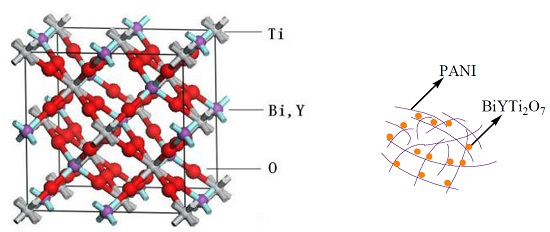
2.1. Preparation Method of BiYTi2O7
2.2. Preparation of Polyaniline–Hybridized BiYTi2O7
2.3. Characterization of BiYTi2O7 and PANI–BiYTi2O7
2.4. Photocatalytic Characterizations of BiYTi2O7 and PANI–BiYTi2O7
3. Results and Discussion
3.1. Characterization
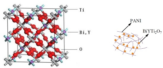
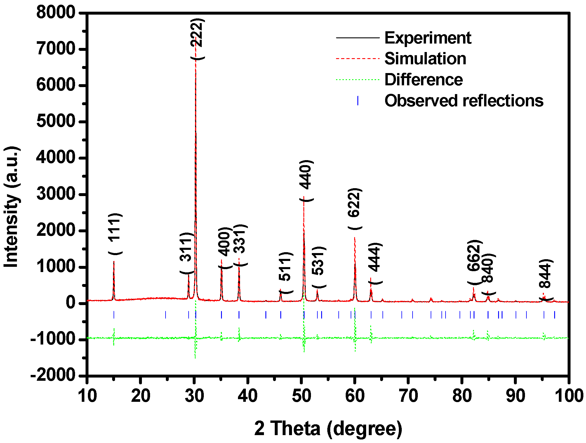
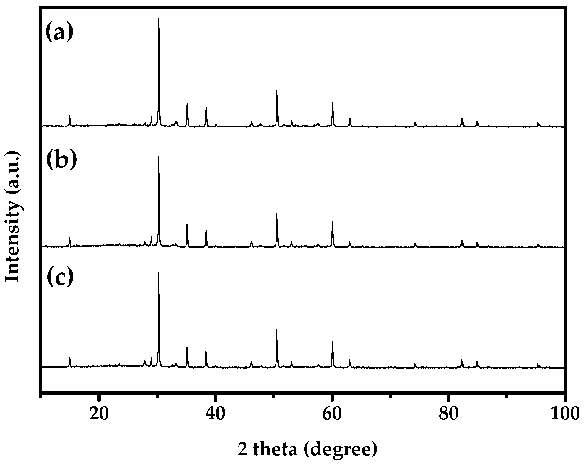
3.1.1. XRD Analysis
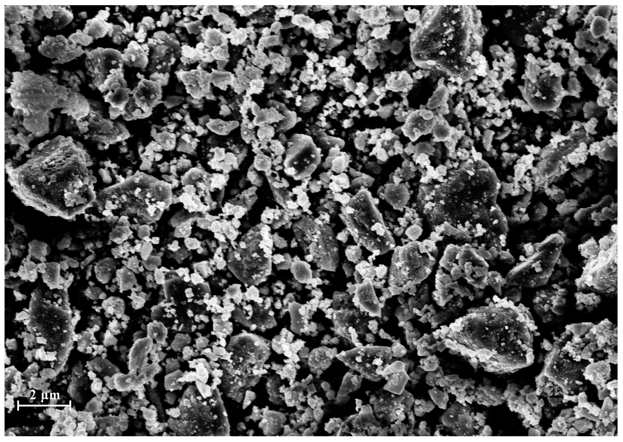
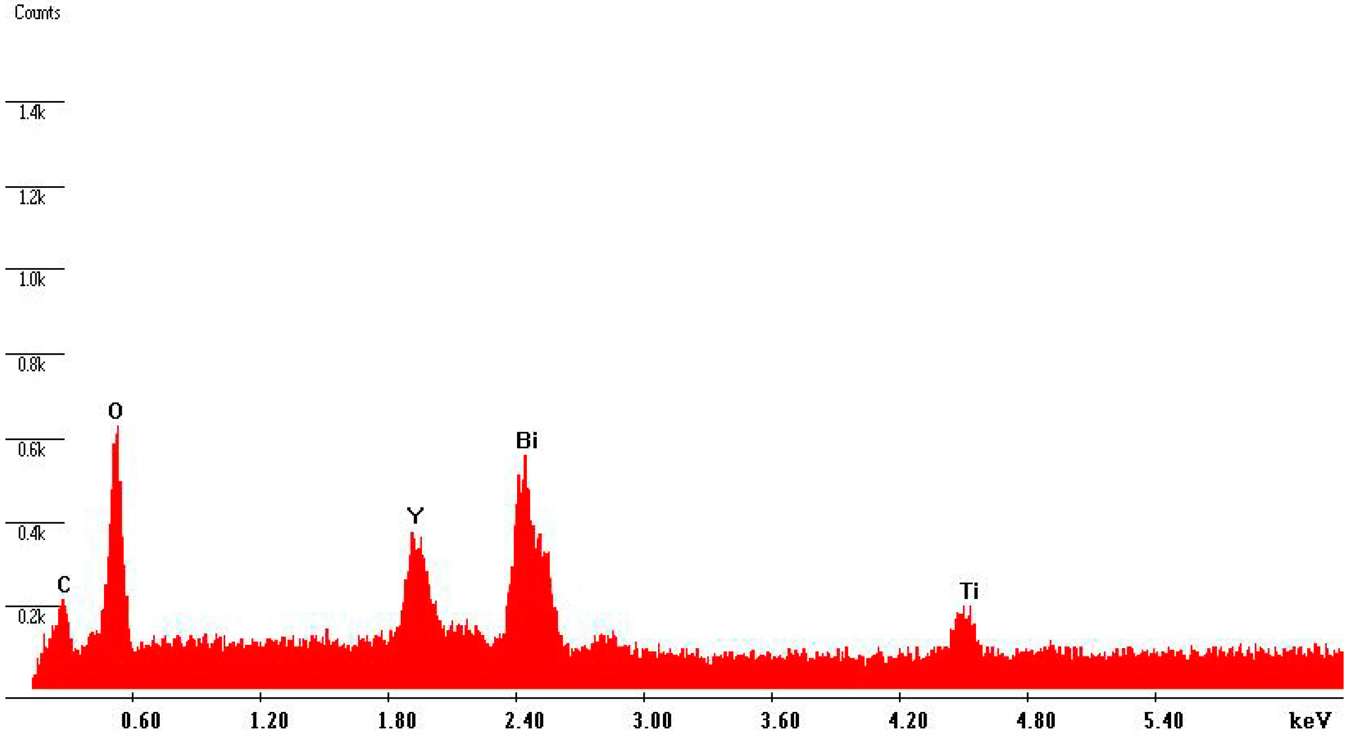
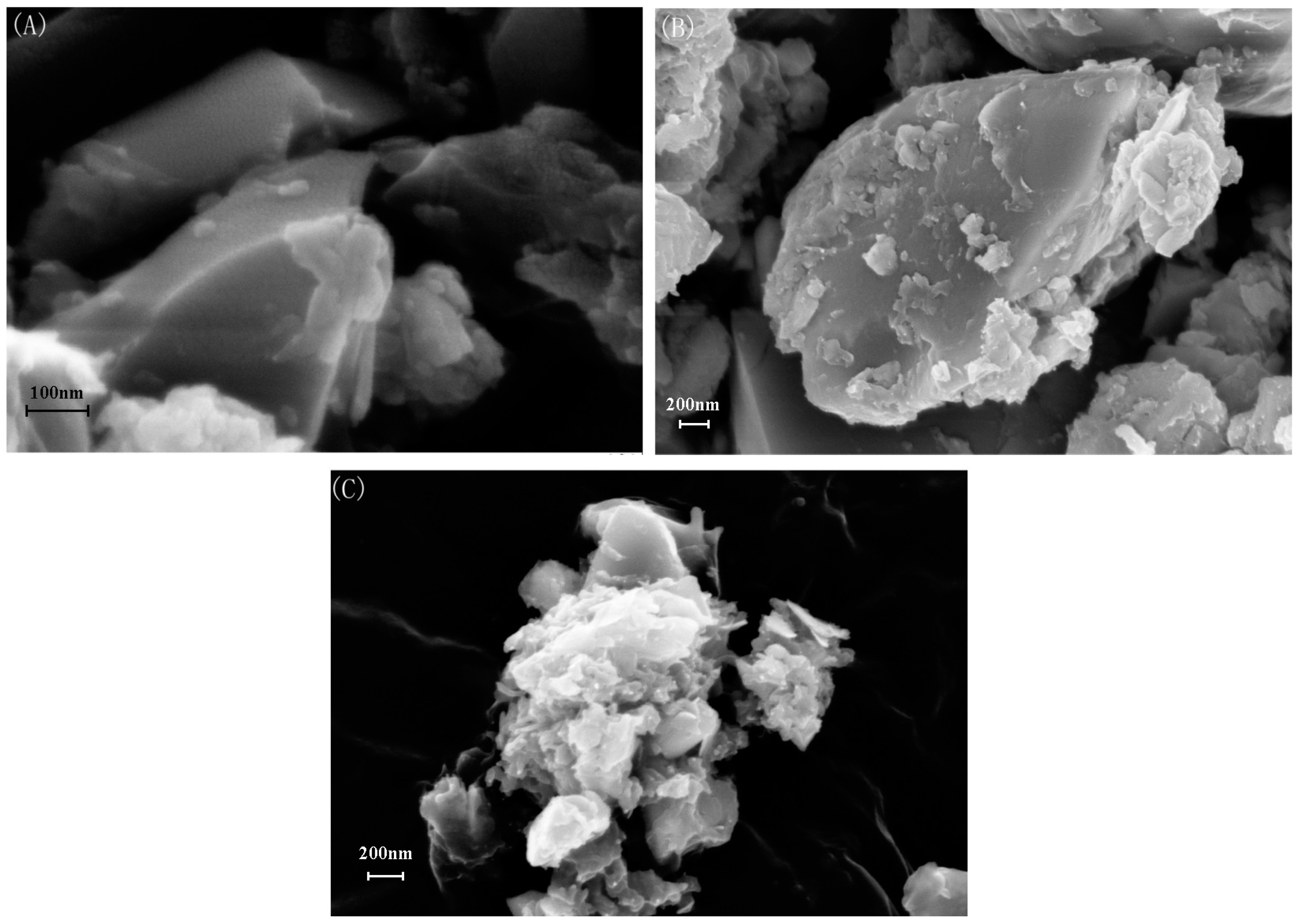
3.1.2. SEM Analysis
3.1.3. XPS Analysis
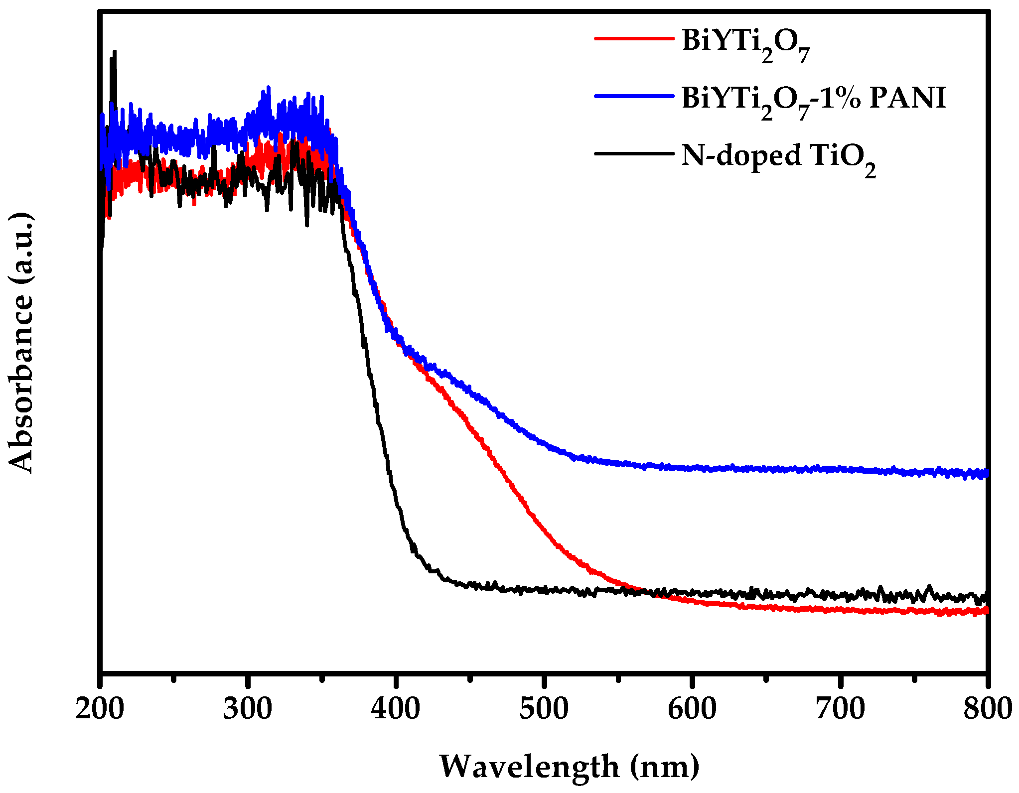
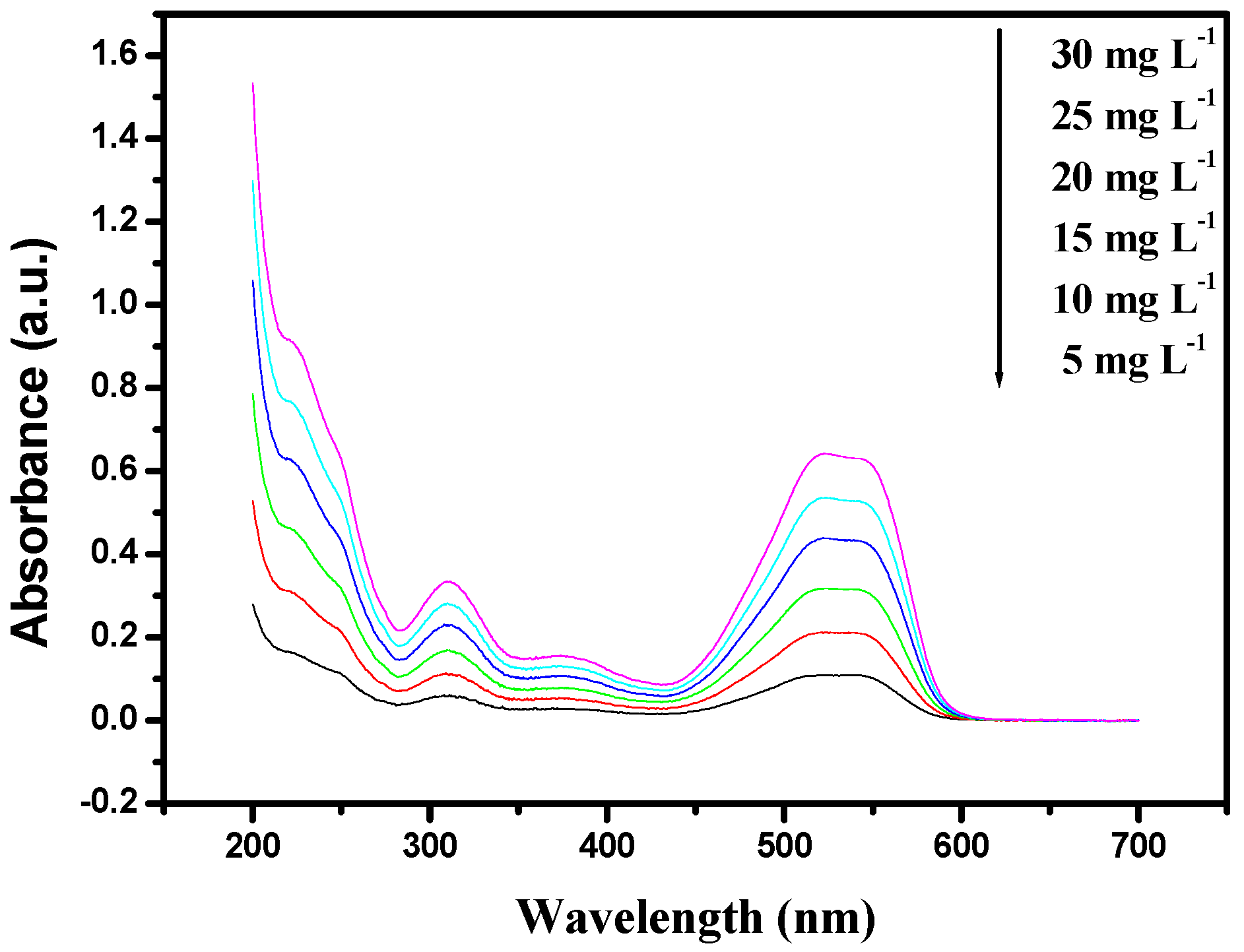
3.1.4. UV-Vis Diffuse Reflectance Spectra
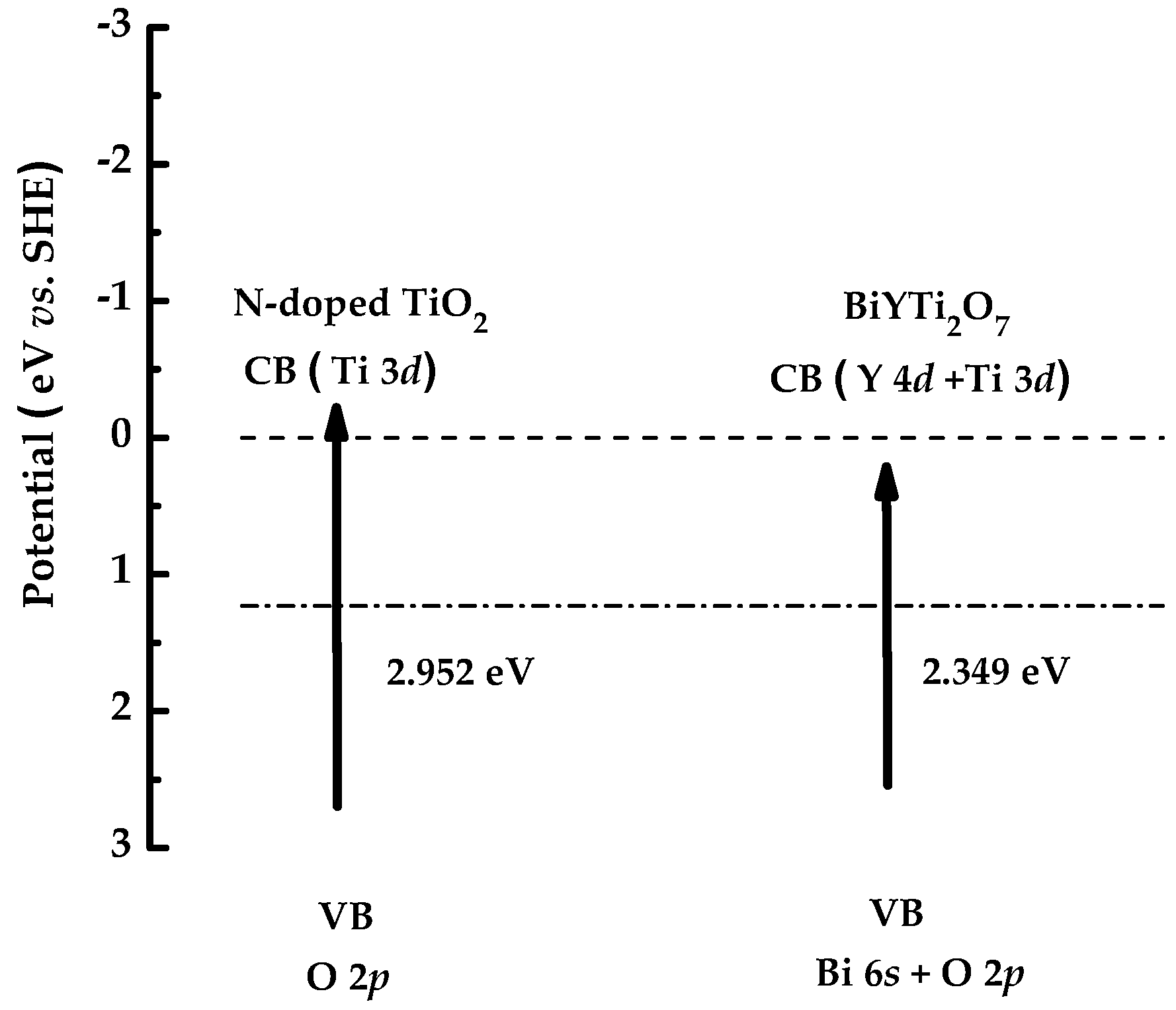
3.1.5. Band Structures
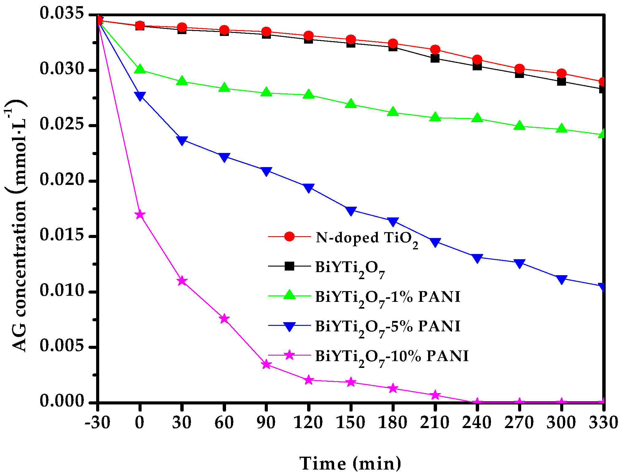
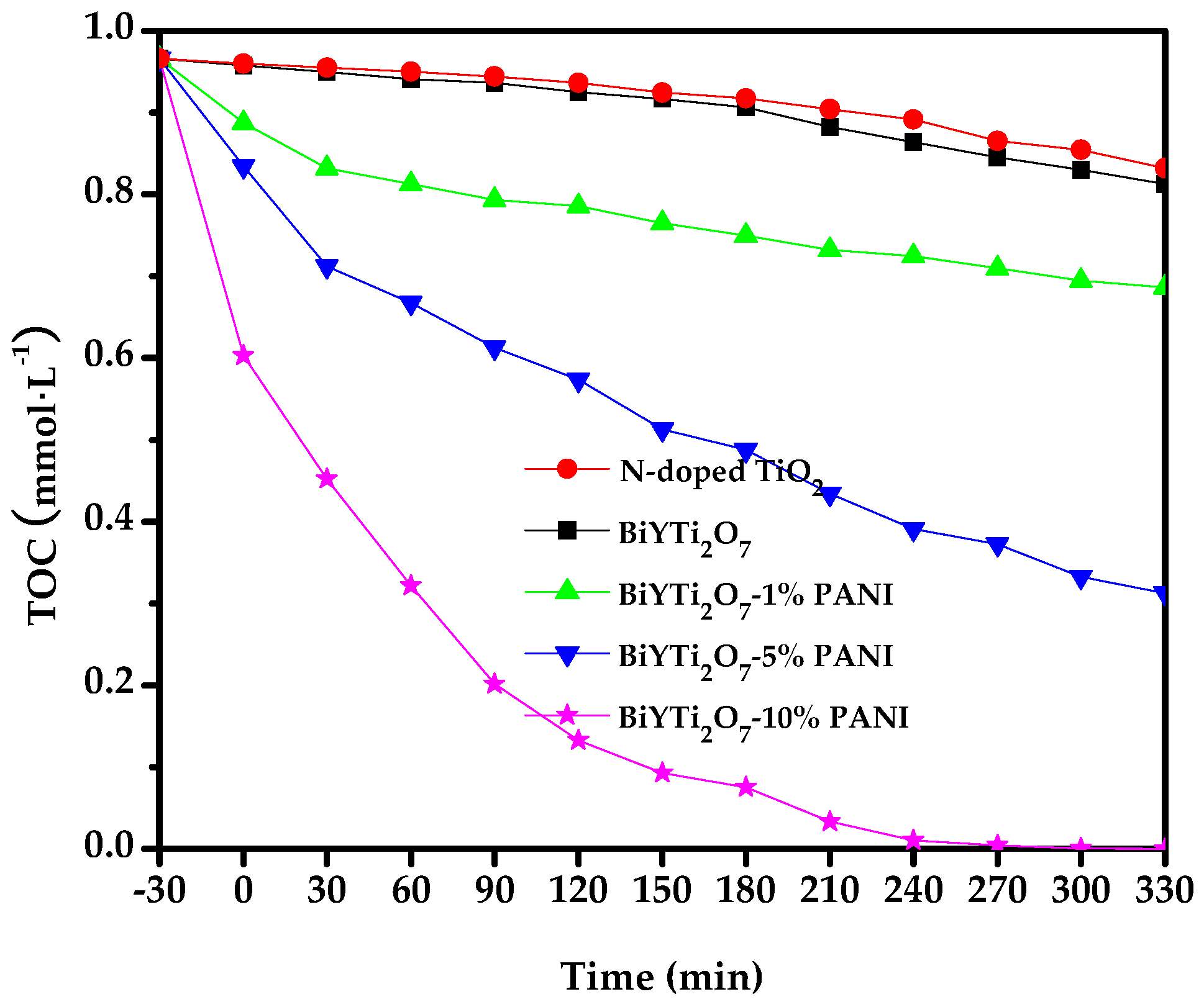
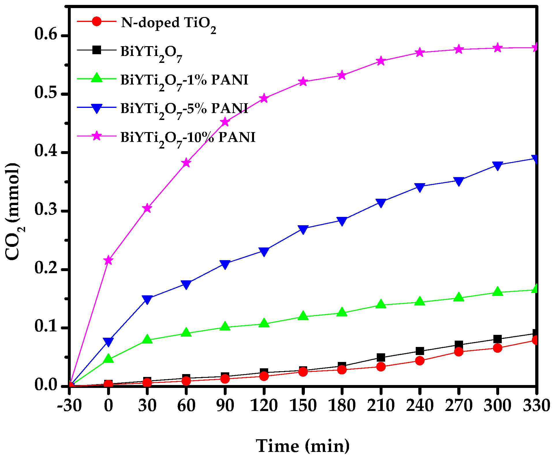
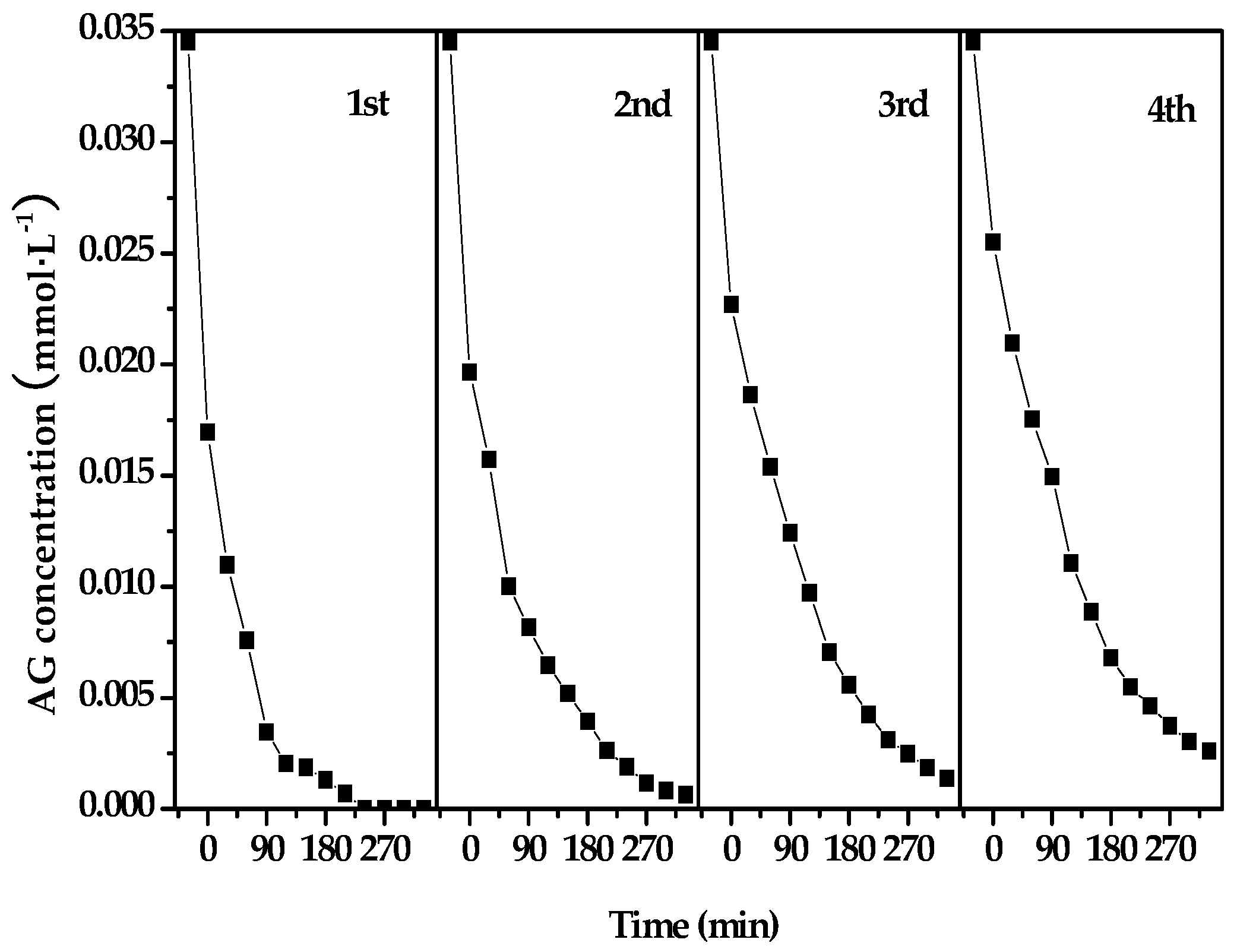
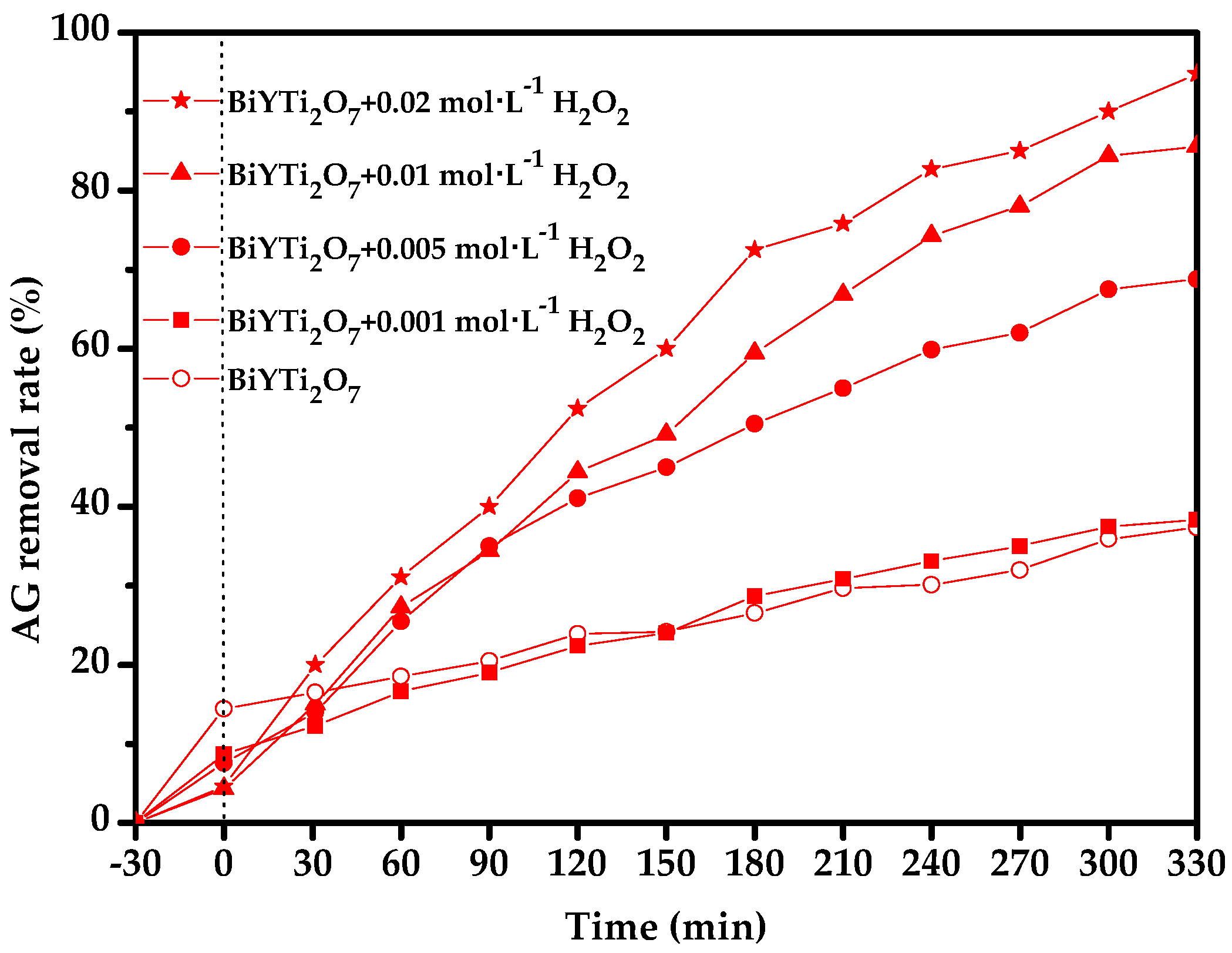
3.2. Photocatalytic Properties of Different PANI-Modified BiYTi2O7 Photocatalysts
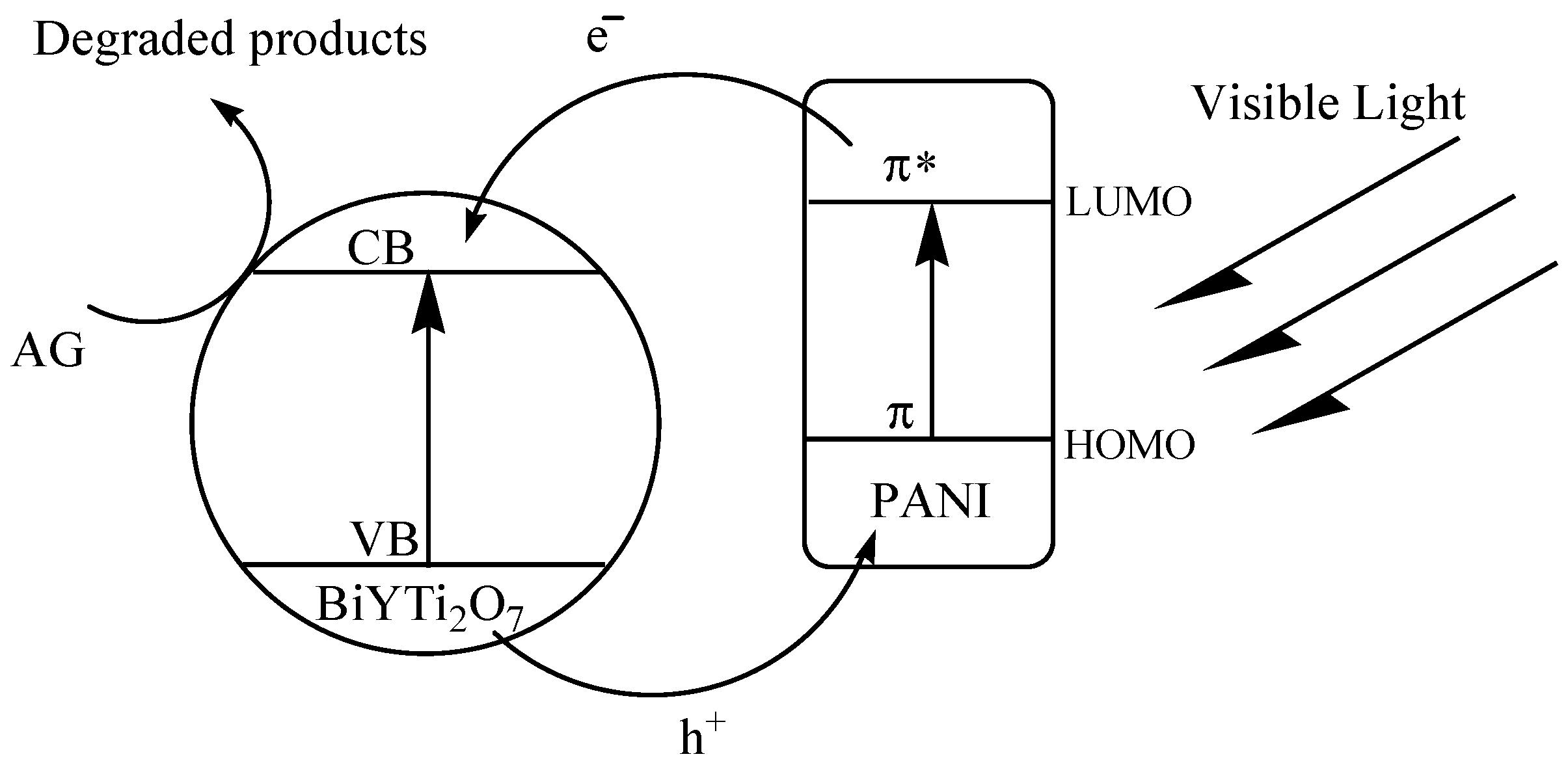
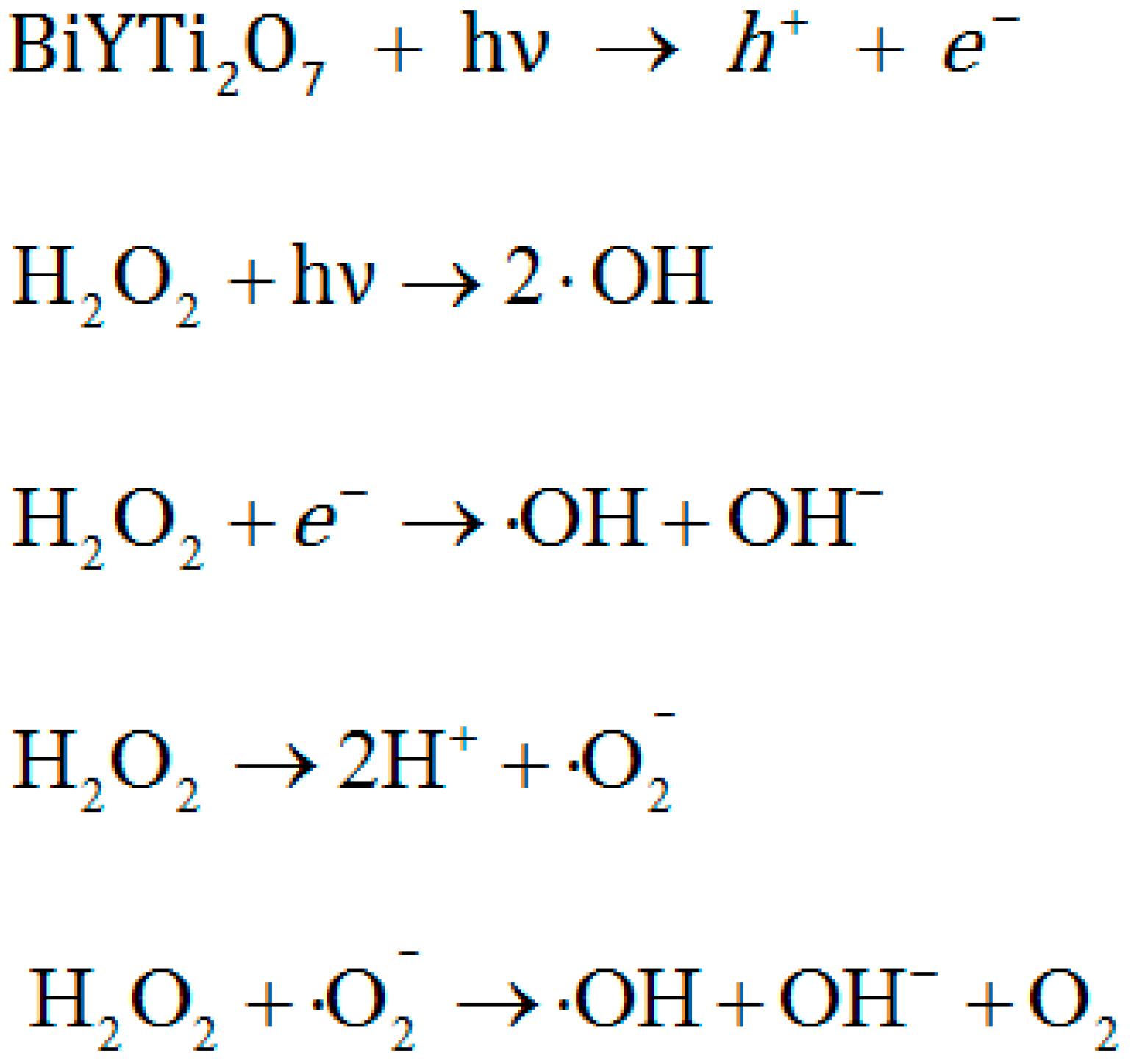
3.3. Photocatalytic Degradation Mechanism
4. Conclusions
Acknowledgments
Author Contributions
Conflicts of Interest
References
- Jin, R.C.; Cao, Y.W.; Mirkin, C.A.; Kelly, K.L.; Schatz, G.C.; Zheng, J.G. Photoinduced conversion of silver nanospheres to nanoprisms. Science 2001, 294, 1901–1903. [Google Scholar] [CrossRef]
- Lahiri, D.; Subramanian, V.; Shibata, T.; Wolf, E.E.; Bunker, B.A.; Kamat, P.V. Photoinduced transformations at semiconductor/metal interfaces: X-ray absorption studies of titania/gold films. J. Appl. Phys. 2003, 93, 2575–2582. [Google Scholar] [CrossRef]
- Kamat, P.V. Photoinduced transformations in semiconductor-metal nanocomposite assemblies. Pure Appl. Chem. 2002, 74, 1693–1706. [Google Scholar] [CrossRef]
- Chandrasekharan, N.; Kamat, P.V. Improving the photoelectrochemical performance of nanostructured TiO2 films by adsorption of gold nanoparticles. J. Phys. Chem. B 2000, 104, 10851–10857. [Google Scholar]
- Ghosh, S.; Sahu, K.; Mondal, S.K.; Sen, P.; Bhattacharyya, K. A femtosecond study of photoinduced electron transfer from dimethylaniline to coumarin dyes in a cetyltrimethylammonium bromide micelle. J. Chem. Phys. 2006, 125, 054509. [Google Scholar] [CrossRef] [PubMed]
- Jaeger, J.B.; Pillai, E.D.; Jaeger, T.D.; Duncan, M.A. Ultraviolet and infrared photodissociation of Si+(C6H6)n and Si+(C6H6)n Ar clusters. J. Phys. Chem. A 2005, 109, 2801–2808. [Google Scholar] [CrossRef] [PubMed]
- Kempa, T.; Farrer, R.A.; Giersig, M.; Fourkas, J.T. Photochemical synthesis and multiphoton luminescence of monodisperse silver nanocrystals. Plasmonics 2006, 1, 45–51. [Google Scholar] [CrossRef]
- Bhatkhande, D.S.; Pangarkar, V.G.; Beenackers, A.A.C.M. Photocatalytic degradation for environmental applications—A review. J. Chem. Technol. Biotechnol. 2002, 77, 102–116. [Google Scholar] [CrossRef]
- Breault, T.M.; Bartlett, B.M. Lowering the band gap of anatase-structured TiO2 by coalloying with Nb and N: Electronic structure and photocatalytic degradation of methylene blue dye. J. Phys. Chem. C 2012, 116, 5986–5994. [Google Scholar] [CrossRef]
- Kuwahara, Y.; Aoyama, J.; Miyakubo, K.; Eguchi, T.; Kamegawa, T.; Mori, K.; Yamashita, H. TiO2 photocatalyst for degradation of organic compounds in water and air supported on highly hydrophobic FAU zeolite: Structural, sorptive, and photocatalytic studies. J. Catal. 2012, 285, 223–234. [Google Scholar] [CrossRef]
- Da Dalt, S.; Alves, A.K.; Bergmann, C.P. Photocatalytic degradation of methyl orange dye in water solutions in the presence of MWCNT/TiO2 composites. Mater. Res. Bull. 2013, 48, 1845–1850. [Google Scholar] [CrossRef]
- Sinha, A.K.; Pradhan, M.; Sarkar, S.; Pal, T. Large-scale solid-state synthesis of Sn-SnO2 nanoparticles from layered SnO by sunlight: A material for dye degradation in water by photocatalytic reaction. Environ. Sci. Technol. 2013, 47, 2339–2345. [Google Scholar] [CrossRef] [PubMed]
- Breault, T.M.; Bartlett, B.M. Composition dependence of TiO2:(Nb,N)-x compounds on the rate of photocatalytic methylene blue dye degradation. J. Phys. Chem. C 2013, 117, 8611–8618. [Google Scholar] [CrossRef]
- Iyyapushpam, S.; Nishanthi, S.T.; Pathinettam Padiyan, D. Photocatalytic degradation of methyl orange using alpha-Bi2O3 prepared without surfactant. J. Alloy. Compd. 2013, 563, 104–107. [Google Scholar] [CrossRef]
- Jin, S.Q.; Wang, X.; Wang, X.L.; Ju, M.G.; Shen, S.; Liang, W.Z.; Zhao, Y.; Feng, Z.C.; Playford, H.Y.; Walton, R.I. Effect of phase junction structure on the photocatalytic performance in overall water splitting: Ga2O3 photocatalyst as an example. J. Phys. Chem. C 2015, 119, 18221–18228. [Google Scholar] [CrossRef]
- Srinivasu, K.; Modak, B.; Ghosh, S.K. Porous graphitic carbon nitride: A possible metal-free photocatalyst for water splitting. J. Phys. Chem. C 2014, 118, 26479–26484. [Google Scholar] [CrossRef]
- Lee, K.M.; Lai, C.W.; Ngai, K.S.; Juan, J.C. Recent developments of zinc oxide based photocatalyst in water treatment technology: A review. Water Res. 2016, 88, 428–448. [Google Scholar] [CrossRef] [PubMed]
- Liyanage, A.D.; Perera, S.D.; Tan, K.; Chabal, Y.; Balkus, K.J. Synthesis, characterization, and photocatalytic activity of Y-doped CeO2 nanorods. ACS Catal. 2014, 4, 577–584. [Google Scholar] [CrossRef]
- Yu, X.L.; Shavel, A.; An, X.Q.; Luo, Z.S.; Ibanez, M.; Cabot, A. Cu2ZnSnS4-Pt and Cu2ZnSnS4-Au heterostructured nanoparticles for photocatalytic water splitting and pollutant degradation. J. Am. Chem. Soc. 2014, 136, 9236–9239. [Google Scholar] [CrossRef] [PubMed]
- Zhou, X.M.; Xu, Q.L.; Lei, W.Y.; Zhang, T.T.; Qi, X.Y.; Liu, G.; Deng, K.; Yu, J.G. Origin of tunable photocatalytic selectivity of well-defined alpha-Fe2O3 nanocrystals. Small 2014, 10, 674–679. [Google Scholar] [CrossRef] [PubMed]
- Wang, L.L.; Wang, J.F.; Zhang, Y.; Ding, Z.T.; Cao, Q.E. Investigation on the determination of proteins by spectrophotometry with azocarmine G. J. Yunnan Univ. Nat. Sci. 2008, 30, 83–86. [Google Scholar]
- Chen, Z.G.; Ren, F.L.; Si, S.H.; Liao, X.H.; Ding, W.F.; Liu, J.B. Sensitive determination of DNA based on resonance light scattering enhancement of Azocarmine G and CTAB. J. Cent. South Univ. Technol. 2005, 12, 688–692. [Google Scholar] [CrossRef]
- Murakami, T.; Mahmut, N.; Hitomi, S.; Ohtsuka, A. Dark and light neurons in the human brain, with special reference to their reactions to Golgi’s silver nitrate, luxol fast blue MBS and azocarmine G. Arch. Histol. Cytol. 1997, 60, 265–274. [Google Scholar] [CrossRef] [PubMed]
- Low, G.K.C.; Mcevoy, S.R.; Matthews, R.W. Formation of nitrate and ammonium-ions in titanium-dioxideI mediated photocatalytic degradation of organic-compounds containing nitrogen-atoms. Environ. Sci. Technol. 1991, 25, 460–467. [Google Scholar] [CrossRef]
- Matos, J.; Laine, J.; Herrmann, J.M. Effect of the type of activated carbons on the photocatalytic degradation of aqueous organic pollutants by UV-irradiated titania. J. Catal. 2001, 200, 10–20. [Google Scholar] [CrossRef]
- Khodja, A.A.; Lavedrine, B.; Richard, C.; Sehili, T. Photocatalytic degradation of metoxuron in aqueous suspensions of TiO2 analytical and kinetic studies. Int. J. Photoenergy 2002, 4, 147–151. [Google Scholar] [CrossRef]
- Zhao, W.; Ma, W.H.; Chen, C.C.; Zhao, J.C.; Shuai, Z.G. Efficient degradation of toxic organic pollutants with Ni2O3/TiO2-xBx under visible irradiation. J. Am. Chem. Soc. 2004, 126, 4782–4783. [Google Scholar] [CrossRef] [PubMed]
- Murashkina, A.A.; Murzin, P.D.; Rudakova, A.V.; Ryabchuk, V.K.; Emeline, A.V.; Bahnemann, D.W. Influence of the dopant concentration on the photocatalytic activity: Al-doped TiO2. J. Phys. Chem. C 2015, 119, 24695–24703. [Google Scholar] [CrossRef]
- Lu, F.; Cai, W.P.; Zhang, Y.G. ZnO Hierarchical micro/nanoarchitectures: Solvothermal synthesis and structurally enhanced photocatalytic performance. Adv. Funct. Mater. 2008, 18, 1047–1056. [Google Scholar] [CrossRef]
- Kitano, M.; Takeuchi, M.; Matsuoka, M.; Thomas, J.M.; Anpo, M. Preparation of visible light-responsive TiO2 thin film photocatalysts by an RF magnetron sputtering deposition method and their photocatalytic reactivity. Chem. Lett. 2005, 34, 616–617. [Google Scholar] [CrossRef]
- Chatterjee, D.; Dasgupta, S. Visible light induced photocatalytic degradation of organic pollutants. J. Photochem. Photobiol. C. 2005, 6, 186–205. [Google Scholar] [CrossRef]
- Zhao, J.C.; Chen, C.C.; Ma, W.H. Photocatalytic degradation of organic pollutants under visible light irradiation. Top. Catal. 2005, 35, 269–278. [Google Scholar] [CrossRef]
- Zou, Z.G.; Ye, J.H.; Sayama, K.; Arakawa, H. Direct splitting of water under visible light irradiation with an oxide semiconductor photocatalyst. Nature 2001, 414, 625–627. [Google Scholar] [CrossRef] [PubMed]
- Sivalingam, G.; Madras, G. Photocatalytic degradation of poly(bisphenol-A-carbonate) in solution over combustion-synthesized TiO2: Mechanism and kinetics. Appl. Catal. A. 2004, 269, 81–90. [Google Scholar] [CrossRef]
- Maeda, K.; Terashima, H.; Kase, K.; Higashi, M.; Tabata, M.; Domen, K. Surface modification of TaON with monoclinic ZrO2 to produce a composite photocatalyst with enhanced hydrogen evolution activity under visible light. Bull. Chem. Soc. Jpn. 2008, 81, 927–937. [Google Scholar] [CrossRef]
- Shi, H.X.; Chen, J.Y.; Li, G.Y.; Nie, X.; Zhao, H.J.; Wong, P.K.; An, T.C. Synthesis and characterization of novel plasmonic Ag/AgX-CNTs (X = Cl, Br, I) nanocomposite photocatalysts and synergetic degradation of organic pollutant under visible light. ACS Appl. Mater. Interfaces 2013, 5, 6959–6967. [Google Scholar] [CrossRef] [PubMed]
- Ge, L.; Han, C.C.; Liu, J. In situ synthesis and enhanced visible Light photocatalytic activities of novel PANI-g-C3N4 composite photocatalysts. J. Mater. Chem. 2012, 22, 11843–11850. [Google Scholar] [CrossRef]
- He, G.H.; Liang, C.J.; Ou, Y.D.; Liu, D.N.; Fang, Y.P.; Xu, Y.H. Preparation of novel Sb2O3/WO3 photocatalysts and their activities under visible light irradiation. Mater. Res. Bull. 2013, 48, 2244–2249. [Google Scholar] [CrossRef]
- Wang, J.X.; Ruan, H.; Li, W.J.; Li, D.Z.; Hu, Y.; Chen, J.; Shao, Y.; Zheng, Y. Highly efficient oxidation of gaseous benzene on novel Ag3VO4/TiO2 nanocomposite photocatalysts under visible and simulated solar light irradiation. J. Phys. Chem. C 2012, 116, 13935–13943. [Google Scholar] [CrossRef]
- Kameyama, T.; Takahashi, T.; Machida, T.; Kamiya, Y.; Yamamoto, T.; Kuwabata, S.; Torimoto, T. Controlling the electronic energy structure of ZnS-AgInS2 solid solution nanocrystals for photoluminescence and photocatalytic hydrogen evolution. J. Phys. Chem. C 2015, 119, 24740–24749. [Google Scholar] [CrossRef]
- Teng, F.; Liu, Z.L.; Zhang, A.; Li, M. Photocatalytic performances of Ag3PO4 polypods for degradation of dye pollutant under natural indoor weak light irradiation. Environ. Sci. Technol. 2015, 49, 9489–9494. [Google Scholar] [CrossRef] [PubMed]
- Han, S.C.; Hu, L.F.; Liang, Z.Q.; Wageh, S.; Al-Ghamdi, A.A.; Chen, Y.S.; Fang, X.S. One-step hydrothermal synthesis of 2D hexagonal nanoplates of alpha-Fe2O3/graphene composites with enhanced photocatalytic activity. Adv. Funct. Mater. 2014, 24, 5719–5727. [Google Scholar] [CrossRef]
- Luan, J.F.; Li, Y.Y. Photocatalytic water splitting for hydrogen production with Gd2MSbO7 (M = Fe, In, Y) photocatalysts under visible light irradiation. Materials 2015, 8, 16–30. [Google Scholar] [CrossRef]
- Liu, W.; Ji, M.S.; Chen, S.F. Preparation, characterization and activity evaluation of Ag2Mo4O13 photocatalyst. J. Hazard. Mater. 2011, 186, 2001–2008. [Google Scholar] [CrossRef] [PubMed]
- Zhao, J.; Dong, W.W.; Wu, Y.P.; Wang, Y.N.; Wang, C.; Li, D.S.; Zhang, Q.C. Two (3,6)-connected porous metal-organic frameworks based on linear trinuclear [Co-3(COO)(6)] and paddlewheel dinuclear [Cu-2(COO)(4)] SBUs: Gas adsorption, photocatalytic behaviour, and magnetic properties. J. Mater. Chem. A 2015, 3, 6962–6969. [Google Scholar] [CrossRef]
- Gao, J.K.; Miao, J.W.; Li, P.Z.; Teng, W.Y.; Yang, L.; Zhao, Y.L.; Liu, B.; Zhang, Q.C. A p-type Ti( IV)-based metal-organic framework with visible-light photo-response. Chem. Commun. 2014, 50, 3786–3788. [Google Scholar] [CrossRef] [PubMed]
- Gao, J.K.; Cao, S.W.; Tay, Q.L.; Liu, Y.; Yu, L.M.; Ye, K.Q.; Mun, P.C.S.; Li, Y.X.; Rakesh, G.; Loo, S.C.J.; et al. Molecule-based water-oxidation catalysts (WOCs): Cluster-size-dependent dye-sensitized polyoxometalates for visible-light-driven O2 evolution. Sci. Rep. 2013, 3, 1853. [Google Scholar] [CrossRef] [PubMed]
- Luan, J.F.; Ma, K.; Pan, B.C.; Li, Y.M.; Wu, X.S.; Zou, Z.G. Synthesis and catalytic activity of new Gd2BiSbO7 and Gd2YSbO7 nanocatalysts. J. Mol. Catal. A Chem. 2010, 321, 1–9. [Google Scholar] [CrossRef]
- Luan, J.F.; Ma, K.; Li, Y.M.; Zou, Z.G. Photophysical and photocatalytic properties of novel Y2GaSbO7 and Y2YbSbO7 photocatalysts under visible light irradiation. J. Mater. Sci. 2011, 46, 813–823. [Google Scholar] [CrossRef]
- Luan, J.F.; Li, M.; Ma, K.; Li, Y.M.; Zou, Z.G. Photocatalytic activity of novel Y2InSbO7 and Y2GdSbO7 nanocatalysts for degradation of environmental pollutant rhodamine B under visible light irradiation. Chem. Eng. J. 2011, 167, 162–171. [Google Scholar] [CrossRef]
- Luan, J.F.; Cai, H.L.; Zheng, S.R.; Hao, X.P.; Luan, G.Y.; Wu, X.S.; Zou, Z.G. Structural and photocatalytic properties of novel Bi2GaVO7. Mater. Chem. Phys. 2007, 104, 119–124. [Google Scholar] [CrossRef]
- Luan, J.F.; Cai, H.L.; Hao, X.P.; Zhang, J.B.; Luan, G.Y.; Wu, X.S.; Zou, Z.G. Structural characterization and photocatalytic properties of novel Bi2FeVO7. Res. Chem. Intermed. 2007, 33, 487–500. [Google Scholar] [CrossRef]
- Barka, N.; Assabbane, A.; Nounah, A.; Ichou, Y.A. Photocatalytic degradation of indigo carmine in aqueous solution by TiO2-coated non-woven fibres. J. Hazard. Mater. 2008, 152, 1054–1059. [Google Scholar] [CrossRef] [PubMed]
- Chen, X.; Mao, S.S. Titanium dioxide nanomaterials: Synthesis, properties, modifications, and applications. Chem. Rev. 2007, 107, 2891–2959. [Google Scholar] [CrossRef] [PubMed]
- Cheng, H.; Huang, B.; Dai, Y.; Qin, X.; Zhang, X.; Wang, Z.; Jiang, M. Visible-light photocatalytic activity of the metastable Bi20TiO32 synthesized by a high-temperature quenching method. J. Solid State Chem. 2009, 182, 2274–2278. [Google Scholar] [CrossRef]
- Guillard, C.; Disdier, J.; Monnet, C.; Dussaud, J.; Malato, S.; Blanco, J.; Maldonado, M.I.; Herrmann, J.M. Solar efficiency of a new deposited titania photocatalyst: Chlorophenol, pesticide and dye removal applications. Appl. Catal. B 2003, 46, 319–332. [Google Scholar] [CrossRef]
- Dong, F.; Li, Q.Y.; Sun, Y.J.; Ho, W.K. Noble metal-like behavior of plasmonic Bi particles as a cocatalyst deposited on (BiO)2CO3 microspheres for efficient visible light photocatalysis. ACS Catal. 2014, 4, 4341–4350. [Google Scholar] [CrossRef]
- Yang, D.R.; Feng, J.; Jiang, L.L.; Wu, X.L.; Sheng, L.Z.; Jiang, Y.T.; Wei, T.; Fan, Z.J. Photocatalyst interface engineering: Spatially confined growth of ZnFe2O4 within graphene networks as excellent visible-light-driven photocatalysts. Adv. Funct. Mater. 2015, 25, 7080–7087. [Google Scholar] [CrossRef]
- Li, G.Y.; Nie, X.; Chen, J.Y.; Jiang, Q.; An, T.C.; Wong, P.K.; Zhang, H.M.; Zhao, H.J.; Yamashita, H. Enhanced visible-light-driven photocatalytic inactivation of Escherichia coli using g-C3N4/TiO2 hybrid photocatalyst synthesized using a hydrothermal-calcination approach. Water Res. 2015, 86, 17–24. [Google Scholar] [CrossRef] [PubMed]
- Zhang, G.; Hu, Z.Y.; Sun, M.; Liu, Y.; Liu, L.M.; Liu, H.J.; Huang, C.P.; Qu, J.H.; Li, J.H. Formation of Bi2WO6 bipyramids with vacancy pairs for enhanced solar-driven photoactivity. Adv. Funct. Mater. 2015, 25, 3726–3734. [Google Scholar] [CrossRef]
- Li, N.; Li, Y.M.; Li, W.J.; Ji, S.D.; Jin, P. One-step hydrothermal synthesis of TiO2@MoO3 core-shell nanomaterial: Microstructure, growth mechanism, and improved photochromic property. J. Phys. Chem. C 2016, 120, 3341–3349. [Google Scholar] [CrossRef]
- Gnayem, H.; Sasson, Y. Nanostructured 3D sunflower-like bismuth doped BiOClxBr1−x solid solutions with enhanced visible light photocatalytic activity as a remarkably efficient technology for water Purification. J. Phys. Chem. C 2015, 119, 19201–19209. [Google Scholar] [CrossRef]
- Li, H.L.; Yu, K.; Lei, X.; Guo, B.J.; Fu, H.; Zhu, Z.Q. Hydrothermal synthesis of novel MoS2/BiVO4 hetero-nanoflowers with enhanced photocatalytic activity and a mechanism investigation. J. Phys. Chem. C 2015, 119, 22681–22689. [Google Scholar] [CrossRef]
- Grigioni, I.; Stamplecoskie, K.G.; Selli, E.; Kamat, P.V. Dynamics of photogenerated charge carriers in WO3/BiVO4 heterojunction photoanodes. J.Phys. Chem. C 2015, 119, 20792–20800. [Google Scholar] [CrossRef]
- Luan, J.F.; Wang, S.; Ma, K.; Li, Y.M.; Pan, B.C. Structural property and catalytic activity of new In2YbSbO7 and Gd2YbSbO7 nanocatalysts under visible light irradiation. J. Phys. Chem. C 2010, 114, 9398–9407. [Google Scholar] [CrossRef]
- El Jaouhari, A.; Laabd, M.; Aouzal, Z.; Bouabdallaoui, M.; Bazzaoui, E.A.; Albourine, A.; Martins, J.I.; Wang, R.; Bazzaoui, M. Effect of electrolytic conditions on PANi electrosynthesis on stainless steel: A new application to polycarboxy-benzoic acids removal from industrial effluents. Prog. Org. Coat. 2016, 101, 233–239. [Google Scholar] [CrossRef]
- Negi, Y.S.; Adhyapak, P.V. Development in polyaniline conducting polymers. J. Macromol. Sci. Polym. Rev. 2002, 42, 35–53. [Google Scholar] [CrossRef]
- Xie, J.; Zhao, C.E.; Lin, Z.Q.; Gu, P.Y.; Zhang, Q.C. Nanostructured conjugated polymers for energy-related applications beyond solar cells. Chem. Asian J. 2016, 11, 1489–1511. [Google Scholar] [CrossRef] [PubMed]
- Pandiselvi, K.; Fang, H.F.; Huang, X.B.; Wang, J.Y.; Xu, X.C.; Li, T. Constructing a novel carbon nitride/polyaniline/ZnO ternary heterostructure with enhanced photocatalytic performance using exfoliated carbon nitride nanosheets as supports. J. Hazard. Mater. 2016, 314, 67–77. [Google Scholar] [CrossRef] [PubMed]
- Shang, M.; Wang, W.Z.; Sun, S.M.; Ren, J.; Zhou, L.; Zhang, L. Efficient visible light-induced photocatalytic degradation of contaminant by spindle-like PANI/BiVO4. J. Phys. Chem. C 2009, 113, 20228–20233. [Google Scholar] [CrossRef]
- Hidalgo, D.; Bocchini, S.; Fontana, M.; Saracco, G.; Hernandez, S. Green and low-cost synthesis of PANI-TiO2 nanocomposite mesoporous films for photoelectrochemical water splitting. RSC Adv. 2015, 5, 49429–49438. [Google Scholar] [CrossRef]
- Zou, Z.G.; Ye, J.H.; Arakawa, H. Compositional dependence of crystallization in the glass-ceramics system Bi2O3-In2O3-MnO2-B2O3. J. Mater. Sci. Lett. 2000, 19, 1909–1911. [Google Scholar] [CrossRef]
- Zhang, H.; Zong, R.L.; Zhao, J.C.; Zhu, Y.F. Dramatic visible photocatalytic degradation performances due to synergetic effect of TiO2 with PANI. Environ. Sci. Technol. 2008, 42, 3803–3807. [Google Scholar] [CrossRef] [PubMed]
- Chen, L.X.; Rajh, T.; Wang, Z.Y.; Thurnauer, M.C. XAFS studies of surface structures of TiO2 nanoparticles and photocatalytic reduction of metal ions. J. Phys. Chem. B 1997, 101, 10688–10697. [Google Scholar] [CrossRef]
- Chen, W.F.; Koshy, P.; Huang, Y.; Adabifiroozjaei, E.; Yao, Y.; Sorrell, C.C. Effects of precipitation, liquid formation, and intervalence charge transfer on the properties andphotocatalytic performance of cobalt- or vanadium-doped TiO2 thin films. J. Hydrogen Energy 2016, 41, 19025–19056. [Google Scholar] [CrossRef]
- Liao, Y.H.B.; Wang, J.X.; Lin, J.S.; Chung, W.H.; Lin, W.Y.; Chen, C.C. Synthesis, photocatalytic activities and degradation mechanism of Bi2WO6 toward crystal violet dye. Catal. Today 2011, 174, 148–159. [Google Scholar] [CrossRef]
- Tauc, J.; Grigorov, R.; Vancu, A. Optical properties and electronic structure of amorphous germanium. Phys. Status Solidi 1966, 15, 627–637. [Google Scholar] [CrossRef]
- Butler, M.A. Photoelectrolysis with YFeO3 electrodes. J. Appl. Phys. 1977, 48, 1914–1920. [Google Scholar] [CrossRef]
- Oshikiri, M.; Boero, M.; Ye, J.H.; Zou, Z.G.; Kido, G. Electronic structures of promising photocatalysts InMO4 (M=V, Nb, Ta) and BiVO4 for water decomposition in the visible wavelength region. J. Chem. Phys. 2002, 117, 7313–7318. [Google Scholar] [CrossRef]
- Liu, G.M.; Wu, T.X.; Zhao, J.C.; Hidaka, H.; Serpone, N. Photoassisted degradation of dye pollutants. 8. Irreversible degradation of alizarin red under visible light radiation in air-equilibrated aqueous TiO2 dispersions. Environ. Sci. Technol. 1999, 33, 2081–2087. [Google Scholar] [CrossRef]
- Reddy, V.R.; Hwang, D.W.; Lee, J.S. Effect of Zr substitution for Ti in KLaTiO4 for photocatalytic water splitting. Catal. Lett. 2003, 90, 39–43. [Google Scholar] [CrossRef]
- Sun, H.; Qiu, G.H.; Wang, Y.; Feng, X.H.; Yin, H.; Liu, F. Effects of Co and Ni co-doping on the physicochemical properties of cryptomelane and its enhanced performance on photocatalytic degradation of phenol. Mater. Chem. Phys. 2014, 148, 783–789. [Google Scholar] [CrossRef]
- Liu, L.; Ding, L.; Liu, Y.G.; An, W.J.; Lin, S.L.; Liang, Y.H.; Cui, W.Q. A stable Ag3PO4@PANI core@shell hybrid: Enrichment photocatalytic degradation with pi-pi conjugation. Appl. Catal. B 2017, 210, 92–104. [Google Scholar] [CrossRef]
- Williams, G.; Seger, B.; Kamat, P.V. TiO2-graphene nanocomposites. UV-assisted photocatalytic reduction of graphene oxide. ACS Nano 2008, 2, 1487–1491. [Google Scholar] [CrossRef] [PubMed]
- Zhu, S.B.; Xu, T.G.; Fu, H.B.; Zhao, J.C.; Zhu, Y.F. Synergetic effect of Bi2WO6 photocatalyst with C-60 and enhanced photoactivity under visible irradiation. Environ. Sci. Technol. 2007, 41, 6234–6239. [Google Scholar] [CrossRef] [PubMed]
- Ameen, S.; Akhtar, M.S.; Kim, Y.S.; Yang, O.B.; Shin, H.S. An effective nanocomposite of polyaniline and ZnO: Preparation, characterizations, and its photocatalytic activity. Colloid Polym. Sci. 2011, 289, 415–421. [Google Scholar] [CrossRef]
- Hou, J.G.; Cao, R.; Jiao, S.Q.; Zhu, H.M.; Kumar, R.V. PANI/Bi12TiO20 complex architectures: Controllable synthesis and enhanced visible-light photocatalytic activities. Appl. Catal. B 2011, 104, 399–406. [Google Scholar] [CrossRef]
- Chen, H.H.; Xiong, X.Q.; Hao, L.L.; Zhang, X.; Xu, Y.M. Improved visible light photocatalytic activity of WO3 through CuWO4 for phenol degradation. Appl. Surf. Sci. 2016, 389, 491–495. [Google Scholar] [CrossRef]
- Neppolian, B.; Kanel, S.R.; Choi, H.C.; Shankar, M.V.; Arabindoo, B.; Murugesan, V. Photocatalytic degradation of reactive yellow 17 dye in aqueous solution in the presence of TiO2 with cement binder. Int. J. Photoenergy 2003, 5, 45–49. [Google Scholar] [CrossRef]
- Muruganandham, M.; Swaminathan, M. Solar photocatalytic degradation of a reactive azo dye in TiO2-suspension. Sol. Energy Mater. Sol. Cells 2004, 81, 439–457. [Google Scholar] [CrossRef]
- Wang, G.L.; Xu, X.F.; Wu, X.M.; Cao, G.X.; Dong, Y.M.; Li, Z.J. Visible-light-stimulated enzymelike activity of graphene oxide and its application for facile glucose sensing. J. Phys. Chem. C 2014, 118, 28109–28117. [Google Scholar] [CrossRef]
- Ng, T.W.; Zhang, L.S.; Liu, J.S.; Huang, G.C.; Wang, W.; Wong, P.K. Visible-light-driven photocatalytic inactivation of escherichia coli by magnetic Fe2O3-AgBr. Water Res. 2016, 90, 111–118. [Google Scholar] [CrossRef] [PubMed]


| Atom | x | y | z | Occupation factor |
|---|---|---|---|---|
| Bi | 0.5 | 0.5 | 0.5 | 0.5 |
| Y | 0.5 | 0.5 | 0.5 | 0.5 |
| Ti | 0 | 0 | 0 | 1 |
| O(1) | 0.375 | 0.375 | 0.375 | 1 |
| O(2) | 0.125 | 0.125 | 0.125 | 1 |
| BiYTi2O7 | Bi4f | Y3d | Ti2p | O1s | C1s | |||
|---|---|---|---|---|---|---|---|---|
| Bi4f5/2 | Bi4f7/2 | Y3d3/2 | Y3d5/2 | Ti2p1/2 | Ti2p3/2 | |||
| Experimental (eV) | 163.3 | 156.5 | 157.9 | 158.6 | 463.8 | 457.2 | 528.8 | 283.9 |
| After carbon revision (eV) | 164.0 | 157.2 | 158.6 | 159.3 | 464.5 | 457.2 | 529.5 | 284.8 |
| Photocatalysts | R | Regression equation | K (h−1) |
|---|---|---|---|
| BiYTi2O7 | 0.9697 | Y = 0.0337X − 0.0052 | 0.0337 |
| BiYTi2O7-1%PANI | 0.9877 | Y = 0.0379X + 0.1515 | 0.0379 |
| BiYTi2O7-5%PANI | 0.9648 | Y = 0.2441X + 0.1698 | 0.2441 |
| BiYTi2O7-10%PANI | 0.9874 | Y = 0.9004X + 0.7508 | 0.9004 |
© 2017 by the authors. Licensee MDPI, Basel, Switzerland. This article is an open access article distributed under the terms and conditions of the Creative Commons Attribution (CC BY) license ( http://creativecommons.org/licenses/by/4.0/).
Share and Cite
Luan, J.; Shen, Y.; Wang, S.; Guo, N. Synthesis, Property Characterization and Photocatalytic Activity of the Polyaniline/BiYTi2O7 Polymer Composite. Polymers 2017, 9, 69. https://doi.org/10.3390/polym9030069
Luan J, Shen Y, Wang S, Guo N. Synthesis, Property Characterization and Photocatalytic Activity of the Polyaniline/BiYTi2O7 Polymer Composite. Polymers. 2017; 9(3):69. https://doi.org/10.3390/polym9030069
Chicago/Turabian StyleLuan, Jingfei, Yue Shen, Shu Wang, and Ningbin Guo. 2017. "Synthesis, Property Characterization and Photocatalytic Activity of the Polyaniline/BiYTi2O7 Polymer Composite" Polymers 9, no. 3: 69. https://doi.org/10.3390/polym9030069