Article

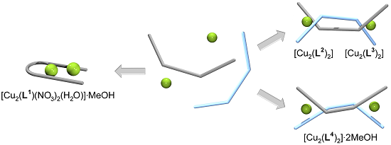
Spacer-Controlled Supramolecular Assemblies of Cu(II) with Bis(2-Hydroxyphenylimine) Ligands. from Monoligand Complexes to Double-Stranded Helicates and Metallomacrocycles
 †

1
Department of Chemistry and Food Chemistry, Technische Universität Dresden, 01062 Dresden, Germany
2
Institute of Organic Chemistry, Technische Universität Bergakademie Freiberg, 09596 Freiberg, Germany
*
Authors to whom correspondence should be addressed.
Dedicated to Professor Peter Mühl on the occasion of his 85th birthday.
Crystals 2016, 6(9), 120; https://doi.org/10.3390/cryst6090120
Submission received: 28 August 2016 / Revised: 13 September 2016 / Accepted: 14 September 2016 / Published: 21 September 2016
(This article belongs to the Special Issue Crystal Structure of Complex Compounds)

Abstract

:
Reaction of Cu(NO3)2·3H2O or Cu(CH3COO)2·H2O with the bis(2-hydroxyphenylimine) ligands H2L1-H2L4 gave four Cu(II) complexes of composition [Cu2(L1)(NO3)2(H2O)]·MeOH, [Cu2(L2)2], [Cu2(L3)2] and [Cu2(L4)2]·2MeOH. Depending on the spacer unit, the structures are characterized by a dinuclear arrangement of Cu(II) within one ligand (H2L1), by a double-stranded [2+2] helical binding mode (H2L2 and H2L3) and a [2 + 2] metallomacrocycle formation (H2L4). In these complexes, the Cu(II) coordination geometries are quite different, varying between common square planar or square pyramidal arrangements, and rather rare pentagonal bipyramidal and tetrahedral geometries. In addition, solution studies of the complex formation using UV/Vis and ESI-MS as well as solvent extraction are reported.

Graphical Abstract

1. Introduction

The coordination chemistry of multifunctional Schiff base ligands has been the focus of a considerable number of investigations because they offer application possibilities in catalysis, optics, magnetic materials, sensing, separation, etc. [1,2,3,4,5,6,7,8]. One of the main areas of research relates to the self-assembly of corresponding metal complexes; gaining an understanding of the factors that affect such processes remains a challenge. Among the ligand types studied, tetradentate and hexadentate bis(2-hydroxyphenylimine) ligands are of considerable interest and several examples have been described in the literature [9,10,11,12]. Recently, we have published two double-stranded Cu(II) helicates with methylene and sulfurdiphenylene bridged 3-ethoxy-2-hydroxyphenyl substituted diimines. These recognize and bind two water molecules in their peripheral O4 cavities [13]. Here we report four further Cu(II) complexes with structure-related hexadentate (H2L1 and H2L3) and tetradentate diimine ligands (H2L2 and H2L4) having cyclohexylidene, cyclohexylidenemethylene, hexamethylene and dioxotriphenylene spacer units varying in size and flexibility (Scheme 1). Whereas in case of H2L1 a dinuclear monoligand Cu(II) complex having an interesting new coordination pattern was formed, for H2L2 and H2L3 double-stranded helicates and for H2L4 a metallomacrocycle have been isolated and structurally characterized. The obtained complex structures demonstrate the strong influence of the spacer function on the resulting structure.

2. Results and Discussions

2.1. Synthesis of the Ligands and Their Cu(II) Complexes

The four diimine ligands H2L1-H2L4 have been synthesized by standard Schiff base condensation of salicylaldehyde or 3-ethoxysalicylaldehyde with the different diamines in methanol [14].
Ligand H2L1 was firstly described by Tozzo et al. [15]. However, in this paper, the elemental analysis is incorrect. The correct data are given here in the Experimental Section. For the introduction of the cyclohexylidene spacer group, the corresponding (±)-trans-1,2-diaminocyclohexane was used. A detailed study by Constable et al. using the enantiopure (R,R)-cyclohexylidene bridged diimine ligand H2L1, showed the formation of two relevant Cu(II) complexes, [CuL1] and [(CuL1)(H2O)], by reaction of Cu(CH3COO)2·H2O and this ligand in methanol [16,17]. Both complexes have a 1:1 (Cu:L1) composition with a square planar Cu(II) arrangement based on the inner N2O2 donor set. The first of these shows interesting molecular recognition behavior towards a water molecule in the outer O4 coordination sphere forming the second complex site. This O4 pocket has also been used for the additional binding of s- or f-block metal ions leading to heterodinuclear Cu(II) complexes [17,18,19]. During our studies, we found that the reaction of the (±)-trans-1,2-cyclohexylidene bridged H2L1 with Cu(NO3)2·3H2O (molar ratio 1:1) in methanol/tetrahydrofuran (v:v = 1:1) leads to a dinuclear Cu(II) complex. Slow diffusion of diethyl ether/hexane into this solution gave single crystals of composition [Cu2(L1)(NO3)2(H2O)]·MeOH that were suitable for X-ray structure determination. However, solids from repeated experiments always gave different analytical results, suggesting the formation of more than one complex species and possibly side products in changing ratios. Besides this 2:1 species (Cu:L1), particularly 1:1 and 2:2 species were each identified in the bulk material by ESI-MS.
H2L2 has been reported by Berkessel et al. [20]. The trimethylcyclohexylidenemethylene spacer group in our studies is based on cis/trans-5-amino-1,3,3-trimethylcyclohexanemethylamine. Whereas H2L3 was first described by Ha [21], H2L4 was synthesized for the first time in this work with this last reaction being catalyzed by addition of a small amount of sulfuric acid.
In contrast to H2L1 the diimine ligands H2L2-H2L4 gave homogeneous dinuclear copper complexes under comparable experimental conditions when reacted with Cu(CH3COO)2·H2O (molar ratio 1:1); namely, the double-stranded helicates [Cu2(L2)2] and [Cu2(L3)2] and the metallomacrocycle [Cu2(L4)2]·2MeOH, respectively. In these cases, single crystals formed on allowing the reaction mixture to stand for one week.

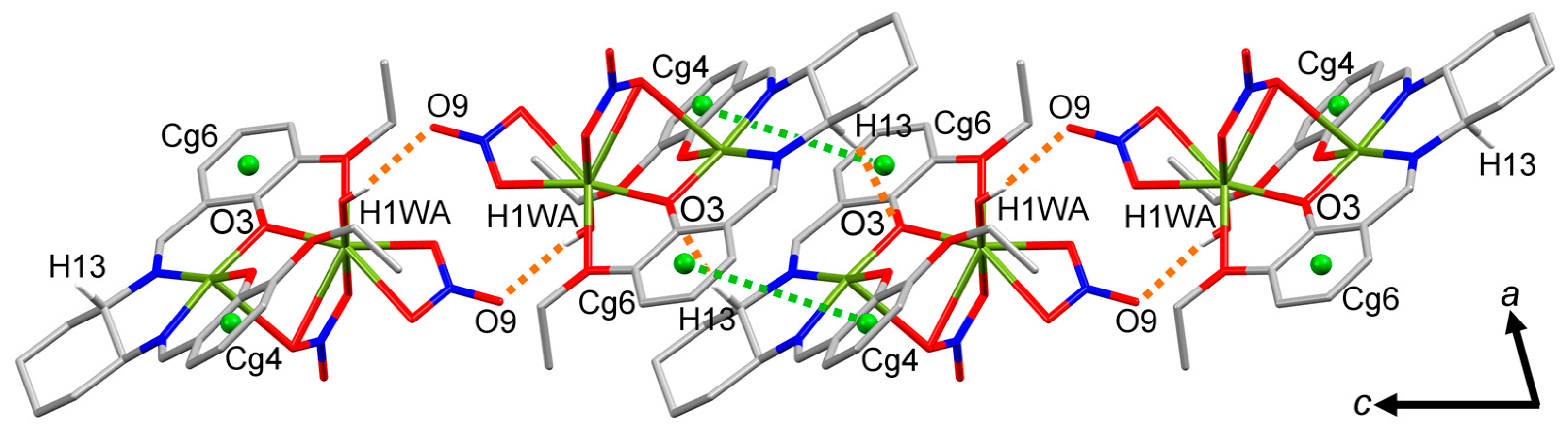
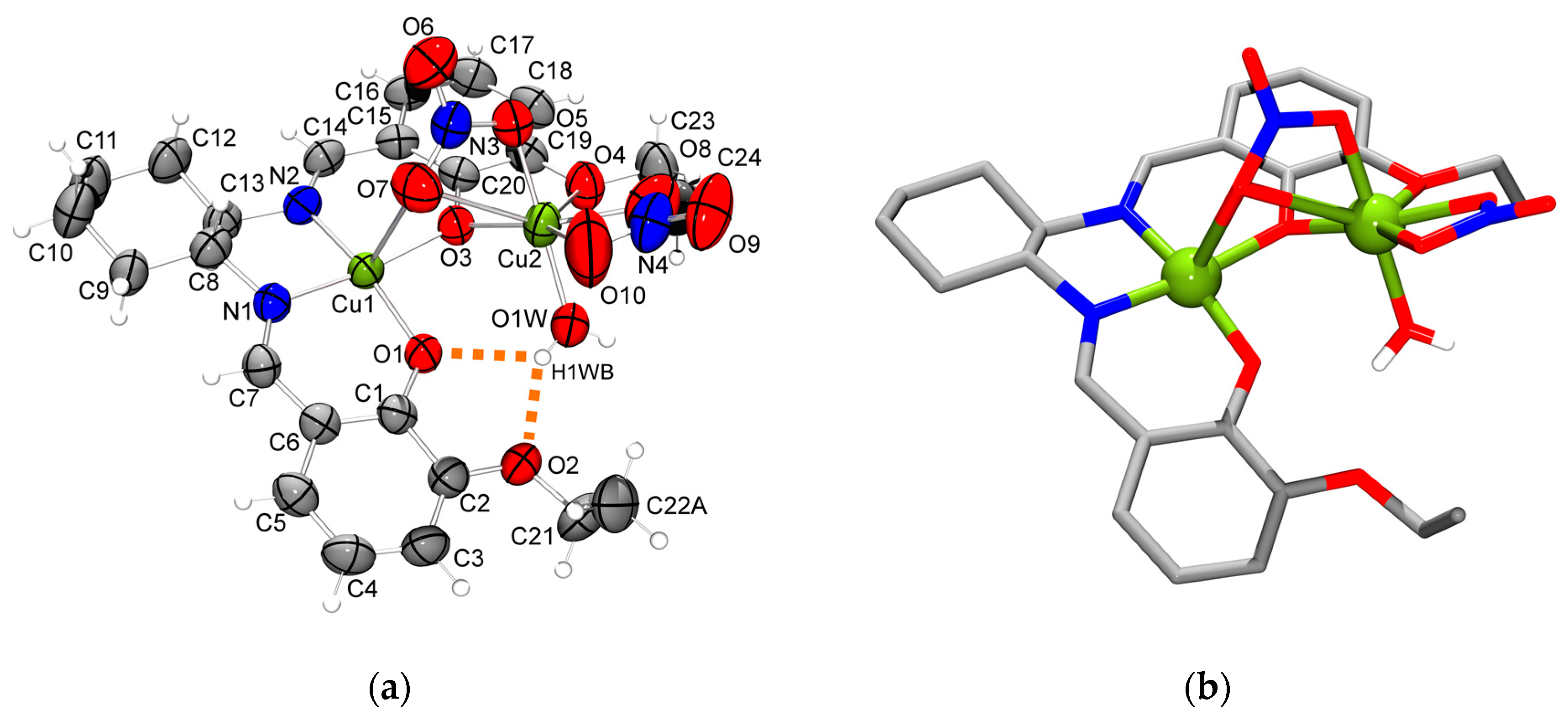
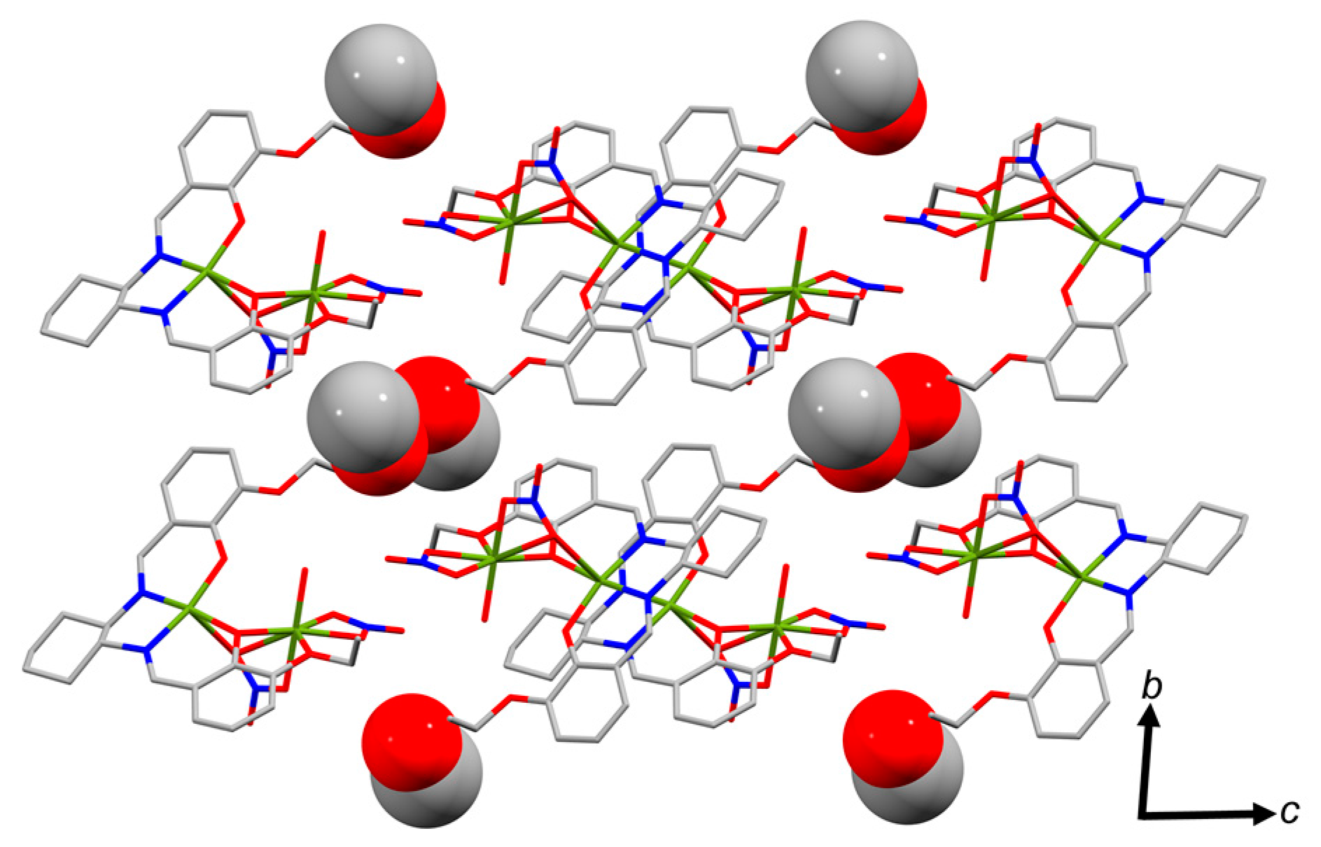
2.2. Molecular Structure of the Monoligand Cu(II) Complex with H2L1

Figure 1 shows the molecular structure of the dinuclear monoligand complex [Cu2(L1)(NO3)2(H2O)]·MeOH. It is crystallized in the triclinic space group P-1. In this compound both Cu(II) centers show a quite different coordination mode. Cu1 is five-coordinated by the inner square planar N2O2 donor set of imine nitrogen atoms and oxygen atoms of deprotonated hydroxyl groups together with one apical nitrate oxygen whereas Cu2 is surrounded by seven oxygen donor atoms involving one deprotonated hydroxyl and one ethoxy group, two bidentate nitrate ions and one water molecule. This results in a distorted square pyramidal arrangement for Cu1 and in a very rare distorted pentagonal bipyramidal coordination geometry for Cu2 [22,23,24,25,26]. The four bond lengths for Cu1-N and Cu1-O in the plane lie in the expected range of strong interactions (Table 1; 1.893–1.951 Å); however, the length of the apical Cu1-O bond (2.658 Å) with the nitrate ion is substantial longer indicating a weak interaction. The seven Cu2-O bond lengths fall within the range 1.951–2.723 Å; stronger interactions with Cu2 occur for the apical water molecule (1.951 Å), the two nitrate ions (1.955 and 2.084 Å) and the hydroxy oxygen donor atom of the ligand (1.987 Å). The remaining Cu2-O interactions with bond lengths of 2.341 Å for the C2H5O-substituent, 2.522 Å and 2.723 Å for the second interactions of both bidentate nitrate ions are all rather weak. Cu1 and Cu2 are directly linked in the structure by two bridging modes, first by strong Cu-O interactions with the hydroxyl oxygen and second by weak Cu-O contacts with one nitrate ion. As a result the distance between both Cu(II) centers is relatively short (3.276 Å). The unique coordination pattern for Cu(II) in the molecule is further stabilized by the water ligand forming two moderate hydrogen bonds O1W-H1WB···O1/O2 with bond lengths of 2.15 Å and 2.05 Å, respectively (Table S1).
The coordinated water molecule is also involved in a moderate hydrogen bond O1W-H1WA···O9 (2.11 Å) with a nitrate ion of an adjacent molecule resulting in the formation of dimeric complex molecules oriented along the crystallographic c-axis (Figure 2; Table S2). Each dimer represents a pair of enantiomers introduced in the ligand by the (±)-trans-1,2-diaminocyclohexane during the Schiff base synthesis. The dimers are interconnected via weak hydrogen bonds C13-H13···O3 and π···π contacts forming polymeric strands (Tables S2 and S3). Comparable weak hydrogen bonds CH···O and CH···π exist between neighboring strands (Figure S1; Tables S2 and S4). The resulting void space in the packing is occupied by strongly disordered methanol molecules. The packing motif is shown in Figure 3 and Figure S2.
It is noted that the above Cu(II) complex is structurally comparable to a Zn(II) complex, incorporating the doubly deprotonated form of the related 3-methoxy-2-hydroxyphenylene substituted cis/trans-1,2-cyclohexylidene bridged diimine [27]. However, in this case, the coordination number of Zn(II) is five and both Zn(II) centers have a square pyramidal geometry.

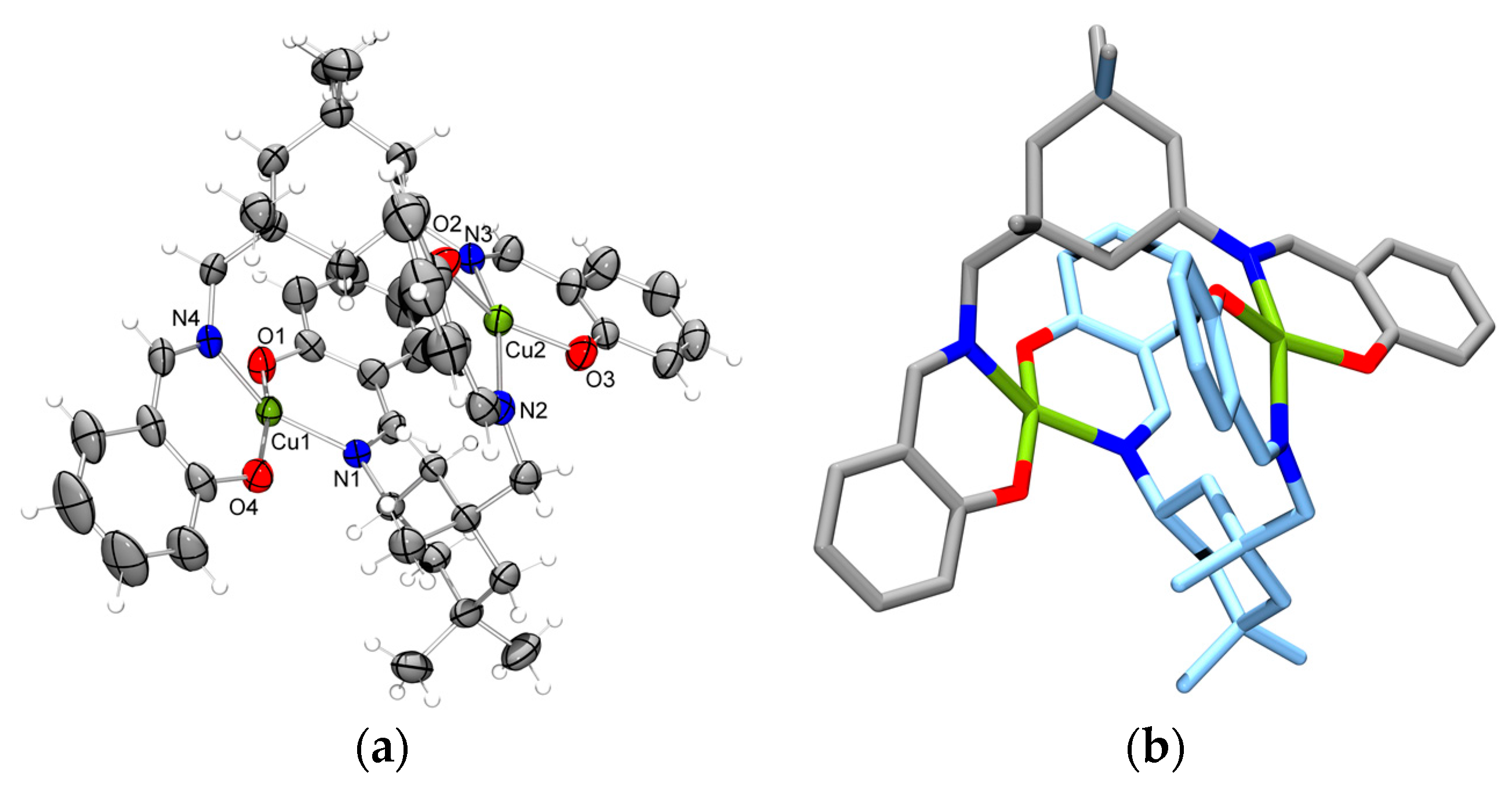
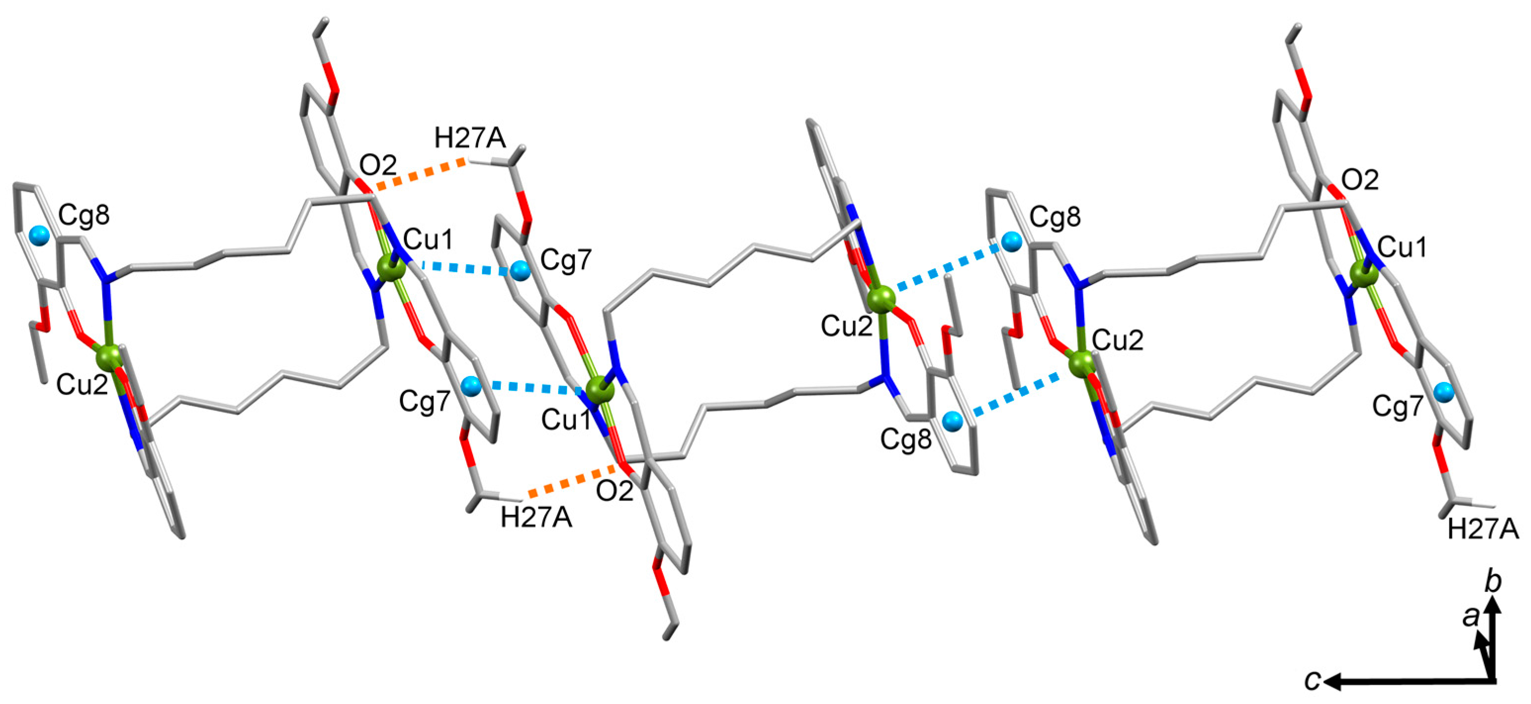
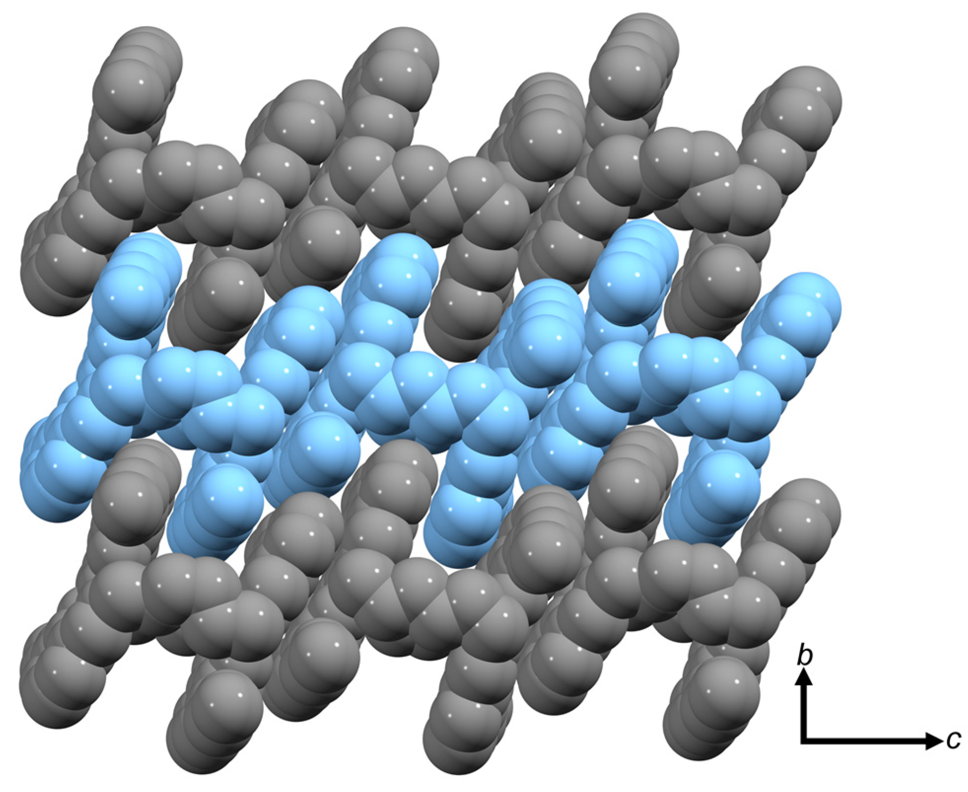
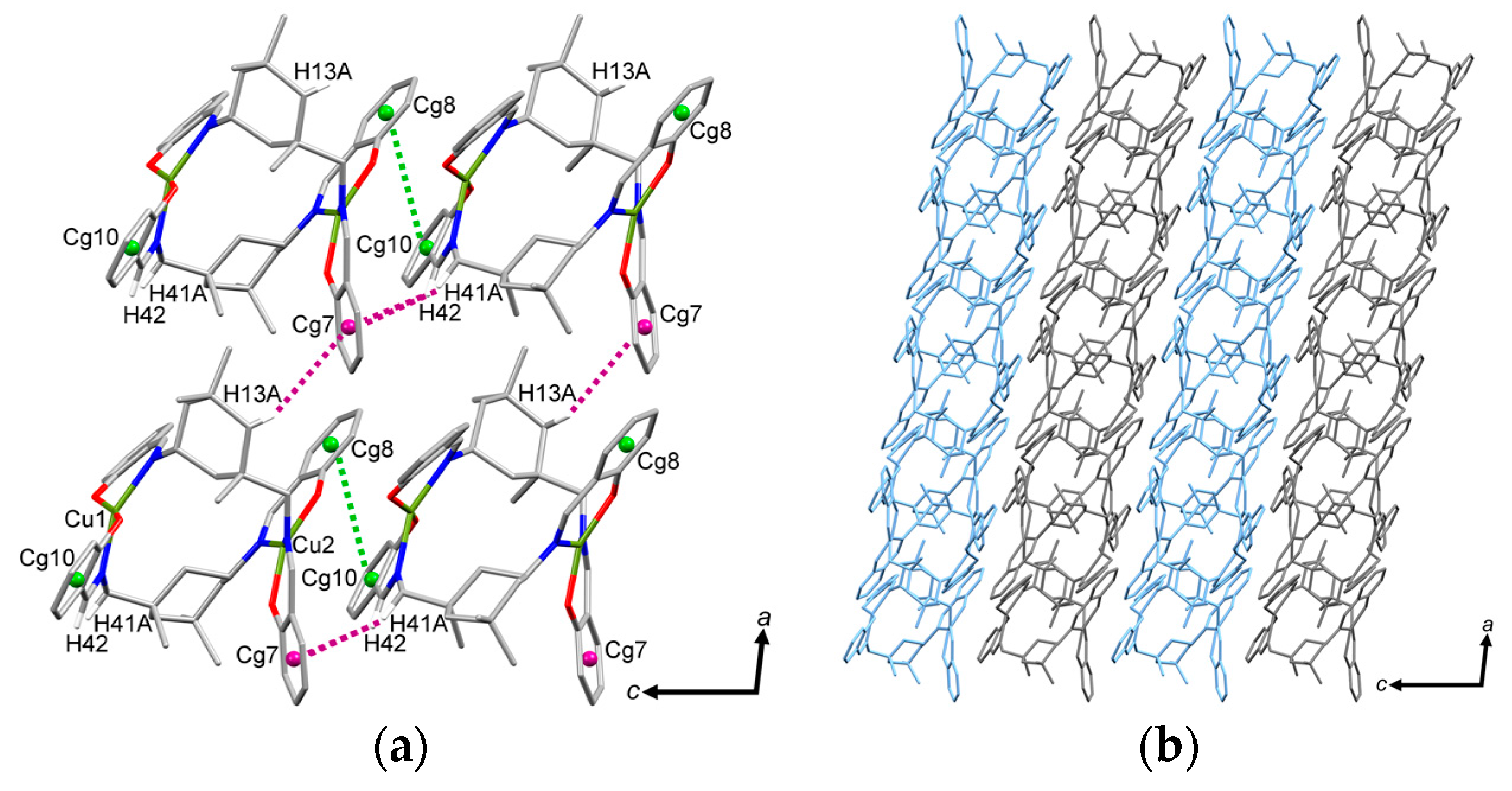
2.3. Molecular Structure of the Double-Stranded Cu(II) Helicates with H2L2 and H2L3

In contrast to H2L1 the modified cyclohexylidene spacer function in H2L2 gives some changes in the latter’s size and flexibility. This is clearly reflected in the resulting Cu(II) complex structure (monoclinic space group P21/c) shown in Figure 4. In this case, Cu(II) forms a neutral dinuclear double-stranded helicate of composition [Cu2(L2)2]. Every helicate unit contains of two deprotonated ligand molecules. Each Cu(II) ion in the complex is coordinated in a strongly distorted square planar arrangement by an N2O2 donor set. The distortion of the Cu(II) centers is indicated by the parameter τ4 [28] which is 0.29 for Cu1 and 0.31 for Cu2. The Cu-O bond lengths are slightly shorter (1.88 Å and 1.89 Å) than the Cu-N bonds (1.99 Å and 2.01 Å; Table 2). Furthermore, the Cu···Cu separation is 5.33 Å and is significantly longer than between the complex molecules (4.13 Å). The presence of weak intramolecular hydrogen bonds C-H···O between CH2 groups on the cyclohexane ring and the four oxygen atoms of the hydroxyl groups helps to stabilize the helicate structure (Table S5).
Interestingly, only the cis-isomers of the ligand have been identified in the crystal structure of [Cu2(L2)2], although both cis- and trans-isomers were present in the diamine used. Along the crystallographic c-axis adjacent helicates are linked by regular stacking of both enantiomers forming polymeric strands. Within these strands, the molecules are connected via CH···π and π···π interactions (Figure 5 and Figure S3; Tables S6 and S7). In addition, the short distance Cu2···Cg10 of 3.90 Å indicates an additional cation···π interaction potentially including the imine double bond (Cu2···Cimine = 3.46 Å). Furthermore, within the crystallographic ac-plane CH···π interactions occur between neighboring strands resulting in the formation of 2D layers. However, no interactions were found between adjacent layers. In contrast to the strands mentioned before, the neighboring helicates possess the same handedness along the crystallographic a-axis.
In analogy to H2L2, the ligand H2L3, possessing a long flexible hexamethylene spacer unit, also reacts with Cu(II) to form a double-stranded helicate of composition [Cu2(L3)2] whose molecular structure (triclinic space group P-1) is shown in Figure 6. Both Cu(II) centers possess distorted square planar coordination environments bound to N2O2 donor sets of two deprotonated ligand molecules (τ4: 0.10 for Cu1; 0.17 for Cu2) [28]. Compared to [Cu2(L2)2], the Cu-N and Cu-O bond lengths are only slightly different (Table 3). However, in [Cu2(L3)2], the Cu···Cu separation is 7.09 Å, longer than in [Cu2(L2)2], while the Cu-Cu distances between adjacent helicates are 5.80 Å and 5.61 Å, respectively. Again, intramolecular hydrogen bonds between CH2 groups and nitrogen or oxygen atoms contribute to the stabilization of the helicate (Table S8).
Remarkably, there are few intermolecular contacts between the complex molecules. Within the crystallographic c-axis the helicates are linked by Cu···π interactions to form strand-like arrangements (Figure 7; Table S9). Furthermore, a weak hydrogen bond occurs involving an ethoxy substituent and the oxygen atom O2 (Table S10). The strands contain both left-handed as well as right-handed helices. Due to the long-chain acyclic spacer unit the strands are interlocked forming 2D layers in the bc-plane (Figure 8).

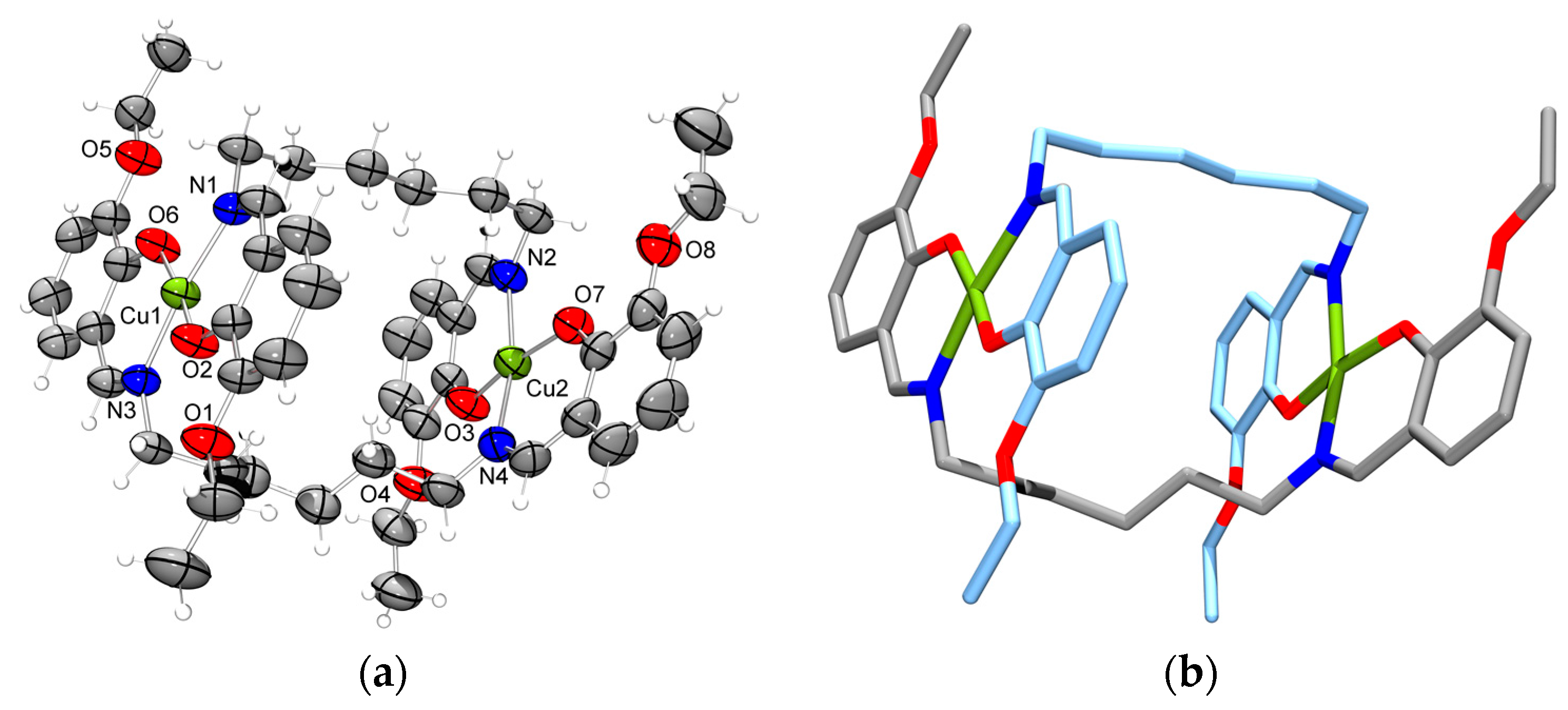
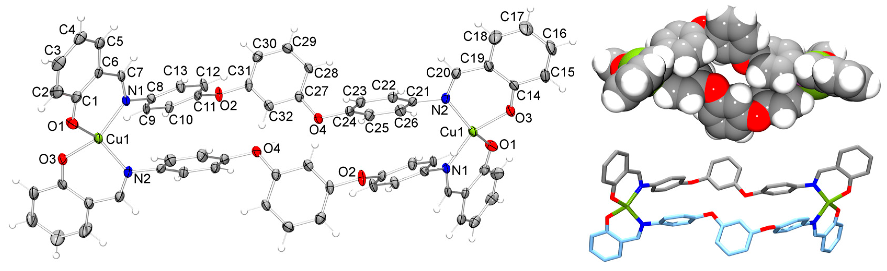
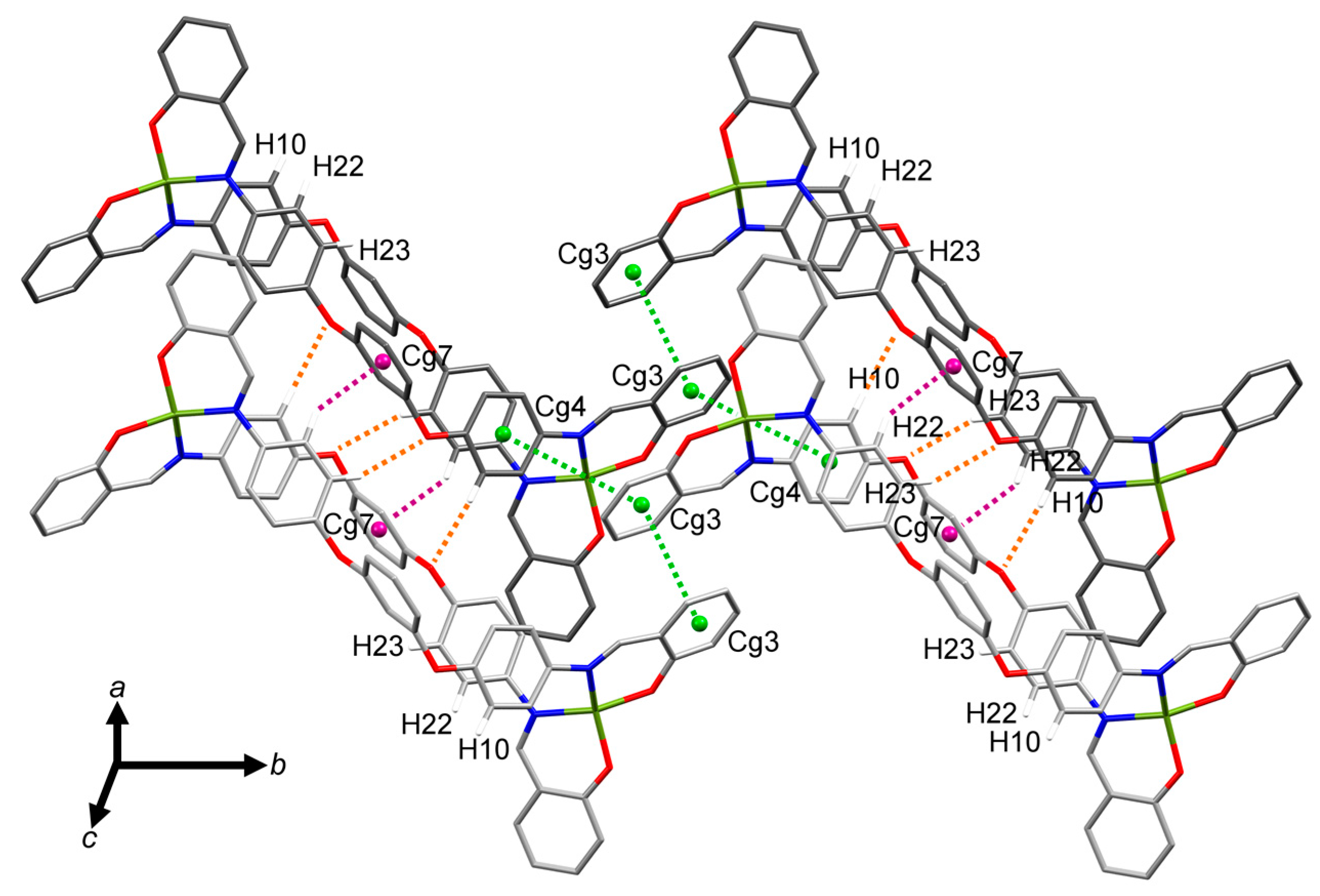
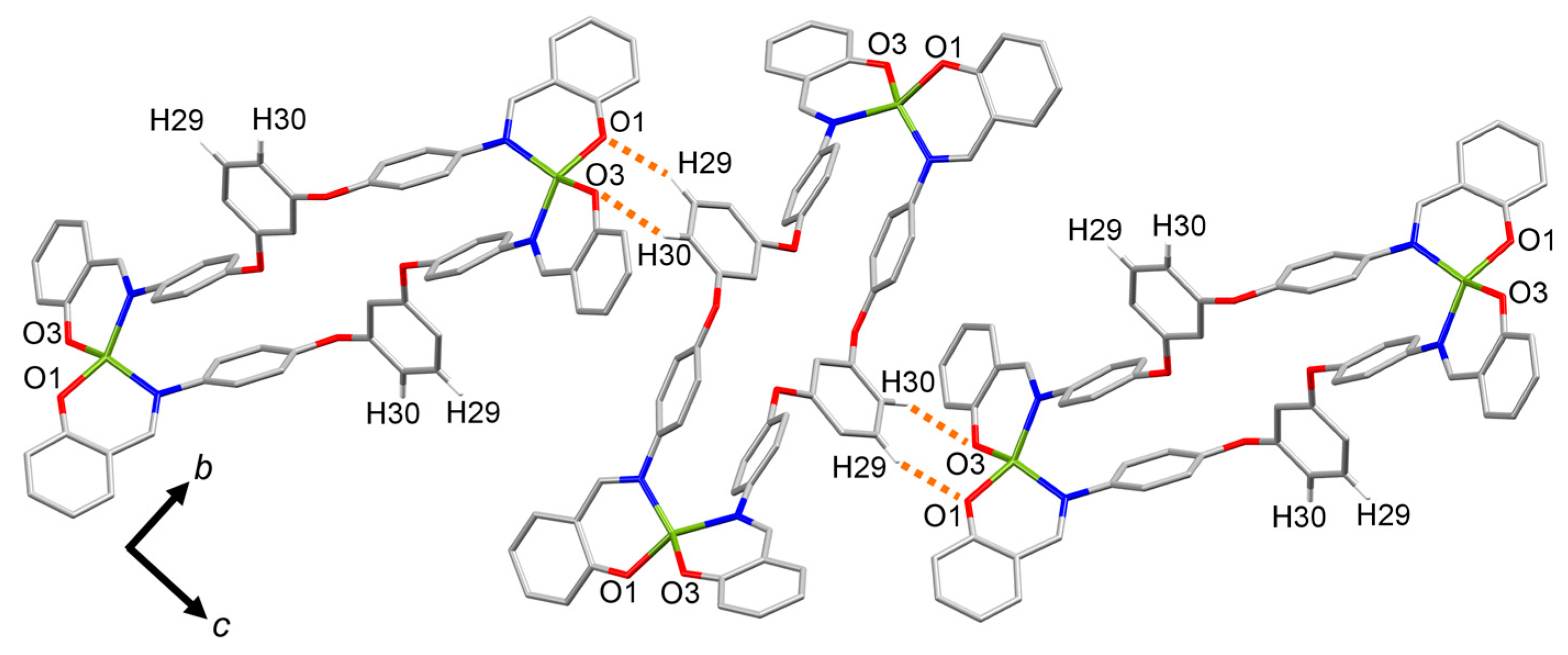
2.4. Molecular Structure of the Cu(II) Metallomacrocycle with H2L4

Ligand H2L4 is a diimine ligand with a large but limited flexible spacer based on aromatic subunits. In contrast to the other complexes obtained in this study, we were able to isolate a Cu(II) metallomacrocycle of composition [Cu2(L4)2]·2MeOH employing this ligand. Figure 9 shows the molecular structure (monoclinic space group P21/c) of this compound. The two Cu(II) ions in this dinuclear complex are coordinated in a strongly distorted tetrahedral arrangement (τ4 = 0.49) [28] by N2O2 donor sets from two deprotonated ligand molecules. The Cu-O and Cu-N bond lengths are in the expected range and comparable with the three other complexes discussed so fare (Table 4). Furthermore, weak CH···π contacts contribute to the stabilization of the metallomacrocycle (Table S11). This is in agreement with a structurally related Cu(II) complex discussed by Yoshida et al. [29]. The Cu···Cu separation within the metallomacrocycle is 16.17 Å and the Cg···Cg distance between the two in 1,3-position bridging central phenylene rings is 6.08 Å. However, this metallomacrocycle cannot act as a “molecular box” for guest inclusion, because of its flat arrangement. This results from the intramolecular π···π interactions between the in 1,4-position bridging aromatic units in the central part of the spacer (Table S12). Therefore any binding of small solvent molecules can only occur in the packing void space, with a methanol molecule bound to an adjacent metallomacrocycle via moderate bifurcated hydrogen bonds (O1A-H1A···O1, Table S13). However, it is possible to replace methanol by other small solvent molecules like water. This is indicated by the elemental analysis of the bulk material where four water molecules were found instead of two molecules of methanol (Figure S4).
The complex molecules are connected along the crystallographic a-axis by hydrogen bonds (C10-H10···O4 and C23-H23···O2; Table S13) and CH···π contacts (C22-H22···Cg7; Table S14), in which the central aromatic ring acts as an acceptor (Figure 10). This leads to a regular stacking of the compact metallomacrocycles. The peripheral aromatic units (Cg3) are involved in two π···π interactions between adjacent molecules. Additionally, the short Cu···Cg distance of 3.91 Å indicates the presence of a weak metal-aromatic ring contact.
As shown in Figure 11, the complex molecules possess two different orientations along the crystallographic c-axis resulting in a sheet-like assembly. Within the bc-plane, the metallomacrocycles are connected via two different hydrogen bonds. Further CH···π interactions (Table S14) contribute to the organization of this packing motif.

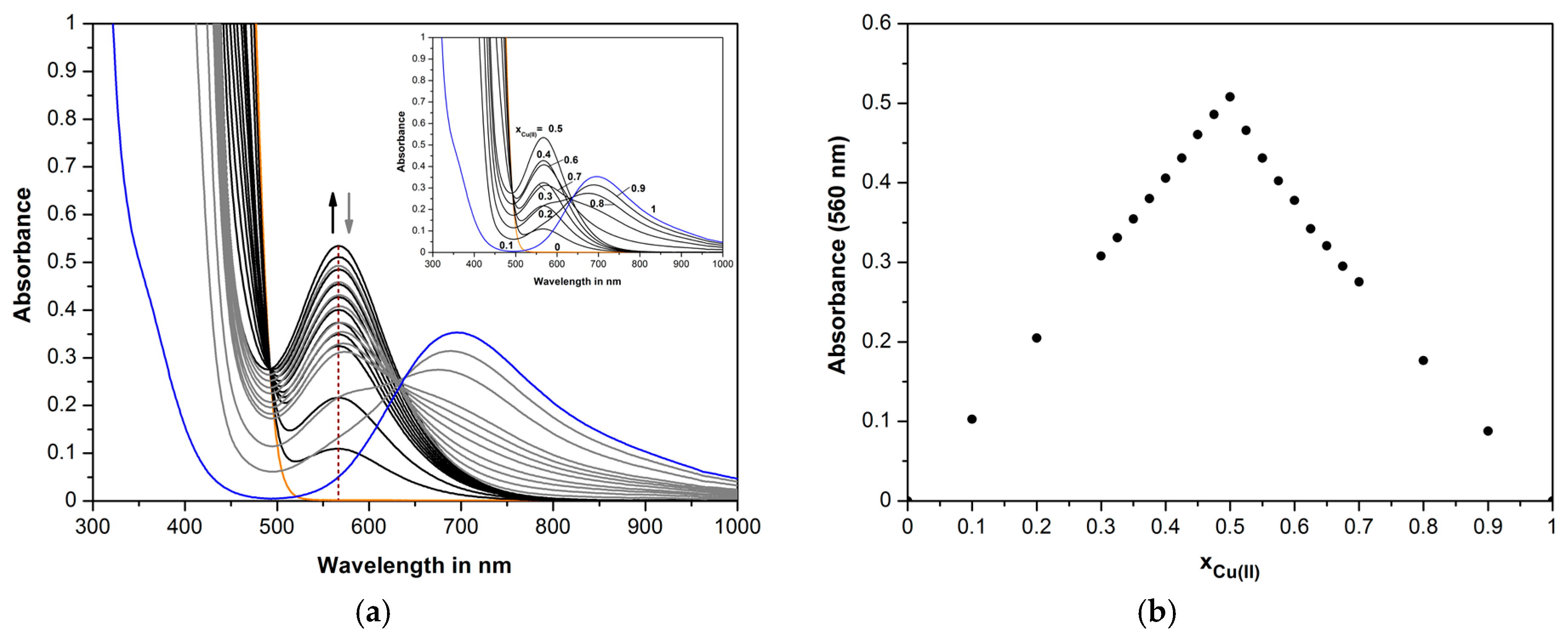
2.5. UV/Vis Studies of Cu(II) with the Diimines H2L1-H2L4

A series of UV/Vis spectroscopic studies in combination with electrospray ionization mass spectrometry measurements was performed in order to characterize the complex formation of Cu(II) with the diimine ligands H2L1-H2L4 in solution. Figure 12 shows the UV/Vis spectra of H2L1 in dichloromethane/methanol with changing concentration ratios of Cu(II) and ligand; the constant total concentration was 3 × 10⁻³ M. The absorption spectrum of H2L1 exhibits no absorption bands at wavelengths above 520 nm, which is consistent with the yellow color of the ligand solution. The addition of Cu(II) leads to a new band at 560 nm representing the formed Cu(II) complex. The resulting Job plot at this wavelength has a maximum in absorbance for the Cu(II) complexation with H2L1 at xCu(II) = 0.5 and points to a preferred formation of 1:1 complex species between Cu(II) and H2L1 under the chosen experimental conditions. Furthermore, the absorption spectra show an isosbestic point at 492 nm, which reaches a maximum at xCu(II) = 0.5 demonstrating the formation of a 1:1 complex. The ESI-MS spectrum of this sample solution shows a peak at m/z = 472.3 which corresponds to the 1:1 complex species [HL + Cu]+. Similar results were obtained using Cu(II) nitrate and tetrahydrofuran or dichloromethane/methanol as solvent. Figures S5, S6 and S7 depict the UV/Vis spectroscopic results for complex formation of Cu(II) with H2L2-H2L4, respectively. In all cases, the addition of Cu(II) leads to a new broad shoulder in the wavelength range 400–600 nm. Again, the maximum in absorbance at xCu(II) = 0.5 demonstrates the preferred formation of 1:1 complex species. For H2L2 and H2L3, the ESI-MS spectra of a sample solution show peaks corresponding to dinuclear complexes ([HL + L + 2Cu]+: m/z = 879.5 for H2L2, m/z = 947.4 for H2L3). For H2L4, the peak at m/z = 663.6 ([HL + Cu]+) could also be a result of fragmentation of a dinuclear complex in the gas-phase.
Preliminary liquid-liquid extraction experiments for Cu(II) using the extraction system Cu(NO3)2-NaNO3-HEPES buffer-H2O/ligand-CHCl3 were performed for all four ligands (Figure 13). Only the 3-ethoxy-2-hydroxyphenyl substituted ligand H2L3 extracts Cu(II) quantitatively. However, H2L1 and H2L4 also possess a high extraction capability for Cu(II). This result is a proof of sufficient complex stability and lipophilicity of the relevant extracted complexes in solution. Interestingly, H2L2 shows a significant lower extractability for Cu(II), presumably caused by the complicated steric requirements of the bulky spacer unit affecting the aqueous-organic phase transfer.

3. Experimental Section

3.1. Reagents and Methodology

All reagents and solvents were purchased from commercial sources and used as received. Elemental analyses were performed with an EA 3000 Euro Vector (HEKATECH, Wegberg, Germany) and a Vario Micro Cube (Elementar, Langenselbold, Germany). The melting points were determined with a Melting Point M-560 (Büchi, Essen, Germany). The NMR spectra were measured with a DRX-500 (Bruker, Karlsruhe, Germany). For UV/Vis studies, a Lambda 25 (PerkinElmer, Rodgau, Germany) spectrophotometer was used. Mass spectra were recorded with an ESQUIRE-detector (Bruker) operated in positive or negative ion electrospray ionization mode.

3.2. X-ray Structure Determinations

Crystals employed for the X-ray determinations were obtained directly from the respective reactions solutions and were used without further drying. Intensities were collected on a Kappa Apex II CCD diffractometer (Bruker AXS) with ω and ψ scans to approximately 60° (2θmax) at 143(2) or 296(2) K using graphite-monochromated Mo-Kα radiation generated from a sealed tube (0.71073 Å). The Apex Suite [30] program package was used for data collection, integration and data reduction Multi-scan empirical absorption corrections were applied to all data sets using the program SADABS [31]. The structures were solved by direct methods with the program SHELXS-97 [32] and the full-matrix least squares refinement was performed with SHELXL using the SHELXle user interface [33,34]. In general, ordered non-hydrogen atoms with occupancies greater than or equal to 0.5 were refined anisotropically; partial occupancy solvent oxygen atoms were refined isotropically. Carbon-bound hydrogen atoms were included in idealized positions and refined using a riding model. Oxygen-bound hydrogen atoms that were structurally evident in the difference Fourier map were included and refined with bond length and angle restraints. Parameters for data collection and structure refinement data for all structures are summarized in Table S16. ORTEP [35] depictions of the three structures are given in Figure 1, Figure 4, Figure 6 and Figure 9, selected bond lengths and angles in Table 1, Table 2, Table 3, Table 4 and Tables S1–S15.
CCDC 1488122 ([Cu2(L2)2]), 1488123 ([Cu2(L3)2]), 1488124 ([Cu2(L4)2]·2MeOH) and 1488125 ([Cu2(L1)(NO3)2(H2O)]·MeOH) contain the Supplementary Materials crystallographic data for this paper. These data can be obtained free of charge via www.ccdc.cam.ac.uk, or by contacting The Cambridge Crystallographic Data Centre, 12, Union Road, Cambridge CB21EZ, UK; Fax: +44-1223-336033.

3.3. Ligand Syntheses

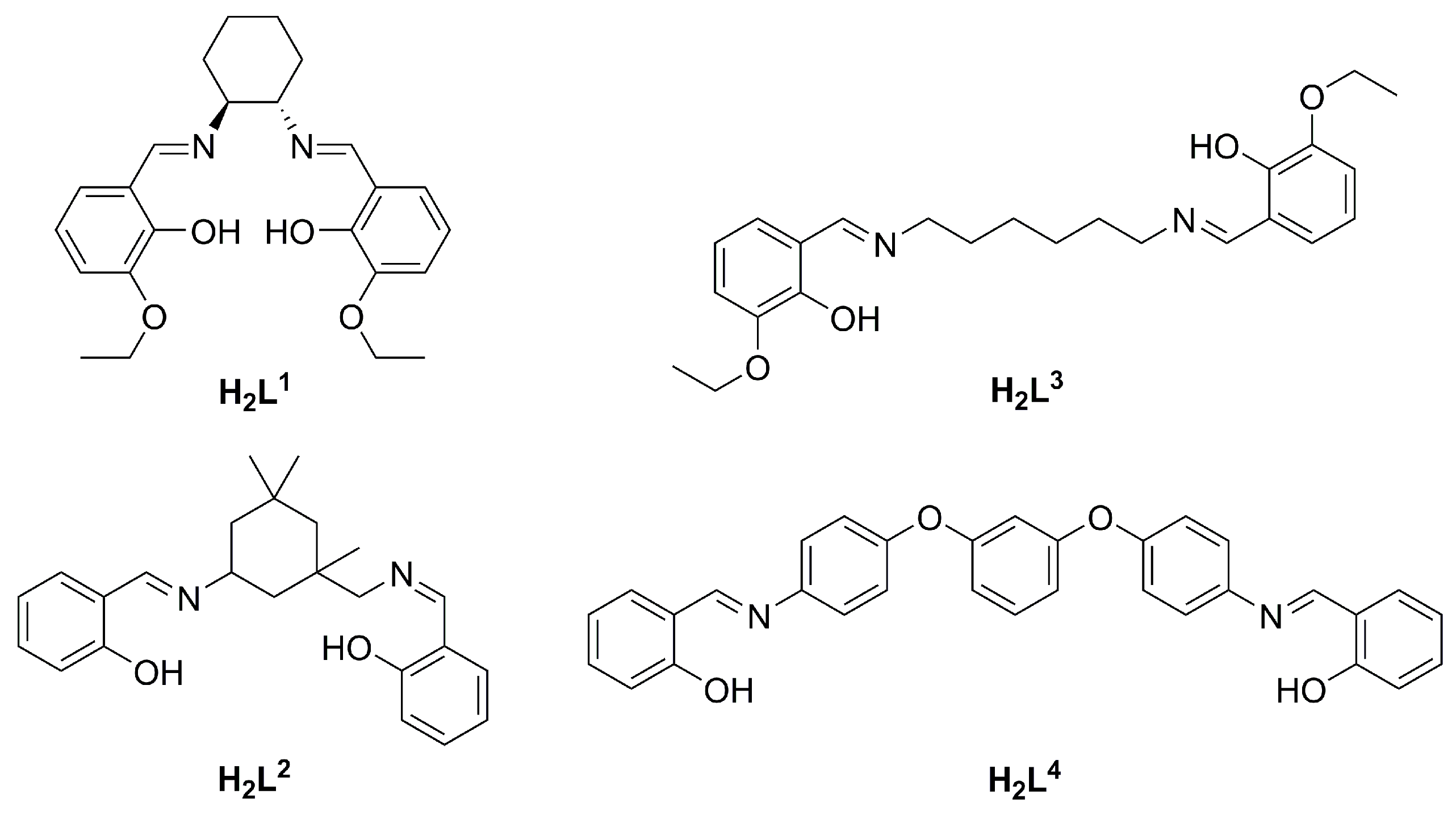
3.3.1. (±)-trans-N,N′-Bis(3-ethoxysalicylidene)-1,2-diaminocyclohexane (H2L1)

To a solution of (±)-trans-diaminocyclohexane (0.57 g, 5.00 mmol) in methanol (20 mL) was added a methanol solution (10 mL) of 3-ethoxysalicylaldehyde (1.66 g, 10.00 mmol). The mixture was stirred for 48 h at room temperature. The solvent was removed under reduced pressure to give a yellow solid residue which was recrystallized from methanol. The solid was washed with methanol and diethyl ether and dried in vacuo. Yellow powder. Yield: 1.80 g (4.40 mmol, 88%). m.p. 125 °C. Calculated for C24H30N2O4 (410.51 g·mol−1): C 70.22; H 7.37; N 6.82. Found: C 69.51; H 7.43; N 7.21. UV/Vis: λmax (lg ε): 221 nm (52398); 259 nm (24885); 327 nm (5510); 414 nm (325). ESI-MS: m/z = 411.3 [M + H]+; 821.6 [2M + H]+. 1H-NMR (500 MHz, CDCl3, 25 °C): δ = 13.90 (br. s, 2H, OH), 8.22 (s, 2H), 6.84 (dd, 3JH-H = 7.8 Hz, 4JH-H = 1.4 Hz, 2H), 6.76 (dd, 3JH-H = 7.8 Hz, 4JH-H = 1.3 Hz, 2H), 6.72 (t, 3JH-H = 7.9 Hz, 2H), 4.06 (q, 3JH-H = 7.0 Hz, 4H), 3.27–3.29 (m, 2H), 1.85–1.93 (m, 4H), 1.68–1.69 (m, 2H), 1.46 (m, 8H). 13C-NMR (125 MHz, CDCl3, 25 °C): δ = 164.7 (2CH), 151.5 (2Cq), 148.2 (2Cq), 123.1 (2CH), 118.4 (2Cq), 117.8 (2CH), 115.0 (2CH), 72.4 (2CH), 64.3 (2CH2), 33.0 (2CH2), 24.0 (2CH2), 14.9 (2CH3).

3.3.2. 2-{[(5-(2-Hydroxybenzylideneamino)-1,3,3-trimethylcyclohexyl)methylimino]methyl}phenol (H2L2)

To a solution of 5-amino-1,3,3-trimethylcyclohexanemethylamine (mixture of cis and trans; 0.85 g, 5.00 mmol) in methanol (30 mL) was added a methanol solution (20 mL) of salicylaldehyde (1.22 g, 10.00 mmol). The mixture was stirred for 24 h at room temperature. The precipitate was filtered off, washed with methanol and diethyl ether and dried in vacuo. Yellow powder. Yield: 1.05 g (2.77 mmol, 55%). m.p. 141 °C. Calculated for C24H30N2O2 (378.51 g·mol−1): C 76.16; H 7.99; N 7.40. Found: C 76.03; H 7.81; N 7.39. UV/Vis: λmax (lg ε): 256 nm (34733); 317 nm (11885); 407 nm (204). ESI-MS: m/z = 379.3 [M + H]+; 757.6 [2M + H]+; 376.9 [M − H]. 1H-NMR (500 MHz, CDCl3, 25 °C): δ = 13.59 (br. s, 2H, OH), 8.41 (s, 1H), 8.30 (s, 1H), 7.23–7.32 (m, 4H), 6.96 (d, 3JH-H = 8.5 Hz, 2H), 6.86 (d, 3JH-H = 7.6 Hz, 2H), 3.57–3.62 (m, 1H), 3.32–3.39 (m, 2H), 1.63 (d, 2JH-H = 12.6 Hz, 2H), 1.39–1.44 (m, 2H), 1.23–1.26 (m, 2H), 1.20 (s, 3H), 1.11 (s, 3H), 1.00 (s, 3H). 13C-NMR (125 MHz, CDCl3, 25 °C): δ = 165.4 (CH), 163.0 (CH), 161.3 (Cq), 161.2 (Cq), 132.3 (CH), 131.3 (2CH), 131.2 (CH), 118.7 (2Cq), 118.54 (2CH), 118.50 (CH), 117.01 (CH), 116.98 (CH), 75.0 (CH2), 61.9 (CH), 48.0 (CH2), 47.2 (CH2), 43.7 (CH2), 35.1 (Cq), 31.5 (Cq), 28.0 (CH3), 24.3 (CH3).

3.3.3. N,N′-Bis(3-ethoxysalicylidene)-1,6-diaminohexane (H2L3)

To a solution of 3-ethoxysalicylaldehyde (1.83 g, 11.00 mmol) in ethanol (40 mL) was added an ethanol solution (20 mL) of hexamethylenediamine (0.58 g, 5.00 mmol). The mixture was stirred for 24 h at room temperature. The precipitate was filtered off, washed with ethanol and diethyl ether and dried in vacuo. Yellow powder. Yield: 1.73 g (4.19 mmol, 84%). m.p. 115 °C. Calculated for C24H32N2O4 (412.52 g·mol−1): C 69.88; H 7.82; N 6.79. Found: C 70.27; H 8.04; N 6.77. UV/Vis: λmax (lg ε): 222 nm (53860); 260 nm (25893); 327 nm (5503); 416 nm (831). ESI-MS: m/z = 413.3 [M + H]+; 411.0 [M − H]. 1H-NMR (500 MHz, CDCl3, 25 °C): δ = 14.17 (br. s, 2H, OH), 8.29 (s, 2H), 6.89 (dd, 3JH-H = 7.9 Hz, 4JH-H = 1.6 Hz, 2H), 6.84 (dd, 3JH-H = 7.7 Hz, 4JH-H = 1.4 Hz, 2H), 6.74 (t, 3JH-H = 7.9 Hz, 2H), 4.10 (q, 3JH-H = 7.1 Hz, 4H), 3.58 (m, 4H), 1.66–1.69 (m, 4H), 1.47 (t, 3JH-H = 7.1 Hz, 6H), 1.41–1.43 (m, 4H). 13C-NMR (125 MHz, CDCl3, 25 °C): δ = 164.6 (2CH), 152.7 (2Cq), 147.8 (2Cq), 122.8 (2CH), 118.5 (2Cq), 117.5 (2CH), 115.1 (2CH), 64.4 (2CH2), 58.7 (2CH2), 30.6 (2CH2), 26.7 (2CH2), 14.9 (2CH3).

3.3.4. 1,3-Bis[4-(salicylideneamino)phenyloxy]benzol (H2L4)

To a suspension of 4,4′-(1,3-phenylenedioxy)dianiline (1.46 g, 5.00 mmol) in methanol (50 mL) was added a methanol solution (10 mL) of salicylaldehyde (1.22 g, 10.00 mmol). Two drops of concentrated sulfuric acid were added and the mixture was refluxed for 8 h. After cooling the precipitate was filtered off, washed with methanol and diethyl ether and dried in vacuo. Pale yellow powder. Yield: 2.18 g (4.36 mmol, 87%). m.p. 131 °C. Calculated for C32H24N2O4 (500.54 g·mol−1): C 76.78; H 4.83; N 5.60. Found: C 76.11; H 4.89; N 5.59. UV/Vis: λmax (lg ε): 306 nm (21692); 327 nm (27861); 347 nm (30934). ESI-MS: m/z = 501.3 [M + H]+; 499.0 [M − H]. 1H-NMR (500 MHz, CDCl3, 25 °C): δ = 13.22 (br. s, 2H, OH), 8.61 (s, 2H), 7.35–7.39 (m, 4H), 7.31 (s, 1H), 7.27–7.30 (m, 4H), 7.07–7.10 (m, 4H), 7.01 (d, 3JH-H = 7.9 Hz, 2H), 6.94 (td, 3JH-H = 7.4 Hz, 4JH-H = 0.9 Hz, 2H), 6.77 (dd, 3JH-H = 8.2 Hz, 4JH-H = 2.2 Hz, 2H), 6.72 (t, 4JH-H = 2.2 Hz, 1H). 13C-NMR (125 MHz, CDCl3, 25 °C): δ = 161.9 (2CH), 161.0 (2Cq), 158.6 (2Cq), 155.6 (2Cq), 144.1 (2Cq), 133.1 (2CH), 132.2 (2CH), 130.6 (1CH), 122.6 (2CH), 120.0 (4CH), 119.2 (2Cq), 119.1 (2CH), 117.2 (4CH), 113.4 (2CH), 109.3 (1CH).

3.4. Complex Syntheses

3.4.1. [Cu2(L1)(NO3)2(H2O)]·MeOH

A THF solution of H2L1 (41 mg, 0.1 mmol, 2 mL) was over-layered with a solution of copper nitrate trihydrate (24 mg, 0.1 mmol, 2 mL) in THF/methanol (v/v = 1:1). Within three days suitable single crystals were obtained by slow diffusion of diethyl ether/hexane (v/v = 1:1). The brown-colored crystals were filtered off, washed with ethanol and diethyl ether and dried in vacuo. Brown crystals. Yield: 23 mg (0.032 mmol, 64%). C25H34Cu2N4O12 (709.66 g·mol–1). ESI-MS: m/z = 472.3 [HL + Cu]+, 943.3 [HL + L + 2Cu]+, 960.5 [2L + 2Cu + NH4]+, 941.4 [2L + 2Cu − H].

3.4.2. [Cu2(L2)2]

Two methanol solutions of H2L2 (38 mg, 0.1 mmol, 10 mL) and copper acetate monohydrate (20 mg, 0.1 mmol, 10 mL) were combined. After two hours brown-colored plates could be obtained. To complete the precipitation the mixture was left for 24 h. The crystals were filtered off, washed with methanol and diethyl ether and dried in vacuo. Dark brown plates. Yield: 11 mg (0.013 mmol, 25%). Anal. Calcd. for C48H56Cu2N4O4 (880.07 g·mol−1): C 65.51; H 6.41; N 6.37. Found: C 65.02; H 6.42; N 6.23. ESI-MS: m/z = 440.3 [HL + Cu]+, 879.5 [HL + L + 2Cu]+.

3.4.3. [Cu2(L3)2]

Two solutions of H2L3 (41 mg, 0.1 mmol, 50 mL) and copper acetate monohydrate (20 mg, 0.1 mmol, 50 mL) in acetonitrile/methanol (v/v = 1:1) were combined and the mixture was left for one week. Brown crystals could be obtained, which were filtered off, washed with acetonitrile and diethyl ether and dried in vacuo. Brown plates. Yield: 30 mg (0.032 mmol, 63%). Calculated for C48H60Cu2N4O8 (948.10 g·mol−1): C 60.81; H 6.38; N 5.91. Found: C 60.40; H 6.36; N 5.68. ESI-MS: m/z = 474.3 [HL + Cu]+, 947.4 [HL + L + 2Cu]+.

3.4.4. [Cu2(L4)2]·4H2O

Two solutions of H2L4 (50 mg, 0.1 mmol, 100 mL) and copper acetate monohydrate (20 mg, 0.1 mmol, 100 mL) in dichloromethane/methanol (v/v = 1:1) were combined and the mixture was left for one week. Brown crystals could be obtained, which were filtered off, washed with ethanol and diethyl ether and dried in vacuo. Brown green crystals. Yield: 43 mg (0.035 mmol, 70%). Calculated for C65H56Cu2N4O13 (1228.27 g·mol−1): C 63.56; H 4.60; N 4.56. Found: C 63.79; H 4.14; N 4.28. ESI-MS: m/z = 501.3 [H2L + H]+, 663.6 [HL + Cu]+, 661.2 [L + Cu − H].

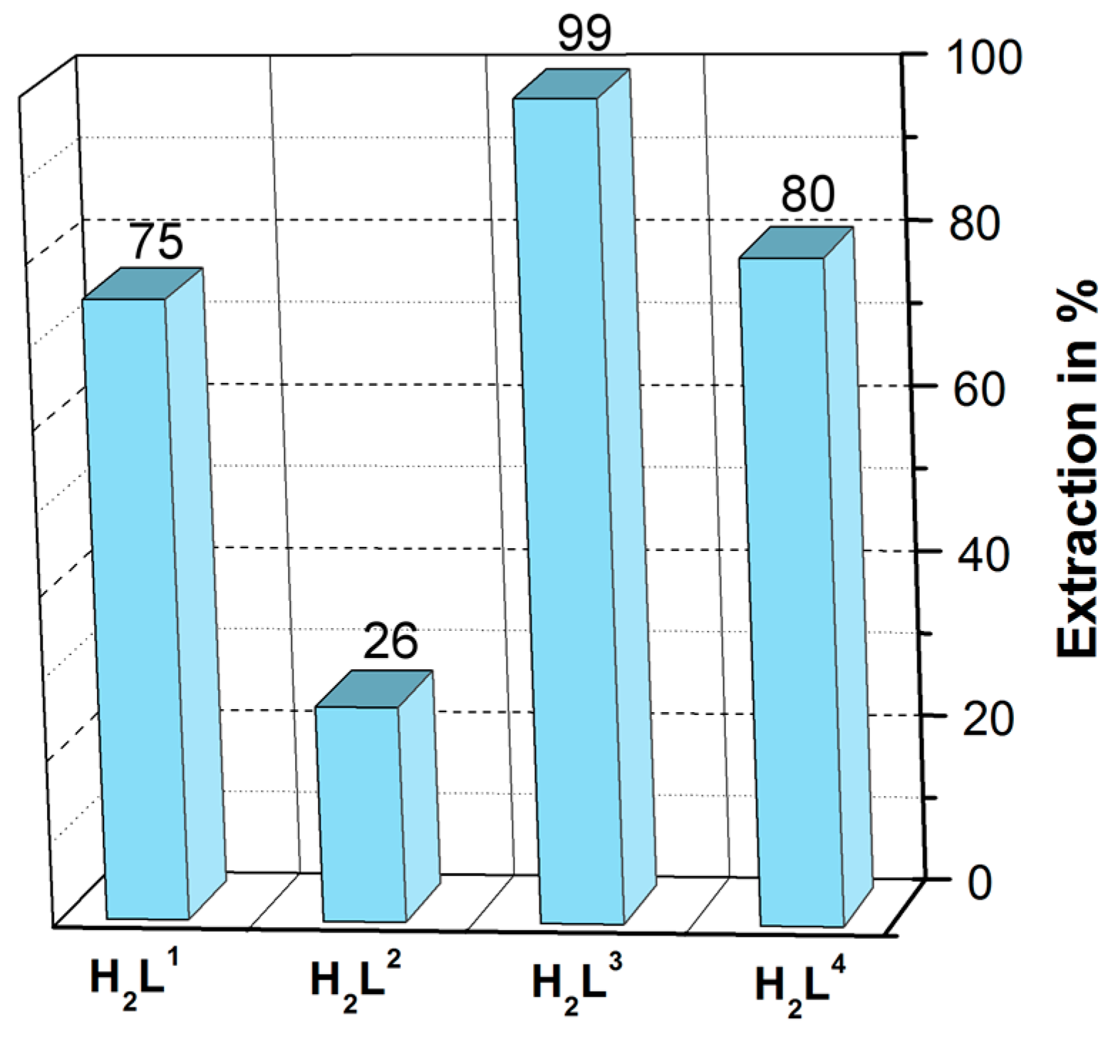
3.5. Liquid-Liquid Extraction

All extraction experiments were performed at room temperature in microcentrifuge tubes (2 mL) by mechanical shaking. The phase ratio V(w):V(org) was 1:1 (0.5 mL each); the shaking time was 30 min. The organic phase consists of CHCl3 as diluent and the ligand (1 × 10⁻³ M). The aqueous phase contained the buffer HEPES/NaOH (pH = 7.7), NaNO3 (5 × 10⁻³ M) and Cu(NO3)2 (1 × 10⁻⁴ M). All samples were centrifuged after extraction. The metal concentration in both phases (100 µL samples each) was determined radiometrically using γ-radiation (64Cu; NaI(Tl) scintillation counter Cobra II/Canberra-Packard, Detroit, MI, USA).

4. Conclusions

In this study, dinuclear Cu(II) complexes with four diimine ligands were synthesized and characterized in solution and the solid state. The characterized complexes range from a monoligand complex with H2L1 to [2 + 2] helicates with H2L2 and H2L3 and a [2 + 2] metallomacrocycle with H2L4. We have demonstrated that the complex structures are strongly influenced by the size and flexibility of the spacer unit. A quite interesting result is the formation of the unique 2:1 (Cu:L) complex by reaction of Cu(NO3)2 with H2L1. This reaction leads to different coordination patterns for the two Cu(II) centers, a commonly observed square pyramidal and a rare pentagonal bipyramidal arrangement. Typically, this ligand forms 1:1 complexes with Cu(II) having a square planar geometry [16,17]. The differences in the literature experimental conditions and our reaction are the counter anion of the Cu(II) salt and the slightly modified solvent system employed; we have used nitrate instead of acetate and the solvent mix methanol/tetrahydrofuran instead of pure methanol.
It is also interesting to note that in contrast to the Cu(II) helicates with the structurally related methylene or sulfurdiphenylene bridged 3-ethoxy-2-hydroxyphenyl substituted diimines [13], the related helicate from H2L3 having a long hexamethylene spacer cannot bind a water molecule. This is obviously caused by the closely folded structure of this Cu(II) helicate which prevents the preorganization of an outer O4 coordination sphere. However, such an environment is able to be generated in the more stretched Cu(II) helicates.
Finally, the UV/Vis and ESI-MS studies performed in dichloromethane/methanol solutions are broadly consistent with the solid state crystallographic results. They also provide some evidence for a preferred formation of dinuclear Cu(II) complex species in solution. However, in the case of H2L1 the results indicate the presence of several complex species in solution as well as in the solid state. Since the overall reasons for the different behavior of the systems investigated remain unclear, further detailed studies are necessary in order to clarify these results.

Supplementary Materials

The following are available online at https://www.mdpi.com/2073-4352/6/9/120/s1, Figure S1: CH···O and CH···π interactions between neighboring strands in [Cu2(L1)(NO3)2(H2O)]·MeOH; Figure S2: The packing motif of [Cu2(L1)(NO3)2(H2O)]·MeOH; Figure S3: The packing motif of [Cu2(L2)]; Figure S4: Methanol molecules in the void space of [Cu2(L4)]·2MeOH packing; Figure S5: UV/Vis spectra and Job plot for complexation of copper(II) acetate with ligand H2L2 in THF; Figure S6: UV/Vis spectra and Job plot for complexation of copper(II) acetate with ligand H2L3 in dichloromethane/methanol; Figure S7: UV/Vis spectra and Job plot for complexation of copper(II) acetate with ligand H2L4 in dichloromethane/methanol; Table S1: Intramolecular hydrogen bonds in [Cu2(L1)(NO3)2(H2O)]·MeOH; Table S2: Intermolecular hydrogen bonds in [Cu2(L1)(NO3)2(H2O)]·MeOH, Table S3: Intermolecular π···π interactions in [Cu2(L1)(NO3)2(H2O)]·MeOH; Table S4: Intermolecular CH···π interactions in [Cu2(L1)(NO3)2(H2O)]·MeOH; Table S5: Intramolecular hydrogen bonds in [Cu2(L2)2]; Table S6: Intermolecular CH···π interactions in [Cu2(L2)2]; Table S7: Intermolecular π···π interactions in [Cu2(L2)2]; Table S8: Intramolecular hydrogen bonds in [Cu2(L3)2]; Table S9: Intermolecular cation···π interactions in [Cu2(L3)2]; Table S10: Intermolecular hydrogen bonds in [Cu2(L3)2]; Table S11: Intramolecular CH···π interactions in [Cu2(L4)2]·2MeOH; Table S12: Intramolecular π···π interactions in [Cu2(L4)2]·2MeOH; Table S13: Intermolecular hydrogen bonds in [Cu2(L4)2]·2MeOH; Table S14: Intermolecular CH···π interactions in [Cu2(L4)2]·2MeOH; Table S15: Intermolecular π···π interactions in [Cu2(L4)2]·2MeOH; Table S16: Crystal and structure refinement data for [Cu2(L1)(NO3)2(H2O)]·MeOH, [Cu2(L2)2], [Cu2(L3)2] and [Cu2(L4)2]·2MeOH.

Acknowledgments

The authors thank the German Federal Ministry of Education and Research (BMBF project 02NUK014A) and the German Federation of Industrial Research Associations (AIF/ZIM project KF2807202RH3) for financial support. Norman Kelly and Franziska Taube thank Margret Acker for assistance in the radiotracer experiments.

Author Contributions

Norman Kelly, Kerstin Gloe and Karsten Gloe conceived and designed the experiments; Franziska Taube, Axel Heine and Norman Kelly performed the experiments; Thomas Doert and Willy Seichter were responsible for the single crystal structures; Kerstin Gloe, Karsten Gloe and Jan J. Weigand analyzed the data; and Norman Kelly and Karsten Gloe wrote the paper. All authors discussed the results and commented on the manuscript.

Conflicts of Interest

The authors declare no conflict of interest.

References

  1. Yoshida, N.; Oshio, H.; Ito, T. Self-assembling tetranuclear copper(II) complex of a bis-bidentate Schiff base: Double-helical structure induced by aromatic π···π and CH···π interactions. Chem. Commun. 1998, 63–64. [Google Scholar] [CrossRef]
  2. Yoshida, N.; Oshio, H.; Ito, T. Copper(II)-assisted self-assembly of bis-N,O-bidentate Schiff bases: New building blocks for a double-helical supramolecular motif. J. Chem. Soc. Perkin Trans. 2 1999, 975–983. [Google Scholar] [CrossRef]
  3. Lacroix, P.G. Second-order optical nonlinearities in coordination chemistry: The case of bis(salicylaldiminato)metal Schiff base complexes. Eur. J. Inorg. Chem. 2001, 2001, 339–348. [Google Scholar] [CrossRef]
  4. Cozzi, P.G. Metal-salen Schiff base complexes in catalysis: Practical aspects. Chem. Soc. Rev. 2004, 33, 410–421. [Google Scholar] [CrossRef] [PubMed]
  5. Che, C.-M.; Huang, J.-S. Metal complexes of chiral binaphthyl Schiff-base ligands and their application in stereoselective organic transformations. Coord. Chem. Rev. 2003, 242, 97–113. [Google Scholar] [CrossRef]
  6. Sakamoto, M.; Manseki, K.; Okawa, H. d–f Heteronuclear complexes: Synthesis, structures and physicochemical aspects. Coord. Chem. Rev. 2001, 219–221, 379–414. [Google Scholar] [CrossRef]
  7. Whiteoak, C.J.; Salassaa, G.; Kleij, A.W. Recent advances with π-conjugated salen systems. Chem. Soc. Rev. 2012, 41, 622–631. [Google Scholar] [CrossRef] [PubMed]
  8. Lacroix, P.G.; Malfant, I.; Real, J.-A.; Rodriguez, V. From magnetic to nonlinear optical switches in spin-crossover complexes. Eur. J. Inorg. Chem. 2013, 2013, 615–627. [Google Scholar] [CrossRef]
  9. Vigato, P.A.; Tamburini, S. The challenge of cyclic and acyclic Schiff bases and related derivatives. Coord. Chem. Rev. 2004, 248, 1717–2128. [Google Scholar] [CrossRef]
  10. Vigato, P.A.; Tamburini, S.; Bertolo, L. The development of compartmental macrocyclic Schiff bases and related polyamine derivatives. Coord. Chem. Rev. 2007, 251, 1311–1492. [Google Scholar] [CrossRef]
  11. Biswas, S.; Ghosh, A. Use of Cu(II)-di-Schiff bases as metalloligands in the formation of complexes with Cu(II), Ni(II) and Zn(II) perchlorate. Polyhedron 2013, 65, 322–331. [Google Scholar] [CrossRef]
  12. Nathan, L.C.; Koehne, J.E.; Gilmore, J.M.; Hannibal, K.A.; Dewhirst, W.E.; Mai, T.D. The X-ray structures of a series of copper(II) complexes with tetradentate Schiff base ligands derived from salicylaldehyde and polymethylenediamines of varying chain length. Polyhedron 2003, 22, 887–894. [Google Scholar] [CrossRef]
  13. Kelly, N.; Schulz, J.; Gloe, K.; Doert, T.; Gloe, K.; Wenzel, M.; Acker, M.; Weigand, J.J. Self-assembly of double-stranded copper(II) helicates with 2-ethoxy-2-hydroxyphenyl substituted diimines. Synthesis, molecular structure, and host-guest recognition of H2O. Z. Anorg. Allg. Chem. 2015, 641, 2215–2221. [Google Scholar] [CrossRef]
  14. Qin, W.; Long, S.; Panunzio, M.; Biondi, S. Schiff Bases: A short survey on an evergreen chemistry tool. Molecules 2013, 18, 12264–12289. [Google Scholar] [CrossRef] [PubMed]
  15. Tozzo, E.; Romera, S.; dos Santos, M.P.; Muraro, M.; de A. Santos, R.H.; Liao, L.M.; Vizotto, L.; Dockal, E.R. Synthesis, spectral studies and X-ray crystal structure of N,N′-(±)-trans-1,2-cyclohexylenebis(3-ethoxysalicylideneamine). J. Mol. Struct. 2008, 876, 210–220. [Google Scholar] [CrossRef]
  16. Constable, E.C.; Zhang, G.; Housecroft, C.E.; Neuburger, M.; Zampese, J.A. Hierarchical multicomponent assembly utilizing sequential metal-ligand and hydrogen-bonding interactions. CrystEngComm 2009, 11, 657–662. [Google Scholar] [CrossRef]
  17. Constable, E.C.; Zhang, G.; Housecroft, C.E.; Neuburger, M.; Zampese, J.A. Host-guest chemistry of a chiral Schiff base copper(II) complex: Can chiral information be transferred to the guest cation. CrystEngComm 2010, 12, 1764–1773. [Google Scholar] [CrossRef]
  18. Koner, R.; Lee, G.-H.; Wang, Y.; Wei, H.-H.; Mohanta, S. Two new diphenoxo-bridged discrete dinuclear CuIIGdIII compounds with cyclic diimino moieties: Syntheses, structures, and magnetic properties. Eur. J. Inorg. Chem. 2005, 2005, 1500–1505. [Google Scholar] [CrossRef]
  19. Hazra, S.; Koner, R.; Nayak, M.; Sparkes, H.A.; Howard, J.A.K.; Mohanta, S. Cocrystallized dinuclear-mononuclear CuII3NaI and double-decker-triple-decker CuII5KI3 complexes derived from N,N′-ethylenebis(3-ethoxysalicylaldimine). Cryst. Growth Des. 2009, 9, 3603–3608. [Google Scholar] [CrossRef]
  20. Berkessel, A.; Roland, K.; Schröder, M.; Neudörfl, J.M.; Lex, J. Enantiomerically pure isophorone diamine [3-(aminomethyl)-3,5,5-trimethylcyclohexylamine]:  A chiral 1,4-diamine building block made available on large scale. J. Org. Chem. 2006, 71, 9312–9318. [Google Scholar] [CrossRef] [PubMed]
  21. Ha, K. 6,6′-Diethoxy-2,2′-[hexane-1,6-diylbis(nitrilomethanylylidene)]diphenol. Acta Cryst. 2011, E67. [Google Scholar] [CrossRef] [PubMed]
  22. Wester, D.; Palenik, G.J. Pentagonal bipyramidal complexes of nickel(II) and copper(II). The relative importance of ligand geometry vs. crystal field effects. J. Am. Chem. Soc. 1974, 96, 7565–7566. [Google Scholar] [CrossRef]
  23. Nelson, S.M. Developments in the synthesis and coordination chemistry of macrocyclic Schiff base ligands. Pure Appl. Chem. 1980, 52, 2461–2476. [Google Scholar] [CrossRef]
  24. Drew, M.G.B.; Nelson, J.; Nelson, S.M. Synthesis of some pentagonal-bipyramidal complexes of manganese(II), iron(II), cobalt(II), nickel(II), copper(II), and zinc(II) with a heptadentate Schiff-base ligand and the crystal and molecular structures of a copper(II) complex. Dalton Trans. 1981, 1685–1690. [Google Scholar] [CrossRef]
  25. Sakurai, T.; Kobayashi, K.; Tsuboyama, S.; Konyo, Y.; Nagao, A.; Ishizu, K. Structure of dichloro(5,6,8,9,11,12,14,15-octahydro-2,3-benzo-1,4,7,10,13-pentaoxacyclopentadec-2-ene) copper(II) chloroform solvate, a Cu(II) crown ether complex with pentagonaI-bipyramidal geometry. Acta Cryst. 1983, C39, 206–208. [Google Scholar]
  26. Diouf, O.; Gaye, M.; Sail, A.S.; Tamboura, F.; Barry, A.H.; Jouini, T. Crystal structure of diaqua-2,6-diacetylpyridine-bis(acetylhydrazone)copper(II) complex dinitrate hydrate. Z. Kristallogr. NCS 2001, 216, 421–422. [Google Scholar]
  27. Zhang, Y.; Feng, W.; Liu, H.; Zhang, Z.; Lü, X.; Song, J.; Fan, D.; Wong, W.-K.; Jones, R.A. Photo-luminescent hetero-trinuclear Zn2Ln (Ln = Nd, Yb, Er or Gd) complexes based on the binuclear Zn2L precursor. Inorg. Chem. Commun. 2012, 24, 148–152. [Google Scholar] [CrossRef]
  28. Yang, L.; Powell, D.R.; Houser, R.P. Structural variation in copper(I) complexes with pyridylmethylamide ligands: structural analysis with a new four-coordinate geometry index, τ4. Dalton Trans. 2007, 955–964. [Google Scholar] [CrossRef] [PubMed]
  29. Yoshida, N.; Oshio, H.; Ito, T. Supramolecular motifs in metal complexes of Schiff bases. Part 6.1 Topology of two types of self-assembly of bis-N,O-bidentate Schiff base ligands by copper(II) ions. J. Chem. Soc. Perkin Trans. 2001, 1674–1678. [Google Scholar] [CrossRef]
  30. Bruker. Apex Suite (V. 2012.4); Bruker AXS Inc.: Madison, WI, USA, 2012. [Google Scholar]
  31. Sheldrick, G.M. Bruker AXS Area Detector Scaling and Absorption Correction; University of Göttingen: Göttingen, Germany, 2008. [Google Scholar]
  32. Sheldrick, G.M. SHELXS-97, Program for the Solution of Crystal Structures; University of Göttingen: Göttingen, Germany, 1997. [Google Scholar]
  33. Sheldrick, G.M. A short history of SHELX. Acta Cryst. 2008, A64, 112–122. [Google Scholar] [CrossRef] [PubMed]
  34. Hübschle, C.B.; Sheldrick, G.M.; Dittrich, B. ShelXle: A Qt graphical user interface for SHELXL. J. Appl. Cryst. 2011, 44, 1281–1284. [Google Scholar] [CrossRef] [PubMed]
  35. Farrugia, L.J. ORTEP-3 for Windows-a version of ORTEP-III with a Graphical User Interface (GUI). J. Appl. Cryst. 1997, 30, 565–566. [Google Scholar] [CrossRef]
Scheme 1. Structural formulas of H2L1-H2L4.
Crystals 06 00120 sch001
Figure 1. ORTEP drawing (50% probability level) of the molecular structure of: [Cu2(L1)(NO3)2(H2O)]·MeOH (a); and simplified stick representation (b).
Crystals 06 00120 g001
Figure 2. Formation of complex dimers and polymeric strands by [Cu2(L1)(NO3)2(H2O)]·MeOH via hydrogen bonding and π···π interactions.
Crystals 06 00120 g002
Figure 3. Packing of [Cu2(L1)(NO3)2(H2O)]·MeOH showing the disordered methanol molecules in the void space.
Crystals 06 00120 g003
Figure 4. ORTEP drawing (50% probability level) of the molecular structure of: [Cu2(L2)2] (a); and a simplified stick representation of the helicate (b).
Crystals 06 00120 g004
Figure 5. Intermolecular CH···π and π···π interactions in [Cu2(L2)2] resulting in the formation of 2D layers (a); and the resulting packing motif with complex molecules of consistent handedness in the same color (b).
Crystals 06 00120 g005
Figure 6. ORTEP drawing (50% probability level) of the molecular structure of: [Cu2(L3)2] (a); and stick representation of the helicate (b).
Crystals 06 00120 g006
Figure 7. Illustration of the formation of polymeric strands by [Cu2(L3)2] involving Cu(II)···π interactions.
Crystals 06 00120 g007
Figure 8. 2D layers within the bc-plane in [Cu2(L3)2].
Crystals 06 00120 g008
Figure 9. ORTEP drawing of the molecular structure of [Cu2(L4)2]·2MeOH (Ellipsoids are given at the 50% probability level; disordered CH3OH is not shown) and the corresponding space fill and capped stick representations.
Crystals 06 00120 g009
Figure 10. Hydrogen bonds, CH···π and π···π interactions between adjacent metallomacrocycles in [Cu2(L4)2]·2MeOH.
Crystals 06 00120 g010
Figure 11. Different orientations of the metallomacrocycles in the bc-plane in [Cu2(L4)2]·2MeOH.
Crystals 06 00120 g011
Figure 12. UV/Vis spectra (a) and Job plot (560 nm) (b) for the complexation of Cu(II) with H2L1 in dichloromethane/methanol (v/v = 1:1); [H2L1] + [Cu(II)] = 3 × 10⁻³ M (xCu(II) = 0–1), t = 30 min. (copper(II) acetate spectrum in blue, ligand spectrum in orange).
Crystals 06 00120 g012
Figure 13. Liquid-liquid extraction of Cu(II) by H2L1, H2L2, H2L3 and H2L4 at pH = 7.7 (HEPES/NaOH buffer); [Cu(II)] = 1 × 10⁴ M, [NaNO3] = 5 × 10³ M; [L] = 1 × 10³ M in CHCl3; t = 30 min, T = 22 ± 1 °C.
Crystals 06 00120 g013
Table 1. Selected bond lengths (Å) in [Cu2(L1)(NO3)2(H2O)]·MeOH.
Bond Lengths
Cu1-N11.926(3)Cu2-O31.987(2)
Cu1-N21.951(3)Cu2-O42.341(3)
Cu1-O11.893(2)Cu2-O51.955(2)
Cu1-O31.951(2)Cu2-O72.723(3)
Cu1-O72.658(3)Cu2-O82.084(4)
Cu2-O102.522(7)
Cu2-O1W1.951(3)
Table 2. Selected bond lengths (Å) in [Cu2(L2)2].
Bond Lengths
Cu1-N12.0065(17)
Cu1-N41.9875(19)
Cu1-O11.8947(15)
Cu1-O41.8828(15)
Cu2-N21.9903(19)
Cu2-N31.9963(17)
Cu2-O21.9007(16)
Cu2-O31.8958(17)
Table 3. Selected bond lengths (Å) in [Cu2(L3)2].
Bond Lengths
Cu1-N12.0022(17)
Cu1-N31.9991(17)
Cu1-O21.8791(14)
Cu1-O61.8722(15)
Cu2-N21.9897(19)
Cu2-N41.989(2)
Cu2-O31.8900(16)
Cu2-O71.8838(15)
Table 4. Selected bond lengths (Å) in [Cu2(L4)2]·2MeOH.
Bond Lengths
Cu1-N11.976(3)
Cu1-N21.982(3)
Cu1-O11.900(2)
Cu1-O31.899(2)

