Article

Preclinical Cancer Chemoprevention Studies Using Animal Model of Inflammation-Associated Colorectal Carcinogenesis

1
Cytopatholgy Division, Tohkai Cytopathology Institute, Cancer Research and Prevention (TCI-CaRP), 5-1-2 Minami-uzura, Gifu 500-8285, Japan
2
Department of Tumor Pathology, Gifu University Graduate School of Medicine, 1-1 Yanagido, Gifu 501-1194, Japan
Cancers 2012, 4(3), 673-700; https://doi.org/10.3390/cancers4030673
Submission received: 5 April 2012 / Revised: 14 June 2012 / Accepted: 6 July 2012 / Published: 16 July 2012
(This article belongs to the Special Issue Advances in Cancer Chemoprevention)

Abstract

:
Inflammation is involved in all stages of carcinogenesis. Inflammatory bowel disease, such as ulcerative colitis and Crohn’s disease is a longstanding inflammatory disease of intestine with increased risk for colorectal cancer (CRC). Several molecular events involved in chronic inflammatory process are reported to contribute to multi-step carcinogenesis of CRC in the inflamed colon. They include over-production of free radicals, reactive oxygen and nitrogen species, up-regulation of inflammatory enzymes in arachidonic acid biosynthesis pathway, up-regulation of certain cytokines, and intestinal immune system dysfunction. In this article, firstly I briefly introduce our experimental animal models where colorectal neoplasms rapidly develop in the inflamed colorectum. Secondary, data on preclinical cancer chemoprevention studies of inflammation-associated colon carcinogenesis by morin, bezafibrate, and valproic acid, using this novel inflammation-related colorectal carcinogenesis model is described.

Abbreviations

ACF
aberrant crypt foci
AD
adenoma
ADC
adenocarcinoma
AOM
azoxymetheane
5-ASA
5-aminosalicylic acid
CD
Crohn’s disease
COX
cyclooxygenase
CRC
colorectal cancer
DMH
1,2-dimethylhydrazine
DSS
dextran sodium sulfate
H&E
hematoxylin and eosin
HATs
histone acetylases
HCC
hepatocellular carcinoma
HDACs
histone decarboxylases
HDACIs
histone decarboxylase inhibitors
HIF
hypoxia-inducible factor
HMG-CoA
3-hydroxy-3-methylglutary coenzyme A
IBD
inflammatory bowel disease
IL
interleukin
iNOS
inducible nitric oxide synthase
KAD
Kyoto Apc Delta
MAM
methylazoxymethanol
NF-κB
nuclear factor-kappaB
NSAIDs
non-steroidal anti-inflammatory drugs
PhIP
2-amino-1-methyl-6-phenylimidazo[4,5-b]pyridine
PPARs
peroxisome proliferator-activated receptors
PSC
primary sclerosing cholangitis
RXR
retinoid X receptor
TNF
tumor necrosis factor
Stat3
signal transducer and activator of transcription
UC
ulcerative colitis
UDCA
ursodeoxycholic acid
VPA
valproic acid

1. Introduction

An association between inflammation and cancer has been suggested for a long time [1] and it is now well-recognized that inflammation is involved in carcinogenesis in several tissues [2,3]. Patients with inflammatory bowel disease (IBD), especially major types of IBD ulcerative colitis (UC) and Crohn’s disease (CD) have a significantly increased risk of developing premalignancy (dysplastic lesions) and malignancy (adenocarcinoma, ADC) in the colorectum [4,5,6]. Although UC-associated colorectal cancer (CRC) accounts for only less than 2% of all CRCs in the general population, it is responsible for 10–15% of deaths in the UC patients [7]. The risk of CRC increases in relation to the degrees of inflammation and the disease duration (duration/risk = 10 years/1.6%, 20 years/8.3%, and 30 years/18.4%) in UC patients [8]. Even younger patients with UC have high risk of CRC [9]. CD is also associated with an increased risk of large and small bowel ADC [10]. Patients with CD have an increased cumulative risk for CRC, from 2.9% at 10 years to 8.3% after 30 years of disease [10]. Patients with UC as well as those with CRC have been increasing in Asian countries including Japan, similarly to Western countries [11]. Therefore, it is necessary to investigate the mechanisms of CRC development with the background of inflammation for establishing the countermeasure strategy such as chemoprevention [12,13,14]. Also, a novel animal model is required [15,16,17,18,19], as until now there have been few useful ones.
Along with the development of surveillance colonoscopy or prophylactic colonoscopy, recently the concept of chemoprevention has gained increasing importance [20]. Many natural or synthetic pharmacological agents have been evaluated for their chemopreventive efficacy for UC-associated CRC using animal models. The ideal chemopreventive agents would be effective for preventing neoplastic progression, safe (without or low side-effects), and inexpensive [20,21]. The most frequently used chemopreventive agents in UC patients are 5-aminosalicylic acid (5-ASA) compounds (mesalazine and sulfasalazine) as well as ursodeoxycholic acid (UDCA), which is applied in patients with primary sclerosing cholangitis (PSC) [22] being one of the extra-intestinal manifestations. Although the chemopreventive role for UDCA in PSC patients is generally accepted [23,24], there is still debate regarding the chemopreventive capability of 5-ASA derivatives in the patients without PSC [21]. While there are numerous studies supporting the chemopreventive efficacy of 5-ASA [25,26], several studies [27,28] could not find significant reduction in UC-associated dysplasia and CRC. We have reported that chemopreventive efficacy of UDCA is superior to that of 5-ASA in the mouse azoxymethane (AOM)/dextran sodium sulfate (DSS) model [15,17,18] of colitis-related colorectal carcinogenesis [29]. Therefore, further pharmacological candidates and potential targets should be evaluated for chemoprevention in UC-associated CRC [12,13,14,17,30].
This article describes our short-term mouse and rat CRC models with the background of colitis mimicking human UC and our exploration of chemopreventive agents [12,13,14], and finally summarizes our recent data on chemopreventive abilities of morin, bezafibrate, and valproic acid (VPA) against inflammation-related mouse or rat colorectal carcinogenesis.

2. Development of an Inflammation-Associated CRC Model

2.1. AOM/DSS Mouse Model

Rats have mostly been employed for animal colorectal carcinogenesis models, and AOM, methylazoxymethanol (MAM) acetate, and 1,2-dimethylhydrazine (DMH) have been widely used as colorectal carcinogens [15]. About 30 weeks are required for development of CRC in about half of rats that are initiated with these colonic carcinogens. On the other hand, in experiments and studies using mice, multiple administrations of the colorectal carcinogens are required and it takes a long-term of 40 weeks or longer to develop CRC [31]. Therefore, I have developed a novel mouse model that would produce CRC in a short-term in the inflamed colorectum [18]. AOM-DSS, here called the TANAKA model [15,17,18], is a well-characterized experimental model for UC-associated colorectal carcinogenesis (Figure 1a–e) [18,32,33,34,35,36]. Mice that received a single injection of a low dose of the classic colon carcinogen AOM prior to administration of DSS in drinking water develop inflammation and mucosal ulcer (Figure 1b) [18,37,38] as well as dysplasia (Figure 1c), adenoma (AD) (Figure 1d) and ADC (Figure 1e) with pathologic features that resemble those of human UC-associated neoplasia [18,33,34,36]. The extent of neoplastic lesions depends on several factors like strain susceptibility as well as duration, dosage, and schedule of cyclic DSS application [39,40,41].
To settle the issue of the influence of peroxisome proliferator-activated receptor (PPAR) agonists on colorectal carcinogenesis, which has been a topic since 1998 [42,43,44], we confirmed that colitis induced by DSS, which is a non-genotoxic carcinogen [45,46], using aberrant crypt foci (ACF) as a biomarker [16,47,48,49], had tumor promoter activity to enhance development of ACF in rats and hypothesized that a combination of DSS and AOM would induce CRC in a short-term period in mice as well [50].

2.2. DMH/DSS and 2-Amino-1-methyl-6-phenylimidazo[4,5-b]pyridine (PhIP)/DSS Mouse Models

Instead of AOM, experiments with DMH [51] or a heterocyclic amine, PhIP [52] as an initiator (colon carcinogen) and followed by DSS treatment resulted in rapid development of colorectal neoplasms. Histopathologically, ADC induced by DMH/DSS showed severer atypia and more aggressive biological nature than that induced by AOM/DSS. As noticed in the cancers induced by AOM/DSS, the ADC cells developed in the inflamed colon of mice that received DMH and DSS were positive for cyclooxygenase (COX)-2, inducible nitric oxide synthase (iNOS), and β-catenin. Mutation patterns of the β-catenin gene slightly differ among the ADCs that were induced by the different treatment regimens: AOM/DSS, codon 32–34, 37, and 41; DMH/DSS, codon 32, 34, 37, and 41; and PhIP/DSS, codon 32 and 34. However, these mutations was limited to the codon 32–34, 37, 41, and 45 that played an important role in degradation of β-catenin protein.
Figure 1. (a) Macroscopic view of colorectal tumors developed in mice that received AOM and DSS. Histopathology of (b) severe mucosal ulcer; (c) dysplastic crypts, (d) colonic adenoma (AD), and (e) adenocarcinoma (ADC) in a mouse that received AOM and DSS.
Cancers 04 00673 g001

2.3. DSS Promotes CRC Development in ApcMin/+ Mice

In ApcMin/+ mice, known as an animal model for familial adenomatous polyposis (FAP), multiple tumors (tubular ADs) develop in the small intestine, instead of the large intestine in human FAP, and markedly few tumors develop in the large bowel. However, dysplastic crypts are observed in the colonic mucosa of ApcMin/+ mice [53,54]. Therefore, DSS possibly enhances the growth of dysplastic crypts, and finally the lesions progress to ADCs. To investigate whether DSS-induced inflammation in the colonic mucosa would accelerate the growth of dysplastic crypts, ApcMin/+ mice were given drinking water containing 2% DSS for one week without the initiation (carcinogen) treatment [53]. Surprisingly, multiple colorectal tumors, which were histopathologically tubular ADs and ADCs, developed four weeks after the end of DSS treatment. Immunohistochemistry showed that the developed colorectal ADCs were positive against β-catenin, COX-2, iNOS, and p53 antibodies, suggesting that these factors were involved in the development of colorectal neoplasms in the ApcMin/+ mice by the DSS treatment, in addition to oxidative stress and nitrosative stress. The findings suggested that DSS-induced inflammation in the large bowel of ApcMin/+ mice exert powerful tumor-promotion and/or progression effects on the growth of dysplastic crypts, which had already existed after the birth [53,54].

2.4. AOM/DSS and DMH/DSS Rat Models

The mouse inflammation-associated colorectal carcinogenesis model was named the TANAKA model. With this model it was possible to induce colorectal tumors in a short-term period in rats as well as by similar treatment regimens (AOM/DSS and DMH/DSS) [55,56]. The administration dose of colon carcinogens for initiation is too low to induce colon tumors, but it can initiate or induce DNA modification [18]. Treatment with DSS followed by AOM did not produce colonic neoplasms [18]. The TANAKA model will help advance the research on elucidation of the mechanisms of inflammation-associated colorectal carcinogenesis, inhibition of carcinogenesis, and clarification of the mechanisms of the tumor-promotion ability of DSS. In particular, development of challenging research using the Kyoto Apc Delta (KAD) rats (Figure 2a–e) [57] and gpt delta rats [58] will give new insight in the pathogenesis of CRC development in the inflamed colon [57]. Interestingly, atypical and neoplastic cells of dysplastic crypts, AD, and ADC in the KAD rats that received AOM and DSS contain Paneth’s granules (Figure 2f), which consist of several anti-microbial compounds and other compounds that are known to be important in immunity and host-defense, in their cytoplasm. We have thus confirmed that in rats with or without genetically alterations colonic tumors are rapidly produced as observed in mice and the rat AOM/DSS and DMH/DSS models can be applied to determine the chemopreventive ability of target compounds.
Figure 2. (a) Macroscopic view of colorectal tumors developed in KAD rats that received AOM and DSS. Histopathology of (b) severe mucosal ulcer; (c) dysplastic crypts; (d) colonic adenoma (AD); (e) adenocarcinoma (ADC), and (f) cryptal cells containing Paneth’s granules in a mouse that received AOM and DSS.
Cancers 04 00673 g002

2.5. Detection of Initiators and Promoters in Colorectal Carcinogenesis

We could identify initiators and promoters of colorectal carcinogenesis by modifying the regimens of AOM/DSS and DMH/DSS. When applied test compounds to the initiation treatment instead of AOM or DMH and followed by DSS treatment, we could evaluate initiation activity of test compounds [59]. When test chemicals are applied after initiation treatment with AOM or DMH, we could determine tumor promotion activity of test compounds (unpublished data and [60]). Thus, modification of two regimens in the TANAKA model will determine environmental carcinogens and tumor promoters in the colorectum.

3. Exploration of Chemopreventive Agents Using an Inflammation-Associated Colorectal Carcinogenic Model and Elucidation of the Mechanisms

Studies on chemoprevention of inflammation-associated colorectal carcinogenesis by several natural and synthetic compounds against have been reported using the AOM/DSS-induced mouse and rat colorectal carcinogenesis models. Several are promising compounds and their clinical application is expected. Representative compounds are: auraptene and nobiletin from citrus fruits [61], collinin [61], β-cyclodextrin inclusion compounds of auraptene and 4'-geranyloxyferulic acid [62], tricin [35], melatonin [55], urosodeoxycholic acid [29], COX-2 selective inhibitor nimesulide [63], iNOS selective inhibitors [64], PPAR ligands (troglitazone and bezafibrate) [63], and the lipophilic statin pitavastatin [65]. All these compounds have anti-inflammatory activity and are able to suppress the expression of COX-2, iNOS, and inflammatory cytokines.

4. Preclinical in Vivo Chemoprevention Studies
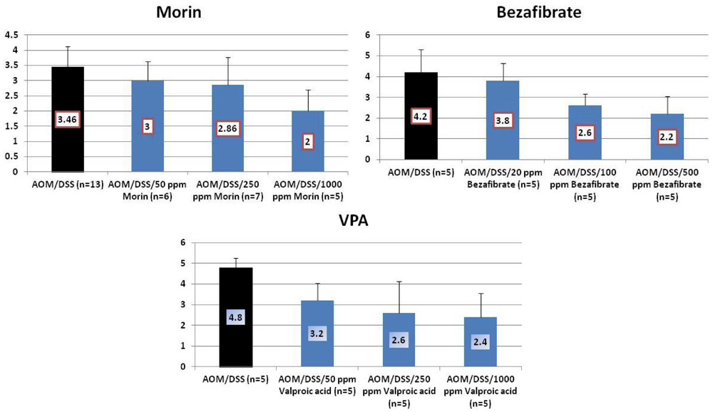
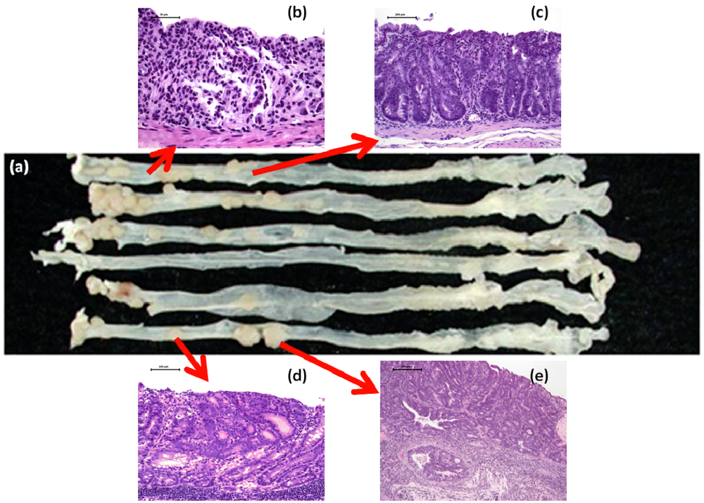
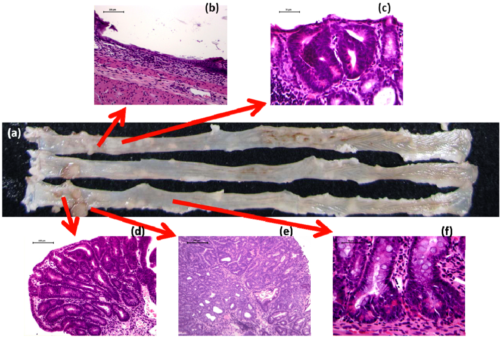
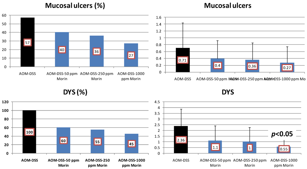
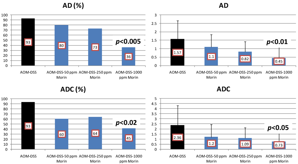
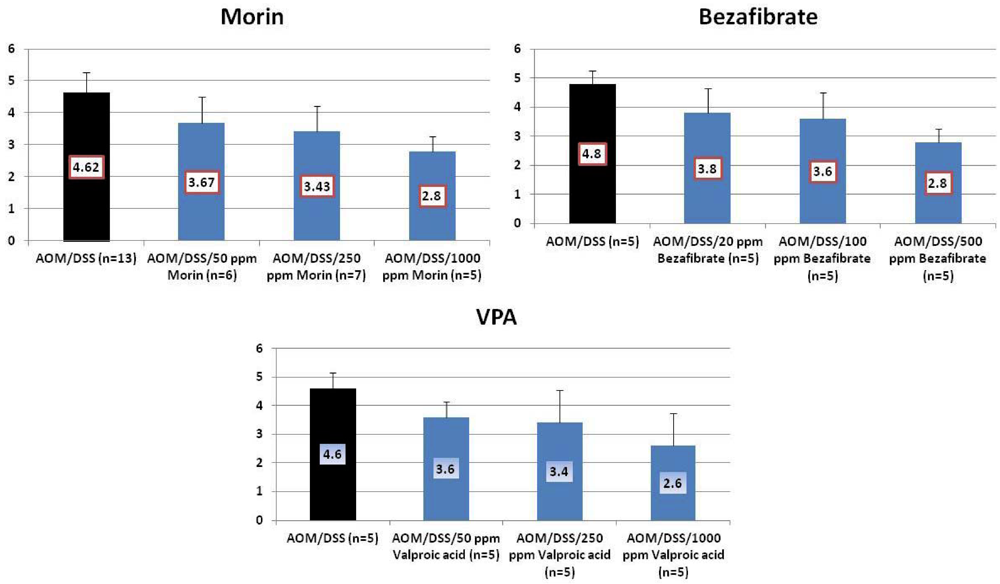
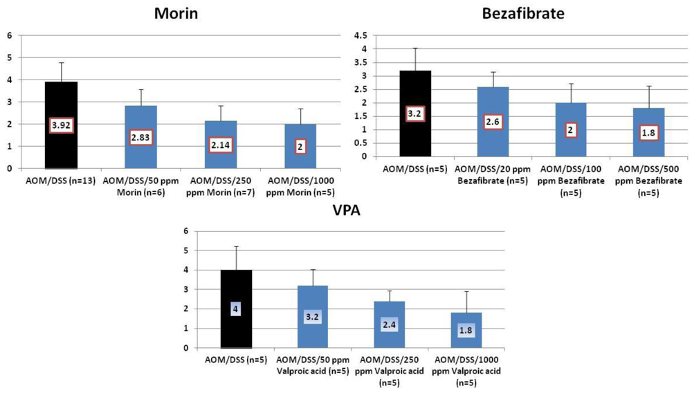
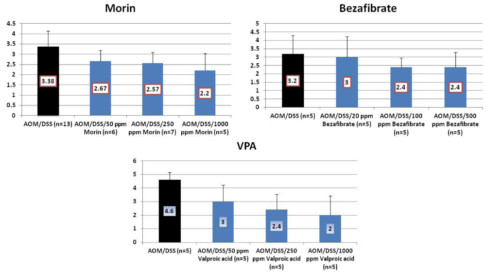
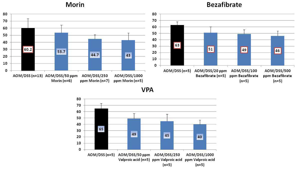
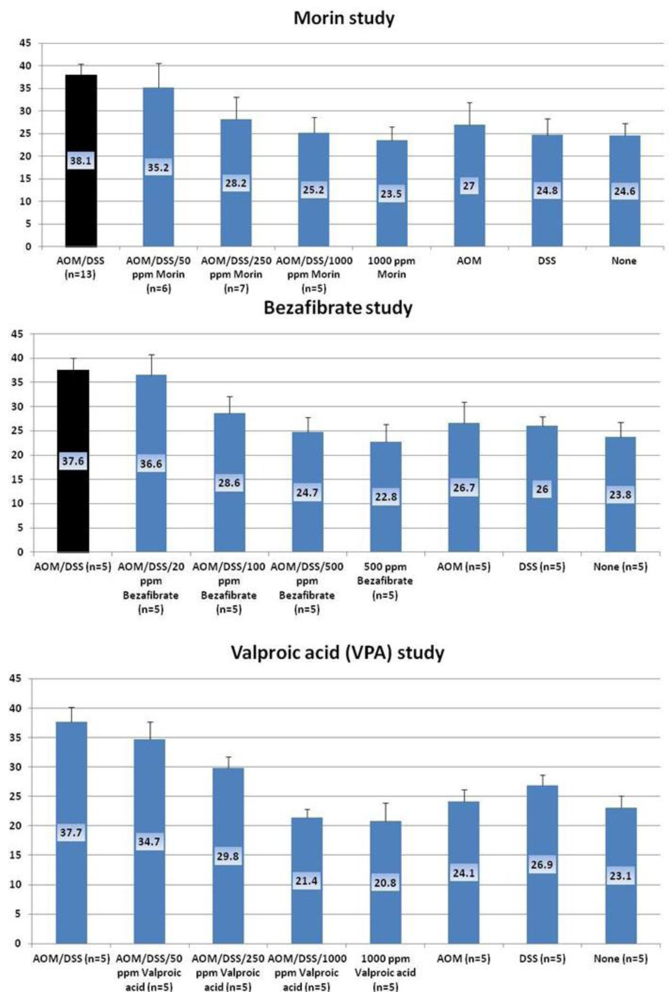
Using our animal models of inflammation-associated colorectal carcinogenesis, cancer chemopreventive abilities of candidate compounds, morin, bezafibrate, and VPA in mice or rats were investigated. All animal experiments were performed in accordance with protocols approved by the Animal Care and Use Committee of the Institute, TCI-CaRP.

4.1. Morin Study

A flavonol, morin (3,5,7,2',4'-pentahydroxyflavone) found in almonds, mill, fig, mulberry, and other Moraceae, acts as a potent antioxidant, inhibitor of xanthine oxidase, protein kinase C and proliferation, apoptosis inducer and modulator of lipoxygenase and cyclooxygenase activities. This flavone has been reported to inhibit the growth of COLO205 cells in nude mice [66], exhibit intestinal anti-inflammatory activity in the acute phase of rat colitis induced by trinitrobenzenesulfonic acid [67,68]. We have previously reported that morin inhibits AOM-induced putative precursor lesions, ACF, in rats [69] and inhibit chemically-induced rat tongue carcinogenesis [70]. Morin exerts anti-inflammatory effects on septic shock induced by lipopolysaccharide [71]. This study aimed to determine possible inhibitory potential of morin in colitis-associated colon carcinogenesis initiated with AOM and promoted by DSS in male F344 rats.
Materials and methods: A total of 66 male rats (5-week-old) were initiated with a single s.c. injection of AOM (20 mg/kg bw), and then they were given promotion stimuli by the treatment with 1.5% DSS in their drinking water for seven days. They were then given a basal diet containing 50, 250 and 1,000 ppm of morin for 17 weeks. Experimental groups included the AOM/1.5% DSS (n = 14), AOM/1.5% DSS/50 ppm morin (n = 10), AOM/1.5% DSS/250 ppm morin (n = 11), AOM/1.5% DSS/1,000 ppm morin (n = 11), AOM alone (n = 5), 1.5% DSS alone (n = 5), 500 ppm morin alone (n = 5), and untreated (n = 5) groups. At the end (week 20) of the study histopathological analysis of colorectum was performed on hematoxylin and eosin (H&E)-stained histologic sections (3 μm thickness). Proliferation activity of colonic ADCs was determined by immunofluorescence technique using anti-Mcm2 antibody (BD Biosciences PharMingen, Tokyo, Japan). Apoptotic cells were detected by fluorescein in situ tunnel method, TACS TdT kit (R&D Systems, Inc., Minneapolis, MN, USA). Polyamine levels [72] and mRNA expression of nuclear factor-kappaB (NF-κB), tumor necrosis factor (TNF)-α, interleukin (IL)-1β, Stat3, and hypoxia-inducible factor (HIF)-1α [73] in colonic mucosa were determined in mice randomly selected from each group. All measurements were statistically analyzed using either the Tukey multiple comparison post test or Fisher’s extract probability test. Differences were considered to be statistically significant at p < 0.05.
Results: At week 20, the treatment with morin inhibited colonic mucosal ulcer (Figure 3) and dysplastic crypts (p < 0.05 at 1,000 ppm, Figure 3). The incidence (p < 0.005) and multiplicity (p < 0.01) of colonic AD were significantly reduced by feeding with 1000 ppm morin (Figure 4). Also, dietary administration with 1,000 ppm morin significantly inhibited the incidence (p < 0.02) and multiplicity (p < 0.05) of colonic ADC (Figure 4), when compared to the AOM/DSS group (93% incidence and 2.36 ± 1.95 multiplicity). Feeding with 50 ppm and 250 ppm morin lowered the incidence and multiplicity of ADC, but the inhibition rates did not reach statistically significance. The treatments also modulated proliferation and apoptosis in ADCs. Mcm2 positive rates (%) of ADCs in rats fed the diets containing 50 ppm morin (n = 6, 78.7 ± 6.8), 250 ppm morin (n = 7, 63.4 ± 6.3, p < 0.001), and 1,000 ppm morin (n = 5, 55.6 ± 12.5, p < 0.001) were lower than that of rats given AOM and DSS (n = 13, 83.4 ± 8.8). When compared with the AOM and DSS group (n = 13, 8.23 ± 1.24), apoptotic index (%) of ADCs was increased by feeding with morin: 50 ppm morin (n = 6, 10.83 ± 3.66), 250 ppm morin (n = 7, 11.29 ± 3.95), and 1,000 ppm morin (n = 5, 13.00 ± 2.74, p < 0.05).
Growth inhibition and apoptosis induction by morin in CRC might be caused by activation of caspase 3 and increase of p21 protein [66]. Suppression of NF-κB-regulated gene products and enhancement of apoptosis induced by TNF [74] also contribute to inhibition of colitis-related colorectal carcinogenesis by morin. Our findings suggest that dietary morin is able to inhibit colitis-related colon carcinogenesis in rats and a flavonol morin is one of the candidates for clinical application of chemoprevention against CRC development in patients with ulcerative colitis.
Because morin can inhibit inflammation, inhibit tumor promotion, suppress tumor growth, and down-regulate the expression of certain genes regulated by NF-κB, it may be possible that morin modulates the activation of NF-κB and NF-κB-regulated gene expression induced by carcinogens, inflammatory agents, and immune modulators. In fact, Manna et al. [74] have recently reported morin suppresses the activation of NF-κB and NF-κB-regulated gene expression that leads to enhancement of apoptosis.
Figure 3. Incidences and multiplicities of mucosal ulcer and dysplastic crypts in the morin study.
Cancers 04 00673 g003
Figure 4. Incidences and multiplicities of adenoma (AD) and adenocarcinoma (ADC) in the morin study.
Cancers 04 00673 g004

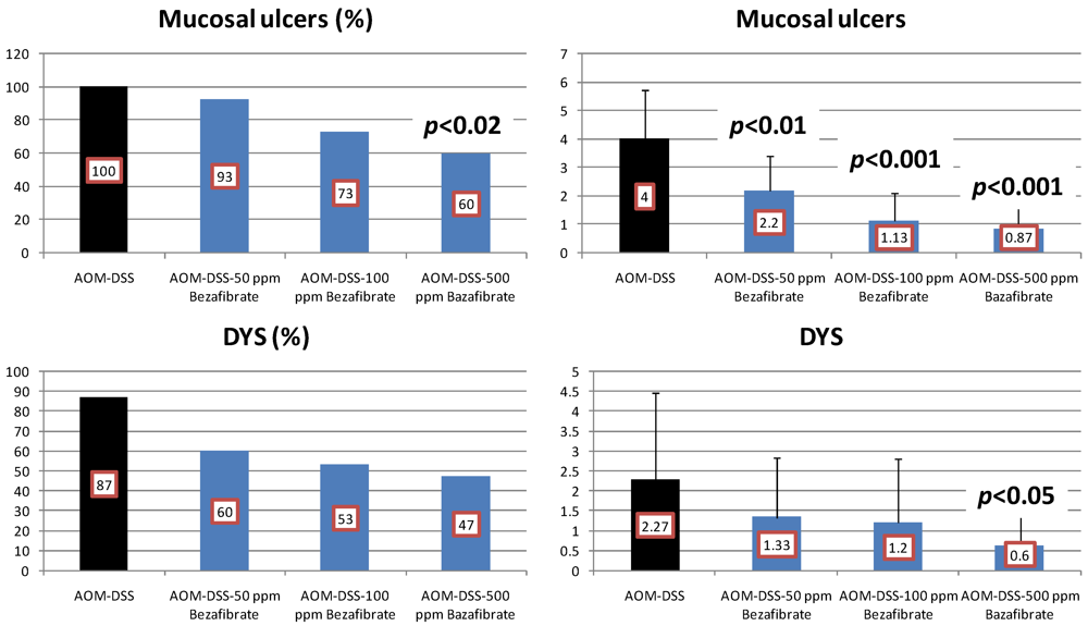
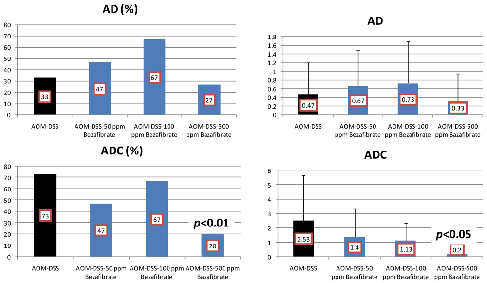
4.2. Bezafibrate Study

CRC is one of the leading forms of malignancy in the developed countries. Epidemiologic and animal studies have suggested that risk factors for coronary artery disease like insulin resistance and dyslipidemia are probably related to the development of colon cancer [75,76,77]. In particular, nuclear peroxisome proliferator-activated receptors (PPARs), mainly PPARα and PPARγ, which play a central role in lipid and glucose metabolism, had been hypothesized as being involved in colon carcinogenesis [50,78,79]. Furthermore, synthetic PPAR ligands (glitazones and bezafibrate) with proven beneficial effects on insulin resistance and triglyceride levels had been proposed to be candidates as tumor preventive agents [50,63,79]. This study aimed to determine inhibitory potential of bezafibrate in colitis-associated colon carcinogenesis initiated with AOM and promoted by DSS in mice.
Materials and methods: A total of 100 male ICR mice (5-week old) initiated with a single s.c. injection of AOM (10 mg/kg bw) were promoted by 1.5% DSS in their drinking water for seven days. They were then given a basal diet containing 50, 100 and 500 ppm of bezafibrate for 17 weeks. Mice were divided into 8 groups: AOM/1.5% DSS (n = 20), AOM/1.5% DSS/50 ppm bezafibrate (n = 20), AOM/1.5% DSS/100 ppm bezafibrate (n = 20), AOM/1.5% DSS/500 ppm bezafibrate (n = 20), AOM alone (n = 5), 1.5% DSS alone (n = 5), 500 ppm bezafibrate alone (n = 5), and untreated (n = 5) groups. At the end (week 20) of the study histopathological analysis of colorectum was performed on H&E-stained histological sections of 3 μm in thickness. Immunofluorescence technique using anti-Mcm2 antibody (BD Biosciences PharMingen) and fluorescein in situ tunnel method, TACS TdT kit (R&D Systems, Inc.) were used for determination of proliferation activity and apoptosis index of colonic ADCs, respectively. Polyamine levels [72] and mRNA expression of NF-κB, TNF-α, IL-1β, Stat3, and HIF-1α [73] in colonic mucosa were assayed in some mice of each group. Measurements were statistically analyzed using either the Tukey multiple comparison post-test or Fisher’s extract probability test. Differences were considered to be statistically significant at p < 0.05.
Results: At week 20, the bezafibrate feeding inhibited the occurrence of mucosal ulcer (the incidence at 500 ppm, p < 0.02; and the multiplicity at 50, 100 and 500 ppm, p < 0.01 or p < 0.001, Figure 5) and dysplastic crypts (the multiplicity at 500 ppm, p < 0.05, Figure 5). As illustrated in Figure 6, he development of colonic ADC was significantly inhibited by feeding with 500 ppm bezafibrate (incidence: 73% reduction, p < 0.01; and multiplicity: 92%, p < 0.05), when compared to the AOM/DSS group (73% incidence and 2.53 ± 3.14 multiplicity). Feeding with 50 ppm (47% incidence with a multiplicity of 1.40 ± 1.92) and 100 ppm bezafibrate (67% incidence with a multiplicity of 1.13 ± 1.19) also lowered the incidence and multiplicity of ADC, but the inhibition was not statistically significant when compared to the AOM/DSS group (Figure 6). Although statistically insignificant, feeding with 50 and 100 ppm bezafibrate increased the incidence and multiplicity of ADs, but 500 ppm bezafibrate decreased the development of ADs (Figure 6). These findings may suggest the threshold of chemopreventive ability of bezafibrate. Feeding with bezafibrate lowered Mcm2 positive rates (%) of ADCs, when compared with the AOM and DSS group (n = 10, 72.9 ± 10.1): 50 ppm bezafibrate (n = 7, 54.3 ± 9.5), 100 ppm bezafibrate (n = 4, 38.8 ± 7.4, p < 0.01), and 500 ppm bezafibrate (n = 3, 22.7 ± 3.2, p < 0.001) were lower than that of rats given AOM and DSS (n = 13, 83.4 ± 8.8). When compared with the AOM and DSS group (n = 10, 8.18 ± 1.17), apoptotic index (%) of ADCs was increased by feeding with bezafibrate: 50 ppm bezafibrate (n = 7, 10.71 ± 3.35), 100 ppm bezafibrate (n = 4, 13.20 ± 2.49, p < 0.05), and 500 ppm bezafibrate (n = 3, 13.25 ± 3.10, p < 0.05).
Anti-angiogenic effects and anti-inflammatory activity of PPARα agonist [80] are considered to contribute to inhibition of CRC growth. Down-regulation of the anti-apoptotic gene Mcl-1 by PPARα agonist [81] causes apoptosis. The findings suggest that dietary bezafibrate is able to inhibit colitis-related colon carcinogenesis in mice and a hypolipidemic drug bezafibrate is one of the candidates for clinical application of chemoprevention against CRC development in patients with ulcerative colitis.
Figure 5. Incidences and multiplicities of mucosal ulcer and dysplastic crypts in the bezafibrate study.
Cancers 04 00673 g005
Figure 6. Incidences and multiplicities of adenoma (AD) and adenocarcinoma (ADC) in the bezafibrate study.
Cancers 04 00673 g006
Accumulating animal experimental, human laboratory and epidemiologic data [50,63,79,82,83] support the hypothesis linking triglyceride levels and insulin resistance to the development of colon cancer. These facts emphasize the potential for this cancer to become a preventable disease not only via screening and removal of polyps but through relevant lifestyle changes and pharmacological interventions which can provide even more avenues for prevention [83,84,85].
The incidence of insulin resistance has been increasing in the Western world where colon cancer is the second leading cause of cancer death. This suggests the interrelationship of these conditions. The biological role of PPARs in various diseases, including inflammation and cancer, has been highlighted recently [50,63,78,85,86,87]. PPARs are members of the nuclear hormone receptor family of ligand-activated transcription factors that play a prominent role in the regulation of many metabolic processes. The PPAR isoformes α and γ are important regulators in lipid and glucose metabolism, cell differentiation and inflammatory response [87,88]. These data propose that PPAR may be associated with many aspects of colon cancer development including insulin- and inflammation-related mechanisms. The fibric acid derivative bezafibrate is the pan PPAR (α, β/δ, and γ) activator with predominantly PPARα (as all fibrates) and β/δ effects but also with perceptible PPARγ properties [88,89,90]. The use of bezafibrate is associated with triglyceride-lowering and high density-cholesterol raising effects resulting in decreased systemic availability of fatty acid, diminished of fatty acid uptake by muscle and improvement of insulin sensitization [91,92]. These direct and indirect effects may have contributed to the suppression of the development of colonic tumors in rodents by bezafibrate [50,63,79]. Recently, Tenenbaum et al. [93] have reported possible preventive effects of bezafibrate on the development of CRC from patients with coronary artery disease.

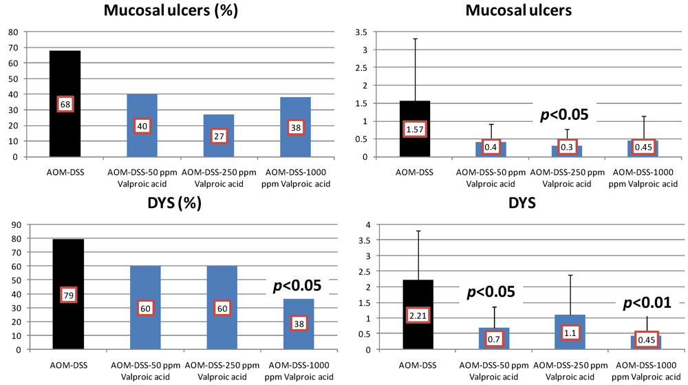
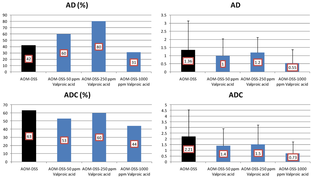
4.3. Valproic Acid (VPA) Study

Epigenetic modification plays an important role in tumorigenesis. Affecting epigenetic and tumorigenic alterations is a promising strategy for anticancer targeted therapy [94,95,96]. Among the key chromatin modifying enzymes which influence gene expression, histone acetyltransferases (HATs) and histone deacetylases (HDACs) have attracted interest because of their impact on tumor development and progression. Histone deacetylase inhibitors (HDACIs) represent a new and promising class of antitumor drugs that influence gene expression by enhancing acetylation of histones in specific chromatin domains. HDACIs also exert potent anticancer activities inducing cell cycle arrest and apoptosis. Moreover, HDACIs down-regulate genes involved in tumor progression, invasion and angiogenesis. Based on the ability of HDACIs to regulate many signaling pathways, co-treatment of these compounds that are currently under clinical investigation with molecular targeted drugs is a promising strategy against many types of tumors.
VPA (2-propylpentanoic acid) [97] is a well-established drug for the therapy of epilepsy. It is teratogenic when administered during early pregnancy and can induce birth defects such as neural tube closure defects and other malformations. This well-tolerated antiepileptic drug was found to be a powerful HDAC-1 inhibitor [97]. VPA induces differentiation of carcinoma cells, transformed hematopoietic progenitor cells and leukemic blasts from acute myeloid leukemia patients [98]. Our microarray analysis during the AOM-DSS carcinogenesis revealed alteration of Wif-1 expression [34]. VPA has been reported to modify the Wif-1 expression [99]. These findings may suggest possible modifying effects of VPA on AOM-DSS colorectal carcinogenesis. In this study, we determined whether VPA is able to inhibit colitis-associated colon carcinogenesis in mice.
Materials and methods: A total of 85 mice aged five weeks was used and they were divided into 8 groups: AOM/2% DSS (n = 19), AOM/2% DSS/50 ppm VPA (n = 15), AOM/2% DSS/250 ppm VPA (n = 15), AOM/2% DSS/1,000 ppm VPA (n = 16), AOM alone (n = 5), 2% DSS alone (n = 5), 1,000 ppm VPA alone (n = 5), and untreated (n = 5) groups. Mice were initiated with a single s.c. injection of AOM (10 mg/kg bw) were promoted by 2% DSS in their drinking water for seven days. They were then given a basal diet containing 50, 250 or 1,000 ppm of VPA for 17 weeks. At the end (week 20) of the study histopathological examination of large bowel was performed on H&E-stained histological sections (3 μm in thickness). Immunofluorescence technique using anti-Mcm2 antibody (BD Biosciences PharMingen) for evaluating proliferating activity and fluorescein in situ tunnel method, TACS TdT kit (R&D Systems, Inc.) for detecting apoptosis cells were applied on histological sections of colonic ADCs. Polyamine levels [72] and mRNA expression of NF-κB, TNF-α, IL-1β, Stat3, and HIF-1α [73] in colonic mucosa were assayed in some mice of each group.At the end of the study (week 20), Measurements were statistically analyzed using either the Tukey multiple comparison post test or Fisher’s extract probability test. Differences were considered to be statistically significant at p < 0.05.
Results: VPA feeding inhibited the development of mucosal ulcer (Figure 7) and dysplastic crypts (the incidence at 1,000 ppm VPA, p < 0.05; and the multiplicity at 50 ppm, p < 0.05 and at 1,000 ppm, p < 0.01, Figure 7).
Figure 7. Incidences and multiplicities of mucosal ulcer and dysplastic crypts in the Valproic Acid (VPA) Study.
Cancers 04 00673 g007
The development of colonic AD and ADC was lowered by feeding with VPA, but the reduction rates were statistically insignificant (Figure 8). When fed with VPA-containing diet, Mcm2 positive rates (%) of ADCs were lower than that of the AOM and DSS group (n = 9, 80.1 ± 6.8): 125 ppm VPA (n = 7, 73.7 ± 8.0), 250 ppm VPA (n = 6, 64.2 ± 2.5, p < 0.01), and 1,000 ppm VPA (n = 5, 61.6 ± 9.6, p < 0.001). As to apoptotic index (%) of ADCs, the values of mice fed with 125 ppm VPA (n = 7, 10.67 ± 3.44), 250 ppm VPA (n = 6, 12.33 ± 1.75, p < 0.05), and 1,000 ppm VPA (n = 5, 12.80 ± 2.39, p < 0.05) were higher thab the AOM and DSS group (n = 9, 8.44 ± 1.33).
Figure 8. Incidences and multiplicities of adenoma (AD) and adenocarcinoma (ADC) in the Valproic Acid (VPA) Study.
Cancers 04 00673 g008
Our findings suggest slight chemopreventive effects of VPA on colitis-related colon carcinogenesis in mice, suggesting that single use of a HDAC inhibitor VPA is not practical for inhibiting CRC development in inflamed colon. VPA combined with other known chemopreventive or chemotherapeutic agent(s) may exert to inhibit CRC that develop in colitic mucosa.
VPA was studied in combination with all-trans retinoid acid in patients with acute myeloid leukemia who were not candidates for intensive chemotherapy [100]. Using human hepatocellular carcinoma (HCC) cells, HepG2, combination treatment with acyclic retinoid and VPA is demonstrated to be an effective regimen for the chemoprevention and chemotherapy of HCC [101]. Acyclic retinoid and VPA cooperatively increase the expression of retinoid X receptor (RXR)-β and p21 (CIP1), while inhibiting the phosphorylation of RXRα, and these effects were associated with induction of apoptosis and the inhibition of cell growth in HepG2 cells. Several HDACIs seem to exert an antitumor effect in a synergistic manner with different anticancer compounds and to overcome the resistance induced by conventional chemotherapeutic drugs [102,103,104,105]. A phase I trial of single agent VPA was reported in patients with newly diagnosed cervical cancer [106]. Twelve patients were included. VPA doses ranged from 20 mg/kg to 40 mg/kg daily for five days. Tumor HDAC activity decreased in 8 patients. Many lines of evidence suggest that tumor cells are characterized by histone hypoacetylation and that over-expression of HDACs is involved in tumorigenesis of various human malignancies [107,108]. Recently, a population-based case-control study with long-term users of VPA has not supported HDAC inhibition by VPA as a pharmacologic principle for general chemoprevention [109]. However, VPA doses (0.35–0.70 mmol/L) used in clinical practice could be too low to achieve cancer preventive effects in contrast the doses (0.50–3.0 mmol/L) that inhibit HDAC. Since VPA is reported to suppress progression of urological malignancies [110,111], it is worthy to evaluate the modifying effects of VPA on different stage of carcinogenesis.

5. Effects of Morin, Bezafibrate and VPA on Expression of Pro-Inflammatory Cytokines and HIF-1α and Content of Tissue Polyamines in the Inflamed Colon

IBD represents a dysregulated mucosal immune response to antigens derived from the commensal microbiota in a genetically susceptible host that initially derives from innate immune abnormalities leading to an excessive pro-inflammatory cytokines (T-helper 1, T-helper 2, and T-helper 17 cytokines) derived from CD4+ T cells [9,112]. A key point in understanding IBD pathophysiology is to understand the immunoregulatory pathways associated with the intestinal immune system as they apply to IBD. Therefore, in addition to immunotherapy, pro-inflammatory cytokines secreted by innate and adaptive immune cells are targets for IBD treatment [73]. Similarly, cytokines, including NF-κB [73,113], TNF-α [9,35,64], and interleukin (IL)-1β [64,114,115] are potentially molecular targets for inflammation-associated CRC [14]. Potential cancer chemopreventive agents also modulate expression of signal transducer and activator of transcription (Stat3) [55,114], HIF-1α [116,117], and survivin [73] in the target tissues.
Polyamines are organic cations that control gene expression at the transcriptional, posttranscriptional, and translational levels. Multiple cellular carcinogenesis pathways are involved in regulation of transcription and translation of polyamine-metabolizing enzymes [118]. We have reported he importance of research utilizing pharmaceutical inhibitors and cancer chemoprevention against CRC targeting the polyamine pathway [72,119,120].
The findings in in vivo studies using the TANAKA models suggest that the order of chemopreventive potential of test compounds was bezafibrate > morin > VPA by estimating inhibition rate of CRC development. To investigate the effects of these chemicals on the molecules that are involved in carcinogenesis, we determined mRNA expression of the NF-κB (Figure 9), TNF-α, (Figure 10), IL-1β (Figure 11). Stat 3 (Figure 12), HIF-1α (Figure 13) in colorectal mucosa of mice or rats from the experiments with morin, bezafibrate, and VPA. At sacrifice, each colon was cut open longitudinally, and was flushed clean with PBS. Five animals of each group from three experiments were used for real-time quantitative RT-PCR analysis. Their distal colon was taken for total RNA isolation. For total RNA isolation, epithelial cells were scraped from the underlying muscle layer with a glass microscope slide, were homogenized on ice in lysis buffer (Qiagen, Tokyo, Japan), and were frozen at −80°C until RNA was isolated. Total RNA was extracted from colonic mucosa using the RNeasy Mini Kit (Qiagen) according to the manufacturer’s protocol. The cDNA was then synthesized from total RNA using the High-Capacity cDNA Reverse Transcription Kit (Applied Biosystems Japan Ltd., Tokyo, Japan). Quantitative real time PCR analysis of individual cDNA was performed with ABI Prism 7500 (Applied Biosystems Japan Ltd., Tokyo, Japan) using TaqMan Gene Expression Assays (Applied Biosystems Japan Ltd., Tokyo, Japan) and primers, which were chosen on the basis of rat or mouse nucleotide sequences in the GenBank database (Table 1). PCR cycling conditions were 50 °C for 2 min, 95 °C for 10 min, followed by 40 cycles of 95 °C for 15 s and 60 °C for 1 min. The relative mRNA expression was normalized by b-actin mRNA. All the test chemicals lowered mRNA expression of these proteins. Therefore, effects of these three test compounds on the development of mucosal ulcer, dysplastic crypts, and colonic neoplasms may be related to the expression that was modified by feeding with test chemicals.
Figure 9. mRNA expression of NF-κB in the colorectum of mice or rats from the morin, bezafibrate, and VPA studies.
Cancers 04 00673 g009
Figure 10. mRNA expression of TNF-α in the colorectum of mice or rats from the morin, bezafibrate, and VPA studies.
Cancers 04 00673 g010
Figure 11. mRNA expression of IL-1β in the colorectum of mice or rats from the morin, bezafibrate, and VPA studies.
Cancers 04 00673 g011
Figure 12. mRNA expression of Stat3 in the colorectum of mice or rats from the morin, bezafibrate, and VPA studies.
Cancers 04 00673 g012
Figure 13. mRNA expression of HIF-1α in the colorectum of mice or rats from the morin, bezafibrate, and VPA studies.
Cancers 04 00673 g013
Table 1. Primer sequences used for real-time PCR assays.
Gene symbolPrimers
RatMouse
NF-κBForward: 5'-ctggcagctcttctcaaagc-3'Mm00476361_m1 *
Reverse: 5'-ccaggtcatagagaggctcaa-3'
Tnf-αForward: 5'-cgagatgtggaactggcaga-3'Mm00443258_m1 *
Reverse: 5'-ctacgggcttgtcactcga-3'
IL-1βRn00 580432_m1 *Mm00434228_m1 *
Stat3Forward: 5'-ttgtgatgcctccttgattgtc-3'Mm00456961_m1 *
Reverse: 5'-atcggaggcttagtgaagaagttc-3'
Hif-1αRn00577560_ml *Forward: 5'-cctggaaacgagtgaaagga-3'Reverse: 5'-tggtcagctgtggtaatcca-3'
Cancers 04 00673 i001actinForward: 5'-tcaggtcatcactatcggcaat-3'Mm00607939_s1*
Reverse: 5'-aaagaaagggtgtaaaacgca-3'
* TaqMan (Assay ID#).
Treatments with morin, bezafibrate, and VPA also lowered immunohistochemical positivity of survivin (Figure 14) in ADCs and polyamine contents (Figure 15) of colorectal mucosa. These effects may be also responsible for modulatory effects of these three compounds on colonic tumor development. However, further studies including dose selection and toxicity of the compounds should be conducted before going to clinical trials.
Figure 14. Immunohistochemical positivity (%) against survivin in colonic ADC cells that developed in mice or rats in the morin, bezafibrate, and VPA studies.
Cancers 04 00673 g014
Figure 15. Polyamine content (nmol/mg) in the colonic mucosa of mice or rats in the morin, bezafibrate, and VPA studies.
Cancers 04 00673 g015

6. Conclusions

Chemoprevention is an important approach to decreasing cancer morbidity and mortality by the use of non-toxic natural or synthetic substances to reverse the processes of initiation, promotion, and subsequent progression of cancer. Evidence is rapidly accumulating that chronic inflammation contributes to carcinogenesis through increase of cell proliferation, angiogenesis, and metastasis in a number of neoplasms, including colorectal carcinoma. To investigate pathobiology of CRC developed in inflamed colorectum and search effective cancer chemopreventive agents against such colitis-related CRC, we developed mouse and rat models (TANAKA models) of inflammation-associated colorectal carcinogenesis. In this article, powerful tumor-promotion effect of inflammation induced by DSS in rodents that are initiated with a low dose of a colonic carcinogen is described. Also, our recent data on the modifying effects of morin, bezafibrate, and VPA on AOM/DSS-induced colorectal carcinogenesis is presented. I would stress that inflammatory stress and several cytokines produced by inflammatory cells are important in pathobiology of CRC development in colitic mucosa. These could be molecular targets for chemoprevention and/or therapy in CRC in IBD patients.

Acknowledgements

This work was partly supported by a Grant-in-Aid for the 2nd and 3rd Terms Comprehensive 10-year Strategy for Cancer Control, Cancer Prevention, from the Ministry of Health and Welfare of Japan, a Grant-in-Aid for Cancer Research from the Ministry of Health and Welfare of Japan, and a Grant-in-Aid (No. 13671986 and No. 23501324) from the Ministry of Education, Science, Sports and Culture of Japan.

Conflict of interest

None declared.

References

  1. Balkwill, F.; Mantovani, A. Inflammation and cancer: Back to Virchow? Lancet 2001, 357, 539–545. [Google Scholar] [CrossRef]
  2. Marshall, B.J.; Warren, J.R. Unidentified curved bacilli in the stomach of patients with gastritis and peptic ulceration. Lancet 1984, 1, 1311–1315. [Google Scholar]
  3. Tanaka, T.; Suzuki, R. Inflammation and cancer. In Cancer: Disease Progression and Chemoprevention 2007; Tanaka, T., Ed.; Research Signpost: Kerala, India, 2007; pp. 27–44. [Google Scholar]
  4. Lakatos, P.L.; Lakatos, L. Risk for colorectal cancer in ulcerative colitis: Changes, causes and management strategies. World J. Gastroenterol. 2008, 14, 3937–3947. [Google Scholar] [CrossRef] [Green Version]
  5. Itzkowitz, S.H.; Yio, X. Inflammation and cancer IV. Colorectal cancer in inflammatory bowel disease: The role of inflammation. Am. J. Physiol. Gastrointest. Liver Physiol. 2004, 287, G7–G17. [Google Scholar] [CrossRef]
  6. Ullman, T.A.; Itzkowitz, S.H. Intestinal inflammation and cancer. Gastroenterology 2011, 140, 1807–1816. [Google Scholar] [CrossRef]
  7. Munkholm, P. Review article: The incidence and prevalence of colorectal cancer in inflammatory bowel disease. Aliment. Pharmacol. Ther. 2003, 18, S1–S5. [Google Scholar] [CrossRef]
  8. Eaden, J.A.; Abrams, K.R.; Mayberry, J.F. The risk of colorectal cancer in ulcerative colitis: A meta-analysis. Gut 2001, 48, 526–535. [Google Scholar] [CrossRef]
  9. Tanaka, T.; Kohno, H.; Murakami, M.; Shimada, R.; Kagami, S. Colitis-related rat colon carcinogenesis induced by 1-hydroxy-anthraquinone and methylazoxymethanol acetate (Review). Oncol. Rep. 2000, 7, 501–508. [Google Scholar]
  10. Canavan, C.; Abrams, K.R.; Mayberry, J. Meta-analysis: Colorectal and small bowel cancer risk in patients with Crohn’s disease. Aliment. Pharmacol. Ther. 2006, 23, 1097–1104. [Google Scholar] [CrossRef]
  11. Sung, J.J.; Lau, J.Y.; Goh, K.L.; Leung, W.K. Increasing incidence of colorectal cancer in Asia: Implications for screening. Lancet Oncol. 2005, 6, 871–876. [Google Scholar] [CrossRef]
  12. Tanaka, T.; Oyama, T.; Yasui, Y. Dietary supplements and colorectal cancer. Curr. Topics Neutraceut. Res. 2008, 6, 165–188. [Google Scholar]
  13. Tanaka, T.; Sugie, S. Inhibition of colon carcinogenesis by dietary non-nutritive compounds. J. Toxicol. Pathol. 2007, 20, 215–235. [Google Scholar] [CrossRef]
  14. Yasui, Y.; Kim, M.; Oyama, T.; Tanaka, T. Colorectal carcinogenesis and suppression of tumor development by inhibition of enzymes and molecular targets. Curr. Enzym. Inhib. 2009, 5, 1–26. [Google Scholar] [CrossRef]
  15. Rosenberg, D.W.; Giardina, C.; Tanaka, T. Mouse models for the study of colon carcinogenesis. Carcinogenesis 2009, 30, 183–196. [Google Scholar]
  16. Tanaka, T. Colorectal carcinogenesis: Review of human and experimental animal studies. J. Carcinog. 2009, 8, 5. [Google Scholar] [CrossRef]
  17. Tanaka, T. Development of an inflammation-associated colorectal cancer model and its application for research on carcinogenesis and chemoprevention. Int. J. Inflamm. 2012, 2012, 658786. [Google Scholar]
  18. Tanaka, T.; Kohno, H.; Suzuki, R.; Yamada, Y.; Sugie, S.; Mori, H. A novel inflammation-related mouse colon carcinogenesis model induced by azoxymethane and dextran sodium sulfate. Cancer Sci. 2003, 94, 965–973. [Google Scholar] [CrossRef]
  19. Tanaka, T.; Yasui, Y.; Ishigamori-Suzuki, R.; Oyama, T. Citrus compounds inhibit inflammation- and obesity-related colon carcinogenesis in mice. Nutr. Cancer 2008, 60, S70–S80. [Google Scholar] [CrossRef]
  20. Levine, J.S.; Burakoff, R. Chemoprophylaxis of colorectal cancer in inflammatory bowel disease: Current concepts. Inflamm. Bowel Dis. 2007, 13, 1293–1298. [Google Scholar] [CrossRef]
  21. Zisman, T.L.; Rubin, D.T. Colorectal cancer and dysplasia in inflammatory bowel disease. World J. Gastroenterol. 2008, 14, 2662–2669. [Google Scholar] [CrossRef]
  22. Das, D.; Arber, N.; Jankowski, J.A. Chemoprevention of colorectal cancer. Digestion 2007, 76, 51–67. [Google Scholar] [CrossRef]
  23. Pardi, D.S.; Loftus, E.V., Jr.; Kremers, W.K.; Keach, J.; Lindor, K.D. Ursodeoxycholic acid as a chemopreventive agent in patients with ulcerative colitis and primary sclerosing cholangitis. Gastroenterology 2003, 124, 889–893. [Google Scholar]
  24. Tung, B.Y.; Emond, M.J.; Haggitt, R.C.; Bronner, M.P.; Kimmey, M.B.; Kowdley, K.V.; Brentnall, T.A. Ursodiol use is associated with lower prevalence of colonic neoplasia in patients with ulcerative colitis and primary sclerosing cholangitis. Ann. Intern. Med. 2001, 134, 89–95. [Google Scholar]
  25. Andrews, J.M.; Travis, S.P.; Gibson, P.R.; Gasche, C. Systematic review: Does concurrent therapy with 5-ASA and immunomodulators in inflammatory bowel disease improve outcomes? Aliment. Pharmacol. Ther. 2009, 29, 459–469. [Google Scholar] [CrossRef]
  26. Velayos, F.S.; Terdiman, J.P.; Walsh, J.M. Effect of 5-aminosalicylate use on colorectal cancer and dysplasia risk: A systematic review and metaanalysis of observational studies. Am. J. Gastroenterol. 2005, 100, 1345–1353. [Google Scholar] [CrossRef]
  27. Bernstein, C.N.; Blanchard, J.F.; Metge, C.; Yogendran, M. Does the use of 5-aminosalicylates in inflammatory bowel disease prevent the development of colorectal cancer? Am. J. Gastroenterol. 2003, 98, 2784–2788. [Google Scholar]
  28. Bernstein, C.N.; Eaden, J.; Steinhart, A.H.; Munkholm, P.; Gordon, P.H. Cancer prevention in inflammatory bowel disease and the chemoprophylactic potential of 5-aminosalicylic acid. Inflamm. Bowel Dis. 2002, 8, 356–361. [Google Scholar] [CrossRef]
  29. Kohno, H.; Suzuki, R.; Yasui, Y.; Miyamoto, S.; Wakabayashi, K.; Tanaka, T. Ursodeoxycholic acid versus sulfasalazine in colitis-related colon carcinogenesis in mice. Clin. Cancer Res. 2007, 13, 2519–2525. [Google Scholar]
  30. Tanaka, T.; Shnimizu, M.; Moriwaki, H. Cancer chemoprevention by carotenoids. Molecules 2012, 17, 3202–3242. [Google Scholar]
  31. Takahashi, M.; Wakabayashi, K. Gene mutations and altered gene expression in azoxymethane-induced colon carcinogenesis in rodents. Cancer Sci. 2004, 95, 475–480. [Google Scholar] [CrossRef]
  32. Boivin, G.P.; Washington, K.; Yang, K.; Ward, J.M.; Pretlow, T.P.; Russell, R.; Besselsen, D.G.; Godfrey, V.L.; Doetschman, T.; Dove, W.F.; et al. Pathology of mouse models of intestinal cancer: Consensus report and recommendations. Gastroenterology 2003, 124, 762–777. [Google Scholar]
  33. Clapper, M.L.; Cooper, H.S.; Chang, W.C. Dextran sulfate sodium-induced colitis-associated neoplasia: A promising model for the development of chemopreventive interventions. Acta Pharmacol. Sin. 2007, 28, 1450–1459. [Google Scholar] [CrossRef]
  34. Suzuki, R.; Miyamoto, S.; Yasui, Y.; Sugie, S.; Tanaka, T. Global gene expression analysis of the mouse colonic mucosa treated with azoxymethane and dextran sodium sulfate. BMC Cancer 2007, 7, 84. [Google Scholar] [CrossRef]
  35. Oyama, T.; Yasui, Y.; Sugie, S.; Koketsu, M.; Watanabe, K.; Tanaka, T. Dietary tricin suppresses inflammation-related colon carcinogenesis in male Crj: CD-1 mice. Cancer Prev. Res. (Phila) 2009, 2, 1031–1038. [Google Scholar] [CrossRef]
  36. Yasui, Y.; Tanaka, T. Protein expression analysis of inflammation-related colon carcinogenesis. J. Carcinog. 2009, 8, 10. [Google Scholar] [CrossRef]
  37. Cooper, H.S.; Murthy, S.N.; Shah, R.S.; Sedergran, D.J. Clinicopathologic study of dextran sulfate sodium experimental murine colitis. Lab. Invest. 1993, 69, 238–249. [Google Scholar]
  38. Murthy, S.N.; Cooper, H.S.; Shim, H.; Shah, R.S.; Ibrahim, S.A.; Sedergran, D.J. Treatment of dextran sulfate sodium-induced murine colitis by intracolonic cyclosporin. Dig. Dis. Sci. 1993, 38, 1722–1734. [Google Scholar] [CrossRef]
  39. Suzuki, R.; Kohno, H.; Sugie, S.; Nakagama, H.; Tanaka, T. Strain differences in the susceptibility to azoxymethane and dextran sodium sulfate-induced colon carcinogenesis in mice. Carcinogenesis 2006, 27, 162–169. [Google Scholar]
  40. Suzuki, R.; Kohno, H.; Sugie, S.; Tanaka, T. Sequential observations on the occurrence of preneoplastic and neoplastic lesions in mouse colon treated with azoxymethane and dextran sodium sulfate. Cancer Sci. 2004, 95, 721–727. [Google Scholar] [CrossRef]
  41. Suzuki, R.; Kohno, H.; Sugie, S.; Tanaka, T. Dose-dependent promoting effect of dextran sodium sulfate on mouse colon carcinogenesis initiated with azoxymethane. Histol. Histopathol. 2005, 20, 483–492. [Google Scholar]
  42. Lefebvre, A.M.; Chen, I.; Desreumaux, P.; Najib, J.; Fruchart, J.C.; Geboes, K.; Briggs, M.; Heyman, R.; Auwerx, J. Activation of the peroxisome proliferator-activated receptor gamma promotes the development of colon tumors in C57BL/6J-APCMin/+ mice. Nat. Med. 1998, 4, 1053–1057. [Google Scholar]
  43. Saez, E.; Tontonoz, P.; Nelson, M.C.; Alvarez, J.G.; Ming, U.T.; Baird, S.M.; Thomazy, V.A.; Evans, R.M. Activators of the nuclear receptor PPARgamma enhance colon polyp formation. Nat. Med. 1998, 4, 1058–1061. [Google Scholar] [CrossRef]
  44. Sarraf, P.; Mueller, E.; Jones, D.; King, F.J.; DeAngelo, D.J.; Partridge, J.B.; Holden, S.A.; Chen, L.B.; Singer, S.; Fletcher, C.; et al. Differentiation and reversal of malignant changes in colon cancer through PPARgamma. Nat. Med. 1998, 4, 1046–1052. [Google Scholar]
  45. Hirono, I.; Kuhara, K.; Hosaka, S.; Tomizawa, S.; Golberg, L. Induction of intestinal tumors in rats by dextran sulfate sodium. J. Natl. Cancer Inst. 1981, 66, 579–583. [Google Scholar]
  46. Mori, H.; Ohbayashi, F.; Hirono, I.; Shimada, T.; Williams, G.M. Absence of genotoxicity of the carcinogenic sulfated polysaccharides carrageenan and dextran sulfate in mammalian DNA repair and bacterial mutagenicity assays. Nutr. Cancer 1984, 6, 92–97. [Google Scholar]
  47. Alrawi, S.J.; Schiff, M.; Carroll, R.E.; Dayton, M.; Gibbs, J.F.; Kulavlat, M.; Tan, D.; Berman, K.; Stoler, D.L.; Anderson, G.R. Aberrant crypt foci. Anticancer Res. 2006, 26, 107–119. [Google Scholar]
  48. Bird, R.P. Role of aberrant crypt foci in understanding the pathogenesis of colon cancer. Cancer Lett. 1995, 93, 55–71. [Google Scholar] [CrossRef]
  49. Gupta, A.K.; Pretlow, T.P.; Schoen, R.E. Aberrant crypt foci: What we know and what we need to know. Clin. Gastroenterol. Hepatol. 2007, 5, 526–533. [Google Scholar] [CrossRef]
  50. Tanaka, T.; Kohno, H.; Yoshitani, S.; Takashima, S.; Okumura, A.; Murakami, A.; Hosokawa, M. Ligands for peroxisome proliferator-activated receptors alpha and gamma inhibit chemically induced colitis and formation of aberrant crypt foci in rats. Cancer Res. 2001, 61, 2424–2428. [Google Scholar]
  51. Kohno, H.; Suzuki, R.; Sugie, S.; Tanaka, T. Beta-Catenin mutations in a mouse model of inflammation-related colon carcinogenesis induced by 1,2-dimethylhydrazine and dextran sodium sulfate. Cancer Sci. 2005, 96, 69–76. [Google Scholar] [CrossRef]
  52. Tanaka, T.; Suzuki, R.; Kohno, H.; Sugie, S.; Takahashi, M.; Wakabayashi, K. Colonic adenocarcinomas rapidly induced by the combined treatment with 2-amino-1-methyl-6-phenylimidazo[4,5-b]pyridine and dextran sodium sulfate in male ICR mice possess beta-catenin gene mutations and increases immunoreactivity for beta-catenin, cyclooxygenase-2 and inducible nitric oxide synthase. Carcinogenesis 2005, 26, 229–238. [Google Scholar]
  53. Tanaka, T.; Kohno, H.; Suzuki, R.; Hata, K.; Sugie, S.; Niho, N.; Sakano, K.; Takahashi, M.; Wakabayashi, K. Dextran sodium sulfate strongly promotes colorectal carcinogenesis in Apc(Min/+) mice: Inflammatory stimuli by dextran sodium sulfate results in development of multiple colonic neoplasms. Int. J. Cancer 2006, 118, 25–34. [Google Scholar] [CrossRef]
  54. Yamada, Y.; Hata, K.; Hirose, Y.; Hara, A.; Sugie, S.; Kuno, T.; Yoshimi, N.; Tanaka, T.; Mori, H. Microadenomatous lesions involving loss of Apc heterozygosity in the colon of adult Apc(Min/+) mice. Cancer Res. 2002, 62, 6367–6370. [Google Scholar]
  55. Tanaka, T.; Yasui, Y.; Tanaka, M.; Oyama, T.; Rahman, K.M. Melatonin suppresses AOM/DSS-induced large bowel oncogenesis in rats. Chem. Biol. Interact. 2009, 177, 128–136. [Google Scholar] [CrossRef]
  56. Toyoda-Hokaiwado, N.; Yasui, Y.; Muramatsu, M.; Masumura, K.; Takamune, M.; Yamada, M.; Ohta, T.; Tanaka, T.; Nohmi, T. Chemopreventive effects of silymarin against 1,2-dimethylhydrazine plus dextran sodium sulfate-induced inflammation-associated carcinogenicity and genotoxicity in the colon of gpt delta rats. Carcinogenesis 2011. [Google Scholar] [CrossRef]
  57. Yoshimi, K.; Tanaka, T.; Takizawa, A.; Kato, M.; Hirabayashi, M.; Mashimo, T.; Serikawa, T.; Kuramoto, T. Enhanced colitis-associated colon carcinogenesis in a novel Apc mutant rat. Cancer Sci. 2009, 100, 2022–2027. [Google Scholar] [CrossRef]
  58. Toyoda-Hokaiwado, N.; Yasui, Y.; Muramatsu, M.; Masumura, K.; Takamune, M.; Yamada, M.; Ohta, T.; Tanaka, T.; Nohmi, T. Chemopreventive effects of silymarin against 1,2-dimethylhydrazine plus dextran sodium sulfate-induced inflammation-associated carcinogenicity and genotoxicity in the colon of gpt delta rats. Carcinogenesis 2011, 32, 1512–1517. [Google Scholar]
  59. Kohno, H.; Totsuka, Y.; Yasui, Y.; Suzuki, R.; Sugie, S.; Wakabayashi, K.; Tanaka, T. Tumor-initiating potency of a novel heterocyclic amine, aminophenylnorharman in mouse colonic carcinogenesis model. Int. J. Cancer 2007, 121, 1659–1664. [Google Scholar] [CrossRef]
  60. Hata, K.; Tanaka, T.; Kohno, H.; Suzuki, R.; Qiang, S.H.; Kuno, T.; Hirose, Y.; Hara, A.; Mori, H. Lack of enhancing effects of degraded lambda-carrageenan on the development of beta-catenin-accumulated crypts in male DBA/2J mice initiated with azoxymethane. Cancer Lett. 2006, 238, 69–75. [Google Scholar] [CrossRef]
  61. Kohno, H.; Suzuki, R.; Curini, M.; Epifano, F.; Maltese, F.; Gonzales, S.P.; Tanaka, T. Dietary administration with prenyloxycoumarins, auraptene and collinin, inhibits colitis-related colon carcinogenesis in mice. Int. J. Cancer 2006, 118, 2936–2942. [Google Scholar] [CrossRef]
  62. Kim, M.; Murakami, A.; Miyamoto, S.; Tanaka, T.; Ohigashi, H. The modifying effects of green tea polyphenols on acute colitis and inflammation-associated colon carcinogenesis in male ICR mice. Biofactors 2010, 36, 43–51. [Google Scholar]
  63. Kohno, H.; Suzuki, R.; Sugie, S.; Tanaka, T. Suppression of colitis-related mouse colon carcinogenesis by a COX-2 inhibitor and PPAR ligands. BMC Cancer 2005, 5, 46. [Google Scholar] [CrossRef] [Green Version]
  64. Kohno, H.; Takahashi, M.; Yasui, Y.; Suzuki, R.; Miyamoto, S.; Kamanaka, Y.; Naka, M.; Maruyama, T.; Wakabayashi, K.; Tanaka, T. A specific inducible nitric oxide synthase inhibitor, ONO-1714 attenuates inflammation-related large bowel carcinogenesis in male Apc(Min/+) mice. Int. J. Cancer 2007, 121, 506–513. [Google Scholar] [CrossRef]
  65. Yasui, Y.; Suzuki, R.; Miyamoto, S.; Tsukamoto, T.; Sugie, S.; Kohno, H.; Tanaka, T. A lipophilic statin, pitavastatin, suppresses inflammation-associated mouse colon carcinogenesis. Int. J. Cancer 2007, 121, 2331–2339. [Google Scholar] [CrossRef]
  66. Chen, Y.C.; Shen, S.C.; Chow, J.M.; Ko, C.H.; Tseng, S.W. Flavone inhibition of tumor growth via apoptosis in vitro and in vivo. Int. J. Oncol. 2004, 25, 661–670. [Google Scholar]
  67. Galvez, J.; Coelho, G.; Crespo, M.E.; Cruz, T.; Rodriguez-Cabezas, M.E.; Concha, A.; Gonzalez, M.; Zarzuelo, A. Intestinal anti-inflammatory activity of morin on chronic experimental colitis in the rat. Aliment. Pharmacol. Ther. 2001, 15, 2027–2039. [Google Scholar] [CrossRef]
  68. Ocete, M.A.; Galvez, J.; Crespo, M.E.; Cruz, T.; Gonzalez, M.; Torres, M.I.; Zarzuelo, A. Effects of morin on an experimental model of acute colitis in rats. Pharmacology 1998, 57, 261–270. [Google Scholar] [CrossRef]
  69. Tanaka, T.; Kawabata, K.; Honjo, S.; Kohno, H.; Murakami, M.; Shimada, R.; Matsunaga, K.; Yamada, Y.; Shimizu, M. Inhibition of azoxymethane-induced aberrant crypt foci in rats by natural compounds, caffeine, quercetin and morin. Oncol. Rep. 1999, 6, 1333–1340. [Google Scholar]
  70. Kawabata, K.; Tanaka, T.; Honjo, S.; Kakumoto, M.; Hara, A.; Makita, H.; Tatematsu, N.; Ushida, J.; Tsuda, H.; Mori, H. Chemopreventive effect of dietary flavonoid morin on chemically induced rat tongue carcinogenesis. Int. J. Cancer 1999, 83, 381–386. [Google Scholar] [CrossRef]
  71. Fang, S.H.; Hou, Y.C.; Chang, W.C.; Hsiu, S.L.; Chao, P.D.; Chiang, B.L. Morin sulfates/glucuronides exert anti-inflammatory activity on activated macrophages and decreased the incidence of septic shock. Life Sci. 2003, 74, 743–756. [Google Scholar] [CrossRef]
  72. Tanaka, T.; Kawabata, K.; Kakumoto, M.; Hara, A.; Murakami, A.; Kuki, W.; Takahashi, Y.; Yonei, H.; Maeda, M.; Ota, T.; et al. Citrus auraptene exerts dose-dependent chemopreventive activity in rat large bowel tumorigenesis: The inhibition correlates with suppression of cell proliferation and lipid peroxidation and with induction of phase II drug-metabolizing enzymes. Cancer Res. 1998, 58, 2550–2556. [Google Scholar]
  73. Yasui, Y.; Hosokawa, M.; Mikami, N.; Miyashita, K.; Tanaka, T. Dietary astaxanthin inhibits colitis and colitis-associated colon carcinogenesis in mice via modulation of the inflammatory cytokines. Chemico. Biol. Interact. 2011, 193, 79–87. [Google Scholar] [CrossRef]
  74. Manna, S.K.; Aggarwal, R.S.; Sethi, G.; Aggarwal, B.B.; Ramesh, G.T. Morin (3,5,7,2',4'-Pentahydroxyflavone) abolishes nuclear factor-kappaB activation induced by various carcinogens and inflammatory stimuli, leading to suppression of nuclear factor-kappaB-regulated gene expression and up-regulation of apoptosis. Clin. Cancer Res. 2007, 13, 2290–2297. [Google Scholar] [CrossRef]
  75. Giovannucci, E. Insulin and colon cancer. Cancer Causes Control 1995, 6, 164–179. [Google Scholar] [CrossRef]
  76. Mutoh, M.; Niho, N.; Wakabayashi, K. Concomitant suppression of hyperlipidemia and intestinal polyp formation by increasing lipoprotein lipase activity in Apc-deficient mice. Biol. Chem. 2006, 387, 381–385. [Google Scholar] [CrossRef]
  77. Tabuchi, M.; Kitayama, J.; Nagawa, H. Hypertriglyceridemia is positively correlated with the development of colorectal tubular adenoma in Japanese men. World J. Gastroenterol. 2006, 12, 1261–1264. [Google Scholar]
  78. Matthiessen, M.W.; Pedersen, G.; Albrektsen, T.; Adamsen, S.; Fleckner, J.; Brynskov, J. Peroxisome proliferator-activated receptor expression and activation in normal human colonic epithelial cells and tubular adenomas. Scand. J. Gastroenterol. 2005, 40, 198–205. [Google Scholar]
  79. Niho, N.; Takahashi, M.; Kitamura, T.; Shoji, Y.; Itoh, M.; Noda, T.; Sugimura, T.; Wakabayashi, K. Concomitant suppression of hyperlipidemia and intestinal polyp formation in Apc-deficient mice by peroxisome proliferator-activated receptor ligands. Cancer Res. 2003, 63, 6090–6095. [Google Scholar]
  80. Panigrahy, D.; Kaipainen, A.; Huang, S.; Butterfield, C.E.; Barnes, C.M.; Fannon, M.; Laforme, A.M.; Chaponis, D.M.; Folkman, J.; Kieran, M.W. PPARalpha agonist fenofibrate suppresses tumor growth through direct and indirect angiogenesis inhibition. Proc. Natl. Acad. Sci. USA 2008, 105, 985–990. [Google Scholar]
  81. Xiao, S.; Anderson, S.P.; Swanson, C.; Bahnemann, R.; Voss, K.A.; Stauber, A.J.; Corton, J.C. Activation of peroxisome proliferator-activated receptor alpha enhances apoptosis in the mouse liver. Toxicol. Sci. 2006, 92, 368–377. [Google Scholar] [CrossRef]
  82. Bruce, W.R.; Wolever, T.M.; Giacca, A. Mechanisms linking diet and colorectal cancer: The possible role of insulin resistance. Nutr. Cancer 2000, 37, 19–26. [Google Scholar] [CrossRef]
  83. Colangelo, L.A.; Gapstur, S.M.; Gann, P.H.; Dyer, A.R.; Liu, K. Colorectal cancer mortality and factors related to the insulin resistance syndrome. Cancer Epidemiol. Biomarkers Prev. 2002, 11, 385–391. [Google Scholar]
  84. Cowey, S.; Hardy, R.W. The metabolic syndrome: A high-risk state for cancer? Am. J. Pathol. 2006, 169, 1505–1522. [Google Scholar] [CrossRef]
  85. Giovannucci, E. Insulin, insulin-like growth factors and colon cancer: A review of the evidence. J. Nutr. 2001, 131, 3109S–3120S. [Google Scholar]
  86. Thompson, E.A. PPARgamma physiology and pathology in gastrointestinal epithelial cells. Mol. Cells 2007, 24, 167–176. [Google Scholar]
  87. Yasui, Y.; Kim, M.; Tanaka, T. PPAR Ligands for Cancer Chemoprevention. PPAR Res. 2008, 2008, 548919. [Google Scholar]
  88. Willson, T.M.; Brown, P.J.; Sternbach, D.D.; Henke, B.R. The PPARs: From orphan receptors to drug discovery. J. Med. Chem. 2000, 43, 527–550. [Google Scholar] [CrossRef]
  89. Peters, J.M.; Aoyama, T.; Burns, A.M.; Gonzalez, F.J. Bezafibrate is a dual ligand for PPARalpha and PPARbeta: Studies using null mice. Biochim. Biophys. Acta 2003, 1632, 80–89. [Google Scholar]
  90. Tenenbaum, A.; Motro, M.; Fisman, E.Z. Dual and pan-peroxisome proliferator-activated receptors (PPAR) co-agonism: The bezafibrate lessons. Cardiovasc. Diabetol. 2005, 4, 14. [Google Scholar] [CrossRef]
  91. Robillard, R.; Fontaine, C.; Chinetti, G.; Fruchart, J.C.; Staels, B. Fibrates. Handb. Exp. Pharmacol. 2005, 170, 389–406. [Google Scholar] [CrossRef]
  92. Tenenbaum, A.; Motro, M.; Fisman, E.Z.; Schwammenthal, E.; Adler, Y.; Goldenberg, I.; Leor, J.; Boyko, V.; Mandelzweig, L.; Behar, S. Peroxisome proliferator-activated receptor ligand bezafibrate for prevention of type 2 diabetes mellitus in patients with coronary artery disease. Circulation 2004, 109, 2197–2202. [Google Scholar]
  93. Tenenbaum, A.; Boyko, V.; Fisman, E.Z.; Goldenberg, I.; Adler, Y.; Feinberg, M.S.; Motro, M.; Tanne, D.; Shemesh, J.; Schwammenthal, E.; et al. Does the lipid-lowering peroxisome proliferator-activated receptors ligand bezafibrate prevent colon cancer in patients with coronary artery disease? Cardiovasc. Diabetol. 2008, 7, 18. [Google Scholar] [CrossRef]
  94. Cang, S.; Ma, Y.; Liu, D. New clinical developments in histone deacetylase inhibitors for epigenetic therapy of cancer. J. Hematol. Oncol. 2009, 2, 22. [Google Scholar] [CrossRef]
  95. Tanji, N.; Ozawa, A.; Kikugawa, T.; Miura, N.; Sasaki, T.; Azuma, K.; Yokoyama, M. Potential of histone deacetylase inhibitors for bladder cancer treatment. Expert Rev. Anticancer Ther. 2011, 11, 959–965. [Google Scholar] [CrossRef]
  96. Wanczyk, M.; Roszczenko, K.; Marcinkiewicz, K.; Bojarczuk, K.; Kowara, M.; Winiarska, M. HDACi—Going through the mechanisms. Front. Biosci. 2011, 16, 340–359. [Google Scholar] [CrossRef]
  97. Blaheta, R.A.; Cinatl, J., Jr. Anti-tumor mechanisms of valproate: A novel role for an old drug. Med. Res. Rev. 2002, 22, 492–511. [Google Scholar] [CrossRef]
  98. Gottlicher, M.; Minucci, S.; Zhu, P.; Kramer, O.H.; Schimpf, A.; Giavara, S.; Sleeman, J.P.; Lo Coco, F.; Nervi, C.; Pelicci, P.G.; et al. Valproic acid defines a novel class of HDAC inhibitors inducing differentiation of transformed cells. EMBO J. 2001, 20, 6969–6978. [Google Scholar] [CrossRef]
  99. Milutinovic, S.; D’Alessio, A.C.; Detich, N.; Szyf, M. Valproate induces widespread epigenetic reprogramming which involves demethylation of specific genes. Carcinogenesis 2007, 28, 560–571. [Google Scholar]
  100. Kuendgen, A.; Schmid, M.; Schlenk, R.; Knipp, S.; Hildebrandt, B.; Steidl, C.; Germing, U.; Haas, R.; Dohner, H.; Gattermann, N. The histone deacetylase (HDAC) inhibitor valproic acid as monotherapy or in combination with all-trans retinoic acid in patients with acute myeloid leukemia. Cancer 2006, 106, 112–119. [Google Scholar]
  101. Tatebe, H.; Shimizu, M.; Shirakami, Y.; Sakai, H.; Yasuda, Y.; Tsurumi, H.; Moriwaki, H. Acyclic retinoid synergises with valproic acid to inhibit growth in human hepatocellular carcinoma cells. Cancer Lett. 2009, 285, 210–217. [Google Scholar] [CrossRef]
  102. Drexler, H.C.; Euler, M. Synergistic apoptosis induction by proteasome and histone deacetylase inhibitors is dependent on protein synthesis. Apoptosis 2005, 10, 743–758. [Google Scholar] [CrossRef]
  103. Emanuele, S.; Lauricella, M.; Carlisi, D.; Vassallo, B.; D’Anneo, A.; di Fazio, P.; Vento, R.; Tesoriere, G. SAHA induces apoptosis in hepatoma cells and synergistically interacts with the proteasome inhibitor Bortezomib. Apoptosis 2007, 12, 1327–1338. [Google Scholar]
  104. Fandy, T.E.; Shankar, S.; Ross, D.D.; Sausville, E.; Srivastava, R.K. Interactive effects of HDAC inhibitors and TRAIL on apoptosis are associated with changes in mitochondrial functions and expressions of cell cycle regulatory genes in multiple myeloma. Neoplasia 2005, 7, 646–657. [Google Scholar] [CrossRef]
  105. Yu, C.; Rahmani, M.; Conrad, D.; Subler, M.; Dent, P.; Grant, S. The proteasome inhibitor bortezomib interacts synergistically with histone deacetylase inhibitors to induce apoptosis in Bcr/Abl+ cells sensitive and resistant to STI571. Blood 2003, 102, 3765–3774. [Google Scholar] [CrossRef]
  106. Chavez-Blanco, A.; Segura-Pacheco, B.; Perez-Cardenas, E.; Taja-Chayeb, L.; Cetina, L.; Candelaria, M.; Cantu, D.; Gonzalez-Fierro, A.; Garcia-Lopez, P.; Zambrano, P.; et al. Histone acetylation and histone deacetylase activity of magnesium valproate in tumor and peripheral blood of patients with cervical cancer. A phase I study. Mol. Cancer 2005, 4, 22. [Google Scholar]
  107. Liu, T.; Kuljaca, S.; Tee, A.; Marshall, G.M. Histone deacetylase inhibitors: Multifunctional anticancer agents. Cancer Treat. Rev. 2006, 32, 157–165. [Google Scholar] [CrossRef]
  108. Mahlknecht, U.; Hoelzer, D. Histone acetylation modifiers in the pathogenesis of malignant disease. Mol. Med. 2000, 6, 623–644. [Google Scholar]
  109. Hallas, J.; Friis, S.; Bjerrum, L.; Stovring, H.; Narverud, S.F.; Heyerdahl, T.; Gronbaek, K.; Andersen, M. Cancer risk in long-term users of valproate: A population-based case-control study. Cancer Epidemiol. Biomarkers Prev. 2009, 18, 1714–1719. [Google Scholar] [CrossRef]
  110. Chen, C.L.; Sung, J.; Cohen, M.; Chowdhury, W.H.; Sachs, M.D.; Li, Y.; Lakshmanan, Y.; Yung, B.Y.; Lupold, S.E.; Rodriguez, R. Valproic acid inhibits invasiveness in bladder cancer but not in prostate cancer cells. J. Pharmacol. Exp. Ther. 2006, 319, 533–542. [Google Scholar] [CrossRef]
  111. Xia, Q.; Sung, J.; Chowdhury, W.; Chen, C.L.; Hoti, N.; Shabbeer, S.; Carducci, M.; Rodriguez, R. Chronic administration of valproic acid inhibits prostate cancer cell growth in vitro and in vivo. Cancer Res. 2006, 66, 7237–7244. [Google Scholar] [CrossRef]
  112. Blumberg, R.S. Inflammation in the intestinal tract: Pathogenesis and treatment. Dig. Dis. 2009, 27, 455–464. [Google Scholar] [CrossRef]
  113. Kim, M.; Miyamoto, S.; Yasui, Y.; Oyama, T.; Murakami, A.; Tanaka, T. Zerumbone, a tropical ginger sesquiterpene, inhibits colon and lung carcinogenesis in mice. Int. J. Cancer 2009, 124, 264–271. [Google Scholar] [CrossRef]
  114. Tanaka, T.; de Azevedo, M.B.; Duran, N.; Alderete, J.B.; Epifano, F.; Genovese, S.; Tanaka, M.; Curini, M. Colorectal cancer chemoprevention by 2 beta-cyclodextrin inclusion compounds of auraptene and 4'-geranyloxyferulic acid. Int. J. Cancer 2010, 126, 830–840. [Google Scholar]
  115. Tanaka, T.; Sugiura, H.; Inaba, R.; Nishikawa, A.; Murakami, A.; Koshimizu, K.; Ohigashi, H. Immunomodulatory action of citrus auraptene on macrophage functions and cytokine production of lymphocytes in female BALB/c mice. Carcinogenesis 1999, 20, 1471–1476. [Google Scholar] [CrossRef]
  116. Shimizu, M.; Shirakami, Y.; Sakai, H.; Yasuda, Y.; Kubota, M.; Adachi, S.; Tsurumi, H.; Hara, Y.; Moriwaki, H. (−)-Epigallocatechin gallate inhibits growth and activation of the VEGF/VEGFR axis in human colorectal cancer cells. Chem. Biol. Interact. 2010, 185, 247–252. [Google Scholar] [CrossRef]
  117. Ben-Shoshan, M.; Amir, S.; Dang, D.T.; Dang, L.H.; Weisman, Y.; Mabjeesh, N.J. 1alpha,25-dihydroxyvitamin D3 (Calcitriol) inhibits hypoxia-inducible factor-1/vascular endothelial growth factor pathway in human cancer cells. Mol. Cancer Ther. 2007, 6, 1433–1439. [Google Scholar] [CrossRef]
  118. Paz, E.A.; Garcia-Huidobro, J.; Ignatenkos, N.A. Polyamines in cancer. Adv. Clin. Chem. 2011, 54, 45–70. [Google Scholar] [CrossRef]
  119. Kohno, H.; Tanaka, T.; Kawabata, K.; Hirose, Y.; Sugie, S.; Tsuda, H.; Mori, H. Silymarin, a naturally occurring polyphenolic antioxidant flavonoid, inhibits azoxymethane-induced colon carcinogenesis in male F344 rats. Int. J. Cancer 2002, 101, 461–468. [Google Scholar] [CrossRef]
  120. Tanaka, T.; Kawabata, K.; Kakumoto, M.; Makita, H.; Ushida, J.; Honjo, S.; Hara, A.; Tsuda, H.; Mori, H. Modifying effects of a flavonoid morin on azoxymethane-induced large bowel tumorigenesis in rats. Carcinogenesis 1999, 20, 1477–1484. [Google Scholar] [CrossRef]

