Molecular Subtype Conversion between Primary and Metastatic Breast Cancer Corresponding to the Dynamics of Apoptotic and Intact Circulating Tumor Cells


 , ,
, ,
Abstract
:1. Introduction
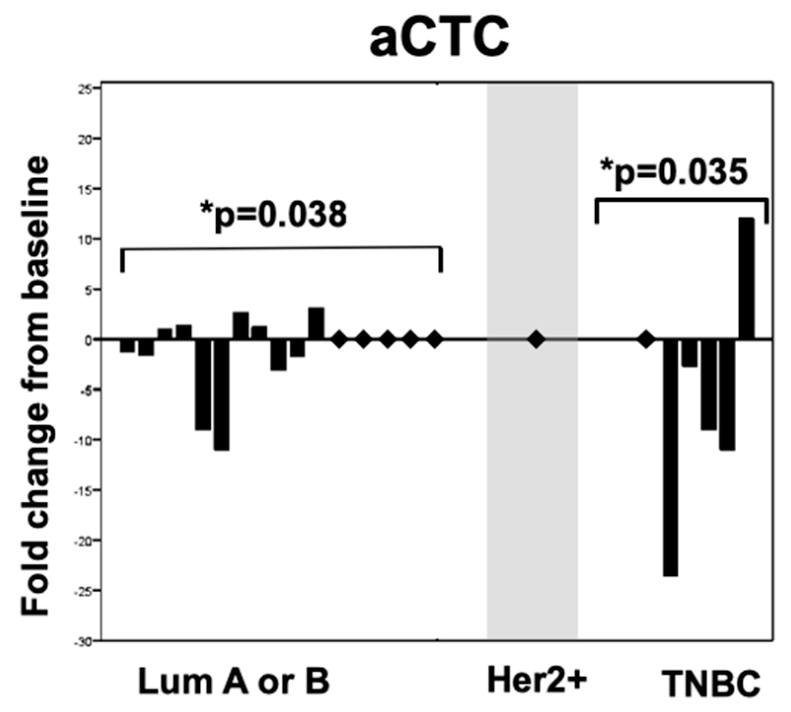
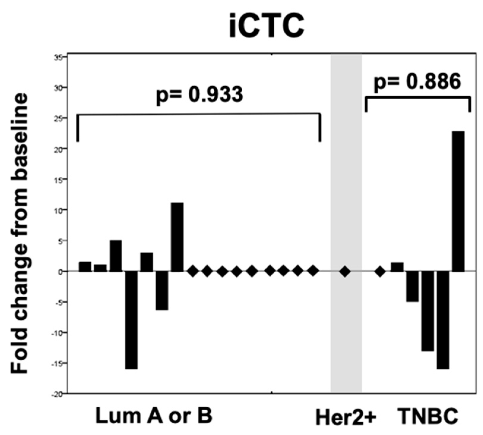
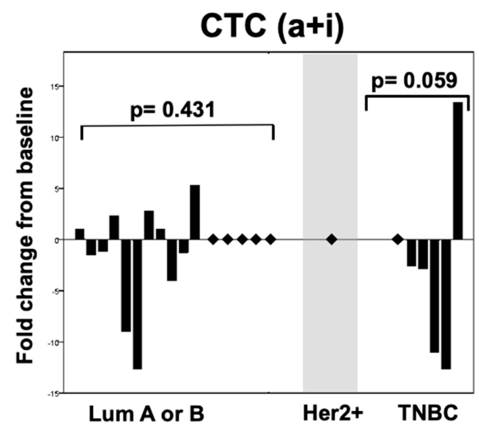
2. Results
3. Discussion
3.1. Role of Apoptotic CTCs as a Potential Parameter of Treatment Response
3.2. Therapy Modalities and Possible Implications for CTC Dynamics
3.3. Limitations
4. Materials and Methods
4.1. Patients
4.2. Real Time qPCR (RT-qPCR) and Defining Intrinsic Subtypes
4.3. Quantification of Circulating Tumor Cells
5. Conclusions
Author Contributions
Funding
Conflicts of Interest
References
- Hayes, D.F.; Cristofanilli, M.; Budd, G.T.; Ellis, M.J.; Stopeck, A.; Miller, M.C.; Matera, J.; Allard, W.J.; Doyle, G.V.; Terstappen, L.W. Circulating Tumor Cells at Each Follow-up Time Point during Therapy of Metastatic Breast Cancer Patients Predict Progression-Free and Overall Survival. Clin. Cancer Res. 2006, 12, 4218–4224. [Google Scholar] [CrossRef] [Green Version]
- Giuliano, M.; Giordano, A.; Jackson, S.; Hess, K.R.; Giorgi, U.D.; Mego, M.; Handy, B.C.; Ueno, N.T.; Alvarez, R.H.; Laurentiis, M.D.; et al. Circulating tumor cells as prognostic and predictive markers in metastatic breast cancer patients receiving first-line systemic treatment. Breast Cancer Res. 2011, 13, R67. [Google Scholar] [CrossRef] [PubMed]
- Dawood, S.; Broglio, K.; Valero, V.; Reuben, J.; Handy, B.; Islam, R.; Jackson, S.; Hortobagyi, G.N.; Fritsche, H.; Cristofanilli, M. Circulating tumor cells in metastatic breast cancer. Cancer 2008, 113, 2422–2430. [Google Scholar] [CrossRef] [Green Version]
- Polasik, A.; Tzschaschel, M.; Schochter, F.; Gregorio, A.D.; Friedl, T.; Rack, B.; Hartkopf, A.; Fasching, P.; Schneeweiss, A.; Müller, V.; et al. Circulating Tumour Cells, Circulating Tumour DNA and Circulating MicroRNA in Metastatic Breast Carcinoma—What is the Role of Liquid Biopsy in Breast Cancer? Geburtshilfe und Frauenheilkunde 2017, 77, 1291–1298. [Google Scholar] [CrossRef] [PubMed]
- Cristofanilli, M.; Budd, G.T.; Ellis, M.J.; Stopeck, A.; Matera, J.; Miller, M.C.; Reuben, J.M.; Doyle, G.V.; Allard, W.J.; Terstappen, L.W.; et al. Circulating Tumor Cells, Disease Progression, and Survival in Metastatic Breast Cancer. N. Engl. J. Med. 2004, 351, 781–791. [Google Scholar] [CrossRef] [PubMed]
- Baccelli, I.; Schneeweiss, A.; Riethdorf, S.; Stenzinger, A.; Schillert, A.; Vogel, V.; Klein, C.; Saini, M.; Bäuerle, T.; Wallwiener, M.; et al. Identification of a population of blood circulating tumor cells from breast cancer patients that initiates metastasis in a xenograft assay. Nat. Biotechnol. 2013, 31, 539–544. [Google Scholar] [CrossRef] [PubMed]
- Gerlinger, M.; Rowan, A.J.; Horswell, S.; Larkin, J.; Endesfelder, D.; Gronroos, E.; Martinez, P.; Matthews, N.; Stewart, A.; Tarpey, P.; et al. Intratumor Heterogeneity and Branched Evolution Revealed by Multiregion Sequencing. N. Engl. J. Med. 2012, 366, 883–892. [Google Scholar] [CrossRef] [PubMed] [Green Version]
- Meng, X.; Song, S.; Jiang, Z.-F.; Sun, B.; Wang, T.; Zhang, S.; Wu, S. Receptor conversion in metastatic breast cancer: A prognosticator of survival. Oncotarget 2016, 7, 71887–71903. [Google Scholar] [CrossRef] [PubMed]
- Stefanovic, S.; Wirtz, R.; Deutsch, T.M.; Hartkopf, A.; Sinn, P.; Varga, Z.; Sobottka, B.; Sotiris, L.; Taran, F.-A.; Domschke, C.; et al. Tumor biomarker conversion between primary and metastatic breast cancer: mRNA assessment and its concordance with immunohistochemistry. Oncotarget 2017, 8, 51416–51428. [Google Scholar] [CrossRef] [Green Version]
- Ross, J.S.; Slodkowska, E.A. Circulating and Disseminated Tumor Cells in the Management of Breast Cancer. Am. J. Clin. Pathol. 2009, 132, 237–245. [Google Scholar] [CrossRef] [PubMed] [Green Version]
- Bidard, F.C.; Peeters, D.J.; Fehm, T.; Nolé, F.; Gisbert-Criado, R.; Mavroudis, D.; Grisanti, S.; Generali, D.; Garcia-Saenz, J.A.; Stebbing, J.; et al. Clinical validity of circulating tumour cells in patients with metastatic breast cancer: A pooled analysis of individual patient data. Lancet Oncol. 2014, 15, 406–414. [Google Scholar] [CrossRef]
- Wallwiener, M.; Riethdorf, S.; Hartkopf, A.D.; Modugno, C.; Nees, J.; Madhavan, D.; Sprick, M.R.; Schott, S.; Domschke, C.; Baccelli, I.; et al. Serial enumeration of circulating tumor cells predicts treatment response and prognosis in metastatic breast cancer: A prospective study in 393 patients. BMC Cancer 2014, 14, 512. [Google Scholar] [CrossRef] [PubMed]
- Bidard, F.C.; Jacot, W.; Dureau, S. Clinical utility of circulating tumor cells (CTC) count to choose between first line hormone therapy & chemotherapy in ER+ HER2- metastatic breast cancer. In Proceedings of the 2018 San Antonio Breast Cancer Symposium, San Antonio, TX, USA, 4–8 December 2018. Abstract GS3-07. [Google Scholar]
- Deutsch, T.M.; Riethdorf, S.; Nees, J.; Hartkopf, A.D.; Schönfisch, B.; Domschke, C.; Sprick, M.R.; Schütz, F.; Brucker, S.Y.; Stefanovic, S.; et al. Impact of apoptotic circulating tumor cells (aCTC) in metastatic breast cancer. Breast Cancer Res. Treat. 2016, 160, 277–290. [Google Scholar] [CrossRef]
- Giordano, A.; Gao, H.; Anfossi, S.; Cohen, E.; Mego, M.; Lee, B.-N.; Tin, S.; Laurentiis, M.D.; Parker, C.A.; Alvarez, R.H.; et al. Epithelial-Mesenchymal Transition and Stem Cell Markers in Patients with HER2-Positive Metastatic Breast Cancer. Mol. Cancer Ther. 2012, 11, 2526–2534. [Google Scholar] [CrossRef] [PubMed] [Green Version]
- Giuliano, A.E.; Hunt, K.K.; Ballman, K.V.; Beitsch, P.D.; Whitworth, P.W.; Blumencranz, P.W.; Leitch, A.M.; Saha, S.; McCall, L.M.; Morrow, M. Axillary Dissection vs. No Axillary Dissection in Women with Invasive Breast Cancer and Sentinel Node Metastasis. JAMA 2011, 305, 569. [Google Scholar] [CrossRef]
- Fehm, T.; Sagalowsky, A.; Clifford, E.; Beitsch, P.; Saboorian, H.; Euhus, D.; Meng, S.; Morrison, L.; Tucker, T.; Lane, N.; et al. Cytogenetic Evidence That Circulating Epithelial Cells in Patients with Carcinoma Are Malignant. Clin. Cancer Res. 2002, 8, 2073–2084. [Google Scholar] [PubMed]
- Hoefnagel, L.D.; Vijver, M.J.V.D.; Slooten, H.-J.V.; Wesseling, P.; Wesseling, J.; Westenend, P.J.; Bart, J.; Seldenrijk, C.A.; Nagtegaal, I.D.; Oudejans, J.; et al. Receptor conversion in distant breast cancer metastases. Breast Cancer Res. 2010, 12, R75. [Google Scholar] [CrossRef]
- Kimbung, S.; Kovács, A.; Danielsson, A.; Bendahl, P.-O.; Lövgren, K.; Stolt, M.F.; Tobin, N.P.; Lindström, L.; Bergh, J.; Einbeigi, Z.; et al. Contrasting breast cancer molecular subtypes across serial tumor progression stages: Biological and prognostic implications. Oncotarget 2015, 6, 33306–33318. [Google Scholar] [CrossRef] [PubMed]
- Hoefnagel, L.D.C.; Moelans, C.B.; Meijer, S.L.; Slooten, H.-J.V.; Wesseling, P.; Wesseling, J.; Westenend, P.J.; Bart, J.; Seldenrijk, C.A.; Nagtegaal, I.D.; et al. Prognostic value of estrogen receptor α and progesterone receptor conversion in distant breast cancer metastases. Cancer 2012, 118, 4929–4935. [Google Scholar] [CrossRef] [Green Version]
- Janni, W.; Rack, B.K.; Fasching, P.A.; Haeberle, L.; Tesch, H.; Lorenz, R.; Schochter, F.; Tzschaschel, M.; Gregorio, A.D.; Fehm, T.N.; et al. Persistence of circulating tumor cells in high risk early breast cancer patients five years after adjuvant chemotherapy and late recurrence: Results from the adjuvant SUCCESS A trial. J. Clin. Oncol. 2018, 36, 515. [Google Scholar] [CrossRef]
- Bidard, F.-C.; Proudhon, C.; Pierga, J.-Y. Circulating tumor cells in breast cancer. Mol. Oncol. 2016, 10, 418–430. [Google Scholar] [CrossRef] [PubMed]
- Aktas, B.; Kasimir-Bauer, S.; Müller, V.; Janni, W.; Fehm, T.; Wallwiener, D.; Pantel, K.; Tewes, M. Comparison of the HER2, estrogen and progesterone receptor expression profile of primary tumor, metastases and circulating tumor cells in metastatic breast cancer patients. BMC Cancer 2016, 16, 522. [Google Scholar] [CrossRef] [PubMed]
- Schramm, A.; Friedl, T.W.P.; Schochter, F.; Scholz, C.; Gregorio, N.D.; Huober, J.; Rack, B.; Trapp, E.; Alunni-Fabbroni, M.; Müller, V.; et al. Therapeutic intervention based on circulating tumor cell phenotype in metastatic breast cancer: Concept of the DETECT study program. Arch. Gynecol. Obstet. 2015, 293, 271–281. [Google Scholar] [CrossRef] [PubMed]
- Krawczyk, N.; Hartkopf, A.; Banys, M.; Meier-Stiegen, F.; Staebler, A.; Wallwiener, M.; Röhm, C.; Hoffmann, J.; Hahn, M.; Fehm, T. Prognostic relevance of induced and spontaneous apoptosis of disseminated tumor cells in primary breast cancer patients. BMC Cancer 2014, 14, 394. [Google Scholar] [CrossRef] [PubMed]
- Russo, A.; Terrasi, M.; Agnese, V.; Santini, D.; Bazan, V. Apoptosis: A relevant tool for anticancer therapy. Ann. Oncol. 2006, 17, vii115–vii123. [Google Scholar] [CrossRef] [PubMed]
- Kallergi, G.; Konstantinidis, G.; Markomanolaki, H.; Papadaki, M.A.; Mavroudis, D.; Stournaras, C.; Georgoulias, V.; Agelaki, S. Apoptotic Circulating Tumor Cells in Early and Metastatic Breast Cancer Patients. Mol. Cancer Ther. 2013, 12, 1886–1895. [Google Scholar] [CrossRef] [PubMed] [Green Version]
- Smerage, J.B.; Budd, G.T.; Doyle, G.V.; Brown, M.; Paoletti, C.; Muniz, M.; Miller, M.C.; Repollet, M.I.; Chianese, D.A.; Connelly, M.C.; et al. Monitoring apoptosis and Bcl-2 on circulating tumor cells in patients with metastatic breast cancer. Mol. Oncol. 2013, 7, 680–692. [Google Scholar] [CrossRef] [PubMed]
- Meng, S.; Tripathy, D.; Frenkel, E.; Shete, S.; Naftalis, E.; Huth, J.; Beitsch, P.; Leitch, M.; Hoover, S.; Euhus, D.; et al. Circulating Tumor Cells in Patients with Breast Cancer Dormancy. Clin. Cancer Res. 2004, 10, 8152–8162. [Google Scholar] [CrossRef] [Green Version]
- Wolff, A.C.; Hammond, M.E.H.; Hicks, D.G.; Dowsett, M.; Mcshane, L.M.; Allison, K.H.; Allred, D.C.; Bartlett, J.M.; Bilous, M.; Fitzgibbons, P.; et al. Recommendations for Human Epidermal Growth Factor Receptor 2 Testing in Breast Cancer: American Society of Clinical Oncology/College of American Pathologists Clinical Practice Guideline Update. J. Clin. Oncol. 2013, 31, 3997–4013. [Google Scholar] [CrossRef] [PubMed] [Green Version]
- Wirtz, R.M.; Sihto, H.; Isola, J.; Heikkilä, P.; Kellokumpu-Lehtinen, P.-L.; Auvinen, P.; Turpeenniemi-Hujanen, T.; Jyrkkiö, S.; Lakis, S.; Schlombs, K.; et al. Biological subtyping of early breast cancer: A study comparing RT-qPCR with immunohistochemistry. Breast Cancer Res. Treat. 2016, 157, 437–446. [Google Scholar] [CrossRef] [PubMed]
- Laible, M.; Schlombs, K.; Kaiser, K.; Veltrup, E.; Herlein, S.; Lakis, S.; Stöhr, R.; Eidt, S.; Hartmann, A.; Wirtz, R.M.; et al. Technical validation of an RT-qPCR in vitro diagnostic test system for the determination of breast cancer molecular subtypes by quantification of ERBB2, ESR1, PGR and MKI67 mRNA levels from formalin-fixed paraffin-embedded breast tumor specimens. BMC Cancer 2016, 16, 398. [Google Scholar] [CrossRef] [PubMed]
- Goldhirsch, A.; Winer, E.P.; Coates, A.S.; Gelber, R.D.; Piccart-Gebhart, M.; Thürlimann, B.; Senn, H.-J.; Albain, K.S.; André, F.; Bergh, J.; et al. Personalizing the treatment of women with early breast cancer: Highlights of the St Gallen International Expert Consensus on the Primary Therapy of Early Breast Cancer 2013. Ann. Oncol. 2013, 24, 2206–2223. [Google Scholar] [CrossRef]
- Fehm, T.; Müller, V.; Aktas, B.; Janni, W.; Schneeweiss, A.; Stickeler, E.; Lattrich, C.; Löhberg, C.R.; Solomayer, E.; Rack, B.; et al. HER2 status of circulating tumor cells in patients with metastatic breast cancer: A prospective, multicenter trial. Breast Cancer Res. Treat. 2010, 124, 403–412. [Google Scholar] [CrossRef] [PubMed]
- Fehm, T.; Hoffmann, O.; Aktas, B.; Becker, S.; Solomayer, E.F.; Wallwiener, D.; Kimmig, R.; Kasimir-Bauer, S. Detection and characterization of circulating tumor cells in blood of primary breast cancer patients by RT-PCR and comparison to status of bone marrow disseminated cells. Breast Cancer Res. 2009, 11, R59. [Google Scholar] [CrossRef] [PubMed]
- Rossi, E.; Basso, U.; Celadin, R.; Zilio, F.; Pucciarelli, S.; Aieta, M.; Barile, C.; Sava, T.; Bonciarelli, G.; Tumolo, S.; et al. M30 Neoepitope Expression in Epithelial Cancer: Quantification of Apoptosis in Circulating Tumor Cells by CellSearch Analysis. Clin. Cancer Res. 2010, 16, 5233–5243. [Google Scholar] [CrossRef] [PubMed] [Green Version]
- Fehm, T.; Becker, S.; Becker-Pergola, G.; Sotlar, K.; Gebauer, G.; Dürr-Störzer, S.; Neubauer, H.; Wallwiener, D.; Solomayer, E.-F. Presence of apoptotic and nonapoptotic disseminated tumor cells reflects the response to neoadjuvant systemic therapy in breast cancer. Breast Cancer Res. 2006, 8, R60. [Google Scholar] [CrossRef]


| Patient characteristic | n (%) | |
| Total, n | 34 | |
| Mean age at primary tumor biopsy, years | 52.3 | |
| Mean age at metastatic tumor biopsy, years | 58.1 | |
| Phenotype of primary tumor, immunohistochemistry | n (%) | |
| Luminal A | 18 (53%) | |
| Luminal B | 9 (26.5%) | |
| HER2 positive | 2 (6%) | |
| Triple-negative | 4 (12%) | |
| NA | 1 (2.9%) | |
| Intrinsic subtype of primary tumor by RT-qPCR | n (%) | |
| Luminal A | 16 (47.1%) | |
| Luminal B | 9 (26.5%) | |
| HER2 positive | 1 (2.9%) | |
| Triple-negative | 7 (20.6%) | |
| NA | 1 (2.9%) | |
| Intrinsic subtype of metastatic tumor by RT-qPCR | n (%) | |
| Luminal A | 11 (32.4%) | |
| Luminal B | 11 (32.4%) | |
| HER2 positive | 1 (2.9%) | |
| Triple-negative | 10 (29.4%) | |
| NA | 1 (2.9%) | |
| Intrinsic subtype conversion assessed by RT-qPCR | n (%) | |
| Converters | 19 (55.9%) | |
| Stable | 14 (41.2%) | |
| NA | 1 (2.9%) | |
| Grading of primary tumor | n (%) | |
| G1 | 0 (0%) | |
| G2 | 18 (53%) | |
| G3 | 12 (35%) | |
| GX | 4 (12%) | |
| ER/ESR1 status of primary tumor | IHC, n (%) | RT-qPCR, n (%) |
| Positive | 24 (70.6%) | 26 (76.5%) |
| Negative | 10 (29.4%) | 8 (23.5%) |
| Unknown | 0 (0%) | 0 (0%) |
| PR/PGR status of primary tumor | IHC, n (%) | RT-qPCR, n (%) |
| Positive | 26 (76.5%) | 17 (50%) |
| Negative | 8 (23.5%) | 17 (50%) |
| Unknown | 0 (0%) | 0 (0%) |
| HER2/ERBB2 status of primary tumor | IHC, n (%) | RT-qPCR, n (%) |
| Positive | 2 (5.9%) | 1 (2.9%) |
| Negative | 29 (85.3%) | 32 (94.1%) |
| Unknown | 3 (8.8%) | 1 (2.9%) |
| Ki67/MKI67 status of primary tumor | RT-qPCR, n (%) | |
| Positive | 6 (17.6%) | |
| Negative | 27 (8.8%) | |
| Unknown | 1 (2.9%) | |
| CTC positive patients at baseline | n (%) | |
| CTC(a+i) intact and apoptotic | 23 (67.6%) | |
| CTC(a) apoptotic | 16 (47.1%) | |
| CTC(i) intact | 23 (67.6%) | |
| CTC positive patients after 1 cycle of systemic therapy | n (%) | |
| CTC(a+i) | 11 (32.4%) | |
| CTC (a) | 7 (20.6%) | |
| CTC (i) | 11 (32.4%) | |
| Characteristic | Chemotherapy | Endocrine Therapy | Anti-HER2 Therapy |
|---|---|---|---|
| Total patients, n (%) | 24 (70.6%) | 9 (26.5%) | 1 (2.9%) |
| Age at first diagnosis | 51.2 | 53 | 46 |
| PFS since study recruitment (months) | 7 | 22 | 10 |
| OS since study recruitment (months) | 16 | 32 | 20 |
| CTC positive patients at baseline, n (%) | |||
| CTC(a+i) intact and apoptotic | 20 (58.8%) | 3 (8.8%) | 0 |
| CTC(a) apoptotic | 14 (41.2%) | 2 (5.9%) | 0 |
| CTC(i) intact | 20 (58.8%) | 3 (8.8%) | 0 |
| CTC positive patients after 1 therapy cycle, n (%) | |||
| CTC(a+i) intact and apoptotic | 10 (29.4%) | 1 (2.9%) | 0 |
| CTC (a) apoptotic | 6 (17.6%) | 1 (2.9%) | 0 |
| CTC (i) intact | 10 (29.4%) | 1 (2.9%) | 0 |
© 2019 by the authors. Licensee MDPI, Basel, Switzerland. This article is an open access article distributed under the terms and conditions of the Creative Commons Attribution (CC BY) license (http://creativecommons.org/licenses/by/4.0/).
Share and Cite
Stefanovic, S.; Deutsch, T.M.; Wirtz, R.; Hartkopf, A.; Sinn, P.; Schuetz, F.; Sohn, C.; Bohlmann, M.K.; Sütterlin, M.; Schneeweiss, A.; et al. Molecular Subtype Conversion between Primary and Metastatic Breast Cancer Corresponding to the Dynamics of Apoptotic and Intact Circulating Tumor Cells. Cancers 2019, 11, 342. https://doi.org/10.3390/cancers11030342
Stefanovic S, Deutsch TM, Wirtz R, Hartkopf A, Sinn P, Schuetz F, Sohn C, Bohlmann MK, Sütterlin M, Schneeweiss A, et al. Molecular Subtype Conversion between Primary and Metastatic Breast Cancer Corresponding to the Dynamics of Apoptotic and Intact Circulating Tumor Cells. Cancers. 2019; 11(3):342. https://doi.org/10.3390/cancers11030342
Chicago/Turabian StyleStefanovic, Stefan, Thomas M. Deutsch, Ralph Wirtz, Andreas Hartkopf, Peter Sinn, Florian Schuetz, Christof Sohn, Michael K. Bohlmann, Marc Sütterlin, Andreas Schneeweiss, and et al. 2019. "Molecular Subtype Conversion between Primary and Metastatic Breast Cancer Corresponding to the Dynamics of Apoptotic and Intact Circulating Tumor Cells" Cancers 11, no. 3: 342. https://doi.org/10.3390/cancers11030342