Article

Refined Method for Droplet Microfluidics-Enabled Detection of Plasmodium falciparum Encoded Topoisomerase I in Blood from Malaria Patients

by Marianne Smedegaard Hede 1, Patricia Nkem Okorie 2, Signe Kirk Fruekilde 3, Søren Fjelstrup 3,4, Jonas Thomsen 1, Oskar Franch 3,4, Cinzia Tesauro 3, Magnus Tobias Bugge 3, Mette Christiansen 5, Stéphane Picot 6, Felix Lötsch 7,11, Ghyslain Mombo-Ngoma 7,8, Johannes Mischlinger 7, Ayôla A. Adegnika 7,8, Finn Skou Pedersen 3,4, Yi-Ping Ho 3,4, Eskild Petersen 9, Magnus Stougaard 10, Michael Ramharter 7,8,11 and Birgitta Ruth Knudsen 3,4,*
1
Zymonostics, 8000 Aarhus C, Denmark
2
Institute for Advanced Medical Research and Training, College of Medicine, University of Ibadan, 200001 Ibadan, Nigeria
3
Department of Molecular Biology and Genetics, University of Aarhus, 8000 Aarhus C, Denmark
4
NANO, University of Aarhus, 8000 Aarhus C, Denmark
5
Department of Clinical Immunology, Aarhus University Hospital, 8200 Aarhus N, Denmark
6
Malaria Research Unit, SMITh, ICBMS, UMR 5246 CNRS-INSA-CPE-UCBL1 & Institutde Parasitologie et Mycologie Médicale, Hospices Civils de Lyon, 69373 Lyon Cedex 08, France
7
Centre de Recherches Médicales de Lambaréné, Lambaréné, Gabon
8
Institut für Tropenmedizin, Universität Tübingen, 72074 Tübingen, Germany
9
Department of Infectious Diseases, Aarhus University Hospital, 8200 Aarhus N, Denmark
10
Department of Clinical Medicine, University of Aarhus, 8000 Aarhus C, Denmark
11
Department of Medicine I, Division of Infectious Diseases and Tropical Medicine, Medical University of Vienna, 1090 Vienna, Austria
*
Author to whom correspondence should be addressed.
Micromachines 2015, 6(10), 1505-1513; https://doi.org/10.3390/mi6101432
Submission received: 17 August 2015 / Revised: 23 September 2015 / Accepted: 29 September 2015 / Published: 5 October 2015
(This article belongs to the Special Issue Droplet Microfluidics: Techniques and Technologies)

Abstract

:
Rapid and reliable diagnosis is essential in the fight against malaria, which remains one of the most deadly infectious diseases in the world. In the present study we take advantage of a droplet microfluidics platform combined with a novel and user-friendly biosensor for revealing the main malaria-causing agent, the Plasmodium falciparum (P. falciparum) parasite. Detection of the parasite is achieved through detection of the activity of a parasite-produced DNA-modifying enzyme, topoisomerase I (pfTopoI), in the blood from malaria patients. The assay presented has three steps: (1) droplet microfluidics-enabled extraction of active pfTopoI from a patient blood sample; (2) pfTopoI-mediated modification of a specialized DNA biosensor; (3) readout. The setup is quantitative and specific for the detection of Plasmodium topoisomerase I. The procedure is a considerable improvement of the previously published Rolling Circle Enhanced Enzyme Activity Detection (REEAD) due to the advantages of involving no signal amplification steps combined with a user-friendly readout. In combination these alterations represent an important step towards exploiting enzyme activity detection in point-of-care diagnostics of malaria.

1. Introduction

Diagnosis of malaria is traditionally achieved using direct visualization of the disease-causing Plasmodium parasite of which P. falciparum is the most predominant [1]. In a previous paper it was shown that malaria diagnostics might also be reached by droplet microfluidics-enabled lysis of blood from malaria patients followed by detection of an enzyme, topoisomerase I (pfTopoI), expressed by the malaria parasite [2]. Unlike other methods, which detect parasitic proteins using antibodies, this assay, which is denoted Rolling Circle Enhanced Enzyme Activity Detection (REEAD), detects the activity of the biomarker protein and not merely its presence [2]. This makes REEAD extremely sensitive since one enzyme molecule can convert many substrate molecules into a detectable product. Furthermore, pfTopoI is an essential enzyme and disease detection will therefore not fail due to biomarker mutations. Spurred on by the very promising aspects of the REEAD assay, the present study aimed at further developing enzyme-based detection of malaria towards point-of-care diagnosis.
pfTopoI belongs to a group of DNA-modifying enzymes, DNA topoisomerases, that cleave and ligate one strand of a DNA double helix in order to exert its important biological functions [3]. It is this reaction that is exploited in the presented assay where P. falciparum is detected using a biosensor made from a labeled and partly double-stranded DNA substrate that is converted to a detectable product only by the cleavage-ligation reactions of pfTopoI. This biosensor is combined with a droplet microfluidics platform to allow efficient diagnosis of malaria in blood samples from infected individuals.

2. Experimental Section

2.1. Cloning and Purification of Topoisomerase I

pfTopoI or hTopoI were expressed and purified essentially as described previously [2,4]. The storage buffer of the enzymes was changed for 10 mM Tris-HCl, pH 7.4, using Bio-Spin 6 columns from Biorad (Copenhagen, Denmark). Before storage the enzymes were supplemented with glycerol to a final concentration of 50% and NaCl to a final concentration of 150 mM.

2.2. Blood Sampling

Blood samples from patients diagnosed with malaria were collected at the Aarhus University Hospital, Denmark, and at Centre de Recherches Médicales de Lambaréné (CERMEL), Albert Schweitzer Hospital, Lambaréné, Gabon. Blood samples from uninfected individuals were collected at the Aarhus University Hospital, Denmark.

2.3. Enzyme Extraction Using Droplet Microfluidics

The droplet microfluidic devices were fabricated by conventional soft lithography techniques [5], casting and curing the PDMS prepolymer on a SU-8 3025 (MicroChem, Westborough, MA, USA) master of a channel height at around 25 μm. PDMS prepolymer (Sylgard 184) was prepared in a 10:1 (base:curing agent) ratio and cured at 65 °C for 1 h. Prior to the experiments, the channels were wetted with oil/surfactant (Pico-Surf 1, 1% in HFE-7500, Dolomite Microfluidics, Royston, UK) for at least 15 min. Two syringe pumps (Harvard Apparatus, Holliston, MA, USA) were used to control the flow rates of oil/surfactant and reagents independently. The droplet volume and generation frequency were controlled by the flow rate ratio, determined by the competition between continuous phase and disperse phase [2,6,7]. Subsequently, blood and lysis buffer (10 mM Tris pH 7.5, 5 mM EDTA supplemented with protease inhibitors) were subjected to droplet microfluidics essentially as described previously [2]. Upon harvesting, the droplets were left at room temperature for 15 min after which the droplets were broken by addition of 25% (v/v) 1H,1H,2H,2H-Perfluoro-1-octanol (Sigma-Aldrich, Copenhagen, Denmark).

2.4. Lysis of Blood Using Bead Beating

Blood and lysis buffer (10 mM Tris pH 7.5, 5 mM EDTA supplemented with protease inhibitors) were mixed 1:4 and bead beated for 3 min using a Disruptor Genie from Scientific Industries (New York, NY, USA) and acid-washed glass beads (150–212 μm) from Sigma-Aldrich. The resulting lysate was left on ice for 15 min and centrifuged for 5 min at 1000 g.

2.5. Substrate Preparation

Oligonucleotides (see Table 1) were purchased at Sigma-Aldrich. A labeled PCR product was produced using the following conditions: Template: plasmid pYES2.1 (Life Technologies, Waltham, MA, USA), primers 1 and 2 (see Table 1), DreamTaq Polymerase, and DreamTaq buffer (Life Technologies). The reaction was spiked with either dATP, [alpha-32P] from Perkin Elmer (Figure 4) or 20 µM Aminoallyl-dUTP-XX-ATTO-488 (Jena Bioscience, Jena, Germany) (Figure 2 and Figure 3). After cycling, the PCR product was purified using the E.Z.N.A.® Cycle-Pure Kit (Omega Biotek, Norcross, GA, USA).
Table 1. Oligonucleotides used in the present study.
NameSequence5ʹ Modification
Primer 1ATTTTTCTAAGTCTTTTAGATCGAACGACTCAGAATGATGCATGTATACTAAACTCACAAATTAGAGC
Primer 2TTTTTTTTTTTTTTTTTTTTTTTTTGCTTTCTCATAGCTCACGCTG
Am-Cl[AmC6F]ACTACCATTCTGAGTCGTTCGATCTAAAAGACTTAGAAmine
Biotin-ClBiotin-ACTACCATTCTGAGTCGTTCGATCTAAAAGACTTAGABiotin
ClACTACCATTCTGAGTCGTTCGATCTAAAAGACTTAGA
NClATTTTTCTAAGTCTTTTAGATCGAACGACTCAGAATG

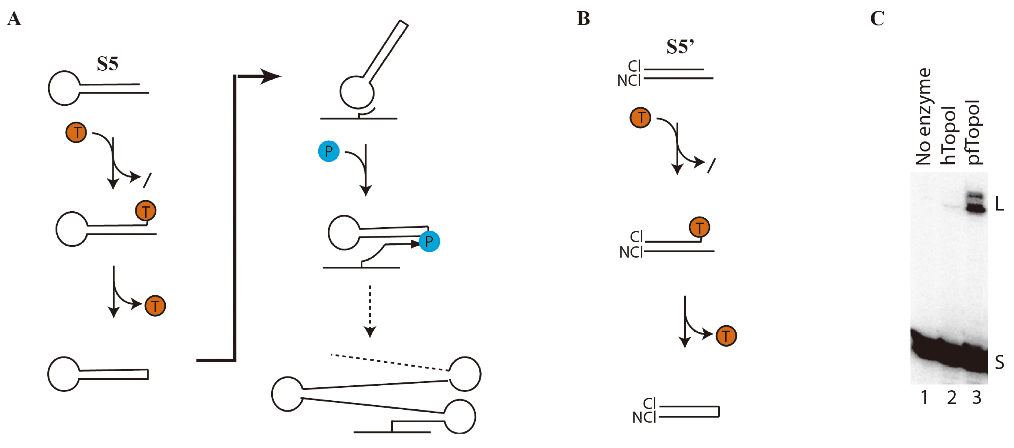
2.6. Polyacrylamide Gel-Enabled Detection of pfTopoI Activity

To prepare the substrate S5', the oligonucleotide Cl (see Table 1) was radiolabeled at the 5'-end using T4 polynucleotide kinase (NEB, Ipswich, MA, USA) as described by the supplier. The radiolabeled oligonucleotide was hybridized to NCl (see Table 1) by heating to 95 °C and slowly cooling to room temperature. Then, 2 pmol of the assembled substrate was incubated for 1 h at 37 °C with either 10 ng of hTopoI, 10 ng of pfTopoI, or no enzyme (reaction buffer: 10 mM Tris-HCl (pH 7.5), 1 mM EDTA, 100 mM NaCl). The reaction products were separated in a 8% polyacrylamide gel in TBE (acrylamide 7 M urea gels in TBE (48 mM Tris, 45.5 mM Boric Acid, 1 mM EDTA)). The gel was dried and the reaction products visualized by phosphorimaging.

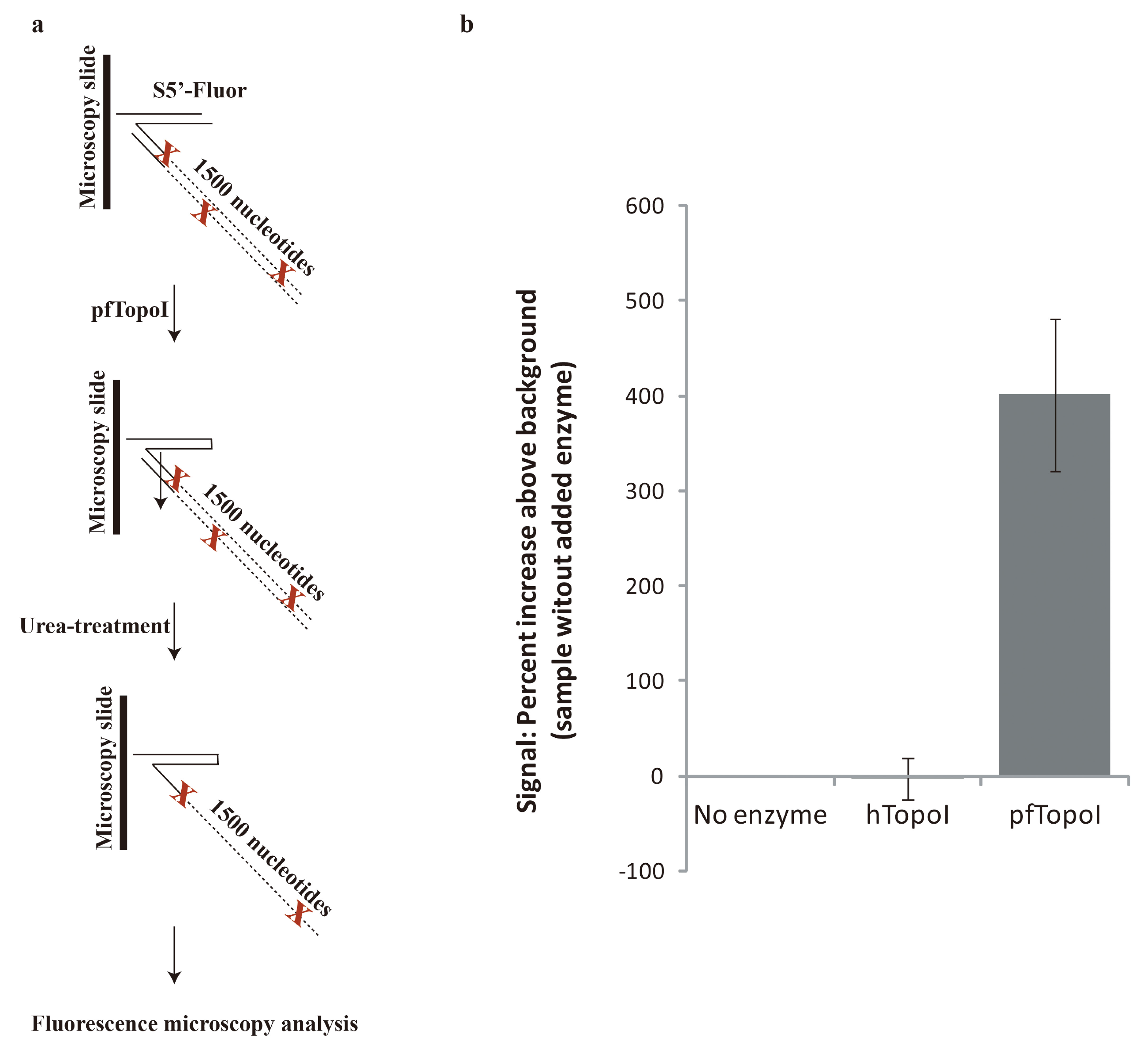
2.7. Fluorescence Microscopy-Enabled Detection of pfTopoI Activity

A 5'-amine conjugated version of the cleaved strand Cl (Am-Cl) was immobilized by spotting on CodeLink® HD Activated Slides (Surmodics, Eden Prairie, MN, USA) as described by the supplier (10 µL, 5 µM amine conjugated oligonucleotide per spot). To assemble the substrate S5'-flour, 2.5 ng of fluorescent PCR product (See Section 2.5) was added to each printed spot and left to hybridize to the printed oligo for 1 h at 50 °C in a humidity chamber. After washing for 1 min in wash buffer A (100 mM Tris-HCl pH 7.5; 150 mM NaCl; 0.3% SDS) and 1 min in wash buffer B (100 mM Tris-HCl pH 7.5; 150 mM NaCl; 0.05% Tween 20), the microscopy slide was dehydrated by washing in 96% ethanol for 1 min and left to air dry.
The reaction buffer (10 mM Tris-HCl (pH 7.5), 1 mM EDTA, 300 mM NaCl) was supplemented with 10 ng of purified hTopoI or pfTopoI (Figure 2) or 1 µL of bead beating-produced blood lysate (Figure 3) pr. 10 µL reaction volume. Then, 10 µL of the reaction mixture was added to each spot. After a 1 h incubation at 37 °C in a humidity chamber the slides were washed for 1 min in wash buffer A, 1 min in wash buffer B, 1 min in water, 2 × 30 min in 8 M urea, 1 min in wash buffer A, 1 min in wash buffer B, and 1 min 96% ethanol. After air drying, the slide was mounted with VECTASHIELD® and subjected to fluorescence microscopy. The number of fluorescent signals per microscopic image was determined using Image J.

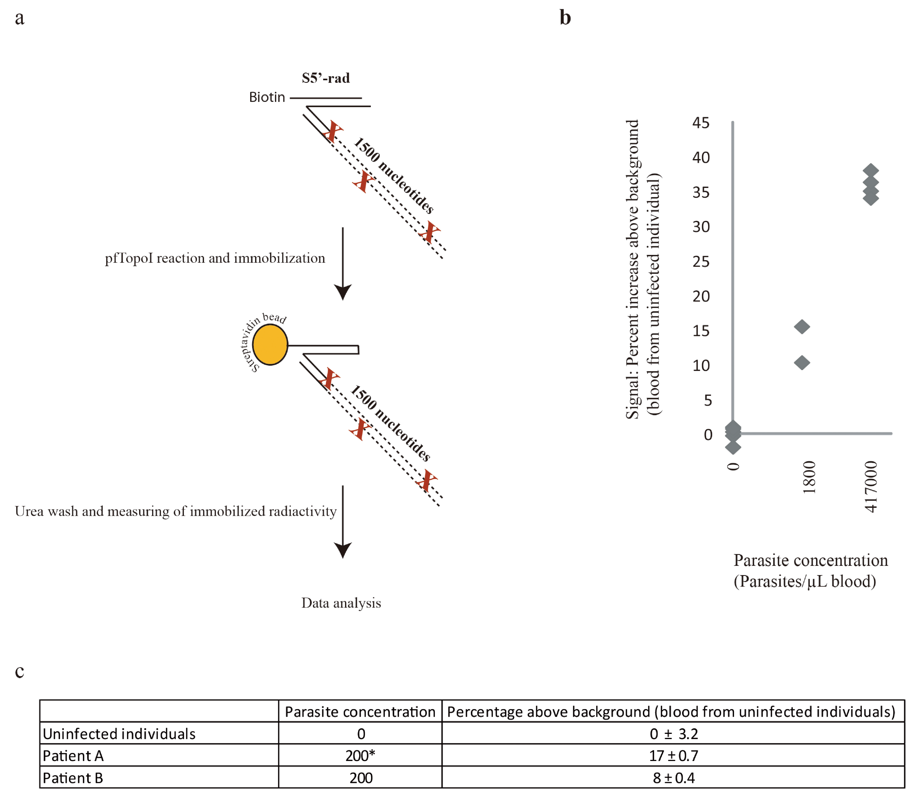
2.8. Radioactivity-Enabled Detection of pfTopoI Activity Using Magnetic Beads

To prepare the substrate S5'-rad, a radiolabeled PCR product (see Section 2.5) was hybridized to a 5'-biotin labeled oligonucleotide (Biotin-Cl, see Table 1) by heating to 95 °C and slowly cooling to room temperature. Hybridization was carried out in a buffer containing 10 mM tris pH 7.5; 0.25 mM EDTA; and 150 mM NaCl. Then, 1 fmol of the assembled substrate was incubated with 5 µL of blood lysate (see droplet microfluidics) in a 50 µL reaction volume. The reaction buffer was 10 mM Tris-HCl (pH 7.5), 1 mM EDTA, and 300 mM NaCl.
After incubation, the reaction product was mixed with 100 µg of streptavidin beads (Dynabeads® MyOne™ Streptavidin C1) and left at room temperature for 20 min. The beads were subsequently washed in water (1 min), 8 M urea (2 × 15 min), and water (1 min). After each wash the washing agent was removed by placing the beads on a magnet and carefully removing the supernatant. Upon washing the tube containing the magnetic beads was placed on top of a photographic film (MG-SR PLUS medical X-ray film from Konica Minolta, Åbyhøj, Denmark). After a three-day exposure time the film was developed and the signals quantified using the Image J software (http://imagej.nih.gov/ij/).

3. Results

3.1. Specific and Efficient DNA Substrate for pfTopoI

In the previously published REEAD setup, the ability of pfTopoI to cleave and ligate a short protruding DNA-end of a double-stranded substrate (composed of one DNA strand that folds on itself) was utilized to generate a circular DNA product that could be amplified by a so-called phi-polymerase to generate a detectable product (Figure 1a) [2].
To eliminate the requirement for a DNA polymerase-driven signal amplification step, the established REEAD substrate [2] termed S5 was modified. On the modified substrate (S5'), pfTopoI facilitates the ligation of two independent DNA strands that are annealed to form a duplex (Figure 1b). To confirm the specificity and efficiency of this reaction we incubated radiolabeled S5' with either human (hTopoI) or plasmodium (pfTopoI) topoisomerase I. Note that hTopoI is the enzyme most likely to interfere with the detection assay in the clinical samples used for diagnosis of malaria. The reaction products were separated in an 8% denaturing sequencing gel. As evident from Figure 1b, upon incubation with pfTopoI we do observe low mobility bands (marked “L”) at the top of the substrate (marked “S”) (compare lane 3 with lane 1), which is consistent with pfTopoI-mediated joining of the 5'-radiolabeled Cl strand and the NCl strand of the substrate as a result of the cleavage-ligation activities of the enzyme. This additional band is not observed upon incubation with hTopoI, supporting the specificity of the substrate S5' towards pfTopoI (compare lane 3 with lane 2).
Figure 1. Specific detection of pfTopoI activity using S5' (a) REEAD [2]: S5 is specifically cleaved and ligated by pfTopoI (indicated by a T in red circle) to form a covalently closed DNA circle (left panel). These DNA circles can be detected by rolling circle amplification (right panel) where the circle is hybridized to an immobilized DNA oligonucleotide that is extended by a DNA polymerase (indicated by a P in a blue circle) using the DNA circle as template. The result is a long detectable DNA molecule. (b) A DNA substrate (S5') for pfTopoI consisting of one strand that is cleaved by pfTopoI (cleaved strand, Cl) hybridized to its complementary (non-cleaved) strand (NCl) is used. Incubation with pfTopoI (indicated by a T in red circle) leads to cleavage of the substrate and subsequent fusion of the two DNA strands (see text for details). (c) 5'-radiolabeled, assembled substrate was incubated with either pfTopoI (lane 3), hTopoI (lane 2) or no enzyme (lane 1). The reaction products were separated in an 8% denaturing polyacrylamide gel. The bands corresponding to the radiolabeled substrate are indicated with S and the bands corresponding to the ligation product are indicated with L.
Micromachines 06 01432 g001

3.2. Towards Development of a User-Friendly Readout

To facilitate the detection of the pfTopoI-specific reaction on S5' without the need for the sophisticated and specialized laboratory equipment necessary for gelelectrophoresis, the non-cleaved strand of S5' was elongated with a 1500 nucleotide fluorescent tail to generate the substrate “S5'-fluor” as a first step towards a user-friendly readout format (see Figure 2a). The substrate was immobilized on a modified microscopy slide (see experimental section) and incubated with either purified hTopoI or pfTopoI (Figure 2) or extracts of blood from a malaria-infected patient, serial diluted in blood from an uninfected individual (Figure 3). Following pfTopoI incubation, the samples were treated with urea that removed the fluorescent tail of unreacted S5'-fluor whereas substrates that were ligated by pfTopoI retained fluorescent labeling due to covalent coupling between one strand of the fluorescent tail to the surface anchored strand of the substrate (Figure 2a). Subsequently, the reaction outcomes were analyzed using fluorescent microscopy.
Incubation with pfTopoI (Figure 2b) gives rise to a 400% increase in the number of signals compared to the sample without any added enzyme. The addition of hTopoI, on the other hand, does not increase the number of signals. This result clearly shows that the present assay, like its predecessor REEAD [2], is highly specific for the detection of pfTopoI.
A blood sample from a malaria patient with 1150 parasites/µL blood was diluted in blood from an uninfected individual to create blood with decreasing concentrations of parasites. Extracts from these samples were incubated with the immobilized substrate S5ʹ-fluor. The slide was washed with urea and subjected to fluorescent microscopy. A clear correlation between parasite concentration and the number of signals was observed, indicating that the assay is quantitative (Figure 3).
Figure 2. Test of substrate S5ʹ-fluor using purified enzymes. (a) S5'-flour immobilized on a microscopy slide was incubated with pfTopoI. A subsequent urea wash removes unreacted fluorescent tails prior to fluorescence microscopic analysis. (b) S5ʹ-fluor was incubated with no enzyme hTopoI, or pfTopoI, as indicated and the results analyzed in a fluorescent microscope. The results are shown as the percent increase in fluorescent signal compared to the sample without enzyme. The graph shows the average of three independent experiments.
Micromachines 06 01432 g002
Figure 3. Test of substrate S5'-fluor using blood extracts. S5'-fluor was incubated with extracts prepared from blood with increasing concentrations of P. falciparum as indicated and the results determined by fluorescent microscopy. The results are shown as percent increase in fluorescent signals compared to blood samples negative for plasmodium. The graph shows the average of two independent experiments. The inset zooms in on the lowest parasite concentrations.
Micromachines 06 01432 g003

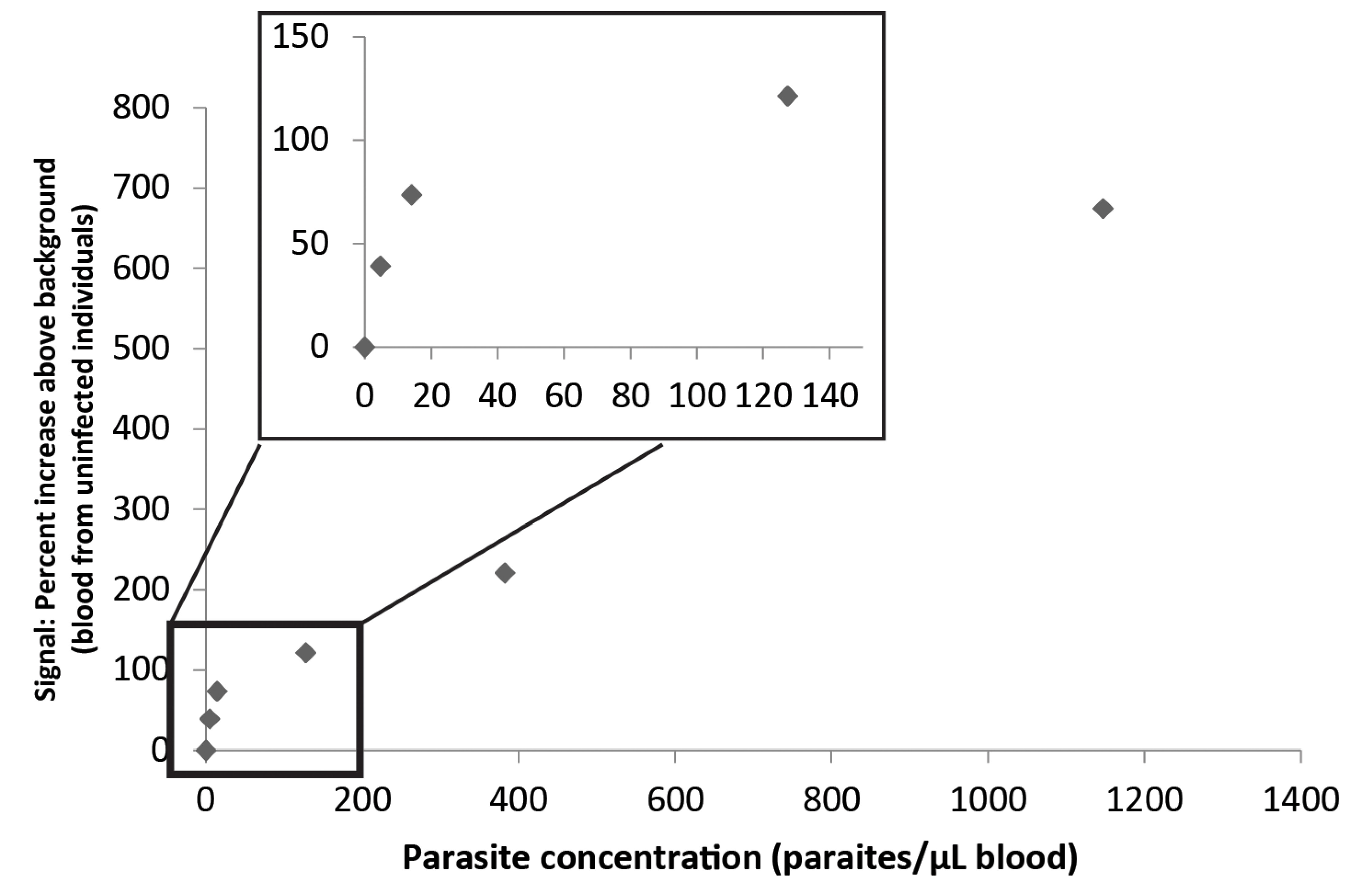
3.3. Combination of the Enzyme-Based Malaria Detection with a Droplet Microfluidics Platform

To eliminate the need for fluorescence microscopy and eventually also other electricity-driven devices such as a bead beater, we further optimized the described assay to use droplet microfluidics to extract active pfTopoI enzyme from blood from malaria patients. Furthermore, the DNA-substrate was modified to generate S5ʹ-rad on which pfTopoI reaction lead to the covalent fusion of a biotin-labeled DNA oligonucleotide and a radiolabeled DNA tail (that substitutes the fluorescent tail of S5ʹ-fluor). After immobilization onto streptavidin-coated magnetic beads, a urea wash denatures the DNA and the radiolabeled DNA tail will be removed from the unreacted S5ʹ-rad. The amount of radioactivity left after the wash will hence be a measure for the pfTopoI activity. This strategy was tested (see Figure 4b) on two positive blood samples (with 1800 parasites/µL blood or 417,000 parasites/µL blood, as determined by microscopy). Furthermore, the usability of the assay in patients with low parasite density was tested using two samples with parasite concentrations adjusted to 200 parasites/µL blood (see Figure 4c). All blood samples were extracted using droplet microfluidics. In Figure 4 the usability of droplet microfluidics is confirmed. Furthermore, the P. falciparum positive samples give significantly higher signals than do the uninfected samples. This demonstrates that P. falciparum infection, e.g., malaria, can be detected in a low-technological manner that could be easily adapted to low resource settings.
Figure 4. Detection of malaria using the enzyme-based assay in combination with droplet microfluidics-facilitated extraction of active pfTopoI. Extracts from blood with different parasite concentrations were prepared by droplet microfluidics and analyzed using the S5ʹ-rad substrate system (shown in (a), see text for details). (b) The results are shown as percent increase compared to blood samples negative for Plasmodium. The graph shows the results of two independent experiments. The sample with 417,000 parasites pr. µL blood was tested in triplicate in one of the experiments. (c) The results from blood samples with low parasite concentrations are shown as percent increase compared to blood samples negative for Plasmodium. * Sample adjusted to 200 parasites/µL blood by dilution of a sample with 400 parasites/µL blood with blood from a healthy individual.
Micromachines 06 01432 g004

4. Conclusions

In the current paper we present a droplet microfluidics-enabled method for detection of the most predominant malaria-causing agent, the P. falciparum parasite, in blood from infected patients. The presented method is simple, quantitative, and requires no external enzymes or other labile reagents. Moreover, although the microfluidic setup currently is driven by electricity-powered syringe pumps, a handheld and hand-operated device could be envisioned [8]. The presented assay is thus very suitable for adaptation to point-of-care diagnostics. The technique described here is an extension of the previously published assay REEAD [2]. Both in REEAD and in the modified assay presented here, detection of P. falciparum is achieved by detecting the activity of a parasitic enzyme, pfTopoI. pfTopoI is able to cleave and ligate DNA. More specifically, it has been shown capable of covalently joining the two strands of a DNA hairpin structure (see Figure 1 and previous publication [2]). In REEAD, the hairpin-shaped DNA substrate is formed by a single DNA molecule, and the product of the pfTopoI-mediated hairpin ligation reaction is consequently a covalently closed DNA circle [2,9]. The pfTopoI-generated DNA circles are subsequently detected through rolling circle amplification of a primer attached to a microscopy slide, hybridization of fluorescent probes recognizing the rolling circle amplification product, and fluorescence microscopy [2,9]. In contrast, the biosensor presented in this paper is made from two separate DNA strands that are joined by pfTopoI to form one long, linear DNA strand. In the presence of pfTopoI, a biotin moiety allowing immobilization can be fused to a radiolabeled DNA molecule allowing ultrasensitive detection. Conjugation of the reaction product to streptavidin beads followed by washing under DNA denaturing conditions ensures immobilization of radiolabeled DNA on the beads if—and only if—pfTopoI has ligated the two DNA molecules. The novel assay hence distinguishes itself from REEAD in two important ways: (1) no polymerase or other labile reagents are needed; and (2) no fluorescence microscopy is required. In the current setup we use radiolabeling to detect the DNA product produced by pfTopoI. To allow point-of-care diagnostics, this readout could, however, be replaced by a colorimetric readout based on, for instance, horseradish peroxidase in order to allow visualization of pfTopoI activity without the use of radioactivity. Although further optimization and testing are still needed, the combination of these two alterations represents an important step towards using enzyme activity detection in point-of-care diagnostics.

Acknowledgments

We are thankful to technicians Noriko Y. Hansen and Lone Højgaard Nielsen for technical assistance during this project and to Mirjam Groger, Rella Manego Zoleko, Selidji T Agnandji, Lilian Endame, Johanna Kim for contributing with sample collection. This work was supported by the Danish Research Councils (11-116325/FTP), the Lundbeck Foundation (R95-A10275), Arvid Nilsson’s Foundation, The Augustinus Foundation, Marie & M. B. Richters Foundation, Aage and Johanne Louis-Hansens Foudation, Aase and Ejnar Danielsens Foundation, Familien Erichsens Mindefond, and Familien Hede Nielsens Fond.

Author Contributions

M.S.H. designed and performed experiments, analyzed data, and wrote the paper. P.N.O., S.K.F., S.F., J.T., O.F., C.T. and M.T.B. performed experiments and analyzed data. M.C., S.P., F.L., G.M.N., J.M., A.A.A., E.P. and M.R. contributed to manuscript writing as well as with blood samples from malaria patients and uninfected individuals. F.S.P. supervised and contributed to manuscript writing. M.S. and Y.P.H. supervised and contributed to assay design. B.R.K. supervised, contributed to assay design, and wrote the paper.

Conflicts of Interest

The authors declare no conflict of interest.

Abbreviations

P. falciparumPlasmodium falciparum
pfTopoIP. falciparum topoisomerae I
hTopoIhuman topoisomerase I
REEADrolling circle enhanced enzyme activity detection
PCRpolymerase chain reaction

References

  1. Tangpukdee, N.; Duangdee, C.; Wilairatana, P.; Krudsood, S. Malaria diagnosis: A brief review. Korean J. Parasitol. 2009, 47, 93–102. [Google Scholar] [CrossRef] [PubMed]
  2. Juul, S.; Nielsen, C.J.; Labouriau, R.; Roy, A.; Tesauro, C.; Jensen, P.W.; Harmsen, C.; Kristoffersen, E.L.; Chiu, Y.L.; Frohlich, R.; et al. Droplet microfluidics platform for highly sensitive and quantitative detection of malaria-causing plasmodium parasites based on enzyme activity measurement. ACS Nano 2012, 6, 10676–10683. [Google Scholar] [CrossRef] [PubMed]
  3. Champoux, J.J. DNA topoisomerases: Structure, function, and mechanism. Ann. Rev. Biochem. 2001, 70, 369–413. [Google Scholar] [CrossRef] [PubMed]
  4. Lisby, M.; Olesen, J.R.; Skouboe, C.; Krogh, B.O.; Straub, T.; Boege, F.; Velmurugan, S.; Martensen, P.M.; Andersen, A.H.; Jayaram, M.; et al. Residues within the N-terminal domain of human topoisomerase I play a direct role in relaxation. J. Biol. Chem. 2001, 276, 20220–20227. [Google Scholar] [CrossRef] [PubMed]
  5. Qin, D.; Xia, Y.; Whitesides, G.M. Soft lithography for micro- and nanoscale patterning. Nat. Protoc. 2010, 5, 491–502. [Google Scholar] [CrossRef] [PubMed]
  6. Ho, Y.P.; Grigsby, C.L.; Zhao, F.; Leong, K.W. Tuning physical properties of nanocomplexes through microfluidics-assisted confinement. Nano Lett. 2011, 11, 2178–2182. [Google Scholar] [CrossRef] [PubMed]
  7. Juul, S.; Ho, Y.P.; Koch, J.; Andersen, F.F.; Stougaard, M.; Leong, K.W.; Knudsen, B.R. Detection of single enzymatic events in rare or single cells using microfluidics. ACS Nano 2011, 5, 8305–8310. [Google Scholar] [CrossRef] [PubMed]
  8. Abate, A.R.; Weitz, D.A. Syringe-vacuum microfluidics: A portable technique to create monodisperse emulsions. Biomicrofluidics 2011, 5, 14107. [Google Scholar] [CrossRef] [PubMed]
  9. Tesauro, C.; Juul, S.; Arno, B.; Nielsen, C.J.; Fiorani, P.; Frohlich, R.F.; Andersen, F.F.; Desideri, A.; Stougaard, M.; Petersen, E.; et al. Specific detection of topoisomerase I from the malaria causing P. falciparum parasite using isothermal rolling circle amplification. In Proceedings of 2012 Annual International Conference of the IEEE Engineering in Medicine and Biology Society (EMBC), San Diego, CA, USA, 28 August–1 September 2012; pp. 2416–2419.

